Embed Size (px)
Citation preview
Image Registration for Digital Radiographic Images and its Applications to Industrial
NDT Inspection
Jianxin GAO, Gary PENNEY, Martyn WRIGHT, TWI, Cambridge, UK
Abstract. Radiographic images taken under different conditions of the engineering structures in service need to be aligned properly prior to performing effectively any of the automatic defect detection. To this end, image registration techniques based on intensity correlation were employed in this paper in a coarse-to-fine multilevel manner to speed up the time-consuming calculation process. The original image with high spatial resolution was downscaled to several images, each with the resolution half of the previous level. The registration for the coarsest image pair resulted in the detection of predominant part of the displacement field associated with the image pairs, while the registration for the finest image pairs generated delicate corrections for the previously calculated displacement field. This image registration procedure was streamlined into a whole framework for digital radiographic processing for industrial NDT inspection. The aligned image sequences can be used subsequently for 3D defect assessment. Preliminary applications showed that the implementation of image registration improved considerably the effectiveness of the automatic defect detection.
1.Introduction
Radiographic inspection is a well-established non-destructive testing technique for quality control and integrity assurance in a wide range of industries. With the development of imaging and computer techniques in terms of hardware and software, the interpretation of radiographic images is increasingly becoming an automated process through digital image processing. This greatly reduces human interaction which is prone to fatigue and subjectivity. One of the simple but robust image processing techniques is image subtraction. This is particularly useful when a golden image can be obtained, and subsequently an active image of a component being inspected can be subtracted from it, leading to the potential flaws being highlighted for automatic defect recognition. When a golden image is not available, image subtraction still can be used to reveal the difference between relevant images.
It is apparent that when there is deformation between the reference and target images,
direct image subtraction would be of little help in flaw detection. Those deformations might be the result of different imaging geometry during radiographic exposure, or the inevitable misalignment of the component on a convey belt in a production line. In order to get better inspection results, it is imperative that these deformations or distortions between the reference and target images be corrected for as much as possible. Image registration is the
ECNDT 2006 - Poster 50
1
very method for this purpose, which has been developed by researchers both from computer vision and image processing community and has been applied to single modal and multi-modal imaging tasks in medical imaging, robotic navigation, human gesture recognition [1,2,3].
The purpose of this paper is to apply intensity based image registration in the
interpretation of industrial radiographic images for automated flaw detection. A multiresolution scheme is adopted in a coarse- to-fine manner. The original image with high spatial resolution was downscaled to several images, each with the resolution half of the previous level. The registration for the coarsest image pair resulted in the detection of predominant part of the displacement field associated with the image pairs, while the registration for the finest image pairs generated delicate corrections for the previously calculated displacement field. This image registration procedure was streamlined into a whole framework for digital radiographic processing for industrial NDT inspection. The aligned image sequences were then used for three-dimensional defect measurement. Preliminary applications showed that the implementation of image registration improved considerably the effectiveness of the automatic defect detection.
2. Multiresolution Image Registration
Registering a target image to a reference image is to find a function (also called a deformation field) that performs the backward mapping of the former onto the latter as close as possible. Generally, the deformation can be locally and globally. A multiresolution approach can handle effectively both the global and local deformations. The basic idea is that the deformations within the two misaligned radiographic images can be decomposed into their respective components in each resolution level. At a low-resolution image level, only the predominant part of the deformation components is extracted. This can be thought as a rough approximation to the optimal geometric transformation, and is then eliminated by image transformation. Subsequently, a higher resolution registration is followed, from which the finer deformations on this resolution level can be calculated [1]. The whole image registration process can be conducted in such a recursive manner to progressively approach the final optimal deformation field that best transformed the target image to the reference image.
Let 1I and 2I represent the reference and target image, respectively. The registration of these two images is to find a transformation T , such that the difference between the reference image 1I and the affine-transformed image TI 2 is minimal:
}min{ 21TIIT −= (1)
It is dealt with as an optimisation problem such that the parameter vector c , which represents the ideal transformation Tc , will maximise a similarity function
),()( 21TcIIsimilaritycF = (2)
The choice of the transformation to be used depends on the knowledge of the deformation to be recovered. For 2D radiographic images, a Piecewise Bilinear Maps (PBM) is used to represent the associated deformation. Multiresolution decomposition can be applied to PBMs resulting in a hierarchy of transformation spaces, where transformations from spaces with higher dimension can model more localised and finer detail of the distortion.
2
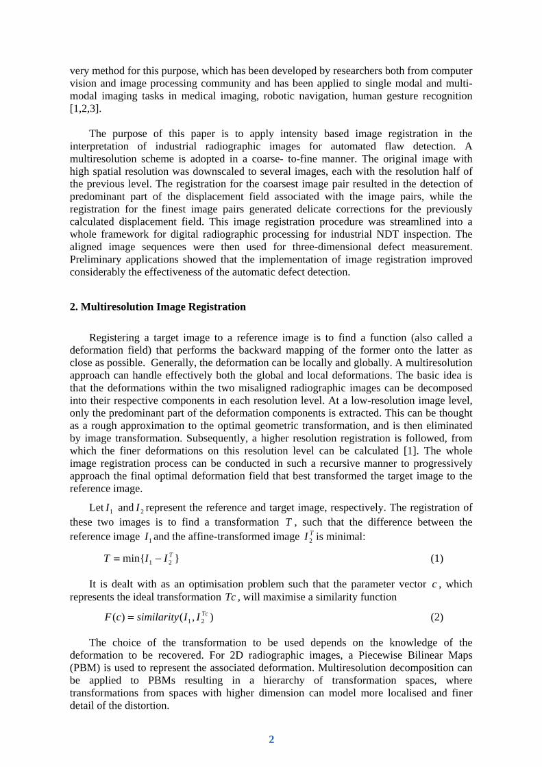
The mapping of a subset in a target image onto the reference image is dictated by the displacement components at each control points of the subset, as illustrated in Fig.1 as follows:
1,11,11,1,,1,1,, ),(),(),(),()( ++++++++ +++= jijijijijijijiji cvucvucvucvupm ωωωω (3)
where the weighting functions are
uvvuvuvu
vuvuvuvu
ji
ji
ji
ji
=
−=
−=
−−=
++
+
+
),()1(),()1(),(
)1)(1(),(
1,1
1,
,1
,
ωωωω
(4)
The values of u and v are the relative proportion of the point with regard to the control points in horizontal and vertical direction, respectively.
Different similarity measures had been used in image registration. The mostly used in intensity based image registration is the normalised correlation function, defined as:
>><−<•>><−<
><•><−>•<=
222
211
2121
)()( TcTc
TcTc
IIII
IIIIC (5)
where <..> denotes ensemble average. This similarity function eliminates the effect of background differences between the two images, and varies between [-1, 1], with 1 indicating an exact match between the affine transformed image and the reference image.
Image registration is realised through an optimisation process that maximises the similarity function (5). Since there are a number of factors affecting the similarity function, the optimisation tends to be trapped into local maxima in the vicinity of global minima.
Fig.1. Bilinear affine transformation of a target image onto a reference image. Control point c(i,j) are mapped to a(i,j) by taking into account of the corresponding deformation field.
3
This leads to erroneous registration results. The use of multiresolution algorithm will help avoid such kind of situation by adopting an appropriate scheme.
3. Methods and Results
A framework of radiographic image processing has been established, which contains most conventional image processing techniques such as image segmentation, intensity thresholding, image convolution, fast Fourier transformation (FFT), image addition and subtraction, plus multiresolution image registration. Source image can be loaded from a CCD camera or from image files. Once the target radiographic images had been registered to the reference image, an image subtraction is performed they are subtracted from the latter to highlight the difference between them and the common features being eliminated.
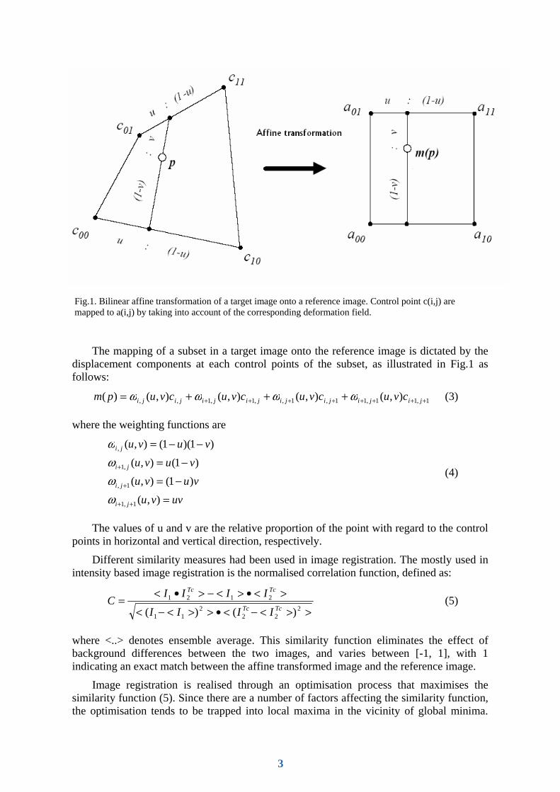
A verification test had been conducted to validate the framework. Fig.2 is one of the test results using a pair of radiographs taken from a Printed Circuit Board (PCB) with a slight different exposure setting. The predominant part of the registered image Fig.2 (c) was recovered precisely to the reference image Fig.2 (a) as was indicated by the almost dark appearance of the remaining image Fig.2 (d) generated by subtracting the registered image Fig.2 (c) from the reference Fig.2 (a). The only parts that had substantial bright pixels were the up-right and bottom-left part in which the original image pair differed considerably as a result of focusing shift. In contrast, direct image subtraction of the original target image Fig.2 (b) from reference Fig.2 (a) resulted in an image Fig.2 (e) of little use, which necessitated image registration.
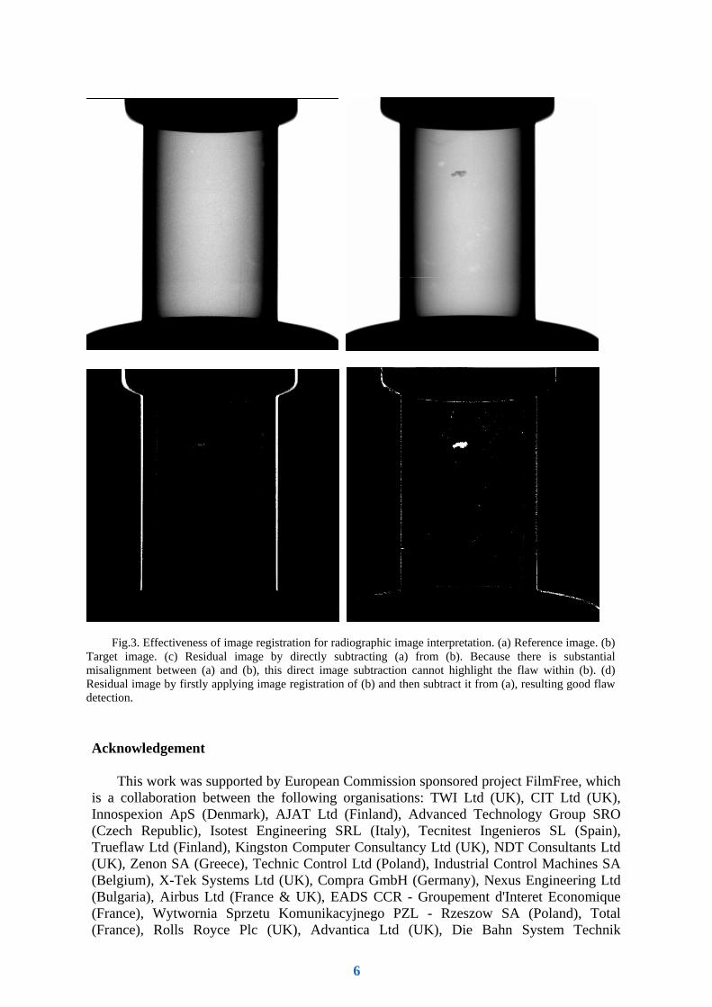
Fig.3 shows the preliminary application result for a series of pipe radiographic images for defect detection. Fig.3 (a) and (b) are the X-ray images of two pipe samples taken by X-Tek 450KV Radiographic System at 350KV. Without image registration, the operation of image subtraction produced a result image Fig.2(c) in which potential flaw was hardly discernible for human eye visualisation. With image registration, however, potential flaw was highlighted in the subtracted image Fig.3(d), which helped for subsequent automatic recognition.
4. Conclusion Remarks
A multiresolution image registration algorithm has been applied to radiographic image
interpretation, which is implemented in a coarse-to-fine manner. The registration of the coarsest image results in the detection of predominant part of the deformation field associated with the image pairs, while the registration for the finest image pairs generated delicate corrections for the previously calculated deformation field. This image registration procedure was streamlined into a whole framework for digital radiographic processing for industrial NDT inspection. The aligned image sequences were subsequently used for 3D defect assessment. Preliminary applications showed that the use of image registration in interpretation of radiographic images improves substantially the flaw detection capability by image subtraction. Further work will be focused on practical application of the proposed image processing system to a variety of industrial sectors to develop a highly automated flaw detection system.
4
Fig.2. Verification result of a pair of PCB radiographic images. The predominant part of the registered image (c) is recovered to the reference image (a) as was indicated by the almost dark appearance image (d) generated by subtracting (c) from (a). The only parts that have substantial bright pixels are the up-right and bottom-left part in which the original image pair differs considerably. Direct subtracting the original target image (b) from (a) results in an image (e) of little use.
(a) (b) (c)
(d) (e)
5
Acknowledgement
This work was supported by European Commission sponsored project FilmFree, which is a collaboration between the following organisations: TWI Ltd (UK), CIT Ltd (UK), Innospexion ApS (Denmark), AJAT Ltd (Finland), Advanced Technology Group SRO (Czech Republic), Isotest Engineering SRL (Italy), Tecnitest Ingenieros SL (Spain), Trueflaw Ltd (Finland), Kingston Computer Consultancy Ltd (UK), NDT Consultants Ltd (UK), Zenon SA (Greece), Technic Control Ltd (Poland), Industrial Control Machines SA (Belgium), X-Tek Systems Ltd (UK), Compra GmbH (Germany), Nexus Engineering Ltd (Bulgaria), Airbus Ltd (France & UK), EADS CCR - Groupement d'Interet Economique (France), Wytwornia Sprzetu Komunikacyjnego PZL - Rzeszow SA (Poland), Total (France), Rolls Royce Plc (UK), Advantica Ltd (UK), Die Bahn System Technik
Fig.3. Effectiveness of image registration for radiographic image interpretation. (a) Reference image. (b) Target image. (c) Residual image by directly subtracting (a) from (b). Because there is substantial misalignment between (a) and (b), this direct image subtraction cannot highlight the flaw within (b). (d) Residual image by firstly applying image registration of (b) and then subtract it from (a), resulting good flaw detection.
6

(Germany), Helenic Society of Non Destructive Testing (Greece), Castings Technology International (UK), Bulgarian Welding Society (Bulgaria), Commissariat a l'Energie Atomique (France), Federal Institute for Materials Research and Testing (Germany), Technical University of Sofia (Bulgaria), Technical University of Szczecin (Poland), LME Ltd (UK), and ICL Ltd (UK).
The Project is co-ordinated and managed by TWI Ltd and is partly funded by the EC under the IP SME programme, reference number: NMP2-CT-2005-515746.
References
[1] Stefan Veeser, Michael J. Dunn, Guang-ZhongYang. Multiresolution image registration for two-dimensional gel electrophoresis. Proteomics, 2001, Vol.1(7), 856-870 [2] Carlos Sorzano, Philippe Thevenaz and Michael Unser, Elastic registration of biological images using vector-spline regularization. IEEE trans on Biomed Eng. 2005, Vol.52(4), 652-663 [3] Vincent Noblet, Christian Heinrich, Fabrice Heitz, and Jean-Paul Armspach, 3-D deformable image registration: a topology preservation scheme based on hierarchical deformation models and interval analysis optimization. IEEE Trans. Image Process, 2005, Vol. 14(5), 553-566
7