Embed Size (px)
Citation preview
154 Images: Pulmonary Fibrosis and Emphysema
journal.copdfoundation.org JCOPDF © 2018 Volume 5 • Number 2 • 2018
For personal use only. Permission required for all other uses.
Chronic Obstructive Pulmonary Diseases:
Journal of the COPD Foundation
Combined Pulmonary Fibrosis and EmphysemaMary Salvatore, MD1 Kevin Kwon, BA1 Robert M. Steiner, MD2
Abbreviations: combined pulmonary fibrosis and emphysema, CPFE; chronic obstructive pulmonary disease, COPD; idiopathic pulmonary fibrosis, IPF; usual interstitial pneumonitis, UIP; computed tomography, CTCitation: Salvatore M, Kwon K, Steiner RM. Images in COPD: combined pulmonary fibrosis and emphysema. Chronic Obstr Pulm Dis. 2018;5(2):154-157. doi: https://doi.org/10.15326/jcopdf.5.2.2018.0137
Images in COPD
1 Department of Radiology, Mount Sinai Medical Center, New York, New York
2 Columbia University Medical Center, New York, New York
Address correspondence to:
Mary Salvatore, MDDepartment of RadiologyMount Sinai Medical Center1 Gustave L. Levy PlaceNew York, NY [email protected]
Keywords:chronic obstructive pulmonary disease; COPD; combined pulmonary fibrosis and emphysema, CPFE; idiopathic pulmonary fibrosis; IPF
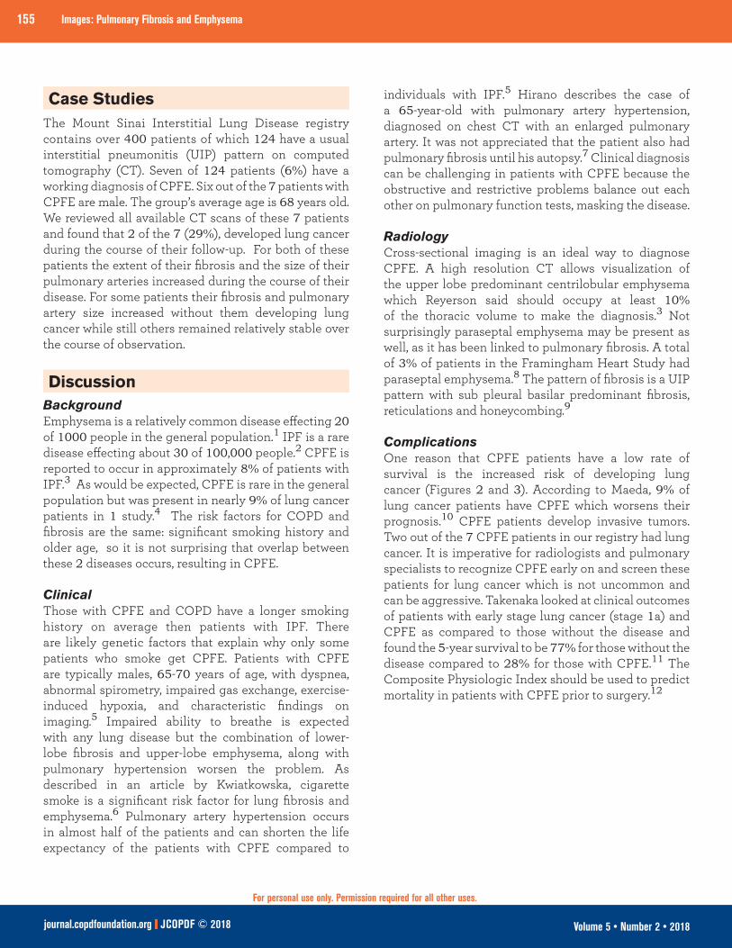
IntroductionCombined pulmonary fibrosis and emphysema (CPFE) is a disease characterized by both centrilobular emphysema (usually in the upper lobes) and fibrosis (typically in the lower lobes) (Figure 1). CPFE is believed to be distinct from idiopathic pulmonary fibrosis (IPF) and chronic obstructive pulmonary disease (COPD). Patients with CPFE tend to be older males with significant smoking histories. This disease is important clinically because of the increased risk for lung cancer and poor overall survival.
155 Images: Pulmonary Fibrosis and Emphysema
journal.copdfoundation.org JCOPDF © 2018 Volume 5 • Number 2 • 2018
For personal use only. Permission required for all other uses.
Case StudiesThe Mount Sinai Interstitial Lung Disease registry contains over 400 patients of which 124 have a usual interstitial pneumonitis (UIP) pattern on computed tomography (CT). Seven of 124 patients (6%) have a working diagnosis of CPFE. Six out of the 7 patients with CPFE are male. The group’s average age is 68 years old. We reviewed all available CT scans of these 7 patients and found that 2 of the 7 (29%), developed lung cancer during the course of their follow-up. For both of these patients the extent of their fibrosis and the size of their pulmonary arteries increased during the course of their disease. For some patients their fibrosis and pulmonary artery size increased without them developing lung cancer while still others remained relatively stable over the course of observation.
DiscussionBackgroundEmphysema is a relatively common disease effecting 20 of 1000 people in the general population.1 IPF is a rare disease effecting about 30 of 100,000 people.2 CPFE is reported to occur in approximately 8% of patients with IPF.3 As would be expected, CPFE is rare in the general population but was present in nearly 9% of lung cancer patients in 1 study.4 The risk factors for COPD and fibrosis are the same: significant smoking history and older age, so it is not surprising that overlap between these 2 diseases occurs, resulting in CPFE.
Clinical Those with CPFE and COPD have a longer smoking history on average then patients with IPF. There are likely genetic factors that explain why only some patients who smoke get CPFE. Patients with CPFE are typically males, 65-70 years of age, with dyspnea, abnormal spirometry, impaired gas exchange, exercise-induced hypoxia, and characteristic findings on imaging.5 Impaired ability to breathe is expected with any lung disease but the combination of lower-lobe fibrosis and upper-lobe emphysema, along with pulmonary hypertension worsen the problem. As described in an article by Kwiatkowska, cigarette smoke is a significant risk factor for lung fibrosis and emphysema.6 Pulmonary artery hypertension occurs in almost half of the patients and can shorten the life expectancy of the patients with CPFE compared to
individuals with IPF.5 Hirano describes the case of a 65-year-old with pulmonary artery hypertension, diagnosed on chest CT with an enlarged pulmonary artery. It was not appreciated that the patient also had pulmonary fibrosis until his autopsy.7 Clinical diagnosis can be challenging in patients with CPFE because the obstructive and restrictive problems balance out each other on pulmonary function tests, masking the disease.
Radiology Cross-sectional imaging is an ideal way to diagnose CPFE. A high resolution CT allows visualization of the upper lobe predominant centrilobular emphysema which Reyerson said should occupy at least 10% of the thoracic volume to make the diagnosis.3 Not surprisingly paraseptal emphysema may be present as well, as it has been linked to pulmonary fibrosis. A total of 3% of patients in the Framingham Heart Study had paraseptal emphysema.8 The pattern of fibrosis is a UIP pattern with sub pleural basilar predominant fibrosis, reticulations and honeycombing.9
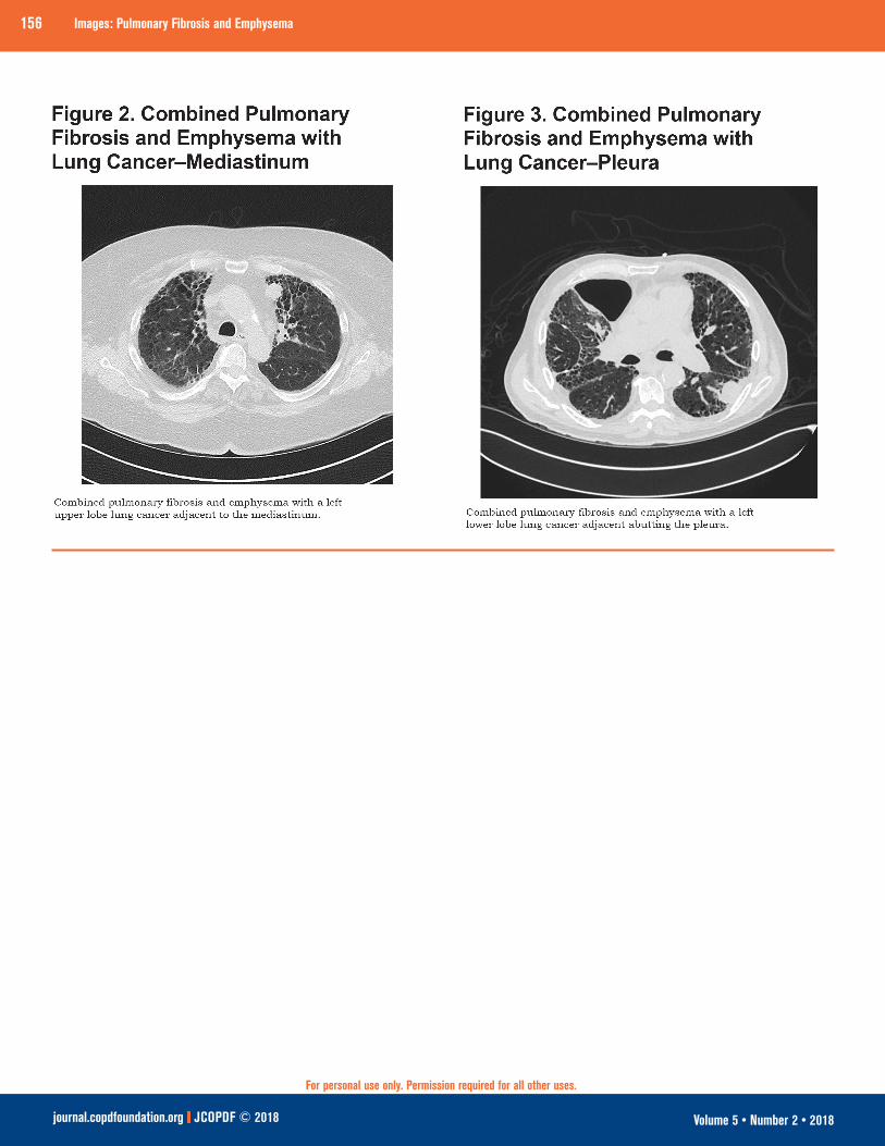
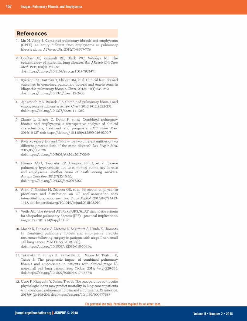
ComplicationsOne reason that CPFE patients have a low rate of survival is the increased risk of developing lung cancer (Figures 2 and 3). According to Maeda, 9% of lung cancer patients have CPFE which worsens their prognosis.10 CPFE patients develop invasive tumors. Two out of the 7 CPFE patients in our registry had lung cancer. It is imperative for radiologists and pulmonary specialists to recognize CPFE early on and screen these patients for lung cancer which is not uncommon and can be aggressive. Takenaka looked at clinical outcomes of patients with early stage lung cancer (stage 1a) and CPFE as compared to those without the disease and found the 5-year survival to be 77% for those without the disease compared to 28% for those with CPFE.11 The Composite Physiologic Index should be used to predict mortality in patients with CPFE prior to surgery.12
156 Images: Pulmonary Fibrosis and Emphysema
journal.copdfoundation.org JCOPDF © 2018 Volume 5 • Number 2 • 2018
For personal use only. Permission required for all other uses.
157 Images: Pulmonary Fibrosis and Emphysema
journal.copdfoundation.org JCOPDF © 2018 Volume 5 • Number 2 • 2018
For personal use only. Permission required for all other uses.
Lin H, Jiang S. Combined pulmonary fibrosis and emphysema (CPFE): an entity different from emphysema or pulmonary fibrosis alone. J Thorac Dis. 2015;7(4):767-779. Coultas DB, Zumwalt RE, Black WC, Sobonya RE. The epidemiology of interstitial lung diseases. Am J Respir Crit Care Med. 1994;150(4):967–972.doi: https://doi.org/10.1164/ajrccm.150.4.7921471
Ryerson CJ, Hartman T, Elicker BM, et al. Clinical features and outcomes in combined pulmonary fibrosis and emphysema in idiopathic pulmonary fibrosis. Chest. 2013;144(1):234–240. doi: https://doi.org/10.1378/chest.12-2403
Jankowich MD, Rounds SIS. Combined pulmonary fibrosis and emphysema syndrome: a review. Chest. 2012;141(1):222-231. doi: https://doi.org/10.1378/chest.11-1062
Zhang L, Zhang C, Dong F, et al. Combined pulmonary fibrosis and emphysema: a retrospective analysis of clinical characteristics, treatment and prognosis. BMC Pulm Med. 2016;16:137. doi: https://doi.org/10.1186/s12890-016-0300-7
Kwiatkowska S. IPF and CPFE — the two different entities or two different presentations of the same disease? Adv Respir Med. 2017;86(1):23-26.doi: https://doi.org/10.5603/ARM.a2017.0049
Hirano ACG, Targueta EP, Campos FPFD, et al. Severe pulmonary hypertension due to combined pulmonary fibrosis and emphysema: another cause of death among smokers. Autops Case Rep. 2017;7(2):15-26.doi: https://doi.org/10.4322/acr.2017.022
Araki T, Nishino M, Zazueta OE, et al. Paraseptal emphysema: prevalence and distribution on CT and association with interstitial lung abnormalities. Eur J Radiol. 2015;84(7):1413-1418. doi: https://doi.org/10.1016/j.ejrad.2015.03.010
Wells AU. The revised ATS/ERS/JRS/ALAT diagnostic criteria for idiopathic pulmonary fibrosis (IPF) - practical implications. Respir Res. 2013;14(Suppl 1):S2. Maeda R, Funasaki A, Motono N, Sekimura A, Usuda K, Uramoto H. Combined pulmonary fibrosis and emphysema predicts recurrence following surgery in patients with stage I non-small cell lung cancer. Med Oncol. 2018;35(3). doi: https://doi.org/10.1007/s12032-018-1091-x
Takenaka T, Furuya K, Yamazaki K, Miura N, Tsutsui K, Takeo S. The prognostic impact of combined pulmonary fibrosis and emphysema in patients with clinical stage IA non-small cell lung cancer. Surg Today. 2018; 48(2):229-235. doi: https://doi.org/10.1007/s00595-017-1577-8
Ueno F, Kitaguchi Y, Shiina T, et al. The preoperative composite physiologic index may predict mortality in lung cancer patients with combined pulmonary fibrosis and emphysema. Respiration. 2017;94(2):198-206. doi: https://doi.org/10.1159/000477587
1.
2.
3.
4.
5.
6.
7.
8.
9.
References
10.
12.
11.