Embed Size (px)
Citation preview
Breast augmentation with an unknown substanceLamya Ebrahim,1 David Morrison,2 Alan Kop,2 Donna Taylor1,3
1Department of Radiology,Royal Perth Hospital, Perth,Western Australia, Australia2Department of MedicalEngineering and Physics, RoyalPerth Hospital, Perth, WesternAustralia, Australia3School of Surgery, Universityof Western Australia, Perth,Australia
Correspondence toDr Donna Taylor,[email protected]
Accepted 25 May 2014
To cite: Ebrahim L,Morrison D, Kop A, et al.BMJ Case Rep Publishedonline: [please include DayMonth Year] doi:10.1136/bcr-2014-204535
SUMMARYBefore the widespread use of silicone implants variousforeign substances were injected directly into the breasts.The nature of these materials sometimes remainsunknown and can cause various complications requiringsurgical intervention. Preoperative diagnostic imagingcan help characterise the type and distribution of theinjected material, thereby assisting in making decisionsregarding treatment. We report a case of breastaugmentation with an unknown substance, aiming tohighlight some imaging characteristics of different breastaugmentation substances.
BACKGROUNDIn the 1950s, synthetic materials called organogenscomposed of petroleum jelly, paraffin or injectablesilicone gel were commonly used for breast aug-mentation, particularly in Asian countries. In the1970s, silicone mammary prostheses becamepopular. Hydrogel filled implants or injectablehydrogel, for example polyacrylamide, have beenin use since the 1990s.1
Various complications are seen following breastaugmentation, including failure of the implant shell(where present) and spread of the implant material.The latter may either be contained within thefibrous capsule or dispersed into the surroundingtissues. Identification of the type of implant mater-ial is important in planning treatment, in particularsurgical removal of the material.The exact number of patients who have suffered
from polyacrylamide hydrogel injection complica-tions is unknown. However, the rising number of
lawsuits in courts led the Chinese State Food andDrug Administration to call for an immediate banon the production, sale and use of polyacrylamidehydrogel in 2006.2
While mammography can distinguish betweensaline and silicone material due to differences inradio-opacity, it is unable to distinguish betweenother substances; ultrasound is also of little value.MRI is the technique of choice for assessment of
breast implant integrity and complications, and theuse of different pulse sequences may help differen-tiate between some of these implanted materials.Nuclear magnetic resonance (NMR) spectroscopyshows promise in the accurate identification of aug-mentation material, thus aiding in planningtreatment.
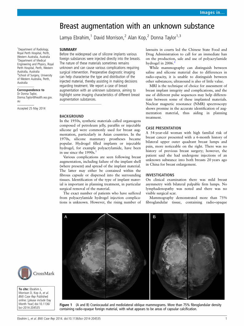
CASE PRESENTATIONA 54-year-old woman with high familial risk ofbreast cancer presented with a 6-month history ofbilateral upper outer quadrant breast lumps andpain, more noticeable on the right. There was nohistory of previous breast surgery; however, thepatient said she had undergone injections of anunknown substance into both breasts 20 years agoin China for breast enlargement.
INVESTIGATIONSOn clinical examination there was mild breastasymmetry with bilateral palpable firm lumps. Nolymphadenopathy was noted and there was novisible surgical scar.Mammography demonstrated more than 75%
fibroglandular tissue, containing radio-opaque
Figure 1 (A and B) Craniocaudal and mediolateral oblique mammograms. More than 75% fibroglandular densitycontaining radio-opaque foreign material, with what appears to be areas of capsular calcification.
Ebrahim L, et al. BMJ Case Rep 2014. doi:10.1136/bcr-2014-204535 1
Images in…
foreign material, with what appeared to be areas of capsular cal-cification. This was thought to represent silicone implants withareas of dystrophic calcification (figure 1).
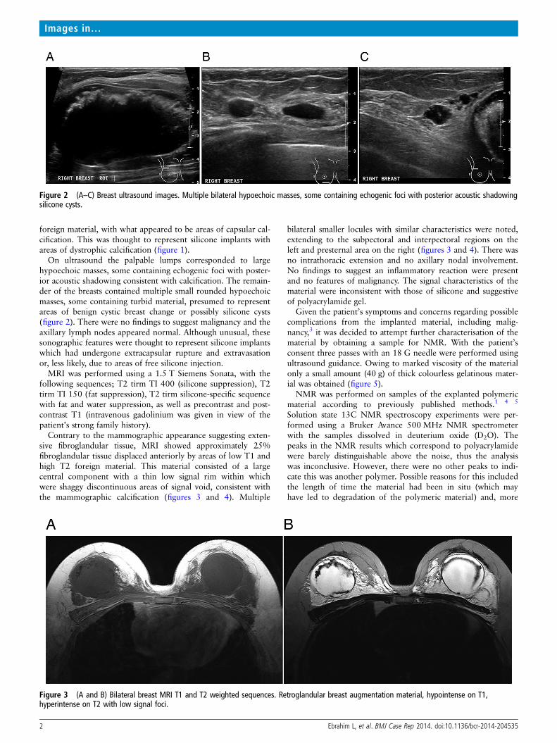
On ultrasound the palpable lumps corresponded to largehypoechoic masses, some containing echogenic foci with poster-ior acoustic shadowing consistent with calcification. The remain-der of the breasts contained multiple small rounded hypoechoicmasses, some containing turbid material, presumed to representareas of benign cystic breast change or possibly silicone cysts(figure 2). There were no findings to suggest malignancy and theaxillary lymph nodes appeared normal. Although unusual, thesesonographic features were thought to represent silicone implantswhich had undergone extracapsular rupture and extravasationor, less likely, due to areas of free silicone injection.
MRI was performed using a 1.5 T Siemens Sonata, with thefollowing sequences; T2 tirm TI 400 (silicone suppression), T2tirm TI 150 (fat suppression), T2 tirm silicone-specific sequencewith fat and water suppression, as well as precontrast and post-contrast T1 (intravenous gadolinium was given in view of thepatient’s strong family history).
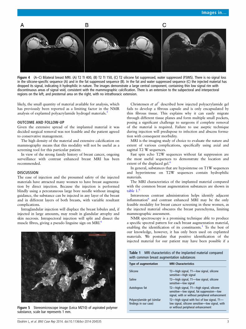
Contrary to the mammographic appearance suggesting exten-sive fibroglandular tissue, MRI showed approximately 25%fibroglandular tissue displaced anteriorly by areas of low T1 andhigh T2 foreign material. This material consisted of a largecentral component with a thin low signal rim within whichwere shaggy discontinuous areas of signal void, consistent withthe mammographic calcification (figures 3 and 4). Multiple
bilateral smaller locules with similar characteristics were noted,extending to the subpectoral and interpectoral regions on theleft and presternal area on the right (figures 3 and 4). There wasno intrathoracic extension and no axillary nodal involvement.No findings to suggest an inflammatory reaction were presentand no features of malignancy. The signal characteristics of thematerial were inconsistent with those of silicone and suggestiveof polyacrylamide gel.
Given the patient’s symptoms and concerns regarding possiblecomplications from the implanted material, including malig-nancy,3 it was decided to attempt further characterisation of thematerial by obtaining a sample for NMR. With the patient’sconsent three passes with an 18 G needle were performed usingultrasound guidance. Owing to marked viscosity of the materialonly a small amount (40 g) of thick colourless gelatinous mater-ial was obtained (figure 5).
NMR was performed on samples of the explanted polymericmaterial according to previously published methods.1 4 5
Solution state 13C NMR spectroscopy experiments were per-formed using a Bruker Avance 500 MHz NMR spectrometerwith the samples dissolved in deuterium oxide (D2O). Thepeaks in the NMR results which correspond to polyacrylamidewere barely distinguishable above the noise, thus the analysiswas inconclusive. However, there were no other peaks to indi-cate this was another polymer. Possible reasons for this includedthe length of time the material had been in situ (which mayhave led to degradation of the polymeric material) and, more
Figure 2 (A–C) Breast ultrasound images. Multiple bilateral hypoechoic masses, some containing echogenic foci with posterior acoustic shadowingsilicone cysts.
Figure 3 (A and B) Bilateral breast MRI T1 and T2 weighted sequences. Retroglandular breast augmentation material, hypointense on T1,hyperintense on T2 with low signal foci.
2 Ebrahim L, et al. BMJ Case Rep 2014. doi:10.1136/bcr-2014-204535
Images in…
likely, the small quantity of material available for analysis, whichhas previously been reported as a limiting factor in the NMRanalysis of explanted polyacrylamide hydrogel materials.5
OUTCOME AND FOLLOW-UPGiven the extensive spread of the implanted material it wasdecided surgical removal was not feasible and the patient agreedto conservative management.
The high density of the material and extensive calcification onmammography means that this modality will not be useful as ascreening tool for this particular patient.
In view of the strong family history of breast cancer, ongoingsurveillance with contrast enhanced breast MRI has beenrecommended.
DISCUSSIONThe ease of injection and the presumed safety of the injectedmaterials have attracted many women to have breast augmenta-tion by direct injection. Because the injection is performedblindly using a percutaneous large bore needle without imagingguidance, the substance can be injected in any layer of the breastand in different layers of both breasts, with variable resultantcomplications.
Intraglandular injection will displace the breast lobules and, ifinjected in large amounts, may result in glandular atrophy andskin necrosis. Intrapectoral injection will split and dissect themuscle fibres, giving a pseudo linguine sign on MRI.6
Christensen et al7 described how injected polyacrylamide gelfails to develop a fibrous capsule and is only encapsulated bythin fibrous tissue. This explains why it can easily migratethrough different tissue planes and form multiple small pockets,posing a significant challenge to surgeons if complete removalof the material is required. Failure to use aseptic techniqueduring injection will predispose to infection and abscess forma-tion with consequent morbidity.
MRI is the imaging study of choice to evaluate the nature andextent of various complications, specifically using axial andsagittal T2 W sequences.
Fast spin echo T2W sequences without fat suppression arethe most useful sequences to demonstrate the location andextent of the displaced gel.6
In general, substances that are hypointense on T1W sequencesand hyperintense on T2W sequences contain hydrophilicmaterials.
The MRI characteristics of the implanted material comparedwith the common breast augmentation substances are shown intable 1.8
Intravenous contrast administration helps identify adjacentinflammation6 and contrast enhanced MRI may be the onlyfeasible modality for breast cancer screening in these women, asthe injected material obscures the breast parenchyma, limitingmammographic assessment.
NMR spectroscopy is a promising technique able to producea specific spectral pattern for each breast augmentation material,enabling the identification of its constituents.1 To the best ofour knowledge, however, it has only been used on explantedmaterials. We postulate that positive identification of theinjected material for our patient may have been possible if a
Figure 5 Stereomicroscope image (Leica MZ10) of aspirated polymersubstance, scale bar represents 1 mm.
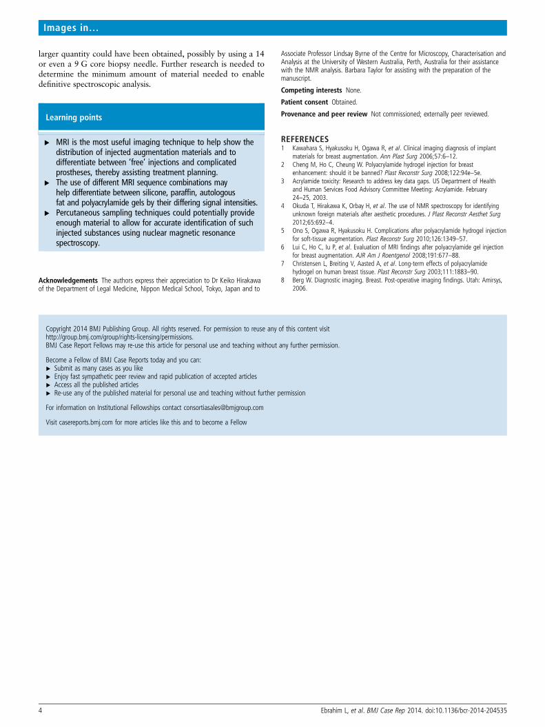
Figure 4 (A–C) Bilateral breast MRI. (A) T2 TI 400, (B) T2 TI 150, (C) T2 silicone fat suppressed, water suppressed (FSWS). There is no signal lossin the silicone-specific sequence (A) and in the fat suppressed sequence (B). In the fat and water suppressed sequence (C) the injected material hasdropped its signal, indicating it hydrophilic in nature. The images demonstrate a large central component, containing thin low signal rim withdiscontinuous areas of signal void, consistent with the mammographic calcification. There is an extension to the subpectoral and interpectoralregions on the left, and presternal area on the right, with no intrathoracic extension.
Table 1 MRI characteristics of the implanted material comparedwith common breast augmentation substances
Type of augmentation MRI Characteristics
Silicone T2—high signal, T1—low signal, siliconesensitive—high signal
Saline T2—high signal, T1—low signal, siliconesensitive—low signal
Autologous fat T2—high signal, T1—high signal, siliconesensitive—low signal, fat suppression—lowsignal, with or without peripheral enhancement
Polyacrylamide gel (similarfindings in our case)
T2—high signal with foci of low signal, T1—low signal, silicone sensitive—low signal, withor without peripheral enhancement
Ebrahim L, et al. BMJ Case Rep 2014. doi:10.1136/bcr-2014-204535 3
Images in…
larger quantity could have been obtained, possibly by using a 14or even a 9 G core biopsy needle. Further research is needed todetermine the minimum amount of material needed to enabledefinitive spectroscopic analysis.
Learning points
▸ MRI is the most useful imaging technique to help show thedistribution of injected augmentation materials and todifferentiate between ‘free’ injections and complicatedprostheses, thereby assisting treatment planning.
▸ The use of different MRI sequence combinations mayhelp differentiate between silicone, paraffin, autologousfat and polyacrylamide gels by their differing signal intensities.
▸ Percutaneous sampling techniques could potentially provideenough material to allow for accurate identification of suchinjected substances using nuclear magnetic resonancespectroscopy.
Acknowledgements The authors express their appreciation to Dr Keiko Hirakawaof the Department of Legal Medicine, Nippon Medical School, Tokyo, Japan and to
Associate Professor Lindsay Byrne of the Centre for Microscopy, Characterisation andAnalysis at the University of Western Australia, Perth, Australia for their assistancewith the NMR analysis. Barbara Taylor for assisting with the preparation of themanuscript.
Competing interests None.
Patient consent Obtained.
Provenance and peer review Not commissioned; externally peer reviewed.
REFERENCES1 Kawahara S, Hyakusoku H, Ogawa R, et al. Clinical imaging diagnosis of implant
materials for breast augmentation. Ann Plast Surg 2006;57:6–12.2 Cheng M, Ho C, Cheung W. Polyacrylamide hydrogel injection for breast
enhancement: should it be banned? Plast Reconstr Surg 2008;122:94e–5e.3 Acrylamide toxicity: Research to address key data gaps. US Department of Health
and Human Services Food Advisory Committee Meeting: Acrylamide. February24–25, 2003.
4 Okuda T, Hirakawa K, Orbay H, et al. The use of NMR spectroscopy for identifyingunknown foreign materials after aesthetic procedures. J Plast Reconstr Aesthet Surg2012;65:692–4.
5 Ono S, Ogawa R, Hyakusoku H. Complications after polyacrylamide hydrogel injectionfor soft-tissue augmentation. Plast Reconstr Surg 2010;126:1349–57.
6 Lui C, Ho C, Iu P, et al. Evaluation of MRI findings after polyacrylamide gel injectionfor breast augmentation. AJR Am J Roentgenol 2008;191:677–88.
7 Christensen L, Breiting V, Aasted A, et al. Long-term effects of polyacrylamidehydrogel on human breast tissue. Plast Reconstr Surg 2003;111:1883–90.
8 Berg W. Diagnostic imaging. Breast. Post-operative imaging findings. Utah: Amirsys,2006.
Copyright 2014 BMJ Publishing Group. All rights reserved. For permission to reuse any of this content visithttp://group.bmj.com/group/rights-licensing/permissions.BMJ Case Report Fellows may re-use this article for personal use and teaching without any further permission.
Become a Fellow of BMJ Case Reports today and you can:▸ Submit as many cases as you like▸ Enjoy fast sympathetic peer review and rapid publication of accepted articles▸ Access all the published articles▸ Re-use any of the published material for personal use and teaching without further permission
For information on Institutional Fellowships contact [email protected]
Visit casereports.bmj.com for more articles like this and to become a Fellow
4 Ebrahim L, et al. BMJ Case Rep 2014. doi:10.1136/bcr-2014-204535
Images in…