Embed Size (px)
Citation preview
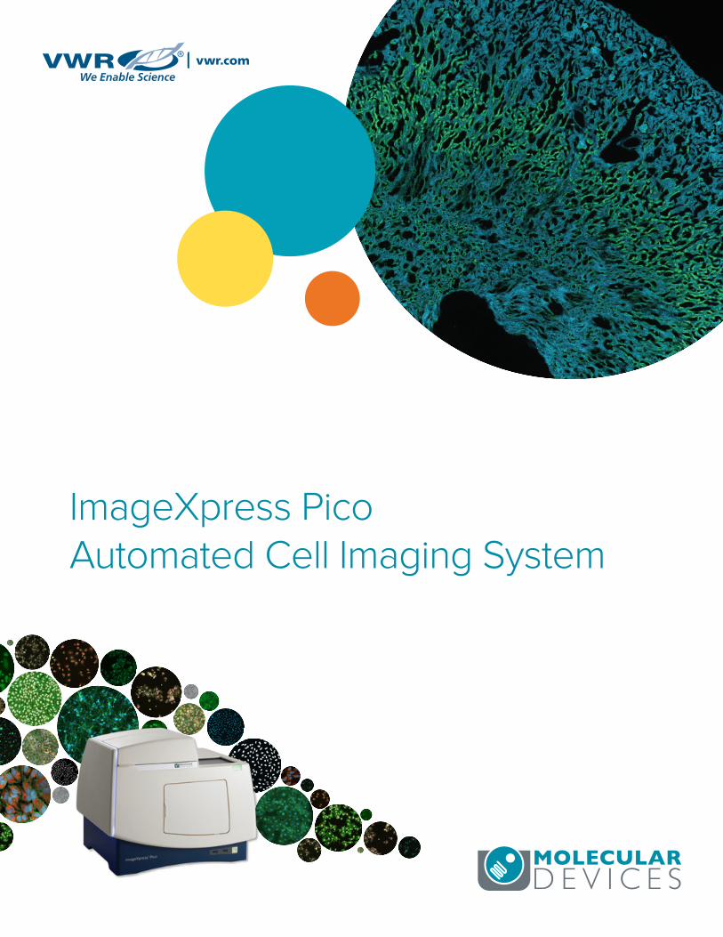
ImageXpress Pico Automated Cell Imaging System
www.moleculardevices.com2
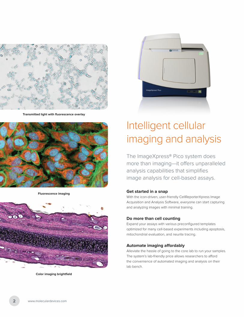
Intelligent cellular imaging and analysis
The ImageXpress® Pico system does more than imaging—it offers unparalleled analysis capabilities that simplifies image analysis for cell-based assays.
Get started in a snapWith the icon-driven, user-friendly CellReporterXpress Image
Acquisition and Analysis Software, everyone can start capturing
and analyzing images with minimal training.
Do more than cell countingExpand your assays with various preconfigured templates
optimized for many cell-based experiments including apoptosis,
mitochondrial evaluation, and neurite tracing.
Automate imaging affordablyAlleviate the hassle of going to the core lab to run your samples.
The system’s lab-friendly price allows researchers to afford
the convenience of automated imaging and analysis on their
lab bench.
Transmitted light with fluorescence overlay
Fluorescence imaging
Color imaging brightfield
ImageXpress Pico Automated Cell Imaging System 3
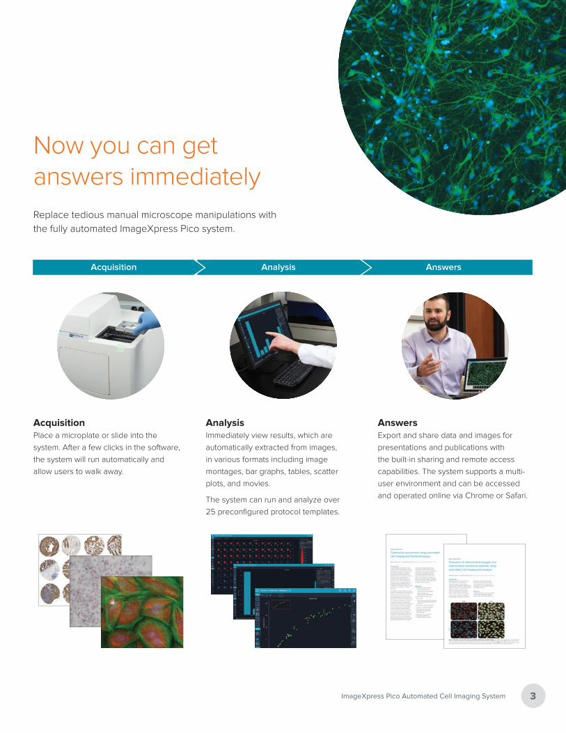
Now you can get answers immediatelyReplace tedious manual microscope manipulations with
the fully automated ImageXpress Pico system.
AcquisitionPlace a microplate or slide into the
system. After a few clicks in the software,
the system will run automatically and
allow users to walk away.
AnalysisImmediately view results, which are
automatically extracted from images,
in various formats including image
montages, bar graphs, tables, scatter
plots, and movies.
The system can run and analyze over
25 preconfigured protocol templates.
AnswersExport and share data and images for
presentations and publications with
the built-in sharing and remote access
capabilities. The system supports a multi-
user environment and can be accessed
and operated online via Chrome or Safari.
Acquisition Analysis Answers
www.moleculardevices.com4
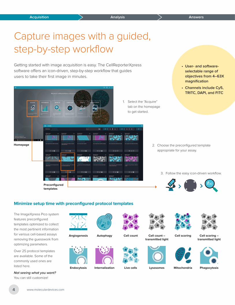
Capture images with a guided, step-by-step workflowGetting started with image acquisition is easy. The CellReporterXpress
software offers an icon-driven, step-by-step workflow that guides
users to take their first image in minutes.
Acquisition Analysis Answers
Angiogenesis Autophagy Cell count Cell count – transmitted light
Cell scoring Cell scoring – transmitted light
Endocytosis Internalization Live cells Lysosomes Mitochondria Phagocytosis
Minimize setup time with preconfigured protocol templates
Acquisition
1. Select the “Acquire”
tab on the homepage
to get started.
2. Choose the preconfigured template
appropriate for your assay.
• User- and software-selectable range of objectives from 4–63X magnification
• Channels include Cy5, TRITC, DAPI, and FITC
3. Follow the easy icon-driven workflow.
f(x)
The ImageXpress Pico system
features preconfigured
templates optimized to collect
the most pertinent information
for various cell-based assays
removing the guesswork from
optimizing parameters.
Over 25 protocol templates
are available. Some of the
commonly used ones are
listed here.
Not seeing what you want?
You can still customize!
Homepage
Preconfigured templates
Answers
5ImageXpress Pico Automated Cell Imaging System
Acquisition Analysis Answers
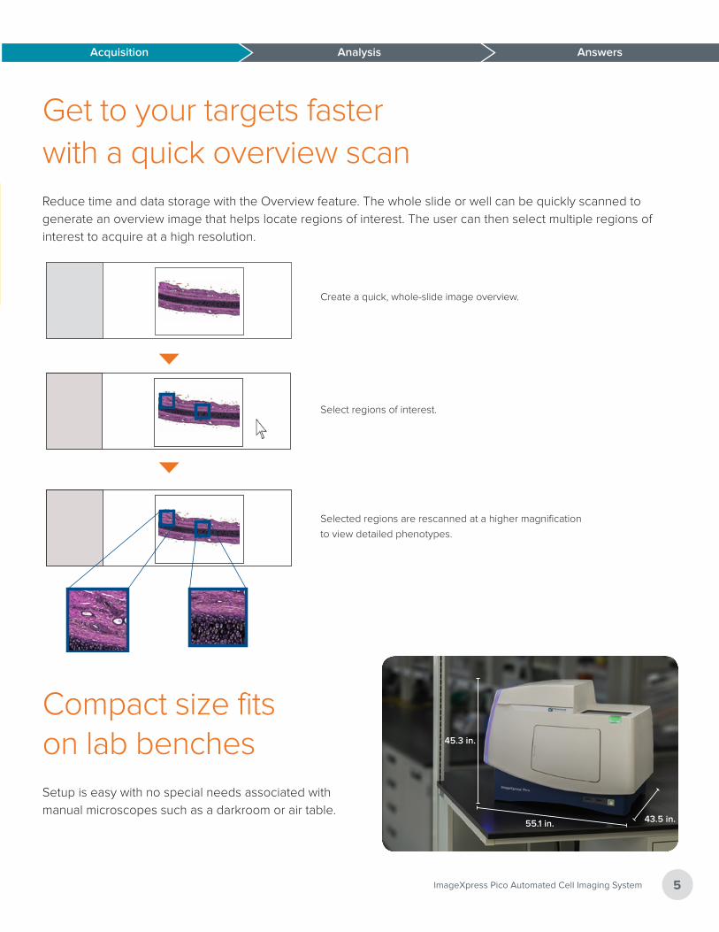
Get to your targets faster with a quick overview scanReduce time and data storage with the Overview feature. The whole slide or well can be quickly scanned to
generate an overview image that helps locate regions of interest. The user can then select multiple regions of
interest to acquire at a high resolution.
Selected regions are rescanned at a higher magnification
to view detailed phenotypes.
Select regions of interest.
Create a quick, whole-slide image overview.Create a quick, whole-slide image overview.
Selected regions are rescanned at a higher magnification to view detailed phenotypes.
Select regions of interest.
Compact size fits on lab benchesSetup is easy with no special needs associated with
manual microscopes such as a darkroom or air table.
45.3 in.
55.1 in. 43.5 in.
Mitochondria
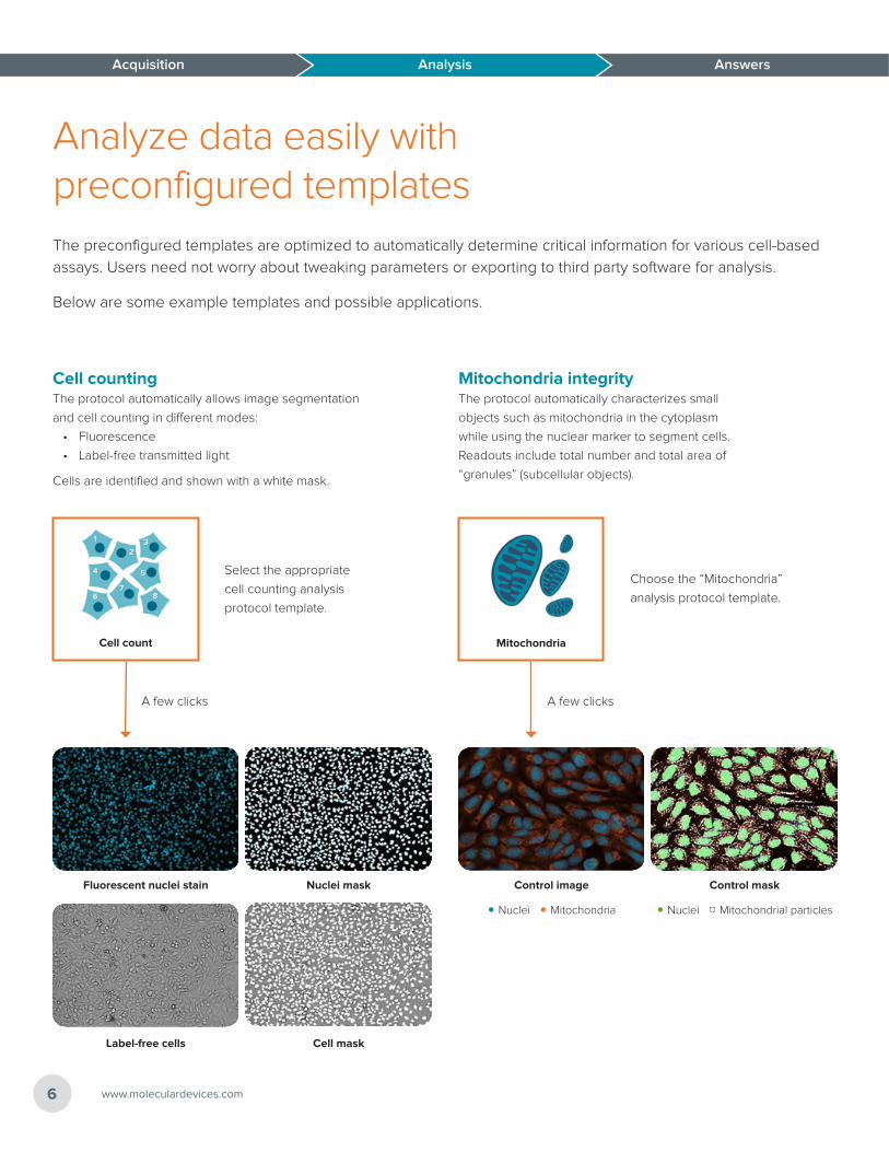
Analyze data easily with preconfigured templatesThe preconfigured templates are optimized to automatically determine critical information for various cell-based
assays. Users need not worry about tweaking parameters or exporting to third party software for analysis.
Below are some example templates and possible applications.
Acquisition Analysis Answers
Cell countingThe protocol automatically allows image segmentation
and cell counting in different modes:
• Fluorescence
• Label-free transmitted light
Cells are identified and shown with a white mask.
Fluorescent nuclei stain Nuclei mask
Label-free cells Cell mask
Cell count
Select the appropriate
cell counting analysis
protocol template.
A few clicks
Mitochondria integrityThe protocol automatically characterizes small
objects such as mitochondria in the cytoplasm
while using the nuclear marker to segment cells.
Readouts include total number and total area of
“granules” (subcellular objects).
Control image Control mask
Acquisition
• Nuclei • Mitochondria • Nuclei • Mitochondrial particles
Choose the “Mitochondria”
analysis protocol template.
A few clicks
www.moleculardevices.com6
Neurite tracing
Cell scoring
Answers
7ImageXpress Pico Automated Cell Imaging System
Answers
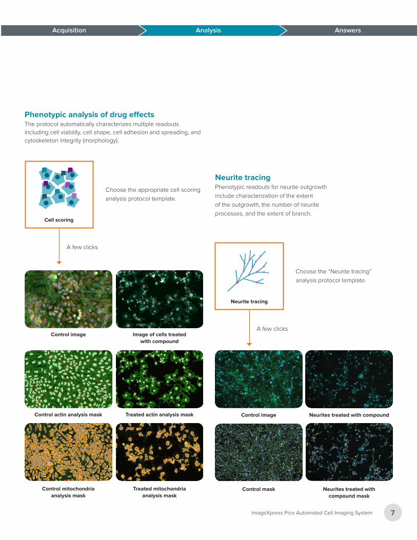
Phenotypic analysis of drug effectsThe protocol automatically characterizes multiple readouts including cell viability, cell shape, cell adhesion and spreading, and cytoskeleton integrity (morphology).
Control image Image of cells treated with compound
Control actin analysis mask Treated actin analysis mask
Acquisition Analysis
Control mitochondria analysis mask
Treated mitochondria analysis mask
Choose the appropriate cell scoring
analysis protocol template.
A few clicks
Neurite tracingPhenotypic readouts for neurite outgrowth
include characterization of the extent
of the outgrowth, the number of neurite
processes, and the extent of branch.
Control image Neurites treated with compound
Control mask Neurites treated with compound mask
Choose the “Neurite tracing”
analysis protocol template.
A few clicks
www.moleculardevices.com8
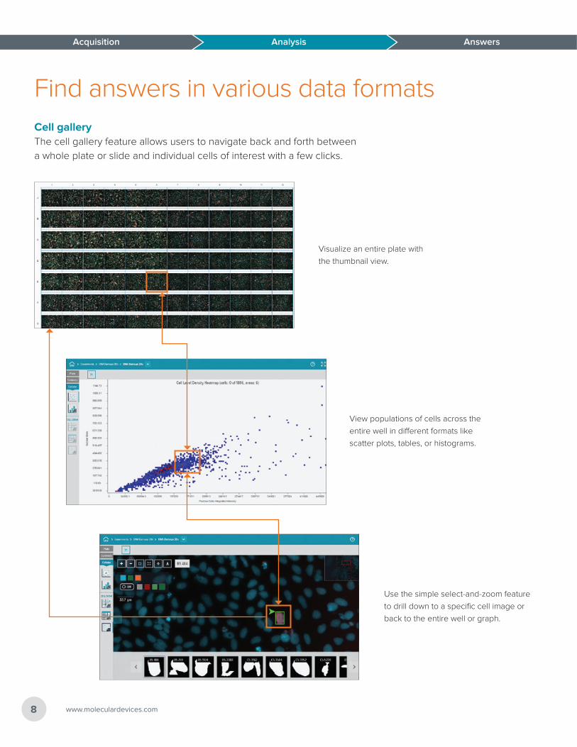
Find answers in various data formatsCell galleryThe cell gallery feature allows users to navigate back and forth between
a whole plate or slide and individual cells of interest with a few clicks.
Visualize an entire plate with
the thumbnail view.
Use the simple select-and-zoom feature
to drill down to a specific cell image or
back to the entire well or graph.
View populations of cells across the
entire well in different formats like
scatter plots, tables, or histograms.
Acquisition Analysis Answers Acquisition
9ImageXpress Pico Automated Cell Imaging System
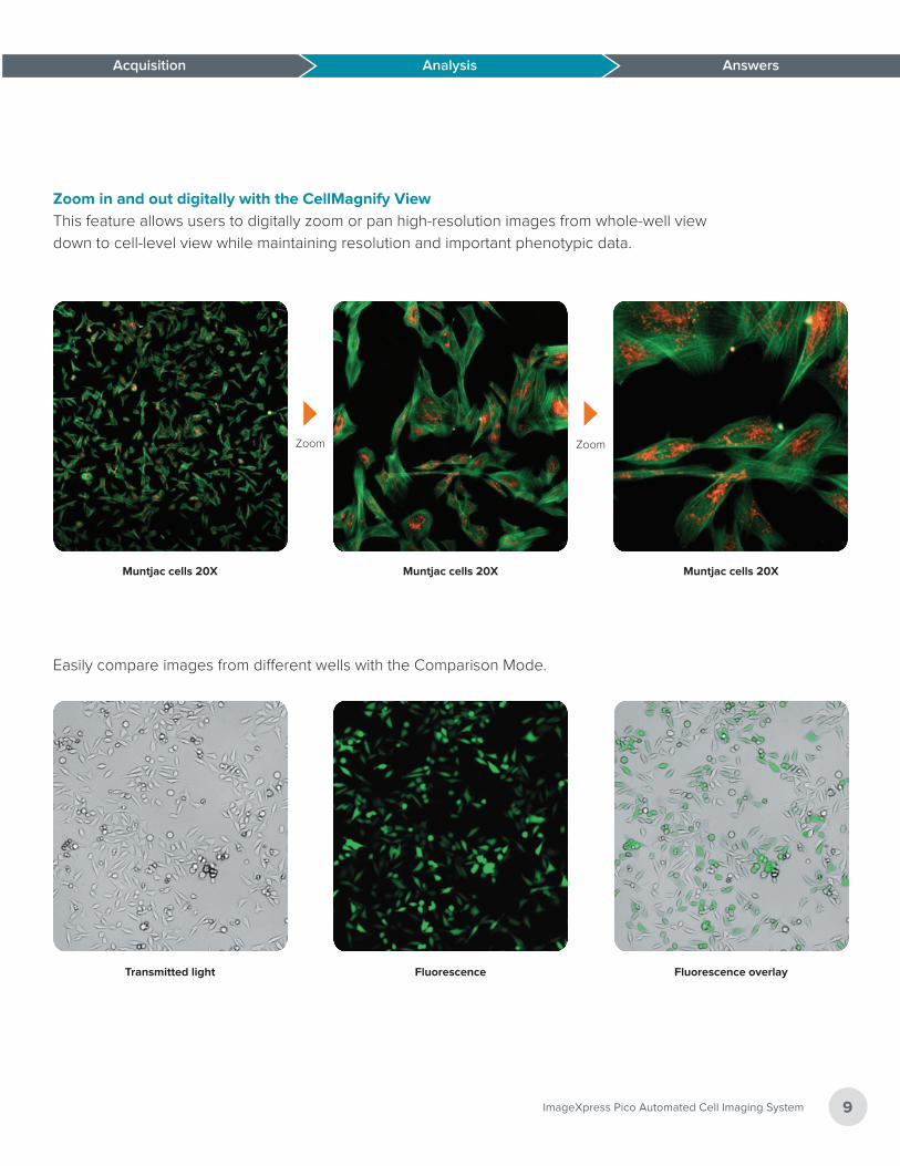
Zoom in and out digitally with the CellMagnify ViewThis feature allows users to digitally zoom or pan high-resolution images from whole-well view
down to cell-level view while maintaining resolution and important phenotypic data.
Zoom Zoom
Muntjac cells 20X
Easily compare images from different wells with the Comparison Mode.
Transmitted light Fluorescence overlay
Muntjac cells 20XMuntjac cells 20X
Fluorescence
Answers AnswersAcquisition Analysis
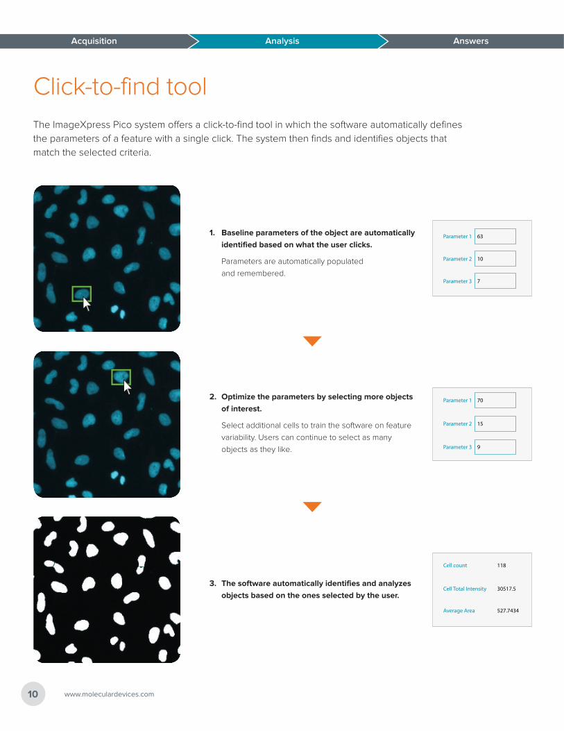
Click-to-find toolThe ImageXpress Pico system offers a click-to-find tool in which the software automatically defines
the parameters of a feature with a single click. The system then finds and identifies objects that
match the selected criteria.
1. Baseline parameters of the object are automatically
identified based on what the user clicks.
Parameters are automatically populated
and remembered.
Parameter 1
Parameter 2
Parameter 3
63
10
7
2. Optimize the parameters by selecting more objects
of interest.
Select additional cells to train the software on feature
variability. Users can continue to select as many
objects as they like.
Parameter 1
Parameter 2
Parameter 3
70
15
9
3. The software automatically identifies and analyzes
objects based on the ones selected by the user.
Cell count
Cell Total Intensity
Average Area
118
30517.5
527.7434
www.moleculardevices.com10
Acquisition Analysis Answers
Acquisition Analysis Answers
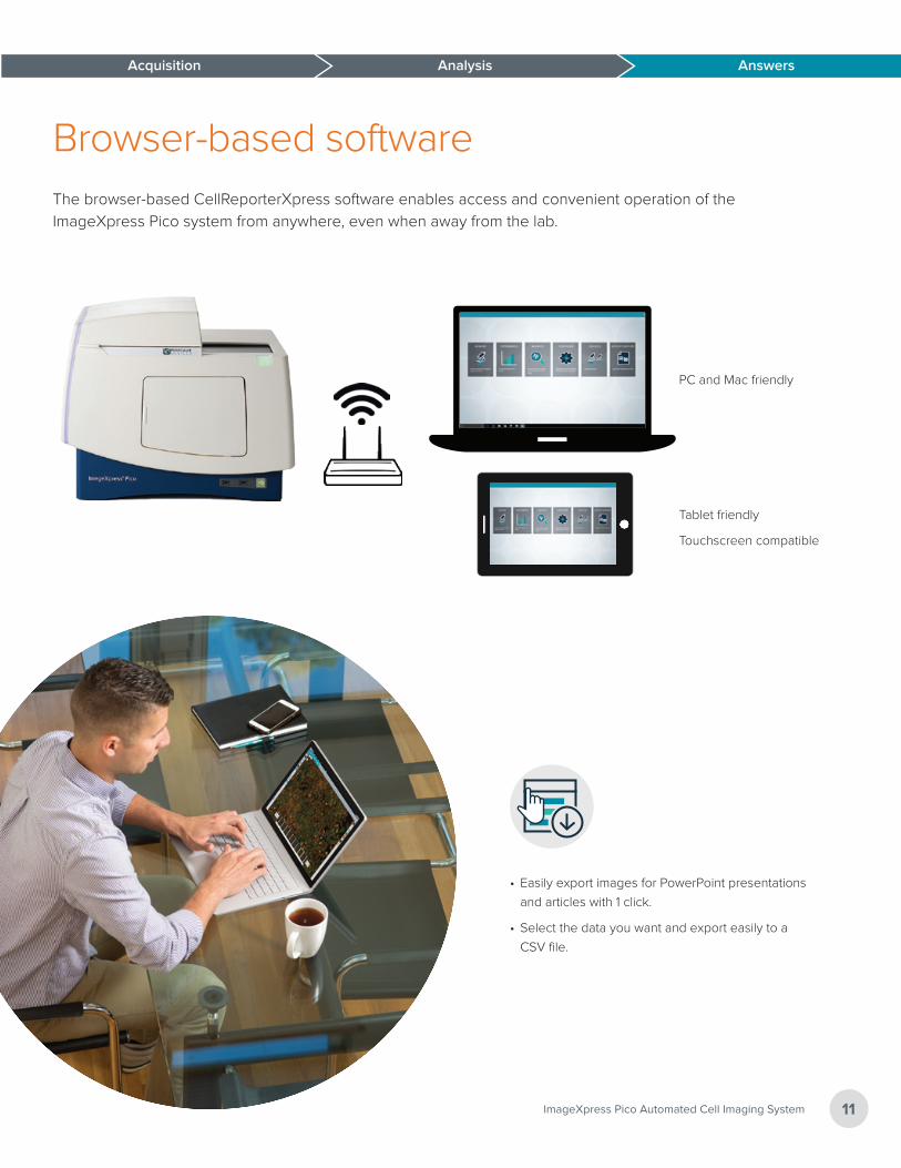
Browser-based softwareThe browser-based CellReporterXpress software enables access and convenient operation of the
ImageXpress Pico system from anywhere, even when away from the lab.
PC and Mac friendly
Tablet friendly
Touchscreen compatible
11ImageXpress Pico Automated Cell Imaging System
• Easily export images for PowerPoint presentations
and articles with 1 click.
• Select the data you want and export easily to a
CSV file.
Answers
The trademarks used herein are the property of Molecular Devices, LLC or their respective owners. Specifications subject to change without notice. Patents: www.moleculardevices.com/productpatents FOR RESEARCH USE ONLY. NOT FOR USE IN DIAGNOSTIC PROCEDURES. 5/18 2184A-VWR
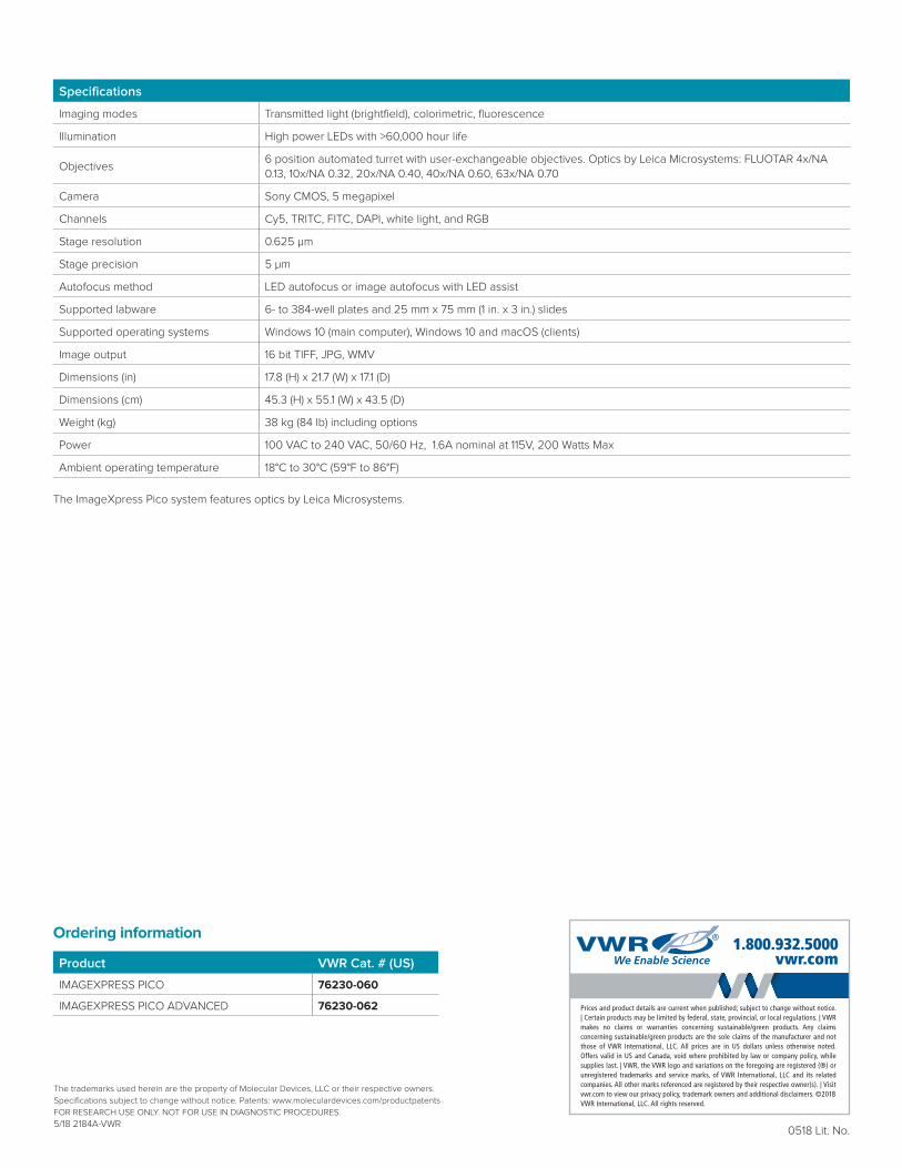
Specifications
Imaging modes Transmitted light (brightfield), colorimetric, fluorescence
Illumination High power LEDs with >60,000 hour life
Objectives6 position automated turret with user-exchangeable objectives. Optics by Leica Microsystems: FLUOTAR 4x/NA 0.13, 10x/NA 0.32, 20x/NA 0.40, 40x/NA 0.60, 63x/NA 0.70
Camera Sony CMOS, 5 megapixel
Channels Cy5, TRITC, FITC, DAPI, white light, and RGB
Stage resolution 0.625 µm
Stage precision 5 µm
Autofocus method LED autofocus or image autofocus with LED assist
Supported labware 6- to 384-well plates and 25 mm x 75 mm (1 in. x 3 in.) slides
Supported operating systems Windows 10 (main computer), Windows 10 and macOS (clients)
Image output 16 bit TIFF, JPG, WMV
Dimensions (in) 17.8 (H) x 21.7 (W) x 17.1 (D)
Dimensions (cm) 45.3 (H) x 55.1 (W) x 43.5 (D)
Weight (kg) 38 kg (84 lb) including options
Power 100 VAC to 240 VAC, 50/60 Hz, 1.6A nominal at 115V, 200 Watts Max
Ambient operating temperature 18°C to 30°C (59°F to 86°F)
The ImageXpress Pico system features optics by Leica Microsystems.
0518 Lit. No.
Ordering information
Product VWR Cat. # (US)
IMAGEXPRESS PICO 76230-060
IMAGEXPRESS PICO ADVANCED 76230-062