Embed Size (px)
Citation preview

1
Imaging in Gynecology: What is
AppropriateFrancisco A. Quiroz, MD
Appropriate• Right or suitable• To set apart for a specific use
Appropriateness• The quality or state for being especially
suitable or fitting

2
Imaging Modalities
Ultrasound Pelvis• Trans abdominal• Transvaginal
Doppler 3-D
• Hysterosonogram Computed Tomography MR PET
Practice Guidelines
Describe recommended conduct in specific areas of clinical practice. They are based on analysis of current literature, expert opinion, open forum commentary and informal consensus
Consensus Conference
National Institutes of Health (NIH) U.S. Preventive Services Task Force Centers for Disease Control (CDC) National Comprehensive Cancer
Network (NCCN) American College of Physicians American College of Radiology Specialty Societies

3
Methodology
Steps in consensus development ?• Formulation of the question or
topic selection• Panel composition – requirements• Literature review• Assessment of scientific evidence
or critical appraisal• Presentation and discussion
• Drafting of document• Recommendations for future
research• Peer review• Statement document• Publication – Dissemination• Periodic review and updating
ACR Appropriateness Criteria
Evidence based guidance to assist referring physicians and other providers in making the most appropriate imaging or treatment decision for a specific clinical condition

4
Appropriateness Criteria
Expert panels • Diagnostic imaging• Medical specialty organizations
American Congress of Obstetricians and Gynecologists
Society of Gynecologic Oncologists
Structured process for development of criteria
Availability Expertise Radiation Use of contrast Cost
Clinical Information
ACR Criteria – Rating Scale
Scale 1-9 3 Categories
• 1 – 3 “usually not appropriate”• 4 – 6 “may be appropriate”• 7 – 9 “usually appropriate”

5
Usually appropriate• Study indicated in certain clinical
settings at a favorable risk-benefit ratio for patients, as supported by published peer-review scientific studies, supplemented by expert opinion
Clinical Scenarios
Abnormal vaginal bleeding Acute pelvic pain Adnexal mass Staging and follow up of ovarian
cancer Evaluation and follow up of
endometrial cancer Cancer cervix
VAGINAL BLEEDING

6
Vaginal Bleeding
Endometrial sampling the most appropriate initial step in evaluation
vaginal bleeding depending on clinical situation (Endometrial Bx, D&C)
Vaginal Bleeding Role of imaging
• Screening• Detection and characterization of focal
structural abnormalities• Direction appropriate patient care• Inconclusive biopsy results• Persistent bleeding despite negative findings
Sampling error ~ 60 % endometrial cavity curetted with D&C
Acta Obstet Gynecol Scand 2001
www.acr.org/Quality-Safety/Appropriateness-Criteria/Diagnostic
7
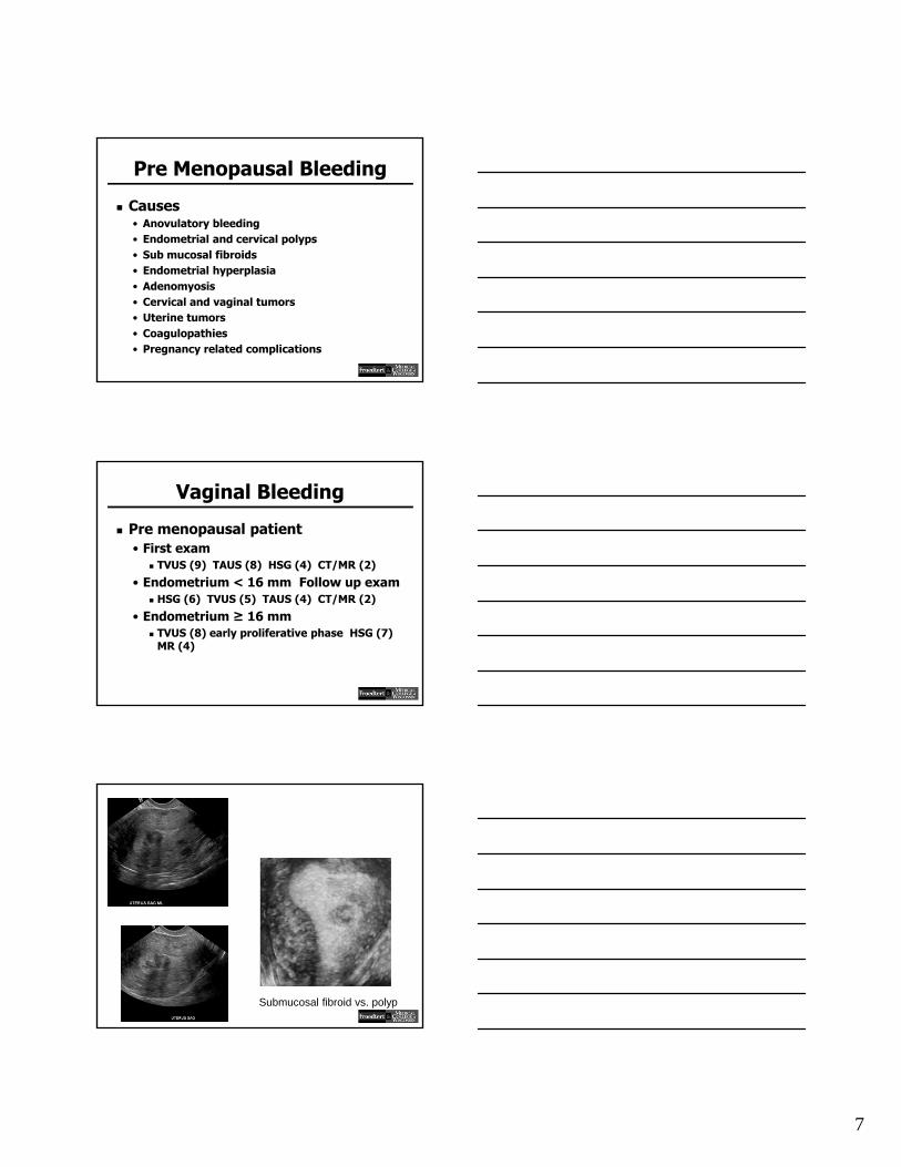
Pre Menopausal Bleeding
Causes• Anovulatory bleeding• Endometrial and cervical polyps• Sub mucosal fibroids• Endometrial hyperplasia• Adenomyosis• Cervical and vaginal tumors• Uterine tumors• Coagulopathies• Pregnancy related complications
Vaginal Bleeding
Pre menopausal patient• First exam
TVUS (9) TAUS (8) HSG (4) CT/MR (2)
• Endometrium < 16 mm Follow up exam HSG (6) TVUS (5) TAUS (4) CT/MR (2)
• Endometrium ≥ 16 mm TVUS (8) early proliferative phase HSG (7)
MR (4)
Submucosal fibroid vs. polyp

8
Submucosal leiomyoma
Submucosal leiomyoma
Vaginal Bleeding
Heterogeneous endometrium
Suspected focal abnormality
Endometrium not adequately visualized at transvaginal exam
Hysterosonogram (Rating 8 ACR)

9
Heterogeneous Endometrium ? Focal Abnormality Not adequately visualized
MR (5-6)
Heterogeneous endometrium - ? Focal abnormality Endometrium not adequately visualized
Endometrial polyp and submucosal leiomyoma HSG - ACR (8)
Pre Menopausal Bleeding

10
TAUS (4)
TVUS (8)
US Doppler (5)
Endometrium 16 mm Doppler added value –further characterize endometrial abnormality
Blood flow in intracavitary lesion excludes retained blood clot
TAUS
Wider field of view Increased depth of penetration Evaluation adjacent organs
• Uterus in neutral position• Poor penetration by TVUS• Markedly enlarged fibroid uterus
Subserosal or pedunculated
• Intolerance to vaginal probe
Uterine Leiomyoma
11
Uterine leiomyomas
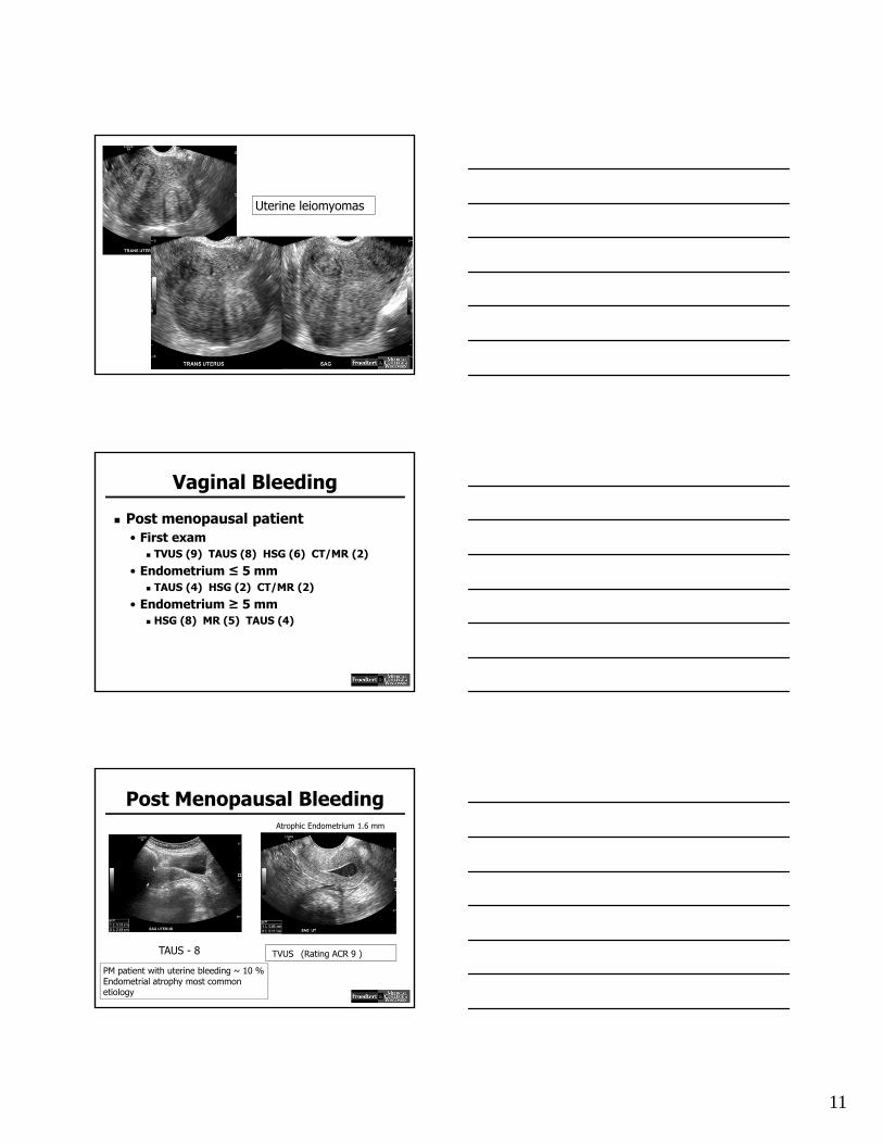
Vaginal Bleeding
Post menopausal patient• First exam
TVUS (9) TAUS (8) HSG (6) CT/MR (2)
• Endometrium ≤ 5 mm TAUS (4) HSG (2) CT/MR (2)
• Endometrium ≥ 5 mm HSG (8) MR (5) TAUS (4)
Post Menopausal BleedingAtrophic Endometrium 1.6 mm
TAUS - 8 TVUS (Rating ACR 9 )
PM patient with uterine bleeding ~ 10 % Endometrial atrophy most common etiology

12
Post Menopausal Bleeding
Endometrioid endometrial Ca stage 1A
Post Menopausal Bleeding
Hysterosonogram - (Rating ACR 8)
Endometrial Ca
Pre Menopausal Bleeding
Endometrial polyp

13
Thickened Endometrium with cysts
Nov 2008
28 y.o dysfunctional bleeding. Endometrium 8 mm
Premenopausal Endometrial Bleeding Endometrium < 16 mm F/U Dec. 2011
Submucosal fundal leiomyoma
HSG (8)
14
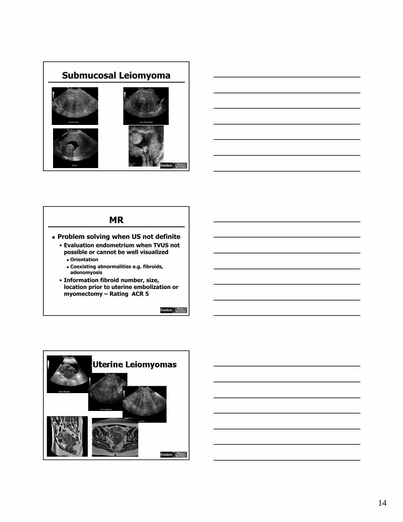
Submucosal Leiomyoma
MR
Problem solving when US not definite• Evaluation endometrium when TVUS not
possible or cannot be well visualized Orientation Coexisting abnormalities e.g. fibroids,
adenomyosis
• Information fibroid number, size, location prior to uterine embolization or myomectomy – Rating ACR 5
Uterine Leiomyomas

15
Evaluation Pre Uterine Embolization
Poorly visualized fibroids in US. Multiple pedunculated fibroids and closeness to endometrium prevents intervention
AJR 2006;187:1499
Evaluation Pre Uterine Embolization
AJR 2006;187:1499
ACUTE PELVIC PAIN

16
Acute Pelvic Pain
Obstetrical causes Gynecologic causes
• Simple ovarian cysts• Ruptured or hemorrhagic ovarian cysts• Pelvic inflammatory disease• Ovarian torsion• Malposition of intrauterine devices
Non gynecologic causes• Appendicitis• Inflammatory bowel disease• Diverticulitis• Urinary tract calculi• Pyelonephritis
Imaging
Choice of Imaging modality determined by clinically suspected differential diagnosis• Clinical history• Physical exam• Laboratory tests

17
Imaging
Pelvic Ultrasound (TAUS and TVS)Preferred modalities for initial evaluation when obstetric or gynecologic causes are suspected
Computed Tomography or Magnetic Resonance (MR) Gastrointestinal or urinary tract etiology is suspected
Acute Pelvic Pain
Reproductive age group• Gynecological etiology suspected
positive serum β-hCG negative serum β-hCG
• Non gynecological etiology suspected Positive serum β-hCG Negative serum β-hCG
Gynecological and non gynecologic etiology suspected and positive serum β-hCG
• TAUS/TVS – Rating ACR 9 Ectopic pregnancy
• MR Abdomen/Pelvis – Rating ACR 6 Appendicitis MR Urography – detection obstructive
uropathy vs. physiologic dilatation of pregnancy
18
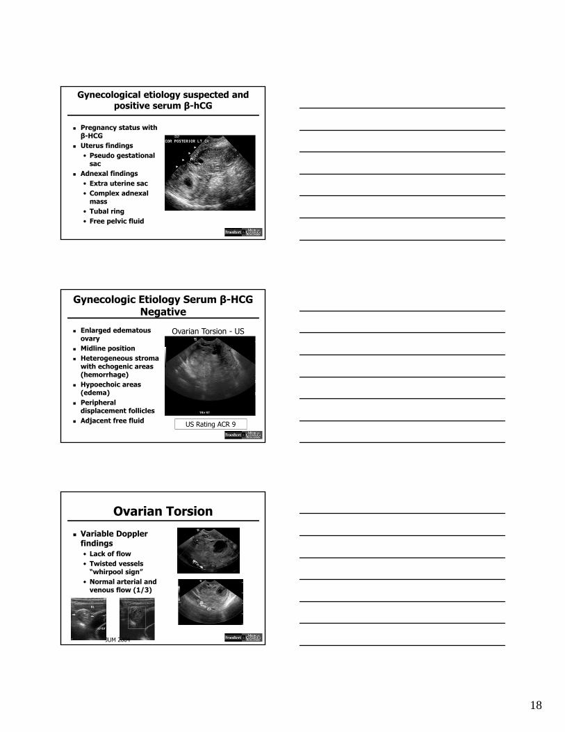
Gynecological etiology suspected and positive serum β-hCG
Pregnancy status with β-HCG
Uterus findings• Pseudo gestational
sac Adnexal findings
• Extra uterine sac• Complex adnexal
mass• Tubal ring• Free pelvic fluid
Gynecologic Etiology Serum β-HCG Negative
Enlarged edematous ovary
Midline position Heterogeneous stroma
with echogenic areas (hemorrhage)
Hypoechoic areas (edema)
Peripheral displacement follicles
Adjacent free fluid US Rating ACR 9
Ovarian Torsion - US
Ovarian Torsion
Variable Doppler findings• Lack of flow• Twisted vessels
“whirpool sign”• Normal arterial and
venous flow (1/3)
JUM 2004

19
Ovarian Torsion CT findings
• Enlarged ovary with or without associated ovarian mass
• Ipsilateral twisted pedicle – rare
• Deviation uterus on twisted side
• Sub acute ovarian hemorrhage
• Abnormal enhancement with contrast
CT/MR rating ACR 4
Gynecologic Etiology Serum β-HCG Negative
Ruptured or hemorrhagic ovarian cyst.Most common gynecologic cause acute pelvic pain
US – ACR 9
Hemorrhagic Ovarian Cyst
September 17
July 29

20
Hemorrhagic Ovarian Cyst
Gynecologic Etiology Serum β-HCG Negative
Pelvic Inflammatory Disease
Spectrum STD involves cervix, uterus, fallopian tubes and ovaries
US - ACR 9
PID - USTubo ovarian complex Tubo ovarian abscess

21
PID - CT
CT - ACR 6
pyosalpinx
Pts with diffuse pelvic pain, peritonitis or difficult or equivocal USEarly or mild inflammatory changes may be better appreciated on CT
PID - CT
Global view of disease process and extension
PID- TOA

22
Bilateral TOA
Rarely used in PIDVery sensitive to detection inflammationComplementary problem solving method
MR – ACR 6
Bilateral TOA TVUS Drainage
Image guided abscess drainage TVUS - CT
Bilateral TOA TVUS & CT Drainage

23
Intramural IUD
Patients often with pelvic pain3D-US more accurate and sensitive than TVUS
IUD – PT with Pelvic Pain
Malposition IUD in lower uterine segment with intramural extension
Chronic pelvic pain• Adenomyosis• Endometriosis• Pelvic congestion syndrome

24
Adenomyosis
Sub endometrial/myometrial cysts dilated cystic glands
Echogenic linear striations heterotopic endometrium extending into inner myometrium
Adenomyosis
Decreased echogenicity endometrium hyperplasia smooth muscle
Heterogeneity small echogenic islands of heterotopic endometrial tissue surrounded by smooth muscle
Reinhold. RG 1999
Adenomyosis - MR
Diffuse/focal thickening junctional zone smooth muscle hyperplasia
Embedded bright foci T2W ectopic endometrial tissue + cystic dilatation glands

25
Endometriosis US
Unilocular homogenously hypoechoic cyst with diffuse low level echoes and increased through transmission. Echogenic mural foci
Endometriosis MR
T1W
T1 fat suppressed
T2WWoodward. RG 2001
High signal intensity T1-T2 blood products and concentrated protein
Shading – loss signal within lesion (chronic)
Pelvic Congestion Syndrome
Tortuous dilated veins incompetent valvesUS/CT multiple dilated varicose veins surrounding pelvic organs

26
Non Gynecologic etiology serum βpositive
Pregnant + RLQ ? Appendicitis• TAUS/TVUS usually appropriate (ACR 9) no
radiation but poor visualization appendix (normal appendix 13-50 %), limited graded compression variable sensitivity and specificity
• MR without contrast (ACR 8) no ionizing radiation > sensitivity and specificity than US
• CT when US non diagnostic, MR unavailable or equivocal. Need prompt Dx of potentially life-threatening condition (ACR 4)
Non Gynecologic Etiology Serum βPositive
Normal appendix on MR
Pregnant Patient – RLQ Pain ? Appendicitis
US – ACR 9
MR ACR 8
CT ACR 4
Non Gynecologic etiology serum βnegative
US – ACR 7
Avoid radiation exposure younger patients

27
Non Gynecologic etiology serum βnegative
Appendicitis Diverticulitis
Inflammatory bowel disease Litiasis ureteral
CT – ACR 9
Non Gynecologic etiology serum βnegative
19 y.o female RLQ pain CT preferred modality for detecting bowel pathology
Crohn’s Disease
CT – ACR 9
Non Gynecologic etiology serum βnegative
Acute Pyelonephritis CT – ACR 9

28
ADNEXAL MASS
Adnexal Mass
US exam of choice for evaluation patient with suspected adnexal mass
Characterization mass as cystic, solid or complex
Color/power Doppler adjunct to gray scale imaging
Spectral Doppler not reliable in differentiate malignant from benign masses
Adnexal Mass
Reproductive age• First exam
TVUS, TAUS, Doppler – (ACR 9), MR (ACR 6)
Reproductive age• Complex or solid mass detected prior
pelvic US. Follow-up recommendations Ultrasound (ACR 9), MR (ACR 5)

29
Ovarian Cyst
Simple cyst benign process 100 % pre menopausal women most resolve spontaneously
Simple cyst PM woman (17-24 %) ≥ 5 cm rarely malignant
Ovarian Masses
Ovarian Masses
Confidence characterization lesions such as cystic teratoma

30
Adnexal Mass
Determine origin mass uterine vs. ovarianPedunculated leiomyoma
MR – ACR 5
Adnexal Mass
Complex or solid mass detected by prior pelvic US getting smaller at short-term follow-up• TVUS (ACR 9) if resolved, no further
imaging required Complex or solid mass persistent or
enlarging on pelvic US at short term F/U• MR (ACR 8), US (ACR 5), CT (ACR 4) or
surgery in the appropriate clinical setting
Adnexal Mass
Reproductive age• Initial US large cyst > 5 cm apparently
simple TVUS (ACR 9) > 5 cm but ≤ 7 cm annual F/U
SRU Consensus Conf. Radiology 2010
MR (ACR 4) – indeterminate cyst or inadequate US
• Origin of the mass• Characterization – benign vs. malignant features?

31
Adnexal mass Post menopausal patient
• Initial evaluation US (ACR – 9) MR (ACR 5)
• Simple ovarian cyst > 1 cm by pelvic US Follow-up recommendations Annual F/U to ensure stability > 7 cm consider MR
• Complex or solid mass by pelvic US Follow-up recommendations MR (ACR 5) – Consider surgical evaluation
SRU Consensus Conf. Radiology 2010
Post menopausal Patient
Post Menopausal Patient
32
STAGING AND FOLLOW UP
OVARIAN CANCER
Staging & FU Ovarian Ca
Pre treatment staging of ovarian cancer• CT (ACR 9) MR (ACR 7) PET (ACR 4)
US (ACR 3) Rule out recurrence ovarian cancer
• CT abd/pelvis (ACR 9) PET-CT (ACR 8) CT c/a/p (ACR 6) MR (ACR 4) US (ACR 3)
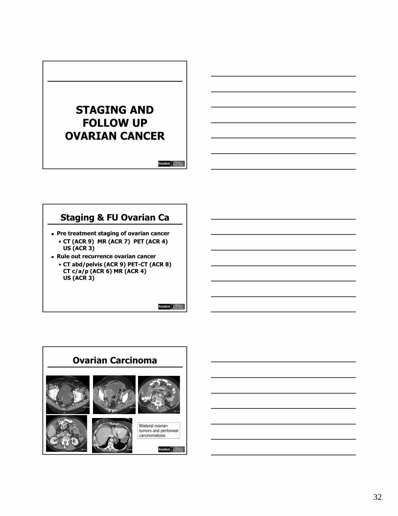
Ovarian Carcinoma
Bilateral ovarian tumors and peritoneal carcinomatosis
33
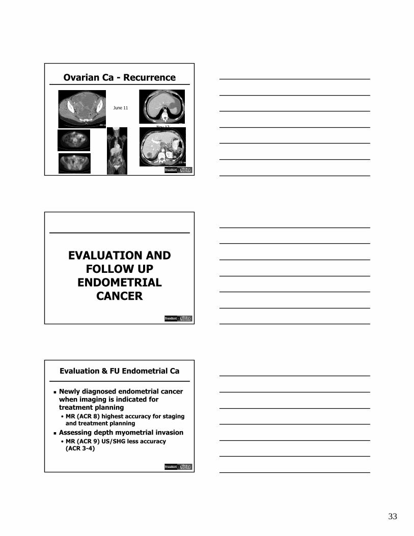
Ovarian Ca - Recurrence
June 11
Nov 12
EVALUATION AND FOLLOW UP
ENDOMETRIAL CANCER
Evaluation & FU Endometrial Ca
Newly diagnosed endometrial cancer when imaging is indicated for treatment planning• MR (ACR 8) highest accuracy for staging
and treatment planning
Assessing depth myometrial invasion• MR (ACR 9) US/SHG less accuracy
(ACR 3-4)

34
Endometrial Carcinoma
Endocervical extension
Evaluation & FU Endometrial Ca
Lymph node evaluation• PET-CT (ACR – 9) CT (ACR 8)
MR (ACR -8) pre and post treatment
Assessing endocervical extent• MR pelvis (ACR – 9)
Post therapy evaluation in patients with clinically suspected recurrence• PET (ACR 9) MR (ACR 8) CT (ACR 8)
INVASIVE CANCER CERVIX

35
FIGO Classification
Cervical CA is staged by the International Federation of Gynecology and Obstetrics (FIGO) classification, based on clinical examination including physical exam under anesthesia, colposcopy, endocervical curettage, hysteroscopy, cystoscopy, proctoscopy, intravenous urography, barium enema, and radiographs of lungs and skeleton
Invasive Ca Cervix
Role • Pre treatment evaluation• Assess tumor size and location• Detect involvement parametrium• Detect involvement sidewall and
adjacent organs• Evaluate for lymph node metastases
FIGO Staging Cervix Ca Stage IB clinically
visible lesions limited to the cervix or pre clinical cancers > than stage IA
IB1 clinically visible lesion < 4.0 cm
IB2 clinically visible lesion > 4.0 cm

36
Invasive Ca Cervix
Pre treatment planning• FIGO stage 1b1, tumor size < 4 cm
MR (ACR – 8) PET-CT (8) CT (ACR 5) US (ACR 2)
• FIGO stage 1b2, tumor size > 4 cm MR (ACR – 9) PET-CT (ACR 9) CT (ACR – 5)
• FIGO stage greater than 1b MR (ACR – 9) PET-CT (ACR – 9) CT c/a/b
(ACR – 7)
Ca Cervix
Poorly differentiated neuroendocrine tumor
Ca Cervix – LT Iliac Lymphadenopathy
Pre treatment nov 2012
Post treatment feb 2013

37
Conclusions
Gynecologic Imaging useful in wide variety of clinical presentations
Ultrasound usually the most appropriate modality
Modalities such as MR, CT are problem solving and improved characterization
Major role of MR, CT and PET-CT in Gyn Oncologic imaging


![Enema Administration[1]](https://img.pdfslide.net/doc/110x75/55289a1f49795921048b4a43/enema-administration1.jpg)