Embed Size (px)
Citation preview
PICTORIAL REVIEW
Imaging of patellar fractures
Mohamed Jarraya1,2 & Luis E. Diaz3 & William F. Arndt3 & Frank W. Roemer4 &
Ali Guermazi2
Received: 15 September 2016 /Revised: 9 November 2016 /Accepted: 15 November 2016 /Published online: 30 November 2016# The Author(s) 2016. This article is published with open access at Springerlink.com
Abstract Patellar fractures account for approximately 1% ofall skeletal fractures and may result from direct, indirect, orcombined trauma. Because of the importance of patellar in-tegrity for knee extension and the risk of associated injury tothe extensor mechanism, accurate reporting and description offracture type is paramount for appropriate management. Thispictorial essay aims to review the normal anatomy of the pa-tella, the mechanisms of injury and different types of patellarfractures, with a brief introduction to therapeutic management.Teaching Points• Patellar fractures are classified according to their morphologyand degree of displacement.
• Direct trauma results in stellate fractures.• Indirect trauma results in transverse fractures.• Displacement should raise suspicion for retinacular injury.
Keywords Patella . Fracture . Extensor mechanism rupture .
Conventional radiograph .MRI
Introduction
Patellar fractures account for approximately 1% of all skeletalfractures and may result from direct, indirect, or combinedinjuries [1]. They are most prevalent in individuals between20 and 50 years of age, and they occur twice as often in men asin women [2]. Patellar fractures are the most common cause ofdisruption of the extensor mechanism, six times as frequent assoft tissue injuries such as quadriceps or patellar tendon rup-ture [3]. Fractures may be caused either by excessive forcethrough the extensor mechanism or by a direct blow.Complications include stiffness, extension weakness, andpatellofemoral osteoarthritis.
Diagnosis on conventional radiography is usually easy.However, awareness of the different morphologic types andmechanisms of injury is important for subsequent manage-ment. Because of the crucial role of the patella in maximizingknee extension, treatment directed toward anatomical restora-tion is preferred and has been shown to result in improvedoutcomes [4]. This pictorial essay reviews the relevant anato-my and normal biomechanics of the patella, as well as differ-ent mechanisms of injury and types of fractures. An overviewof conservative and surgical management is also presented.
Anatomy and function
The patella is the largest sesamoid bone of the body. Despitelarge variations in shape, the patella is typically ovoid and flaton its anterior non-articular surface. Its proximal margin istermed the basis, and the rounded inferior margin, the apex.The proximal three-fourths of the patella is covered with thickarticular cartilage, while the distal pole is entirely devoid ofarticular cartilage. The proximal articular cartilage is dividedinto medial and lateral facets by a longitudinal ridge. The
* Mohamed [email protected]
1 Department of Radiology, Mercy Catholic Medical Center, 1500Lansdowne Avenue, Darby 19023, PA, USA
2 Department of Radiology, Boston University School of Medicine,Boston, MA, USA
3 Department of Radiology, VA Boston Healthcare System,Boston, MA, USA
4 Department of Radiology, University of Erlangen-Nuremberg,Erlangen, Germany
Insights Imaging (2017) 8:49–57DOI 10.1007/s13244-016-0535-0
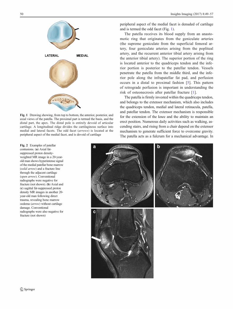
peripheral aspect of the medial facet is denuded of cartilageand is termed the odd facet (Fig. 1).
The patella receives its blood supply from an anasto-motic ring that originates from the geniculate arteries(the supreme geniculate from the superficial femoral ar-tery, four geniculate arteries arising from the poplitealartery, and the recurrent anterior tibial artery arising fromthe anterior tibial artery). The superior portion of the ringis located anterior to the quadriceps tendon and the infe-rior portion is posterior to the patellar tendon. Vesselspenetrate the patella from the middle third, and the infe-rior pole along the infrapatellar fat pad, and perfusionoccurs in a distal to proximal fashion [5]. This patternof retrograde perfusion is important in understanding therisk of osteonecrosis after patellar fracture [1].
The patella is firmly invested within the quadriceps tendon,and belongs to the extensor mechanism, which also includesthe quadriceps tendon, medial and lateral retinacula, patella,and patellar tendon. The extensor mechanism is responsiblefor the extension of the knee and the ability to maintain anerect position. Numerous daily activities such as walking, as-cending stairs, and rising from a chair depend on the extensormechanism to generate sufficient force to overcome gravity.The patella acts as a fulcrum for a mechanical advantage. In
Fig. 1 Drawing showing, from top to bottom, the anterior, posterior, andaxial views of the patella. The proximal part is termed the basis, and thedistal part, the apex. The distal pole is entirely devoid of articularcartilage. A longitudinal ridge divides the cartilaginous surface intomedial and lateral facets. The odd facet (arrows) is located at theperipheral aspect of the medial facet, and is devoid of cartilage
Fig. 2 Examples of patellarcontusions. (a) Axial fat-suppressed proton density-weighted MR image in a 20-year-old man shows hyperintense signalof the medial patellar bone marrow(solid arrow) and a fracture linethrough the adjacent cartilage(open arrow). Conventionalradiographs were negative forfracture (not shown). (b) Axial and(c) sagittal fat-suppressed protondensity MR images in another 20-year-old man following directtrauma, revealing bone marrowoedema (arrow) without cartilagedamage. Conventionalradiographs were also negative forfracture (not shown)
50 Insights Imaging (2017) 8:49–57
fact, without a patella, more force would be required toachieve knee extension [6].
Imaging modalities
Evaluation and classification of patellar fractures is based onanteroposterior (AP), lateral, and skyline view radiographs ofthe knee. A recent study, however, showed that adding com-puted tomography (CT) to the evaluation led to changes inmanagement plans in almost half of the cases [7]. In that study,CT provided more accurate evaluation of comminuted frac-tures of the lower pole than did conventional radiography [7].Magnetic resonance imaging (MRI) is commonly used when aradiographically occult patellar fracture is suspected (Fig. 2).MRI is also highly sensitive for detection of cartilage damageand subchondral fracture and contusion, and provides
additional information on the integrity of the soft tissue com-ponents of the extensor mechanism.
Mechanism of injury and classification of patellarfractures
Patellar fractures are the result of direct, indirect, orcombined injury [1]. Direct injuries can be secondaryto low-energy trauma (fall on the knee from sitting orstanding height) or high-energy trauma (dashboard im-pact in a motor vehicle accident) [1]. Most commonly,though, the mechanism of injury combines direct andindirect patterns, i.e., culmination of a direct blow, quad-riceps contraction, and secondary joint collapse [1]. Thefracture pattern is not determined solely by the mecha-nism of injury, but also depends on factors such as
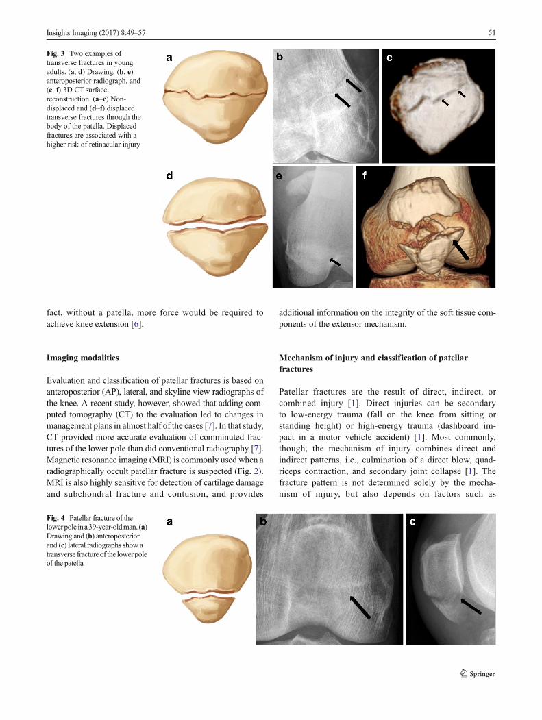
Fig. 3 Two examples oftransverse fractures in youngadults. (a, d) Drawing, (b, e)anteroposterior radiograph, and(c, f) 3D CT surfacereconstruction. (a–c) Non-displaced and (d–f) displacedtransverse fractures through thebody of the patella. Displacedfractures are associated with ahigher risk of retinacular injury
Fig. 4 Patellar fracture of thelowerpole ina39-year-oldman. (a)Drawing and (b) anteroposteriorand (c) lateral radiographs show atransverse fractureof the lowerpoleof the patella
Insights Imaging (2017) 8:49–57 51
patient age, bone quality, and degree of knee flexion [1].Patellar fractures are commonly classified according totheir morphologic pattern and degree of displacement.Melvin and colleagues defined displacement as separa-tion of fracture fragments by more than 3 mm, and/orarticular incongruity of more than 2 mm [1].
With indirect injuries, the mechanism of fracture involvesfailure of the extensor mechanism due to eccentric overload,typically a forceful contraction mechanism of the quadricepswith the knee in flexed position. A classic example is a fall onthe feet, in which the quadriceps eccentrically contracts todecelerate the body. When the force of the fall overwhelmsthe resistance to knee flexion, the extensor mechanism fails,resulting in patellar fracture [8].
Transverse fractures
Approximately 80% of these fractures occur in the middle tolower third of the patella [9]. While the mechanism may bemultifactorial, these injuries are typically associated with in-direct longitudinal forces [1]. Up to two-thirds of transversepatellar fractures are displaced [10], which should raise suspi-cion for retinacular and extensor mechanism injury, i.e., tear tothe medial and lateral patellar retinacula [1] (Fig. 3).
Pole fractures
Small proximal or distal avulsion-type fractures are importantto recognize, since they are often associated with substantial
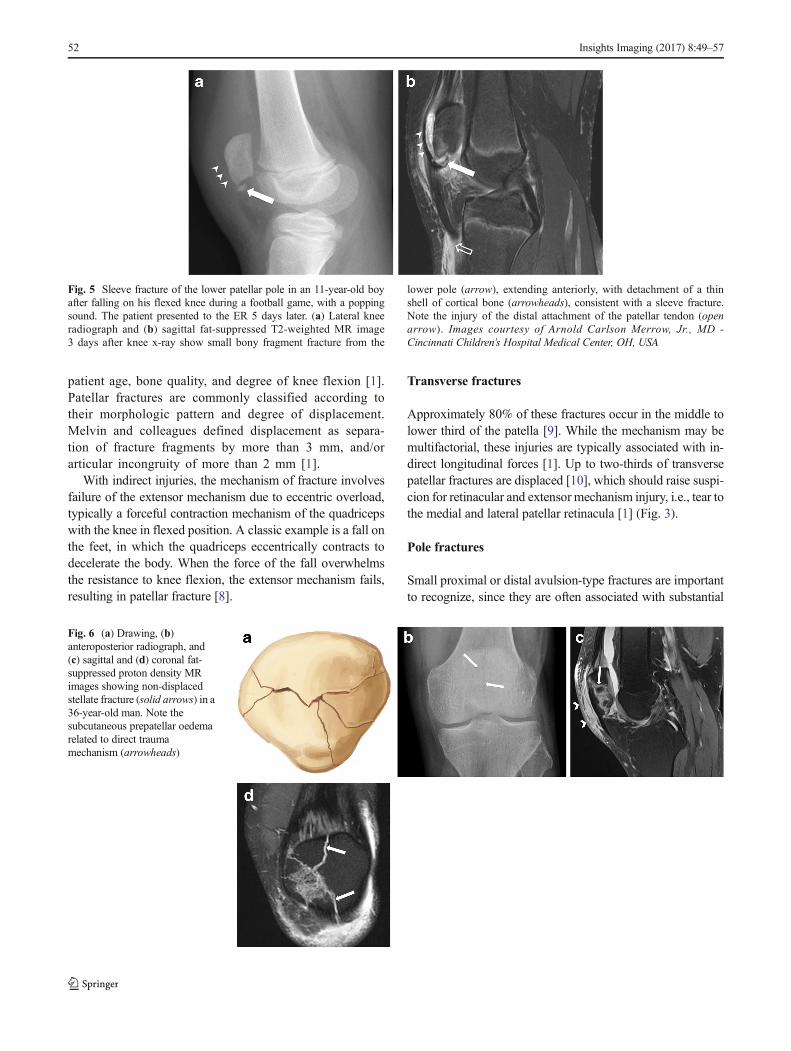
Fig. 5 Sleeve fracture of the lower patellar pole in an 11-year-old boyafter falling on his flexed knee during a football game, with a poppingsound. The patient presented to the ER 5 days later. (a) Lateral kneeradiograph and (b) sagittal fat-suppressed T2-weighted MR image3 days after knee x-ray show small bony fragment fracture from the
lower pole (arrow), extending anteriorly, with detachment of a thinshell of cortical bone (arrowheads), consistent with a sleeve fracture.Note the injury of the distal attachment of the patellar tendon (openarrow). Images courtesy of Arnold Carlson Merrow, Jr., MD -Cincinnati Children’s Hospital Medical Center, OH, USA
Fig. 6 (a) Drawing, (b)anteroposterior radiograph, and(c) sagittal and (d) coronal fat-suppressed proton density MRimages showing non-displacedstellate fracture (solid arrows) in a36-year-old man. Note thesubcutaneous prepatellar oedemarelated to direct traumamechanism (arrowheads)
52 Insights Imaging (2017) 8:49–57
soft tissue injury to the quadriceps or patellar tendon [1].Distal pole fractures (bony avulsion of the patellar tendon)are extra-articular, since the distal pole is devoid of articularcartilage [1]. In this case, lateral radiographs demonstrate pa-tella alta and increased Insall-Salvati ratio [1] (Fig. 4), whileupper pole bony avulsions are associated with low-riding
patella, also termed patella baja (reduced Insall-Salvati ratio)[1]. In the paediatric population, adolescents are most vulner-able to avulsion fractures because of their increase in musclestrength and relative weakness of osteocartilaginous junctions.In this case, there is a large sleeve of unossified patellar carti-lage, which may or may not be accompanied by tiny ossific
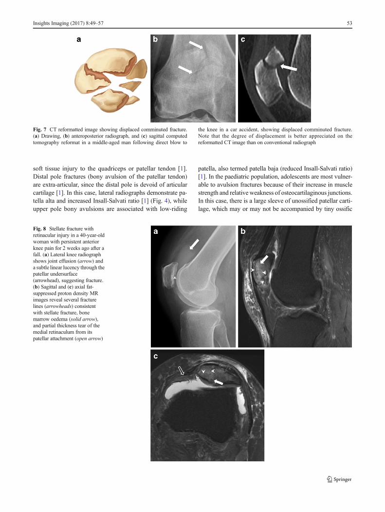
Fig. 7 CT reformatted image showing displaced comminuted fracture.(a) Drawing, (b) anteroposterior radiograph, and (c) sagittal computedtomography reformat in a middle-aged man following direct blow to
the knee in a car accident, showing displaced comminuted fracture.Note that the degree of displacement is better appreciated on thereformatted CT image than on conventional radiograph
Fig. 8 Stellate fracture withretinacular injury in a 40-year-oldwoman with persistent anteriorknee pain for 2 weeks ago after afall. (a) Lateral knee radiographshows joint effusion (arrow) anda subtle linear lucency through thepatellar undersurface(arrowhead), suggesting fracture.(b) Sagittal and (c) axial fat-suppressed proton density MRimages reveal several fracturelines (arrowheads) consistentwith stellate fracture, bonemarrow oedema (solid arrow),and partial thickness tear of themedial retinaculum from itspatellar attachment (open arrow)
Insights Imaging (2017) 8:49–57 53
fragments (Fig. 5). Patellar alignment (alta or baja), location ofsoft tissue swelling, and joint effusion provide clues to diag-nosis [11].
Stellate fractures
Stellate fractures result from a direct blow to the patella withthe knee in a partially flexed position [1]. Approximately 65%of these injuries are non-displaced [12, 13]. In the case of anextensive comminuted fracture with displacement, the trans-verse component may extend into the medial and lateral reti-nacula (Figs. 6, 7, and 8). Associated articular cartilage dam-age of the patellar and trochlear surfaces is not uncommon[14].
Vertical fractures
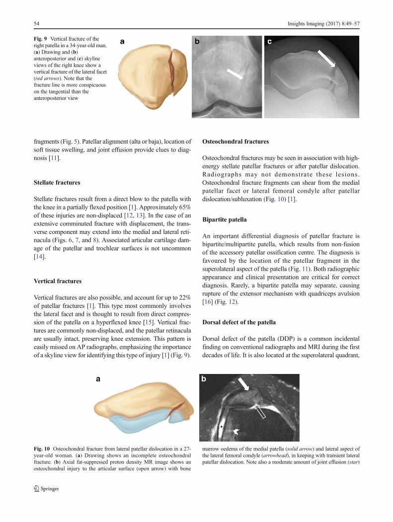
Vertical fractures are also possible, and account for up to 22%of patellar fractures [1]. This type most commonly involvesthe lateral facet and is thought to result from direct compres-sion of the patella on a hyperflexed knee [15]. Vertical frac-tures are commonly non-displaced, and the patellar retinaculaare usually intact, preserving knee extension. This pattern iseasily missed on AP radiographs, emphasizing the importanceof a skyline view for identifying this type of injury [1] (Fig. 9).
Osteochondral fractures
Osteochondral fractures may be seen in association with high-energy stellate patellar fractures or after patellar dislocation.Radiographs may not demonstrate these lesions.Osteochondral fracture fragments can shear from the medialpatellar facet or lateral femoral condyle after patellardislocation/subluxation (Fig. 10) [1].
Bipartite patella
An important differential diagnosis of patellar fracture isbipartite/multipartite patella, which results from non-fusionof the accessory patellar ossification centre. The diagnosis isfavoured by the location of the patellar fragment in thesuperolateral aspect of the patella (Fig. 11). Both radiographicappearance and clinical presentation are critical for correctdiagnosis. Rarely, a bipartite patella may separate, causingrupture of the extensor mechanism with quadriceps avulsion[16] (Fig. 12).
Dorsal defect of the patella
Dorsal defect of the patella (DDP) is a common incidentalfinding on conventional radiographs and MRI during the firstdecades of life. It is also located at the superolateral quadrant,
Fig. 9 Vertical fracture of theright patella in a 34-year-old man.(a) Drawing and (b)anteroposterior and (c) skylineviews of the right knee show avertical fracture of the lateral facet(red arrows). Note that thefracture line is more conspicuouson the tangential than theanteroposterior view
Fig. 10 Osteochondral fracture from lateral patellar dislocation in a 27-year-old woman. (a) Drawing shows an incomplete osteochondralfracture. (b) Axial fat-suppressed proton density MR image shows anosteochondral injury to the articular surface (open arrow) with bone
marrow oedema of the medial patella (solid arrow) and lateral aspect ofthe lateral femoral condyle (arrowhead), in keeping with transient lateralpatellar dislocation. Note also a moderate amount of joint effusion (star)
54 Insights Imaging (2017) 8:49–57
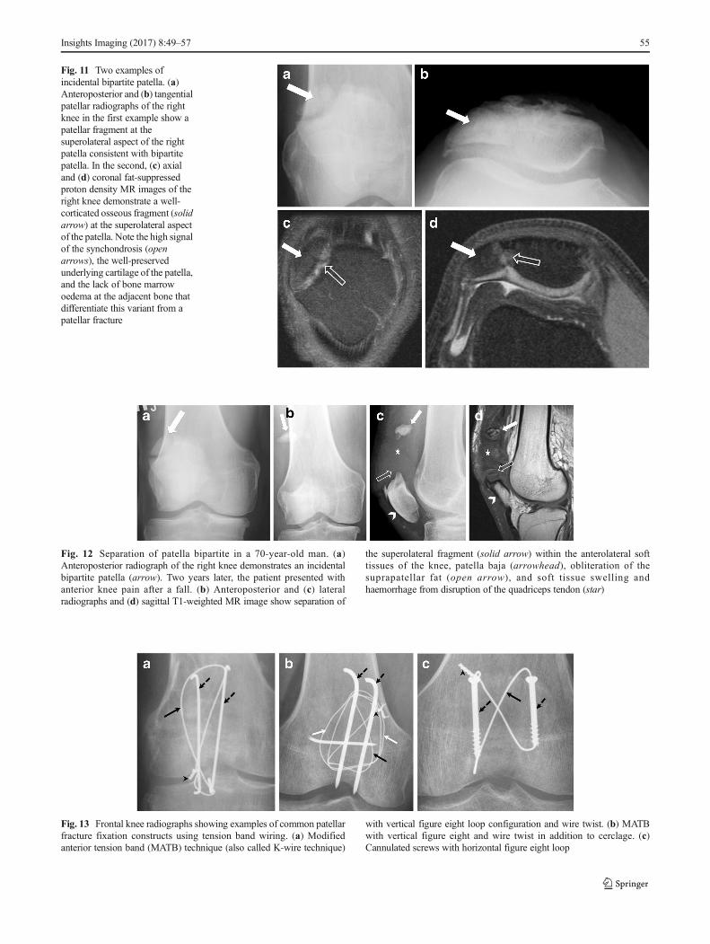
Fig. 13 Frontal knee radiographs showing examples of common patellarfracture fixation constructs using tension band wiring. (a) Modifiedanterior tension band (MATB) technique (also called K-wire technique)
with vertical figure eight loop configuration and wire twist. (b) MATBwith vertical figure eight and wire twist in addition to cerclage. (c)Cannulated screws with horizontal figure eight loop
Fig. 12 Separation of patella bipartite in a 70-year-old man. (a)Anteroposterior radiograph of the right knee demonstrates an incidentalbipartite patella (arrow). Two years later, the patient presented withanterior knee pain after a fall. (b) Anteroposterior and (c) lateralradiographs and (d) sagittal T1-weighted MR image show separation of
the superolateral fragment (solid arrow) within the anterolateral softtissues of the knee, patella baja (arrowhead), obliteration of thesuprapatellar fat (open arrow), and soft tissue swelling andhaemorrhage from disruption of the quadriceps tendon (star)
Fig. 11 Two examples ofincidental bipartite patella. (a)Anteroposterior and (b) tangentialpatellar radiographs of the rightknee in the first example show apatellar fragment at thesuperolateral aspect of the rightpatella consistent with bipartitepatella. In the second, (c) axialand (d) coronal fat-suppressedproton density MR images of theright knee demonstrate a well-corticated osseous fragment (solidarrow) at the superolateral aspectof the patella. Note the high signalof the synchondrosis (openarrows), the well-preservedunderlying cartilage of the patella,and the lack of bone marrowoedema at the adjacent bone thatdifferentiate this variant from apatellar fracture
Insights Imaging (2017) 8:49–57 55
and is commonly associated with bipartite/multipartite patella[17]. DDP is believed to be an ossification anomaly, possiblystress-induced considering histological evidence of avascularnecrosis at the site of the dorsal defect [18, 19]. DDP usuallyheals spontaneously and is rarely noted in adults [20].
On conventional radiographs, the dorsal defect appears as around, well-defined radiolucency, with sclerotic margins, lo-cated at the dorsal subchondral bone of the superolateral as-pect of the patella [18]. The MR signal intensity of the defectusually mirrors that of the overlying cartilage. The cartilageoverlying the defect should be closely inspected, as it mightfissure and thin [21].
Management
Because of the vital mechanical role of the patella in achievingknee extension, the goal of management is directed towardrestoring the extensor mechanism while maximizing articularcongruency. Non-surgical management is indicated for frac-tures with a clinically intact extensor mechanism and minimalstep-off (<2–3 mm) and/or fracture displacement (<1–4 mm)[12, 22]. Surgical management is indicated in the case of anincompetent extensor mechanism, fracture separation, intra-articular loose bodies, or osteochondral fracture [4]. Whensurgery is indicated, open reduction with internal fixationmay use one or a combination of the following: tension bands,K-wires, cerclage wires, cannulated screws, and fixation plate(Fig. 13) [4]. Partial patellectomy has been described fordisplaced transverse and comminuted fractures. Retention ofa portion of the patella is thought to preserve some of thepatellar moment and improve strength [6]. Total patellectomyis indicated in rare cases of failed internal fixation, infection,tumour, or patellofemoral arthritis (Fig. 14) [4].
Summary
Patellar fractures are rare but can lead to significant clinicaldeficits, especially when associated with an injury to the
extensor mechanism. Diagnosis and accurate classificationare important for timely and appropriate management. Themost common patellar fractures are transverse, which are sec-ondary to indirect injury, while direct blows typically result incomminuted fractures. Vertical fractures are less common, butmay easily be missed on AP radiographs, warranting carefulassessment of skyline views. Special care should be taken toexamine for displacement, as it may indicate an associatedretinacular injury and the need for surgical management.Bipartite patella is an important differential diagnosis of pa-tellar fracture. Management of patellar fractures depends on anumber of issues, including clinical presentation and degree ofdisplacement. Awide variety of surgical and non-surgical op-tions are available.
Acknowledgments We thank Dr. Arnold Carlson Merrow, Jr.,Cincinnati Children’s Hospital Medical Center, OH, USA, for hiscontribution.
References
1. Melvin JS, Karunakar MA (2004) Patella fractures and extensormechanism injuries. In: Court-Brown CB, Heckman JD,McQueen MM, Ricci WM, Tornetta P III (eds) Rockwood andGreen’s Fractures in adults. Wolters Kluwer, Philadelphia, pp2269–2302
2. Gwinner C,Mardian S, Schwabe P, Schaser KD, Krapohl BD, JungTM (2016) Current concepts review: Fractures of the patella. GMSInterdiscip Plast Reconstr Surg DGPW 5:Doc01
3. Pengas IP, Assiotis A, Khan W, Spalding T (2016) Adult nativeknee extensor mechanism ruptures. Injury. doi:10.1016/j.injury.2016.06.032
4. Melvin JS, Mehta S (2011) Patellar fractures in adults. J Am AcadOrthop Surg 19:198–207
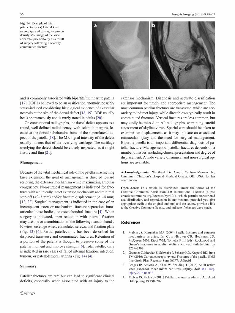
Fig. 14 Example of totalpatellectomy. (a) Lateral kneeradiograph and (b) sagittal protondensity MR image of the kneeafter total patellectomy as a resultof surgery following a severelycomminuted fracture
56 Insights Imaging (2017) 8:49–57
Open Access This article is distributed under the terms of theCreative Commons Attribution 4.0 International License (http://creativecommons.org/licenses/by/4.0/), which permits unrestricteduse, distribution, and reproduction in any medium, provided you giveappropriate credit to the original author(s) and the source, provide a linkto the Creative Commons license, and indicate if changes were made.
5. Scapinelli R (1967) Blood supply of the human patella. Its relationto ischaemic necrosis after fracture. J Bone Joint Surg (Br) 49:563–570
6. Albanese SA, Livermore JT, Werner FW, Murray DG, Utter RG(1992) Knee extensor mechanics after subtotal excision of the pa-tella. Clin Orthop Relat Res: 217–222
7. Lazaro LE, Wellman DS, Pardee NC et al (2013) Effect of comput-erized tomography on classification and treatment plan for patellarfractures. J Orthop Trauma 27:336–344
8. Scolaro J, Bernstein J, Ahn J (2011) Patellar fractures. Clin OrthopRelat Res 469:1213–1215
9. Nummi J (1971) Operative treatment of patellar fractures. ActaOrthop Scand 42:437–438
10. Andrews JR, Hughston JC (1977) Treatment of patellar fractures bypartial patellectomy. South Med J 70(809–813):817
11. Merrow AC, Reiter MP, Zbojniewicz AM, Laor T (2014) Avulsionfractures of the pediatric knee. Pediatr Radiol 44:1436–1445, quiz1433–1436
12. Bostrom A (1972) Fracture of the patella. A study of 422 patellarfractures. Acta Orthop Scand Suppl 143:1–80
13. Ashby ME, Shields CL, Karmy JR (1975) Diagnosis ofosteochondral fractures in acute traumatic patellar dislocationsusing air arthrography. J Trauma 15:1032–1033
14. Bono JV, Haas BH, Scuderi GR (1995) Traumatic maladies of theextensor mechanism. In: Scuderi GR (ed) The patella. Springer-Verlag, New York, pp 253–275
15. Dowd GS (1982) Marginal fractures of the patella. Injury 14:287–291
16. Tonotsuka H, Yamamoto Y (2008) Separation of a bipartite patellacombined with quadriceps tendon rupture: a case report. Knee 15:64–67
17. Mellado JM, Salvado E, Ramos A, Camins A, Sauri A (2001)Dorsal defect on a multi-partite patella: imaging findings. EurRadiol 11:1136–1139
18. Ho VB, Kransdorf MJ, Jelinek JS, Kim CK (1991) Dorsal defect ofthe patella: MR features. J Comput Assist Tomogr 15:474–476
19. van Holsbeeck M, Vandamme B, Marchal G, Martens M, Victor J,Baert AL (1987) Dorsal defect of the patella: concept of its originand relationship with bipartite and multipartite patella. SkeletRadiol 16:304–311
20. Safran MR, McDonough P, Seeger L, Gold R, Oppenheim WL(1994) Dorsal defect of the patella. J Pediatr Orthop 14:603–607
21. Dwek JR, Chung CB (2008) The patellar extensor apparatus of theknee. Pediatr Radiol 38:925–935
22. Carpenter JE, Kasman R, Matthews LS (1994) Fractures of thepatella. Instr Course Lect 43:97–108
Insights Imaging (2017) 8:49–57 57