Embed Size (px)
Citation preview
ANTISENSE & NUCLEIC ACID DRUG DEVELOPMENT 8:401^14 (1998)Mary Ann Liebert, Inc.
Direct Gene Transfer to the Respiratory Tract of Micewith Pure Plasmid and Lipid-Formulated DNA
MICHAEL J. McCLUSKIE,1'2 YONGLIANG CHU,5 JIU-LIN XIA,5 JOEL JESSEE,5 GULILAT GEBYEHU,5and HEATHER L. DAVIS123'4
ABSTRACT
Direct gene transfer into the respiratory system could be carried out for either therapeutic or immunizationpurposes. Here we demonstrate that cells in the lung can take up and express plasmid DNA encoding a lu-ciferase reporter gene whether it is administered in naked form or formulated with cationic liposomes. De-pending on the lipid used, the transfection efficiency with liposome-formulated DNA may be higher, the same
as, or less than that with pure plasmid DNA. Tetramethy ltetraalkylspermine analogs with alkyl groups of 16 or
18 carbons and DMRIE/cholesterol formulations proved particularly effective. Similar results for reportergene expression in the lung were obtained whether the DNA (naked or lipid formulated) was administered byindirect, noninvasive intranasal delivery (inhaled or instilled) or by invasive, direct intratracheal delivery (in-jected or via a cannula). Reporter gene expression peaks around 4 days, then falls off dramatically by 9 days.The dose-response is linear, at least up to 100 /¿g plasmid DNA, suggesting better transfection efficienciesmight be realized if there was not a volume limitation. For a given dose of DNA, the best results are obtainedwhen the DNA is mixed with the minimum amount of lipid that can complex it completely. These results are
discussed in the context of direct gene transfer for either gene therapy or delivery of a mucosa! DNA vaccine.
INTRODUCTION
The respiratory system is attractive for direct gene de-livery, as it has an extensive surface area of pulmonary ep-
ithelium that is readily accessible via the bronchiolar and alveo-lar network. This easy and noninvasive method of gene deliveryhas the potential to transfect a large number of cells and allowhigh local concentrations of expressed gene products to be at-tained. Gene delivery into the respiratory system could be use-
ful for either therapeutic or immunization purposes.There are several potential therapeutic applications for direct
gene transfer in the lung. Perhaps the most obvious is gene ther-apy for inborn errors of metabolism, such as delivery of the cys-tic fibrosis (CF) transmembrane conductance regulator gene(CFTR), to treat CF (Hyde et al, 1993; Yoshimura et al., 1992;Alton et al., 1993; Caplen et al., 1995). It might also be possibleto treat allergy-induced asthma, for example, by expressing In-
terferon-y (IFN-y) to shift a T helper type 2 (Tfi2) immune re-
sponse, which is associated with IgE, to a TTil response (Li etal., 1996). Lung cancer could perhaps be treated by direct trans-fer of suicide genes or tumor repressor genes (Woll and Hart,1995). Other local or systemic, acquired diseases might bemanaged by short-term, low-dose therapy through expressionof genes encoding cytokines or coactivation factors.
Although most therapeutic strategies aim to avoid an im-mune response against the expressed gene product, the purpose-ful introduction of antigen-encoding genes can be used for ei-ther immunotherapy or prophylactic vaccination. Intranasaladministration of such DNA vaccines would be simple andnoninvasive and should also induce specific mucosal immunity,which is desirable to prevent entry of pathogens via the exten-sive mucosal surfaces of the body.
Direct gene transfer may be carried out in many tissues byvarious means. The use of plasmid DNA is preferable to live vi-
'Loeb Research Institute, Ottawa, Canada.department of Cellular and Molecular Medicine, 'Department of Microbiology and Immunology, Faculty of Medicine, and 4School of Reha-
bilitation Sciences, Faculty of Health Sciences, University of Ottawa, Ottawa, Canada.5Life Technologies, Inc., Rockville, MD 20850.
401
402 McCLUSKIE ET AL.
ral or bacterial vectors owing to ease of production, the nonim-munogenic nature of the vector itself (Krieg, 1996; Pisetsky,1996; Klinman et al., 1997), and the absence of risk of inadver-tent infection. Plasmid DNA may be administered in pure or
naked form, complexed with cationic lipids, microencapsu-lated, or coated onto gold particles introduced biolistically (La-sic and Templeton, 1996; Miller, 1992; Ledley, 1995). Deliveryof genes to the respiratory system may be hampered by poortransport of molecules across the epithelium owing to biochem-ical (e.g., secreted enzymes), physical (e.g., mucus, cilia), or
mechanical (e.g., coughing) pulmonary defense mechanisms.Previous reports have indicated that gene transfer may be car-
ried out in the lung tissue using plasmid DNA either alone or
formulated with cationic lipids. However, there are conflictingreports as to the relative efficiencies of these two methods.Here, we examine direct gene transfer in the lungs of mice byadministration of plasmid DNA using different techniques andlipid formulations.
MATERIALS AND METHODS
Plasmid DNA vectors
Gene transfer studies were carried out with plasmid DNA inwhich the immediate early promoter of the cytomegalovirus(CMV) drove expression of firefly Photinus pyralis luciferase(pCMV-luc) (Davis et al., 1993b), /3-galactosidase (pCMV-lacZ) (Davis et al., 1993b), or green fluorscent protein (pEGFP-Cl, Clontech Laboratories, Inc., Palo Alto, CA). The DNA was
purified on Qiagen anion-exchange chromatography columns(Qiagen GmbH, Hilden, Germany) and resuspended in sterilesaline (0.15 M NaCl, Sigma-Aldrich, Oakville, Canada). DNAprepared in this way has only low levels of endotoxin (Davis etal., 1996b). The concentration of DNA was calculated based on
absorbance of ultraviolet light (OD 260), with final concentra-tions usually being 5-10 mg/ml. DNA solutions were stored at
—
20°C until required for in vivo delivery. DNA was adminis-tered either as pure plasmid DNA in saline (naked DNA) or for-mulated with the cationic lipids described below.
Cationic and neutral lipidsTetramethyltetraalkylspermine analogs were provided by
Life Technologies, Inc. (Gaithersburg, MD) (J. Jessee et al., un-
published observations). In brief, spermine was acylated withthe desired acyl chloride (Nu-Chek-Prep, Inc., Eysian, MN),
S
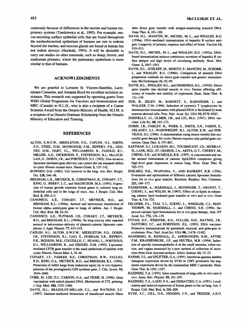
FIG. 1. Structure of tetramethyltetraalkylspermine analogues. Tetram-ethyltetralaurylspermine (TMTLS) R = (CH2)10CH3. Tetramethyltetra-mvristvlspermine (TMTMS) R = (CH2)12CH3. Tetramethyltetrapa-lmitylspermine (TMTPS) R = (CH2)14CH3. Tetramethyltetraoleoyl-spermine (TMTOS) R = (CH^CH = CH(CH2)7CH3.
Table 1. Lipid Formulations Used forDirect Gene Transfer into Lungs of Mice
Formulation* Molar ratio Other name
TetramethyltetraalkylsperminesTMTLS: DOPE 1:1.5TMTLS :DLPE 1:1.5TMTLS :DPAPE 1:1.5TMTMS: DOPE 1:1.5TMTMS :DMPE 1:1.5TMTMS :DPAPE 1:1.5TMTOS: DOPE 1:1.5TMTOS :DPAPE 1:1.5TMTPS: DOPE 1:1.5TMTPS :DPAPE 1:1.5
OthersDOTMA:DOPE 1:1DOSPA:DOPE 1.5:1DMRIE:C 1:1GAP-DLRIE:DOPE 1:1
Cellfectin®
Lipofectin®LipofectAMINE®
"C, cholesterol; DLP, dilauroylphosphatidylethanolamine;DMPE, dimyristoylphosphatidylethanolamine; DMRIE (+)-N-(2-hydroxyethyl)-N,N-dimethyl-2,3-bis-(tetradecyloxy)-1-propanaminium bromide; DOPE, dioleylphosphatidyl-ethanolamine; DOSP, 2,3-dioleyloxy-N-[2(sperminecarboxa-mido)ethyl]-N,N-dimethyl-l-propaniniumtrifluoracetate;DOTMA, N-[ 1 -(2,3-dioleoyloxy)propyl]-N,N,N-trimethyl-ammonium chloride; DPAPE, dipalmitoleoylphosphati-dylethanolamine; GAP-DLRIE, (+)-N-(3-aminopropyl)-N,N-dimethyl-2,3-bis(dodecyloxy)-1 -propanaminium bromide;TMTLS, tetramethyltetralaurylspermine; TMTMS, tetra-methyltetramyristylspeimine; TMTOS, tetramethyltetraoleoyl-spermine; TMTPS, tetramethyltetrapalmitylspermine.
followed with lithium aluminium hydride reduction to obtaintetraalkylspetmine. The tetraalklyspermine was alkylated fur-ther with methyl iodide to obtain the tetramethyltetraalkylsper-mine, which was then characterized by infrared and nuclearmagnetic resonance (NMR) spectroscopy and fast atomic bom-bardment-mass spectrometry. Four different tetramethylte-traalkylspermine derivatives were produced (Fig. 1), whichwere then formulated with one of DOPE, DPAPE, DMPE, andDLPE (Avanti Polar Lipids, Albaster, AL) at ratios as outlinedin Table 1.
Other lipid formulations tested were Lipofectin®, LipofectAMINE®, DMRIE:C, and Cellfectin® as well as a mixture ofGAP-DLRIE (Wheeler et al., 1996) and DOPE (Life Technolo-gies, Inc.) (Table 1).
Preparation of liposomesThe lipids were formulated into liposomes using the reverse
evaporation method. A known amount of cationic lipid was
mixed with the neutral colipid in a 2-L round-bottom flask toobtain the desired molar ratio of cationic/neutral lipid. Méthyl-ène chloride or 10% methanol in méthylène chloride was addedto make a 10-30 mg/ml solution. Distilled water was added to
give approximately 1 or 2 mg/ml solution. The mixture was
shaken 3-5 minutes, then let stand for approximately 3 minutes.The solution was subjected to rotary evaporation to remove
DIRECT GENE TRANSFER TO LUNG 403
méthylène chloride and methanol. The volume was adjustedwith distilled water to obtain the desired concentration (1 or 2mg/ml). The liposome solution was flushed with nitrogenstored at 4°C.
Preparation of plasmid DNA-liposome complexesLipid-formulated DNA was administered in a total volume of
150 fi\ containing 1-100 fig DNA and 0.2-5 times that weightof lipid. For each formulation, the required volumes of stockDNA solution, stock lipid solution, and diluent (0.15 M NaCl)were calculated.
The lipid, the diluent, and the DNA were added in sequentialsteps, with brief vortexing after each step. The final mixturewas left undisturbed (either on ice, at ambient temperature, or at37°C) for 30 minutes to allow DNA-liposome complex forma-tion. Immediately before administration, the DNA-lipid mix-ture was mixed gently by tapping on the side of the microtube.
To determine whether all the DNA was complexed with thelipid, mixtures were run on a 0.5% agarose gel and stained withethidium bromide (0.04 fig/ml). The presence of free plasmidwas detected as a band at the expected distance, whereas DNA-lipid complexes failed to enter the gel.
Determination of size and £ potentialParticle size and £ potential were measured with complexes
formed using pCMV-luc mixed with an equal amount of thefollowing five lipid formulations: Cellfectin, LipfectAMINE,DMRIE-C, TMTLS-DPAPE, and TMTOS-DOPE. These fiveformulations were chosen as they represented the range of effi-ciencies of lung transfection obtained when complexes were
administered by i.n. inhalation to mice. Additional measure-
ments were recorded using 100 fig DNA mixed with 0.2-5times that weight of Cellfectin to determine the effect of differ-ent relative proportions of DNA and lipid components. Mea-surements were taken 30 minutes after mixing for all lipid for-mulations, and for Cellfectin, measurements were also taken at
regular time intervals up to 4 hours.Particle size measurements were made at a scattering angle
of 90° with a Brookhaven 90Plus Particle Size Analyzer(Brookhaven Instruments Corp., Holtsville, NY). The homo-dyne intensity-intensity correlation function G(q,t) was ob-tained. The amplitude of the scattering vector
q = (4im/A)sin(0/2)
where n is the refractive index of the medium, À is the wave-
length of the excitation light in a vacuum, 8 is the scattering an-
gle, and t is the time.Autocorrelation functions were analyzed using the CONTIN
program (Brookhaven Instruments Corp.), which employs theconstrained regularization method to calculate the mean diffu-sion time, (t), related to the diffusion coefficient by
D = A2l6irW(0/2)<f)
From each D value, we obtained Stokes' radius, Rs, by Stokes-Einstein equation.
R. = *T6tttjD
where k is Boltzmann's constant, T is the absolute temperature,and T) is the viscosity of the solvent.
£ potential measurements were made .with the same
Brookhaven instrument as for the particle size measurements.In £ potential measurements, the measured Doppler shift fre-quency, Aft) is given by
À Eusin 6
where E (V/cm) and u [(/urn s_1)/(V cm"1)] are the appliedelectric field strength and electrophoretic mobility, respec-tively. Therefore, u can be directly obtained from the Ao. Val-ues of £ potential were calculated by
e
where e is permitivity of solution.
In vivo gene transferAnimals. Experiments were carried out using female
BALB/c mice (Charles River, Montreal, Quebec, Canada) or
IFN-y knockout mice (Jackson Labs, Bar Harbor, ME) aged6-8 weeks, with a total of 10-15 mice per experimental or con-
trol group. Each experiment was repeated two or three times(n = 5) to ensure reproducibility of results. Control groups forluciferase expression consisted of untreated mice and mice ad-ministered plasmid encoding a nonrelevant gene (i.e., /3-galac-tosidase, /3-gal). Each animal received naked or lipid-formu-lated DNA in a total volume of 150 pi, which was given bysingle or multiple administrations and which contained differ-ent doses of DNA and different relative proportions of DNAand lipid. The DNA was administered to the respiratory systemof the mice via intranasal (i.n.) or intratracheal (i.t.) routes.
i.n. Instillation. Mice were lightly anesthetized withHalothane® (Halocarbon Laboratories, River Edge, NJ) andheld on their backs with the head suspended inferiorly. TheDNA solution was then instilled bilaterally into the nasal cavityusing a gel-loading tip and a Gilson pipette. The tip was in-serted a few millimeters into the nasal cavity, and each nostrilwas instilled three times at 15-minute intervals with 25 /xl ofDNA solution (for a total volume of 150 /¿,1/mouse).
i.n. Inhalation. Droplets of DNA solution were deposited bypipette directly over both external nares of mice under eitherHalothane or Somnotol® (75 mg/kg i.p.) (MTC Pharmaceuti-cals, Cambridge, Ontario, Canada) anesthesia. A total volumeof 150 fi\ was given as either a single (1 X 150 p\) or triple(3 X 50 fi\) administration. For the triple administration, 15-minute intervals were allowed between administrations.
i.t. Injection. Mice fully anesthetized with Somnotol had thetrachea exposed through an anterior midline incision. The DNAsolution (150 fi\) was then injected through the anterior wall ofthe trachea using a 0.3-ml insulin syringe with a 29-gauge nee-
dle attached (Becton Dickinson, Franklin Lakes, NJ). The inci-sion was sutured, and the mouse was placed in an incubator un-
til fully recovered from the anesthetic (approximately 45minutes).
i.t. Cannulation. Mice under Somnotol anesthesia had thetrachea exposed via an anterior midline incision, and this was
used to visualize the insertion of the cannula into the trachea. A
404 McCLUSKIE ET AL.
20-gauge olive tip steel feeding tube (Fine Science Tools Inc.,North Vancouver, BC, Canada) attached to a 1-ml tuberculinsyringe (Becton Dickinson) was passed through the oral cavityand into the trachea. The DNA solution (150 pi) was thenslowly injected directly into the lungs.
Evaluation of reporter gene expressionMice were killed by cervical dislocation under Halothane
anesthesia at various times from 6 hours to 19 days after genetransfer. For determination of luciferase reporter gene activity,the lungs and bronchial tree were dissected free and storedfrozen (—20°C) until assayed using the Promega LuciferaseAssay System (Madison, WI). This was carried out as describedpreviously for muscle (Davis et al., 1993b) except that the lungswere homogenized in 350 pi of 1 X lysis buffer. Measurementswere taken with a Monolight 2010 luminometer (Analytical Lu-minescence Laboratory, San Diego, CA). The protein contentof the lungs was determined by the Bio-Rad microassay proce-dure using the Bio-Rad Protein Assay Reagent (Hercules, CA).Values for luciferase activity were expressed in relative lightunits (RLU) sec/mg protein.
For visualization of green fluorescent protein, the lungs andbronchial tree were dissected free and thinly sectioned. The headof the mouse was severed between the upper and lower jaws, fa-cial skin was removed, and the head was split sagitally to revealthe nasal passage. The lungs, bronchial tree, and nasal passageswere examined for fluorescence using a Zeiss Axioskop light mi-croscope (Carl Zeiss Canada Ltd., North York, Ontario, Canada).
PCR analysisLungs were removed and homogenized, and genomic plus
plasmid DNA was isolated using QIAamp Tissue Kit (Qiagen).PCR was performed in a GeneAmp PCR System 2400 (PerkinElmer, Norwalk, CT) using upstream (5'-ATG GAA GAC GCCAAA AAC AT-3') and downstream (5'-TTA CAA TTT GGACTT TCC GC-3') primers (Life Technlogies, Inc.) designed to
amplify the luciferase gene. The components of the reaction mix-ture were as indicated in the Basic PCR Protocol supplied withTaq DNA polymerase (Life Technologies, Inc.). After an initialdenaturation at 94°C for 2 minutes, the samples were subjected to35 cycles of the following: denaturation at 94°C for 30 seconds,annealing at 52°C for 45 seconds, and extension at 72°C for 2minutes. This was followed by a 7-minute extension at 72°C.PCR products were evaluated by 0.8% agarose gel electrophore-sis stained with 0.04 pg/ml ethidium bromide. As a positive con-
trol, 1 /ig plasmid DNA was added to lungs of untreated mice be-fore tissue homogenization and DNA isolation.
Assay of humoral response to expressed proteinsPlasma taken from mice at 2, 4, 8, and 12 weeks after immu-
nization by i.n. inhalation of 100 pg pCMV-lacZ mixed withCellfectin was evaluated for total IgG antibodies specific to ß-gal as previously described (Davis et al., 1997).
Experimental groups
Effect ofDNA/Lipid Ratio. Mice were administered by i.n. in-halation with a total of 25 pg pCMV-luc DNA mixed with Cell-
fectin at DNA/lipid (w/w) ratios of 1:0 (pure DNA), 5:1, 3:1,2:1, 1:1, 1:2, 1:3, and 1:5 (equivalent to charge ratios of 0, 0.05,0.08, 0.12, 0.23, 0.46, 0.69, and 1.16 nmol cationic lipid/nmolnucleotide, respectively). Lungs were removed 72 hours later.
DNA Dose. Mice received a total of 1, 10, 25, 50, or 100 pgof pCMV-luc DNA, formulated with Cellfectin at a 1:1DNA/lipid ratio (w/w). This was given by i.n. inhalation in a to-tal volume of 150 pi. Lungs were removed at 72 hours.
Longevity of Reporter Gene Expression. A total of 100 pgpCMV-luc DNA formulated with Cellfectin at a 1:1 DNA/lipid(w/w) ratio was administered by i.n. inhalation to each mouse.
Lungs were removed from mice killed at 6 hours and 1, 2, 3, 4,5, 7, 9, 14, or 19 days after gene transfer and assayed for lu-ciferase activity.
Method ofDNA Administration. A total of 100 pg pCMV-lucDNA formulated with Cellfectin at a 1:1 DNA/lipid (w/w) ratiowas administered by each of the four methods described (i.n.instillation, i.n. inhalation, i.t. injection, i.t. cannulation). Lungswere removed 24, 48, or 72 hours later.
Lipid Composition. A total of 100 pg pCMV-luc DNA was
formulated with each of the 14 different lipids (as described) at a
1:1 DNA/lipid (w/w) ratio. These solutions were administered tomice by i.n. inhalation, and lungs were removed 72 hours later.
StatisticsData were expressed as group means ± standard error of the
means (SEM) and were analyzed using the GraphPAD InStatprogram (Graph PAD Software, San Diego, CA). The signifi-cance of the differences between groups was determined by theMann-Whitney test for comparison of two groups or by a 1-fac-tor analysis of variance, followed by the Krustal-Wallis test forcomparison of three or more groups. Differences were consid-ered to be not significant at p > 0.05. In no case were resultssignificantly different for the same treatment carried out on twoor three separate occasions (n = 5). Therefore, these data were
pooled (n = 10 or 15).
RESULTS
For most of the studies, Cellfectin (TMTPS:DOPE [1:1.5w/w]) (Table 1) was used as a model lipid, as it allowed efficienttransfection of the mouse lung, and i.n. inhalation was used as a
model delivery method, as it was easy and noninvasive.
Effect of DNA/lipid ratio
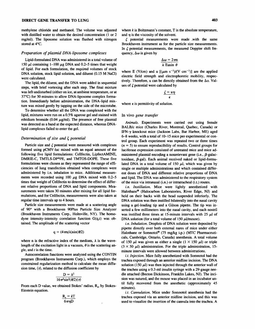
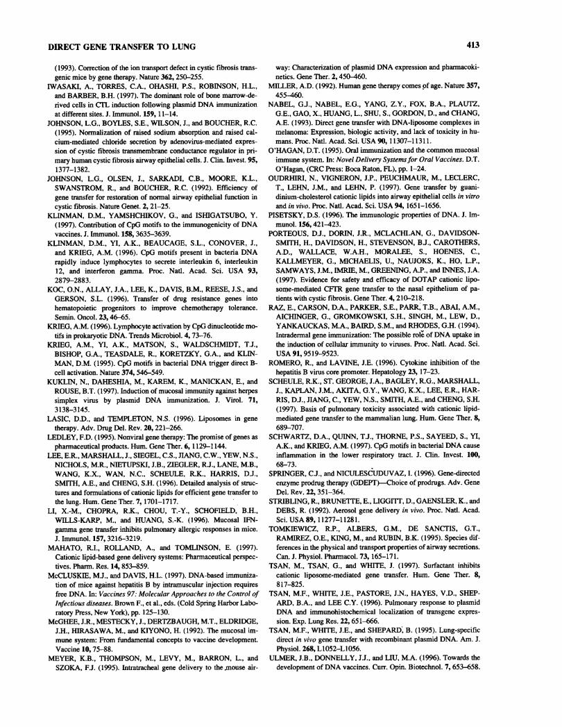
The efficiency of lung tissue transfection after i.n. inhalationof DNA mixed with lipid depended on the relative amounts ofDNA and lipid. For example, with Cellfectin, the highest mean
luciferase expression was obtained using a DNA/lipid ratio(w/w) of 1:2, although ratios of 1:1 and 1:3 also gave good re-
sults (Fig. 2). We have shown previously by visualization offree plasmid DNA on an agarose gel (Van der Woude et al.,1997) that all DNA was complexed when mixed with at leasttwice as much (by weight) Cellfectin (McCluskie and Davis,1997). Thus, the present results indicate that the most efficient
DIRECT GENE TRANSFER TO LUNG 405
150
100SI=
-
06
50 Jj_FT
1:0 5:1 3:1 2:1 1:1 1:2l)NA:lipid (mass ratio)
1:3 1:5
FIG. 2. Effect of DNA/lipid ratio on luciferase expression. DNA (25fig pCMV-luc) was mixed with increasing amounts of Cellfectin andadministered by i.n. inhalation to mice. Lungs were harvested at 72hours and assayed for luciferase activity. Bars represent the mean lu-ciferase activity in relative light units (RLU)/sec/mg of lung protein,and T bars indicate SEM (n = 10).
gene transfer for a given dose of DNA was obtained when itwas mixed with the minimum amount of the lipid that couldcompletely complex it. The presence of excess uncomplexedDNA (e.g., DNA/lipid at 5:1 or 3:1, w/w) resulted in lower lev-els of luciferase activity, and the lowest luciferase activity was
detected with pure plasmid DNA. Transfection efficiency alsodecreased with excess lipid (1:5), possibly owing to toxicity ofthe lipid.
There was no difference between expression levels in lungsremoved 72 hours after i.n. inhalation of 100 fig pCMV-luccomplexed with Cellfectin when complexes were formed on iceor at ambient temperature (mean ± SD, 466 ± 242 and 506 ±
180 RLU/sec/mg lung protein, respectively, p = 0.97). How-ever, when the temperature was increased to 37°C, expressionlevels decreased significantly (220 ± 203 RLU/sec/mg protein,p < 0.05).
For further studies, we chose a DNA/lipid ratio of 1:1, whichwas the lowest amount of lipid to give a relatively high trans-fection efficiency, to minimize any possible lipid-associatedtoxicity. Cationic lipids have been shown to be toxic in vivo in a
dose-dependent manner (Scheule et al., 1997).
Effect of DNA dose
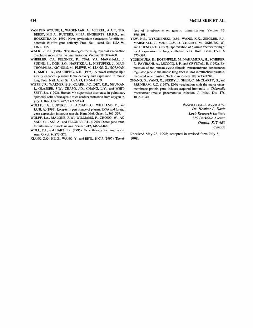
The level of luciferase activity in the lung 48 hours after i.n.inhalation of pCMV-luc DNA (mixed 1:1 w/w with Cellfectin)was directly proportional to the amount of DNA from 1 to 100fig (r = 0.99) (Fig. 3). The lack of plateau in reporter gene ac-
tivity indicated that uptake and expression of DNA were notlimited by saturation with DNA but rather by the maximumvolume that could be given without causing undue stress to theanimal. There was no significant difference between expressionlevels in lungs removed 72 hours after i.n. inhalation of 100 pgpCMV-luc complexed 1:1 w/w with Cellfectin when the com-
plexes were given by a single (1 X 150 /xl) or triple (3 X 50 tilover 30 minutes) administration schedule (mean ± SD, 595 ±326 and 506 ± 180 RLU/sec/mg protein, respectively, p =
0.63). On the other hand, animals were less distressed with thetriple than the single administration method. A 100-/xg dose ofDNA given as a triple administration was used in all subsequentexperiments.
Longevity of reporter gene expressionLow levels of luciferase expression could be detected as
early as 6 hours after i.n. inhalation of 100 fig pCMV-luc(mixed 1:1 w/w with Cellfectin). The reporter gene activity
400
.S 300°3oca
g> 200
S
3-
06 100HT
10 25 50pCMV-luc dose (ng)
100
FIG. 3. Effect of DNA dose on luciferase expression. DNA (1-100fig pCMV-luc) was mixed with increasing amounts of Cellfectin at a
DNA/lipid w/w ratio of 1:1 and administered by i.n. inhalation to mice.Lungs were harvested at 48 hours and assayed for luciferase activity.Bars represent the mean luciferase activity in RLU/sec/mg of lung pro-tein, and T bars indicate SEM (n = 10).
800
a 60<H•5ouaM
ua»VI
-
OS
400-
200H
123456789 19Time (d) after DNA administration
FIG. 4. Time course of reporter gene expression. DNA (100 figpCMV-luc) mixed with Cellfectin (1:1 w/w) was administered to miceby i.n. inhalation. Lungs were harvested at various times after adminis-tration and assayed for luciferase activity. Each value represents themean luciferase activity in RLU/sec/mg of lung protein, and T bars in-dicate SEM (n = 10).
406 McCLUSKIE ET AL.
10000
âj 1000ouawoS 100
10
31D0Q DNA in saline H B DNA with lipid
Time (hr)24 0 48 Q B 72
IN instillation IN inhalation IT injection IT çannulationFIG. 5. Routes of administration. DNA ( 100 fig pCMV-luc) was administered to mice either in saline or mixed with Cellfectin (1:1 w/w) by i.n.instillation, i.n. inhalation, i.t. injection, and i.t. cannulation. Lungs were harvested at 24, 48, or 72 hours and assayed for luciferase activity. Barsrepresent the luciferase activity in RLU/sec/mg of lung protein, and T bars indicate SEM (n = 10).
increased steadily for 4 days but declined rapidly betweendays 4 and 5. Only very low levels of luciferase were de-tected at 9 days, although these were sustained for at least a
further 10 days (Fig. 4). Because luciferase is unstable andhas a short half-life, we considered the presence of high lu-ciferase activity to indicate recent expression from plasmid
none (in saline)Lipofectin
LipofectamineGAP-DLRIE:DOPE
DMRIE-CTMTLS:DOPETMTLSrDLPETMTLS:DP^PETMTMS:DOPE
TMTMS:DMPETMTMS:DP PETMTOS:DOPETMTOS:DP*PETMTPSrDOPETMTPS:DP^PE
DNA rather than persistence of gene product in the absenceof new synthesis. The kinetics of expression was the same inIFN-y knockout mice as in normal (BÁLB/c) mice (mean ±SD, 519 ± 36 and 466 ± 242 RLU/sec/mg protein, respec-tively, at day 3, p > 0.99; 92 ± 69 and 95 ± 10 at day l,p =
0.81).
200 400 600RLU/sec/mg protein
800
FIG. 6. Gene transfer with different cationic lipids. DNA (100 fig pCMV-luc) in saline or mixed with 1 of 14 different lipids at a 1:1 DNAflipid(w/w) ratio was administered to mice by i.n. inhalation. Lungs were harvested at 72 hours and assayed for luciferase activity. Bars represent themean luciferase activity in RLU/sec/mg of lung protein, and T bars indicate SEM (n = 5).
DIRECT GENE TRANSFER TO LUNG 407
Lipofectin-Lipofectamine
GAP-DLRIE:DOPEDMRIE-C
TMTLS:DOPETMTLS:DLPE
TMTLS:DPAPETMTMS:DOPETMTMS:DMPEHTMTMS:DPAPETMTOS:DOPE-TMTOS:DPAPE-TMTPS:DOPE-
TMTPS:DPAPE-50 40 30 20
Naked better than lipid complexée! Lipid complexedbetter than naked
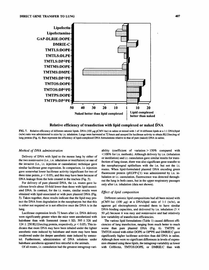
Relative efficiency of transfection with lipid complexed or naked DNAFIG. 7. Relative efficiency of different cationic lipids. DNA (100 pg pCMV-luc) in saline or mixed with 1 of 14 different lipids at a 1:1 DNA/lipid(w/w) ratio was administered to mice by i.n. inhalation. Lungs were harvested at 72 hours and assayed for luciferase activity to obtain RLU/sec/mg oflung protein (Fig. 6). Bars represent the efficiency of lipid-complexed DNA formulations relative to that of pure (naked) DNA in saline.
Method of DNA administration
Delivery of DNA with lipid to the mouse lung by either ofthe two noninvasive (i.e., i.n. inhalation or instillation) or one ofthe invasive (i.e., i.t. injection or cannulation) technique gavesimilar luciferase gene expression. In comparison, i.t. injectiongave somewhat lower luciferase activity (significant for two ofthree time points, p < 0.05), and this may have been because ofDNA leakage from the hole created in the trachea (Fig. 5).
For delivery of pure plasmid DNA, the i.n. routes gave lu-ciferase levels about 10-fold lower than those with lipid-associ-ated DNA. In contrast, for the i.t. routes, similar results were
obtained with lipid-associated or with pure plasmid DNA (Fig.5). Taken together, these results indicate that the lipid may pro-tect the DNA from degradation in the nasopharynx but that thisis either not required or is not effective once the DNA is in thelung.
Luciferase expression levels 72 hours after i.n. DNA deliverywere significantly greater when the mice were anesthetized withhalothane than with Somnotol (mean ± SD, 595 ± 326 and124 ± 250 RLU/sec/mg protein, respectively,/? < 0.05). This in-dicates that more DNA may have been inhaled under the lighteranesthetic state induced by halothane and more may have beenswallowed under the deeper anesthetic state induced by somno-
tol. Furthermore, administration of DNA solutions underhalothane anesthesia appeared less stressful to the animals.
Of all routes, i.t. cannulation had the greatest intragroup vari-
ability (coefficient of variation > 150% compared with< 100% for i.n. methods). Although delivery by i.n. (inhalationor instillation) and i.t. cannulation gave similar results for trans-fection of lung tissue, there was also significant gene transfer tothe nasopharyngeal epithelium with the i.n. but not the i.t.routes. When lipid-formulated plasmid DNA encoding greenfluorescent protein (pEGFP-Cl) was administered by i.n. in-halation or i.t. cannulation, fluorescence was detected through-out the lung in both cases, but in the upper respiratory passagesonly after i.n. inhalation (data not shown).
Effect of lipid compositionDifferent cationic lipid compositions had all been mixed with
pCMV-luc (100 pg) at a DNA/lipid ratio of 1:1 (w/w), as
agarose gel electrophoresis revealed them to have similarDNA-binding capacities, and delivered by i.n. inhalation (3 X50 pi) because it was easy and noninvasive and had relativelylow variability of transfection efficiencies.
The various lipid formulations (Table 1) caused different effi-ciencies of lung transfection, ranging from much better to muchworse than pure plasmid DNA (Fig. 6). TMTPS orTMTOS mixed with either DOPE or DP^PE and DMRIE:C gavesignificantly higher luciferase activities than did DNA in saline.Although there were no significant differences in levels of expres-sion obtained using these lipids, the intragroup variability as lowerwith Cellfectin, TMTOS:DOPE, or DMPJEC than with
408 McCLUSKIE ET AL.
Table 2. Size and £ Potential of Various FormulationsUsed for Gene Transfer to Respiratory Tract
Sample Size (nm)( potential
(mV)
LipofectAMINE 93 60DMRIE:C 309 42TMTLS:DPAPE 386 30TMTOS:DOPE 505 36Cellfectin 365 41pCMV-luc 128 -50pCMV-luc : LipofectAMINE ( 1:1f 1711 32pCMV-luc:DMRIE:C (1:1) 658 -20pCMV-luc : TMTLS : DPâPE (1:1) 848
-
36pCMV-luc : TMTOS : DOPE (1:1) 751 20pCMV-luc : Cellfectin (1:5) 1217 29pCMV-luc : Cellfectin (1:2) 803 15pCMV-luc : Cellfectin (1:1) 758 -17pCMV-luc:Cellfectin (2:1) 1232 -27pCMV-luc:Cellfectin (5:1) 1409 -35
"The w/w ratio of DNA/lipid.
TMTPS:DPAPE or TMTOS:DPAPE. The next group ofTMTMS:DPAPE, TMTLS^P^PE, and TMTLS:DLPE resultedin luciferase expression equivalent to that obtained with pure plas-mid DNA. Finally, Lipofectin, LipofectAMINE, TMTMS:DOPE,TMTMS:DMPE, TMTLS:DOPE, and GAP-DLRIE:DOPE all re-
sulted in luciferase activities significandy less than that obtainedwith naked DNA (p < 0.05). The actual relative efficiencies com-
pared with pure plasmid DNA are shown in Figure 7.
Physicochemical characteristics ofDNA/lipid complexes
DNA/liposome complexes formed using five different lipidformulations had different sizes (~650->1700 nm) and £ po-
tential (—36-+32 mV) (Table 2). Maximum levels of expres-sion in lung at 72 hours after i.n. inhalation of lipid/pCMV-luccomplexes were obtained with those of 700-800 nm and with a
£ potential of —16-—20 mV (Fig. 8). There is, however, no
clear correlation between complex size and Ç potential (r =
0.55).For any of the five formulations there was no difference in
the size or the f potential at 30 minutes vs. 4 hours, indicating a
certain stability over time. All in vivo administrations were car-
ried out within 30-60 minutes of complex formation.
Antibodies against expressed proteinsAntibodies against /3-gal were detected after administration
of Cellfectin-complexed pCMV-lacZ DNA. However, the titersinduced were much lower than those by i.m. injection of thesame dose (100 pg) of DNA (data not shown).
DISCUSSION
Direct gene transfer into the lung might be carried out for thepurpose of gene therapy or DNA-based immunization. Depend-ing on the application, requirements may vary for which regionof lung and cell type are transfected, as well as for the effi-ciency of transfection and longevity of gene expression. Manyfactors may influence these outcomes (Table 3).
Naked vs. lipid-formulated DNA for genedelivery to lungs
Pure plasmid (naked) DNA is very inefficient for in vitrotransfection of cells, a procedure that is usually carried out withtransfection reagents (e.g., cationic lipids, dendrimers) or phys-ical facilitation (e.g., calcium phosphate precipitation or elec-troporation). In contrast, pure plasmid DNA can transfect ma-
ture muscle fibers in vivo with a much higher efficiency than
B
o—
a.ons
-¡OS
800
600
400-^ x,
200 H
500 1000 2000 40size (nm) Ç potential (mV)
FIG. 8. Effect of size and f potential. DNA (100 fig pCMV-luc) mixed with different lipids at various DNA/lipid (w/w) ratios was administeredto mice by i.n. inhalation. Lungs were harvested at 72 hours and assayed for luciferase activity (mean RLU/sec/mg lung protein, n = 5). Size and r
potential of DNA/lipid complexes prepared in an identical manner were measured. (A) The effect of size on expression levels. (B) The effect of fpotential on expression levels.
DIRECT GENE TRANSFER TO LUNG 409
lipid-formulated DNA (McCluskie and Davis, 1997; Davis etal., 1993a; Wolff et al., 1990). Pure plasmid DNA may also beused for gene delivery by intradermal, subcutaneous, i.n., andi.v. routes (Wolff et al., 1992; Fynan et al., 1993; Donnelly etal., 1997; Raz et al., 1994; Kuklin et al., 1997), although trans-fection efficiency in these tissues is considerably less than inmuscle.
Cationic lipids have been shown to be effective for directgene transfer by numerous routes, including i.t., i.n., i.v., in-traarterial, intratumor, intracranial, intraocular, and by topicaladministration to the hair follicle (Lasic and Templeton, 1996;
Miller, 1992; Ledley, 1995). The liposomes form complexeswith the plasmid DNA via ionic interactions, and although theexact mechanism of DNA/lipid uptake is unknown, it has beensuggested that liposomes bring about enhanced gene transfer byattaching to cell surfaces, where they either fuse directly or are
endocytosed with subsequent intracellular fusion with endoso-mal membranes. Furthermore, it has been proposed that as thelipid diffuses into the membrane, lipid/DNA ionic interactionsare disrupted, thereby releasing the DNA into the cytoplasm or
endosóme (Felgner et al., 1996).Liposomes may also improve transfection efficiency by in-
Table 3. Factors to Consider for Gene Transfer to Lungs
Factor
Outcome that may be influenced1Area/cell
type Transfection Longevity oftransfected efficiency expression References
Transfection agent for lipid-mediated transferChoice of cationic lipids • • O
and colipidsAmounts of cationic 9 O
lipids and colipidsToxicityInteraction of lipid with • 0
surfactantDNA/lipid ratio • O
Suspension buffer 9
Mixing procedures 0Temperature for complex • O
formationSize, C potential of • O
complexesStability of complexes • OStorage of time and • O
conditionsPlasmid DNA
Choice of promoter/enhancer elements
Presence of intronsNature and number of
CpG motifsAntigenicity of encoded
proteinPurification procedure/ •
purityAdministration protocol
Route of administration O
Anesthetic usedNumber and volume of
administrationsInterspecies differences •
• O
O»O
• o
Felgner et al., 1996; Wheeler et al., 1996;Tsan et al., 1995, 1997; Lee et al., 1996;Eastman et al., 1997
Felgner et al., 1996; Wheeler et al., 1996;Tsan et al., 1997
Caplen et al., 1995; Scheule et al., 1997;Eastman et al., 1997; Porteous et al., 1997
Fasbender et al., 1997
Felgner et al, 1996; Meyer et al., 1995;Wheeler et al., 1996; Tsan et al., 1995;Lee et al., 1996; Eastman et al., 1997
Felgner et al., 1996; Meyer et al., 1995;Eastman et al., 1997
Felgner et al., 1996Felgner et al., 1996
Meyer et al., 1995; Mahato et al., 1997;Eastman et al., 1997
Lee et al, 1996Lee et al., 1996
Tsan et al., 1995; Xiang et al., 1997;Yew et al., 1997
Yew et al., 1997Schwartz et al., 1997
Lasic and Templeton, 1996; Kuklin et al.,1997
Felgner et al., 1996; Meyer et al., 1995
Meyer et al., 1995; Wheeler et al., 1996;Canónico et al., 1994; Eastman et al., 1997
Wheeler et al., 1996
Fynan et al, 1993; Tomkiewicz, et al.,1995; Hazinski, 1993
"Outcome that may be affected by a given factor is indicated by a closed circle (•), and factors addressed in this work are indi-cated by an open circle (O).
410 McCLUSKIE ET AL.
creasing the retention time and reducing the rate of DNA degra-dation by extracellular nucleases (Meyer et al., 1995). In thepresent study, it appears that the lipid protected the DNA fromdegradation caused by factors encountered in the nasopharynxbut not in the lung.
There have been conflicting reports as to the relative effi-ciencies of pure plasmid DNA and liposome-formulated DNAfor direct gene transfer into the lung. Some reports state thatnaked DNA cannot transfect lung tissue (Hazinski et al., 1991;Stribling et al., 1992), whereas others say that it can with lower(Yoshimura et al, 1992; Felgner et al., 1996; Wheeler et al.,1996) or equal efficiency to that of lipids (Meyer et al., 1995;Tsan et al., 1995). Many studies evaluate only lipid-formulatedDNA, precluding a comparison with pure plasmid DNA. Thepresent study is the most extensive and comprehensive to dateto compare pure plasmid DNA and lipid-formulated DNA fordirect gene transfer into the lung. Our findings, in agreementwith others, indicate that level of expression varies greatly withdifferent lipid formulations and thus can explain why such dis-crepant results have been reported in the literature. Of the 14lipids tested in the present study, 5 were more efficient thanpure plasmid DNA, 3 were about the same, and 6 were less ef-ficient.
Method of gene deliveryNumerous approaches for carrying out direct gene transfer
into the lung have been reported, including i.t. injection(Yoshimura et al., 1992), i.n. inhalation of droplets applied tothe nares (Fynan et al., 1993), aspiration with the tongue pulledout (Li et al., 1996), i.t. or i.n. instillation through a catheter(Hyde et al., 1993; Meyer et al., 1995; Wheeler et al., 1996;Tsan et al., 1995), and aerosol delivery (Stribling et al., 1992;Canónico et al., 1994b). However, this is the first report to com-
pare several different methods! We show comparable efficien-cies with i.n. (inhalation or instillation) and i.t. (cannulation)methods of delivery, i.n. inhalation is our preferred method, as
it is easy and quick to perform and noninvasive and offers shortrecovery time and the potential for repeated administrations. Aswell, i.n. approaches cause transfection of cells in the nasophar-ynx, a situation that may prove beneficial with DNA vaccinesbecause of the large number of antigen-presenting cells (APC)in the extensive lymphoid tissue of the nasal cavity and phar-ynx. APC have been shown to play a central role in the induc-tion of immune repsonses after DNA immunization (Doe et al.,1996; Corr et al., 1996; Iwasaki et al., 1997).
Liposome compositionCationic liposomes are composed of a mixture of a cationic
lipid for charge and a neutral colipid for stability and reducedcytotoxicity. The net positively charged liposome and nega-tively charged DNA spontaneously form complexes. We testeda series of cationic tetramethyltetraalkylspermine analogsmixed with phosphatidylethanolamine derivatives with variouscommercially available and experimental lipids (Table 1).
We observed the most efficient transfection with two novellipids, TMTPS and TMTOS, which are composed of 16 and 18carbon chain fatty acids, respectively, and also with the 14 car-
bon chain DMRIE. However, the novel lipids TMTLS andTMTMS, which have 12 and 14 carbon chain fatty acids, re-
spectively, were considerably less efficient. This suggests thatfor tetramethyltetraalkylspermine analogs, transfection effi-ciency is proportional to carbon chain length and that even bet-ter results might be obtained using fatty acid chains longer than18.
We have found GAP-DLRIE, from the 2,3-dioxypropani-minium class of lipids, and the two commercially availablelipids, Lipofectin and LipofectAMTNE, to be less efficient fortransfection of the respiratory epithelium than pure plasmidDNA alone. Our results with GAP-DLRIE contrast with thoseof Wheeler et al. (1996), who reported it to give better lungtransfection than such lipids as DMRIE, DOTAP, DOTMA,and DOSPA. Other studies with Lipofectin and LipofectAMINE have reported varied results (Yoshimura et al., 1992;Li et al., 1996; Meyer et al., 1995; Canónico et al., 1994a;Egilmez et al., 1996; Brigham et al., 1993).
We looked only at total gene expression in the lung, but it ispossible that different lipids may preferentially transfect differ-ent regions and cell types of the respiratory system. With thelipids tested here, we detected relatively high expression in thelungs but were unable to detect any in the trachea. In contrast,another study found trachéal but not lung transfection usingguanidium-cholesterol liposome preprations (Oudrhiri et al.,1997), and this difference may be caused by numerous factorsother than the lipid used (Table 3).
Physicochemical characteristics
It is thought that the physical (i.e., size) and chemical (i.e.,charge) properties of DNA/lipid complexes will influence vari-ous aspects of direct gene transfer, such as transfection effi-ciency and type of cell taking up the DNA. The size and £ po-tential of DNA/lipid complexes have been shown to depend on
(1) the amount and type of cationic and neutral colipids, (2) thecationic lipid/DNA ratio, (3) the final concentration of lipid andDNA, (4) the composition of the suspending substance, (5) themixing procedures, and (6) the molecular weight, type, andform of plasmid DNA (Felgner et al., 1996; Mahato et al.,1997). Our results indicate that both size and charge of theDNA/lipid complexes affect transfection efficiency, but owingto the lack of correlation of these two properties and the limitednumber of complexes tested, it is not possible to determine theirexact roles. Furthermore, because size and £ potential ofDNA/lipid complexes may vary within a given lipid formula-tion, it is possible that the effects seen may have been due to a
minority population of a specific size.
Use of animal models to evaluaterespiratory gene transfer
Animal models have been used to evaluate DNA/lipid for-mulations for direct gene transfer into the lung, particularly forgene therapy applications (Hyde et al., 1993; Yoshimura et al.,1992; Alton et al., 1993). Our results indicate several factors, inaddition to those affecting the physicochemical properties dis-cussed, that can play a role in such studies. For example, opti-mal results appear to be obtained when just enough lipid is usedto complex all of the DNA. An excess of cationic liposome mayinterfere with the release of DNA, whereas an excess of DNAwould leave some DNA uncomplexed and may overneutralizethe cationic charge. Although the lipids reported in this study
DIRECT GENE TRANSFER TO LUNG 411
all have similar DNA-binding capacities, this is not true forother lipids we have tested. Thus, for a given lipid, it is impor-tant to determine the optimal DNA/lipid ratio by checking fornonassociated DNA with gel electrophoresis (Van der Woudeetal., 1997).
It is necessary as well to consider the rather brief period ofpeak gene expression and its rapid rate of rise and fall. Thus, as-
says should be timed appropriately to ensure measurement ofpeak expression, and groups to be compared must be evaluatedafter identical periods of time.
Finally, it is wise to include pure plasmid DNA as a control,as this will provide a point of reference and better comparisonbetween different studies.
Potential of respiratory gene transferfor clinical application
DNA vaccines delivered parenterally can induce potent sys-temic immune responses, even with transfection of relativelyfew cells (Donnelly et al., 1997; Ulmer et al., 1996; Davis et al.,1997), and even protection against subsequent mucosal chal-lenge (Zhang et al., 1997). However, specific mucosal immu-nity is considered more desirable (O'Hagan, 1995; McGhee etal., 1992; Walker, 1994), and this might be possible with deliv-ery of DNA vaccines to mucosal surfaces. To date, few exam-
ples of respiratory DNA vaccine delivery have been reported.However, i.n. administration of plasmid DNA expressing in-fluenza virus hemagglutinin glycoproteins protected both miceand chickens against lethal influenza challenge (Fynan et al.,1993), and both systemic and mucosal immunity have beengenerated in mice following i.n. immunization with plasmidDNA expressing herpes simplex virus type 1 glycoprotein B(Kuklin et al., 1997). Similarly, we detect antibodies against ß-gal following administration by i.n. inhalation of /acZ-encodingplasmid DNA complexes with our most effective lipid. How-ever, these are lower than those induced by i.m. delivery (Daviset al., 1997), and this may be due to the lower level and shorterduration of expression or different types of cells transfected(Davis etal, 1993a, 1996a).
In contrast to the use of DNA for immunization, gene trans-fer to the respiratory system for therapeutic purposes will likelyrequire a larger number of cells to be transfected. For example,gene therapy for CF may require transfection of nearly 100% ofepithelial cells if correction of both chloride transport andsodium conductance is necessary for therapeutic effects (John-son et al, 1995) or < 10% if only the former is required (John-son et al., 1992). In either event, expression may not have to beat physiologic levels, as in CF transgenic mice, about 5% ofwild-type levels of CFTR mRNA can correct the pathologiccondition (Dorin et al., 1996). For therapeutic expression ofsome polypeptides, it may also be necessary to transfect a rela-tively large number of cells, and it is difficult to predict whetherthe approaches used herein will be adequate for such applica-tions. In mice, the maximum dose of DNA was limited by theconcentrations of the reagents, and thus volume, so it is possi-ble that relatively more efficient transfection could be realizedin the human with larger volumes of concentrated DNA solu-tions.
Another factor of obvious concern for therapeutic applicationis the brief period of gene expression in the lung. We show high
luciferase reporter gene expression for only a 3-day period(days 2-4 after gene transfer), similar to previously reportedfindings using reporter genes in animals (Meyer et al., 1995;Hazinski et al., 1991; Wheeler et al., 1996) and the CFTR genein humans (Caplen et al., 1995). Although we and others(Yoshimura et al., 1992; Stribling et al., 1992; Wheeler et al.,1996; Tsan et al., 1995) show very low levels of gene expres-sion for at least several weeks, this is unlikely to be adequatefor most therapeutic effects. Nevertheless, a brief period of ex-
pression might be adequate for certain therapeutic applications,including protection of the lungs for oxidant stress by the super-oxide dismutase gene (Wispe et al., 1992), transfer of drug re-sistance genes to decrease chemotherapy toxicity (Koc et al,1996), protection of the lungs from endotoxin injury by theprostaglandin G/H synthase gene (Conary et al., 1994), or acti-vation of prodrugs (Springer and Niculescuduvaz, 1996).
In the present study, the decrease in gene expression is un-
likely to be immune mediated, as luciferase is essentially non-
immunogenic in a mouse, and its expression continues for atleast 60 days in muscle (Davis et al., 1997). Also, gene expres-sion in the lungs of nude (no T cells) and SCID (no B or T cells)mice is similarly short-lived (Lee et al., 1996). The loss is un-
likely to be due solely to normal cell turnover, which is about4-5 weeks for type II alveolar epithelial cells (Bowden, 1983),although this could be accelerated by the DNA/lipid complexes.Although the turnover rate is less than 1 week for bronchial andtrachéal epithelial cells (Bowden, 1983), we jiave found highreporter gene expression in the lung but not the trachea. We can
detect by PCR plasmid DNA in lung extracts for only 6 hoursafter administration, as reported elsewhere (Meyer et al., 1995).Luciferase is unstable, and this likely reflects the rapid degrada-tion of extracellular DNA rather than the loss of DNA being ex-
pressed.Expression might decline becuase of promoter tumoff. Lipo-
some-formulated DNA induces, in mouse lungs, elevated levelsof cytokines, such as IFN-y and tumor necrosis factor-a (TNF-a) (Scheule et al., 1997; Lee et al., 1996), that are known todownregulate viral (e.g., CMV) (Xiang et al., 1997; Harms andSplitter, 1995; Romero and Lavine, 1996) as well as some non-
viral promoters (Yew et al., 1997). In the present study, IFN-ywas found not to be responsible for the loss of expression, butother cytokines could have played a role. Increased cytokineexpression can be induced by immunostimulatory CpG motifswithin the plasmid DNA vectors (Krieg et al., 1995; Klinman etal., 1996; Krieg, 1996; Schwartz et al., 1997) as well as by thecationic and possibly the neutral lipids (Scheule et al., 1997).We have noted no long-term adverse effects with i.n. adminis-tration of up to 1 mg plasmid DNA alone but death within 48hours with the addition of 0.5-1 mg of Cellfectin (M.J. Mc-Cluskie and H.L. Davis, unpublished observations). However,others have noted that a given dose of lipid-complexed DNAwas toxic when given by IN instillation but not as an aerosol(Eastman et al., 1997). Nevertheless, various studies haveshown liposome-formulated DNA to be safe and nontoxic inanimal models and humans at doses producing physiologic re-
sponses (Caplen et al., 1995; Canónico et al., 1994b; Nabel etal., 1993; Tsan et al., 1996; Porteous et al., 1997).
The results of the present study in mice are useful for estab-lishing animal models of direct gene transfer into the respira-tory system. However, extrapolation to humans should be made
412 McCLUSKIE ET AL.
cautiously because of differences in the murine and human res-
piratory systems (Tomkiewicz et al., 1995). For example, mu-
cus-secreting surface epithelial cells that are found throughoutthe tracheobronchial epithelium of humans are rare in rodentsbeyond the trachea, and mucous glands are found in human butnot rodent airways (Hazinski, 1993). It will be desirable tocarry out studies on other mammals, such as sheep, ferrets, andnonhuman primates, where the pulmonary epithelium is moresimilar to that of humans.
ACKNOWLEDGMENTS
We are grateful to Lorraine St. Vincent-Hamblin, Lacri-mioara Comanita, and Amanda Boyd for excellent technical as-
sistance. This research was supported by operating grants fromWHO Global Programme for Vaccines and Immunization andMRC (Canada) to H.L.D., who is also a recipient of a CareerScientist Award from the Ontario Ministry of Health. M.J.M. isa recipient of an Ontario Graduate Scholarship from the OntarioMinistry of Education and Training.
REFERENCES
ALTON, E.W.F.W., MIDDLETON, P.G., CAPLEN, N.J., SMITH,S.N., STEEL, D.M., MUNKONGE, F.M., JEFFERY, P.K., GED-DES, D.M., HART, S.L., WILLIAMSON, R., FASOLD, K.I.,MILLER, A.D., DICKINSON, P., STEVENSON, B.J., McLACH-LAN, G., DORIN, J.R., and PORTEOUS, D.J. (1993). Non-invasiveliposome-mediated gene delivery can correct the ion transport defectin cystic fibrosis mutant mice. Nature Genet. 5, 135-142.
BOWDEN, D.H. (1983). Cell turnover in the lung. Am. Rev. Respir.Dis. 128, S46-48.
BRIGHAM, L.K., MEYRICK, B., CHRISTMAN, B., CONARY, J.T.,KING, G., BERRY, L.C., and MAGNUSON, M.A. (1993). Expres-sion of human growth hormone fusion genes in cultured lung en-
dothelial cells and in the lungs of mice. Am. J. Respir. Cell. Mol.Biol. 8, 209-213.
CANÓNICO, A.E., CONARY, J.T., MEYRICK, B.O., andBRIGHAM, K.L. (1994a). Aerosol and intravenous transfection ofhuman alpha,-antitrypsin gene to lungs of rabbits. Am. J. Respir.Cell. Mol. Biol. 10,24-29.
CANÓNICO, A.E., PLITMAN, J.D., CONARY, J.T., MEYRICK,B.O., and BRIGHAM, K.L. (1994b). No lung toxicity after repeatedaerosol or intravenous delivery of plasmid-cationic liposome com-
plexes. J. Appl. Physiol. 77,415^119.CAPLEN, N.J., ALTON, E.W.F.W., MIDDLETON, P.G., DORIN,
I.R., STEVENSON, B.J., GAO, X., DURHAM, S.R., JEFFREY,P.K., HODSON, M.E., COUTELLE, C, HUANG, L., PORTEOUS,D.J., WILLIAMSON, R„ and GEDDES, D.M. (1995). Liposome-mediated CFTR gene transfer to the nasal epithelium of patients withcystic fibrosis. Nature Med. 1, 39-46.
CONARY, J.T., PARKER, R.E., CHRISTMAN, B.W., FAULKS,R.D., KING, G.A., MEYRICK, B.O., and BRIGHAM, K.L. (1994).Protection of rabbit lungs from endotoxin injury by in vivo hyperex-pression of the prostaglandin G/H synthase gene. J. Clin. Invest. 93,1834-1840.
CORR, M., LEE, D.J., CARSON, D.A., and TIGHE, H. (1996). Genevaccination with naked plasmid DNA: Mechanism of CTL priming.J. Exp. Med. 184, 1555-1560.
DAVIS, H.L., BRAZOLOT-MILLAN, C.L., and WATK1NS, S.C.(1997). Immune-mediated destruction of transfected muscle fibers
after direct gene transfer with antigen-expressing plasmid DNA.GeneTher. 4, 181-188.
DAVIS, H.L., MANCINI, M., MICHEL, M.-L., and WHALEN, R.G.(1996a). DNA-mediated immunization tö hepatitis B surface anti-gen: Longevity of primary response and effect of boost. Vaccine 14,910-915.
DAVIS, H.L., MICHEL, M.-L., and WHALEN, R.G. (1993a). DNA-based immunization induces continuous secretion of hepatitis B sur-
face antigen and high levels of circulating antibody. Hum. Mol.Genet. 2, 1847-1851.
DAVIS, H.L., SCHLEEF, M., MORITZ, P., MANCINI, M., SCHORR,J„ and WHALEN, R.G. (1996b). Comparison of plasmid DNApreparation methods for direct gene transfer and genetic immuniza-tion. BioTechniques 21, 92-99.
DAVIS, H.L., WHALEN, H.L., and DEMENEIX, H.L. (1993b). Directgene transfer into skeletal muscle in vivo: Factors affecting effi-ciency of transfer and stability of expression. Hum. Gene Ther. 4,151-159.
DOE, B., SELBY, M., BARNETT, S., BAENZIGER, J., andWALKER, CM. (1996). Induction of cytotoxic T lymphocytes byintramuscular immunization with plasmid DNA is facilitated by bonemarrow-derived cells. Proc. Nati. Acad. Sei. USA 93, 8578-8583.
DONNELLY, J.J., ULMER, J.B., and LIU, M.A. (1997). DNA vac-
cines. Life Sei. 60, 163-172.DORIN, J.R., FARLEY, R., WEBB, S„ SMITH, S.N., FARINI, E.,
DELANEY, S.J., WAINWRIGHT, B.J., ALTON, E.W., and POR-TEOUS, D.J. (1996). A demonstration using mouse models that suc-cessful gene therapy for cystic fibrosis requires only partial gene cor-
rection. Gene Ther. 3, 797-801.EASTMAN, S.J., LUKASON, M.J., TOUSIGNANT, J.D., MURRAY,
H„ LANE, M.D., ST. GEORGE, J.A., AKTTA, G.Y., CHERRY, M.,CHENG, S.H., and SCHEULE, R.K. (1997). A concentrated and sta-ble aerosol formulation of cationic lipid:DNA complexes givinghigh-level gene expression in mouse lung. Hum. Gene Ther. 8,765-773.
EGILMEZ, N.K., IWANUMA, Y., AND BANKERT, R.B. (1996).Evaluation and optimization of different cationic liposome formula-tions for in vivo gene transfer. Biochem. Biophys. Res. Commun.221, 169-173.
FASBENDER, A., MARSHALL, J., MONINGER, T., GRÜNST, T.,CHENG, S., and WELSH, M. (1997). Effect of co-lipids in enhanc-ing cationic lipid-mediated gene transfer in vitro and in vivo. GeneTher. 4,716-725.
FELGNER, P.L., TSAI, Y.J., SUKHU, L., WHEELER, C.J., MAN-THORPE, M., MARSHALL, J., and CHENG, S.H. (1996). Im-proved cationic lipid formulations for in vivo gene therapy. Ann. NYAcad. Sei. 772, 126-139.
FYNAN, E.F., WEBSTER, R.G., FULLER, D.H., HAYNES, J.R.,SANTORO, J.C, and ROBINSON, H.L. (1993). DNA vaccines:Protective immunizations by parenteral, mucosal, and gene-gun in-oculations. Proc. Nati. Acad.Sci. USA 90, 11478-11482.
HANEBERG, B., KENDALL, D., AMERONGEN, H.M., AFTER,F.M., KRAEHENBUHL, J.P., and NEUTRA, M.R. (1994). Induc-tion of specific immunoglobulin A in the small intestine, colon-rec-tum, and vagina measured by a new method of collection of secre-
tions from local mucosal surfaces. Infect. Immun. 62, 15-23.HARMS, J.S., and SPLITTER, G.A. (1995). Interferon-gamma inhibits
transgene expression driven by SV40 or CMV promoters but aug-ments expression driven by the mammalian MHC I promoter. Hum.GeneTher. 6, 1291-1297.
HAZINSKI, T.A. (1993). Gene transfection of lung cells in vitro and invivo. Anna. Rev. Physiol. 55, 181-207.
HAZINSKI, T.A., LADD, P.A., and DEMATTEO, CA. (1991). Local-ization and induced expression of fusion genes in the rat lung. Am. J.Respir. Cell. Mol. Biol. 4, 206-209.
HYDE, S.C, GILL, D.R., HIGGINS, CF., and TREZISE, A.E.O.
DIRECT GENE TRANSFER TO LUNG 413
(1993). Correction of the ion transport defect in cystic fibrosis trans-
genic mice by gene therapy. Nature 362, 250-255.IWASAKI, A., TORRES, CA., OHASHI, P.S., ROBINSON, H.L.,
and BARBER, B.H. (1997). The dominant role of bone marrow-de-rived cells in CTL induction following plasmid DNA immunizationat different sites. J. Immunol. 159, 11-14.
JOHNSON, L.G., BOYLES, S.E., WILSON, J., and BOUCHER, R.C.(1995). Normalization of raised sodium absorption and raised cal-cium-mediated chloride secretion by adenovirus-mediated expres-sion of cystic fibrosis transmembrane conductance regulator in pri-mary human cystic fibrosis airway epithelial cells. J. Clin. Invest. 95,1377-1382.
JOHNSON, L.G., OLSEN, J., SARKADI, C.B., MOORE, K.L.,SWANSTROM, R., and BOUCHER, R.C. (1992). Efficiency ofgene transfer for restoration of normal airway epithelial function incystic fibrosis. Nature Genet. 2, 21-25.
KLINMAN, D.M., YAMSHCHIKOV, G., and ISHIGATSUBO, Y.(1997). Contribution of CpG motifs to the immunogenicity of DNAvaccines. J. Immunol. 158, 3635-3639.
KLINMAN, D.M., YI, A.K., BEAUCAGE, S.L., CONOVER, J.,and KRIEG, A.M. (1996). CpG motifs present in bacteria DNArapidly induce lymphocytes to secrete interleukin 6, interleukin12, and interferon gamma. Proc. Nati. Acad. Sei. USA 93,2879-2883.
KOC, O.N., ALLAY, J.A., LEE, K., DAVIS, B.M., REESE, J.S., andGERSON, S.L. (1996). Transfer of drug resistance genes intohematopoietic progenitors to improve chemotherapy tolerance.Semin. Oncol. 23,46-65.
KRIEG, A.M. (1996). Lymphocyte activation by CpG dinucleotide mo-
tifs in prokaryotic DNA. Trends Microbiol. 4, 73-76.KRIEG, A.M., YI, A.K., MATSON, S., WALDSCHMIDT, T.J.,
BISHOP, G.A., TEASDALE, R., KORETZKY, G.A., and KLIN-MAN, D.M. (1995). CpG motifs in bacterial DNA trigger direct B-cell activation. Nature 374, 546-549.
KUKLIN, N., DAHESHIA, M., KAREM, K., MANICKAN, E., andROUSE, B.T. (1997). Induction of mucosal immunity against herpessimplex virus by plasmid DNA immunization. J. Virol. 71,3138-3145.
LASIC, D.D., and TEMPLETON, N.S. (1996). Liposomes in genetherapy. Adv. Drug Del. Rev. 20, 221-266.
LEDLEY, F.D. (1995). Nonviral gene therapy: The promise of genes as
pharmaceutical products. Hum. Gene Ther. 6, 1129-1144.LEE, E.R., MARSHALL, J., SIEGEL, C.S., JIANG, C.W., YEW, N.S.,
NICHOLS, M.R., NIETUPSKI, J.B., ZIEGLER, R.J., LANE, M.B.,WANG, K.X., WAN, N.C., SCHEULE, R.K., HARRIS, D.J.,SMITH, A.E., and CHENG, S.H. (1996). Detailed analysis of struc-tures and formulations of cationic lipids for efficient gene transfer tothe lung. Hum. Gene Ther. 7, 1701-1717.
LI, X.-M., CHOPRA, R.K., CHOU, T.-Y., SCHOFIELD, B.H.,WILLS-KARP, M., and HUANG, S.-K. (1996). Mucosal IFN-gamma gene transfer inhibits pulmonary allergic responses in mice.J. Immunol. 157, 3216-3219.
MAHATO, R.I., ROLLAND, A., and TOMLINSON, E. (1997).Cationic lipid-based gene delivery systems: Pharmaceutical perspec-tives. Pharm. Res. 14, 853-859.
McCLUSKIE, M.J., and DAVIS, H.L. (1997). DNA-based immuniza-tion of mice against hepatitis B by intramuscular injection requiresfree DNA. In: Vaccines 97: Molecular Approaches to the Control ofInfectious diseases. Brown F., et al., eds. (Cold Spring Harbor Labo-ratory Press, New York), pp. 125-130.
McGHEE, J.R., MESTECKY, J., DERTZBAUGH, M.T., ELDRIDGE,J.H., HIRASAWA, M., and KIYONO, H. (1992). The mucosal im-mune system: From fundamental concepts to vaccine development.Vaccine 10, 75-88.
MEYER, K.B., THOMPSON, M., LEVY, M., BARRON, L., andSZOKA, F.J. (1995). Intratracheal gene delivery to the .mouse air-
way: Characterization of plasmid DNA expression and pharmacoki-netics. Gene Ther. 2,450-460.
MILLER, A.D. (1992). Human gene therapy comes of age. Nature 357,455^60.
NABEL, G.J., NABEL, E.G., YANG, Z.Y., FOX, B.A., PLAUTZ,G.E., GAO, X., HUANG, L., SHU, S., GORDON, D., and CHANG,A.E. (1993). Direct gene transfer with DNA-liposome complexes inmelanoma: Expression, biologic activity, and lack of toxicity in hu-mans. Proc. Nati. Acad. Sei. USA 90, 11307-11311.
O'HAGAN, D.T. (1995). Oral immunization and the common mucosalimmune system. In: Novel Delivery Systems for Oral Vaccines. D.T.O'Hagan, (CRC Press: Boca Raton, FL), pp. 1-24.
OUDRHTRI, N., VIGNERON, J.P., PEUCHMAUR, M., LECLERC,T., LEHN, J.M., and LEHN, P. (1997). Gene transfer by guani-dinium-cholesterol cationic lipids into airway epithelial cells in vitroand in vivo. Proc. Nati. Acad. Sei. USA 94, 1651-1656.
PISETSKY, D.S. (1996). The immunologie properties of DNA. J. Im-munol. 156,421^23.
PORTEOUS, D.J., DORIN, J.R., MCLACHLAN, G, DAVIDSON-SMITH, H., DAVIDSON, H., STEVENSON, B.J., CAROTHERS,A.D., WALLACE, W.A.H., MORALEE, S., HOENES, C,KALLMEYER, G, MICHAELIS, U., NAUJOKS, K., HO, L.P.,SAMWAYS, J.M., IMRIE, M., GREENING, A.P., and INNES, J.A.(1997). Evidence for safety and efficacy of DOTAP cationic lipo-some-mediated CFTR gene transfer to the nasal epithelium of pa-tients with cystic fibrosis. Gene Ther. 4, 210-218.
RAZ, E., CARSON, D.A., PARKER, S.E., PARR, T.B., ABAI, A.M.,AICHINGER, G, GROMKOWSKI, S.H., SINGH, M., LEW, D.,YANKAUCKAS, M.A., BAIRD, S.M., and RHODES, G.H. (1994).Intradermal gene immunization: The possible role of DNA uptake inthe induction of cellular immunity to viruses. Proc. Nati. Acad. Sei.USA 91,9519-9523.
ROMERO, R„ and LAVINE, J.E. (1996). Cytokine inhibition of thehepatitis B virus core promoter. Hepatology 23, 17-23.
SCHEULE, R.K., ST. GEORGE, J.A., BAGLEY, R.G., MARSHALL,J., KAPLAN, J.M., AKITA, G.Y., WANG, K.X., LEE, E.R., HAR-RIS, D.J., JIANG, C, YEW, N.S., SMITH, A.E., and CHENG, S.H.(1997). Basis of pulmonary toxicity associated with cationic lipid-mediated gene transfer to the mammalian lung. Hum. Gene Ther. 8,689-707.
SCHWARTZ, D.A., QUTNN, T.J., THORNE, P.S., SAYEED, S., YI,A.K., and KRIEG, A.M. (1997). CpG motifs in bacterial DNA cause
inflammation in the lower respiratory tract. J. Clin. Invest. 100,68-73.
SPRINGER, C.J., and NICULESCUDUVAZ, I. (1996). Gene-directedenzyme prodrug therapy (GDEPT)—Choice of prodrugs. Adv. GeneDel. Rev. 22, 351-364.
STRIBLING, R., BRUNETTE, E., LIGGITT, D., GAENSLER, K., andDEBS, R. (1992). Aerosol gene delivery in vivo. Proc. Nati. Acad.Sei. USA 89, 11277-11281.
TOMKIEWICZ, R.P., ALBERS, G.M., DE SANCTIS, G.T.,RAMIREZ, O.E., KING, M., and RUBIN, B.K. (1995). Species dif-ferences in the physical and transport properties of airway secretions.Can. J. Physiol. Pharmacol. 73, 165-171.
TSAN, M., TSAN, G, and WHITE, J. (1997). Surfactant inhibitscationic liposome-mediated gene transfer. Hum. Gene Ther. 8,817-825.
TSAN, M.F., WHITE, J.E., PASTORE, J.N., HAYES, V.D., SHEP-ARD, B.A., and LEE C.Y. (1996). Pulmonary response to plasmidDNA and immunohistochemical localization of transgene expres-sion. Exp. Lung Res. 22, 651—666.
TSAN, M.F., WHITE, J.E., and SHEPARD, B. (1995). Lung-specificdirect in vivo gene transfer with recombinant plasmid DNA. Am. J.Physiol. 268, L1052-L1056.
ULMER, J.B., DONNELLY, J.J., and LIU, M.A. (1996). Towards thedevelopment of DNA vaccines. Curr. Opin. Biotechnol. 7, 653-658.
414
VAN DER WOUDE, L, WAGENAAR, A., MEEKEL, A.A.P., TER,BEEST, M.B.A., RUITERS, M.H.J., ENGBERTS, J.B.F.N., andHOEKSTRA, D. (1997). Novel pyridinium surfactants for efficient,nontoxic in vitro gene delivery. Proc. Nati. Acad. Sei. USA 94,1160-1165.
WALKER, R.I. (1994). New strategies for using mucosal vaccinationto achieve more effective immunization. Vaccine 12, 387^100.
WHEELER, C.J., FELGNER, P., TSAI, Y.J., MARSHALL, J.,SUKHU, L., DOH, S.G., HARTIKKA, J., NTETUPSKI, J., MAN-THORPE, M., NICHOLS, M., PLEWE, M., LIANG, X., NORMAN,J., SMITH, A., and CHENG, S.H. (1996). A novel cationic lipidgreatly enhances plasmid DNA delivery and expression in mouse
lung. Proc. Nati. Acad. Sei. USA 93, 11454-11459.WISPE, J.R., WARNER, B.B., CLARK, J.C, DEY, C.R., NEUMAN,
J., GLASSER, S.W., CRAPO, J.D., CHANG, L.Y., and WHIT-SETT, J.A. (1992). Human Mn-superoxide dismutase in pulmonaryepithelial cells of transgenic mice confers protection from oxygen in-jury. J. Biol. Chem. 267,23937-23941.
WOLFF, J.A., LUDTKE, J.J., ACSADI, G, WILLIAMS, P., andJAM, A. (1992). Long-term persistence of plasmid DNA and foreigngene expression in mouse muscle. Hum. Mol. Genet. 1, 363-369.
WOLFF, J.A., MALONE, R.W., WILLIAMS, P., CHONG, W., AC-SADI, G, JAM, A., and FELGNER, P.L. (1990). Direct gene trans-fer into mouse muscle in vivo. Science 247, 1465-1468.
WOLL, P.J., and HART, I.R. (1995). Gene therapy for lung cancer.
Ann. Oncol. 6, S73-S77.XIANG, Z.Q., HE, Z., WANG, Y., and ERTL, H.C.J. (1997). The ef-
McCLUSKIE ET AL.
feet of interferon-y on genetic immunization. Vaccine 15,896-898.
YEW, N.S., WYSOKENSKI, D.M., WANG, K.X., ZIEGLER, R.J.,MARSHALL, J., McNEILLY, D., CHERRY, M., OSBURN, W.,and CHENG, S.H. (1997). Optimization of plasmid vectors for high-level expression in lung epithelial cells. Hum. Gene Ther. 8,575-584.
YOSHIMURA, K., ROSENFELD, M., NAKAMURA, H., SCHERER,E., PAVIRAM, A., LECOCQ, J.-P., and CRYSTAL, R. (1992). Ex-pression of the human cystic fibrosis transmembrane conductanceregulator gene in the mouse lung after in vivo intratracheal plasmid-mediated gene transfer. Nucleic Acids Res. 20, 3233-3240.
ZHANG, D., YANG, X., BERRY, J., SHEN, C, McCLARTY, G, andBRUNHAM, R.C. (1997). DNA vaccination with the major outer-membrane protein gene induces acquired immunity to Chlamydiatrachomatis (mouse pneumonitis) infection. J. Infect. Dis. 176,1035-1040.
Address reprint requests to:Dr. Heather L. Davis
Loeb Research Institute725 Parkdale Avenue
Ottawa, K1Y 4E9Canada
Received May 28, 1998; accepted in revised form July 6,1998.
This article has been cited by:
1. Chenchen Peng, Lihong Wang, Zizhu Chen, Lei Ma, Yan Wei, Zhangfu Long. 2011. Construction of Porcine Growth HormoneEukaryotic Expression Vector and Its Transfection Mediated by Cationic Liposome in Mice. Animal Biotechnology 22:4, 223-235.[CrossRef]
2. Shailja Tiwari, Govind P Agrawal, Suresh P Vyas. 2010. Molecular basis of the mucosal immune system: from fundamentalconcepts to advances in liposome-based vaccines. Nanomedicine 5:10, 1617-1640. [CrossRef]
3. Steve PascoloPlasmid DNA and Messenger RNA for Therapy . [CrossRef]4. Zohar Bromberg, Clifford S. Deutschman, Yoram G. Weiss. 2005. Heat shock protein 70 and the acute respiratory distress
syndrome. Journal of Anesthesia 19:3, 236-242. [CrossRef]5. Liz Haygreen, Fred Davison, Pete Kaiser. 2005. DNA vaccines for poultry: the jump from theory to practice. Expert Review of
Vaccines 4:1, 51-62. [CrossRef]6. Qing-an Xu, Fei Yu, Mingwen Fan, Zhuan Bian, Jihua Guo, Rong Jia, Zhi Chen, Bin Peng, Bing Fan. 2005. Immunogenicity
and Protective Efficacy of a Targeted Fusion DNA Construct against Dental Caries. Caries Research 39:5, 422-431. [CrossRef]7. Jennifer L Harcourt, Larry J Anderson, Wayne Sullender, Ralph A Tripp. 2004. Pulmonary delivery of respiratory syncytial virus
DNA vaccines using macroaggregated albumin particles. Vaccine 22:17-18, 2248-2260. [CrossRef]8. John Donnelly, Karin Berry, Jeffrey B Ulmer. 2003. Technical and regulatory hurdles for DNA vaccines. International Journal
for Parasitology 33:5-6, 457-467. [CrossRef]9. B.I. Loehr, R. Rankin, R. Pontarollo, T. King, P. Willson, L.A. Babiuk, S. van Drunen Littel-van den Hurk. 2001. Suppository-
Mediated DNA Immunization Induces Mucosal Immunity against Bovine Herpesvirus-1 in Cattle. Virology 289:2, 327-333.[CrossRef]
10. Maria Grazia Cusi, Rinaldo Zurbriggen, Marcello Valassina, Silvia Bianchi, Peter Durrer, Pier Egisto Valensin, Marco Donati,Reinhard Glück. 2000. Intranasal Immunization with Mumps Virus DNA Vaccine Delivered by Influenza Virosomes ElicitsMucosal and Systemic Immunity. Virology 277:1, 111-118. [CrossRef]
11. GUIDO DIETRICH, SIMONE SPRENG, IVAYLO GENTSCHEV, WERNER GOEBEL. 2000. Bacterial Systems for theDelivery of Eukaryotic Antigen Expression Vectors. Antisense and Nucleic Acid Drug Development 10:5, 391-399. [Abstract] [FullText PDF] [Full Text PDF with Links]
12. Dagmara Mohuczy, Xiaoping Tang, M.Ian Phillips[3] Delivery of antisense DNA by vectors for prolonged effects in vitro andin vivo 314, 32-51. [CrossRef]
13. Harriet L Robinson, Tamera M PertmerDNA vaccines for viral infections: Basic studies and applications 55, 1-74. [CrossRef]14. Cecil Czerkinsky, Fabienne Anjueie, Jerry R. McGhee, Annie Geoige-Chundy, Jon Holmgren, Marie-Paule Kieny, Kohlaro
Fujiyashi, Jiri F. Mestecky, Valerie Pierrefite-Carle, Carok Rusk, Jia-Bin Sun. 1999. Mucosal immunity and tolerance: relevanceto vaccine development. Immunological Reviews 170:1, 197-222. [CrossRef]
15. M McCluskie. 1999. Mucosal immunization with DNA vaccines. Microbes and Infection 1:9, 685-698. [CrossRef]