Embed Size (px)
Citation preview
271
imitations of Intravenous Digital Subtraction Angiography Patrick A. Turski ,1 William J . Zwiebel, 1 Charles M. Strother ,1 Andrew B. CrummY,1 Gastone G. Celesia,2 and Joseph F. Sackett1
Eighteen digital subtraction angiography (DSA) examinations were retrospectively evaluated for factors that led to their erroneous interpretation. Overlapping vessels obscured pathologic conditions in five cases. In four cases the lesions were not adequately profiled by the DSA projections. Eight lesions were rendered inconspicuous by misregistration artifacts attributable to motion, either from swallowing or from pulsation of vessel walls. One diagnostic error was caused by poor opacification from a degraded contrast bolus secondary to low cardiac output.
Digital subtraction angiography (DSA) permits visualization of vascular structures after intravenous inject ion of contrast material [1-4]. The method is sensitive , specific, and accurate in its abil ity to demonstrate pathologic condit ions involving the extrac ranial carotid arteries [5, 6]. The low risk associated with the procedure has resulted in rapid acceptance of the technique for the evaluat ion of vascular disease. However, there are situations in which interpretation of the DSA examination results in an incorrect diagnosis. The purpose of this investigation was to identify factors that may contribute to diagnostic error.
Materials and Methods
The arteriographic images of patients referred fo r standard angiography and intravenous DSA between May 1981 and October 1982 were reviewed retrospectively. The pat ients comprised a select popu lation in which atherosc lerotic lesions were common. Eighteen DSA examinati ons performed on members of this population were found to have been erroneously interpreted. Sixteen diagnostic errors involved lesions of the carot id bifurcation or proximal internal carotid artery, one involved the proximal right common carotid artery, and one involved the left subclavian artery. All DSA examinations reviewed were considered adequate for d iagnosis at the time the examinat ion was performed. All lesions studied were atherosclerotic in nature and were c lassified as either stenoses or su rface irregularities, which were considered ulcerations. Stenoses were graded as minimal (0-30%), moderate (30%-70%), or severe (70%- 99%). The DSA diagnosis was considered incorrect if subsequent standard angiog raphy changed the grade of the stenosis (e.g ., from minimal to moderate) or revea led previously undetected surface irregularities suggesting ulcerations. The DSA examinations that had been erroneously interpreted were then reviewed to identify the causes of the incorrect diagnoses.
The DSA system using a time subtrac tion algorithm (also called mask mode radiography) has been described in detail elsewhere
[1, 3, 4]. Forty ml meglumine d iatrizoate (Renograf in 76, Squibb) was injected at a rate of 14 ml / sec through a 5.3 French pigtail catheter inserted in th e superior vena cava via a brachial ve in. The carotid bifurcation was evaluated in left and right posterior oblique projections with angulat ion of the patient and x-ray tube of approximately 60°. If the initial oblique views were inadequate, anteroposterior or repeat oblique views were obtained . When symptoms were referable to the prox imal brach iocephalic vessels, a right posterior oblique view of the aorti c arch was obtained. The examination was limited to four injections, thus restr ict ing the number of projections that could be attempted. The size of the image intensifier (1 5.2 cm or 22.9 cm mode) required that each anatomic region (aortic arch , carotid bifurcations, or intracranial vessels) be examined separately.
Results
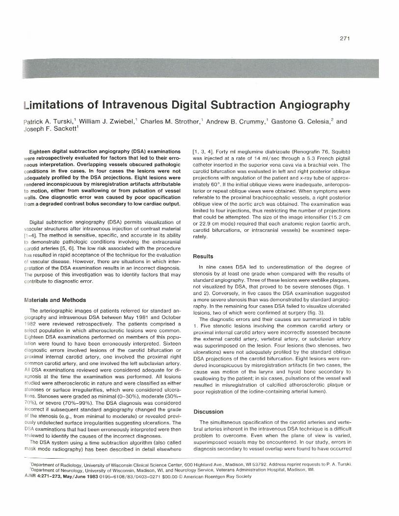
In nine cases DSA led to underestimat ion of the degree of stenosis by at least one grade when compared with the resu lts of standard angiography. Three of these lesions were weblike plaques, not visualized by DSA , that proved to be severe stenoses (figs. 1 and 2). Conversely, in five cases the DSA examinat ion suggested a more severe stenosis than was demonstrated by standard angiography. In the remain ing four cases DSA failed to visualize ulcerated lesions, two of which were confirmed at su rgery (fig. 3).
The diagnostic errors and their causes are summarized in table 1. Five stenotic lesions involving the common carotid artery or proximal internal carotid artery were incorrectly assessed because th e external carotid artery, vertebral artery, or subclavian artery was superimposed on the lesion. Four lesions (two stenoses , two ulcerations) were not adequately profil ed by the standard oblique DSA projections of the carot id bi furcation . Eight lesions were rendered inconspicuous by misregistration artifacts (in two cases, the cause was motion of the larynx and hyoid bone secondary to swallowing by the patient ; in six cases, pulsat ions of the vessel wall resul ted in misregistrat ion of ca lcified atherosc lerotic plaque or poor registrat ion of the iod ine-contain ing arter ial lumen).
Discussion
The simu ltaneous opacification of the carotid arteries and vertebral arteries inherent in the intravenous DSA technique is a difficu lt problem to overcome. Even when the plane of view is varied, superimposed vessels may be encountered . In our study , errors in diagnosis secondary to vessel overlap were found to have occurred
'Department of Rad iology, University of Wisconsin Clin ical Science Center, 600 Highland Ave., Madison, WI 53792. Address reprint requests to P. A. Turski . ' Department of Neurology, University of Wisconsin , Madison , WI , and Neurology Service, Veterans Administration Hospital, Madison, WI.
AJNR 4:271-273, May/ June 1983 0195- 6108/ 83 / 0403- 0271 $00.00 © American Roentgen Ray Society
272 DIGITAL RADIOGRAPHY AND DSA AJNR:4, May/ June 1983
1A 18 2A 28 Fig. 1 .-A, Intravenous DSA of aortic arch . Left subclavian artery appears free of significant stenosis. Fig . 2.-A, Carotid DSA compromised by po!)r
vascular opacification from low cardiac output. C 1-
rotid artery appears free of obstruction. B, Standald angiography reveals high-grade stenosis secondary to vascular web (arrow) .
B, Intraarterial DSA reveals high-grade stenosis secondary to vascular web (arrow) .
3A 38 4
Fig . 3.-A, DSA fai ls to show small ulcerations because of vessel overlap, lack of profile view, and pulsation of vessel wall. B, Standard angiography demonstrates multiple small ulcerations.
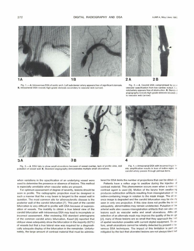
Fig . 4. -lntracranial DSA with incorrect logarI thmic amplification results in loss of iodine signal as carotid artery passes through petrous bone.
when variations in the opacification of an underlying vessel were used to determine the presence or absence of lesions. This method is especially unreliable when vascu lar webs are present.
For optimum assessment of degree of severity, lesions shou ld be seen in profile. The radiographic projection must be designed in such a manner that the x-ray beam is tangent to the vessel wall in question. The most common site for atherosclerotic disease is the posterior wall of the carotid bifurcation [7]. This part of the carotid bifurcation is very difficult to profile with DSA because of superposit ion of vessels . The inability to obtain a true lateral view of the carotid bifurcation with intravenous DSA may lead to incomplete or incorrect assessment. After reviewing 200 standard arteriograms of the common carotid artery bifurcat ion, Kasef [8] reported that oblique views adequately show the bifurcation in the majority (62%) of vessels but that a true lateral view was required for a diagnostically adequate display of the bifurcation in the remainder. Unfortunately, the large amount of contrast material that must be adminis-
tered for DSA limits the number of projections that can be obtainl,d. Patients have a reflex urge to swallow during the injectior of
contrast material. This phenomenon occurs even when a nonionic contrast agent is used [9]. Motion of the larynx from swallow'ng produces subtraction artifacts resulting from misregistration of he iodine-containing image in relation to the mask image. The dif'erence image is degraded and the carot id bifurcation may be cle;',rly seen in only one projection . If this view does not profile the lefl on adequately, abnormalities may remain undetected. Pulsation of the arterial walls also causes misregistration artifacts th at can obscure lesions such as vascular webs and small ulcerations. Althol19h
selection of an alternate mask may improve the quality of the im.lge [2] , many of these lesions are so small that they approach the limit of spatial resolution possible with current digital equipment. Thf'refore, small ulcerations cannot be reliably detected by present in iravenous DSA techniques. The impact of this limitation is perh 1Ps
mitigated by the fact that ulcerated lesions are not always idenli' ied
AJNR:4, May / June 1983 DIGITAL RADIOGRAPHY AND DSA 273
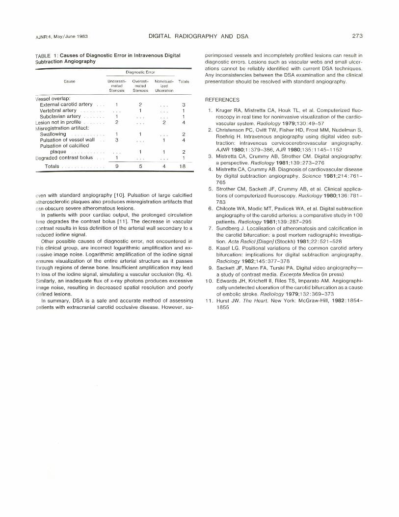
TABLE 1: Causes of Diagnostic Error in Intravenous Digital subtraction Angiography
Diagnostic Error
Cause Underesli- Overest i- Nonvisual- Totals mated mated ized
Stenosis Stenosis Ulceration
Vessel overlap: External carotid artery 2 3 Vertebral artery . . . . . . . . Subclavian artery 1 1
Lesion not in profile 2 2 4 Misregistration artifact:
Swallowing 1 2 Pulsation of vessel wall 3 4 Pulsat ion of calc ified
plaque 2 Degraded contrast bolus
Totals 9 5 4 18
even with standard angiography [10]. Pu lsation of large calc ified atherosclerotic plaques also produces misregistration artifacts that can obscure severe atheromatous lesions.
In patients with poor cardiac output , the prolonged circulation time degrades the contrast bolus [11]. The decrease in vascular contrast results in less definition of th e arterial wall secondary to a reduced iodine signal.
Other possible causes of diagnostic error, not encountered in th is clin ical group, are incorrect logarithmic amplification and excessive image noise. Logarithmic amplification of the iodine signal ensures visualization of the entire arterial struc ture as it passes through regions of dense bone. Insuffic ient amplification may lead to loss of the iodine signal, simulating a vascu lar occlusion (fig. 4) . Simi larly, an inadequate flu x of x-ray photons produces excessive image noise , resulting in decreased spatial resolution and poorly defined lesions.
In summary, DSA is a safe and accurate method of assessing patients with extracran ial carotid occlusive disease. However, su-
perimposed vessels and incompletely profil ed lesions can result in diagnostic errors. Lesions such as vascular webs and small ulceration s cannot be reliably identified with current DSA techniques. Any inconsistencies between the DSA examinat ion and the clinica l presentation should be reso lved with standard angiography.
REFERENCES
1. Kruger RA, Mistretta CA, Houk TL, et aL Computerized fluoroscopy in rea l time for noninvasive visualizat ion of the cardiovascu lar system. Radiology 1979;130 : 49-57
2. Christen son PC, Ovitt TW, Fisher HD, Frost MM, Nudelman S, Roehrig H. Intravenous angiography using digital video subtract ion: intravenous cervicocerebrovascular angiography. AJNR 1980;1 : 3 79-386 , AJR 1980;135: 1145-1152
3. Mistretta CA, Crummy AB, Strother CM . Digital angiography: a perspective . Radiology 198 1 ; 139: 273-276
4. Mistretta CA, Crummy AB. Diagnosis of cardiovascular d isease by digita l subtraction ang iog raphy. Science 1981 ;214: 761-765
5. Strother CM , Sackett JF, Crummy AB , et aL Clinica l applications of com puterized fluoroscopy. Radiology 1980;1 36: 781 -783
6. Ch ilcote WA, Modic MT, Pavlicek WA, et aL Digital subtract ion angiography of the carot id arteries: a comparat ive study in 100 patients. Radiology 1981 ;139: 287 -295
7. Sundberg J. Localisation of atheromatosis and calc ification in the carotid bifurcation: a post mortem rad iographic investigation . Acta Radiol [Oiagn] (Stockh) 1981 ;22: 52 1-528
8 . Kasef LG . Positional variat ions of the common carot id artery bifurcation: implications for digital subtract ion angiography. Radiology 1982;145 :377 - 378
9. Sackett JF, Mann FA, Tursk i PA. Digital video angiographya study of contrast media . Excerp ta Medica (in press)
10. Edward s JH , Krichefi II, Riles TS, Imparato AM . Angiographically undetected ulcerat ion of the carotid bifu rcation as a cause of embolic stroke. Radiology 1979;132 : 369- 373
11 . Hurst JW. The Heart . New York: McGraw-Hi li , 1982: 1854-1855