Embed Size (px)
Citation preview
CASE REPORT
432THE EUROPEAN JOURNAL OF ESTHETIC DENTISTRY
VOLUME 8 • NUMBER 3 • AUTUMN 2013
Correspondence to: José Carlos Martins da Rosa
Av. São Leopoldo 680 – CEP 95097-350 – Caxias do Sul, Rio Grande do Sul, Brazil;
Tel: +55 54 99711313; E-mail: [email protected]
Immediate Dentoalveolar Restoration
of compromised sockets:
a novel technique
José Carlos Martins da Rosa Specialist in Periodontics, São Paulo Association of Dental Surgeons,
Bauru, São Paulo, Brazil
MSc in Prosthetics, São Leopoldo Mandic Dental Research Center,
Campinas, São Paulo, Brazil
PhD Student in Oral Implantology, São Leopoldo Mandic Dental Research Center,
Campinas, São Paulo, Brazil
Ariádene Cristina Pértile de Oliveira RosaSpecialist in Implant Dentistry, São Leopoldo Mandic Dental Research Center,
Campinas, São Paulo, Brazil
Specialist in Acupuncture, Brazilian College of Systemic Studies, CBES,
Porto Alegre, Rio Grande de Sul, Brazil
MSc Student in Oral Implantology, São Leopoldo Mandic Dental Research Center,
Campinas, São Paulo, Brazil
Darcymar Martins da RosaSpecialist in Prosthetics, Pontifical Catholic University,
Porto Alegre, Rio Grande de Sul, Brazil
Specialist in Oral Implantology, Pontifical Catholic University,
Campinas, São Paulo, Brazil
Carla Mônica ZardoSpecialist in Orthodontics and Facial Orthopedics, HRAC,
University of São Paulo, Bauru, São Paulo SP, Brazil
MSc Student in Oral Implantology, São Leopoldo Mandic Dental Research Center,
Campinas, São Paulo, Brazil
ROSA ET AL
433THE EUROPEAN JOURNAL OF ESTHETIC DENTISTRY
VOLUME 8 • NUMBER 3 • AUTUMN 2013
Abstract
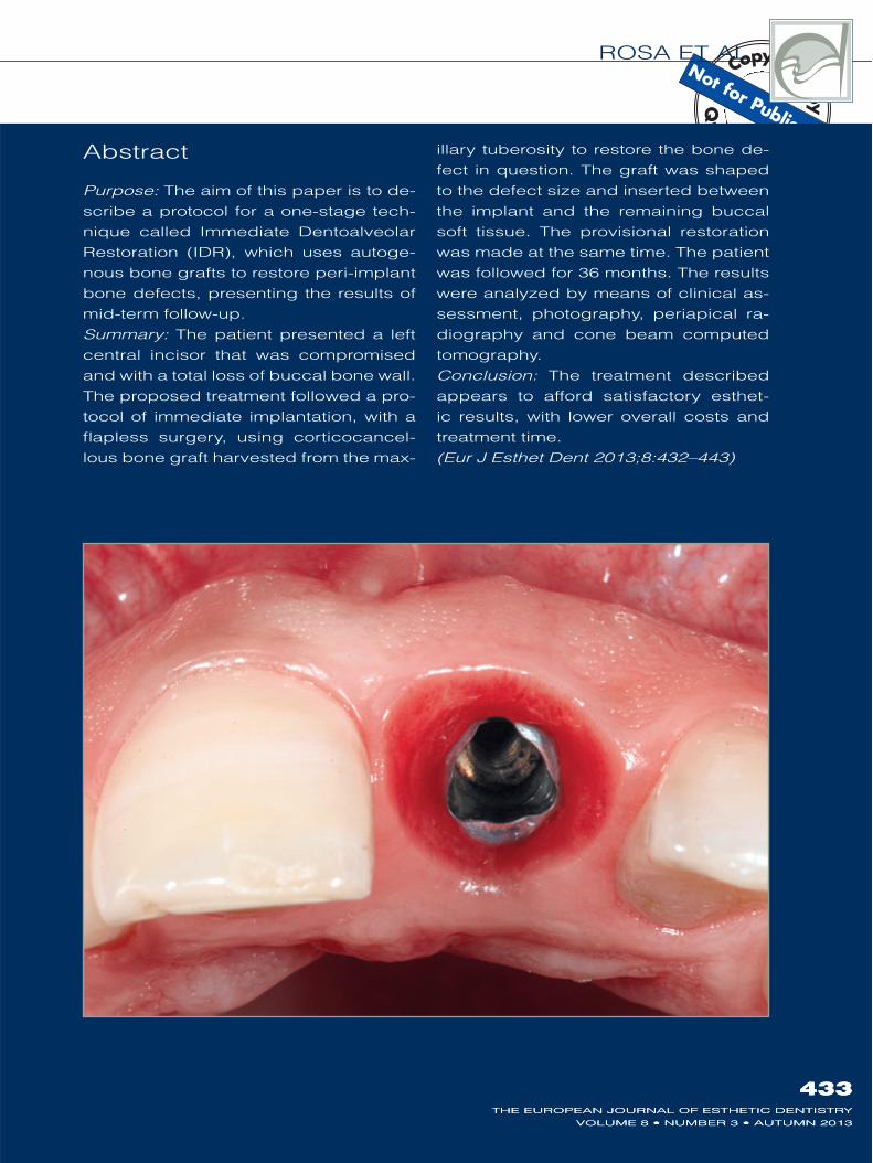
Purpose: The aim of this paper is to de-scribe a protocol for a one-stage tech-nique called Immediate Dentoalveolar Restoration (IDR), which uses autoge-nous bone grafts to restore peri-implant bone defects, presenting the results of mid-term follow-up. Summary: The patient presented a left central incisor that was compromised and with a total loss of buccal bone wall. The proposed treatment followed a pro-tocol of immediate implantation, with a flapless surgery, using corticocancel-lous bone graft harvested from the max-
illary tuberosity to restore the bone de-fect in question. The graft was shaped to the defect size and inserted between the implant and the remaining buccal soft tissue. The provisional restoration was made at the same time. The patient was followed for 36 months. The results were analyzed by means of clinical as-sessment, photography, periapical ra-diography and cone beam computed tomography. Conclusion: The treatment described appears to afford satisfactory esthet-ic results, with lower overall costs and treatment time. (Eur J Esthet Dent 2013;8:432–443)
433THE EUROPEAN JOURNAL OF ESTHETIC DENTISTRY
VOLUME 8 • NUMBER 3 • AUTUMN 2013
CASE REPORT
434THE EUROPEAN JOURNAL OF ESTHETIC DENTISTRY
VOLUME 8 • NUMBER 3 • AUTUMN 2013
Introduction
Immediate implantation and provision-alization requires the maintenance of the supporting tissues during dental extraction procedures.1-4 Nevertheless, implant placement in a fresh extraction socket is often associated with the pres-ence of peri-implant defects at the time of surgery.
Several procedures have been pro-posed to reestablish the compromised gingival and alveolar bone architecture, such as orthodontic forced eruption,5,6 Guided Bone Regeneration (GBR),7-11 and block bone graft with or without sub-epithelial connective tissue grafts.12,13 All of these treatments can be used to treat bone defects before, during, or after tooth removal, in two or three surgical stages. However, the possibility of reconstruction through grafting and immediate restor-ation in a single operation has not been supported by several clinical studies.
We propose that these cases could be successfully treated using a technique that allows dental extraction, implant-ation and provisionalization to occur in the same procedure as the flapless bone reconstruction.
The aim of this paper is to report results from a mid-term follow-up of a clinical case in which dental implantation and immedi-ate provisionalization in a compromised fresh socket was performed, describing a technique that we refer to as Immedi-ate Dentoalveolar Restoration (IDR). This technique uses corticocancellous bone grafts harvested from the maxillary tuber-osity to restore peri-implant bone defects at the same procedure and can be used in postextraction sites with minimal or se-vere loss of bone walls.
Report
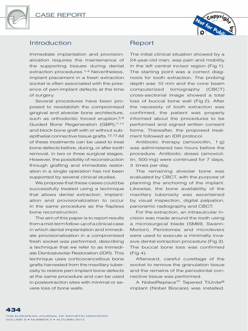
The initial clinical situation showed by a 24-year-old man, was pain and mobility in the left central incisor region (Fig 1). The starting point was a correct diag-nosis for tooth extraction. The probing depth was 10 mm and the cone beam computerized tomography (CBCT) cross-sectional image showed a total loss of buccal bone wall (Fig 2). After the necessity of tooth extraction was confirmed, the patient was properly informed about the procedures to be performed and signed written consent forms. Thereafter, the proposed treat-ment followed an IDR protocol.
Antibiotic therapy (amoxicillin, 1 g) was administered two hours before the procedure. Antibiotic doses (amoxicil-lin, 500 mg) were continued for 7 days, 3 times per day.
The remaining alveolar bone was evaluated by CBCT, with the purpose of planning the anchoring of the implant. Likewise, the bone availability of the maxillary tuberosity was ascertained by visual inspection, digital palpation, panoramic radiography and CBCT.
For the extraction, an intrasulcular in-cision was made around the tooth using a microsurgical blade (SM69, Swann-Morton). Periotomes and microlevers were used to execute a minimally inva-sive dental extraction procedure (Fig 3). The buccal bone loss was confirmed (Fig 4).
Afterward, careful curettage of the socket to remove the granulation tissue and the remains of the periodontal con-nective tissue was performed.
A NobelReplace™ Tapered TiUnite® implant (Nobel Biocare) was installed.
ROSA ET AL
435THE EUROPEAN JOURNAL OF ESTHETIC DENTISTRY
VOLUME 8 • NUMBER 3 • AUTUMN 2013
Fig 1 Initial clinical assessment of the compro-mised left central incisor.
Fig 3 The root fracture could be seen after the minimally invasive dental extraction procedure.
Fig 5 The implant was installed with palatal an-chorage and with a 3 mm distance between implant platform and gingival margin (NobelReplace™ Ta-pered TiUnite® implant, 16 mm X 5.0 mm).
Fig 2 Total loss of buccal bone wall as ob-served by cone beam computerized tomog-raphy.
Fig 4 Absence of buccal bone wall confirmed af-ter dental extraction.
The final insertion torque was 50 Ncm. The implant was placed by means of a palatal approach with ideal three-di-mensional positioning. The implant plat-form was inserted 3 mm apically of the gingival margin (Fig 5).
A provisional crown was constructed, establishing the ideal emergence pro-file to allow for the accommodation of the soft tissues and to promote a thicker and more stable margin of gingival tis-sue. In addition, the provisional crown was made out of occlusion. All steps of the provisionalization were performed
CASE REPORT
436THE EUROPEAN JOURNAL OF ESTHETIC DENTISTRY
VOLUME 8 • NUMBER 3 • AUTUMN 2013
before bone graft procedures so as to not risk contaminating the graft while handling the materials used to construct the crown.
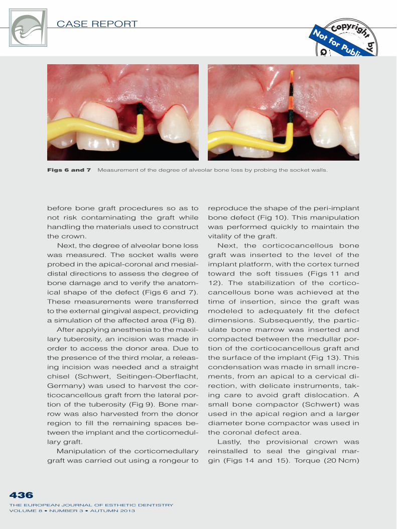
Next, the degree of alveolar bone loss was measured. The socket walls were probed in the apical-coronal and mesial-distal directions to assess the degree of bone damage and to verify the anatom-ical shape of the defect (Figs 6 and 7). These measurements were transferred to the external gingival aspect, providing a simulation of the affected area (Fig 8).
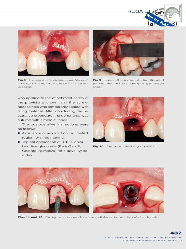
After applying anesthesia to the maxil-lary tuberosity, an incision was made in order to access the donor area. Due to the presence of the third molar, a releas-ing incision was needed and a straight chisel (Schwert, Seitingen-Oberflacht, Germany) was used to harvest the cor-ticocancellous graft from the lateral por-tion of the tuberosity (Fig 9). Bone mar-row was also harvested from the donor region to fill the remaining spaces be-tween the implant and the corticomedul-lary graft.
Manipulation of the corticomedullary graft was carried out using a rongeur to
reproduce the shape of the peri-implant bone defect (Fig 10). This manipulation was performed quickly to maintain the vitality of the graft.
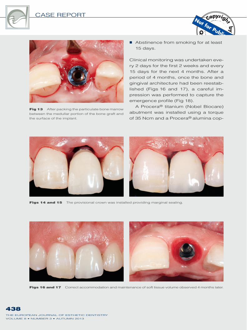
Next, the corticocancellous bone graft was inserted to the level of the implant platform, with the cortex turned toward the soft tissues (Figs 11 and 12). The stabilization of the cortico-cancellous bone was achieved at the time of insertion, since the graft was modeled to adequately fit the defect dimensions. Subsequently, the partic-ulate bone marrow was inserted and compacted between the medullar por-tion of the corticocancellous graft and the surface of the implant (Fig 13). This condensation was made in small incre-ments, from an apical to a cervical di-rection, with delicate instruments, tak-ing care to avoid graft dislocation. A small bone compactor (Schwert) was used in the apical region and a larger diameter bone compactor was used in the coronal defect area.
Lastly, the provisional crown was reinstalled to seal the gingival mar-gin (Figs 14 and 15). Torque (20 Ncm)
Figs 6 and 7 Measurement of the degree of alveolar bone loss by probing the socket walls.
ROSA ET AL
437THE EUROPEAN JOURNAL OF ESTHETIC DENTISTRY
VOLUME 8 • NUMBER 3 • AUTUMN 2013
Fig 9 Bone graft being harvested from the lateral portion of the maxillary tuberosity using an straight chisel.
Figs 11 and 12 Placing the corticocancellous bone graft shaped to match the defect configuration.
Fig 8 The area to be reconstructed was “outlined” at the soft tissue region using blood from the alveo-lar socket.
Fig 10 Simulation of the final graft position.
was applied to the attachment screw of the provisional crown, and the screw- access hole was temporarily sealed with filling material. After concluding the re-storative procedure, the donor area was sutured with simple stitches.
The postoperative instructions were as follows:!" Avoidance of any load on the treated
region for three months;!" Topical application of 0.12% chlor-
hexidine gluconate (PerioGard®, Colgate-Palmolive) for 7 days, twice a day.
CASE REPORT
438THE EUROPEAN JOURNAL OF ESTHETIC DENTISTRY
VOLUME 8 • NUMBER 3 • AUTUMN 2013
Fig 13 After packing the particulate bone marrow between the medullar portion of the bone graft and the surface of the implant.
Figs 14 and 15 The provisional crown was installed providing marginal sealing.
!" Abstinence from smoking for at least 15 days.
Clinical monitoring was undertaken eve-ry 2 days for the first 2 weeks and every 15 days for the next 4 months. After a period of 4 months, once the bone and gingival architecture had been reestab-lished (Figs 16 and 17), a careful im-pression was performed to capture the emergence profile (Fig 18).
A Procera® titanium (Nobel Biocare) abutment was installed using a torque of 35 Ncm and a Procera® alumina cop-
Figs 16 and 17 Correct accommodation and maintenance of soft tissue volume observed 4 months later.
ROSA ET AL
439THE EUROPEAN JOURNAL OF ESTHETIC DENTISTRY
VOLUME 8 • NUMBER 3 • AUTUMN 2013
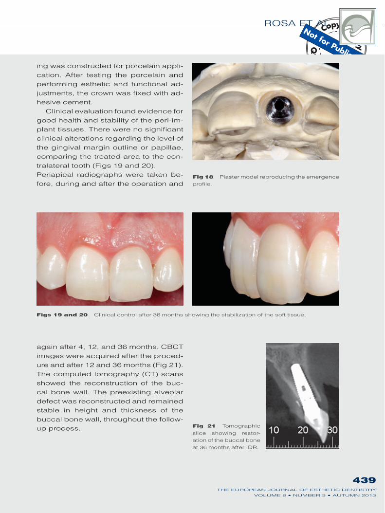
Fig 18 Plaster model reproducing the emergence profile.
Figs 19 and 20 Clinical control after 36 months showing the stabilization of the soft tissue.
Fig 21 Tomographic slice showing restor-ation of the buccal bone at 36 months after IDR.
ing was constructed for porcelain appli-cation. After testing the porcelain and performing esthetic and functional ad-justments, the crown was fixed with ad-hesive cement.
Clinical evaluation found evidence for good health and stability of the peri-im-plant tissues. There were no significant clinical alterations regarding the level of the gingival margin outline or papillae, comparing the treated area to the con-tralateral tooth (Figs 19 and 20). Periapical radiographs were taken be-fore, during and after the operation and
again after 4, 12, and 36 months. CBCT images were acquired after the proced-ure and after 12 and 36 months (Fig 21). The computed tomography (CT) scans showed the reconstruction of the buc-cal bone wall. The preexisting alveolar defect was reconstructed and remained stable in height and thickness of the buccal bone wall, throughout the follow-up process.
CASE REPORT
440THE EUROPEAN JOURNAL OF ESTHETIC DENTISTRY
VOLUME 8 • NUMBER 3 • AUTUMN 2013
Discussion
Immediate restoration with dental im-plants after dental extraction is sug-gested to achieve esthetic restoration, maintenance of the bone, gingival archi-tecture and reduction of patient discom-fort.1-4,14-20 However, an impediment to immediate restoration after dental extrac-tion may be the morphology of the dam-aged socket, if there is greater involve-ment of the buccal cortical bone, due to thickness of the bone and reduced vas-cularization. Because the buccal wall is fragile, total loss of this cortical bone can often be seen, even without the involve-ment of the other walls.
The treatment alternatives for reso-lution of alveolar defects after tooth re-moval are widely documented in the literature,7,9-11,13 and recommended as viable solutions before or after de-layed implant placement. However, the esthetic results of these techniques are less predictable, when combined to ad-ditional risk factors, for example, a high lip line or thin gingival biotype. In addi-tion, such techniques require long treat-ment time and present high morbidity.
As an alternative to block grafts and guided bone regeneration, the idea of the IDR technique is to promote a bar-rier with the corticancellous graft in the shape of the bone wall defect, stabilizing the particulate bone graft around the im-plant. This procedure represents signifi-cant gains in esthetic results and in total treatment time, since it enables the re-covery of an alveolar bone defect in the same surgical implant installation and immediate provisionalization, without opening the flap and keeping the gin-gival architecture in the same position.
It is suggested that the maxillary tu-berosity is an excellent donor area for alveolar reconstruction after dental ex-traction. In spite of providing a limited quantity of available bone for grafting in some cases, the use of the tuberosity has the advantages of excellent post-operative recovery and ease of graft ad-aptation in the receptor bed because of bone malleability. However, the harvest-ing of this graft may involve some risks, such as exposure of sinus membrane and damage of the last molar roots. In order to avoid these risks, besides the detailed preliminary assessment, care-ful technical execution and use of ade-quate instruments are necessary for the removal of this type of graft.
The vascularization pattern is known to be vital for bone grafting success. Because of the trabecular nature of grafts harvested from the tuberosity, these grafts have a high capacity for revascularization.21-25 In addition, one study has indicated that the maxillary and mandibular periosteum and bone marrow from the maxillary tuberosity can effectively serve as reliable and easy-to-harvest intraoral sources of os-teoprogenitor cells.26 Graft cell survival is related to the efficiency of the sur-gical technique and the time taken to transfer the graft to the receptor area.23 In fact, such grafts need to be manipu-lated quickly so that their vitality is not decreased.
It is known that early, low-intensity stimulation of a graft, without loss of mechanical stability, increases local blood flow and contact osteogenesis, thereby accelerating the process of bone graft incorporation.24,27,28 There-fore, the immediate construction of the
ROSA ET AL
441THE EUROPEAN JOURNAL OF ESTHETIC DENTISTRY
VOLUME 8 • NUMBER 3 • AUTUMN 2013
provisional crown is fundamental in the described technique.
Otherwise, in undamaged sock-ets, the implant should be inserted by means of a palatal approach to achieve better bone anchoring, with three-di-mensional positioning to spread the occlusal forces and enhance the es-thetic results.29
The size of the peri-implant gap determines whether filling it with par-ticulate bone is needed. Such a filling would preferably be autogenous, as this yields the best results with regard to bone healing.14 When the diameter of the implant is smaller than the sock-et opening, the peri-implant gap must be filled with particulate bone, thereby minimizing contraction of the tissues involved.
Stabilization and close contact be-tween the bone graft and the compro-mised site receiving the graft facilitate the revascularization process and fa-vor early incorporation of the graft into the host vascular bed.23-25,30,31 For this reason, particulate bone is required for filling the gap between the implant, the socket walls and the corticocancellous bone when the IDR technique is used. The main risk involved in performing this technique is not obtaining the correct adjustment and stabilization of the graft to the receptor site. This is more criti-cal in cases of thin periodontal biotype. Therefore, special care must be taken in obtaining the correct adaptation of the corticocancellous graft and the conden-sation of particulate bone.
Upon reconstruction of the alveolar anatomy, and in accordance with the lit-erature regarding the immediate restor-ation of implants,32 the prosthetic crown
used in this case was planned so that it would fit closely but without compres-sion of the gingival margin, providing support and stability to the soft tissue. A concavity was sculpted subgingivally on the prosthetic crown emergence profile to prevent facial tissue compression and to assist in physiological remodeling of the soft tissue complex.
Using the technique described in this paper, buccal bone wall thickening due to palatal anchoring of the implant and grafting of corticocancellous bone was observed. Because of the greater thick-ness of the buccal bone crest, along with an adequate prosthetic crown emergence profile, a greater volume of soft tissue was obtained, thus providing better and more stable gingival margin outlines. Furthermore, the concavity of the definitive prosthetic crown emer-gence profile may influence soft tissue volume preservation. These results re-mained stable throughout the period of monitoring.
Conclusion
In the reported clinical case, the sta-bility of hard and soft tissue had been observed throughout the period of fol-low-up. This data indicate that the IDR technique may promote restoration of freshly damaged sockets, thus making immediate provisionalization of an im-plant possible, saving the patients from having several surgical interventions and avoiding the esthetic risks related to these procedures.
CASE REPORT
442THE EUROPEAN JOURNAL OF ESTHETIC DENTISTRY
VOLUME 8 • NUMBER 3 • AUTUMN 2013
References
1. Wöhrle PS. Single-tooth replacement in the aes-thetic zone with immediate provisionalization: fourteen consecutive case reports. Pract Periodontics Aesthet Dent 1998;10:1107–1114.
2. Touati B, Guez G. Immedi-ate implantation with provi-sionalization: from literature to clinical implications. Pract Proced Aesthet Dent 2002;14:699–707.
3. Kan JY, Rungcharassaeng K, Lozada J. Immediate place-ment and provisionalization of maxillary anterior single implants: 1-year prospective study. Int J Oral Maxillofac Implants 2003;18:31–39.
4. De Rouck T, Collys K, Cosyn J. Single-tooth replacement in the anterior maxilla by means of immedi-ate implantation and provi-sionalization: a review. Int J OralMaxillofac Implants. 2008;23:897–904.
5. Chambrone L, Cham-brone LA. Forced ortho-dontic eruption of fractured teeth before implant place-ment: case report. J of the Canadian Dental Assoc 2005;71:257–261.
6. Salama H, Salama M. The role of orthodontic extrusive remodeling in the enhance-ment of soft and hard tissue profiles prior to implant placement: a systematic ap-proach to the management of the extraction site defects. Int J Periodontics Rest Dent 1993;13:312–333.
7. Jung RE, Kokovic V, Juri-sic M, Yaman D, Subramani K, Weber FE. Guided bone regeneration with a synthetic biodegradable membrane: a comparative study in dogs. Clin Oral Implants Res 2011;22:802–807.
8. De Santis E, Botticelli D, Pantani F, Pereira FP, Beol-chini M, Lang NP. Bone regeneration at implants placed into extraction sock-ets of maxillary incisors in dogs. Clin Oral Implants Res 2011;22:430–437.
9. Berglungh T, Lindhe J. Heal-ing arround implants placed in bone defects treated with Bio-Oss: an experimental study in the dog. Clin Oral Implants Res 1997;8: 117–124.
10. Ormianer Z, Palti A, Shif-man A. Survival of immedi-ately loaded dental implants in deficient alveolar bone sites augmented with !-Tricalcium Phosphate. Im-plant Dentistry 2006;15: 395–403.
11. Van Steenberghe D, Cal-lens A, Geers L, Jacobs R. The clinical use of deprotein-ized bovine bone mineral on bone regeneration in conjunc-tion with immediate implant installation. Clin Oral Implants Res 2000;11:210–216.
12. Schneider D, Grunder U, Ender A, Hämmerle C H, Jung RE. Volume gain and stability of peri-implant tis-sue following bone and soft tissue augmentation: 1-year results from a prospec-tive cohort study. Clin Oral Implants Res 2011;22:28–37.
13. Rebaudi A, Massei G, Trisi P, Calvari F. A new technique for bone augmentation and papilla reconstruction with autogenous free gingival-bone grafts. Int J Periodon-tics Rest Dent 2007;27: 429–439.
14. Schropp L, Kostopoulos L, Wenzel A, Isidor F. Clinical and radiographic perfor-mance of delayed-immediate single-tooth implant place-ment associated with peri-implant bone defects. A 2-year prospective, con-trolled, randomized follow-up report. J Clin Periodontol 2005;32:480–487.
15. Tupac RG. When is an implant ready for a tooth? J Calif Dent Assoc 2003 Dec;31:911–915.
16. De Kok IJ, Chang SS, Mori-arty JD, Cooper LF. A retro-spective analysis of periim-plant tissue responses at immediate load/provisional-ized microthreaded implants. Int J Oral Maxillofac Implants 2006;21:405–412.
17. Barone A, Rispoli L, Vozza I, Quaranta A, Covani U. Immediate restoration of sin-gle implants placed immedi-ately after tooth extraction. J Periodontol 2006;77: 1914–1920.
18. Degidi M, Piattelli A, Carin-ci F. Immediate loaded dental implants: comparison between fixtures inserted in postextractive and healed bone sites. J Craniofac Surg 2007;18:965–971.
19. Crespi R, Capparè P, Gherlone E, Romanos GE. Immediate occlusal loading of implants placed in fresh sockets after tooth extrac-tion. Int J Oral Maxillofac Implants 2007;22:955–962.
20. Villa R, Rangert B. Immedi-ate and early function of implants placed in extraction sockets of maxillary infected teeth: a pilot study. J Pros-thet Dent 2007;97:96–108. Erratum in: J Prosthet Dent 2008;99:167.
21. Prolo DJ, Rodrigo JJ. Contemporary bone graft physiology and surgery. Clin Orthop Relat Res 1985;200:322–342.
22. Goldberg VM, Stevenson S. Natural history of autografts and allografts. Clin Orthop Relat Res. Clin Orthop Relat Res 1987;225:7–16.
23. Gordh M, Alberius P. Some basic factors essential to autogeneic nonvascularized onlay bone grafting to the craniofacial skeleton. Scand J Plast Reconstr Surg Hand Surg 1999;33:129–146.
ROSA ET AL
443THE EUROPEAN JOURNAL OF ESTHETIC DENTISTRY
VOLUME 8 • NUMBER 3 • AUTUMN 2013
24. Burchardt H. Biology of bone transplantation. Orthop Clin North Am 1987 Apr;18: 187–196.
25. Burchardt H. The biology of bone graft repair. Clin Orthop Relat Res 1983;174:28–42.
26. Cicconetti A, Sacchetti B, Bartoli A, Michienzi S, Corsi A, Funari A, Robey PG, Bianco P, Riminucci M. Human maxillary tuberos-ity and jaw periosteum as sources of osteoprogenitor cells for tissue engineering. Oral Surg Oral Med Oral Pathol Oral Radiol Endod 2007;104:618.e1–e12.
27. Vandamme K, Naert I, Geris L, Sloten JV, Puers R, Duyck J. Histodynamics of bone tissue formation around immediately loaded cylin-drical implants in the rab-bit. Clin Oral Implants Res 2007;18:471–480.
28. Keller EE, Tolmann DE, Eckert SE. Surgical pros-thetic reconstruction of advanced maxillary bone compromised with autoge-nous onlay block bone grafts and osseointegrated endos-seous implants: 12-year study of 32 consecutive patients. Int J Oral Maxillofac Implants 1999;14:197–209.
29. Buser D, Martin W, Belser UC. Optimizing esthetics for implant restor-ations in the anterior max-illa: anatomic and surgical considerations. Int J Oral Maxillofac Implants 2004;(19 Suppl):43–61.
30. Albrektsson T. In vivo studies of bone grafts. The possibil-ity of vascular anastomoses in healing bone. Acta Orthop Scand 1980;51:9–17.
31. Albrektsson T. Repair of bone grafts. A vital micro-scopic and histological investigation in the rabbit. Scand J Plast Reconstr Surg 1980;14:1–12.
32. Zeren KJ. Minimally invasive extraction and immedi-ate implant placement: the preservation of esthetics. Int J Periodontics Restorative Dent 2006;26:171–181.