Embed Size (px)
Citation preview
Schuster-Amft et al., Cogent Medicine (2016), 3: 1240282http://dx.doi.org/10.1080/2331205X.2016.1240282
PHYSIOLOGY & REHABILITATION | RESEARCH ARTICLE
Immediate effects of different upper limb robot-assisted training modes in patients after stroke: A case seriesCorina Schuster-Amft1,2*, Leni Van Kerckhoven3, Magdalena Berse3 and Geert Verheyden3
Abstract: Purpose: This case series investigated immediate effects of three different robot-assisted training (RAT) modes post stroke. Methods: A repeated measures de-scriptive case series design was applied. Patients after stroke (sub‐acute or chronic) received 4 × 30 min RAT for 3 weeks. Every week, a different randomly selected RAT-mode was applied: passive mobilisation, games, or activities of daily living (ADL). Outcome measures were bilateral upper limb function evaluated with the Chedoke McMaster Arm and Hand Activity Inventory (CAHAI‐9), muscle tone investigated with the Modified Modified Ashworth Scale (MMAS), and active and passive range of motion measured with a goniometer (aROM, pROM). Assessments were con-ducted by a blinded examiner at baseline, before and after each intervention week. Patients qualitatively evaluated RAT. There was no control on the patients receiving other therapies or treatments during the study period. For statistical analyses the Wilcoxon signed-rank test was used. Results: In total, seven patients participated (2 females, 5 males, mean ± SD: age 62.4 ± 6.9; time since stroke 35.4 ± 23.6 months, except for 1 sub-acute patient). CAHAI-9 scores changed: 35.9 ± 17.1 at baseline to 39.4 ± 16.6 after RAT, with a significant improvement after ADL-mode (p = 0.028). Patients reported larger ROM, less muscle tone, increased upper limb motor func-tion, and no adverse events. Conclusions: An overall improvement in upper limb
*Corresponding author: Corina Schuster-Amft, Research Department, Reha Rheinfelden, Rheinfelden, Switzerland; Institute for Rehabilitation and Performance Technology, Bern University of Applied Sciences, Burgdorf, Switzerland E-mail: [email protected]
Reviewing editor:Udo Schumacher, University Medical Center Hamburg-Eppendorf, Germany
Additional information is available at the end of the article
ABOUT THE AUTHORSDr Corina Schuster-Amft is a physiotherapist especially trained in neurological rehabilitation. She is the head of the Research Department at the Reha Rheinfelden in Switzerland. The team includes experts from the therapeutic disciplines, sports and movement scientists, and psychologist. The research of the department focusses on technology-based rehabilitation treatment options, e.g. robot-assisted training, virtual reality-based training, or wearable technology and cognitive assessments to evaluate the stroke recovery process. Furthermore, the team is interested in the technique of motor imagery as a treatment option, and devices and assessments to evaluate postural control.
PUBLIC INTEREST STATEMENTThe World Health Organisation states that every 6 s, someone’s quality of life will forever be changed—they will permanently be physically disabled due to stroke. Recovery of motor and sensory function is essential for activities of daily living (ADL) and the independence of the patient. Recently there has been more attention for technology-based treatment methods, such as robot-assisted training (RAT). In our study patients after stroke (sub‐acute or chronic) received 4 × 30 min RAT for 3 weeks. Every week, a different RAT-mode was applied: passive mobilisation, games, or ADL. In total, two females and five males participated (age 62.4 ± 6.9; time since stroke 35.4 ± 23.6 months) in the study. Each of them showed important functional improvements in particular after the ADL training mode. Patients reported larger ROM, less muscle tone, increased upper limb motor function, and no adverse events. It is recommended to include all three RAT modes in the training.
Received: 10 August 2016Accepted: 14 September 2016First Published: 26 September 2016
© 2016 The Author(s). This open access article is distributed under a Creative Commons Attribution (CC-BY) 4.0 license.
Page 1 of 15
Page 2 of 15
Schuster-Amft et al., Cogent Medicine (2016), 3: 1240282http://dx.doi.org/10.1080/2331205X.2016.1240282
function was found in all cases. Despite the significant improvement after the ADL-mode, it remains recommended to include all three RAT modes. RAT did subjectively but not objectively influence muscle tone and ROM.
Subjects: Assistive Technology; Disability; Physiotherapy; Rehabilitation Medicine
Keywords: upper limb motor function; robot-assisted training; stroke; activities of daily living
1. IntroductionStroke is the most common cause of chronic disability in the developed world (Rossini, Calautti, Pauri, & Baron, 2003). In the US, every 40 s a new stroke occurs which leads up to 790’000 new pa-tients every year (Go et al., 2013).
Significant problems after stroke are hemiplegia and sensory deficits contralateral to the side of brain lesion, in particular limitations in upper limb movement and function. Recovery of motor and sensory function of the upper extremity is essential for activities of daily living (ADL) and the inde-pendence of the patient. A recent study showed that six months after stroke, only 25% of patients showed a good recovery of upper extremity function Kong & Lee, 2013). Therefore, upper extremity training is a key component of stroke rehabilitation.
Classical therapy concepts in stroke rehabilitation include Bobath or the neurodevelopmental treatment concept and proprioceptive neuromuscular facilitation (Brock, Haase, Rothacher, & Cotton, 2011; Hwangbo & Don Kim, 2016; Vaughan-Graham, Cott, & Wright, 2015). However, accord-ing to recent studies these concepts have limitations in terms of intensity and specificity (Huang & Krakauer, 2009; Lo & Xie, 2012). Furthermore, there is little transfer to ADL (Bütefisch, Hummelsheim, Denzler, & Mauritz, 1995; Langhammer & Stanghelle, 2000; Wagenaar & Meyer, 1991). Recently there has been more attention for technology-based treatment methods, such as robot-assisted training (RAT). RAT includes training of (task-)specific movements at an increased intensity (Lo & Xie, 2012; Waldner, Tomelleri, & Hesse, 2009). An additional advantage is the possibility of measuring kinematic parameters during training (Bosecker, Dipietro, Volpe, & Igo Krebs, 2010; Loureiro, Harwin, Nagai, & Johnson, 2011). ARMin III (ETH Zurich, Switzerland) is an exoskeleton robot with the option to align the artificial joint with the corresponding joint of the patient (Balasubramanian, Klein, & Burdet, 2010; Klamroth-Marganska et al., 2013; Staubli, Nef, Klamroth-Marganska, & Riener, 2009). With this design, a large degree of freedom is possible and selective training of specific movements at a single joint is facilitated (Staubli et al., 2009).
A systematic review reported that RAT could improve motor function in the upper limb in patients post stroke (Kwakkel, Kollen, & Krebs, 2008). However, the authors did not detect improvement in ADL function. In a more recent review from Mehrholz, Hadrich, Platz, Kugler, and Pohl (2012) an im-provement in ADL and arm function after RAT arm training could be shown, although no significant effect on upper extremity strength was reported.
From a clinical point of view we know that RAT has promising effects on upper limb function (Mehrholz et al., 2012; Staubli et al., 2009). However, we do not know the effect of the different ARMin modes applied.
In a recent randomised controlled trial it was shown that ARMin III training including three train-ing modes (passive mobilisation, games, ADL) improved motor function of the paretic arm (Klamroth-Marganska et al., 2013). But until now, the three ARMin III training modes have not been systematically investigated.
Therefore, the primary research question of the present study was what the immediate effects of different RAT modes on upper limb functioning in stroke patients were. We hypothesized that all three modes would lead to improvement in upper extremity activity, but mainly the application of
Page 3 of 15
Schuster-Amft et al., Cogent Medicine (2016), 3: 1240282http://dx.doi.org/10.1080/2331205X.2016.1240282
the ADL-mode would lead to improvement in ADL tasks and therefore, to a meaningful change for the patients. We additionally investigated (1) muscle tone in combination with RAT and hypothe-sised that our delivery of RAT would not increase upper extremity muscle tone and (2) the patients’ perspective on RAT.
2. Methods
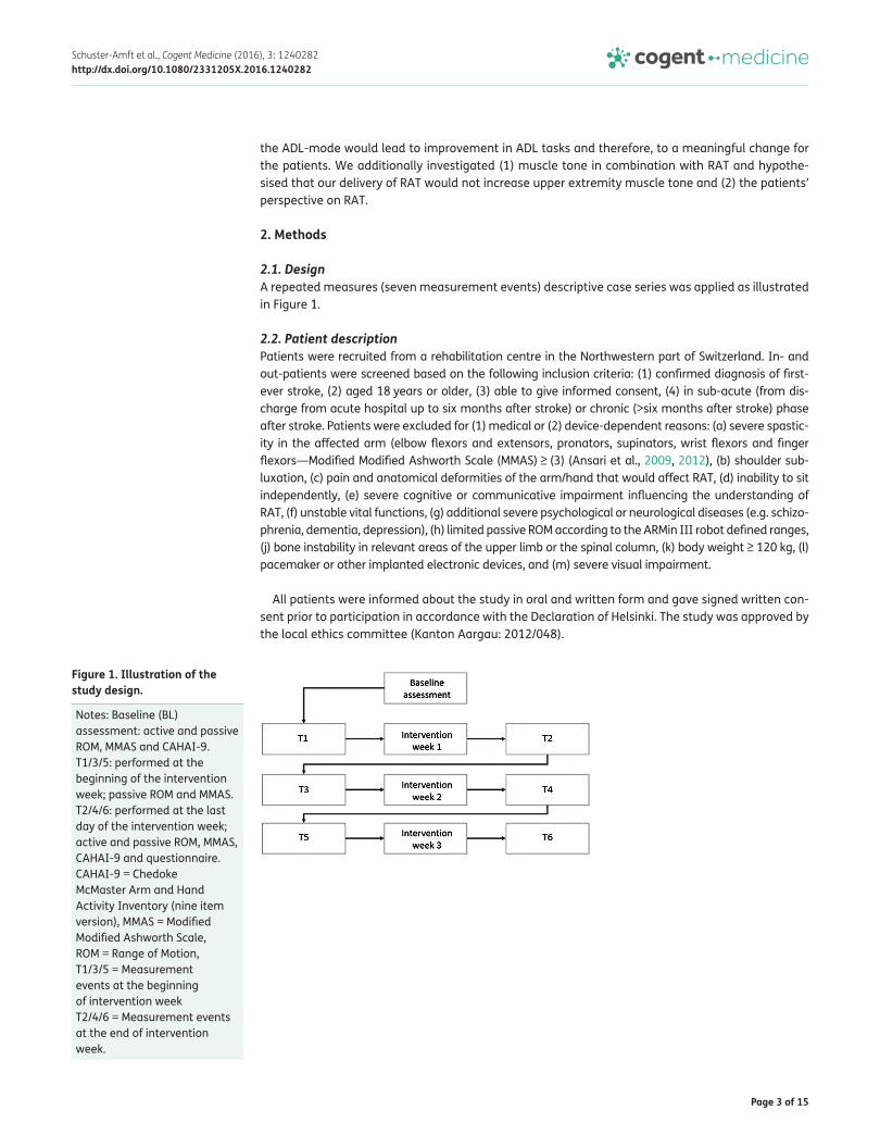
2.1. DesignA repeated measures (seven measurement events) descriptive case series was applied as illustrated in Figure 1.
2.2. Patient descriptionPatients were recruited from a rehabilitation centre in the Northwestern part of Switzerland. In- and out-patients were screened based on the following inclusion criteria: (1) confirmed diagnosis of first-ever stroke, (2) aged 18 years or older, (3) able to give informed consent, (4) in sub-acute (from dis-charge from acute hospital up to six months after stroke) or chronic (>six months after stroke) phase after stroke. Patients were excluded for (1) medical or (2) device-dependent reasons: (a) severe spastic-ity in the affected arm (elbow flexors and extensors, pronators, supinators, wrist flexors and finger flexors—Modified Modified Ashworth Scale (MMAS) ≥ (3) (Ansari et al., 2009, 2012), (b) shoulder sub-luxation, (c) pain and anatomical deformities of the arm/hand that would affect RAT, (d) inability to sit independently, (e) severe cognitive or communicative impairment influencing the understanding of RAT, (f) unstable vital functions, (g) additional severe psychological or neurological diseases (e.g. schizo-phrenia, dementia, depression), (h) limited passive ROM according to the ARMin III robot defined ranges, (j) bone instability in relevant areas of the upper limb or the spinal column, (k) body weight ≥ 120 kg, (l) pacemaker or other implanted electronic devices, and (m) severe visual impairment.
All patients were informed about the study in oral and written form and gave signed written con-sent prior to participation in accordance with the Declaration of Helsinki. The study was approved by the local ethics committee (Kanton Aargau: 2012/048).
Figure 1. Illustration of the study design.
Notes: Baseline (BL) assessment: active and passive ROM, MMAS and CAHAI-9. T1/3/5: performed at the beginning of the intervention week; passive ROM and MMAS. T2/4/6: performed at the last day of the intervention week; active and passive ROM, MMAS, CAHAI-9 and questionnaire. CAHAI-9 = Chedoke McMaster Arm and Hand Activity Inventory (nine item version), MMAS = Modified Modified Ashworth Scale, ROM = Range of Motion, T1/3/5 = Measurement events at the beginning of intervention week T2/4/6 = Measurement events at the end of intervention week.
Page 4 of 15
Schuster-Amft et al., Cogent Medicine (2016), 3: 1240282http://dx.doi.org/10.1080/2331205X.2016.1240282
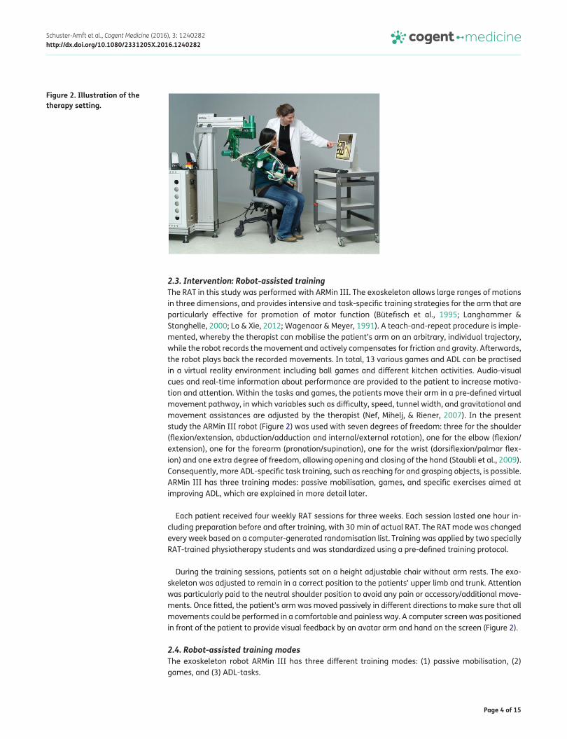
2.3. Intervention: Robot-assisted trainingThe RAT in this study was performed with ARMin III. The exoskeleton allows large ranges of motions in three dimensions, and provides intensive and task-specific training strategies for the arm that are particularly effective for promotion of motor function (Bütefisch et al., 1995; Langhammer & Stanghelle, 2000; Lo & Xie, 2012; Wagenaar & Meyer, 1991). A teach-and-repeat procedure is imple-mented, whereby the therapist can mobilise the patient’s arm on an arbitrary, individual trajectory, while the robot records the movement and actively compensates for friction and gravity. Afterwards, the robot plays back the recorded movements. In total, 13 various games and ADL can be practised in a virtual reality environment including ball games and different kitchen activities. Audio-visual cues and real-time information about performance are provided to the patient to increase motiva-tion and attention. Within the tasks and games, the patients move their arm in a pre-defined virtual movement pathway, in which variables such as difficulty, speed, tunnel width, and gravitational and movement assistances are adjusted by the therapist (Nef, Mihelj, & Riener, 2007). In the present study the ARMin III robot (Figure 2) was used with seven degrees of freedom: three for the shoulder (flexion/extension, abduction/adduction and internal/external rotation), one for the elbow (flexion/extension), one for the forearm (pronation/supination), one for the wrist (dorsiflexion/palmar flex-ion) and one extra degree of freedom, allowing opening and closing of the hand (Staubli et al., 2009). Consequently, more ADL-specific task training, such as reaching for and grasping objects, is possible. ARMin III has three training modes: passive mobilisation, games, and specific exercises aimed at improving ADL, which are explained in more detail later.
Each patient received four weekly RAT sessions for three weeks. Each session lasted one hour in-cluding preparation before and after training, with 30 min of actual RAT. The RAT mode was changed every week based on a computer-generated randomisation list. Training was applied by two specially RAT-trained physiotherapy students and was standardized using a pre-defined training protocol.
During the training sessions, patients sat on a height adjustable chair without arm rests. The exo-skeleton was adjusted to remain in a correct position to the patients’ upper limb and trunk. Attention was particularly paid to the neutral shoulder position to avoid any pain or accessory/additional move-ments. Once fitted, the patient’s arm was moved passively in different directions to make sure that all movements could be performed in a comfortable and painless way. A computer screen was positioned in front of the patient to provide visual feedback by an avatar arm and hand on the screen (Figure 2).
2.4. Robot-assisted training modesThe exoskeleton robot ARMin III has three different training modes: (1) passive mobilisation, (2) games, and (3) ADL-tasks.
Figure 2. Illustration of the therapy setting.
Page 5 of 15
Schuster-Amft et al., Cogent Medicine (2016), 3: 1240282http://dx.doi.org/10.1080/2331205X.2016.1240282
(1) During passive mobilisation, a “teach-and-repeat” mode is used. The therapist moves the ro-bot arm in an elective patient-specific movement sequence emphasising one joint or com-bined joint movements. The robot registers the sequence and afterwards repeats it several times with the patient’s arm.
(2) The game mode includes three games: a ball game, a labyrinth, and table tennis. In all games, active participation of the patient is necessary. To play the games different training move-ments can be selected: shoulder horizontal abduction/adduction, elbow flexion/extension, forearm supination/pronation or wrist dorsiflexion/palmar flexion. To increase difficulty, a ver-tical movement (shoulder flexion/extension) can be incorporated.
(3) The ADL mode includes eight tasks in a virtual environment: wiping the table, slicing bread, getting a train ticket from a vending machine, frying meat, brushing teeth, turning buttons on a stove, opening doors and pouring wine in a glass. All ADL-tasks require an active participa-tion from the patient.
For all three modes, complexity can be raised by increasing speed, range of motion, or lowering the robot support.
All patients were allowed to continue their usual therapy treatments. Kind and amount of sessions are indicated in Table 1.
2.5. Outcome measuresPatients were measured seven times: once at baseline (BL) and once before and after each interven-tion week (Figure 1). The assessor was blinded to the interventions.
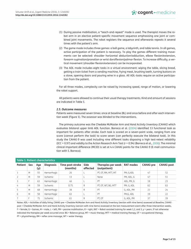
The primary outcome was the Chedoke McMaster Arm and Hand Activity Inventory (CAHAI) which evaluates bilateral upper limb ADL function. Barreca et al. (2004) identified 13 functional task as important for patients after stroke. Each task is scored on a seven-point scale, ranging from one score (cannot perform the task) to score seven (can perfectly execute the bilateral task). In this study the CAHAI-9 was used including nine different tasks disposing a high test-retest reliability (ICC = 0.97) and validity to the Action Research Arm Test (r = 0.94) (Barreca et al., 2006). The minimal clinical important difference (MCID) is set at 4.4 CAHAI points for the CAHAI-9 (E-mail communica-tion with S. Barreca).
Table 1. Patient characteristics
Notes: ADL = Activities of daily living, CAHAI-pre = Chedoke McMaster Arm and Hand Activity Inventory (version with nine items) assessed at Baseline, CAHAI-post = Chedoke McMaster Arm and Hand Activity Inventory (version with nine items) assessed at the last measurement event after three intervention weeks, F = female, G = Games, M = male, L = left, PM = passive mobilisation, R = right, RAT = Robot-assisted training for week 1,2, and 3, y = years. If not otherwise indicated the therapies per week occurred once: BG = Balance group, MT = music therapy, MTT = medical training therapy, OT = occupational therapy, PT = physiotherapy, RM = reflex zone massage, WT = water therapy.
Patient Sex Age (y)
Diagnosis Time post-stroke (months)
Side affected
Therapies per week (outpatient)
RAT modes CAHAI-pre CAHAI-post
1 M 55 Hemorrhagic 26 L PT, OT, RM, MTT, MT PM, G,ADL 47 52
2 M 59 Ischemic 13 L None PM, ADL, G 47 51
3 F 63 Hemorrhagic 46 L PT ADL, PM, G 18 23
4 M 59 Ischemic 0.75 R PT, OT, WT, BG, MTT PM, G, ADL 62 63
5 M 68 Hemorrhagic 38 R PT G, ADL, PM 21 24
6 M 58 Hemorrhagic 63 L PT PM,G, ADL 19 22
7 F 75 Ischemic 61 R PT, OT G, ADL, PM 37 39
Page 6 of 15
Schuster-Amft et al., Cogent Medicine (2016), 3: 1240282http://dx.doi.org/10.1080/2331205X.2016.1240282
Secondary outcomes were passive and active range of motion (aROM, pROM) measurements (Tables 3 and 4), which were assessed with a handheld goniometer and were performed for the fol-lowing movements: shoulder flexion, abduction and outward rotation, elbow flexion and extension, forearm pronation and supination, and wrist palmarflexion and dorsiflexion. Every movement was measured three times and the average value was recorded. If patients reported pain, ROM was lim-ited to the point of pain onset. For shoulder movements, the MCID was determined at eight degrees (Kolber, Fuller, Marshall, Wright, & Hanney, 2012). Changes greater than six degrees for elbow flex-ion, seven degrees for elbow extension, and eight degrees for pronation or supination might be considered as meaningful (Armstrong, MacDermid, Chinchalkar, Stevens, & King, 1998).
The Modified Modified Ashworth Scale measures spasticity. It is performed similar to the Modified Ashworth Scale (MAS) (Bohannon, 1987). However, the scoring is different ranging from zero (no in-crease in muscle tone) to four (affected part(s) rigid in flexion or extension). The MMSA was per-formed three times for the following muscle groups: elbow flexors and extensors, forearm pronators, wrist and finger flexors. For stroke, intrarater reliability for elbow and wrist flexors are good to high (weighted Kappa 0.61 and 0.78 Kw; 0.86 and 0.90 Kw) (Ansari et al., 2009, 2012). Naghdi et al. (2008) recommended the MMAS as a valid spasticity assessment for wrist flexors in patients after stroke.
Finally, patients completed a weekly questionnaire with open questions about their personal im-pression of RAT effects on arm and hand function in their daily living.
2.6. Data analysisFor each training mode a data set was created and descriptive statistics were derived. The Wilcoxon signed-rank test was used to assess whether there were significant differences between pre and post intervention values for the training weeks: passive mobilisation, games, or ADL. If differences were significant, number of patients that showed changes greater than the MCID, were identified. To indicate the level of improvement effect sizes were calculated using a modified version of the Standard Mean Difference method (MBR) applied to the CAHAI-9 using the following formula (Olive & Franco, 2008):
All statistical analyses were performed using STATISTICA (version 12) and Excel (for Mac, version 14.4.0). The significance level was set at p ≤ 0.05 for all analyses.
3. ResultsBetween July and September 2013, 41 patients were screened for study eligibility. Twenty-two pa-tients were excluded and from the remaining 19 patients, only seven were willing to participate (two females, five males, mean ± SD: age 62.4 ± 6.4, CAHAI-9 35.7 ± 15.8). Patient characteristics can be found in Table 1. There were no drop-outs or adverse events during the study.
3.1. Primary research question
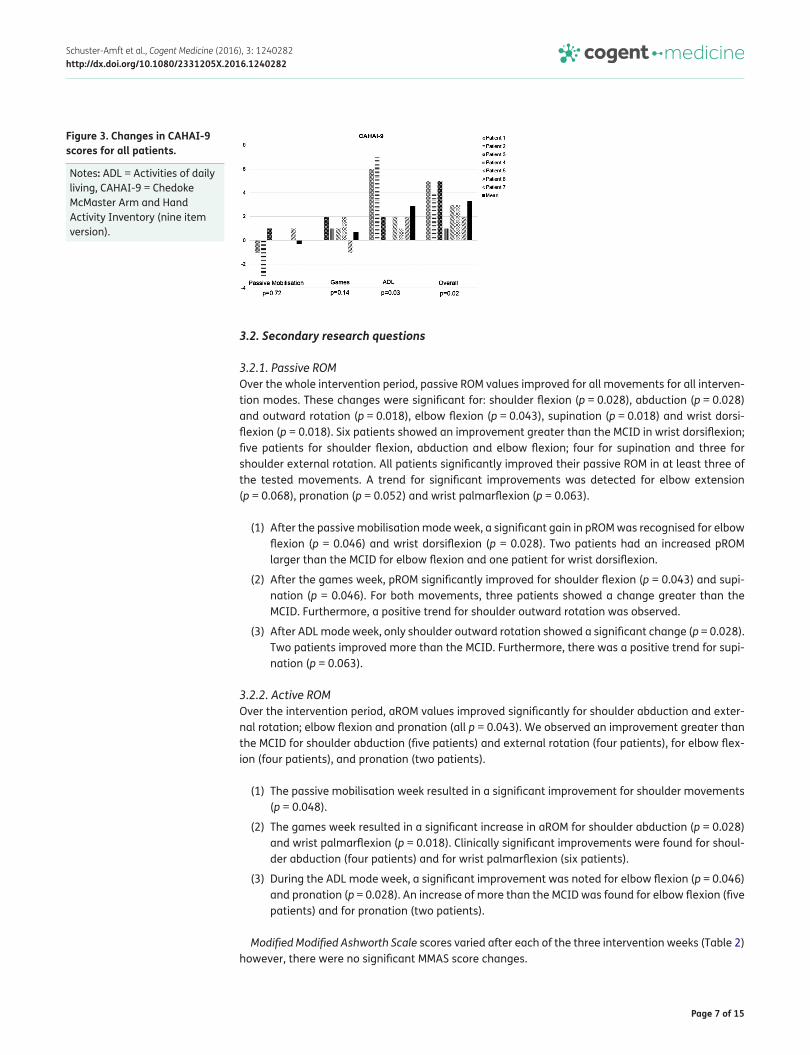
3.1.1. Chedoke McMaster Arm and Hand Activity InventoryCAHAI-9 values at BL ranged from 18 to 62 (mean 35.9 ± 17.1) and at the end of the three-week study period from 22 to 63 (mean 39.1 ± 16.7). Changes in CAHAI-9 score for each patient are dis-played in Figure 3. Overall, patients improved significantly in the CAHAI score ranging from 1 to 5 points (p = 0.018) post intervention. Two patients (P1, P3) improved more than the MCID (4.4 points, P1 and P2). After the ADL training mode week, CAHAI scores were improved significantly (p = 0.028) No significant changes were observed during the passive mobilisation mode (p = 0.715) and games mode (p = 0.138). Patients’ calculated CAHAI-9 effect sizes were as follows: P1 ES = 0.14, P2 ES = 0.11, P3 ES = 0.29, P4 ES = 0.06, P5 ES = 0.18, P6 ES = 0.18, P7 ES = 0.12 indicating small gains.
effect size =(pre-intervention − post intervention CAHAI score)
standard deviation pre− intervention CAHAI score.
Page 7 of 15
Schuster-Amft et al., Cogent Medicine (2016), 3: 1240282http://dx.doi.org/10.1080/2331205X.2016.1240282
3.2. Secondary research questions
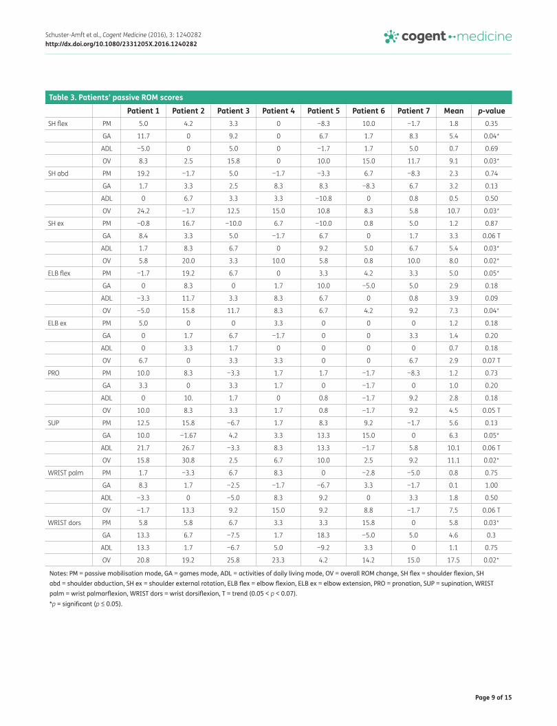
3.2.1. Passive ROMOver the whole intervention period, passive ROM values improved for all movements for all interven-tion modes. These changes were significant for: shoulder flexion (p = 0.028), abduction (p = 0.028) and outward rotation (p = 0.018), elbow flexion (p = 0.043), supination (p = 0.018) and wrist dorsi-flexion (p = 0.018). Six patients showed an improvement greater than the MCID in wrist dorsiflexion; five patients for shoulder flexion, abduction and elbow flexion; four for supination and three for shoulder external rotation. All patients significantly improved their passive ROM in at least three of the tested movements. A trend for significant improvements was detected for elbow extension (p = 0.068), pronation (p = 0.052) and wrist palmarflexion (p = 0.063).
(1) After the passive mobilisation mode week, a significant gain in pROM was recognised for elbow flexion (p = 0.046) and wrist dorsiflexion (p = 0.028). Two patients had an increased pROM larger than the MCID for elbow flexion and one patient for wrist dorsiflexion.
(2) After the games week, pROM significantly improved for shoulder flexion (p = 0.043) and supi-nation (p = 0.046). For both movements, three patients showed a change greater than the MCID. Furthermore, a positive trend for shoulder outward rotation was observed.
(3) After ADL mode week, only shoulder outward rotation showed a significant change (p = 0.028). Two patients improved more than the MCID. Furthermore, there was a positive trend for supi-nation (p = 0.063).
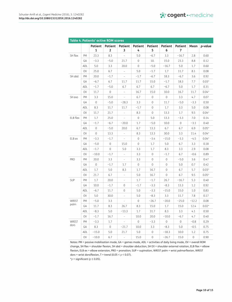
3.2.2. Active ROMOver the intervention period, aROM values improved significantly for shoulder abduction and exter-nal rotation; elbow flexion and pronation (all p = 0.043). We observed an improvement greater than the MCID for shoulder abduction (five patients) and external rotation (four patients), for elbow flex-ion (four patients), and pronation (two patients).
(1) The passive mobilisation week resulted in a significant improvement for shoulder movements (p = 0.048).
(2) The games week resulted in a significant increase in aROM for shoulder abduction (p = 0.028) and wrist palmarflexion (p = 0.018). Clinically significant improvements were found for shoul-der abduction (four patients) and for wrist palmarflexion (six patients).
(3) During the ADL mode week, a significant improvement was noted for elbow flexion (p = 0.046) and pronation (p = 0.028). An increase of more than the MCID was found for elbow flexion (five patients) and for pronation (two patients).
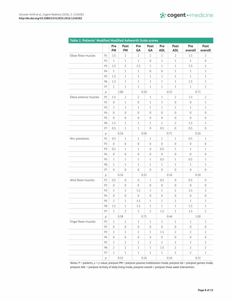
Modified Modified Ashworth Scale scores varied after each of the three intervention weeks (Table 2) however, there were no significant MMAS score changes.
Figure 3. Changes in CAHAI-9 scores for all patients.
Notes: ADL = Activities of daily living, CAHAI-9 = Chedoke McMaster Arm and Hand Activity Inventory (nine item version).
Page 8 of 15
Schuster-Amft et al., Cogent Medicine (2016), 3: 1240282http://dx.doi.org/10.1080/2331205X.2016.1240282
Table 2. Patients’ Modified Modified Ashworth Scale scores
Notes: P = patients, p = p-value, pre/post PM = pre/post passive mobilisation mode, pre/post GA = pre/post games mode, pre/post ADL = pre/post Activity of daily living mode, pre/post overall = pre/post three week intervention.
Pre PM
Post PM
Pre GA
Post GA
Pre ADL
Post ADL
Pre overall
Post overall
Elbow flexor muscles P1 1.5 2 2 2 2 2 1.5 2
P2 1 1 1 0 1 1 1 0
P3 1.5 2 1.5 1 1 1 1.5 2
P4 1 1 1 0 0 1 1 1
P5 1.5 1 1 1 1 1 1 1
P6 1.5 1 1 1 1 1 1.5 1
P7 1 1 1 1 1 1 1 1
p 1.00 0.10 0.32 0.71
Elbow extensor muscles P1 1.5 2 2 1 1 2 1.5 2
P2 0 1 0 1 1 0 0 1
P3 1 1 1 1 1 1 1 1
P4 0 0 0 0 0 0 0 0
P5 0 0 0 0 0 0 0 0
P6 1.5 1 1 1 1 2 1.5 2
P7 0.5 1 1 0 0.5 0 0.5 0
p 0.26 0.56 0.71 0.26
Mm. pronatores P1 0.5 1 1 1 1 1 0.5 1
P2 0 0 0 0 0 0 0 0
P3 0.5 1 1 0 0.5 1 1 1
P4 0 0 0 0 0 0 0 0
P5 1 1 1 1 0.5 1 0.5 1
P6 1 1 1 1 1 1 1 1
P7 0 0 0 0 0 0 0 0
p 0.16 0.32 0.16 0.16
Wrist flexor muscles P1 0.5 0 0 1 0.5 0 0.5 0
P2 0 0 0 0 0 0 0 0
P3 2 2 1.5 1 1 2 1.5 2
P4 0 0 0 0 0 0 0 0
P5 2 1 1.5 1 1 2 1 1
P6 1.5 1 1.5 1 1 1 1.5 1
P7 1 2 2 2 1.5 1 1.5 2
p 0.58 0.71 0.46 1.00
Finger flexor muscles P1 1 1 1 1 1 1 1 1
P2 0 0 0 0 0 0 0 0
P3 2 2 2 1 1.5 2 2 2
P4 0 0 0 0 0 0 0 0
P5 2 2 2 2 2 2 2 2
P6 2 2 2 1 1.5 2 2 2
P7 2 1 1 1 2 2 2 1
p 0.32 0.16 0.16 0.32
Page 9 of 15
Schuster-Amft et al., Cogent Medicine (2016), 3: 1240282http://dx.doi.org/10.1080/2331205X.2016.1240282
Table 3. Patients’ passive ROM scores
Notes: PM = passive mobilisation mode, GA = games mode, ADL = activities of daily living mode, OV = overall ROM change, SH flex = shoulder flexion, SH abd = shoulder abduction, SH ex = shoulder external rotation, ELB flex = elbow flexion, ELB ex = elbow extension, PRO = pronation, SUP = supination, WRIST palm = wrist palmarflexion, WRIST dors = wrist dorsiflexion, T = trend (0.05 < p < 0.07).*p = significant (p ≤ 0.05).
Patient 1 Patient 2 Patient 3 Patient 4 Patient 5 Patient 6 Patient 7 Mean p-valueSH flex PM 5.0 4.2 3.3 0 −8.3 10.0 −1.7 1.8 0.35
GA 11.7 0 9.2 0 6.7 1.7 8.3 5.4 0.04*
ADL −5.0 0 5.0 0 −1.7 1.7 5.0 0.7 0.69
OV 8.3 2.5 15.8 0 10.0 15.0 11.7 9.1 0.03*
SH abd PM 19.2 −1.7 5.0 −1.7 −3.3 6.7 −8.3 2.3 0.74
GA 1.7 3.3 2.5 8.3 8.3 −8.3 6.7 3.2 0.13
ADL 0 6.7 3.3 3.3 −10.8 0 0.8 0.5 0.50
OV 24.2 −1.7 12.5 15.0 10.8 8.3 5.8 10.7 0.03*
SH ex PM −0.8 16.7 −10.0 6.7 −10.0 0.8 5.0 1.2 0.87
GA 8.4 3.3 5.0 −1.7 6.7 0 1.7 3.3 0.06 T
ADL 1.7 8.3 6.7 0 9.2 5.0 6.7 5.4 0.03*
OV 5.8 20.0 3.3 10.0 5.8 0.8 10.0 8.0 0.02*
ELB flex PM −1.7 19.2 6.7 0 3.3 4.2 3.3 5.0 0.05*
GA 0 8.3 0 1.7 10.0 −5.0 5.0 2.9 0.18
ADL −3.3 11.7 3.3 8.3 6.7 0 0.8 3.9 0.09
OV −5.0 15.8 11.7 8.3 6.7 4.2 9.2 7.3 0.04*
ELB ex PM 5.0 0 0 3.3 0 0 0 1.2 0.18
GA 0 1.7 6.7 −1.7 0 0 3.3 1.4 0.20
ADL 0 3.3 1.7 0 0 0 0 0.7 0.18
OV 6.7 0 3.3 3.3 0 0 6.7 2.9 0.07 T
PRO PM 10.0 8.3 −3.3 1.7 1.7 −1.7 −8.3 1.2 0.73
GA 3.3 0 3.3 1.7 0 −1.7 0 1.0 0.20
ADL 0 10. 1.7 0 0.8 −1.7 9.2 2.8 0.18
OV 10.0 8.3 3.3 1.7 0.8 −1.7 9.2 4.5 0.05 T
SUP PM 12.5 15.8 −6.7 1.7 8.3 9.2 −1.7 5.6 0.13
GA 10.0 −1.67 4.2 3.3 13.3 15.0 0 6.3 0.05*
ADL 21.7 26.7 −3.3 8.3 13.3 −1.7 5.8 10.1 0.06 T
OV 15.8 30.8 2.5 6.7 10.0 2.5 9.2 11.1 0.02*
WRIST palm PM 1.7 −3.3 6.7 8.3 0 −2.8 −5.0 0.8 0.75
GA 8.3 1.7 −2.5 −1.7 −6.7 3.3 −1.7 0.1 1.00
ADL −3.3 0 −5.0 8.3 9.2 0 3.3 1.8 0.50
OV −1.7 13.3 9.2 15.0 9.2 8.8 −1.7 7.5 0.06 T
WRIST dors PM 5.8 5.8 6.7 3.3 3.3 15.8 0 5.8 0.03*
GA 13.3 6.7 −7.5 1.7 18.3 −5.0 5.0 4.6 0.3
ADL 13.3 1.7 −6.7 5.0 −9.2 3.3 0 1.1 0.75
OV 20.8 19.2 25.8 23.3 4.2 14.2 15.0 17.5 0.02*
Page 10 of 15
Schuster-Amft et al., Cogent Medicine (2016), 3: 1240282http://dx.doi.org/10.1080/2331205X.2016.1240282
Table 4. Patients’ active ROM scores
Notes: PM = passive mobilisation mode, GA = games mode, ADL = activities of daily living mode, OV = overall ROM change, SH flex = shoulder flexion, SH abd = shoulder abduction, SH EX = shoulder external rotation, ELB flex = elbow flexion, ELB ex = elbow extension, PRO = pronation; SUP = supination, WRIST palm = wrist palmarflexion, WRIST dors = wrist dorsiflexion, T = trend (0.05 < p < 0.07).*p = significant (p ≤ 0.05).
Patient 1
Patient 2
Patient 3
Patient 4
Patient 5
Patient 6
Patient 7
Mean p-value
SH flex PM 23.3 8.3 – 5.0 −6.7 3.3 −16.7 2.8 0.60
GA −3.3 −5.0 21.7 0 10. 15.0 23.3 8.8 0.12
ADL 5.0 3.3 20.0 0 −5.0 −16.7 5.0 1.7 0.60
OV 25.0 6.7 – 5.0 −1.7 1.7 11.7 8.1 0.08
SH abd PM 20.0 −1.7 – −1.7 −6.7 18.3 −6.7 3.6 0.92
GA −6.7 6.7 11.7 11.7 15.0 −1.7 18.3 7.7 0.03*
ADL −1.7 −5.0 6.7 6.7 6.7 −6.7 5.0 1.7 0.31
OV 11.7 0 – 16.7 15.0 10.0 16.7 11.7 0.04*
SH exo PM 3.3 15.0 – 6.7 0 0 3.3 4.7 0.07
GA 0 −5.0 −28.3 3.3 0 11.7 −5.0 −3.3 0.50
ADL 8.3 11.7 11.7 −1.7 0 1.7 3.3 5.0 0.08
OV 11.7 21.7 – 8.3 0 13.3 1.7 9.5 0.04*
ELB flex PM 1.7 25.0 – 0 5.0 13.3 −3.3 7.0 0.14
GA −1.7 −6.7 −20.0 1.7 −5.0 10.0 0 −3.1 0.40
ADL 0 −5.0 20.0 6.7 13.3 6.7 6.7 6.9 0.05*
OV 0 13.3 – 8.3 13.3 30.0 3.3 11.4 0.04*
ELB ex PM −3.3 −1.7 – 0 −3.4 −15.0 −3.3 −4.5 0.04*
GA −5.0 0 15.0 0 1.7 5.0 6.7 3.3 0.18
ADL −1.7 0 5.0 3.3 1.7 8.3 3.3 2.9 0.08
OV −10.0 −1.7 – 3.3 0 −1.7 6.7 −0.6 0.89
PRO PM 20.0 3.3 – 3.3 0 0 −5.0 3.6 0.47
GA 0 −1.7 1.7 0 0 0 5.0 0.7 0.42
ADL 1.7 5.0 8.3 1.7 16.7 0 6.7 5.7 0.03*
OV 21.7 6.7 – 5.0 16.7 0 6.7 9.5 0.05*
SUP PM 1.7 20.0 – 1.7 −1.7 26.7 −16.7 5.3 0.40
GA 10.0 −1.7 0 −1.7 −3.3 −8.3 13.3 1.2 0.92
ADL −6.7 11.7 0 5.0 −3.3 −15.0 15.0 1.0 0.83
OV 5.0 30.0 – 5.0 −8.3 3.3 11.7 7.8 0.17
WRIST palm
PM −5.0 3.3 – 0 −26.7 −20.0 −25.0 −12.2 0.08
GA 11.7 8.3 26.7 8.3 15.0 1.7 15.0 12.4 0.02*
ADL −8.3 5.0 −13.3 1.7 31.7 8.3 3.3 4.1 0.50
OV −1.7 16.7 – 10.0 20.0 −10.0 −6.7 4.7 0.40
WRIST dors
PM −3.3 1.7 – 0 −3.3 0 0 −0.8 0.29
GA 8.3 0 −21.7 10.0 3.3 −8.3 5.0 −0.5 0.75
ADL −15.0 5.0 21.7 5.0 0 −18.3 10.0 1.2 0.75
OV −10.0 6.7 – 15.0 0 −26.7 15.0 0 0.90
Page 11 of 15
Schuster-Amft et al., Cogent Medicine (2016), 3: 1240282http://dx.doi.org/10.1080/2331205X.2016.1240282
3.2.3. Patients’ positive perspective on RATIndependent from the training mode applied, patients mentioned improvements in:
(1) ROM in shoulder and elbow joints,
(2) muscle tone in hand/fingers,
(3) movement coordination and velocity, and controlled voluntary movements,
(4) ability to grasp, manipulate, transport and release objects by the hand and fingers leading to improved ADL movements, e.g. hold a fork, putting on a jacket, and
(5) unconscious usage of the paretic arm and shorter reaction times.
Only one patient mentioned pain in the arm after training. He did not report pain during RAT. No patient mentioned unexpected negative effects. Surprisingly, two patients mentioned a RAT influ-ence on gait, leg loading, and trunk symmetry. All patients did like RAT due to the visual feedback, and the opportunity to experience the exoskeleton’s support against gravity enabling arm/hand movements that are not possible in real life.
3.2.4. Patients’ negative perspective on RATPatients pointed out that the hardware and the software were not as much adaptable as necessary to the individual patients’ needs. Additionally, patients did not want to miss the individual feedback and the correction of body position during RAT from the present supervising therapist. Furthermore, patients missed the whole body therapy approach when working functionally as well as specific muscle relaxation and mobilisation techniques.
4. DiscussionUpper limb RAT has been attributed to a beneficial effect for patients after stroke (Mehrholz et al., 2012). However, differential effects of individual RAT modes have not been evaluated so far. In the present case series three different RAT modes (passive mobilisation, games and ADL) of the ARMin III robot were investigated in one sub-acute and six chronic patients after stroke. As hypothesised, all patients improved over the three-week intervention period in their ADL function ranging between 1 to 5 points in the CAHAI-9 indicating a positive effect on upper limb functioning. Even though, our effects were small, they could be attributed to the principles of task-oriented training (TOT), which was the basis for the development of the virtual ADL mode of ARMin III. TOT implies that training should include functional tasks that are realistic, goal-directed and challenging (Schweighofer, Choi, Winstein, & Gordon, 2012). According to French et al. (2007) repetitive task training, which was part of the ARMin III training too, can lead to ADL improvement. The increase in upper limb function could further be attributed to an increase in aROM, which improved in five patients. Harris and Eng (2007) showed that strength of the paretic upper limb is strongly correlated with ADL function im-provement measured by the CAHAI-13. Using a regression model, the authors found that upper limb strength explained 87% of the variance in CAHAI-scores (Harris & Eng, 2007). Our ADL mode also showed an influence on muscle tone. MMAS scores increased in five patients in one to three muscles with 0.5 or 1 point. A MMAS decrease of about 0.5 points was found in three patients in one to two muscles. Statistical analyses of the MMAS changes were not significant but the changes should be considered in terms of clinical relevance for the patient. Importantly, no patient reported the feeling of an increase in muscle tone after the ADL mode or any negative impact on activities of daily life. Furthermore, it should be noted that the MMAS was measured during the same assessment as the CAHAI. Even though there was an increase in muscle tone after the ADL mode, patients scored bet-ter on the CAHAI assessment, so this suggests that this increase in tone did not affect their func-tional capability. After the three weeks of intervention, the MMAS scores showed only a minor increase in tone. This is supported by findings of Klamroth-Marganska et al. (2013) who showed that training with all three ARMin III training modes for eight weeks led to a small, but not significant, decrease in MAS score.
Page 12 of 15
Schuster-Amft et al., Cogent Medicine (2016), 3: 1240282http://dx.doi.org/10.1080/2331205X.2016.1240282
Overall, passive ROM increased. The greatest change in pROM was expected during the passive mobi-lisation mode due to two reasons: First, passive mobilisation is the only mode where specifically could be worked within a large ROM. This is in contrast to the games and ADL mode, where the ROM was re-stricted and patients’ arm could not go end range. Second, only in passive mobilisation mode the move-ments could be individually adjusted to the individual needs. In the games and ADL mode, the movements had to be selected from pre-defined movements and not all combinations of movements were possible. Nevertheless, the results showed no greater benefit for passive ROM in the passive mobi-lisation mode than during the games or ADL mode. This could imply that for improvement of passive ROM, training can be given in all three training modes. Maybe this can be contributed to the support from the ARMin III during the active training modes. Several of our patients were highly impaired in moving their arm against gravity, so gravity support was set high to allow for movements in the horizontal plane. Active participation of the patient was needed, but the robot assisted the task execution. To support patients’ arm movements against gravity Meadmore et al. (2012, 2013) combined functional electrical stimulation (FES) and RAT equipped with a passive “un-weighing” system. In their studies, FES for M. triceps brachii, the anterior part of the M. deltoideus, and wrist and hand extensor muscles was used to facilitate active contractions to improve arm and hand motor function in ten chronic stroke patients and five patients with Multiple Sclerosis (Meadmore et al., 2012, 2013; Sampson et al., 2016). The combina-tion of FES and RAT also led to improved tracking accuracy and lower required ES amplitude.
In the present study, we expected to find an improvement in active ROM scores during the games mode. Several active ROM scores did improve, but there is no preference for one of the training modes. Beneficial changes were found in both games and ADL mode. One could interpret that active participation of the patient could lead to an improvement in active ROM. The small differences in active ROM could be contributed to the fact that patients could not reach end range ROM during the active training modes. This could possibly be an explanation for less improvement in active ROM compared to the improvements in passive ROM. To increase active ROM Krabben et al. (2012) devel-oped the gravity compensation training (GCT). The researchers investigated seven chronic stroke patients using GCT within 18 half-hour sessions over a six weeks period. Patients practiced three-dimensional, goal-directed arm movements in a gravity compensated, virtual reality augmented environment provided by the Freebal system. Freebal is a gravity compensation device that allows assistive arm movements with the help of two arm slings. Over the training period patients were able to increase their amount of movement repetitions and required less gravity support. However, the synergistic movement patterns of the arm did not change.
4.1. LimitationsThe following limitations have to be considered when interpreting the presented results. Our pa-tients completed the CAHAI-9 at different measurement events in a three-week time period, which could lead to a possible learning effect. However, in some patients, outcome changes were larger than the MCID, which cannot be explained by a learning effect only.
We have a limited number of patients, because of the limited recruitment time and limited num-ber of participants available during that time period. Therefore, it was not possible to analyse influ-ence of the RAT mode order on patient outcomes, recommend a training order or perform subgroup analyses.
Our goal was to include sub-acute as well as chronic patients after stroke. However, only one sub-acute patient was included. Only 50% of the 41 screened patients were eligible for RAT due to limited mobility, shoulder pain, hypersensitivity of the arm or other neurologic diseases. Overall, only seven out of 41 screened patients agreed to participate. Furthermore, the study population is not homoge-neous. We have an age range of 20 years and the level of functionality at baseline is very different. Nevertheless, this shows that the ARMin-robot is adaptable to different kind of patients. There was only one week of training in every training mode, which means only nine hours of actual training in total. Besides, patients kept following their regular training scheme. Overall, these considerations limit the generalizability of our findings about the differential effect of RAT.
Page 13 of 15
Schuster-Amft et al., Cogent Medicine (2016), 3: 1240282http://dx.doi.org/10.1080/2331205X.2016.1240282
In light of our and previous results, we would still recommend RAT as adjunct training to regular stroke rehabilitation (Klamroth-Marganska et al., 2013; Mehrholz et al., 2012; Norouzi-Gheidari, Archambault, & Fung, 2012). Our patients showed good training compliance, probably because of the difference to conventional therapy content and because of the included “fun”-element of RAT. Patients were very motivated throughout the entire study and, in the questionnaire, they reported a more frequent use of their paretic arm during ADL.
Future studies should investigate the ideal training program with regard to functional capability and more extensively study duration with a follow-up period should be considered. Moreover, the researchers could divide the patients in different functionality-categories to avoid great variance of between-group characteristics.
5. ConclusionThe present study is the first study investigating differential effects of RAT modes. Our study showed that all RAT modes with ARMin III were safe, feasible and well accepted by the patients. A three-week long in-tervention improved upper limb functioning of the paretic arm and hand in patients in the sub-acute or chronic phase after stroke. Although we found significant differences after a one-week delivery of ARMin III training in the ADL-mode, we would still suggest to offer all three modes to patients receiving RAT based on the fact that patients are probably also active in the other modes, and thus practice in these modes as well, and because it might be that through offering the passive mode, a potential increase in muscle tone is negated when practicing more actively. Nevertheless, our results also suggest that the ADL mode should be a significant part of RAT when aiming at improving arm and hand functionality post stroke.
AcknowledgementsWe would like to thank Andrea Henneke for her advice and support regarding the robot-assisted training. We are grateful to all patients, who kindly participated in our study.
FundingThe authors received no direct funding for this research.
Competing InterestsThe authors declare no competing interest.
Author detailsCorina Schuster-Amft1,2
E-mail: [email protected] Van Kerckhoven3
E-mail: [email protected] Berse3
E-mail: [email protected] Verheyden3
E-mail: [email protected] Research Department, Reha Rheinfelden, Rheinfelden,
Switzerland.2 Institute for Rehabilitation and Performance Technology,
Bern University of Applied Sciences, Burgdorf, Switzerland.3 KU Leuven - University of Leuven, Department of
Rehabilitation Sciences, 3001 Leuven, Belgium.
Citation informationCite this article as: Immediate effects of different upper limb robot-assisted training modes in patients after stroke: A case series, Corina Schuster-Amft, Leni Van Kerckhoven, Magdalena Berse & Geert Verheyden, Cogent Medicine (2016), 3: 1240282.
ReferencesAnsari, N. N., Naghdi, S., Hasson, S., Fakhari, Z., Mashayekhi, M.,
& Herasi, M. (2009). Assessing the reliability of the Modified Modified Ashworth Scale between two
physiotherapists in adult patients with hemiplegia. NeuroRehabilitation, 25, 235–240.
Ansari, N. N., Naghdi, S., Mashayekhi, M., Hasson, S., Fakhari, Z., & Jalaie, S. (2012). Intra-rater reliability of the Modified Modified Ashworth Scale (MMAS) in the assessment of upper-limb muscle spasticity. NeuroRehabilitation, 31, 215–222.
Armstrong, A. D., MacDermid, J. C., Chinchalkar, S., Stevens, R. S., & King, G. J. (1998). Reliability of range-of-motion measurement in the elbow and forearm. Journal of Shoulder and Elbow Surgery, 7, 573–580. http://dx.doi.org/10.1016/S1058-2746(98)90003-9
Balasubramanian, S., Klein, J., & Burdet, E. (2010). Robot-assisted rehabilitation of hand function. Current Opinion in Neurology, 23, 661–670. http://dx.doi.org/10.1097/WCO.0b013e32833e99a4
Barreca, S., Gowland, C., Stratford, P., Huijbregts, M., Griffiths, J., Torresin, W., ... Masters, L. (2004). Development of the chedoke arm and hand activity inventory: theoretical constructs, item generation, and selection. Topics in Stroke Rehabilitation, 11, 31–42. http://dx.doi.org/10.1310/JU8P-UVK6-68VW-CF3W
Barreca, S., Stratford, P., Masters, L., Lambert, C. L., Griffiths, J., & McBay, C. (2006). Validation of three shortened versions of the chedoke arm and hand activity inventory. Physiotherapy Canada, 58, 148–156. http://dx.doi.org/10.3138/ptc.58.2.148
Bohannon, R. W. (1987). Variability and reliability of the pendulum test for spasticity using a Cybex II isokinetic dynamometer. Physical Therapy, 67, 659–661.
Bosecker, C., Dipietro, L., Volpe, B., & Igo Krebs, H. (2010). Kinematic robot-based evaluation scales and clinical counterparts to measure upper limb motor performance in patients with chronic stroke. Neurorehabilitation and Neural Repair, 24, 62–69. http://dx.doi.org/10.1177/1545968309343214
Brock, K., Haase, G., Rothacher, G., & Cotton, S. (2011). Does physiotherapy based on the Bobath concept, in conjunction with a task practice, achieve greater improvement in walking ability in people with stroke compared to physiotherapy focused on structured task
Page 14 of 15
Schuster-Amft et al., Cogent Medicine (2016), 3: 1240282http://dx.doi.org/10.1080/2331205X.2016.1240282
practice alone? A pilot randomized controlled trial. Clinical Rehabilitation, 25, 903–912. http://dx.doi.org/10.1177/0269215511406557
Bütefisch, C., Hummelsheim, H., Denzler, P., & Mauritz, K. H. (1995). Repetitive training of isolated movements improves the outcome of motor rehabilitation of the centrally paretic hand. Journal of the Neurological Sciences, 130, 59–68. http://dx.doi.org/10.1016/0022-510X(95)00003-K
French, B., Thomas, L. H., Leathley, M. J., Sutton, C. J., McAdam, J., Forster, A., ... Watkins, C. L. (2007). Repetitive task training for improving functional ability after stroke. Cochrane Database Systematic Review, 4, CD006073.
Go, A. S., Mozaffarian, D., Roger, V. L., Benjamin, E. J., Berry, J. D., Borden, W. B., ... Turner, M. B. (2013). Heart disease and stroke statistics–2013 update: A report from the American heart association. Circulation, 127, e6–e245. http://dx.doi.org/10.1161/CIR.0b013e31828124ad
Harris, J. E., & Eng, J. J. (2007). Paretic upper-limb strength best explains arm activity in people with stroke. Physical Therapy, 87, 88–97. http://dx.doi.org/10.2522/ptj.20060065
Huang, V. S., & Krakauer, J. W. (2009). Robotic neurorehabilitation: A computational motor learning perspective. Journal of NeuroEngineering and Rehabilitation, 6, 5. http://dx.doi.org/10.1186/1743-0003-6-5
Hwangbo, P. N., & Don Kim, K. (2016). Effects of proprioceptive neuromuscular facilitation neck pattern exercise on the ability to control the trunk and maintain balance in chronic stroke patients. Journal of Physical Therapy Science, 28, 850–853. http://dx.doi.org/10.1589/jpts.28.850
Klamroth-Marganska, V., Blanco, J., Campen, K., Curt, A., Dietz, V., Ettlin, T., ... Luft, A. (2013). Three-dimensional, task-specific robot therapy of the arm after stroke: A multicentre, parallel-group randomised trial. The Lancet Neurology, 13, 159–166.
Kolber, M. J., Fuller, C., Marshall, J., Wright, A., & Hanney, W. J. (2012). The reliability and concurrent validity of scapular plane shoulder elevation measurements using a digital inclinometer and goniometer. Physiotherapy Theory and Practice, 28, 161–168.
Kong, K. H., & Lee, J. (2013). Temporal recovery and predictors of upper limb dexterity in the first year of stroke: A prospective study of patients admitted to a rehabilitation centre. NeuroRehabilitation, 32, 345–350.
Krabben, T., Prange, G. B., Molier, B. I., Stienen, A. H., Jannink, M. J., Buurke, J. H., & Rietman, J. S. (2012). Influence of gravity compensation training on synergistic movement patterns of the upper extremity after stroke, a pilot study. Journal of NeuroEngineering and Rehabilitation, 9, 44. http://dx.doi.org/10.1186/1743-0003-9-44
Kwakkel, G., Kollen, B. J., & Krebs, H. I. (2008). Effects of robot-assisted therapy on upper limb recovery after stroke: A systematic review. Neurorehabilitation and Neural Repair, 22, 111–121.
Langhammer, B., & Stanghelle, J. K. (2000). Bobath or motor relearning programme? A comparison of two different approaches of physiotherapy in stroke rehabilitation: A randomized controlled study. Clinical Rehabilitation, 14, 361–369. http://dx.doi.org/10.1191/0269215500cr338oa
Lo, H. S., & Xie, S. Q. (2012). Exoskeleton robots for upper-limb rehabilitation: State of the art and future prospects. Medical Engineering & Physics, 34, 261–268. http://dx.doi.org/10.1016/j.medengphy.2011.10.004
Loureiro, R. C., Harwin, W. S., Nagai, K., & Johnson, M. (2011). Advances in upper limb stroke rehabilitation: A technology push. Medical & Biological Engineering &
Computing, 49, 1103–1118. http://dx.doi.org/10.1007/s11517-011-0797-0
Meadmore, K., Exell, T., Freeman, C., Kutlu, M., Rogers, E., Hughes, A. M., ... Burridge, J. (2013). Electrical stimulation and iterative learning control for functional recovery in the upper limb post-stroke. IEEE International Conference on Rehabilation Robotics, 2013, 6650359.
Meadmore, K. L., Hughes, A. M., Freeman, C. T., Cai, Z., Tong, D., Burridge, J. H., & Rogers, E. (2012). Functional electrical stimulation mediated by iterative learning control and 3D robotics reduces motor impairment in chronic stroke. Journal of NeuroEngineering and Rehabilitation, 9, 32. http://dx.doi.org/10.1186/1743-0003-9-32
Mehrholz, J., Hadrich, A., Platz, T., Kugler, J., & Pohl, M. (2012). Electromechanical and robot-assisted arm training for improving generic activities of daily living, arm function, and arm muscle strength after stroke. Cochrane Database Systematic Review, 6, CD006876.
Naghdi, S., Ansari, N. N., Mansouri, K., Asgari, A., Olyaei, G. R., & Kazemnejad, A. (2008). Neurophysiological examination of the Modified Modified Ashworth Scale (MMAS) in patients with wrist flexor spasticity after stroke. Electromyography and Clinical Neurophysiology, 48, 35–41.
Nef, T., Mihelj, M., & Riener, R. (2007). ARMin: A robot for patient-cooperative arm therapy. Medical & Biological Engineering & Computing, 45, 887–900. http://dx.doi.org/10.1007/s11517-007-0226-6
Norouzi-Gheidari, N., Archambault, P. S., & Fung, J. (2012). Effects of robot-assisted therapy on stroke rehabilitation in upper limbs: Systematic review and meta-analysis of the literature. The Journal of Rehabilitation Research and Development, 49, 479–496. http://dx.doi.org/10.1682/JRRD.2010.10.0210
Olive, M. L., & Franco, J. H. (2008). (Effect) size matters: And so does the calculation. The Behavior Analyst Today, 9, 5–10. http://dx.doi.org/10.1037/h0100642
Rossini, P. M., Calautti, C., Pauri, F., & Baron, J. C. (2003). Post-stroke plastic reorganisation in the adult brain. The Lancet Neurology, 2, 493–502. http://dx.doi.org/10.1016/S1474-4422(03)00485-X
Sampson, P., Freeman, C., Coote, S., Demain, S., Feys, P., Meadmore, K., & Hughes, A. M. (2016). Using functional electrical stimulation mediated by iterative learning control and robotics to improve arm movement for people with multiple sclerosis. IEEE Transactions on Neural Systems and Rehabilitation Engineering, 24, 235–248. http://dx.doi.org/10.1109/TNSRE.2015.2413906
Schweighofer, N., Choi, Y., Winstein, C., & Gordon, J. (2012). Task-oriented rehabilitation robotics. American Journal of Physical Medicine & Rehabilitation, 91, S270–S279. http://dx.doi.org/10.1097/PHM.0b013e31826bcd42
Staubli, P., Nef, T., Klamroth-Marganska, V., & Riener, R. (2009). Effects of intensive arm training with the rehabilitation robot ARMin II in chronic stroke patients: Four single cases. Journal of NeuroEngineering and Rehabilitation, 6, 46. http://dx.doi.org/10.1186/1743-0003-6-46
Vaughan-Graham, J., Cott, C., & Wright, F. V. (2015). The Bobath (NDT) concept in adult neurological rehabilitation: What is the state of the knowledge? A scoping review. Part II: Intervention studies perspectives. Disability and Rehabilitation, 37, 1909–1928. http://dx.doi.org/10.3109/09638288.2014.987880
Wagenaar, R., & Meyer, O. (1991). Effects of stroke rehabilitation, I: A critical review of the literature. Journal of Rehabilation Science, 4, 61–73.
Waldner, A., Tomelleri, C., & Hesse, S. (2009). Transfer of scientific concepts to clinical practice: Recent robot-assisted training studies. Functional Neurology, 24, 173–177.
Page 15 of 15
Schuster-Amft et al., Cogent Medicine (2016), 3: 1240282http://dx.doi.org/10.1080/2331205X.2016.1240282
© 2016 The Author(s). This open access article is distributed under a Creative Commons Attribution (CC-BY) 4.0 license.You are free to: Share — copy and redistribute the material in any medium or format Adapt — remix, transform, and build upon the material for any purpose, even commercially.The licensor cannot revoke these freedoms as long as you follow the license terms.
Under the following terms:Attribution — You must give appropriate credit, provide a link to the license, and indicate if changes were made. You may do so in any reasonable manner, but not in any way that suggests the licensor endorses you or your use. No additional restrictions You may not apply legal terms or technological measures that legally restrict others from doing anything the license permits.
Cogent Medicine (ISSN: 2331-205X) is published by Cogent OA, part of Taylor & Francis Group. Publishing with Cogent OA ensures:• Immediate, universal access to your article on publication• High visibility and discoverability via the Cogent OA website as well as Taylor & Francis Online• Download and citation statistics for your article• Rapid online publication• Input from, and dialog with, expert editors and editorial boards• Retention of full copyright of your article• Guaranteed legacy preservation of your article• Discounts and waivers for authors in developing regionsSubmit your manuscript to a Cogent OA journal at www.CogentOA.com