Embed Size (px)
Citation preview
Critical Reviews in Analytical Chemistry, 36:73–106, 2006Copyright c© Taylor and Francis Group, LLCISSN: 1040-8347 print / 1547-6510 onlineDOI: 10.1080/10408340600713652
Immobilization of Biomolecules in Sol–Gels:Biological and Analytical Applications
Vivek Babu Kandimalla, Vijay Shyam Tripathi, and Huangxian JuKey Laboratory of Analytical Chemistry for Life Science (Education Ministry of China),Department of Chemistry, Nanjing University, Nanjing, China
The encapsulation or generation of new surfaces that can fix biomolecules firmly without alteringtheir original conformations and activities is still challenging for the utilization of biochemicalfunctions of active biomolecules. Presently, sol–gel chemistry offers new and interesting pos-sibilities for the promising encapsulation of heat-sensitive and fragile biomolecules (enzyme,protein, antibody and whole cells of plant, animal and microbes); mainly, it is an inherent lowtemperature process and biocompatible. The typical sol–gel process initiates by the hydrolysisof M(OR)4 and is performed in the presence of the active biomolecule. Hydrolysis and conden-sation of the M-monomers in the presence of an acid or base catalyst trigger cross-linking withformation of amorphous MO2, a porous inorganic matrix that grows around the biomolecule ina three-dimensional manner. This class of sol–gel matrices possesses chemical inertness, physicalrigidity, negligible swelling in aqueous solution, tunable porosity, high photochemical and ther-mal stability, and optical transparency. These attractive features have led to intense researchin the optical and electrochemical biosensors, which may be useful for medical, environmen-tal and industrial applications. On the other hand, sol–gel encapsulated organelles have beentransplanted to the living systems, and plant/animal/microbial cells have also been employedfor the production of commercially important metabolites. This review article highlights theadvantages, recent developments, applications and future perspectives of sol–gel immobilizedbiomolecules, which includes enzymes, antibodies, microorganisms, plant and animal cells.
Keywords sol–gel, biosensors, antibodies, biosil, cells, entapment/encapsulation, enzymes, im-mobilization, immunosorbent, proteins
INTRODUCTIONInterdisciplinary knowledge is becoming more and more im-
portant to circumvent the newly emerging problems in medi-cal, environmental and industrial sectors. Critical care is oneof the most challenging (and stressful) areas of medicine, e.g.,while giving the treatment to the patient, careful diagnosis of themarker such as protein and other disease-causing agent in the bi-ological fluids is a deciding factor. Similarly for the early noticeof warfare agents (chemical and biological agents), pollutantsand toxins in the environments and isolation and developmentof new drugs in industrial sectors, the development of highlyspecific detection and quantification methods are challengingtasks for the analytical and bioanalytical scientists and academi-cians. To achieve this task i.e., development of new analyticalmethods/sensors needs—more than ever before—the extendedinterdisciplinary cooperation between the experts in the fields.
Address correspondence to Huangxian Ju, Department of Chem-istry, Nanjing University, Nanjing 210093, P.R. China. E-mail: [email protected]
Therefore, analytical and bioanalytical chemistry has been pro-nounced as an interdisciplinary and practice-oriented area. Inrecent years, much effort and research has been carried out onbiosensors for the development of quick detection tools. Biosen-sors are analytical devices composed of a biological sensing ele-ment (enzyme, antibody, DNA) in intimate contact with a physi-cal transducer (optical, mass or electrochemical), which togetherrelates the concentration of an analyte to a measurable electricalsignal (1–8). Almost similar kinds of biological recognition andcatalytic properties, artificial receptors, have also been devel-oped and used for different applications such as biosensors, chro-matography, etc (9–11). Employing the biomolecules such asenzyme/antibody/microbe/plant/animal cell, as sensing elementis highly beneficial because they had inherent properties such asspecific recognition and catalytic activity (12). Another benefi-cial advantage with whole cells such as microbes/plant/animalcells is production of commercially valuable metabolites bygrowing them in controlled conditions (13, 14). During growth,sometimes cells lose their activity due to the physicochem-ical and mechanical (fermentor agitation/aeration) perturba-tions. Usually at the end of fermentation, cells contain still
73
74 V. B. KANDIMALLA ET AL.
biological activity and can be used when they are in immobilizedform (15).
In aqueous solutions, biomolecules such as enzymes losetheir catalytic activity rather rapidly, because enzymes can sufferoxidation reactions or its tertiary structure could be destroyedat the air/water interface, hence making the use of enzyme andreagents both expensive and complex (16). These problems canbe minimized considerably by enzyme immobilization. Simi-larly the other bioactive molecules such as antibodies, are alsonot much stable in aqueous solutions even though affinity is goodtowards the antigen and functionality. By attachment to an inertsupport material, bioactive molecules may be rendered insol-uble, retaining catalytic activity, thereby extending their usefullife (17–19). In view of the above necessity and advantages, sincethe 1960s, an extensive variety of techniques have been devel-oped to immobilize biomolecules, including adsorption, cova-lent attachment and entrapment in various polymers (20–23).In general, adsorption techniques are easy to perform, but thebonding of the biomolecules is often weak and such biocatalystslack the degree of stabilization and easy leakage from the ma-trix. Perhaps it is possible by entrapment or covalent attachmentmethods. But the covalent linkage method is tedious, often re-quires several chemical steps, and sometimes the involved com-pounds inactivate or reduce the activity of biomolecules. Directimmobilization of active biological substances such as enzyme,proteins, cells antibodies etc., in porous metal oxide carrier byphysical entrapment via the sol–gel processes has drawn greatinterest in recent years. This is due to its simplicity of prepa-ration, low-temperature encapsulation, easy for immobilization,chemical inertness, tunable porosity, optical transparency, me-chanical stability, and negligible swelling behavior (24–29). Thetwo major advantages with a sol–gel system is that it can retaina large content of water; this feature makes the encapsulatedbio-recognition agents or enzyme catalytic centers long-termstable (24, 30) and the process can be performed at room tem-perature (25, 31). Other advantages of silica supports includebiocompatibility and resistance to microbial attack. Moreover,the preparation conditions of a sol–gel have a remarkable ef-fect on the activity of the entrapped active biomolecules. Henceseveral studies have been carried out to examine the proper-ties of the porous sol–gel matrix, such as pore size distribution,surface area, pore geometry, morphology, and polarity (32–37).Using sol–gel techniques different nonrecognition agents suchas enzyme (38–40), antibody (41, 42), and whole cells (43–45)have been successfully immobilized, employed for multiple ap-plications, e.g., biosensors (46, 47), solid phase extraction sor-bents (48, 49), production of commercially valuable enzymesand drugs (13, 50), etc.
In recent years several reviews on sol–gel technology haveappeared with specific applied areas (24–26, 38, 51–56). It is ex-tremely difficult to include all the aspects of sol–gel technologyand cover every report; however, we have tried to incorporate themost important aspects. The present article highlights the advan-tages, recent developments, applications and future perspectives
of sol–gel entrapped biomolecules, including enzymes, antibod-ies, microorganisms, plant and animal cells.
SOL–GEL PROCESS AND MATRIX CHARACTERISTICSThe immobilization of biomolecules in silicate glass formed
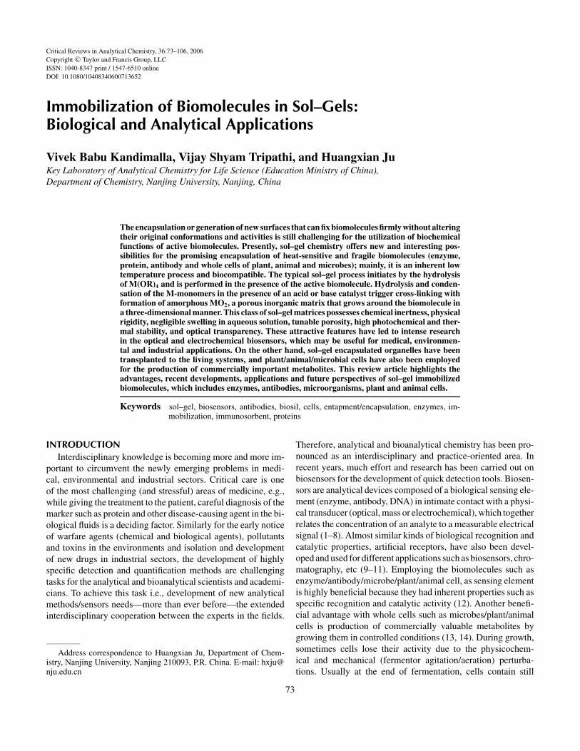
by the sol–gel method has been employed in a number of works(24, 38). In general, the sol–gel process involves hydrolysisof alkoxide precursors under acidic or basic conditions, fol-lowed by condensation and polycondensation of the hydrox-ylated units, which leads to the formation of a porous gel. Typi-cally a low-molecular weight metal alkoxide precursor moleculesuch as tetramethoxy silane or (TMOS) or tetra ethoxysilane(TEOS) is hydrolyzed first in the presence of water, acid catalystand mutual solvent (24, 53). Hydrolysis of metal alkoxide (e.g.,TEOS or TMOS) precursors results in the formation of silanolgroups (Si-OH); through condensation, these silanol moietiesreact further and form siloxanes ( Si O Si ); finally throughpolycondensation of silanol and siloxanes, SiO2 matrices areformed after aging and drying processes as shown in equations1–3 and Figure 1. The resulted sol–gel is an interconnected rigidnetwork with pores of sub-micrometer dimensions and poly-meric chains whose average length is greater than a micrometer.HCl and ammonia are the most generally used catalysts for thehydrolysis; however, other catalysts such as acetic acid, KOH,amines, KF, and HF are also used (31). The rate and extent ofthe hydrolysis are mostly influenced by the strength and concen-tration of the acid or base catalyst (57). Usually weaker acidsrequire longer reaction times to achieve the same extent of reac-tion compared to strong acids. Base-catalyzed hydrolysis of sil-icon alkoxides proceeds much more slowly than acid-catalyzedhydrolysis at equivalent catalyst concentration (32, 57).
(RO)3SiOR + H2O → (RO)3SiOH + ROH [1]
2(RO)3SiOH → (RO)3Si-O-Si(OR)3 + H2O
[2]
(RO)3SiOH + ROSi(OR)3 → (RO)3Si-O-Si(RO)3 + ROH
[3]
When the liquid in the pore is removed at or near ambientpressure by thermal evaporation, drying and shrinkage occurs,the resulted monolith is termed as xerogel. If the liquid is pri-marily alcohol, the monolith is termed as an “alcogel.” Usually,xerogels are superior in mechanical properties and chemical re-sistance to hydrogels in view of cross-linking and densification(24, 25). But the drying of hydrogels inevitably reduces poros-ity, increases steric compression and diffusional limitations, andresults in a reduced bioactivity, especially for inorganic sol-gels(25, 26, 53). The animal/plant cells or organelles cannot surviveat complete dehydration, and are very sensitive to structural dis-turbances caused by invasive polymer matrixes, and cells neededhydrophilic sol–gel/hydrogels to preserve their viability (38).
Silica sol–gels are transparent, chemically inert, negligibleswelling in organic solvents compared to most organic polymersand mechanically stable. In most cases, they are appropriate
BIOMOLECULE IMMOBILIZATION IN SOL–GELS 75
FIG. 1. Schematic diagram of the sol–gel process.
matrices for bio-encapsulation even though they are brittle innature. In recent years, silica sol–gel-based inorganic-organichybrid materials have also been reported (58). The sol–gel ma-terials are generally based on silica, alumina, titania and othercompounds. The introduction of various functional groups intoorganic alkoxide has led to organically modified sol–gel glasses(ormosils) (59). The use of ormosils in bioencapsulation mayprovide interesting properties to the host matrixes from hy-drophobic to hydrophilic (hydrogels) (60, 61). When hydropho-bic silica-forming monomers are used, the resulting electrodesreject water, leaving only segregated islands of carbon at theoutermost surface in contact with electrolyte (62). On the otherhand, when hydrophilic monomers are used, there is an increasein the water-wetted area of the sol–gel glass. Thus the ratio ofhydrophilic and hydrophobic monomers in an organically mod-ified sol–gel glass is crucial in the preparation of biocompatiblematrices and sensor design (63). The structure and properties ofdoped sol-gels depend not only on the chemical compositionsof the starting materials, but also on many operational factorsinvolved in the preparations such as water/silica molar ratio, sol-vent, catalyst, pH and temperature. These parameters highly in-fluence the hydrolysis and condensation and allow the control ofnano- and microstructure of the final material (25). Such controlis essential for achieving a proper balance between non-leachingof the entrapped bioactive molecules and its accessibility to theanalyte. In addition to these the sol sitting time, gel-drying time,and the conditions under which the gel is aged and dried (i.e.,
relative humidity) can also affect the long-term performance ofthese materials (64).
Advances in the Sol–Gel ProcessThe major obstacle with sol–gel entrapment of biomolecules
is the formation of alcohol as a by-product during the hydroly-sis and condensation of the alkoxide precursors, which causes adetrimental effect on the activity of the biomolecules (54, 65). Tocircumvent this problem poly(glyceryl silicate) (PGS) sol–gelprecursors have been introduced by Gill and Ballesteros (38).The stable, water-soluble PGS were prepared by the partial hy-drolysis and condensation of tetramethyl orthosilicate (TMOS)to poly (methyl silicate) (PMS), followed by its transesterifica-tion with glycerol, in a one-pot reaction, catalyzed by hydrochlo-ric acid or poly(antimony(III) ethylene glycoxide). PGS rapidlyhydrolyzed and gelled in aqueous, buffered milieu without theneed for any catalyst, to form silica hydrogels, which producedtransparent, mesoporous, and physically stable silica xerogelsafter aging, washing to remove glycerol, and drying. These sol–gel materials showed good porosity, less shrinkage and highpercentage of bioencapsulation, and the entrapped biomoleculesretained almost complete activity (98%). The poly (silicic acid)entrapped thermolysin, lypoxigenase, silalic acid aldolase, ty-rosinase and S. salmonicolor cells performed poor results due tothe protein precipitation and premature/partial gelation. How-ever, the PGS-derived silical matrices exhibited mild encapsula-tion chemistry and high precursor biocompatibility, and reduced
76 V. B. KANDIMALLA ET AL.
significantly the toxic effects. The biogels retained 83–98% ofthe activity of the native biological as compared with 11–76%for PMS (38). Later Liu and Chen reported alcohol free aque-ous colloidal sol–gel process and encapsulated cytochrome c,catalase, myoglobin and hemoglobin with good retained activi-ties (65). In another alcohol free sol–gel approach sodium sili-cate was employed as starting precursor (66), in which proteinsshowed preserved activity. Perhaps both PGS and sodium sili-cate, routes have inherent limitations in their applications, theglycerol-derivatized silicate precursors need to be synthesized,and in sodium silicate, route precursors release high Na+ con-centration, which must be eliminated through an acidic cation-exhange resin. In addition to this, none of these routes are suit-able for the preparation of hybrid organic-inorganic matrixesthat can provide comfortable environments for a number ofbiomolecules (67, 68). Ferrer et al. reported another alcohol-free and simple aqueous sol–gel method. In this approach thealcohol formed during the hydrolysis was removed through therotavapor method (69). The HRP immobilized in this alcohol-free route exhibited a completely preserved activity and showedhigher specific activity compared with regular sol–gel method.This method was applicable for the preparation of pure silica ma-trixes as well as ormosil. Overall, alcohol-free methods enabledthe encapsulated proteins to retain their structure and biologicalactivity for a prolonged period.
Some non-silica sol–gel materials have also been developedto immobilize bioactive moelcues for the construction of biosen-sors (70) and to synthesize new catalysts for the functional de-vices (71, 72). Liu et al. (70) proved that alumina sol–gel wasa suitable matrix to improve the immobilization of tyrosinasefor detection of trace phenols. Titania is another kind of non-silica material easily obtained from the sol–gel process (73). Yuand Ju (74) reported a simple and mild titania sol–gel thin filmthrough vapor deposition in neutral medium. The titania sol–gel composite film is very efficient in retaining the activity ofhorseradish peroxidase (HRP) and preventing it from leaking outof the film. This method has been extended for the immobiliza-tion of hemoglobin (75), carcinoma antigen 125 (76) and carbo-hydrate antigen 19-9 (77) for the development of biosensors.
Luckarift et al. (78) reported a new enzyme immobiliza-tion (entrapment) method in a biomimetic silica support. Thebiosilicification method was very fast, mild and convenientfor the entrapment of biomolecules at ambient temperatures.In this process, precipitation was catalyzed by the R5 pep-tide, the repeat unit of the silaffin, which was identified fromthe diatom Cylindrotheca fusiformis. During the enzyme im-mobilization in biosilicification, the reaction mixture consistedof silicic acid (hydrolyzed tetramethyl orthosilicate), R5 pep-tide, and enzyme. In the process of the precipitation reaction,the enzyme was entrapped and nm-sized biosilica-immobilizedspheres were formed. This work used butyrylcholine esteraseas a model enzyme. Compared to free enzymes, the biosilica-immobilized enzyme was stable up to 65◦C for 1 hour, whereasfree enzyme lost 85% of activity. These biosilica nano-spheres
could be useful for the immobilization of other fragile andhighly sensitive biomolecules and used for biosensor applica-tions (78). Recently Prieto-Simon et al. (79) reported metal-modified 3-aminopropyltrimethoxysilanes (APTMS) that couldbe employed in the electrochemical monitoring of glucose andhydrogen peroxide. In this study copper-, iron-, zinc-, andcerium-modified APTMS were mixed with tetraethoxysilane(TEOS) and the finally produced xerogels. However, these gelshad potential drawbacks such as lack of homogeneity, low ad-hesion onto the electrode surface, and cracking effects duringaging. The incorporation of poly(ethylene glycol)-cellulose ac-etate composite solution (PEG/CA) improved the characters ofthese gels along with more wettability. The metal modified sol–gels could be more promising in development of interference-free biosensors. Carturan et al. (29, 43, 80) developed a biosilmethod for the encapsulation of plant and animal cells. The gasphase biosil process was recently applied to the bioencapsula-tion of animal cells in silica-alginate beads (81), and reviewd byCoradin et al. (49).
Advantages and Disadvantages with DifferentSol–Gel Matrices
Different kinds of precursors and matrices have been devel-oped to obtain the sol–gel matrices with improved properties likereduced brittle nature, high transparency, improved hydrophilic-ity, flexible porosity, etc. Different sol–gel matrices such as in-organic, organically modified (ormosils), hybrid sol–gels andinterpenetrating polymer networks have been used for the enca-puslation. Perhaps each type of sol–gel has its own advantagesand disadvantages (26, 59). Inorganic sol–gels are good in trans-parency; chemical robustness but brittleness and low porosityin xerogels are major limitations. Similarly organically modi-fied sol–gels have good tunable porosity and electrochemicalactivities, perhaps being relatively fragile and of limited opticaltransparency (27, 63). Hybrid sol–gels can be prepared with flex-ible rigidity, controlled porosity and balance hydrophobicity andhydrophilicity, but poor optical transparency and structural col-lapse on drying are somewhat limiting factors. Interpenetratingpolymer networks are combined matrices of sol–gel with water-soluble polymers such as carrageenan, alginate, agar, PVA andPEG, etc. These matrices are highly biocompatible for fragilemolecules such as organelles and living cells. Compared withalginate and carrageenan beads, sol–gel-layered beads are stableagainst chelating agents and physicochemical perturbations dueto the supporting action of outer sol–gel layer (29, 82). For theimprovement of conductivity and mechanical strength, sol–gelsare generally filled with nano- or micro-particulates platinum,palladium, graphite and methylated silica, clays and celluloseetc., respectively (reinforced matrices). Plant cells encapsulatedin polyester fibers reinforced sol–gel films can be employed inthe production of enzymes in which reinforced polyester fibersserve as a support for the sol–gel films and anchorage for theplant cells (82).
BIOMOLECULE IMMOBILIZATION IN SOL–GELS 77
Porosity of Sol–GelFor the better performance of encapsulated biomolecules,
the doped silica-matrix pores need to meet two requirements.Pores should be large enough to allow unrestricted transport ofmolecules including buffer ions, substrates and products of thereaction and analytes; simultaneously, it should exclude largeparticles such as bacteria. Second, pores should be small enoughto prevent leakage of encapsulated macromolecules (24, 25).Generally most of the entrapped enzymes show an increase inKm, which means high substrate concentration compared to na-tive enzyme (66). Behind this a number of factors are involved,including partitioning of substrates between solution and sup-port, and diffusion resistance to the transport of substrates tothe enzyme. The presence of small pores, or bottlenecks evenin big pores, can reduce the diffusion coefficients of substratesand products significantly in a silica matrix. If the dimensions ofthe matrix are large or if there are a large number of very smallpores, enzyme molecules buried inside the matrix encounter asubstrate concentration significantly lower than that at the sur-face. If the diffusion rate of the substrate is sufficiently slowcompared to enzymatic catalysis, the enzyme molecules closeto the surface can use up most of the substrate molecules enter-ing the matrix, effectively making the substrate concentrationbe zero in the interior of the matrix (66). The similar trans-port or diffusional problems arise in case of antigen/antibodyreaction, plant and animal cell bio-transformation or productionof primary or secondary metabolites, when entrapping the re-spective biomolecules in sol–gel matrixes. To overcome theseproblems, pore size and density should be controlled for theirbetter performance. Different agents including surfactants andnon-surfactants have been used as pore-improving agents. Var-ious alcohols and mixed solvent systems have also been usedfor the improvement of pore size (33). Along with these, manysynthetic routes and strategies have been developed to yield awide diversity of materials of various framework chemical com-positions and pore structures. In most of the studies, ionic andneutral surfactants have been employed as templates (83, 84),which direct the mesophase formation based on the electrostaticand hydrogen-bonding interactions, respectively.
Jie et al. (85) developed macropore-sized (100 µm) sol–gelbioglasses using poly(vinylalcohol) (PVA) as a pore-formingagent. Changing PVA/sol ratio could further control pore size.Takahashi et al. (86) reported that the incorporation of PVAinto lithium niobate (LiNbO3) can obtain thick films along withcrack-free, smooth and good diffusional properties. Xi et al. (32)studied the effect of different catalysts such as HCl, NH4OH,HNO3, HF and NH4F on TEOS sol–gel pore size. RecentlySoares et al. (87) entrapped Candida rugosa lipase in TEOSand MTMS (methyltrimethoxysilane) in presence of PEG. Itshowed considerable esterification activity due to the increasedmean pore size and improved protein accessibility. Wei et al. (88)doped a non-surfactant D-glucose as a pore-forming agent intothe sol–gel. The doping of glucose was highly beneficial as itprovided a biocompatible environment, could be easily removed
under mild conditions and could be used to develop low-cost ma-terials. Tetraalkyl orthosilicate in the presence of non-surfactanttemplate (glucose) could improve the activity of entrapped alka-line phosphatase (ALP) more significantly than in conventionalmicroporous sol–gel materials (89).
On the other hand, during the aging process, cross-linkingof the network increases and the internal solvent is expelledfrom the matrix, causing the internal polarity and viscosity tobe changed and the average pore size to be decreased in a man-ner that depends on the aging conditions (90, 91). The non-uniformity of the pores in silica sol–gel matrices causes cracksand fractures in dry monolithic sensors upon immersion in water(25, 62). To solve this problem, some researchers used surface-active drying control chemical additives such as Triton-X andquaternary ammonium compounds or copolymers to prepare thesol–gel film (92, 93). Previous studies revealed that in usual sol–gel processes, the pore sizes of xeorgels and hydrogels (wet/agedgels) were between 2–20 nm and 4–100 nm (26). Proteins suchas cytochrome c and RNAse (94), and antibodies (95, 96), couldreversibly immerse into the sol–gel and could selectively bindwith the doped moelecules. Rao and Dave (97) reported thatproteins could selectively bind with the sol–gel matrix and dif-fuse in and out. Bis[3-(trimethoxysilyl)-propyl]ethylenediamine(enTMOS) derived sol–gel exhibited the selective affinity be-havior of protein with globular heme proteins (cytochrome c,myoglobin and hemoglobin). Collagen membranes coated withSiO2 layers showed diffusion of proteins, and diffusion coeffi-cient reduced with increasing protein size (molecular weight).The substantial reduction was obtained at MW 150,000. Thiskind of material is beneficial in medical applications for design-ing artificial organs (98).
Interactions Between Sol–Gel Matrix and AnalytesThe interactions between sol–gel matrices and analytes in-
clude electrostatic, hydrogen bonding and hydrophobic inter-actions. They play key roles in the accessibility of analytes tothe entrapped biomolecules even though the pore sizes are quiteenough for the analytes to pass though the matrices, and are im-portant in determining the accessibility of analytes to entrappedproteins. In cases where repulsive or attractive interactions ex-ist between the glass and analytes, the sol–gel matrices take upthe analytes either partially or excessively (25, 28). The study ofthese interactions is very important because they affect the diffu-sional properties significantly. Badjic and Kostic (99, 100) stud-ied the interactions between polar silica and organic compounds.Silica monoliths immersed in solutions containing styrene areevenly dispersed, as styrene cannot form hydrogen bonding withsilica. After soaking of silica matrix in electrolyte solutions ata pH value at which pore walls are negatively charged, anionssuch as [Fe(CN)6]3− are only partially taken up, whereas cationssuch as [Ru(NH3)6]3+ are excessively taken-up by the sol–gelmatrix from the surrounding solution. In either case internaland external concentrations of the ion are unequal even afterequilibrium is reached (101). The protein entrapped in TEOS
78 V. B. KANDIMALLA ET AL.
gels completely showed accessibility to the neutral quenchers,perhaps partial accessibility or repulsion in case of negativelycharged quenchers (102).
Stability of Biomolecules in Sol–GelPhysical entrapment of proteins in a sol–gel matrix preserves
protein structure and functionality and protects the protein fromphysicochemical perturbations. It is mainly due to the sol–gelmatrix “cages,” which provide more rugged environment to thedopant. The sol–gel entrapped heme proteins such as cytochomec (cyt c) and myoglobin (Mb) showed good stability againstpH and thermal perturbations compared to protein in solution(65, 103). Usually globular proteins such as cyt c contain non-covalent interactions that maintain the native folded state underphysiological conditions. The disruption of these interactionsby heating or chemical treatment leads to the conformationalchange and eventually denaturation. But the sol–gel caged cyt cshows high thermal stability due to the exact fitting of the proteininside the cage, which is controlled by the protein size (104). Asthe heme proteins retain their reactivity in optically transparentglass, sol–gel encapsulated Mb can be employed as a sensingelement for the measurement of dissolved oxygen in water usingoptical spectroscopy (105).
ALP entrapped in SiO2 xerogels shows improved thermalstability at basic conditions. The half-life at 70◦C and pH 9.0is 2.6 minutes for free enzyme, 4.7 minutes for the entrappedstate. The entrapped ALP retains activity for 2 months atoptimal conditions. In another study sol–gel encapsulated acidphosphatase also exhibits pronounced protective effect. Enzymetrapped in sol–gel glasses (in presence of PEG 400 and NaF) isstable upto 12 minutes at 70◦C in 0.1 M pH 5.6 citrate buffer,whereas it is stable only for 0.1 min in solution (106, 107).Bovine carbonic anhydrase II (BCA II) is not unfolded even at74◦C, retains 33% activity; perhaps it was unfolded in solutionat 64◦C. The encapsulated BCA II obeys the Michaelis–Mentenkinetics by hydrolyzing the p-nitrophenyl acetate (108). Thisenhanced stability is due to the protective nature of the cageand the rigidity of the SiO2 matrix, which reduces the freedomof peptide-chain refolding molecular motions (24). Trypsin andacid phosphatase entrppled in silicate sol–gel along with PEGhave half-lives 100-fold times higher than that of enzyme insolution at 70◦C (106). More interestingly the creatine kinase(CK) encapsulated in TMOS sol–gel exhibits 4-fold improvedactivity upon short exposure to the elevated temperatures. Cir-cular dichroism results indicate the initial conformations of CKin sol–gel and solution are different, and the entrapped enzymeactivity is not at maximum. However, after heat-treated at47◦C the CK encapsulated in sol–gel matrix reaches maximumactivity after 10 hours. It is mainly due to the improvement inthe pore size, the rearrangement of the CK conformation duringthis process and the interactions between enzyme and sol–gelmatrix (109). Through resonance Raman spectra Das et al. (110)proved that myoglobin could be preserved in native form evenat lower pH by encapsulating them in sol–gel glasses. Chen
et al. (111) studied in-depth the stability of three flavoprotienoxidases, i.e., glucose oxidase (GOD), lactate oxidase, and glu-conate oxidase in hydrated silica gels. The half-life of the GODat 63◦C increased up to 200-fold after immobilization. But thelactate oxidase and gluconate oxidase activities were improvedonly after the doping of weak base poly (N -vinylimidazole) andstrong base poly (ethyleneimine), respectively, prior to the im-mobilization. The protein stabilization depended mainly on thecharges present on the protein and active sites and their interac-tions with anionic silica matrix. GOD is zwitterionic in nature,hence it is more stable in caged gels. Different types of additiveshave been employed as stabilizers to the entrapped proteins,including ligand-based stabilizers (Cod III parvalbumin (112),oncomodulin (113), methyltrimethoxysilane-based materials(to stabilize atrazine chlorohydrolase) (114), the incorporationof organosilanes and polymers into lipase-doped silica (115),poly-(ethylene glycol) (to stabilize acetylcholinesterase and bu-tyrylcholinesterase) (116) and graft copolymers of polyvinylim-idazole and polyvinylpyridine (to stabilize entrapped glucoseoxidase and horseradish peroxidase) (93, 117). RecentlyBrennan et al. (118) reported the addition of sugar (sorbitol) andamino acids (N -methylglycine) increased the thermal stabilityand improved the α-chymostriosin and RNAse T1 activity,because the added osmolytes (sorbitol, N -methylglycine)altered the hydration effects, protein silica interactions and poremorphology.
The sol–gel entrapment method is biocompatible and showsimproved thermal stability to the bioactive molecules, howeverpartial inactivation or loss of activity is a quite common problem.Ferrer et al. (119) observed the activity loss of HRP immobilizedin TEOS gel films and reasoned that the reduction in activitywas due to the detaching of a part of heme groups from activesites during the initial sol–gel formation steps. When these hemegroups (HRP-encapsulated aged gel) were exposed to workingbuffer, they leached out within a day; later, no further reductionin activity was observed even after extensive washing, which wasmonitored though heme characteristic absorption at 280 nm, andtryptophan fluorescence studies. In the cases of some enzymes,active and inactive protein aggregation also causes loss of en-zyme activity. Lloyd and Eyring (120) controlled intermolecularprotein-protein interactions by immobilizing HRP in a silicatematrix. Usually the sol–gel pores were large enough to allowdiffusion of reactants to the enzyme but small enough to preventthe enzyme leakage and interact/aggregate with other enzymemolecules.
Dynamics of Proteins in Sol–GelThe entrapped protein vicinity is completely different from
the native environment. Hence, the potential use and devel-opment of robust analytical devices, the conformational, ro-tational and transalational dynamics and the accessibility ofthe entrapped proteins should be closely monitored. The con-formational and dynamic motions of the entrapped proteinshave been examined widely using absorbance, fluorescence
BIOMOLECULE IMMOBILIZATION IN SOL–GELS 79
(35), resonance Raman (110), dipolar relaxation (37), and time-resolved fluorescence anisotropy (35) measurements. After en-capsulation, proteins such as bovine serum albumin (BSA),human serum albumin (HSA) and monellin retain their confor-mation (112, 113, 121, 122), but small molecules such as myo-globin (Mb) (121) undergo substantial conformational changesduring the entrapment. On the other hand, small proteins suchas cytochrome c and parvalbumin appear to be able to retainfull conformational flexibility upon entrapment and are onlymoderately affected by aging of the matrix (37, 112). It maybe due to the collective effect of high alcohol concentration,aging and pore size. The sol–gel cage restricts the confor-mational change of big molecules, which leads to partial un-folding, but small molecules can easily be gotten more con-formational changes/denaturation (28). Edmiston et al. (121)studied the behavior of myoglobin and acrylodon-labeled bovineserum albumin (BSA-Ac) entrapped in TMOS derived xero-gels. These composites were subjected into the solutions thatcontain ionic quenchers or chemical denaturants. Then the pro-tein response and conformation were detected by following thestatic emission spectra and intensity (121). Jordan et al. (28)observed the nanosecond and picosecond dynamics of BSA-Acand acrylodan-labeled human serum albumin (HSA-Ac) whenthey were sequestered within sol–gel-derived xerogel glasses.These experiments indicated that the “global” protein rotationalmotion was not arrested within a xerogel. In another study Bakeret al. (123) reported that the protein mobility could be modu-lated by using polymer-doped xerogels. The dynamics of HSAentrapped in TEOS-derived materials has recently studied byFlora et al. (102). The entrapped protein showed no global mo-tion (hindered rotation), and performed only segmental and localrotational motions in the region of Trp 214. They reasoned that itwas attributed to electrostatic or other interactions between theprotein and the silica, which restricted global rotational motionsof the protein. It was also suggested that the protein likely un-derwent some unfolding upon entrapment, leading to a greaterdegree of segmental motion in the protein. Brennan and co-workers (124) have recently shown the real-time behavior ofmonellin sequestered within thin TEOS-derived xerogels as theprotein is challenged by the quencher acrylamide and the chem-ical denaturant guanidine hydrochloride. The rotational mobil-ity of GOD and its active site FAD have been investigated byHartnett et al. (125) GOD caged in sol–gel, rotational mobilityis reduced 2-fold times more than in solution, perhaps the activesite pocket is similar to that in solution. Santangelo et al. (126)studied the active site dynamics of horse heart ferricytochromec encapsulated in silica hydrogels. They reported that the dy-namics were highly dependent on the structure of water trappedin the hydrogel. The controlled PEG added in TMOS-derivedsol–gel showed improved dynamics of the pyrene, rhodamine6G (R6G) and acrylodan-labelled bovine serum albumin (BSA-Ac) (123). The time-resolved fluorescence results of R6G andBSA-Ac within TMOS composites showed that relative to free,TMOS R6GB was more mobile in PEG-doped composites. With
the increasing of the PEG doping, the R6G and BSA-Ac also ex-hibited faster dynamics. In this study PEG did not affect the porediameter but was well tuned and the sol–gel processed compos-ite dipolarity and altered the mobility of dopants. These resultswere further interpreted as being due to the preferential adsorp-tion of the PEG to the silica surface, which resulted in enhancedmobility for the entrapped protein, due to less protein–silicainteractions (123). Hence while optimizing the influencing pa-rameter for encapsulated biomolecules, dynamics of the proteinsinside the “cage” should also be taken into consideration for theirbetter performance.
SOL–GEL IMMOBILIZED BIOACTIVE MOLECULES
EnzymesSince emerging in sol–gel bioencapsulation, enzymes have
been widely studied due to their well-known biochemical mech-anisms, commercial availability, wide applications, easy solu-bility in water, good stability, simple molecules compared withanimal, plant or microorganisms. In 1955 Dickey (127) demon-strated for the first time bioencapsulation with partial activity ofurease and catalase, but muscle adenylic acid deaminase com-pletely lost its activity. After 3 decades, considerable attentionhas been paid again towards the bioencapsulation using sol–gelglasses. Avnir’s group (128) successfully encapsulated alkaniephosphatase in silica gel, which retained its activity up to 2months (30% of initial) with improved thermal stability. FurtherShtelzer et al. (129) sequestered trypsin within a binary sol–gel-derived composite using TEOS and PEG. In the same year Zinket al. (130) entrapped other proteins cyt c and Mb in TEOS sol–gel. Later studies on bioencapsulation were accelerated by othergroups, and several proteins such as Mb (25, 104, 105), Hb (25,104, 131, 132), cyt c (103, 133), lactate oxidase (134), GOD(79, 125), ALP (135), Cu-Zn superoxide dimutase (25), urease(136), bacteriorhodopsin (bR) (137, 138), HRP (139, 140) andacetylcholinesterase (141), etc. were immobilized into sol–gelmatrices. Previous reports described the various aspects of sol–gel-entrapped biomolecules in such properties as conformation(122, 142), dynamics (35, 143), accessibility (121), reaction ki-netics (101, 124), activity (24, 144–146), and stability (114, 136,147–149).
Biosensor Applications of Enzymes Entrapped in Sol–Gels.Thus far, sol–gel encapsulated enzymes have been widely em-ployed in the construction of biosensors using different de-tection methods such as electrochemical and optical methods(24, 25, 28, 38). The matrix inherent features such as opticaltransparency, high surface area, chemical and photochemicalinertness and the ability to obtain any desired shape and form(monoliths, thin films, powders, fibers), enabling the design op-tical sensors (62, 150–153). The slow diffusion of the electroac-tive species inside the sol–gel matrix causes a long responsetime. Hence for the improvement of conductivity, metal particlessuch as graphite, gold, palladium, iridium, etc. have been dopedinto sol–gel matrices in the construction of electrochemical
80 V. B. KANDIMALLA ET AL.
FIG. 2. Different configurations employed for the coating of sol–gel films on the electrode surface.
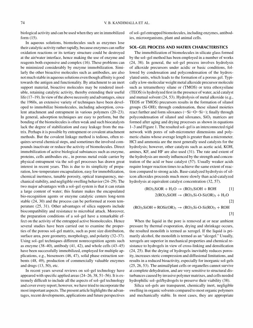
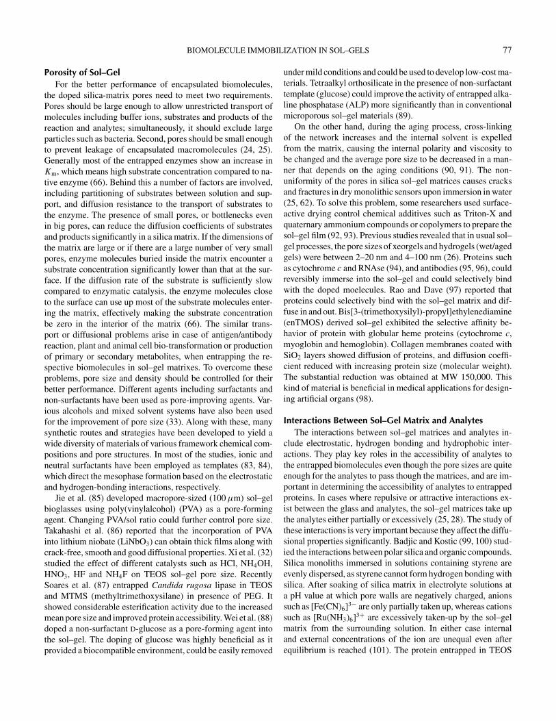
biosensors (154–157). Gavalas et al. (158) doped carbon nan-otubes as conductive materials in to alkoxy silane sol–gels.L-amino oxidase was encapsulated into CNT-sol–gel throughand an aqueous sol–gel process. The added particles improvedmorphology of the composite surface and electrochemical char-acteristics. Sol–gel-derived electrochemical biosensors mainlyrely on two basic configurations: conductive ceramic composites(156, 159, 160), and electrode surface coatings (161, 162). Sincethe pioneering work of Lev and coworkers sol–gel-derived com-posite carbon electrodes (CCEs) (163–165) have been widelyused to develop all kinds of amperometric biosensors. For fixingthe entrapped enzyme on the electrode surface, three main ap-proaches dependent on the convenience, stability and responseare followed; they are single layer (166, 167), bilayer, (154)and sandwich (147, 168) configurations (Figure 2). Along withentrapment inside the sol–gels, few of the studies have been car-ried out by covalently linking the active biomolecules on thesurface of the sol–gel glasses (154). Another important factorin developing biosensors is the thickness of gel film. In mostcases, thin films exhibit quicker response than the thick lay-ers due to the good diffusion properties (42, 169, 170). Withincreasing gel thickness, the signal decays and diffusion of ana-lytes to an biomolecule active site becomes difficult; eventuallythese factors lead to poor response. Homogeneous distribution ofenzymes is possible in sol–gel films compared with regular car-bon paste electrodes; hence, it allows the homogeneous electrontransfer and larger current response in amperometric sensing.
Among all the enzymes studied, GOD and HRP have beenwidely used in sol–gel entrapment studies. The sol–gel immo-bilized GOD exhibits high thermal stability in terms of half-life(200-fold longer than that in the free state) mainly due to thefavorable local polymer–protein interaction between the pos-itive charges on the GOD and the negatively charged silicatematrix (171). Brun et al. (150) reported an optical biosensorbased on the xerogel disk doped with GOD, peroxidase and dye
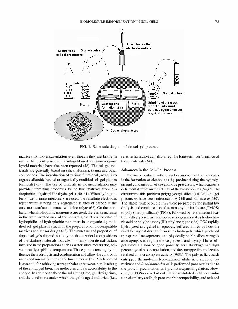
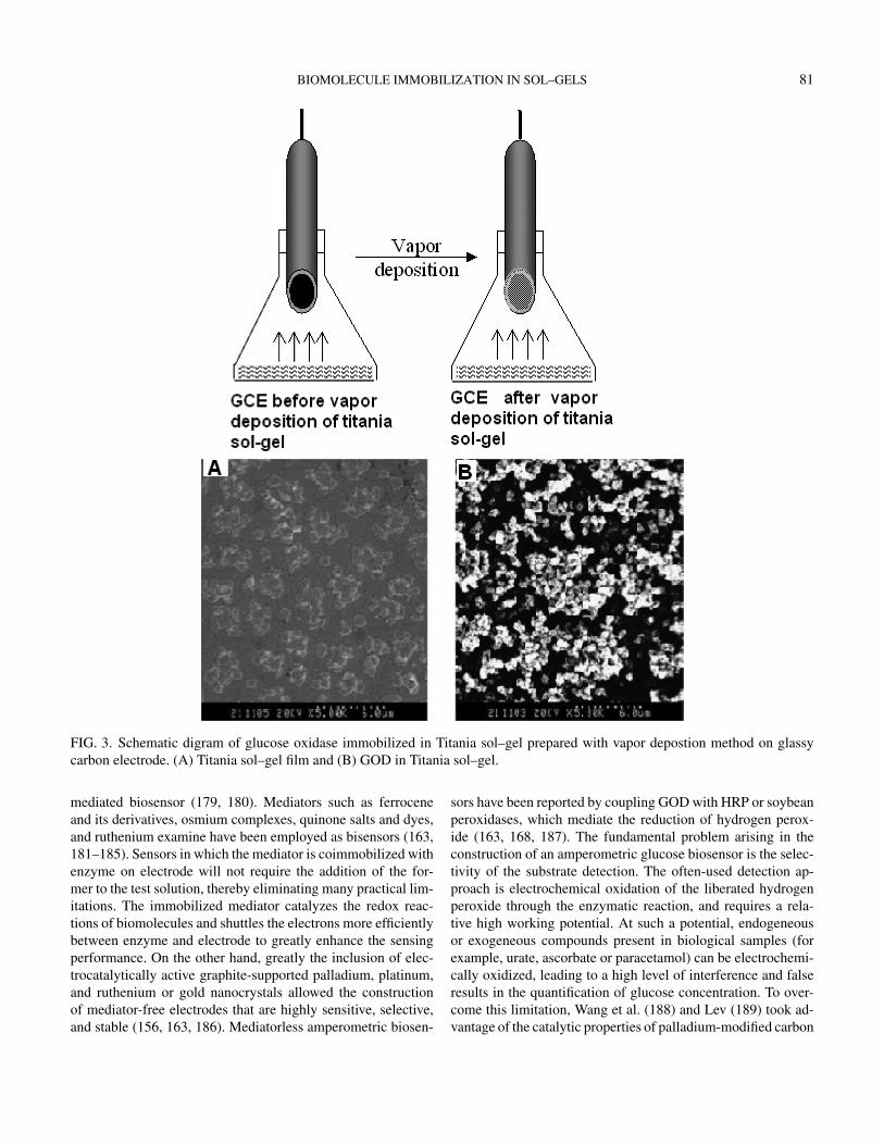
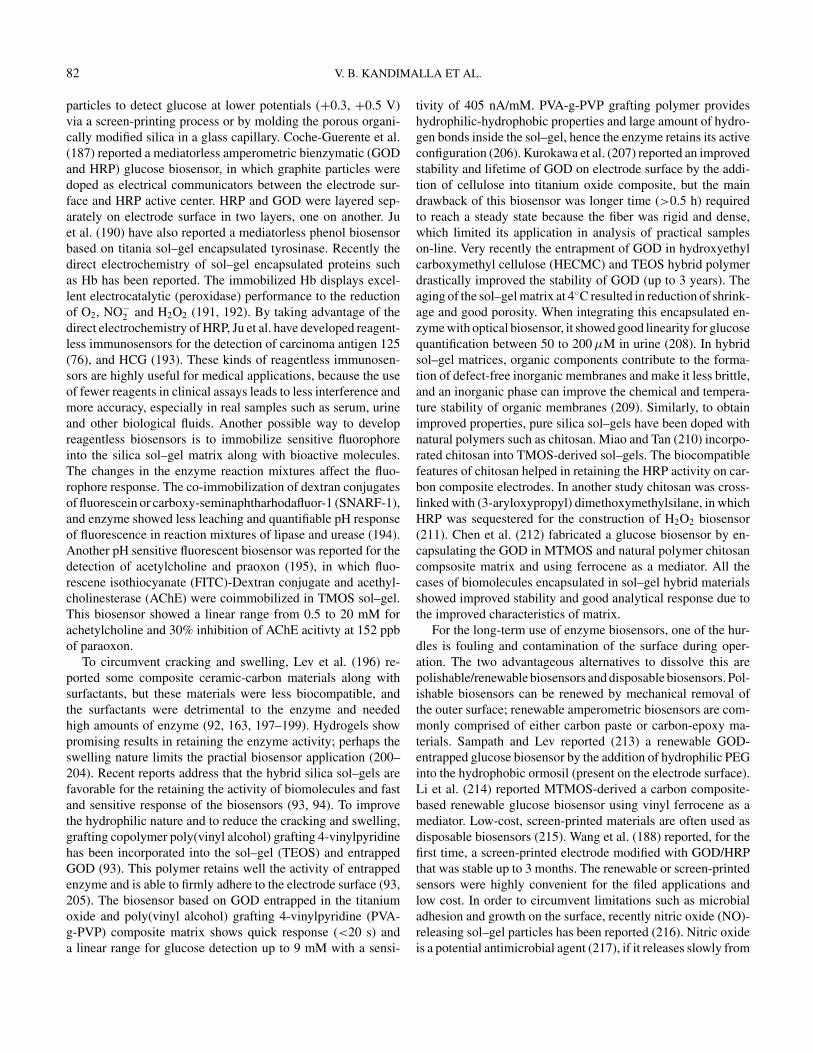
for the detection of glucose. Tatsu (172) prepared tetraethyl or-thosilicate derived silica gel doped with GOD and used it as aglucose recognition element in flow-injection analytical system.Metal alkoxides are more attractive matrices because they pro-vide good conductivity and the possibility of manipulation ofpolarity, rigidity, pore size and distribution and electronic con-ductivity (173). Using the advantages of metal alkoxides, Glezerand Lev (174) prepared platinum electrodes coated with vana-dium pentoxide sol–gel film doped with GOD. Liu et al. (170)reported a glucose biosensor based on immobilization of enzymein alumina (aluminium iso-propoxide) sol–gel films on a pla-tinized glassy carbon electrode. At the GOD/sol–gel/platinizedGCE, glucose response started at +0.2 V approached a maxi-mum value at +0.6 V. The low operating potential greatly min-imized the interference from coexisting electroactive species.Liu et al. (176) immobilized HRP in the ZrO2 sol–gel matrix todevelop a biosensor for the H2O2 determination. Ju and cowork-ers (74, 177) introduced a simple vapor-depsoition method toimmobilise HRP, GOD and other biomolecules titania sol–gel,which retained their catalytic activity and exhibited good re-sponse on electrode surface (Figure 3). The uniform porousstructure of the titania sol–gel matrix had very low mass trans-port barrier, a high catalytic activity, and a fast response (74).Narang et al. (147) developed a glucose biosensor by immobiliz-ing GOD between two sol–gel layers (sandwich). By talking ad-vantage of ormosils, Pandy et al. (178) reported a glucose biosen-sor based on the immobilization of GOD in a sol–gel glassesderived from 3-aminopropyltrimethoxy silane and 2-(3,4-epoxycyclohexyl)-ethyltrimethoxy silane. These films had unre-stricted diffusional properties, and smooth surface face withoutcracking.
The biosensor, which does not require the participation ofredox molecules having reversible electrochemistry, is referredto as nonmediated biosensor, whereas the participation of re-dox mediator in signal transduction generates a category of
BIOMOLECULE IMMOBILIZATION IN SOL–GELS 81
FIG. 3. Schematic digram of glucose oxidase immobilized in Titania sol–gel prepared with vapor depostion method on glassycarbon electrode. (A) Titania sol–gel film and (B) GOD in Titania sol–gel.
mediated biosensor (179, 180). Mediators such as ferroceneand its derivatives, osmium complexes, quinone salts and dyes,and ruthenium examine have been employed as bisensors (163,181–185). Sensors in which the mediator is coimmobilized withenzyme on electrode will not require the addition of the for-mer to the test solution, thereby eliminating many practical lim-itations. The immobilized mediator catalyzes the redox reac-tions of biomolecules and shuttles the electrons more efficientlybetween enzyme and electrode to greatly enhance the sensingperformance. On the other hand, greatly the inclusion of elec-trocatalytically active graphite-supported palladium, platinum,and ruthenium or gold nanocrystals allowed the constructionof mediator-free electrodes that are highly sensitive, selective,and stable (156, 163, 186). Mediatorless amperometric biosen-
sors have been reported by coupling GOD with HRP or soybeanperoxidases, which mediate the reduction of hydrogen perox-ide (163, 168, 187). The fundamental problem arising in theconstruction of an amperometric glucose biosensor is the selec-tivity of the substrate detection. The often-used detection ap-proach is electrochemical oxidation of the liberated hydrogenperoxide through the enzymatic reaction, and requires a rela-tive high working potential. At such a potential, endogeneousor exogeneous compounds present in biological samples (forexample, urate, ascorbate or paracetamol) can be electrochemi-cally oxidized, leading to a high level of interference and falseresults in the quantification of glucose concentration. To over-come this limitation, Wang et al. (188) and Lev (189) took ad-vantage of the catalytic properties of palladium-modified carbon
82 V. B. KANDIMALLA ET AL.
particles to detect glucose at lower potentials (+0.3, +0.5 V)via a screen-printing process or by molding the porous organi-cally modified silica in a glass capillary. Coche-Guerente et al.(187) reported a mediatorless amperometric bienzymatic (GODand HRP) glucose biosensor, in which graphite particles weredoped as electrical communicators between the electrode sur-face and HRP active center. HRP and GOD were layered sep-arately on electrode surface in two layers, one on another. Juet al. (190) have also reported a mediatorless phenol biosensorbased on titania sol–gel encapsulated tyrosinase. Recently thedirect electrochemistry of sol–gel encapsulated proteins suchas Hb has been reported. The immobilized Hb displays excel-lent electrocatalytic (peroxidase) performance to the reductionof O2, NO−
2 and H2O2 (191, 192). By taking advantage of thedirect electrochemistry of HRP, Ju et al. have developed reagent-less immunosensors for the detection of carcinoma antigen 125(76), and HCG (193). These kinds of reagentless immunosen-sors are highly useful for medical applications, because the useof fewer reagents in clinical assays leads to less interference andmore accuracy, especially in real samples such as serum, urineand other biological fluids. Another possible way to developreagentless biosensors is to immobilize sensitive fluorophoreinto the silica sol–gel matrix along with bioactive molecules.The changes in the enzyme reaction mixtures affect the fluo-rophore response. The co-immobilization of dextran conjugatesof fluorescein or carboxy-seminaphtharhodafluor-1 (SNARF-1),and enzyme showed less leaching and quantifiable pH responseof fluorescence in reaction mixtures of lipase and urease (194).Another pH sensitive fluorescent biosensor was reported for thedetection of acetylcholine and praoxon (195), in which fluo-rescene isothiocyanate (FITC)-Dextran conjugate and acethyl-cholinesterase (AChE) were coimmobilized in TMOS sol–gel.This biosensor showed a linear range from 0.5 to 20 mM forachetylcholine and 30% inhibition of AChE acitivty at 152 ppbof paraoxon.
To circumvent cracking and swelling, Lev et al. (196) re-ported some composite ceramic-carbon materials along withsurfactants, but these materials were less biocompatible, andthe surfactants were detrimental to the enzyme and neededhigh amounts of enzyme (92, 163, 197–199). Hydrogels showpromising results in retaining the enzyme activity; perhaps theswelling nature limits the practial biosensor application (200–204). Recent reports address that the hybrid silica sol–gels arefavorable for the retaining the activity of biomolecules and fastand sensitive response of the biosensors (93, 94). To improvethe hydrophilic nature and to reduce the cracking and swelling,grafting copolymer poly(vinyl alcohol) grafting 4-vinylpyridinehas been incorporated into the sol–gel (TEOS) and entrappedGOD (93). This polymer retains well the activity of entrappedenzyme and is able to firmly adhere to the electrode surface (93,205). The biosensor based on GOD entrapped in the titaniumoxide and poly(vinyl alcohol) grafting 4-vinylpyridine (PVA-g-PVP) composite matrix shows quick response (<20 s) anda linear range for glucose detection up to 9 mM with a sensi-
tivity of 405 nA/mM. PVA-g-PVP grafting polymer provideshydrophilic-hydrophobic properties and large amount of hydro-gen bonds inside the sol–gel, hence the enzyme retains its activeconfiguration (206). Kurokawa et al. (207) reported an improvedstability and lifetime of GOD on electrode surface by the addi-tion of cellulose into titanium oxide composite, but the maindrawback of this biosensor was longer time (>0.5 h) requiredto reach a steady state because the fiber was rigid and dense,which limited its application in analysis of practical sampleson-line. Very recently the entrapment of GOD in hydroxyethylcarboxymethyl cellulose (HECMC) and TEOS hybrid polymerdrastically improved the stability of GOD (up to 3 years). Theaging of the sol–gel matrix at 4◦C resulted in reduction of shrink-age and good porosity. When integrating this encapsulated en-zyme with optical biosensor, it showed good linearity for glucosequantification between 50 to 200 µM in urine (208). In hybridsol–gel matrices, organic components contribute to the forma-tion of defect-free inorganic membranes and make it less brittle,and an inorganic phase can improve the chemical and tempera-ture stability of organic membranes (209). Similarly, to obtainimproved properties, pure silica sol–gels have been doped withnatural polymers such as chitosan. Miao and Tan (210) incorpo-rated chitosan into TMOS-derived sol–gels. The biocompatiblefeatures of chitosan helped in retaining the HRP activity on car-bon composite electrodes. In another study chitosan was cross-linked with (3-aryloxypropyl) dimethoxymethylsilane, in whichHRP was sequestered for the construction of H2O2 biosensor(211). Chen et al. (212) fabricated a glucose biosensor by en-capsulating the GOD in MTMOS and natural polymer chitosancompsosite matrix and using ferrocene as a mediator. All thecases of biomolecules encapsulated in sol–gel hybrid materialsshowed improved stability and good analytical response due tothe improved characteristics of matrix.
For the long-term use of enzyme biosensors, one of the hur-dles is fouling and contamination of the surface during oper-ation. The two advantageous alternatives to dissolve this arepolishable/renewable biosensors and disposable biosensors. Pol-ishable biosensors can be renewed by mechanical removal ofthe outer surface; renewable amperometric biosensors are com-monly comprised of either carbon paste or carbon-epoxy ma-terials. Sampath and Lev reported (213) a renewable GOD-entrapped glucose biosensor by the addition of hydrophilic PEGinto the hydrophobic ormosil (present on the electrode surface).Li et al. (214) reported MTMOS-derived a carbon composite-based renewable glucose biosensor using vinyl ferrocene as amediator. Low-cost, screen-printed materials are often used asdisposable biosensors (215). Wang et al. (188) reported, for thefirst time, a screen-printed electrode modified with GOD/HRPthat was stable up to 3 months. The renewable or screen-printedsensors were highly convenient for the filed applications andlow cost. In order to circumvent limitations such as microbialadhesion and growth on the surface, recently nitric oxide (NO)-releasing sol–gel particles has been reported (216). Nitric oxideis a potential antimicrobial agent (217), if it releases slowly from
BIOMOLECULE IMMOBILIZATION IN SOL–GELS 83
the matrix adhesion and the growth of microbes can be con-trolled. NO-releasing glucose biosensors prepared by dopingdiazeniumdiolate-modified sol–gel particles in a polyurethanemembrane exhibits high sensitivity (mention details), repro-ducibility and fast response up to 18 days. To reduce both enzymeinactivation by NO (by minimizing NO exposure) and sol–gelparticle leaching, polyurethane membranes are coated as a layer(217).
Photoactive Proteins. Photoactive proteins such as bacteri-orhodopsin (bR) and phycoerythrin (PE) have had their mech-anisms studied extensively, for potential use as active compo-nents of photonic devies. PE and bR retain their optical activitywhen encapsulated within sol–gel glasses, with enhanced stabil-ity against phtodegradation (218). Bacteriorhodopsin is a natu-rally occurring transmembrane protein that converts light en-ergy into metabolic energy. It was found in the photosyntheticsystem of a salt-marsh bacterium called Halobacterium sali-narium. In its native form, the bR molecule is located in a cellmembrane commonly called “the purple membrane.” Within thebacterial cell, bR is critical to the survival of the organism in anoxygen-deficient environment, as the bR molecules function aslight-driven proton pumps, which transport protons across thecell membrane. This generates a proton, which in turn producesan electrochemical potential used by the organism to synthesizeadenosine triphosphate (ATP). Effectively, bR is used by thebacterium to directly convert sunlight into chemical energy. Theabsorption of light initiates a photocycle in the bR molecule,which accompanies the transportation of protons. The charac-teristics and effects of this photocycle make it a potentiallyuseful material for development as an optically sensitive filmthat is self-developing and erasable. The encapsulation of light-sensitive proteins in transparent matrices is of interest because ofthe potential application to photovoltaic devices, photoimaging,molecular computing (219–221), and chemical sensing (222).Wu et al. (223) and Weetall’s group (224, 225) entrapped bR inwet sol–gel glasses. The D96N mutant bR retained its activity ina dried sol–gel glass (226). Shamansky et al. (227) studied theD96N mutant bR kinetics in dried xerogels. Encapsulation ofphotochromatic proteins in transparent films can be employedin optoelectronic sol–gel devices, which seems to be technicallyfeasible (228). Phycobiliproteins are biomolecular assemblieslocated on the outer thylakoid membranes of marine algae. Theoptical properties of these proteins can be used as fluorescentmarkers in biochemical and biomedical reseach. To interface op-tical properties of PE, Chen et al. (218) entrapped it in sol–gelmaterials. Although PE was stable under ambient light, it de-natured at intense light illumination. The sol–gel entrapped PEretained its conformation, exhibited improved photodegradationcapacity and was more stable than in solution. Such a stablilizedprotoactive proteins could be used in potential applications inbiomolecular sensing, imaging and information processing andstorage.
Use of Sol–Gel Entrapped Enzymes in Drug Screening. An-other potential application of sol–gel-entrapped enzymes is drug
screening. Besanger et al. (229) entrapped clinically importantenzymes cyclooxygenase-2, Factor Xa, dihydrofolate reductase,and A-glutamyl transpeptidase in TEOS and diglycerylsilane(DGS). DGS-entrapped enzymes showed high catalytic activityand stability. Perhaps Km values increased due to the slowerdiffusion and portioning of the substrates within the microp-orous materials. However the interaction between inhibitor andentrapped enzymes was similar to the free enzyme in solution.Similarly in another study (230) for the first time Src proteintyrosine kinase was entrapped in DGS sol–gel. Entrapped Srcprotein tyrosine kinase activity was improved through ligandstabilization effect by the addition of ATP before encapsulation.The IC50 value of Src protein tyrosine kinase entrapped and freeenzyme in solution were close (230). These studies indicates thatthe active sites of sol–gel encapsulated enzymes are accessibleto the inhibitor and substrate in sol–gel “cages.” Hence, such en-trapped enzymes can be employed for the high-throughput drugscreening by paying more attention towards entrapment of otherenzymes. It cannot be generalized that all enzymes can behavesimilarly in sol–gel matrices such as Src protein tyrosine kinase,hence sol–gel parameters should be optimized according to theinteractions of inhibitor with enzyme and sol–gel matrix.
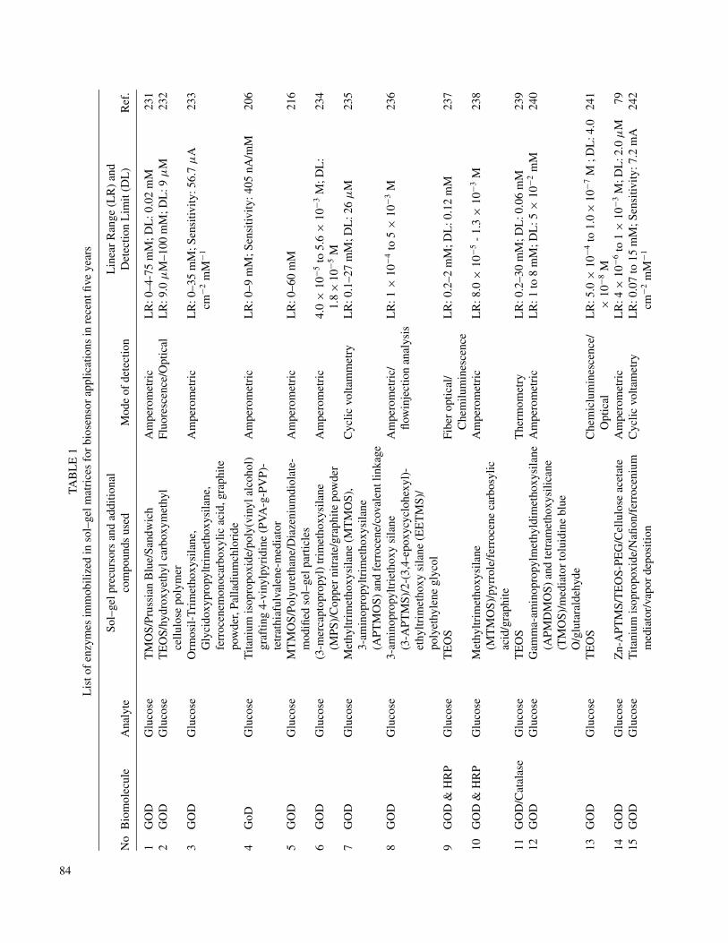
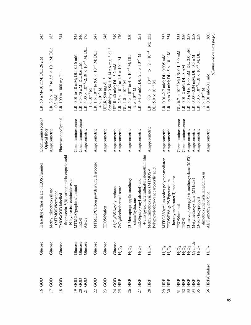
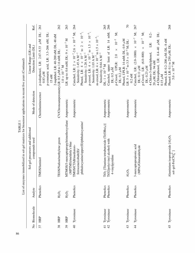
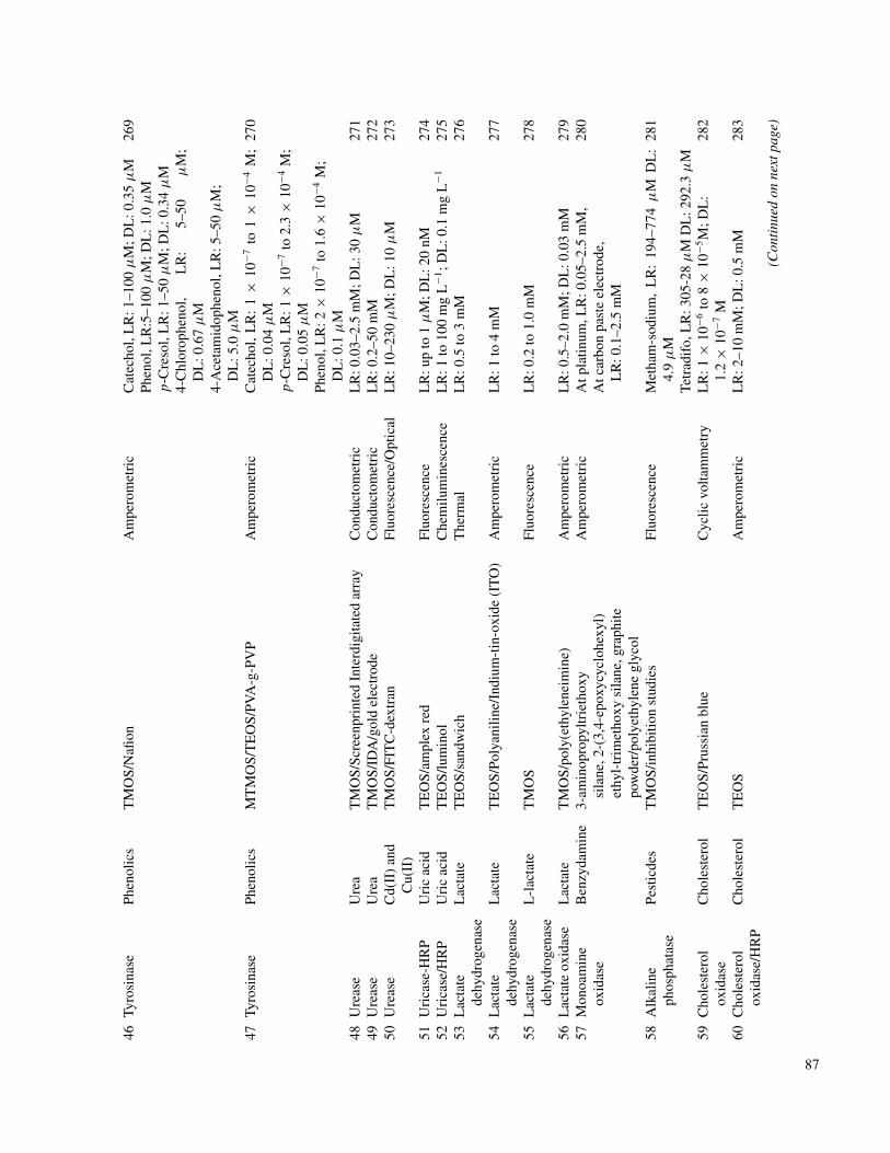
Likely several enzymes have been encapsulated in differentsol–gel matrixes for the different applications. In considerationof space limitations, other reports are presented in Table 1. Pre-vious articles reported by Jin and Brennan (28) and Gill (26) welldocumented the biosensor applications through the year 2000.To avoid repetition in this article, we list biosensor applicationsof the sol–gel-encapsulated enzymes and other proteins from1999 to date, to the best of our knowledge.
Bioconversion Using Sol–Gel Entrapped Enzymes. In thechemical industry a variety of catalysts for well-defined chemi-cal transformations are required. These reactions do not or onlydo unspecifically occur under normal conditions, so that the de-sired product is available only in a small concentration or as amixture with by-products. This often results in the need of costlypurification processes, which makes the chemical synthesis eco-nomically non-efficient. An alternative possibility is the use ofenzymes. These biocatalysts enable highly specific transforma-tions under moderate reaction conditions. Because of the highstereo- or regio-selectivity, the application of enzymes may besuperior to chemical synthesis. Sometimes enzymatic processescan be used for the production of compounds, which are diffi-cult to synthesize chemically. Especially enzymatic methods aresuitable for the synthesis of optically active compounds. Oftenonly one enantiomer of a compound has a pharmacological ef-fect, whereas the other enantiomere has no, or an unwelcome,effect. Therefore, biotechnical syntheses gain importance in theproduction of compounds requiring high enantiomeric purity forpharmaceuticals or commercially important products. A mile-stone in the area of enzymes as catalysts for organic chemistryis the discovery that some of them (e.g., Lipase) retain theircatalytic activity in non-aqueous media (296, 297). Normally,lipases catalyze the hydrolysis of carboxylic acid esters with
TAB
LE
1L
isto
fen
zym
esim
mob
ilize
din
sol–
gelm
atri
ces
for
bios
enso
rap
plic
atio
nsin
rece
ntfiv
eye
ars
No
Bio
mol
ecul
eA
naly
teSo
l–ge
lpre
curs
ors
and
addi
tiona
lco
mpo
unds
used
Mod
eof
dete
ctio
nL
inea
rR
ange
(LR
)an
dD
etec
tion
Lim
it(D
L)
Ref
.
1G
OD
Glu
cose
TM
OS/
Prus
sian
Blu
e/Sa
ndw
ich
Am
pero
met
ric
LR
:0–4
-75
mM
;DL
:0.0
2m
M23
12
GO
DG
luco
seT
EO
S/hy
drox
yeth
ylca
rbox
ymet
hyl
cellu
lose
poly
mer
Fluo
resc
ence
/Opt
ical
LR
:9.0
µM
–100
mM
;DL
:9µ
M23
2
3G
OD
Glu
cose
Orm
osil-
Tri
met
hoxy
sila
ne,
Gly
cido
xypr
opyl
trim
etho
xysi
lane
,fe
rroc
enem
onoc
arbo
xylic
acid
,gra
phite
pow
der,
Palla
dium
chlo
ride
Am
pero
met
ric
LR
:0–3
5m
M;S
ensi
tivity
:56.
7µ
Acm
−2m
M−1
233
4G
oDG
luco
seT
itani
umis
opro
poxi
de/p
oly(
viny
lalc
ohol
)gr
aftin
g4-
viny
lpyr
idin
e(P
VA
-g-P
VP)
-te
trat
hiaf
ulva
lene
-med
iato
r
Am
pero
met
ric
LR
:0–9
mM
;Sen
sitiv
ity:4
05nA
/mM
206
5G
OD
Glu
cose
MT
MO
S/Po
lyur
etha
ne/D
iaze
nium
diol
ate-
mod
ified
sol–
gelp
artic
les
Am
pero
met
ric
LR
:0–6
0m
M21
6
6G
OD
Glu
cose
(3-m
erca
ptop
ropy
l)tr
imet
hoxy
sila
ne(M
PS)/
Cop
per
nitr
ate/
grap
hite
pow
der
Am
pero
met
ric
4.0
×10
−5to
5.6
×10
−3M
;DL
:1.
8×
10−5
M23
4
7G
OD
Glu
cose
Met
hyltr
imet
hoxy
sila
ne(M
TM
OS)
,3-
amin
opro
pyltr
imet
hoxy
sila
ne(A
PTM
OS)
and
ferr
ocen
e/co
vale
ntlin
kage
Cyc
licvo
ltam
met
ryL
R:0
.1–2
7m
M;D
L:2
6µ
M23
5
8G
OD
Glu
cose
3-am
inop
ropy
ltrie
thox
ysi
lane
(3-A
PTM
S)/2
-(3,
4-ep
oxyc
yclo
hexy
l)-
ethy
ltrim
etho
xysi
lane
(EE
TM
S)/
poly
ethy
lene
glyc
ol
Am
pero
met
ric/
flow
inje
ctio
nan
alys
isL
R:1
×10
−4to
5×
10−3
M23
6
9G
OD
&H
RP
Glu
cose
TE
OS
Fibe
rop
tical
/C
hem
ilum
ines
cenc
eL
R:0
.2–2
mM
;DL
:0.1
2m
M23
7
10G
OD
&H
RP
Glu
cose
Met
hyltr
imet
hoxy
sila
ne(M
TM
OS)
/pyr
role
/fer
roce
neca
rbos
ylic
acid
/gra
phite
Am
pero
met
ric
LR
:8.0
×10
−5-
1.3
×10
−3M
238
11G
OD
/Cat
alas
eG
luco
seT
EO
ST
herm
omet
ryL
R:0
.2–3
0m
M;D
L:0
.06
mM
239
12G
OD
Glu
cose
Gam
ma-
amin
opro
pylm
ethy
ldim
etho
xysi
lane
(APM
DM
OS)
and
tetr
amet
hoxy
silic
ane
(TM
OS)
/med
iato
rto
luid
ine
blue
O/g
luta
rald
ehyd
e
Am
pero
met
ric
LR
:1to
8m
M;D
L:5
×10
−2m
M24
0
13G
OD
Glu
cose
TE
OS
Che
mic
lum
ines
cenc
e/O
ptic
alL
R:5
.0×
10−4
to1.
0×
10−7
M;D
L:4
.0×
10−8
M24
1
14G
OD
Glu
cose
Zn-
APT
MS/
TE
OS-
PEG
/Cel
lulo
seac
etat
eA
mpe
rom
etri
cL
R:4
×10
−6to
1×
10−3
M;D
L:2
.0µ
M79
15G
OD
Glu
cose
Tita
nium
isop
ropo
xide
/Nafi
on/f
erro
ceni
umm
edia
tor/
vapo
rde
posi
tion
Cyc
licvo
ltam
etry
LR
:0.0
7to
15m
M;S
ensi
tivity
:7.2
mA
cm−2
mM
−124
2
84
16G
OD
Glu
cose
Tetr
aeth
ylor
thos
ilica
te(T
EO
S)/lu
min
olC
hem
ilum
ines
cenc
e/O
ptic
alfib
erL
R:5
0µ
M–1
0m
M;D
L:2
6µ
M24
3
17G
OD
Glu
cose
Met
hylt
rim
etho
xysi
lane
(MT
MO
S)/f
erro
cene
Am
pero
met
ric
LR
:3.2
×10
−4to
3.5
×10
−2M
;DL
:0.
3m
M18
3
18G
OD
Glu
cose
TM
OS/
GO
Dla
belle
dflu
ores
cein
-5(6
)-ca
rbox
amid
o-ca
proi
cac
idN
-hyd
roxi
su-c
cini
mid
ees
ter
Fluo
resc
ence
/Opt
ical
LR
:100
to10
00m
gL
−124
4
19G
OD
Glu
cose
MT
MO
S/gr
aphi
te/lu
min
olC
hem
ilum
ines
cenc
eL
R:0
.01
to10
mM
;DL
:8.1
6m
M24
520
GO
DG
luco
seT
EO
SC
hem
ilum
ines
cenc
eL
R:3
.5–7
0µ
M;D
L:0
.6µ
M24
621
GO
DG
luco
seA
l 2O
3A
mpe
rom
etri
cL
R:4
.00
×10
−5–2
.19
×10
−2M
;DL
:1
×10
−5M
175
22G
OD
Glu
cose
MT
MO
S/C
arbo
npo
wde
r/vi
nylf
erro
cene
Am
pero
met
ric
LR
:1×
10−4
to9.
6×
10−3
M;D
L:
4×
10−5
M24
7
23G
OD
Glu
cose
TE
OS/
Nafi
onA
mpe
rom
etri
cU
PLR
:500
mg
dl−1
Sens
itivi
ty:0
.54
±0.
14nA
mg−1
dl−1
248
24G
OD
Glu
cose
Al 2
O3/B
SA/p
olyu
reth
ane
Am
pero
met
ric
UPL
R:4
0m
M;D
L:5
.2m
M24
925
HR
PH
2O
2Z
rO2/a
lcoh
othe
rmal
rout
eA
mpe
rom
etri
cL
R:2
.5×
10−7
to1.
5×
10−4
MD
L:1
.0×
10−7
M17
6
26H
RP
H2O
2(3
-Mer
capt
opro
pyl)
trim
etho
xy-
sila
ne/h
ydra
zine
Am
pero
met
ric
LR
:5×
10−6
to4
×10
−3M
;DL
:2
×10
−6M
250
27H
RP
H2O
2T
EO
S/po
ly(v
inyl
alco
hol)
and
4-vi
nylp
yrid
ine/
tetr
athi
aful
vale
ne/th
infil
mA
mpe
rom
etri
cL
R:0
–1.3
mM
;DL
:2.5
×10
−7M
251
28H
RP
H2O
2M
ethy
ltrim
etho
xysi
lane
(MT
MO
S)/
Poly
pyrr
ole/
ferr
ocen
ecar
boxy
licac
idA
mpp
erom
etri
cL
R:
9.0
×10
−7to
2×
10−4
M;
DL
:5.0
×10
−5M
252
29H
RP
H2O
2M
TE
OS/
Osm
ium
redo
xpo
lym
er-m
edia
tor
Am
pero
met
ric
LR
:0.0
1–2.
7m
M;D
L:0
.005
mM
253
30H
RP
H2O
2T
EO
S/PV
A-g
-PV
P/po
tass
ium
hexa
cyan
ofer
rate
(II)
med
iato
rA
mpe
rom
etri
cL
R:u
pto
3.4
mM
;DL
:5×
10−7
M25
4
31H
RP
H2O
2T
EO
S/lu
min
olC
hem
ilum
ines
cenc
eD
L:6
.7×
10−4
M;L
R:0
.1–3
.0m
M25
532
HR
PH
2O
2T
EO
SC
hem
ilum
ines
cenc
eL
R:0
.01–
2m
M;D
L:8
µM
256
33H
RP
H2O
2(3
-mer
capt
opro
pyl)
-tri
met
hoxy
sila
ne(M
PS)
Am
pero
met
ric
LR
:5.0
µM
to10
.0m
M;D
L:2
.0µ
M25
734
HR
PC
yani
deM
etyl
tieth
oxys
ilane
(MT
EO
S)A
mpe
rom
etri
cL
R:0
.004
–0.0
4m
M;D
L:0
.5µ
M25
835
HR
PH
2O
2(3
-aor
ylox
ypro
pyl)
dim
etho
xym
ethy
lsila
ne/c
hito
san
Am
pero
met
ric
LR
:5.0
×10
−9–1
.0×
10−7
M;D
L:
2×
10−9
M.
259
36H
RP/
Cat
alas
eH
2O
2A
l 2O
3/m
ethy
lene
blue
Am
pero
met
ric
LR
:0.0
1m
M–0
.1m
M26
0(C
onti
nued
onne
xtpa
ge)
85
TAB
LE
1L
isto
fen
zym
esim
mob
ilize
din
sol-
gelm
atri
ces
for
bios
enso
rap
plic
atio
nsin
rece
ntfiv
eye
ars
(Con
tinue
d)
No
Bio
mol
ecul
eA
naly
teSo
l-ge
lpre
curs
ors
and
addi
tiona
lco
mpo
unds
used
Mod
eof
dete
ctio
nL
inea
rR
ange
(LR
)an
dD
etec
tion
Lim
it(D
L)
Ref
.
37H
RP
Phen
olic
sT
MO
S/lu
min
olC
hem
ilum
ines
cenc
ep-
iodo
phen
ol,
LR
:2.
67–9
.33
µM
DL
:0.
83µ
Mp-
coum
aric
acid
,L
R:
3.3–
266
nM;
DL
:15
nM2-
naph
thol
.LR
:40-
200
nM;D
L:4
8nM
261
38H
RP
H2O
2T
EO
S/N
afion
/met
hyle
negr
een
CV
/Chr
onoa
mpe
rom
etry
LR
:0.5
µM
to1.
6m
M;D
L:
1.0
×10
−7M
262
39H
RP
H2O
2M
TM
OS/
3-m
erca
ptop
ropy
ltrim
etho
xysi
lane
(MPT
MS)
/mel
dola
’sbl
ueA
mpe
rom
etri
cL
R:u
pto
:0.6
mM
;DL
:9×
10−7
M26
3
40Ty
rosi
nase
Phen
olic
sT
MO
S/C
TAB
/med
iato
rs-
ferr
ocen
e/co
balt(
II)/
phth
aloc
yani
ne/o
pper
(II)
/pht
halo
cyan
ine/
Am
pero
met
ric
Cat
echo
l,L
R:
2×
10−7
-2.6
×10
−5M
;Se
nsiti
vity
:1.4
5A
M−1
Phen
ol,
LR
:4
×10
−7to
2×
10−5
;Se
nsiti
vity
:1.2
8A
M−1
p-cr
esol
,L
R:
4×
10−7
to1.
5×
10−5
;Se
nsiti
vity
:1.0
3A
M−1
m-c
reso
l,L
R:4
×10
−7to
1.5
×10
−5;
Sens
itivi
ty:0
.64
AM
−1
264
41Ty
rosi
nase
Phen
olic
sT
iO2
[Tita
nium
tetr
abut
oxid
e(T
i(O
Bu)
4)]
Am
pero
met
ric
DL
:1×
10−8
M26
542
Tyro
sina
sePh
enol
ics
TiO
2/po
ly(v
inyl
alco
hol)
with
4-vi
nylp
yrid
ine
Am
pero
met
ric
Cat
echo
l,up
per
limit
ofL
R:
14m
M;
DL
:0.5
µM
p-C
reso
l,U
PLR
:2.
0×
10−5
M;
DL
:0.2
µM
Phen
ol,U
PLR
:14
mM
;DL
:0.9
µM
266
43Ty
rosi
nase
Phen
olic
sA
l 2O
3A
mpe
rom
etri
cL
R:5
×10
−9to
3.5
×10
−4M
;DL
:0.
2nM
70
44Ty
rosi
nase
Phen
olic
s3-
mer
capt
opro
pion
icac
id(M
PA)/
Glu
tara
ldeh
yde
Am
pero
met
ric
Cat
echo
l,L
R:
(2.0
–100
0)×
10−7
M;
DL
:0.1
1µ
Mp-
Cre
sol,
LR
:(6
.0–8
00)
×10
−7M
;D
L:0
.23
µM
4-C
hlor
o-3-
met
hylp
heno
l,L
R:
0.2–
20µ
M;D
L:9
4nM
4-C
hlor
ophe
nol,
LR
:0.
4–40
µM
;D
L:
0.15
µM
Phen
ol,L
R:0
.2–2
00µ
M;D
L:8
nM
267
45Ty
rosi
nase
Phen
olic
sA
lum
iniu
mis
opro
poxi
de[A
l 2O
3
sol–
gel-
Fe(C
N)4− 6
]A
mpe
rom
etri
cPh
enol
:LR
:0.1
to25
0µ
M;D
L:
5.0
×10
−8M
268
86
46Ty
rosi
nase
Phen
olic
sT
MO
S/N
afion
Am
pero
met
ric
Cat
echo
l,L
R:1
–100
µM
;DL
:0.3
5µ
MPh
enol
,LR
:5–1
00µ
M;D
L:1
.0µ
Mp-
Cre
sol,
LR
:1–5
0µ
M;D
L:0
.34
µM
4-C
hlor
ophe
nol,
LR
:5–
50µ
M;
DL
:0.6
7µ
M4-
Ace
tam
idop
heno
l,L
R:5
–50
µM
;D
L:5
.0µ
M
269
47Ty
rosi
nase
Phen
olic
sM
TM
OS/
TE
OS/
PVA
-g-P
VP
Am
pero
met
ric
Cat
echo
l,L
R:
1×
10−7
to1
×10
−4M
;D
L:0
.04
µM
p-C
reso
l,L
R:1
×10
−7to
2.3
×10
−4M
;D
L:0
.05
µM
Phen
ol,L
R:2
×10
−7to
1.6
×10
−4M
;D
L:0
.1µ
M
270
48U
reas
eU
rea
TM
OS/
Scre
enpr
inte
dIn
terd
igita
ted
arra
yC
ondu
ctom
etri
cL
R:0
.03–
2.5
mM
;DL
:30
µM
271
49U
reas
eU
rea
TM
OS/
IDA
/gol
del
ectr
ode
Con
duct
omet
ric
LR
:0.2
–50
mM
272
50U
reas
eC
d(II
)an
dC
u(II
)T
MO
S/FI
TC
-dex
tran
Fluo
resc
ence
/Opt
ical
LR
:10–
230
µM
;DL
:10
µM
273
51U
rica
se-H
RP
Uri
cac
idT
EO
S/am
plex
red
Fluo
resc
ence
LR
:up
to1
µM
;DL
:20
nM27
452
Uri
case
/HR
PU
ric
acid
TE
OS/
lum
inol
Che
milu
min
esce
nce
LR
:1to
100
mg
L−1
;DL
:0.1
mg
L−1
275
53L
acta
tede
hydr
ogen
ase
Lac
tate
TE
OS/
sand
wic
hT
herm
alL
R:0
.5to
3m
M27
6
54L
acta
tede
hydr
ogen
ase
Lac
tate
TE
OS/
Poly
anili
ne/I
ndiu
m-t
in-o
xide
(IT
O)
Am
pero
met
ric
LR
:1to
4m
M27
7
55L
acta
tede
hydr
ogen
ase
L-l
acta
teT
MO
SFl
uore
scen
ceL
R:0
.2to
1.0
mM
278
56L
acta
teox
idas
eL
acta
teT
MO
S/po
ly(e
thyl
enei
min
e)A
mpe
rom
etri
cL
R:0
.5–2
.0m
M;D
L:0
.03
mM
279
57M
onoa
min
eox
idas
eB
enzy
dam
ine
3-am
inop
ropy
ltrie
thox
ysi
lane
,2-(
3,4-
epox
ycyc
lohe
xyl)
ethy
l-tr
imet
hoxy
sila
ne,g
raph
itepo
wde
r/po
lyet
hyle
negl
ycol
Am
pero
met
ric
Atp
latin
um,L
R:0
.05–
2.5
mM
,A
tcar
bon
past
eel
ectr
ode,
LR
:0.1
–2.5
mM
280
58A
lkal
ine
phos
phat
ase
Pest
icde
sT
MO
S/in
hibi
tion
stud
ies
Fluo
resc
ence
Met
ham
-sod
ium
,L
R:
194–
774
µM
DL
:4.
9µ
MTe
trad
ifo,
LR
:305
-28
µM
DL
:292
.3µ
M
281
59C
hole
ster
olox
idas
eC
hole
ster
olT
EO
S/Pr
ussi
anbl
ueC
yclic
volta
mm
etry
LR
:1×
10−6
to8
×10
−5M
;DL
:1.
2×
10−7
M28
2
60C
hole
ster
olox
idas
e/H
RP
Cho
lest
erol
TE
OS
Am
pero
met
ric
LR
:2–1
0m
M;D
L:0
.5m
M28
3
(Con
tinu
edon
next
page
)
87
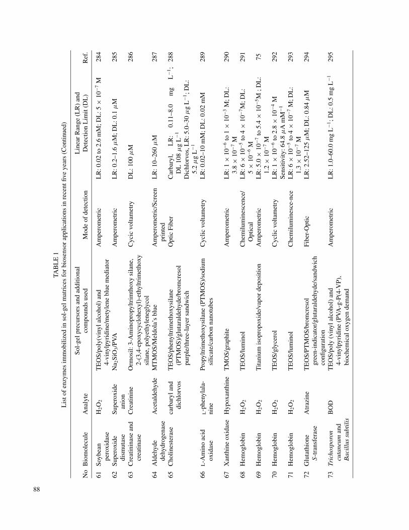
TAB
LE
1L
isto
fen
zym
esim
mob
ilize
din
sol-
gelm
atri
ces
for
bios
enso
rap
plic
atio
nsin
rece
ntfiv
eye
ars
(Con
tinue
d)
No
Bio
mol
ecul
eA
naly
teSo
l-ge
lpre
curs
ors
and
addi
tiona
lco
mpo
unds
used
Mod
eof
dete
ctio
nL
inea
rR
ange
(LR
)an
dD
etec
tion
Lim
it(D
L)
Ref
.
61So
ybea
npe
roxi
dase
H2O
2T
EO
S/po
ly(v
inyl
alco
hol)
and
4-vi
nylp
yrid
ine/
met
ylen
ebl
uem
edia
tor
Am
pero
met
ric
LR
:0.0
2to
2.6
mM
;DL
:5×
10−7
M28
4
62Su
pero
xide
dism
utas
eSu
pero
xide
anio
nN
a 2Si
O3/P
VA
Am
pero
met
ric
LR
:0.2
–1.6
µM
;DL
:0.1
µM
285
63C
reat
inin
ase
and
crea
tinas
eC
reat
inin
eO
rmos
il:3-
Am
inop
ropy
ltrim
thox
ysi
lane
,2-
(3,4
-epo
xycy
cloh
exyl
)-et
hyltr
imet
hoxy
sila
ne,p
olye
thyl
eneg
lyco
l
Cyc
icvo
ltam
etry
DL
:100
µM
286
64A
ldeh
yde
dehy
drog
enas
eA
ceta
ldeh
yde
MT
MO
S/M
eldo
la’s
blue
Am
pero
met
ric/
Scre
enpr
inte
dL
R:1
0–26
0µ
M28
7
65C
holin
este
rase
carb
aryl
and
dich
lorv
osT
EO
S/ph
enyl
trim
etho
xysi
lane
(PT
MO
S)/g
luta
rald
ehyd
e/br
omcr
esol
purp
le/th
ree-
laye
rsa
ndw
ich
Opt
icFi
ber
Car
bary
l,L
R:
0.11
–8.0
mg
L−1
;D
L10
8µ
gL
−1
Dic
hlor
vos,
LR
:5.0
–30
µg
L−1
;DL
:5.
2µ
gL
−1
288
66L-A
min
oac
idox
idas
eL-p
heny
lala
-ni
nePr
opyl
trim
etho
xysi
lane
(PT
MO
S)/s
odiu
msi
licat
e/ca
rbon
nano
tube
sC
yclic
volta
met
ryL
R:0
.02–
10m
M;D
L:0
.02
mM
289
67X
anth
ine
oxid
ase
Hyp
oxan
thin
eT
MO
S/gr
aphi
teA
mpe
rom
etri
cL
R:1
×10
−6to
1×
10−3
M;D
L:
3.8
×10
−7M
290
68H
emog
lobi
nH
2O
2T
EO
S/lu
min
olC
hem
ilum
ines
cenc
e/O
ptic
alL
R:6
×10
−5to
4×
10−7
M;D
L:
5×
10−6
M29
1
69H
emog
lobi
nH
2O
2T
itani
umis
opro
poxi
de/v
apor
depo
sitio
nA
mpe
rom
etri
cL
R:5
.0×
10−7
to5.
4×
10−5
M;D
L:
1.2
×10
−7M
75
70H
emog
lobi
nH
2O
2T
EO
S/gl
ycer
olC
yclic
volta
met
ryL
R:1
×10
−6to
2.8
×10
−4M
Sens
itivi
ty:6
4.8
µA
mM
−129
2
71H
emog
lobi
nH
2O
2T
EO
S/lu
min
olC
hem
ilum
ines
ce-n
ceL
R:6
×10
−5to
4×
10−7
M;D
L:
1.3
×10
−7M
293
72G
luta
thio
neS-
tran
sfer
ase
Atr
azin
eT
EO
S/PT
MO
S/br
omcr
esol
gree
n-in
dica
tor/
glut
aral
dehy
de/s
andw
ich
confi
gura
tion
Fibe
r-O
ptic
LR
:2.5
2–12
5µ
M;D
L:0
.84
µM
294
73Tr
icho
spor
oncu
tane
uman
dB
acil
lus
subt
ilis
BO
DT
EO
S/po
ly(v
inyl
alco
hol)
and
4-vi
nylp
yrid
ine
(PV
A-g
-P(4
-VP)
,bi
oche
mic
alox
ygen
dem
and
Am
pero
met
ric
LR
:1.0
–60.
0m
gL
−1;D
L:0
.5m
gL
−129
5
88
BIOMOLECULE IMMOBILIZATION IN SOL–GELS 89
formation of carboxylic acids and alcohols. Lipases are mostfrequently used enzymes in organic chemistry, because they canmake chemo-, regio- and stereoselective esterification or trans-esterification and also they make possible the catalysis of theenantioselective acylation of amines (298, 299). Reetz et al.(300) successfully immobilized different lipases in hydropho-bic sol–gel matrices, with improved stability and pronounceddegrees of enhanced activity in esterification and transesterifica-tion reactions in organic solvents. The improved activities weredue mainly to the enhanced accessibility of enzyme due to thefine distribution inside matrices, and the interactions betweenhydrophobic groups present in sol–gel matrices and lipase ac-tive sites (115, 300). The lipase entrapped in TMOS (hydrophilicSiO2 gel) retained only 5% activity; however, the incorporationof hydrophobic alkyl groups in the silicon dioxide matrix ledto improved activity. When hydrophobic interfaces are absent,lipases have some elements of secondary structure (“lids”) cov-ering their active sites, making them inaccessible to substrates(301). In the presence of hydrophobic interfaces, conformationalchanges take place, yielding the “open structure” of lipases. Inanother study the lipase entrapped in hydrophobic sol–gel ma-terials was employed in ester hydrolysis in aqueous medium.However during recycle, a reduction in (∼ 5%) activity was re-ported, due to the combined effects of mechanical and loss andleaching (302). Usually after each cycle in transformation reac-tions, the catalyst has to be separated from the reaction mixture,for which the usual method is filtration. To simplify the separa-tion, magnetite nanoparticles were entrapped along with lipasein methyltrimethoxysilane (MTMS) sol–gel matrices. Lipaseswere able to tolerate the presence of magnetite, thus heteroge-neous biocatalysts could be easily separated magnetically fromthe reaction mixture (299).
Phyllosilicate sol–gel entrapped lipase could be used as abiocatalyst in conversion of recycled restaurant grease intoalkylesters. The addition of molecular sieves improved yielddue to the removal of water (303–305). Co-immobilization ofenzymes is a convenient approach for the multi-enzyme pro-cesses. By co-immobilizing formate dehydrogenase, formalde-hyde dehydrogenase and alcohol dehydrogenase in silica gels,together with the electron donor NADH, synthesis of methanolcan be performed at low temperatures and pressures (306, 307).Kuncova et al. (301) studied the effect of different organic sol-vents on stericacid esterification using immobilized lipase. Sol-vents acetone and toluene impaired the activity of free lipase,whereas sol–gel immobilized enzyme partially lost the activity.The thermal stability of lipase increased 55-fold upon entrap-ment in propyltrimethoxysilane (PTMS)/TMOS sol–gel mate-rial. The half-life of immobilized lipase was 4 months at 40◦Cin hexane with efficient esterification capacity. This stabilitywas not only due to the physical entrapment but also to addi-tional multiple interactions with SiO2 through hydrogen, ionic,or hydrophobic interaction, and through multi-point attachmentto the support (308). Invertase can split sucrose into glucoseand fructose. These two products are simple compounds and act
as substrates for the production of aminoacids, organic acids,antibiotics, etc. Nakane et al. (309) entrapped invertase in thethree-dimensional network of cellulose acetate (CA) and ZrO2
fibers. The ZrO2 fiber was formed by coordinated interactionbetween the zirconium and the oxygen of the hydroxyl group orthe carbonyl group on CA molecules. The immobilized inver-tase retained inside the fibers with 92% of initial activity, evenafter 10 recycles and repeated washes. With the increasing ofthe protein concentration the activity of invertase was improveddue to the homogeneous dispersion and completely restrictedswelling. Recently Pierre (55) reviewed the sol–gel encapsu-lated enzymes and their applications. Even though the sol–gelentrapped enzymes show improved stabilities against physico-chemical changes around their vicinity and activity; still, theyare facing diffusion limitations, which lead to the high Km valueand have great impact on conversion at large scale use of immo-bilized enzymes in industrial applications. Hence more effortsare needed for the improved diffusion properties of sol–gel ma-trices for efficient biotransformation/synthetic reactions.
AntibodiesAntibodies (Ab) are immune system-related proteins called
immunoglobulins. Each antibody consists of four polypeptideswith two heavy chains and two light chains joined to form a“Y” shaped molecule. The amino acid sequence in the tips ofthe “Y” varies greatly among different antibodies. This variableregion, composed of 110–130 amino acids, gives the antibodyits specificity for binding antigen. Because of their high speci-ficity and sensitivity they have been widely employed in num-ber of modern diagnostic and therapeutic technologies such asin vitro diagnostics, like radioimmunoassay (RIA), immunora-diometric assay (IRMA), enzyme-linked immunosorbent assay(ELISA), or bloting techniques. In addition to this antibodieshave been employed in affinity chromatography (310, 311), celltyping and graft rejection identification (312), detection of dif-ferent cells and disease markers (313), drug delivery and dis-ease control (314), immunosensors (315, 316) etc. Usually an-tibodies are produced through immunization in vertebrates orhybridoma technology (monoclonal antibodies). In the last 10years, progress in understanding the cellular and molecular ba-sis of the immune response has made it theoretically possible togenerate antibodies against almost any compound. RecombinantDNA technology allows the antibody binding properties to bemodified at the DNA level in suitable organisms such as E.colior yeast cells making it possible to obtain an antibody rapidlywith desired properties without need for the immunization ofthe animals.
Immobilization of Abs on to a solid support was first reportedin 1967 (317), and the technology has widespread application inaffinity chromatography (AC) and other areas. However the ma-jor problem associated with covalent immobilization of antibodyon solid surface is partial loss of biological activity due to the ran-dom orientation of the asymmetric macromolecules, sometimessteric hindrance caused by the neighboring antibody molecules
90 V. B. KANDIMALLA ET AL.
(318). Compared with covalent attachment of the antibodies,entrapment is quite mild and can reduce steric hindrances. Inview of the inertness and biocompatibility of the sol–gel entrap-ment technique antibodies have been successfully encapsulatedin the sol–gel materials for different applications. The Abs en-capsulated in sol–gel-derived glasses can interact with targetmolecules with a high degree of specificity as in solution, andthe signal can be detect using an appropriate sensing scheme(42). Generally antibodies are high molecular weight proteins,if antigen is also high molecular weight compound the interac-tions between antibody and antigen is difficult through the smallpores of the matrix. Sometimes even though antigen is smallcompound, the tagged proteins (enzyme) have high molecularweight, which hinders the antigen-antibody interaction. In such acase, the antigen can be tagged with small signaling compoundssuch as FITC, ferrocene or their derivatives etc, depending uponthe detection mode. Wang et al. (319) encapsulated firstly antiflu-orescein antibodies in TMOS sol–gel. The entrapped antibodiesretained activity significantly and could bind with fluoresceinmolecules, which led to the decrease in the fluorescence.
To evaluate the diffusion of the fluorescein and the decrease influorescence due to the antigen-antibody complex or adsorptionon to the silica marix, sol–gel was prepared with and without an-tibody, and the affinity was checked. The gels dried in desiccatorlost 90% fluorescence due to the conformational changes of thesol–gel entrapped antifluorescein (Ab), and the antifluoresceinwas irreversible even after rehydration. However when stored,the entrapped antibodies at 4◦C in water, is stable up to 4 weeks.In another report (148) antifluorescin antibody was entrappedin sandwich configuration, in aerosol-generated sol–gel-derivedthin films (0.62 ± 0.05 µm). The fluorescent hapten 5-(and 6-)carboxy-4′5-dimethylfluorescein (Me2F) was employed to de-termine the accessibility and viability of the entrapped antiflu-orescein antibody. Antibodies were efficient in recognition andbinding of the Me2F up to 13 weeks when stored in 0.1 M pH 8PBS at 4◦C. For the regeneration, antibodies were treated witha mild chaotrope i.e., 4 M NaCl, with which 40–50% of the re-generation was achieved, up to 6 cycles (143). Yang et al. (320)reported that the addition of PEG did not affect the encapsulationefficiencies of the antigentamicin antibody. But the encapsula-tion efficiencies of the antibody in two kinds of sol–gel materi-als i.e., with and without PEG were 93 and 91%, respectively.But the addition of PEG showed a strong effect on the bindingactivity of the encapsulated antibody. When the antibody en-capsulated in sol–gel along with PEG, 95% of the gentamicinwas bound to the column, in case of without PEG only 42%bound to the column. This improved binding activities probablydue to the high diffusion rate of the gentamicin. Furthermore,addition of PEG prevented the shrinking of the sol–gel glass ma-trix and denaturation of the immobilized antibodies. Similarly,other studies also reported that the addtion of PEG stabilized theAChE and urease and prevented the fouling and adhesion of un-wanted protein to the surface (321, 322). Hence while designingthe matrices for immobilization of biomolecules, it is imperative
to choose dimensions of the support matrices so as to minimizediffusional resistance, thereby making the entire population ofencapsulated biomolecules participate in reaction. Hydatidosisis a disease, which was caused by Echinococcus granulosustapeworm transmits through the dogs and sometimes through thesheep, cattle and pigs. This disease causes cysts on the lungs,liver and other organs in the human body and easily infectsother organs and causes echinoccosis. Hydatidosisis antigenshave been isolated from the cysts, immobilized in the TMOSsol–gel, and coated into ELISA plate wells for the detection ofdisease. The antigens entrapped in the sol–gel matrix retain bio-logical activity, and antibodies in human sera are able to diffusethorough the pores of the silica matrix where they react specif-ically with antigens (323). sol–gel matrices and normal ELISAoptical density measurements are comparable, it indicates thatunder controlled pore size (high porosity 50 A) antibodies caneasily diffuse through the sol–gel and can react with antibodyas in solution.
Shabat et al. (324) successfully encapsulated catalytic anti-boides through sol–gel method and used them in the transforma-tion reactions. Antibody 14D9 is an effective catalyst for varioushydrolytic reactions including the hydrolysis of a cyclic acetal,ketals, epoxides and enolethors. The catalyst has been homo-geneously doped inside the gel matrix and shows a catalysisfollowed Michaelis–Menten kinetics with a slight increase inKm, attributed to the hindrance of reorientational motions nearthe active site. sol–gel encapsulated anti-TNT antibody has beenused as detector for TNT (325). The entrapped antibody is morestable than that immobilized through surface attachment. It isable to differentiate TNT and analogue trinitrobenzene (TNB),and can be employed in different immunoassay formats suchas competitive and displacement assays. The aged gels exhibitsignificantly faster response than xerogels due to the larger porediameter. Tofino et al. (326) developed a flow-through fluoroim-munosensor for the determination of isoproturon. The encapsu-lated antibodies were completely retained the activity and exhib-ited good performance in competitive immunoassay. The FITClabeled and free isoproturon diffused through the pores of sol–gel and bound with antibody without any nonspecific interaction.Detection limit for the immunosensor was 2 µg L−1 with a dy-namic range of 0.002 to 0.2 and 0.004 to 1 mg L−1, dependingon the immobilized antibody concentration. Real samples, her-bicide spiked potato samples and spiked seawater, were quan-tified using this sensor. The antigentamicin Mab entrapped inmesoporous TMOS so-gel monolith was employed for the de-velopment of flow injection fluorescence immunoassay for thequantitative analysis of the gentamicn (320). Gentamicin is abroadspectrum antibiotic and at higher dosages it causes im-paired renal function, hence accurate monitoring of the drug inserum of patients is mandatory. Entapped antigentamicin MAbswere able to detect antibiotic upto 200 ng ml−1 in the serumwith a working range of 250–5000 ng ml−1. The antibodies(immunoreactor) could be reused up to 20 cycles using 5 mMNaOH as an elution buffer. Similarly when employed pH 2.5
BIOMOLECULE IMMOBILIZATION IN SOL–GELS 91
glycine-HCl buffer, the regeneration efficiency was good, butfluorescence intensity of FITC (gentamicin-FITC conjugate) re-duced. Immunoreactor column was stable to up to 1 month atroom temperature and 3 months at 4◦C (320). Hormone (corti-sol) determination through sol–gel entrapped antibody was re-ported recently (42). Anticortisol antibodies were encapsulatedin optically transparent sol–gel silica matrices and competitiveimmunoassays for cortisol were conducted using the antibody-doped silica material as sensing elements. Between the monolithand thin-film, thin films showed good accessibility of antigento the encapsulated antibody and significant reduction in assaytime. The encapsulated antibodies were able to detect cortisol inthe range of 1–100 µg dl−1 with controlled non-specific bind-ing to the sol–gel silica matrix. More importantly the encap-sulated antibody fluorescence signal was 10 times higher thanthe surface-adsorbed antibody. It was attributed to the possibil-ity of more antibody encasulation per unit volume of sol–gel(42).
Wang et al. (327) reported sol–gel derived thick-film am-perometric immunosensor, by encapsulation of antigen (RIgG)in TEOS sol-gels. This thick-film immunosensor showed veryfast response (20 s) due to the effective contact between elec-trode surface and reaction centers though the doped graphitepowder, retained antigenic properties of the sol–gel entrappedantigen and effective binding efficiency between antigen andanti IgG-HRP conjugate. One of the major obstacles in practicaluse of the immunosensing devices is the difficulty associatedwith the regeneration of the sensing surface. Because the bind-ing force between antigen and antibody is relatively strong, in-volved biomolecules are fragile and sensitive. In view of this fewof the researchers has developed renewable electrode surfacesby immobilizing the antigen into the sol–gel matrices. Schisto-soma japonicum antigens (SjAg) were immobilized in TMOSalong with BSA and graphite powder, finally squeezed into thePVC tube with screw of thread at one end. Through screwingup and polishing easy to regenerate the electrode surface (328).Using competitive immunoassay between HRP-SjAb, SjAb andSjAg entrapped on the electrode surface, the immunosensor wasable to detect SjAb up to 4.5 ng mL−1 through fluorescence de-tection. More recently Zhong and Liu (329) reported anothersol–gel amperometric immunosensor for Schistosoma japon-icum antibody assay using the same immobilization procedureby doping with BSA and graphite. The presence of BSA re-duced the nonspecific binding of SjAb/SjAb-HRP conjugate tothe electrode surface. The doping of graphite served as a con-ductor to complete the circuit. The developed immunosensorshowed only 10% reduction in amperometric signal even after 1month and it could be used up to 100 immunoassays. In anotherstudy (330) complement 3 (C3) antibody was immobilized in themethyltrimethoxysilane (MTMOS), BSA and graphite mixtureto construct a renewable amperometric immunosensor for deter-mination of complement 3 in the serum. In this study competitiveimmunoassay was carried out between sol–gel encapsulated an-tibody, C3 antigen and C3-HRP conjugate. Immunosensor had a
linear range between 1.17–35.1 µg mL−1 with a detection limitof 0.56 µg mL−1. Gong et al. (331) designed an amperometricimmunosensor by encapsulating the Newcastle disease antigen(NDAg) in TEOS sol–gel. In this investigation antigen was en-trapped in two different matrices i.e., paraffin-ND Ag-BSA andsol–gel-ND Ag-BSA. sol–gel-NDAg-BSA had a good sensi-tivity in determination of NDAb present in the rabbit serum,and the antigen retained antigenic properties very effectively insol–gel.
Immunosorbents. The accurate determination and the de-velopment of new diagnostic immunoassays for pesticideresidues and environmental contaminants require the develop-ment and the establishment of simple extraction, cleanup, andconcentration procedures. In recent years the development ofnew adsorbent materials and better methodologies for the solidphase extraction (SPE) of trace pollutants from environmen-tal matrixes have been the subject of much research work. Be-cause of their high selectivity, immunosorbents have been be-coming one of the most interesting new SPE tools for environ-mental analysis. Affinity purification method has been widelyemployed for the purification or various compounds success-fully, by attaching the recognition agents on to the solid surface.The sol–gel entrapment method facilitates more simple, low-cost, quick and one-step entrapment of either IgGs or wholeantiserum and no need of any preliminary Ab purification (48,49). Hence sol–gel entrapped Abs employed in affinity purifi-cation by packing into column (immunosorbent). However thefirst step in the development of an IAP procedure is the de-termination of optimal conditions for Ab immobilization. Thesuccessful entrapment of dopants requires substantial screen-ing of the sol–gel preparation procedure parameters, includingexamination of the effects of sol–gel format and compositionon binding (31, 116). Altstein et al. (332) immobilized TNTIgG antibodies in TMOS monoliths. The resulted wet gels werethoroughly crushed and packed into the 1 ml columns for theseparation and quantification of TNT. The entrapped antibodiesexhibited dose dependence, highly reproducible binding, and of-fered capacities higher than or comparable with those of protein-A-agarose-coupled Abs, and allowed simple, quick, and high-recovery elution of the analyte (100–88% for 10 and 90 ng mL−1
TNT respectively), with almost no leaching of the Ab or ana-lyte. Lira et al. (333) reported 2,4-dichlorophenoxyacetic acid(2,4-D) immunosorbent for the extraction of 2,4-D ester. Theimmunosorbent showed good precision, no leakage of the an-tibody and a binding capacity of 130 ng of 2,4-D ester per mgof immobilized antibody, corresponding to 42% of the free an-tibody activity. Using 50% V/V acetonitrile in phosphate buffera good recovery of the 2,4-D ester from the immunosorbent wasobtained.
Bronshtein et al. (48) reported that the presence of non-relevant proteins along with atrazine MAb in sol–gel did not haveany effect on specific and non-specific binding of antibodies. Itindicates that the purification of MAbs from hybridoma cul-ture fluids is not necessary for the immunosorbent’s preparation
92 V. B. KANDIMALLA ET AL.
when employing the sol–gel immobilization process. Howeverthe presence of relatively large amount of proteins in the sol–gel may has an impact on the reproducibility of the systems.Unpurified hybridoma culture fluids showed 16–45% of vari-abilities in sol–gel experiments performed with MAb, againstto a variation of 2–6% in experiments performed with IgGs.Then, 500 µL MAb (2.5 mg protein/ml of sol–gel) entrappedin TMOS (TMOS:aqueous = 1:8, doped with 10% (v/v) PEG)bound with 45 ng of atrazine within 15 minutes at room tem-perature. The entrapped MAb was stable up to 53 days at roomtemperature and did not display any decrease in binding capac-ity. But when kept in solution (with 0.01% thimerosal) at roomtemperature in a sealed tube for 36 and 55 days, 40 and 60% oftheir activity lost, respectively, compared with those that werekept in solution at 4◦C. The elution of antigen from entrappedantibody is another crucial factor in immunosorbent develop-ment, in this study 69% and 86 % of atrazine recovery was re-ported when employing 0.1 M pH 3.5 glycine buffer and 0.1 MpH 11.5 triethylamine buffer, respectively. In another reportBronshtein et al. (49) developed an immunosorbent for the ex-traction of nitroaromatic compounds from the aqueous solutionby the systematic optimization of various important parameters.At the optimized conditions 87% of 2,4-dinitrophenylhydrazine(DNPH) was bound with the encapsulated antibody. Comparedwith xerogels, wet gels show good binding efficiency and poros-ity. The dry gels result in conformational changes of antibodydue to the drastic change in pH and reduced porosity duringthe sol–gel process. Whereas in the case of wet gels, these sideeffects can be minimized and antibodies are always exposed tothe aqueous environment, hence retain good binding efficiencytowards antigen. The doping of PEG improved the binding ef-ficiencey, probably due to higher number of pores, and protec-tive matrix than ordinary silicates. The most important findingin this report is the presence of non-specific proteins (serum)does not show any effect on binding affinity of antibody. Dur-ing Ab purification process the loss of antibody activity occursat the elution steps, this can be avoided by direct encapsula-tion of the antibody in serum. Hence low amount of IgG inserum is enough for immunosorbent compared to the purifiedantibody, which is a great advantage with sol–gel encapsulation.Similarly the presence of non-specific proteins does not impairethe binding efficiency of sol–gel entrapped atrazine MAbs, in-terestingly they does not provide any protective effect on Abs(48). It indicates that while entrapping the biomolecules, theirvicinity should be closely monitored. DNPH is eluted fromentrapped antibody using glycine–HCl (pH 3.5) and absoluteethanol with the recoveries of 69 and 91%, respectively. Theabove studies demonstrate that in the future, sol–gels, entrappedimmunoglobulins can be effectively employed in the develop-ment of biosensors and immunosorbents in simple, quite cheapermanner. sol–gel immunosorbents will become a potential affin-ity sorption tools for the extraction of toxins, pollutants andother proteins from the environmental and biological complexmixtures.
Whole Cell EncapsulationRecent progresses in cell culture and microfabrication tech-
nologies have stressed the development of novel immobilizationtechniques for whole cell stabilization. The development of cell-based biosensors is highly useful for the functional characteriza-tion and detection of various compounds such as drugs, toxins,and drug testing. The industrial development of biotechnologiesis another important applied area in the search for new immo-bilization procedures. They need the immobilization of activebiospecies such as whole cells on the solid substrates with re-stricted growth and mechanical stability. The major advantageswith whole cell immobilization are that it can act as a bag of en-zymes, and the protein conformation is more stable, as it was innative environment. At the same time this technique can reducethe purification costs and easy of encapsulation. The encapsu-lated dead biomass can be used as biosorbents for the removal oftoxic metals. The functionality of the emerging class of live-cellceramic hybrids is largely dependent on the way. The encapsu-lating host influences the cells’ physiology, which arouses newchallenges (29).
Microorganisms. The wild, mutant and recombinant strainsare widely employed for the production of several commerciallyimportant metabolites. Few of the immobilized strains have beenemployed for the commercial industrial applications. Alcohol isgenerally used as a disinfactant, as it solubilizes microbial cellwalls, which causes the death. To improve the alcohol tolerencelimit genetically engineered strains are developed. The physicalcaging in sol–gel is another possible way to improve the alco-hol tolerance. Recently Desimone et al. (320) immobilized Sac-charomyces cerevisiae cells in sol–gel films. These cells exhib-ited improved tolerance limits to ethanol, 1-propanol, 1-butanol,1-pentanol and 1-octonal. The tolerance was due to the tightbinding of the water to silica matrices, it helped in protectingthe cell against the water distorting activity of the surroundingorganic solvent (334). Like proteins, entrapped microbes alsoshowed random distribution in sol–gels without any aggrega-tion. The enzymatic activity decreased rapidly when the gel wasdried under ambient conditions compared to freeze-drying. Im-mobilized E.coli cells followed the Michaelis-Menten equationwhen quantified the β-glucosidase activity via the hydrolysisof 4-nitrophenyl-β-D-galactopyranosdie (4-NPG) (335). It in-dicated that even though enzymes were embedded in the nativeenvironments (inside the cell), the sol–gel conditions should becontrolled for effeicent conversions of enzymes.
Chia et al. (45) reported that sol–gel-encapsulated yeast cellswere live even after dip-coating process and both fresh anddry films that were stored in air at room temperature for up to3 days. Silicate film and entrapped cells were accessible to thesubstrate and fluorescent molecules, which was demonstratedthrough the fluorescent studies by dipping the entrapped cellsinto 4′,6-diamidino-2-phenylindole (DAPI) solution (DAPI isa DNA specific fluorescent molecule). DAPI diffused throughthe sol–gel and cell membranes specifically bound with DNA,which led to 20% increase in the fluorescence. The entrapped
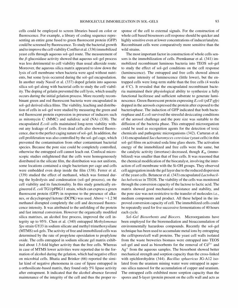
BIOMOLECULE IMMOBILIZATION IN SOL–GELS 93
cells could be employed to screen libraries based on color orfluorescence. For example, a library of coding sequence repre-senting an entire gene fused to green fluorescent protein (GFP)could be screened by fluorescence. To study the bacterial growthand to improve the cell viability Conffier et al. (336) immobilizedyeast cells through aqueous sol–gel route. The measurement ofthe β-glucosidase activity showed that aqueous sol–gel processwas less detrimental to cell viability than usual alkoxide route.Moreover, the aqueous silica matrix appeared to slow down thelysis of cell membrane when bacteria were aged without nutri-ents, but some lysis occurred during the sol–gel encapsulation.In another study Nassif et al. (337) doped gelatin into aqueoussilica sol–gel along with bacterial cells to study the cell viabil-ity. The doping of gelatin prevented the cell lysis, which usuallyoccurs during the initial gelation process. More recently recom-binant green and red fluorescent bacteria were encapsulated insol–gel derived silica films. The viability, leaching and distribu-tion of cells in sol–gel were studied by measuring the green andred fluorescent protein expression in presence of inducers suchas mitomycin C (MMC) and nalidixic acid (NA) (338). Theencapsulated E.coli cells maintained long-term viability with-out any leakage of cells. Even dead cells also showed fluores-cence, due to the perfect caging nature of sol–gel. In addition, thecell proliferation was also controlled by the sol–gel cage, whichprevented the contamination from other contaminant bacterialspecies. Because the pore size could be completely controlled,otherwise the entrapped cell cannot leach out. Confocal micro-scopic studies enlightened that the cells were homogeneouslydistributed in the silicate film, the distribution was not uniform,some locations contained a single bacterium per cage and cellswere embedded even deep inside the film (338). Ferrer et al.(339) studied the effect of methanol, which was formed dur-ing the hydrolysis and condensation (sol–gel process), on thecell viability and its functionality. In this study genetically en-gineered E. coli TG1/pPBG11 strain, which can express a greenfluorescent protein (GFP) in response to the presence of alka-nes, or dicyclopropyl ketone (DCPK) was used. Above ∼1.2 Mmethanol disrupted completely the cell and decreased fluores-cence intensity. It was attributed to the unfolding of the proteinand fast internal conversion. However the organically modifiedsilica matrixes, an alcohol free process, improved the cell in-tegrity up to 95%. Chen et al. (340) entrapped MethylomonasSps strain GYJ3 in sodium silicate and methyl trimethoxysilane(MTMS) sol-gels. The activity of free and immobilized cells wasdetermined by the rate of propylene epoxidation to proplyleneoxide. The cells entrapped in sodium silicate gel matrix exhib-ited about 1.5-fold higher activity than the free cells. Whereasin case of MTMS lower activities were reported due to the for-mation of alcohol during the gelation, which had negative effecton microbial cells. Bhatia and Brinker (66) reported the simi-lar kind of negative phenomena in case of lipase entrapped ina orthosilicate-based matrix, they found only 5% lipase activityafter entrapment. It indicated that the alcohol absence favoredmaintenance of the integrity of the cell and thus the proper re-
sponse of the cell to external signals. For the construction ofwhole cell based biosensors cell response should be quicker andhighly sensitive even at lower concentrations of inducer/analyte.Recombinant cells were comparatively more sensitive than thewild strains.
The most important factor in construction of whole cells sen-sors is the immobilization of cells. Premkumar et al. (341) im-mobilized recombinant luminous bacteria into TEOS sol–gelto study the effect of sol–gel conditions on the cell response(luminescence). The entrapped and free cells showed almostthe same intensity of luminescence (little lower), but the en-trapped cells were long-term stable than the free cells (4 weeksat 4◦C). It revealed that the encapsulated recombinant bacte-ria maintained their physiological ability to synthesize a fullyfunctional luciferase and sufficient substrate to generate lumi-nescence. Green fluorescent protein expressing E.coli (pET-gfp)dopped in the aerosols expressed the protein after exposed to thebacteriophase. The induction of GFP indicated that both bacte-riophase and E.coli survived the stressful desiccating conditionsof the aerosol challenge and the pore size was suitable to thediffusion of the bacteria phase. This entrapped microorganismcould be used as recognition agents for the detection of toxicchemicals and pathogenic microrganisms (342). Carturan et al.(14) encapsulated Saccharomyces cerevisiae (yeast cells) in thinsol–gel films on activated soda lime glass sheets. The activationenergy of the immobilized and free cells were the same, butthe catalytic activity (invertase) decreased, though Km (immo-bilized) was smaller than that of free cells. It was reasoned thatthe chemical modification of the biocatalyst, involving the inter-action of cell membrane with the Si-OH groups. They observedcell aggregation inside the gel layer due to the reduced dispersionof the yeast cells. Betancur et al. (343) encapsulated Lactobacil-lus helveticus in TEOS. The viability of the cells was monitoredthrough the conversion capacity of the lactose to lactic acid. Thematrix showed good mechanical resistance and stability, andthe pore size was not restrictive to the transfer of molecules i.e.,medium components and product. All these helped in the im-proved conversion capacity of cell. The immobilized cells couldbe repeatedly used for five successive fermentations of 40 hourseach cycle.
Sol–Gel Biosorbents and Biocers. Microorganisms havebeen employed for the bioremediation and bioaccumulation ofenvironmentally hazardous compounds. Recently the sol–geltechnique has been used to accumulate metal ions by entrappingthe cell/spores/cell wall proteins. The yeast cell walls isolatedfrom the waste breweries biomass were entrapped into TEOSsol–gel and used as biosorbents for the removal of Cd2+ andAg+ from the aqueous samples. The biosorbent showed bettermechanical strength and sorption capacity than the cross-linkedwith epichlorohydrin (344). Bacillus sphaericus JG-A12 iso-lated from the uranium mining water were entrapped in aque-ous silica nanosol for the accumulation of copper and uranium.The entrapped cells exhibited more sorption capacity than thespores and S-layer (protein present on the cells wall and acts as
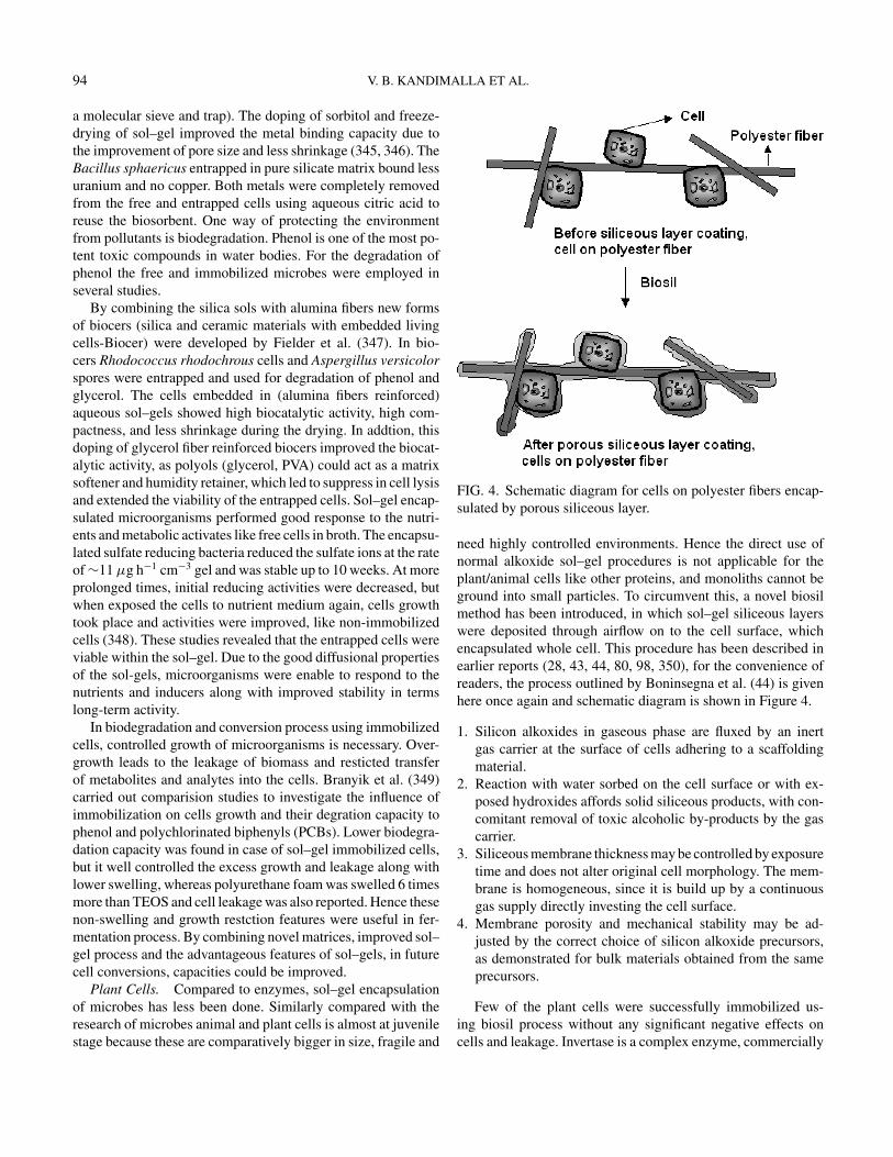
94 V. B. KANDIMALLA ET AL.
a molecular sieve and trap). The doping of sorbitol and freeze-drying of sol–gel improved the metal binding capacity due tothe improvement of pore size and less shrinkage (345, 346). TheBacillus sphaericus entrapped in pure silicate matrix bound lessuranium and no copper. Both metals were completely removedfrom the free and entrapped cells using aqueous citric acid toreuse the biosorbent. One way of protecting the environmentfrom pollutants is biodegradation. Phenol is one of the most po-tent toxic compounds in water bodies. For the degradation ofphenol the free and immobilized microbes were employed inseveral studies.
By combining the silica sols with alumina fibers new formsof biocers (silica and ceramic materials with embedded livingcells-Biocer) were developed by Fielder et al. (347). In bio-cers Rhodococcus rhodochrous cells and Aspergillus versicolorspores were entrapped and used for degradation of phenol andglycerol. The cells embedded in (alumina fibers reinforced)aqueous sol–gels showed high biocatalytic activity, high com-pactness, and less shrinkage during the drying. In addtion, thisdoping of glycerol fiber reinforced biocers improved the biocat-alytic activity, as polyols (glycerol, PVA) could act as a matrixsoftener and humidity retainer, which led to suppress in cell lysisand extended the viability of the entrapped cells. Sol–gel encap-sulated microorganisms performed good response to the nutri-ents and metabolic activates like free cells in broth. The encapsu-lated sulfate reducing bacteria reduced the sulfate ions at the rateof ∼11 µg h−1 cm−3 gel and was stable up to 10 weeks. At moreprolonged times, initial reducing activities were decreased, butwhen exposed the cells to nutrient medium again, cells growthtook place and activities were improved, like non-immobilizedcells (348). These studies revealed that the entrapped cells wereviable within the sol–gel. Due to the good diffusional propertiesof the sol-gels, microorganisms were enable to respond to thenutrients and inducers along with improved stability in termslong-term activity.
In biodegradation and conversion process using immobilizedcells, controlled growth of microorganisms is necessary. Over-growth leads to the leakage of biomass and resticted transferof metabolites and analytes into the cells. Branyik et al. (349)carried out comparision studies to investigate the influence ofimmobilization on cells growth and their degration capacity tophenol and polychlorinated biphenyls (PCBs). Lower biodegra-dation capacity was found in case of sol–gel immobilized cells,but it well controlled the excess growth and leakage along withlower swelling, whereas polyurethane foam was swelled 6 timesmore than TEOS and cell leakage was also reported. Hence thesenon-swelling and growth restction features were useful in fer-mentation process. By combining novel matrices, improved sol–gel process and the advantageous features of sol–gels, in futurecell conversions, capacities could be improved.
Plant Cells. Compared to enzymes, sol–gel encapsulationof microbes has less been done. Similarly compared with theresearch of microbes animal and plant cells is almost at juvenilestage because these are comparatively bigger in size, fragile and
FIG. 4. Schematic diagram for cells on polyester fibers encap-sulated by porous siliceous layer.
need highly controlled environments. Hence the direct use ofnormal alkoxide sol–gel procedures is not applicable for theplant/animal cells like other proteins, and monoliths cannot beground into small particles. To circumvent this, a novel biosilmethod has been introduced, in which sol–gel siliceous layerswere deposited through airflow on to the cell surface, whichencapsulated whole cell. This procedure has been described inearlier reports (28, 43, 44, 80, 98, 350), for the convenience ofreaders, the process outlined by Boninsegna et al. (44) is givenhere once again and schematic diagram is shown in Figure 4.
1. Silicon alkoxides in gaseous phase are fluxed by an inertgas carrier at the surface of cells adhering to a scaffoldingmaterial.
2. Reaction with water sorbed on the cell surface or with ex-posed hydroxides affords solid siliceous products, with con-comitant removal of toxic alcoholic by-products by the gascarrier.
3. Siliceous membrane thickness may be controlled by exposuretime and does not alter original cell morphology. The mem-brane is homogeneous, since it is build up by a continuousgas supply directly investing the cell surface.
4. Membrane porosity and mechanical stability may be ad-justed by the correct choice of silicon alkoxide precursors,as demonstrated for bulk materials obtained from the sameprecursors.
Few of the plant cells were successfully immobilized us-ing biosil process without any significant negative effects oncells and leakage. Invertase is a complex enzyme, commercially
BIOMOLECULE IMMOBILIZATION IN SOL–GELS 95
produced through bacterial fermentation. The disadvantage as-sociated with bacterial invertase is sometimes contaminated withnoxious proteins and presence of prions. The potential alterna-tive for this is production of invertase through the plant cells.Pressi et al. (82) immobilized Ajuga reptans AYN-IRB1 plantcells by biosil method for the invertase production. Ajuga reptanscells were isolated from the callus, which was propagated thor-ough the plant tissue culture and subjected into growth medium.During the second day of growth, fiber disks were added intothe cell suspension for the anchorage. After growth for 7 days,the disks were washed and exposed to an air flow saturatedby a 60/30/10 volumetric mixture of Si(OEt)4, CH3Si(OEt)3
and CH3SiH(OEt)2 for 20 minutes ensuring the deposition ofsiliceous layer on the cell surface. The resulted encapsulatedcells were viable up to 70–75%. Encasulation prevented thecell leakage and suppressed the cell repoduction. About a 40-fold increase in invertase production was reported using biosilencapsulated plant cells. Hata et al. (351) immobilized photo-synthetic pigment chlorophyll a into silica surfactant nanocom-posites films. Chlorophyll a was stable against visible light incomparison with a homogeneous chlorophyll a solution systemeven under an air atmosphere. In another study Carturan et al.(13) immobilized Catharanthus roseus plant cells through biosilmethod for the production of alkaloids vincristin and vinblastin.Most interestingly the immobilized cells were stable up to 100days with continuos production of alkolid. The productivity ofencapsulated cells is two orders of magnitude higher than freecells.
Animal Cells. One of the major challenges of microencap-sulation techniques is to immobilize living cells behind a semipermeable membrane thereby allowing their transplantation. Inrecent years transplantation therapy has emerged as a promisingpotential treatment strategy for curing diseases. For the efficicenttrnasplantation the matrix used for the encapsulation should havegood permeability, which allows the diffusion of small substratesand products, should avoid diffusion of immune system agents,high mechanical strength and non-immunorejection (352). Thesilica and sol–gel materials are biocompatible (nonimmunore-jection). Mechanical features of sol–gel SiO2 coatings in pres-ence of entrapped animal cells was studied by Sglavo et al. (350).In view of the nonimmunorejection and stability of the sol–gelfilms pancreatic islets, which can produce insulin, were immobi-lized thorough biosil process (353). The encapsulated cells wereable to produce insulin up to 3 weeks in vitro conditions. For thein vivo studies encapsulated islets were transplanted peritoneallyin several non-obese diabetic mice. After transplantation of isletsurinary excretion of glucose fell to almost zero with in few days.With similar way Boninsegna et al. (44) immobilized rat pancre-atic islets in siliceous layer and transplanted them into diabeticrats. The encapsulated islets showed long-term constant levelsof glycemia with about immunorejection compared with nonen-casluated islets.
Several proteins, plant and animal cells have been immobi-lized in calcium alginate beads, however the major drawback in
this method is instability of beads in presence of citrate ions andeasy disintegration at simple shear stress. Boninsegna et al. (81)developed alginate silica microcapsules.
In this procedure animal cells or cell aggregates were sus-pended in Na-alginate solution and extruded under a flow ofair saturated with silicon alkoxides. The immediate formationof a sol–gel siliceous membrane on the microdrop surface al-lowed the reaction with Ca2+, leading to solid Ca-alginate andyielding homogeneous, stable microcapsules of 200 µm in di-ameter. In this method the alginate droplet size was controlledby flowing air at the tip of the extrusion nozzle. Human hepa-toblastoma (HepG2) and human T leukaemia Jurkat cells havebeen encapsulated in alginate silica microsperes for the produc-tion of albumin (HSA) human interleukin 2 (IL2), respectively.The important findings in this study were outer siliceous layeracted as a physical barrier and provided more protective cageto calcium-alginate microcapsules. The siliceous layer was sta-ble and preserved the entrapped cells from leakage. Perhapsnon-coated alginate microcapsules were rapidly dissolved. Thesiliceous coating on capsules did not interfere with bioactivity ofthe cells or diffusion of the products, it only provided mechani-cal and chemical stability to the calcium-alginate microcapsules(52).
CONCLUSIONSThe functionality of immobilized biomolecules is governed
mainly by the nature of the biomolecule, the preparation method,as well as the nature and structure of the immobilisation ma-trix. Among the most important properties of the encapsulatedenzymes, good accessibility to analytes and long-term stabil-ity for both operation and storage are prime essentials. Manykinds of bio-components have been preferentially immobilizedby both neat and hybrid silicate gel-processing methods to de-velop sol–gel-derived biomaterials, and this should be intensi-fied in the future. While most sol–gel bioencapsulates reportedto date have used inorganic materials or carbon composite de-rived from either tetramethylorthosilicate (TMOS) or tetraethy-lorthosilicate (TEOS). Even though some other precursors havebeen introduced, still newer more milder sol–gel derived mate-rials are highly desired for improving the analytical capabilitiesof biosensing devices, and for meeting the need of practical ap-plication and commercial production. Maybe the new hybridsol–gel materials and/or methods could provide more electro-chemically active, less brittle nature and biocompatible matrices.For the long-term use and reduction of film fouling though mi-crobial growth or adhesion, new matrices that can inhibit thegrowth have to be explored. This can be possible by developingthe new sol–gel precursor molecules, which are modified withmicrobial inhibition compounds or metal ions.
Even though the plant and animal cells and spores of microbescan be immobilized in silica matrixes, still the main drawback istheir reduced viability. Towards this direction more basic studiesare needed for the potential use of sol–gel advantages and forthe industrial/biotechnological application of cells. More mild,
96 V. B. KANDIMALLA ET AL.
stable gelation conditions should be developed to improve thecells’ viability. However, the studies such as interactions be-tween the sol–gel matrix and whole cells and metabolic changesduing immobilization have to be closely monitorrd for the ex-ploration of new matrices and methods. Such new sol–gel meth-ods/matrices can improve the productivities and be used to de-sign the novel whole cell biosensors and spwan the medicalapplications. Furthermore cells/proteins immobilized in sol–gelmatices can be effectively applied in biochemistry, biotechnol-ogy, environmental monitoring, instrumentation, forensic sci-ence, laboratory automation and pharmaceutical chemistry formultiple applications with practical significance in near future.
ACKNOWLEDGMENTSWe are gratefully acknowledge the financial support of the
Distinguished Young Scholar Fund to HX Ju (20325518), theNational Natural Science Foundation of China (20275017).Kandimalla VB and Tripathi VS are highly thankful to Nan-jing University for providing postdoctoral fellowships.
REFERENCES1. J. Wang, DNA biosensors based on peptide nucleic acid (PNA)
recognition layers. A review. Biosensors and Bioelectronics 13(1998):757–762.
2. S. F. D’Souza, Microbial biosensors. Biosensors and Bioelectron-ics 16 (2001):337–353.
3. B. Alessandra, S. A. Piletsky, P. G. Righetti, and A. P. F., Turner,Capillary electrophoresis coupled to biosensor detection. Journalof Chromatography A 892 (2000):143–153.
4. C. R. Suri, M. Raje, and G. C. Varshney, Immunosensors forpesticide analysis: antibody production and sensor development.Critical Reviews in Biotechnology 22 (2002):15–32.
5. J. E. Pearson, A. Gill, and P. V. Vadgama, Analytical aspects ofbiosensors. Annals of Clinical Biochemistry 37 (2000):119–145.
6. B. X. Li, Z. J. Zhang, and Y. Jin, Plant tissue-based chemilumi-nescence flow biosensor for glycolic acid. Analytical Chemistry73 (2001):1203–1206.
7. M. S. Thakur and N. G. Karanth, Research and development onbiosensors for food analysis in India, in Advances in Biosensorseds. B. D. Malhotra and A. P. F. Turner (Elsevier Science, BV,2003), ch. 5, 131–160.
8. V. B. Kandimalla and H. X. Ju, New horizons with a multidi-mensional tool for applications in analytical chemistry-Aptamer.Analytical Letters 37 (2004):1–19.
9. G. Wulff, Enzyme like catalysis by molecularly imprinted poly-mers. Chemical Reviews 102 (2002):1–27.
10. V. B. Kandimalla and H. X. Ju, Molecular imprinting—A dynamictechnique for multifarious applications in analytical chemistry.Analytical and Bioanalytical Chemistry 380 (2004):587–605.
11. O. Hayden and F. L Dickert, Selective microorganism detectionwith cell surface imprinted polymers. Advanced Materials 13(2001):1480–1483.
12. T. H. Park and M. L. Shuler, Integration of cell culture and micro-fabrication technology. Biotechnology Progress 19 (2003):243–253
13. G. Carturan, R. D. Monte, G. Pressi, S. Secondin, and P. Verza,Production of valuable drugs from plant cells immobilized by
hybrid sol–gel SiO2. Journal of Sol–Gel Science and Technology13 (1998):273–276.
14. G. Carturan, R. Campostrini, S. Dire, V. Scardi, and E. D. Alteriis,Inorganic gels for immobilization of biocatalysts:Inclusion of in-vertase active whole cells of yeast (Saccharomyces cerevisiae)into thin layers of SiO2 gel deposited on glass sheets. Journal ofMolecular Catalysis 57 (1989):L13–L16.
15. V. B. Kandimalla, K. R. S. Sambasivarao, and C. Ayyanna, Im-mobilized whole cell systems for antibiotics production. AsianJournal of Microbiology Biotechnology and Environmental Sci-ences 6 (2004):741–751.
16. N. A. Chaniotakis, Enzyme stabilization strategies based on elec-trolytes and polyelectrolytes for biosensor applications. Analyti-cal and Bioanalytical Chemistry 378 (2002):89–95.
17. T. Bes, C. Gomez–Moreno, J. M. Guisan, and R. Fernandez–Lafuente, Selective enzymatic oxidations: stabilization by multi-point covalent attachment of ferredoxin NAD-reductase: an inter-esting cofactor recycling enzyme. Journal of Molecular Catalysis98 (1995):161–169.
18. R. Fernandez–Lafuente, V. Rodriguez, and J. M. Guisan, Thecoimmobilization of D-amino acid oxidase and catalase en-ables the quantitative transformation of D-amino acids (D-phenylalanine) into keto acids (phenylpyruvic acid). Enzyme Mi-crobial Technology 23 (1998):28–33.
19. M. N. Gupta, Thermostabilization of proteins. Biotechnology andApplied Biochemistry 14 (1991):1–11.
20. L. G. Rody and V. A. Shanke, Immobilized nucleases. CriticalReviews in Biotechnology 13 (1983):255–273.
21. J. M. Palomo, G. Munoz, G. Fernandez-Lorente, C. Mateo, R.Fernandez-Lafuente, and J. M. Guisan. Interfacial adsorption oflipases on very hydrophobic support (octadecyl–Sepabeads): im-mobilization, hyperactivation and stabilization of the open formof lipases. Journal of Molecular Catalysis B: Enzymatic 19–20(2002):279–286.
22. C. Mateo, O. Abian, R. Fernandez–Lafuente, and J. M. Guisan,Increase in conformational stability of enzymes immobilizedon epoxy-activated supports by favoring additional multipointcovalent attachment. Enzyme and Microbial Technology 26(2000):509–515.
23. S. Birgit, R. Annie, and P. U. Abel, Influence of polymerizationparameters and entrapment in poly(hydroxyethyl methacrylate)on activity and stability of GOD. Journal of Molecular CatalysisB: Enzymatic 7 (1999):85–91.
24. D. Avnir, S. Braun, L. Ovadia, and M. Ottolengthi, Enzymes andother proteins entrapped in sol–gel materials. Chemistry of Ma-terials 6 (1994):1605–1614.
25. B. C. Dave, B. Dunn, J. S. Valentine, and J. I. Zink, Sol–gelencapsulation method for biosensors. Analytical Chemistry 66(1994):1120A–1127A.
26. I. Gill, Biodoped nanocomposite polymers: Sol-gel bioencapu-lates. Chemistry of Materials 13 (2001):3404–3421.
27. M. M. Collinson, Recent trends in analytical application of organ-ically modified silicate matrices. Trends in Analytical Chemistry21 (2002):30–37.
28. W. Jin and J. D. Brenan, Properties and applications of proteinsencapsulated within sol–gel derived materials. Analytica ChimicaActa 461 (2002):1–36.
29. G. Carturan, R. D. Toso, S. Boninsegna, and R. D. Monte, Encap-sulation of functional cells by sol–gel silica: actial progress and
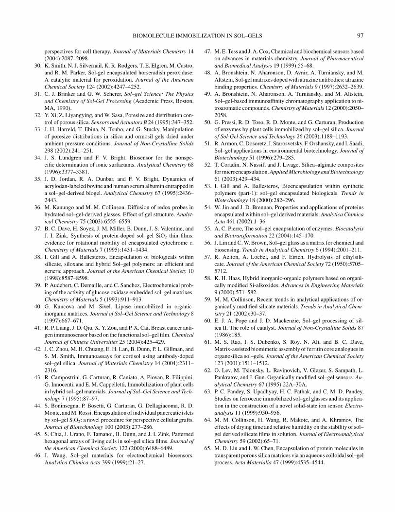
BIOMOLECULE IMMOBILIZATION IN SOL–GELS 97
perspectives for cell therapy. Journal of Materials Chemistry 14(2004):2087–2098.
30. K. Smith, N. J. Silvernail, K. R. Rodgers, T. E. Elgren, M. Castro,and R. M. Parker, Sol-gel encapsulated horseradish peroxidase:A catalytic material for peroxidation. Journal of the AmericanChemical Society 124 (2002):4247–4252.
31. C. J. Brinker and G. W. Scherer, Sol–gel Science: The Physicsand Chemistry of Sol-Gel Processing (Academic Press, Boston,MA, 1990).
32. Y. Xi, Z. Liyangying, and W. Sasa, Poresize and distribution con-trol of porous silica. Sensors and Actuators B 24 (1995):347–352.
33. J. H. Harreld, T. Ebina, N. Tsubo, and G. Stucky, Manipulationof poresize distributions in silica and ormosil gels dried underambient pressure conditions. Journal of Non-Crystalline Solids298 (2002):241–251.
34. J. S. Lundgren and F. V. Bright. Biosensor for the nonspe-cific determination of ionic surfactants. Analytical Chemistry 68(1996):3377–3381.
35. J. D. Jordan, R. A. Dunbar, and F. V. Bright, Dynamics ofacrylodan-labeled bovine and human serum albumin entrapped ina sol–gel-derived biogel. Analytical Chemistry 67 (1995):2436–2443.
36. M. Kanungo and M. M. Collinson, Diffusion of redox probes inhydrated sol–gel-derived glasses. Effect of gel structure. Analyt-ical Chemistry 75 (2003):6555–6559.
37. B. C. Dave, H. Soyez, J. M. Miller, B. Dunn, J. S. Valentine, andJ. I. Zink, Synthesis of protein-doped sol–gel SiO2 thin films:evidence for rotational mobility of encapsulated cytochrome c.Chemistry of Materials 7 (1995):1431–1434.
38. I. Gill and A. Ballesteros, Encapsulation of biologicals withinsilicate, siloxane and hybrid Sol–gel polymers: an efficient andgeneric approach. Journal of the American Chemical Society 10(1998):8587–8598.
39. P. Audebert, C. Demaille, and C. Sanchez, Electrochemical prob-ing of the activity of glucose oxidase embedded sol–gel matrixes.Chemistry of Materials 5 (1993):911–913.
40. G. Kuncova and M. Sivel. Lipase immobilized in organic-inorganic matrices. Journal of Sol–Gel Science and Technology 8(1997):667–671.
41. R. P. Liang, J. D. Qiu, X. Y. Zou, and P. X. Cai, Breast cancer anti-gen immunosensor based on the functional sol–gel film. ChemicalJournal of Chinese Universities 25 (2004):425–429.
42. J. C. Zhou, M. H. Chuang, E. H. Lan, B. Dunn, P. L. Gillman, andS. M. Smith, Immunoassays for cortisol using antibody-dopedsol–gel silica. Journal of Materials Chemistry 14 (2004):2311–2316.
43. R. Campostrini, G. Carturan, R. Caniato, A. Piovan, R. Filippini,G. Innocenti, and E. M. Cappelletti, Immobilization of plant cellsin hybrid sol–gel materials. Journal of Sol–Gel Science and Tech-nology 7 (1995):87–97.
44. S. Boninsegna, P. Bosetti, G. Carturan, G. Dellagiacoma, R. D.Monte, and M. Rossi. Encapsulation of individual pancreatic isletsby sol–gel SiO2: a novel procedure for perspective cellular grafts.Journal of Biotechnology 100 (2003):277–286.
45. S. Chia, J. Urano, F. Tamanoi, B. Dunn, and J. I. Zink, Patternedhexagonal arrays of living cells in sol–gel silica films. Journal ofthe American Chemical Society 122 (2000):6488–6489.
46. J. Wang, Sol–gel materials for electrochemical biosensors.Analytica Chimica Acta 399 (1999):21–27.
47. M. E. Tess and J. A. Cox, Chemical and biochemical sensors basedon advances in materials chemistry. Journal of Pharmaceuticaland Biomedical Analysis 19 (1999):55–68.
48. A. Bronshtein, N. Aharonson, D. Avnir, A. Turniansky, and M.Altstein, Sol-gel matrixes doped with atrazine antibodies: atrazinebinding properties. Chemistry of Materials 9 (1997):2632–2639.
49. A. Bronshtein, N. Aharonson, A. Turniansky, and M. Altstein,Sol–gel-based immunoaffinity chromatography application to ni-troaromatic compounds. Chemistry of Materials 12 (2000):2050–2058.
50. G. Pressi, R. D. Toso, R. D. Monte, and G. Carturan, Productionof enzymes by plant cells immobilized by sol–gel silica. Journalof Sol-Gel Science and Technology 26 (2003):1189–1193.
51. R. Armon, C. Dosoretz, J. Starosvetsky, F. Orshansky, and I. Saadi,Sol–gel applications in environmental biotechnology. Journal ofBiotechnology 51 (1996):279–285.
52. T. Coradin, N. Nassif, and J. Livage, Silica–alginate compositesfor microencapsulation. Applied Microbiology and Biotechnology61 (2003):429–434.
53. I. Gill and A. Ballesteros, Bioencapsulation within syntheticpolymers (part-1): sol–gel encapsulated biologicals. Trends inBiotechnology 18 (2000):282–296.
54. W. Jin and J. D. Brennan, Properties and applications of proteinsencapsulated within sol–gel derived materials. Analytica ChimicaActa 461 (2002):1–36.
55. A. C. Pierre, The sol–gel encapsulation of enzymes. Biocatalysisand Biotransformation 22 (2004):145–170.
56. J. Lin and C. W. Brown, Sol–gel glass as a matrix for chemical andbiosensing. Trends in Analytical Chemistry 6 (1994):2001–211.
57. R. Aelion, A. Loebel, and F. Eirich, Hydrolysis of ethylsili-cate. Journal of the American Chemical Society 72 (1950):5705–5712.
58. K. H. Haas, Hybrid inorganic-organic polymers based on organi-cally modified Si-alkoxides. Advances in Engineering Materials9 (2000):571–582.
59. M. M. Collinson, Recent trends in analytical applications of or-ganically modified silicate materials. Trends in Analytical Chem-istry 21 (2002):30–37.
60. E. J. A. Pope and J. D. Mackenzie, Sol–gel processing of sil-ica II. The role of catalyst. Journal of Non-Crystalline Solids 87(1986):185.
61. M. S. Rao, I. S. Dubenko, S. Roy, N. Ali, and B. C. Dave,Matrix-assisted biomimetic assembly of ferritin core analogues inorganosilica sol–gels. Journal of the American Chemical Society123 (2001):1511–1512.
62. O. Lev, M. Tsionsky, L. Ravinovich, V. Glezer, S. Sampath, L.Pankratov, and J. Gun. Organically modified sol–gel sensors. An-alytical Chemistry 67 (1995):22A–30A.
63. P. C. Pandey, S. Upadhyay, H. C. Pathak, and C. M. D. Pandey,Studies on ferrocene immobilized sol–gel glasses and its applica-tion in the construction of a novel solid-state ion sensor. Electro-analysis 11 (1999):950–956.
64. M. M. Collinson, H. Wang, R. Makote, and A. Khramov, Theeffects of drying time and relative humidity on the stability of sol–gel derived silicate films in solution. Journal of ElectroanalyticalChemistry 59 (2002):65–71.
65. M. D. Liu and I. W. Chen, Encapsulation of protein molecules intransparent porous silica matrices via an aqueous colloidal sol–gelprocess. Acta Materialia 47 (1999):4535–4544.
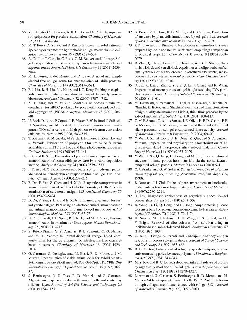
98 V. B. KANDIMALLA ET AL.
66. R. B. Bhatia, C. J. Brinker, A. K. Gupta, and A. P. Singh, Aqueoussol–gel process for protein encapsulation. Chemistry of Materials12 (2000):2434–2441.
67. M. T. Reetz, A. Zonta, and S. Kamp, Efficient immobilization oflipases by entrapment in hydrophobic sol–gel materials. Biotech-nology and Bioengineering 49 (1996):527–534.
68. A. Coiffier, T. Coradin, C. Roux, O. M. Bouvet, and J. Livage, Sol-gel encapsulation of bacteria: comparison between alkoxide andaqueous routes. Journal of Materials Chemistry 11 (2001):2039–2044.
69. M. L. Ferrer, F. del Monte, and D. Levy, A novel and simplealcohol-free sol–gel route for encapsulation of labile proteins.Chemistry of Materials 14 (2002):3619–3621.
70. Z. J. Liu, B. H. Liu, J. L. Kong, and J. Q. Deng, Probing trace phe-nols based on mediator-free alumina sol–gel derived tyrosinasebiosensor. Analytical Chemistry 72 (2000):4707–4712.
71. Z. T. Jiang and Y. M Zuo, Synthesis of porous titania mi-crospheres for HPLC packings by polymerization-induced col-loid aggregation (PICA). Analytical Chemistry 73 (2001):686–688.
72. U. Bach, D. Lupo, P. Comte, J. E. Moser, F. Weissortel, J. Salbeck,H. Spreitzer, and M. Gratzel, Solid-state dye-sensitized meso-porous TiO2 solar cells with high photon-to-electron conversionefficiencies. Nature 395 (1998):583–585.
73. T. Akiyama, A, Miyazaki, M.Sutoh, I. Ichinose, T. Kunitake, andS. Yamada. Fabrication of porphyrin–titanium oxide–fullereneassemblies on an ITO electrode and their photocurrent responses.Colloids Surface:A 169 (2000):137–141.
74. J. Yu and H. X. Ju, Preparation of porous titania sol–gel matrix forimmobilization of horseradish peroxidase by a vapor depositionmethod. Analytical Chemistry 74 (2002):3579–3583.
75. J. Yu and H. X. Ju, Amperometric biosensor for hydrogen perox-ide based on hemolgobin entrapped in titania sol–gel film. Ana-lytica Chimica Acta 486 (2003):209–216.
76. Z. Dai, F. Yan, Z. Chen, and H. X. Ju, Reagentless amperomerticimmunosensor based on direct electrochemistry of HRP for de-termination of carcinoma antigen-125. Analytical Chemistry 75(2003):5429–5434.
77. D. Du, F. Yan, S. Liu, and H. X. Ju, Immunological assay for car-bohydrate antigen 19-9 using an electrochemical immunosensorand antigen immobilization in titania sol–gel matrix. Journal ofImmunological Methods 283 (2003):67–75.
78. H. R. Luckarift, J. C. Spain, R. J. Naik, and M. O. Stone, Enzymeimmobilization in biomimetic silica supports. Nature Biotechnol-ogy 22 (2004):211–213.
79. B. Prieto-Simon, G. S. Armatas, P. J. Pomonis, C. G. Nanos,and M. I. Prodromidis. Metal-dispersed xerogel-based com-posite films for the development of interference free oxidase-based biosensors. Chemistry of Materials 16 (2004):1026–1034.
80. G. Carturan, G. Dellagiacoma, M. Rossi, R. D. Monte, and M.Muraca, Encapsulation of viable animal cells for hybrid bioarti-ficial organs by the Biosil method; Sol–Gel Optics IV. SPIE. TheInternational Society for Optical Engineering 3136 (1997):366–373.
81. S. Boninsegna, R. D. Taso, R. D. Montel, and G. Carturan,Alginate microspheres loaded with animal cells and coated bysilicious layer. Journal of Sol-Gel Science and Technology 26(2003):1154–1157.
82. G. Pressi, R. D. Toso, R. D. Monte, and G. Carturan, Productionof enzymes by plant cells immobilized by sol–gel silica. Journalof Sol-Gel Science and Technology 26 (2003):1189–193.
83. P. T. Tanev and T. J. Pinnavaia, Mesoporous silica molecular sievesprepared by ionic and neutral surfactant templating: comparisonof physical properties. Chemistry of Materials 8 (1996):2068–2079.
84. D. Zhao, Q. Huo, J. Feng, B. F. Chmelka, and G. D. Stucky, Non-ionic triblock and star diblock copolymer and oligomeric surfac-tant syntheses of highly ordered, hydrothermally stable, meso-porous silica structures. Journal of the American Chemical Soci-ety 120 (1998):6024–6036.
85. Q. Jie, K. Lin, J. Zhong, Y. Shi, Q. Li, J. Chang and R. Wang,Preparation of macro porous sol–gel bioglasses using PVA parti-cles as pore former. Journal of Sol–Gel Science and Technology30 (2004):49–61.
86. M. Takahashi, K. Yamauchi, T. Yagi, A. Nishiwaki, K. Wakita, N.Ohnishi, K. Hotta, and I. Shashi, Preparation and characterizationof high-quality stoichiometric LiNbO3 thick films prepared by thesol–gel method. Thin Solid Films 458 (2004):108–113.
87. C. M. F. Soares, O. A. dos Santos, J. E. Olivo, H. F. De Castro, F. F.de Moraes, and G. M. Zanin, Influence of the alkyl substitutedsilane precursor on sol–gel encapsulated lipase activity. Journalof Molecular Catalysis: B Enzymatic 29 (2004):69–70.
88. Y. Wei, J. Xu, H. Dong, J. H. Dong, K. Qiu, and S. A. Jansen-Varnum, Preparation and physisorption characterization of D-glucose-templated mesoporous silica sol–gel materials. Chem-istry of Materials 11 (1999):2023–2029.
89. Y. Wei, J. Xu, Q. Feng, H. Dong, and M. Lin, Encapsulation ofenzymes in meso porous host materials via the nonsurfactant-templated sol–gel process. Materials Letters 44 (2000):6–11.
90. C. J. Brinker and G. W. Scherer, Sol–gel science: The physics andchemistry of sol–gel processing (Academic Press, San Diego, CA,1990).
91. B. Dunn and J. I. Zink, Probes of pore environment and molecule-matrix interactions in sol–gel materials. Chemistry of Materials9 (1997):2280–2291.
92. O. Lev, Diagnostic applications of organically doped sol–gelporous glass. Analusis 20 (1992):543–553.
93. B. Wang, B. Li, Q. Deng, and S. Dong. Amperometric glucosebiosensor based on sol–gel organic-inorganic hybrid material. An-alytical Chemistry 70 (1998):3170–3174.
94. U. Narang, M. H. Rahman, J. H. Wang, P. N. Prasad, and F.V. Bright. Removal of ribonucleases from solution using aninhibitor-based sol–gel-derived biogel. Analytical Chemistry 67(1995):1935–1939.
95. C. Roux, J. Livage, K. Farhati, and L. Monjour, Antibody-antigenreactions in porous sol–gel matrices. Journal of Sol-Gel Scienceand Technology 8 (1997):663–666.
96. D. L. Venton, Entrapment of a highly specific antiprogesteroneantiserum using polysiloxane copolymers. Biochimca et Biophys-ica Acta 797 (1984):343–347.
97. M. S. Rao and B. C. Dave, Selective intake and release of protinsby organically modified silica sol–gels. Journal of the AmericanChemical Society 120 (1998):13270–13271.
98. L. Armanini, G. Carturan, S. Boninsegna, R. D. Monte, and M.Muraca, SiO2 entrapment of animal cells. Part 2: Protein diffusionthrough collagen membranes coated with sol–gel SiO2. Journalof Materials Chemistry 9 (1999):3057–3060.
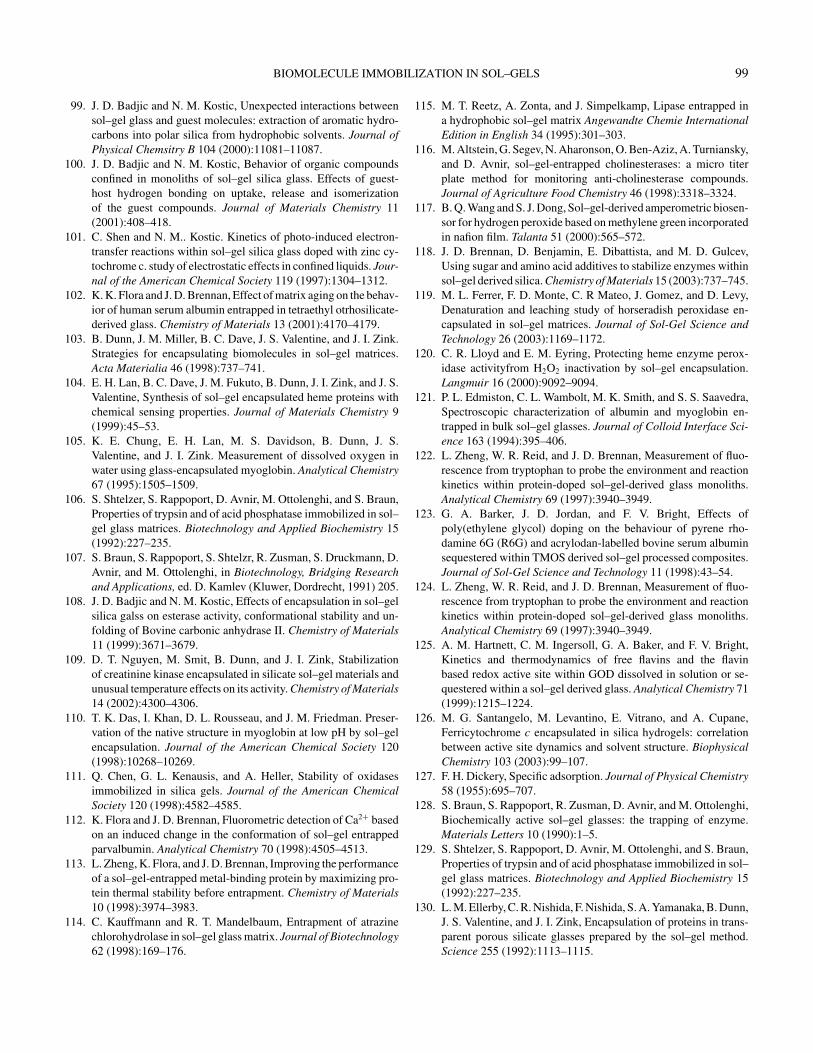
BIOMOLECULE IMMOBILIZATION IN SOL–GELS 99
99. J. D. Badjic and N. M. Kostic, Unexpected interactions betweensol–gel glass and guest molecules: extraction of aromatic hydro-carbons into polar silica from hydrophobic solvents. Journal ofPhysical Chemsitry B 104 (2000):11081–11087.
100. J. D. Badjic and N. M. Kostic, Behavior of organic compoundsconfined in monoliths of sol–gel silica glass. Effects of guest-host hydrogen bonding on uptake, release and isomerizationof the guest compounds. Journal of Materials Chemistry 11(2001):408–418.
101. C. Shen and N. M.. Kostic. Kinetics of photo-induced electron-transfer reactions within sol–gel silica glass doped with zinc cy-tochrome c. study of electrostatic effects in confined liquids. Jour-nal of the American Chemical Society 119 (1997):1304–1312.
102. K. K. Flora and J. D. Brennan, Effect of matrix aging on the behav-ior of human serum albumin entrapped in tetraethyl otrhosilicate-derived glass. Chemistry of Materials 13 (2001):4170–4179.
103. B. Dunn, J. M. Miller, B. C. Dave, J. S. Valentine, and J. I. Zink.Strategies for encapsulating biomolecules in sol–gel matrices.Acta Materialia 46 (1998):737–741.
104. E. H. Lan, B. C. Dave, J. M. Fukuto, B. Dunn, J. I. Zink, and J. S.Valentine, Synthesis of sol–gel encapsulated heme proteins withchemical sensing properties. Journal of Materials Chemistry 9(1999):45–53.
105. K. E. Chung, E. H. Lan, M. S. Davidson, B. Dunn, J. S.Valentine, and J. I. Zink. Measurement of dissolved oxygen inwater using glass-encapsulated myoglobin. Analytical Chemistry67 (1995):1505–1509.
106. S. Shtelzer, S. Rappoport, D. Avnir, M. Ottolenghi, and S. Braun,Properties of trypsin and of acid phosphatase immobilized in sol–gel glass matrices. Biotechnology and Applied Biochemistry 15(1992):227–235.
107. S. Braun, S. Rappoport, S. Shtelzr, R. Zusman, S. Druckmann, D.Avnir, and M. Ottolenghi, in Biotechnology, Bridging Researchand Applications, ed. D. Kamlev (Kluwer, Dordrecht, 1991) 205.
108. J. D. Badjic and N. M. Kostic, Effects of encapsulation in sol–gelsilica galss on esterase activity, conformational stability and un-folding of Bovine carbonic anhydrase II. Chemistry of Materials11 (1999):3671–3679.
109. D. T. Nguyen, M. Smit, B. Dunn, and J. I. Zink, Stabilizationof creatinine kinase encapsulated in silicate sol–gel materials andunusual temperature effects on its activity. Chemistry of Materials14 (2002):4300–4306.
110. T. K. Das, I. Khan, D. L. Rousseau, and J. M. Friedman. Preser-vation of the native structure in myoglobin at low pH by sol–gelencapsulation. Journal of the American Chemical Society 120(1998):10268–10269.
111. Q. Chen, G. L. Kenausis, and A. Heller, Stability of oxidasesimmobilized in silica gels. Journal of the American ChemicalSociety 120 (1998):4582–4585.
112. K. Flora and J. D. Brennan, Fluorometric detection of Ca2+ basedon an induced change in the conformation of sol–gel entrappedparvalbumin. Analytical Chemistry 70 (1998):4505–4513.
113. L. Zheng, K. Flora, and J. D. Brennan, Improving the performanceof a sol–gel-entrapped metal-binding protein by maximizing pro-tein thermal stability before entrapment. Chemistry of Materials10 (1998):3974–3983.
114. C. Kauffmann and R. T. Mandelbaum, Entrapment of atrazinechlorohydrolase in sol–gel glass matrix. Journal of Biotechnology62 (1998):169–176.
115. M. T. Reetz, A. Zonta, and J. Simpelkamp, Lipase entrapped ina hydrophobic sol–gel matrix Angewandte Chemie InternationalEdition in English 34 (1995):301–303.
116. M. Altstein, G. Segev, N. Aharonson, O. Ben-Aziz, A. Turniansky,and D. Avnir, sol–gel-entrapped cholinesterases: a micro titerplate method for monitoring anti-cholinesterase compounds.Journal of Agriculture Food Chemistry 46 (1998):3318–3324.
117. B. Q. Wang and S. J. Dong, Sol–gel-derived amperometric biosen-sor for hydrogen peroxide based on methylene green incorporatedin nafion film. Talanta 51 (2000):565–572.
118. J. D. Brennan, D. Benjamin, E. Dibattista, and M. D. Gulcev,Using sugar and amino acid additives to stabilize enzymes withinsol–gel derived silica. Chemistry of Materials 15 (2003):737–745.
119. M. L. Ferrer, F. D. Monte, C. R Mateo, J. Gomez, and D. Levy,Denaturation and leaching study of horseradish peroxidase en-capsulated in sol–gel matrices. Journal of Sol-Gel Science andTechnology 26 (2003):1169–1172.
120. C. R. Lloyd and E. M. Eyring, Protecting heme enzyme perox-idase activityfrom H2O2 inactivation by sol–gel encapsulation.Langmuir 16 (2000):9092–9094.
121. P. L. Edmiston, C. L. Wambolt, M. K. Smith, and S. S. Saavedra,Spectroscopic characterization of albumin and myoglobin en-trapped in bulk sol–gel glasses. Journal of Colloid Interface Sci-ence 163 (1994):395–406.
122. L. Zheng, W. R. Reid, and J. D. Brennan, Measurement of fluo-rescence from tryptophan to probe the environment and reactionkinetics within protein-doped sol–gel-derived glass monoliths.Analytical Chemistry 69 (1997):3940–3949.
123. G. A. Barker, J. D. Jordan, and F. V. Bright, Effects ofpoly(ethylene glycol) doping on the behaviour of pyrene rho-damine 6G (R6G) and acrylodan-labelled bovine serum albuminsequestered within TMOS derived sol–gel processed composites.Journal of Sol-Gel Science and Technology 11 (1998):43–54.
124. L. Zheng, W. R. Reid, and J. D. Brennan, Measurement of fluo-rescence from tryptophan to probe the environment and reactionkinetics within protein-doped sol–gel-derived glass monoliths.Analytical Chemistry 69 (1997):3940–3949.
125. A. M. Hartnett, C. M. Ingersoll, G. A. Baker, and F. V. Bright,Kinetics and thermodynamics of free flavins and the flavinbased redox active site within GOD dissolved in solution or se-questered within a sol–gel derived glass. Analytical Chemistry 71(1999):1215–1224.
126. M. G. Santangelo, M. Levantino, E. Vitrano, and A. Cupane,Ferricytochrome c encapsulated in silica hydrogels: correlationbetween active site dynamics and solvent structure. BiophysicalChemistry 103 (2003):99–107.
127. F. H. Dickery, Specific adsorption. Journal of Physical Chemistry58 (1955):695–707.
128. S. Braun, S. Rappoport, R. Zusman, D. Avnir, and M. Ottolenghi,Biochemically active sol–gel glasses: the trapping of enzyme.Materials Letters 10 (1990):1–5.
129. S. Shtelzer, S. Rappoport, D. Avnir, M. Ottolenghi, and S. Braun,Properties of trypsin and of acid phosphatase immobilized in sol–gel glass matrices. Biotechnology and Applied Biochemistry 15(1992):227–235.
130. L. M. Ellerby, C. R. Nishida, F. Nishida, S. A. Yamanaka, B. Dunn,J. S. Valentine, and J. I. Zink, Encapsulation of proteins in trans-parent porous silicate glasses prepared by the sol–gel method.Science 255 (1992):1113–1115.
100 V. B. KANDIMALLA ET AL.
131. N. Shibayama and S. Saigo, Kinetics of the allosteric transitionin hemoglobin with in silicate sol–gel. Journal of the AmericanChemical Society 121 (1999):444–445.
132. E. S. Peterson, R. Shinder, and I. Khan, Domain-specific effectorinteractions within the central cavity of human adult hemoglobinin solution and in porous sol–gel matrices: evidence for long-range communication pathways. Biochemistry 43 (2004):4832–4843.
133. G. Fiandaca, E. Vitrano, and A. Cupane, Ferricytochrome c encap-sulated in silica nanoparticles: structural stability and functionalproperties. Biopolymers 74 (2004):55–59.
134. B. Lillis, C. Grogan, H. Berney, and W. A. Lane, Investigation intoimmobilisation of lactate oxidase to improve stability. Sensorsand Actuators B 68 (2000):109–113.
135. A. N. Diaz, F. G. Sanchez, M. C. Ramos, and M. C. Torijas,Horseradish peroxidase sol–gel immobilized for chemilumines-cence measurements of alkaline-phosphatase activity. Sensorsand Actuators B 82 (2002):176–179.
136. U. Narang, P. Prasad, and F. V. Bright, A novel protocol to en-trap active urease in a tetraethoxysilane-derived sol–gel thin-filmarchitecture. Chemistry of Materials 6 (1994):1596–1598 .
137. L. M. Shamansky, K. M. Luong, D. Han, and E. L. Chronister,Photo-induced kinetics of bacteriorhodopsin in a dried xerogelglass. Biosensors and Bioelectronics 17 (2002):227–231.
138. S. Wu, L. M. Ellerby, J. S. Cohan, B. Dunn, M. A. El-Sayed,J. S. Valentine, and J. I. Zink, Bacteriorhodopsin encapsulated intransparent sol–gel glass: A new material. Chemistry of Materials5 (1993):115–120.
139. S. L. Chut, J. Li, and N. Tan, Reagentless amperomertic determi-nation of Hydrogen peroxide by silica sol–gel modified biosensor.Analyst 122 (1997):1431–1434.
140. G. Wang, J. J. Xu, H. Y. Chen, and Z. H. Lu, Amperometrichydrogen peroxide biosensor with sol–gel/chitosannetwork-likefilm as immobilization matrix. Biosensors and Bioelectronics 18(2003):335–343.
141. A. K. Singh, A. W. Flounders, J. V. Volponi, C. S. Ashley,K. Wally, and J. S. Schoeniger, Development of sensors for directdetection of organophosphates. Part I: immobilization, character-ization and stabilization of acetylcholinesterase and organophos-phate hydrolase on silica supports. Biosensors and Bioelectronics14 (1999):703–713.
142. L. Zheng and J. D. Brennan, Measurement of intrinsic fluores-cence to probe the conformational flexibility and thermodynamicstability of a single tryptophan protein entrapped in a sol–gel de-rived glass matrix. Analyst 123 (1998):1735–1744.
143. M. A. Doody, G. A. Baker, S. Pandey, and F. V. Bright, Affinity andmobility of polyclonal anti-donsyl antibodies sequestered withinsol–gel derived biogels. Chemistry of Materials 12 (2000):1142–1147.
144. S. Wu, L. M. Ellerby, J. S. Cohan, B. Dunn, M. A. El-Sayed,J. S. Valentine, and J. I. Zink, Bacteriorhodopsin encapsulatedin transparent sol–gel glass: A new biomaterial. Chemistry ofMaterials 5 (1993):115–120.
145. S. A. Yamanaka, F. Nishida, L. M. Ellerby, C. R. Nishida, B.Dunn, J. S. Valentine, and J. I. Zink, Enzymatic activity of glucoseoxidase encapsulated in transparent glass by the sol–gel method.Chemistry of Materials 4 (1992):495–497.
146. A. K. Williams and J. T. Hupp, Sol–gel encapsulated alcoholdehydrogenase as a versatile, Environmentally stabilized sensor
for alcohols and aldehydes. Journal of the American ChemicalSociety 120 (1998):4366–4371.
147. U. Narang, P. N. Prasad, F. V. Bright, K. Ramanathan, N. D.Kumar, B. D. Malhotra, M. N. Kamalasanan, and S. Chandra,Glucose biosensor based on sol–gel derived platform. AnalyticalChemistry 66 (1994):3139–3144.
148. J. D. Jordan, R. A. Dunbar, and F. V. Bright, Aerosol gener-ated sol–gel derived thin films as biosensing platforms. AnalyticaChimica Acta 332 (1996):83–91.
149. S. A. Yamanaka, B. Dunn, J. S. Valentine, and J. I. Zink, Nicoti-namide adenine dinucleotide phosphate fluorescence and absorp-tion monitoring of enzymatic activity in silicate sol–gels for chem-ical sensing applications. Journal of the American Chemical So-ciety 117 (1995):9095–9096.
150. S. Braun, S. Shtelzer, S. Rappoport, D. Avnir, and M. Ottolenghi,Biocatalysis by sol–gel entrapped enzymes. Non-Crystal Solids147/148 (1992):739–743.
151. H. Wang, A. M. Bardo, M. M. Collinson, and D. A. Higgins, Mi-croheterogeneity in dye-doped silicate and polymer films. Journalof Physical Chemistry B 102 (1998):7231–7237.
152. E. Mei, A. M. Bardo, M. M. Collinson, and D. A. Higgins, Single-molecule studies of sol–gel derived silicate films, microenviron-ments and film-drying conditions. Journal of Physical ChemistryB 104 (2000):9973–9980.
153. M. M. Collinson and A. R. Howells, Sol–gels and electrochem-istry. Analytical Chemistry 72 (2000):702 A–709A.
154. X. Yang, L. Huaa, H. Gonga, and S. N. Tan, Covalent immobiliza-tion of an enzyme (glucose oxidase) onto a carbon sol–gel silicatecomposite surface as a biosensing platform. Analytica ChimicaActa 478 (2003):67–75.
155. S. Bharathi and O. Lev, Sol–gel derived nanocrystalline gold-silicate composite biosensor. Analytical Communications 35(1998):29–31.
156. J. Wang and P. V. A. Pamidi, Sol–gel derived gold compositeelectrodes. Analytical Chemistry 69 (1997):4490–4494.
157. F. Tian and G. Zhu, Sol–gel derived iridium composite glucosebiosensor. Sensors and Actuators B 86 (2002):266–270.
158. V. G. Gavalas, S. A. Law, J. C. Ball, R. Andrews, and L. G.Bachasa, Carbon nanotube aqueous sol–gel composites: enzyme-friendly platforms for the development of stable biosensors. An-alytical Biochemistry 329 (2004):247–252.
159. M. Tsionsky, G. Gun, V. Glezer, and O. Lev, Sol–gel derivedceramic-carbon composite electrodes: introduction and scope ofapplications. Analytical Chemistry 66 (1994):1747–1753.
160. G. Oskam and P. C. Searson, Sol–gel synthesis and characteriza-tion of carbon/ceramic composite electrode. Journal of PhysicalChemistry B 102 (1998):2464–2468.
161. J. Wang, P. V. A. Pamidi, and D. R. Zanette, Self-assembledsilica gel networks. Journal of American Chemical Society 120(1998):5852–5853.
162. B. Wang, B. Li, Z. Wang, G. Xu, Q. Wang, and S. Dong, Sol–gelthin film immobilized soybean peroxidase biosensor for the am-perometric determination of hydrogen peroxide in acid medium.Analytical Chemistry 71 (1999):1935–1939.
163. S. Sampath and O. Lev, Renewable, reagent less glucose sensorbased on a redox modified enzyme and carbon silica composite.Electroanalysis 8 (1996):1112–1116.
164. S. Sampath and O. Lev, Electrochemical oxidation of NADH onsol–gel derived, surface renewable, non-modified and mediator
BIOMOLECULE IMMOBILIZATION IN SOL–GELS 101
modified composite-carbon electrodes. Journal of Electroanalyt-ical Chemistry 446 (1998):57–65.
165. J. Wang, D. S. Park, and P. V. A. Pamidi, Tailoring the macroporosity and performance of sol–gel derived carbon compos-ite glucose sensor. Journal of Electroanalytical Chemistry 434(1997):185–189.
166. B. Wang, J. Zhang, and S. Dong, Silica sol–gel composite filmas an encapsulation matrix for the construction of an amperomet-ric tyrosinase based biosensor. Biosensors and Bioelectronics 15(2000):397–402.
167. Q. Li, G. Luo, Y. Wang, and X. Zhang, Immobilization of glucoseoxidase in sol–gel matrix and its application to fabricate chemi-luminescent glucose sensor. Material Science Engineering C 11(2000):67–70.
168. P. C. Pandey, S. Upadhyay, and H. C. Pathak, A new glucosebiosensor based on sandwich configuration of organically modi-fied sol–gel glass. Electroanalysis 11 (1999):59–64.
169. D. Jiang, J. Tang, B. Liu, P. Yang, and J. Kong, Ultrathin aluminasol–gel derived films allowing direct detection of the liver fibrosismarkers by capacitance measurement. Analytical Chemistry 75(2003):4578–4584.
170. B. Wang, B. Li, Z. Wang, G. Xu, Q. Wang, and S. Dong, Sol–gelthin-film immobilized soybean peroxidase biosensor for the am-perometric determination of hydrogen peroxide in acid medium.Analytical Chemistry 71 (1999):1935–1939.
171. J. Heller and A. Heller, Loss of activity of gain in stability of ox-idases upon their immobilization in hydrated silica: significanceof the electrostatic interactions of surface arginine residues atthe entrances of the reaction channels. Journal of the AmericanChemical Society 120 (1998):4586–4590.
172. Y. Tatsu, K. Yamashita, M. Yamaguchi, S. Yamamura, H.Yamamoto, and S. Yoshikawa, Entrapment of GOD on silica gelby the sol–gel method and its application of glucose sensor. Chem-istry Letters 36 (1992):1615–1618.
173. S. Sakka and T. Yoko, Sol–gel derived coatings and applications,in Chemisty, Spectroscopy and Aplications of Sol–Gel Gasses,eds. R. Reisfeld and C. K. Jorgensen (Springer-Verlag, Berlin,1992), 89–118.
174. V. Glezer and O. Lev, Sol–gel vanadium pentoxide glucosebiosensor. Journal of the American Chemical Society 115(1993):2533–2534.
175. Z. Liu, B. Liu, M. Zhang, J. Kong, and J. Deng Al2O3 sol–gelderived amperometric biosensor for glucose. Analytica ChimicaActa 392 (1999):135–141.
176. B. Liu, Y. Cao, D. Chen, J. Kong, J. Deng, and Z. Liu, Ampero-metric biosensor based on a nanoporous ZrO2 matrix. AnalyticaChimica Acta 478 (2003):59–66.
177. J. Yu, S. Liu, and H. X. Ju, Glucose sensor for flow injection analy-sis of serum glucose based on immobilization of glucose oxidasein titania sol–gel membrane. Biosensors and Bioelectronics 19(2003):401–409.
178. P. C. Pandey, S. Upadhyay, H. C. Pathak, I. Tiwari, and V. S.Tripathi, Studies on glucose biosensors based on nonmediated andmediated electrochemical oxidation of reduced glucose odixaseencapsulated within organically modified sol–gel glasses. Elec-troanalysis 11 (1999):1251–1258.
179. T. Tatsuma, Y. Okawa, and T. Watanane, Enzyme monolayer andbilayer tin-oxide electrodes for the determination of hydrogen per-oxide and glucose. Analytical Chemistry 61 (1989):2352–2355.
180. J. E. Frew, M. A. Harmer, H. A. O. Hill, and S. Ilibor, A method es-timation of hydrogen peroxide based on mediated electron trans-fer reactions of peroxidases at electrodes. Journal of Electroana-lytical Chemistry 201 (1986):1–10.
181. W. Schuhmann, T. J. Ohara, H. K. Schmidt, and A. Heller, Elec-tron transfer between glucoseoxidase and electrodes via redoxmediators bound with flexible chains to the enzyme surface. Jour-nal of the American Chemical Society 113 (1991):1394–1397.
182. P. D. Hale, T. Inagaki, H. I. Karan, Y. Okamoto, and T. Skotheim,A new class of amperometric biosensor incorporating a polymericelectron transfer mediator. Journal of the American ChemicalSociety 111 (1989):3482–3484.
183. J. Niu and J. Y. Lee, Reagent less mediated biosensors basedon polyelectrolyte and sol–gel derived silica matrix. Sensors andActuators B 82 (2002):250–258.
184. P. Janda and J. Weber, Quinone mediated glucose oxidase elec-trode with the enzyme immobilized in polypyrrole. Journal ofElectroanalytical Chemistry 300 (1991):119–127
185. B. Persson and L. Gorton, A comparative study of some 3,7-diamino phenoxazine derivatives and related compounds for theelectrocatalytic oxidation of NADH. Journal of ElectroanalyticalChemistry 292 (1990):115–138.
186. S. Sampath and O. Lev, Membrane-free rhodium-modified,methyl silicate-graphite amperometric biosensor. Journal of Elec-troanalytical Chemistry 426 (1997):131–137.
187. L. Coche-Guerente, S. Cosnier, and P. Labbe, Sol–gel derivedcomposite materials for the construction of oxidase/peroxidasemediator less biosensors. Chemistry of Materials 9 (1997):1348–1352.
188. J. Wang, P. V. A. Pamidi, and D. S. Park, Screen-printable sol–gel enzyme-containing carbon inks. Analytical Chemistry 68(1996):2705–2708.
189. S. Sampath and O. Lev, Inert metal-modified, composite ceramic-carbon, amperometric biosensors: renewable controlled reactivelayer. Analytical Chemistry 68 (1996):2015–2021.
190. H. H. Yu, S. Q. Liu, and H. X. Ju. Mediator-free phenol sensorbased on titania sol–gel encapsulation matrix for immobilizationof tyrosinase by a vapor deposition method. Biosensors and Bio-electronics 19 (2003):509–514.
191. Q. Wang, G. Lu, and B. Yang, Direct electrochemistry and elec-trocatalysis of hemoglobin immobilized on carbon paste elec-trode by silica sol–gel film. Biosensors and Bioelectronics 19(2004):1269–1275.
192. Q. Ji, C. R. Lloyd, W. R. Ellis, and E. M. Eyring, Sol–gel-encapsulated heme proteins. Evidence for CO2 adducts. Journalof the American Chemical Society 120 (1998):221–222.
193. J. Chen, F. Yan, Z. Dai, and H. X. Ju, Reagentless amperomerticimmunosensor for HCG based on direct electrochemistry of HRP.Biosensors and Bioelectronics 2004 (submitted).
194. M. D. Gulcev, G. L. G. Goring, M. Rakic, and J. D. Brennan,Reagentless pH-based biosensing using a fluorescently labelleddextran co-entrapped with a hydrolytic enzyme in sol–gel derivednanocomposite films. Analytica Chimica Acta 457 (2002):47–59.
195. R. A. Doong and H. C. Tsai, Immobilization and characterizationof sol–gel-encapsulated acetylcholinesterase fiber-optic biosen-sor. Analytica Chimica Acta 434 (2001):239–246.
196. M. Tsionsky G. Gun, V. Giezer, and O. Lev, Sol–gel-derivedceramic-carbon composite electrodes: introduction and scope ofapplications. Analytical Chemistry 66 (1994):1747–1753.
102 V. B. KANDIMALLA ET AL.
197. I. Pankrotov and O. Lev, Sol–gel derived renewable-surface biosensors. Journal of Electroanalytical Chemistry 393(1995):35–41.
198. J. Gun and O. Lev, Sol–gel derived, ferrocenyl-modified silicate-graphite composite electrode: wiring of glucose oxidase. Analyt-ica Chimica Acta 336 (1996):95–106.
199. J. Li, S. N. Tan, and H. Ge, Silica sol–gel immobilized ampero-metric biosensor for hydrogen peroxide. Analytica Chimica Acta335 (1996):137–145.
200. B. A. Gregg and A. Heller, Redox polymer films containing en-zymes. 1. A redox-conducting epoxy cement: synthesis, charac-terization, and electrocatalytic oxidation of hydroquinone. Jour-nal of Physical Chemistry 95(1991):5970–5975.
201. N. Vreeke, R. Maidan, and A. Heller, Hydrogen peroxide andβ-nicotinamide adenine dinucleotide sensing amperometric elec-trodes based on electrical connection of horseradish peroxidaseredox centers to electrodes through a three-dimensional electronrelaying polymer network. Analytical Chemistry 64(1992):3084–3090.
202. Q. Deng and S. Dong, Construction of a tyrosinase-based biosen-sor in pure organic phase. Analytical Chemistry 67(1995):1357–1360.
203. T. Lumley-Woodyear, P. Rocca, J. Lindsay, Y. Dror, A. Freeman,and A. Heller, Polyacrylamide-based redox polymer for connect-ing redox centers of enzymes to electrodes. Analytical Chemistry67 (1995):1332–1338.
204. S. Dong and Y. Guo, Organic phase enzyme electrode operated inwater free solvents. Analytical Chemistry 66 (1994):3895–3899.
205. B. Li, N. Liu, W. Kou, Q. Deng, G. Cheng, and S. Dong, Synthesisof a self-gelatinizable grafting copolymer of poly(vinyl alcohol)for construction of an amperometric peroxidase electrode. Ana-lytical Biochemistry 256 (1998):130–132.
206. X. Chen and S. Dong, Sol–gel-derived titanium oxide/copolymercomposite based glucose biosensor. Biosensors and Bioelectron-ics 18 (2003):999–1004.
207. Y. Kurokawa, T. Sano. H. Ohta, and Y. Nakagawa, Immobiliza-tion of enzyme onto cellulose-titanium oxide composite fiber.Biotechnology and Bioenergineering 42 (1993):394–397.
208. X. J. Wu and M. M. F. Choi, An optical glucose biosensor basedon entrapped-glucose oxidase in silicate xerogel hybridized withhydroxyethyl carboxymethyl cellulose. Analytica Chimica Acta514 (2004):219–226.
209. R. A. Zoppi, S. Neves, and S. P. Nunes, Hybrid films of poly(ethylene oxide-b-amide-6) containing sol–gel silicon or titaniumoxide as inorganic fillers: effect of morphology and mechanicalproperties on gas permeability. Polymer 41 (2000):5461–5470.
210. Y. Miao and S. N. Tan, Amperometric hydrogen peroxide biosen-sor with silica sol–gel/chitosan film as immobilization matrix.Analytica Chimica Acta 437 (2001):87–93.
211. G. Wang, J. J. Xu, H. Y. Chen, and Z. H. Lu, Amperometrichydrogen peroxide biosensor with sol–gel/chitosan network-likefilm as immobilization matrix. Biosensors and Bioelectronics 18(2003):335–343.
212. X. Chen, J. Jia, and S. Dong, Organically modified sol–gel chi-tosan composite based glucose biosensor. Electroanalysis 15(2003):608–612.
213. S. Sampath and O. Lev, Inert metal-modified, composite ceramic-carbon, amperometric biosensors: renewable, controlled reactivelayer. Analytical Chemistry 68 (1996):2015–2021.
214. J. Li, L. S. Chia, N. K. Goh, and S. N. Tan, Renewable silica sol–gel derived carbon composite based glucose biosensor. Journalof Electroanalytical Chemistry 460 (1999):234–241.
215. M. Alvarez-Icaza and U. Bilitewski, Mass production of biosen-sors. Analytical Chemistry 65 (1993):525A–533A.
216. J. H. Shin, S. M. Marxer, and M. H. Schoenfisch, Nitric oxide-releasing sol–gel particle/polyurethane glucose biosensors. Ana-lytical Chemistry 76 (2004):4543–4549.
217. B. J. Nablo, T. Y. Chen, and M. H. Schoenfisch, Sol–gel de-rived nitric-oxide releasing materials that reduce bacterial adhe-sion. Journal of the American Chemical Society 123 (2001):9712–9713.
218. Z. Chen, L. A. Samuelson, J. Akkara, D. L. Kaplan, H. Gao,J. Kumar, K. A. Marx, and S. K. Tripathy, Sol–gel Encapsu-lated light-transducing protein phycoerythrin: a new biomaterial.Chemistry of Materials 7 (1995):1779–1783.
219. R. Simmeth and G. W. Rayfield, Evidence that the photoelectricresponse of bacteriorhodopsin occurs in less than 5 picoseconds.Biophysical Journal 57 (1990):1099–1101.
220. T. Miyasaka, K. Koyama, and I. Itoh, Quantum conversion andimage detection by a bacteriorhodopsin-based artificial photore-ceptor. Science 255 (1992):342–344.
221. D. Haronian and A. Lewis, Micro-fabricating bacteriorhodopsinfilms for imaging and computing. Applied Physics Letters 61(1992):2237–2239.
222. P. C. Pandey, S. Singh, B. Upadhyay, H. Weetall, and P. Chen,Reversal in the kinetics of the M state decay of D96N bacteri-orhodopsin: probing of enzyme catalyzed reactions. Sensors andActuators B 36 (1996):470–474.
223. S. Wu, L. M. Ellerby, J. S. Cohan, B. Dunn, M. A. El-Sayed, J.S. Valentine, and J. I. Zink, Bacteriorhodopsin encapsulated intransparent sol–gel glass: a new material. Chemistry of Materials5 (1993):115–120.
224. H. Weetall, D96N mutant bacteriorhodopsin immobilized in sol–gel glass characterization. Applied Biochemistry and Biotechnol-ogy 49 (1994):241–256.
225. H. Weetall, B. Roberston, D. Cullin, J. Brown, and M. Walch,Bacteriorhodopsin immobilized in sol–gel glass. Biochimica etBiophysica Acta 1142 (1993):211–213.
226. H. Weetall, Retention of bacteriorhodopsin activity in dried sol–gel glass. Biosensors and Bioelectronics 11 (1996):327–333.
227. L. M. Shamansky, K M. Luong, D. Han, and E. L. Chronister,Photo induced kinetics of bacteriorhodopsin in a dried xerogelglass. Biosensors and Bioelectronics 17 (2002):227–231.
228. H. Weetall, Retention of bacteriorhodopsin activity in dried sol–gel glass. Biosensors and Bioelectronics 11 (1996):327–333.
229. T. R. Besanger, Y. Chen, A. K. Deisingh, R. Hodgson, W. Jin, S.Mayer, M. A. Brook, and J. D. Brennan, Screening of inhibitorsusing enzymes entrapped in sol–gel-derived materials. AnalyticalChemistry 75 (2003):2382–2391.
230. J. A. C. Aguado, Y. Chen, Z. Zhang, M. A. Brook, and J. D.Brennan, Entrapment of Src protein tyrosine kinase in sugar-modified silica. Analytical Chemistry 76 (2004):4182–4188.
231. T. Li, Z. H. Yao, and L. Ding, Development of an amperometricbiosensor based on glucose oxidase immobilized through silicasol–gel film onto prussian blue modified electrode. Sensors andActuators B 101 (2004):155–160.
232. X. J. Wu and M. M. F. Choi. An optical glucose biosensor basedon entrapped-glucose oxidase in silicate xerogel hybridised with
BIOMOLECULE IMMOBILIZATION IN SOL–GELS 103
hydroxyethyl carboxymethyl cellulose. Analytica Chimica Acta514 (2004):219–226.
233. P. C. Pandey, S. Upadhyay, N. K. Shukla, and S. Sharma, Studieson the electrochemical performance of glucose biosensor basedon ferrocene encapsulated ormosil and glucose oxidase modi-fied graphite paste electrode. Biosensors and Bioelectronics 18(2003):1257–1268.
234. D. R. Shankaran, N. Uehara, and T. Kato. A metal dispersedsol–gel biocomposite amperometric glucose biosensor. Biosen-sors and Bioelectronics 18 (2003):721–728.
235. X. H. Yang, L. Hua, H. Q. Gong, and S. N. Tan, Covalent immo-bilization of an enzyme (glucose oxidase) onto a carbon sol–gelsilicate composite surface as a biosensing platform. AnalyticaChimica Acta 478 (2003):67–75.
236. C. M. Couto, A. N. Araujo, M. C. Montenegro, J. Rohwedder, I.Raimundo, and C. Pasquini, Application of amperometric sol–gelbiosensor to flow injection determination of glucose. Talanta 56(2002):997–1003.
237. Y. X. Li, L. D. Zhu, G. Y. Zhu, and C. Zhao, A chemilumines-cence optical fiber glucose biosensor based on co-immobilizingglucose oxidase and horseradish peroxidase in a sol–gel film.Chemical Research in Chinese Universities 18 (2002):12–15.
238. F. M. Tian and G. Y. Zhu, Bienzymatic amperometric biosen-sor for glucose based on polypyrrole/ceramic carbon aselectrode material. Analytica Chimica Acta 451 (2002):251–258.
239. K. Ramanathan, B. R. Jonsson, and B. Danielsson, Sol–gelbased thermal biosensor for glucose. Analytica Chimica Acta 427(2001):1–10.
240. J. M. Qian, X. X. Li, and H. Y. Huang, Sol–gel-derived am-perometric glucose biosensor based on covalent attachment oftoluidine blue to carrier. Polymers for Advanced Technologies 14(2003):207–211.
241. M. Li, S. H. Lee, Z. U. Bae, K. P. Lee, Y. C. Park, and M. S.Lee, Optical sensor for the determination of glucose based onKIO4 chemiluminescence detection. Journal of Fluorescence 14(2004):597–601.
242. J. H. Yu, S. Q. Liu, and H. X. Ju, Glucose sensor for flow injectionanalysis of serum glucose based on immobilization of glucose ox-idase in titania sol–gel membrane. Biosensors and Bioelectronics19 (2003):401–409.
243. L. Zhu, Y. X. Li, and G. Y. Zhu, A novel flows through opticalfiber biosensor for glucose based on luminol electrochemilumi-nescence. Sensors and Actuators B-Chemical 86 (2002):209–214.
244. S. de Marcos, J. Galindo, J. F. Sierra, J. Galban, and J. R. Castillo,An optical glucose biosensor based on derived glucose oxidaseimmobilized onto a sol–gel matrix. Sensors and Actuators B 57(1999):227–232.
245. L. D. Zhu, Y. X. Li, F. M. Tian, B. Xu, and G. Y. Zhu,Electro-chemiluminescent determination of glucose with a sol–gel derived ceramic-carbon composite electrode as a renewableoptical fiber biosensor. Sensors and Actuators B-Chemical 84(2002):265–270.
246. Q. W. Li, G. A. Luo, Y. M. Wang, and X. R. Zhang, Immobi-lization of glucose oxidase in sol–gel matrix and its applicationto fabricate chemiluminescent glucose sensor. Materials Scienceand Engineering C—Biomimetic and Supramolecular Systems 11(2000):67–70
247. J. Li, L. S. Chia, N. K. Goh, and S. N. Tan, Renewable silica sol–gel derived carbon composite based glucose biosensor. Journalof Electroanalytical Chemistry 460 (1999):234–241.
248. S. Yang, Y. Lu, P. Atanossov, E. Wilkins, and X. Long, Micro-fabricated glucose biosensor with glucose oxidase entrapped insol–gel matrix. Talanta 47 (1998):735–743.
249. X. Chen, Y. Hu, and G. S. Wilson, Glucose microbiosensor basedon alumina sol–gel matrix/electropolymerized composite mem-brane. Biosensors and Bioelectronics 17 (2002):1005–1013.
250. D. R. Shankaran and Y. B. Shim, An amperometric sensor for hy-drogen peroxide based on a (3-mercaptopropyl)trimethoxysilaneself-assembled layer containing hydrazine. Electroanalysis 14(2002):704–707.
251. X. Chen, B. Wang, and S. Dong, Amperometric biosensor forhydrogen peroxide based on sol–gel hydrogel composite thin film.Electroanalysis 13 (2001):1149–1152.
252. F. M. Tian, B. Xu, L. D. Zhu, and G. Y. Zhu, Hydrogen per-oxide biosensor with enzyme entrapped within electrodepositedpolypyrrole based on mediated sol–gel derived composite carbonelectrode. Analytica Chimica Acta, 443 (2001):9–16.
253. T. M. Park, Amperometric determination of hydrogen peroxideby utilizing a sol–gel-derived biosensor incorporating an osmiumredox polymer as mediator. Analytical Letters 32 (1999):287–298.
254. B. Q. Wang, J. Z. Zhang, G. J. Cheng, and S. J. Dong, Ampero-metric enzyme electrode for the determination of hydrogen perox-ide based on sol–gel/hydrogel composite film. Analytica ChimicaActa 407 (2000):111–118.
255. A. N. Diaz, M. C. R. Peinado, and M. C. T. Minguez, sol–gelhorseradish peroxidase biosensor for hydrogen peroxide detectionby chemiluminescence. Analytica Chimica Acta 363 (1998):221–227.
256. J. Li, K. M. Wang, X. H. Yang, and D. Xiao sol–gel horseradishperoxidase biosensor for the chemiluminescent flow determi-nation of hydrogen peroxide. Analytical Communications 36(1999):195–197.
257. J. B. Jia, B. Q. Wang, A. G. Wu, G. J. Cheng, Z. Li, and S.J. Dong, A method to construct a third-generation horseradishperoxidase biosensor: self-assembling gold nanoparticles tothree-dimensional sol–gel network. Analytical Chemistry 74(2002):2217–2223.
258. T. M. Park, I. Iwwuoha, and M. R. Smyth, Development of sa sol–gel enzyme inhibition-based amperometric biosensor for cyanide.Electroanalysis 9 (1997):1120–1123.
259. G. Wang, J. J. Xu, H. Y. Chen, and Z. H. Lu, Amperometrichydrogen peroxide biosensor with sol–gel/chitosan network-likefilm as immobilization matrix. Biosensors and Bioelectronics 18(2003):335–343.
260. D. D. Chen, B. H. Liu, Z. J. Liu, and J. L. Kong, An amper-ometric biosensor for hydrogen peroxidase based on the co-immobilization of catalase and methylene blue in an Al2O3
sol–gel modified electrode. Analytical Letters 34 (2001):687–699.
261. M. C. Ramos, M. C. Torijas, and A. N. Diaz, Enhanced chemi-luminescence biosensor for the determination of phenolic com-pounds and hydrogen peroxide. Sensors and Actuators B 73(2001):71–75.
262. B. Q. Wang and S. J. Dong, Sol–gel-derived amperometric biosen-sor for hydrogen peroxide based on methylene green incorporatedin nafion film. Talanta 51 (2000):565–572.
104 V. B. KANDIMALLA ET AL.
263. J. Z. Zhang, B. Li, Z. X. Wang, G. J. Cheng, and S. J. Dong, Func-tionalized inorganic-organic composite material derivated by sol–gel for construction of mediated amperometric hydrogen peroxidebiosensor. Analytica Chimica Acta 388 (1999):71–78.
264. J. Li, L .S. Chia, N. K. Goh, and S. N. Tan, Silica sol–gelimmobilized amperometric biosensor for the determination ofphenolic compounds. Analytica Chimica Acta 362 (1998):203–211.
265. T. Zhang, B. Z. Tian, J. L. Kong, and B. H. Liu. A sensi-tive mediator-free tyrosinase biosensor based on an inorganic-organic hybrid titania sol–gel matrix. Analytica Chimica Acta489 (2003):199–206.
266. X. Chen, G. J. Cheng, and S. J. Dong, Amperometric tyrosinasebiosensor based on a sol–gel-derived titanium oxide-copolymercomposite matrix for detection of phenolic compounds. Analyst126 (2001):1728–1732.
267. S. Campuzano, B. Serra, M. Pedrero, F. J. M. de Villena,and J. M. Pingarron, Amperometric flow-injection determina-tion of phenolic compounds at self-assembled monolayer-basedtyrosinase biosensors. Analytica Chimica Acta 494 (2003):187–197.
268. Z. J. Liu, J. Q. Deng, and D. Li. A new tyrosinase biosen-sor based on tailoring the porosity of A12O3 sol–gel to co-immobilize tyrosinase and the mediator. Analytica Chimica Acta407 (2000):87–96.
269. M. A. Kim and W. Y. Lee, Amperometric phenol biosensor basedon sol–gel silicate/Nafion composite film. Analytica Chimica Acta479 (2003):143–150.
270. B. Wang, J. Zhang, and S. Dong, Silica sol–gel composite filmas an encapsulation matrix for the construction of an amperomet-ric tyrosinase-based biosensor. Biosensors and Bioelectronics 15(2000):397–402.
271. W. Y. Lee, S. R. Kim, T. H. Kim, K. S. Lee, M. C. Shin, andJ. K. Park, Sol–gel-derived thick-film conductometric biosensorfor urea determination in serum. Analytica Chimica Acta 404(2000):195–203.
272. W. Y. Lee, K. S. Lee, T. H. Kim, M. C. Shin, and J. K. Park,Microfabricated conductometric urea biosensor based on sol–gelimmobilized urease. Electroanalysis 12 (2000):78–82.
273. H. C. Tsai, R. A. Doong, H. C. Chiang, and K. T. Chen, Sol–gelderived urease-based optical biosensor for the rapid determinationof heavy metals. Analytica Chimica Acta 481 (2003):75–84.
274. D. Martinez-Perez, M. L. Ferrer, and C. R. Mateo, A reagent lessfluorescent sol–gel biosensor for uric acid detection in biologicalfluids. Analytical Biochemistry 322 (2003):238–242.
275. Y. Lu, Z. J. Zhang, and F. N. Chen, Chemiluminescence biosensorchip based on a microreactor using carrier air flow for determina-tion of uric acid in human serum. Analyst 127 (2002):1176–1179.
276. A. Chaubey, M. Gerard, V. S. Singh, and B. D. Malhotra, Im-mobilization of lactate dehydrogenase on tetraethylorthosilicate-derived sol–gel films for application to lactate biosensor. AppliedBiochemistry and Biotechnology 96 (2001):293–301.
277. A. Chaubey, K. K. Pande, and B. D. Malhotra, Applica-tion of polyaniline/sol–gel derived. Tetraethylorthosilicate filmsto an amperometric lactate biosensor. Analytical Sciences 19(2003):1477–1480.
278. C. I. Li, Y. H. Lin, C. L. Shih, J. P. Tsaur, and L. K. Chau, Sol–gel encapsulation of lactate dehydrogenase for optical sensing ofL-lactate. Biosensors and Bioelectronics 17 (2002):323–330.
279. J. A. Cox, P. M. Hensley, and C. L. Loch, Evaluation of polycation-stabilized lactate oxidase in a silica sol–gel as a biosensor plat-form. Microchimica Acta 142 (2003):1–5.
280. D. S. de Jesus, C. M. Couto, A. N. Araujo, and M. C. B. S.M. Montenegro. Amperometric biosensor based on monoamineoxidase (MAO) immobilized in sol–gel film for benzydaminedetermination in pharmaceuticals, Journal of Pharmaceutical andBiomedical. Analysis 33 (2003):983–990.
281. F. G. Sanchez, A. N. Diaz, and M. C. R. Peinado, Free and sol–gelimmobilized alkaline phosphatase-based biosensor for the deter-mination of pesticides and inorganic compounds. Analytica Chim-ica Acta 484 (2003):45–51.
282. J. Li, T. Peng, and Y. Pengc, A cholesterol biosensor based on en-trapment of cholesterol oxidase in a silicic sol–gel matrix at a Prus-sian blue modified electrode. Electroanalysis 15 (2003):1031–1037.
283. A. Kumar, R. Malhotra, B. D. Malhotra, and S. K. Grover, Co-immobilization of cholesterol oxidase and horseradish peroxidasein a sol–gel film. Analytica Chimica Acta 414 (2000):43–50.
284. B. Q. Wang, B. Li, Z. X. Wang, G. B. Xu, Q. Wang, and S. J. Dong,Sol–gel thin-film immobilized soybean peroxidase biosensor forthe amperometric determination of hydrogen peroxide in acidmedium. Analytical Chemistry 71 (1999):1935–1939.
285. J. W. Di, S. P. Bi, and M. Zhang, Third-generation superoxide an-ion sensor based on superoxide dismutase directly immobilizedby sol–gel thin film on gold electrode. Biosensors and Bioelec-tronics 19 (2004):1479–1486.
286. P. C. Pandey and A. P. Mishra, Novel potentiornetric sensing ofcreatinine. Sensors and Actuators B 99 (2004):230–235.
287. T. Noguer, A. Tencaliec, C. Calas-Blanchard, A. Avramescu,and J. L. Marty, Interference-free biosensor based on screen-printing technology and sol–gel immobilization for determina-tion of acetaldehyde in wine. Journal of AOAC International 85(2002):1382–1389.
288. V. G. Andreou and Y. D. Clonis. A portable fiber-optic pesti-cide biosensor based on immobilized cholinesterase and sol–gelentrapped bromocresol purple for in-field use. Biosensors andBioelectronics 17 (2002):61–69.
289. V. G. Gavalas, S. A. Law, J. C. Ball, R. Andrews, and L. G.Bachasa, Carbon nanotube aqueous sol–gel composites enzyme-friendly platforms for the development of stable biosensors. An-alytical Biochemistry 329 (2004):247–252.
290. J. J. Niu and J. Y. Lee, Renewable-surface graphite-ceramic en-zyme sensors for the determination of hypoxanthine in fish meat.Analytical Communications 36 (1999):81–83.
291. B. Li, Z. Zhang, and L. Zhao, Chemiluminescent flow-throughsensor for hydrogen peroxide based on sol–gel immobilizedhemoglobin as catalyst. Analytica Chimica Acta 445 (2001):161–167.
292. Q. L. Wang, G. X. Lu, and B. J. Yang, Hydrogen peroxide biosen-sor based on direct electrochemistry of hemoglobin immobilizedon carbon paste electrode by a silica sol–gel film. Sensors andActuators B 99 (2004):50–57.
293. B. X. Li, Z. J. Zhang, and L. X. Zhao, Chemiluminescent flow-through sensor for hydrogen peroxide based on sol–gel immo-bilized hemoglobin as catalyst. Analytica Chimica Acta 445(2001):161–167.
294. V. G. Andreou and Y. D. Clonis, Novel fiber-optic biosensor basedon immobilized glutathione S-transferase and sol–gel entrapped
BIOMOLECULE IMMOBILIZATION IN SOL–GELS 105
bromocresol green for the determination of atrazine. AnalyticaChimica Acta 460 (2002):151–161.
295. J. B. Jia, M. Y. Tang, X. Chen, Q. Li, and S. J. Dong, Co-immobilized microbial biosensor for BOD estimation based onsol–gel derived composite material. Biosensors and Bioelectron-ics 18 (2003):1023–1029.
296. W. Boland, C. Frobl, and M. Lorenz, Esterolytic and liplytic en-zymes in organic synthesis. Synthesis 12 (1991):1049–1072.
297. F. Theil, Lipase supported synthesis of biologically active com-pounds. Chemical Reviews 95 (1995):2203–2227.
298. V. M. Sanchez, F. Rebolledo, and V. Gotor, Candida Antarcticalipase catalyzed resolution of ethyl (±)-3-aminobutyrate. Tetra-hedron Asymmetry 8 (1997):37–40.
299. M. T. Reetz, A. Zonta, V. Vijayakrishnan, and K. Schimossek,Entrapment of lipases in hydrophobic magnetite-containing sol–gel materials: magnetic separation of heterogeneous biocatalysts.Journal of Molecular Catalysis A: Chemical 134 (1998):251–258.
300. M. T. Reetz, Entrapment of biocatalysts in hydrophobic sol–gelmaterials for use in organic chemistry. Advanced Materials 9(1997):943–954.
301. G. Kuncova, J. Szilva, J. Hetflejs, and S. Sabata, Catalysis inorganic solvents with lipase immobilized by sol–gel technique.Journal of Sol–gel Science and Technology 26 (2003):1183–1187.
302. M. T. Reetz, R. Wenkel, and D. Avnir, Entrapment lipases inhydrophobic sol–gel materials: efficient heterogenous biocatalystin aqueous medium. Synthesis 6 (2000):781–783.
303. A. F. Hsu, K. C. Jones, T. A. Foglia, and W. N. Marmer, Op-timization of alkyl ester production from grease using a phyl-losilicate sol–gel immobilized lipase, Biotechnology Letters 25(2003):1713–1716.
304. A. F. Hsu, T. A. Foglia, and S. Shen, Immobilization of Pseu-domonas cepacia lipase in a phyllosilicate sol–gel matrix: effec-tiveness as a biocatalyst. Biotechnology and Applied Biochemistry31 (2000):55–59.
305. A. F. Hsu, K. C. Jones, W. N. Marmer, and T. A. Foglia, Pro-duction of alkyl esters from grease using lipase immobilized in aphyllosilicate sol–gel. Journal of American Oil Chemical Society78 (2001):585–588.
306. Z. Jiang, H. Wu, S. Xu, and S. Huang, Enzymatic conversion ofcarbon dioxide to methanol by dehydrogenases encapsulated insol–gel matrix. ACS Symposium Series (Utilization of GreenhouseGases), 852 (2003):212–218.
307. H. Wu, Z. Y. Jiang, S. W. Xu, and S. F. Huang, A new bio-chemical way for conversion of CO2 to methanol via dehydroge-nases encapsulated in SiO2 matrix. Chinese Chemistry Letters 14(2003):423–425.
308. J. P. Chen and W. S. Lin. Sol–gel powders and supported sol–gelpolymers for immobilization of lipase in ester synthesis. Enzymeand Microbial Technology 32 (2003):801–811.
309. K. Nakane, T. Ogihara, N. Ogata, and Y. Kurokawa, Entrapment ofinvertase on composite gel fiber of cellulose acetate and zirconiumalkoxide by sol–gel process. Journal of Applied Polymer Science81 (2001):2084–2088.
310. D. S. Secher and D. C. Burke, A monoclonal antibody forlarge-scale purification of leukocyte interferon. Nature 285(1981):1345–1348.
311. M. Nisnevitch and M. A. Firer, The solid phase in affinity chro-matography: strategies for antibody attachment. Journal of Bio-chemical and Biophysical Methods 49 (2001):467–480.
312. A. B. Cosimi, R. B. Colvin, R. C. Burton, R. H. Rubin, G.Goldstein, P. C. Kung, W. P. Hansen, F. L. Delmonico, and P.S. Russel, Use of monoclonal antibodies to T-cell subsets forimmunological monitoring and treatment in recipients of renalallografts. Journal of Medicine 305 (1981):308–313.
313. S. F. Schlossmann and J. Ritz, Utilization of monoclonal anti-bodies in the treatment of leukemia and lymphoma. Blood 59(1982):1–11.
314. L. A. Sawyer, Antibodies for the prevention and treatment of viraldiseases. Antiviral Research 47 (2000):57–77.
315. J. Lin, F. Yan, X. Hu, H. X. Ju, Chemiluminescent immunosen-sor for CA 19-9 based on antigen immobilization on cross-linked chitosan membrane, Journal of Immunological Methods291 (2004):165–174.
316. J. Lin and H. X. Ju, Electrochemical and chemiluminescent im-munosensor for tumor markers, Biosensors and Bioelectronics20 (2005):1461–1470.
317. K. Catt and H. D. Niall, Solid phase radioimmunoassay. Nature213 (1967):825–827.
318. R. Walters, Affinity chromatography. Analytical Chemistry 57(1985):1099A–1114A.
319. R. Wang, U. Narang, P. N. Prasad, and F. V. Bright, Affinity ofantifluorescein antibodies encapsulated within a transparent sol–gel glass. Analytical Chemistry 65 (1993):2671–2675.
320. H. H. Yang, Q. Z. Zhu, H. Y. Qu, X. L. Chen, M. T Ding, andJ. G. Xua, Flow injection fluorescence immunoassay for gentam-icin using sol–gel derived mesoporous biomaterial. AnalyticalBiochemistry 308 (2002):71–76.
321. P. Ingshott and H. J. Griesser, Surfaces that resist bio-adhesion.Current Opinion in Solid State Materials Science 4 (1999):403–412.
322. V. K. Yadavalli, W. G. Koh, G. J. Lauzer, and M. V. Pishko, Mi-cro fabricated protein containing poly (ethylene glycol) hydrogelarrys for biosensing. Sensors and Actuators B 97 (2004):290–297.
323. C. Roux, J. Livage, K. Farhati, and L. Monjour, Antibody-antigenreactions in porous sol–gel matrices. Journal of Sol–gel Scienceand Technology 8 (1997):663–666.
324. D. Shabat, F. Grynszpan, S. Saphier, A. Turniansky, D. Avnir,and E. Keinan, An efficient sol–gel reactor for antibody-catalyzedtransformations. Chemistry of Materials 9 (1997):2258–2260.
325. E. H. Lan, B. Dunn, and J. I. Zink, Sol–Gel Encapsulated anti-trinitrotoluene antibodies in immunoassays for TNT. Chemistryof Materials 12 (2000):1874–1878.
326. P. P. Tofino, J. M. B. Moreno1, and M. C. P. Conde, Sol–gelglass doped with isoproturon antibody as selective support for thedevelopment of a flow-through fluoroimmunosensor. AnalyticaChimica Acta 429 (2001):337–345.
327. R. Wang, P. V. A. Pamidi, and K. M. Rogers, Sol–gel derivedthick film amperometric immunosensors. Analytical Chemistry70 (1998):1171–1175.
328. F. C. Gong, Z. J. Zhou, G. L Shen, and R. Q. Yu, Schistosomajaponicum antibody assay by immunosensing with fluorescencedetection using 3,3′,5,5′-tetramethylbenzidine as substrate. Ta-lanta 58 (2002):611–618.
329. T. S. Zhong and G. Liu, Silica sol–gel amperometric immunosen-sor for Schistosoma japonicum antibody assay. Analytical Sci-ences 20 (2004):537–541.
330. G. D. Liu, T. S. Zhong, S. S. Huang, G. L. Shen, and R. Q. Yu,Renewable amperometric immunosensor for complement 3 assay
106 V. B. KANDIMALLA ET AL.
based on the sol–gel technique. Fresenius Journal of AnalyticalChemistry 370 (2001):1029–1034.
331. J. L. Gong, F. C. Gong, G. M. Zeng, G. L. Shen, and R. Q. Yu, Anamperometric immunosensor for the Newcastle disease antibodyassay. Analytical Letters 36 (2003):287–302.
332. M. Altstein, A. Bronshtein, B. Glattstein, A. Zeichner, T. Tamiri,and J. Almog, Immunochemical approaches for purification anddetection of TNT traces by antibodies entrapped in a sol–gel ma-trix. Analytical Chemistry 73 (2001):2461–2467.
333. J. C. V. Lira, E. C. Frias, A. P. Alvarez, and L. E. V. Avila, Prepa-ration and characterization of a sol–gel immunosorbent dopedwith 2,4-D antibodies. Chemistry of Materials 15 (2003):154–161.
334. M. F. Desimone, J. Degrossi, M. D. Aquino and L. E. Diaz, Sol–gelimmobilisation of Saccharomyces cerevisiae enhances viability inorganic media. Biotechnology Letters 25 (2003):671–674.
335. S. Fennouh, S. Guyon, C. Jourdant, J. Livage, and C. Roux, En-capsulation of bacteria in silica gels. (Translated) Comptes RendusAcademy of Sciences Paris 2 (1999):625–630.
336. A. Coiffier, T. Coradin, C. Roux, O. M. M. Bouvet, and J. Li-vage, Sol–gel encapsulation of bacteria: a comparison betweenalkoxide and aqueous routes. Journal of Materials Chemistry 11(2001):2039–2044.
337. N. Nassif, A. Coiffier, T. Coradin, C. Roux, and J. Livage, Via-bility of bacteria in hybrid aqueous silica gels. Journal of Sol–gelScience and Technology 26 (2003):1141–1144.
338. J. R. Premkumar, E. Sagi, R. Rozan, S. Belkin, A. D. Modestove,and O. Lev, Fluorescent bacteria encapsulated in sol–gel de-rived silicate films. Chemistry of Materials 14 (2002):2676–2686.
339. M. L. Ferrer, L. Yuste, F. Rojo, and F. Monte, Biocompatiblesol–gel route for encapsulation of living bacteria in organicallymodified silica matrixes. Chemistry of Materials 15 (2003):3614–3618.
340. J. Chen, Y. Xu, S. Li, C. Xia, and J. Cui, Efficient immobiliza-tion of whole cells of Methylomonas Sps strain GYJ3 by sol–gel entrapment. Journal of Molecular Catalysis B Enzymatic 30(2004):167–172.
341. J. R. Premkumar, O. Lev, R. Rosen, and S. Belkin, Encapsula-tion of Luminous Recombinant E. coli in sol–gel silicate films.Advanced Materials 13 (2001):1773–1775.
342. M. Power, B. Hosticka, E. Black, C. Daitch, and P. Norris, Aero-gels as biosensors: viral particle detection by bacteria immobi-lized on large pore aerogel. Journal of Non-Crystalline Solids285 (2001):303–308.
343. J. F. Betancur, Y. B. Restrepo, P. Abad, and M. Arias, Immobilisa-tion of lactobacillus helveticus using vitreous monoliths from sol–gel technology. (Translated). Boletin De La Sociedad EspanolaDe Ceramica Y Vidrio 42, 2 (2003):79–83.
344. J. Szilva, G. Kuncova, M. Patzak, and P. Dostalek, The applicationof a sol–gel technique to preparation of a heavy metal biosorbentfrom yeast cells. Journal of Sol–gel Science and Technology 13(1998):289–294.
345. U. Soltmann, J. Raff, S. Selenska-Pobell, S. Matys, W. Pompe, andH. Bottcher, Biosorption of heavy metals by sol–gel immobilizedbacillus sphaericus cells, spores and s-layers. Journal of Sol–gelScience and Technology, 26 (2003):1209–1212.
346. J. Raff, U. Soltmann, S. Matys, S. Selenska-Pobell, H. Bottcher,and W. Pompe, Biosorption of uranium and copper by biocers.Chemistry of Materials 15 (2003):240–244.
347. D. Fielder, A. Thron, U. Soltmann, and H. Bottcher, New packingmaterials for bioreactors based on coated and fiber reinforcedbiocers. Chemistry of Materials 16 (2004):3040–3044.
348. K. S. Finnie, J. R. Bartlett, and J. L. Woolfrey, Encapsulationof sulfate reducing bacteria in silica host. Journal of MaterialsChemistry 10 (2000):1099–1101.
349. T. Branyik, G. Kuncova, J. Paca, and K. Demnerova, Encapsula-tion of microbial cells into silica gel. Journal of Sol–gel Scienceand Technology 13 (1998):283–287.
350. V. M. Sglavo, G. Carturan, R. D. Monte, and M. Muraca,. SiO2
entrapment of animal cells. Part I: mechanical features of sol–gelSiO2 coating. Journal of Materials Science 34 (1999):3587–3590.
351. H. Hata, T. Kimura, M. Ogawa, Y. Sugahara, and K. Kuroda,Immobilization of photosynthetic pigments into silica-surfactantnanocomposite films. Journal of Sol–gel Science and Technology19 (2000):543–547.
352. W. T. Ballinger and P. E. Lacy, Transplantation of intact pancreaticislets in rats. Surgery 72 (1972):175–186.
353. E. J. A. Pope, K. Braun, and C. M. Peterson, Bioartificial organsI: silica gel encapsulated pancreatic islets for the treatment ofdiabetes mellitus. Journal of Sol–gel Science and Technology 8(1997):635–639.