Embed Size (px)
Citation preview

Immune Function Determination in Mice
Dermally Exposed to Permethrin
by
Korawuth Punareewattana
Thesis submitted to the Faculty of the Virginia-Maryland Regional College of
Veterinary Medicine, Virginia Polytechnic Institute and State University
in partial fulfillment of the requirements for the degree of
MASTER OF SCIENCE
IN
VETERINARY MEDICAL SCIENCES
APPROVED BY:
____________________
Steven D. Holladay, Chair
____________________ ____________________
Bonnie J. Smith John L. Robertson
October 25, 1999
Blacksburg, Virginia
Keywords : Permethrin, Pyrethroids, Insecticides, Immunotoxicity
Copyright 1999, Korawuth Punareewattana

Immune Function Determination in Mice
Dermally Exposed to Permethrin
BY
Korawuth Punareewattana
Dr. Steven D. Holladay, Chair
Department of Biomedical Sciences and Pathobiology,
Virginia-Maryland Regional College of Veterinary Medicine
(ABSTRACT)
Inhibited immune responses have been observed following occupational, inadvertent, or
therapeutic exposure to chemically diverse xenobiotics. In the present studies,
preliminary data were generated showing limited but significant systemic
immunotoxicity following low-level topical exposure to the pyrethroid insecticide,
permethrin (formerly not considered an immunotoxicant). Permethrin was applied to the
shaved dorsal interscapular region of C57Bl/6N mice at doses of 0.5, 1.5, or 5.0 µl/day.
The highest of these doses was approximately equal to 215 µg/kg/day, which is about
seven times the estimated daily human exposure in individuals wearing permethrin
treated clothing for insect protection. Mice were thus exposed to permethrin daily for 10
or 30 consecutive days, or every other day for 7 or 14 exposures. Body weight was not
affected by the treatment. However thymic weight was decreased and splenic weight
increased 2 days after termination of the topical exposure. Histopathology of immune
organs showed no significant changes. Splenic macrophages showed significantly
depressed chemiluminescent responses up to 10 days following termination of exposure,
but macrophage phagocytic activity was not affected. Cell surface markers of
thymocytes, splenocytes and bone marrow cells were not affected. Antibody production
as shown by plaque forming cell (PFC) assay decreased significantly at 10 days after
dosing termination. Taken together, these data indicate that low-level topical permethrin
exposure may produce systemic immunotoxicity.

iii
ACKNOWLEDGEMENTS
I would like give my deepest thanks to my advisor, Dr. Steven Holladay, for all
his help, guidance, and patience. I would also like to thank the other members of my
committee, Dr. Bonnie Smith and Dr. John Robertson, for their advice and
encouragement.
Special thanks to all those in the lab who have been a great help to me over the
years: Delbert Jones, Kristel Fuhrman, and Lioudmila Sharova.
I also want to thank Joan Kalnitsky who assisted in flow cytometry, and persons
at laboratory animal resourses who took care of all experimental mice.
Special thanks to all those who have been my support outside the lab.
This work was supported by grants from the U.S. army.

iv
TABLE OF CONTENTS
Abstract ii
Acknowledgement iii
List of figures vi
List of tables vii
List of abbreviations viii
Chapter 1.0: Background and Literature Review
1.1: Introduction ......................................................................................................1
1.2: Chemical-induced immunotoxicity ..................................................................3
1.3: Risk assessment in immunotoxicology ............................................................8
1.4: Permethrin: general information.....................................................................10
1.5: Toxicology of permethrin...............................................................................15
1.5.1: Toxicokinetics .................................................................................15
1.5.2: Acute, subacute and chronic toxicity...............................................17
1.5.3: Neurotoxicity...................................................................................20
1.5.4: Cytotoxic and cytogenotoxicity.......................................................21
1.5.5: Immunotoxicity ...............................................................................22
1.6: References ......................................................................................................23
1.7: Specific objectives..........................................................................................31
Chapter 2.0: Effects of topical permethrin exposure on spleen, thymus, and bone
marrow: weights, cell counts, cell-surface antigen expression, and histopathology
2.1: Abstract...........................................................................................................32
2.2: Introduction ....................................................................................................33
2.3: Materials and Methods ...................................................................................34
2.3.1: Mice.................................................................................................34
2.3.2: Permethrin preparation and treatment protocols .............................35
2.3.3: Thymus/body weitht and spleen/body weight ratio.........................35
2.3.4: Cell preparation and cellularity .......................................................35
2.3.5: Immune cell-surface marker analysis assays...................................36

v
2.3.6: Histopathology of spleen and thymus .............................................36
2.3.7: Statistical analysis ...........................................................................36
2.4: Results ............................................................................................................37
2.4.1: Mouse body and organ weights.......................................................37
2.4.2: Histopathology ................................................................................37
2.4.3: Cell-surface antigen expression.......................................................37
2.5: Discussion ......................................................................................................38
2.6: References ......................................................................................................40
Chapter 3.0: Effects of topical permethrin exposure on macrophage function and
antibody production
3.1: Abstract...........................................................................................................54
3.2: Introduction ....................................................................................................55
3.3: Materials and Methods ...................................................................................56
3.3.1: Mice.................................................................................................56
3.3.2: Permethrin preparation and treatment protocols .............................57
3.3.3: Cell preparation ...............................................................................57
3.3.4: Chemiluminescence assay...............................................................57
3.3.5: Phagocytosis of fluorescent microspheres.......................................58
3.3.6: Plaque-forming cell (PFC) assay.....................................................58
3.3.7: Statistical analysis ...........................................................................59
3.4: Results ............................................................................................................59
3.4.1: Chemiluminescent response ............................................................59
3.4.2: Phagocytosis response .....................................................................59
3.4.3: PFC response ...................................................................................59
3.5: Discussion ......................................................................................................60
3.6: References ......................................................................................................61
Chapter 4.0: Conclusion and Significance of the Current Study .....................................68
Curriculum Vitae.............................................................................................................70

vi
LIST OF FIGURES
Figure 1..............................................................................................................................12
Figure 2.1...........................................................................................................................50
Figure 2.2...........................................................................................................................51
Figure 2.3...........................................................................................................................52
Figure 2.4...........................................................................................................................53

vii
LIST OF TABLES
Table 2.1 ............................................................................................................................43
Table 2.2 ............................................................................................................................44
Table 2.3 ............................................................................................................................45
Table 2.4 ............................................................................................................................46
Table 2.5 ............................................................................................................................47
Table 2.6 ............................................................................................................................48
Table 2.7 ............................................................................................................................49
Table 3.1 ............................................................................................................................65
Table 3.2 ............................................................................................................................66
Table 3.3 ............................................................................................................................67

viii
LIST OF ABBREVIATIONS
BaP benzo[a]pyrene
Ca2+ Calcium
CA chromosome aberration
cAMP cyclic adenosine monophosphate
CD cluster of differentiation
Con A concanavalin A
CTL cytotoxic T lymphocyte
DDT dichlorodiphenyltrichloroethane
DEET N,N-diethyl-m-toluamide
DMBA dimethylbenzanthracene
DOD Department of Defense
DTH delayed type hypersensitivity
EPA Environmental Protection Agency
ER endoplasmic reticulum
FITC fluorescein isothiocyanate
γ-GPT γ-glutamyl transpeptidase
HIV human immunodeficiency virus
IFN interferon
Ig immunoglobulin
IL interleukin
IP3 inositol triphosphate
LPS lipopolysaccharide
mAbs monoclonal antibodies
MCA methylcholanthrene
MHC major histocompatibility complex
MLR mixed leukocyte response
NIEHS National Institute of Environmental Health Sciences

ix
NK natural killer
NOEL No-Observable Effect Level
NTP National Toxicology Program
PAHs polycyclic aromatic hydrocarbons
PBB polybrominated biphenyls
PCB polychlorinated biphenyls
PFC Plaque forming cell assay
PLCγ1 phospholipase Cγ1
PTK protein tyrosine kinase
SERCA sarcoplasmic/endoplasmic reticulum calcium-ATPase
SRBC sheep red blood cell
TCDD 2,3,7,8-tetrachlorodibenzo-p-dioxin
TNF tumor necrosis factor
USDA U.S. Department of Agriculture

1
CHAPTER 1.0 : BACKGROUND AND LITERATURE REVIEW
1.1 INTRODUCTION
Immunotoxicity produced by a variety of chemical exposures has been reported
for several years, and the number of recognized immunotoxicants is increasing (Luster
and Rosenthal, 1993). The types of effects shown to occur are often chemical-specific as
well as species-specific and include immunosuppression, targeting either systemic or
local immunity (e.g., lung or skin), hypersensitivity disease, manifested as respiratory
tract allergies or contact dermatitis, and in certain instances autoimmunity.
Immunotoxicants of concern have included not only environmental pollutants but also
certain therapeutics, consumer products, and biologicals (e.g., the therapeutic use of
recombinant materials). Besides, interest has also focused on such diverse materials as
silicone implants and pollutants common to the indoor environment. The latter include
both chemical agents and bioaerosols such as viruses, bacteria, fungi, algae, and protozoa
that have the potential to act as either sensitizing agents or mediators of infectious
disease.
Permethrin, a synthetic pyrethroid insecticide, has been used extensively in
several fields, and may produce immunotoxicity in humans. In agriculture, it is very
effective against a variety of crop-eating insects. In human medicine, it has been shown
to be effective and safe for treatment of body lice, head lice and scabies (Taplin and
Meinking, 1990). In veterinary medicine, many products for treatment of external
parasites such as fleas and ticks contain permethrin. Because it was believed to be safe
for use on the human body, the U.S. army chose permethrin along with another chemical,
N,N-diethyl-m-toluamide (DEET), to impregnate into military clothing as an insect
repellent in order to protect the soldiers from insect-borne disease. Efficacy tests
conducted by the U.S. Department of Agriculture (USDA) and the Department of
Defense (DOD) have shown that permethrin and DEET-treated clothing affords nearly
100% protection against bites from most insect vectors (Snodgrass, 1992).

2
Soldiers exposed to multiple chemicals in the recent war between the United
States and Iraq have developed symptoms related to the exposure (i.e., Gulf War
Syndrome). In particular, increased incidences of infections disease and hypersensitivity
responses (asthma) have been suggested (Proctor et al., 1998; Coker et al., 1999; Das et
al., 1999). The toxicity of a variety of chemicals used in the mission was therefore
examined. Permethrin, previously considered not immunotoxic, was one of the chemicals
that soldiers contacted during the mission in the gulf war, thus this chemical is under
investigation for contributing to the syndrome.
A study by Blaylock et al. (1995) using oral exposure to permethrin in a mouse
model demonstrated immunotoxicity of permethrin in the form of inhibited T lymphocyte
cytotoxic activity. However, the route of exposure was oral rather than the topical
exposure experienced by soldiers, thus it was difficult to extrapolate such results from an
animal study to a human effect.
The present study was supported by the U.S. army in order to determine if
permethrin impregnated in military uniforms has a possibility of causing
immunosuppression to the soldiers wearing those clothes. The experiments were
designed by imitating the actual topical exposure. The highest exposure level used in the
present experiments was about seven times the amount that humans received when
wearing treated clothes (Snodgrass, 1992). The latter author determined that human
exposure from wearing permethrin-treated clothing was about 34 µg/kg/day. Because
soldiers may wear treated clothing intermittently or for several days consecutively,
different exposure protocols were designed corresponding to chemical exposures these
soldiers might receive.
The parameters using for determination of immune function were selected from a
well-accepted immunotoxicity testing battery, which was developed by National
Toxicology Program (NTP) (Luster et al., 1988). Each of these tests has different
estimated predictive values for immunosuppression, and when combining two or more
tests, give higher predictive values (Luster et al., 1992). These tests included
thymus/body weight ratio (predictive value = 0.68), spleen/body weight ratio (predictive
3
value = 0.61), surface marker analysis (predictive value = 0.83), and antibody plaque-
forming cell (PFC) assay (predictive value = 0.78). Combinations of two of these tests
gave predictive values ranging from 0.73 (thymus/body weight ratio with spleen/body
weight ratio) to 0.91 (PFC assay with surface marker analysis). In addition to these tests,
histopathologic changes of thymus and spleen were also used for evaluation. Macrophage
functions were also included as a measure of non-specific immunity, including
phagocytic ability and chemiluminescent activity of macrophages isolated from the
spleen.
The results of this study indicated that dermal exposure to low levels of
permethrin daily for a period of time is able to cause alterations of systemic immune
functions in mice. These alterations included a decrease of thymus weight/body weight
ratio, an increase of spleen weight/body weight ratio, a decrease in the macrophage
chemiluminescent response, and a decrease in antibody production. These immune
changes were present for a short time after permethrin exposure, and then all immune
parameters examined returned to control levels.
1.2 CHEMICAL-INDUCED IMMUNOTOXICITY
Immunotoxicity of chemicals has been of interest for many years as a new
endpoint for human risk assessment, and a large number of chemicals have been
identified to cause immunotoxicity (Luster and Rosenthal, 1993). These chemicals
include the following: polyhalogenated aromatic hydrocarbons (i.e., 2,3,7,8-
tetrachlorodibenzo-p-dioxin [TCDD], polychlorinated biphenyls [PCB], and
polybrominated biphenyls [PBB]); metals (i.e., lead, cadmium, and arsenic); aromatic
hydrocarbons (i.e., benzene and toluene); polycyclic aromatic hydrocarbons (i.e.,
dimethylbenzanthracene [DMBA], benzo[a]pyrene [BaP], and methylcholanthrene
[MCA]); pesticides (i.e., trimethyl phosphorothioate, carbofuran, and chlordane);
organotins (i.e., dibutyltin chloride); aromatic amines (i.e., benzidine, and
acetylaminofluorene); oxidant gases (i.e., NO2, O3, and SO2); particulates (i.e., asbestos,
silica, and beryllium); mycotoxins (i.e., T-2 toxin and ochratoxin); therapeutic drugs (i.e.,

4
cyclosporin, methotrexate, and diphenylhydantoin); and abuse drugs (i.e., cocaine,
alcohol, and marijuana).
Interaction of xenobiotics with the immune system may result in undesirable
effects of three principle types (Luster et al., 1988). The first type is immunosuppression
which is the most common one. The second type is autoimmunity or immune response
against the host itself. The last one is an allergic reaction or contact hypersensitivity in
which there is a response directed against the chemical.
Immunosuppression caused by chemical agents can result in clinical diseases
(Luster and Rosenthal, 1993). Such diseases would more likely be manifested as an
increase in the frequency or severity of infections and increased incidences of certain
cancers such as Kaposi’s sarcoma or non-Hodgkin’s lymphoma. The association between
increased incidence of recurrent infections and cancerous diseases and the chronic low-
level use of immunosuppressive agents has been recognized (Ehrke and Mihich, 1985).
Such agents presumably act by inducing moderate levels of immunosuppression. An
increasing number of reports has described various immune changes in individuals who
have been inadvertently or occupationally exposed to chemical agents. These range from
unconfirmed reports with putative non-immunosuppressive compounds such as
trichloroethylene and methyl isocyanate to more substantiated studies with PCB,
asbestos, and silica. In addition, the results from in vitro and in vivo experimental studies
have suggested that many environmental chemicals can inhibit the immune system and
alter host resistance to infectious agents or tumor cells (Luster and Rosenthal, 1993).
The duration of immunosuppressive states might be transient or long-lasting
depending on the severity and site of the specific xenobiotic effect (Tucker III, 1994).
However, there are no confirmed reports of long-lasting immunosuppression due to
xenobiotics in humans. The immune impairment which results from continued specific
drug therapy with immunosuppressive agents or human immunodeficiency virus (HIV)
infection are the only examples of long-lasting acquired immunodeficiency in humans. In
fact, studies that have reported acquired deficiency of immune function as a result of
xenobiotics or radiation have demonstrated the marked capacity for self-restoring activity

5
of the immune system, so that once the toxic agent has been cleared from the host, the
various cellular components return to a normal state (Tucker III, 1994).
A number of environmental chemicals and therapeutic agents produce
autoimmune responses, which in some cases lead to autoimmune diseases in
experimental animal models and humans (Luster and Rosenthal, 1993; Bigazzi, 1988).
Evidence for drug-induced autoimmunity is more compelling than environmentally
related autoimmunity, as typified by reports of penicillamine- and procainamide-induced
lupus. Autoimmunity from hydrazine occurs in a fairly restricted population (i.e., slow
acetylators). This chemical is found in various natural products including tobacco,
mushrooms, and alfalfa seeds. There have been other reports of drug-induced
autoimmune hemolytic anemia or thrombocytopenia, the most notable being methyldopa.
Similar responses have also been reported in patients receiving chlorpropamide,
procainamide, carbamazepine, and interferon therapy (Bigazzi, 1988). Evidence of
autoimmunity induced by environmental chemicals, especially in humans, is limited.
Occupational or inadvertent exposure to vinyl chloride and/or quartz has been linked to a
disorder resembling scleroderma. Certain metals, such as mercury, cause immune
complex glomerulonephritis in humans, although the extent of mercury-induced
autoimmune disease in humans is unknown. It is well established that low levels of
mercury administered to susceptible strains of mice and rats result in immune-complex
glomerulonephritis and nuclear autoantibodies (Tubbs et al., 1982).
A number of chemical agents has the capacity to produce contact hypersensitivity,
a widely recognized environmental and occupational problem (Luster and Rosenthal,
1993). The examples of these chemicals include polycyclic aromatic hydorocarbons,
platinum salts, cotton dust, formaldehyde, ethylenediamine, and organophosphate
insecticides. The major characteristic that sets allergic responses apart from immune
responses involved in host defense is that the reaction is excessive and often leads to
tissue damage. Chemical-induced hypersensitivities fall into two categories distinguished
not only mechanistically but temporally: delayed-type hypersensitivity, a cell-mediated
response that occurs within 24-48 hr after challenge, and immediate hypersensitivity,

6
which is mediated by immunoglobulin, most commonly IgE, and manifests within
minutes after exposure to an allergen. The type of immediate hypersensitivity response
elicited (i.e., anaphylactic, cytotoxic, Arthus or immune complex) depends on the
interaction of the sensitizing antigen or structurally related compound with antibody. In
contrast, delay-typed hypersensitivity responses are characterized by T-lymphocytes
bearing antigen-specific receptors, which, on contact with cell-associated antigen,
respond by secreting cytokines.
Molecular mechanisms underlying chemical immunotoxicity have been reviewed
by Holsapple et al. (1996). For the vast majority of immunotoxic compounds thus far
identified, disruption of normal immune function is clearly mediated through direct
interaction between the agent, or its metabolite, and immunocompetent cells. Regardless
of whether this interaction occurs at the level of the cell membrane or at intracellular
sites, basic regulatory processes mediated by second messengers are often altered. These
alterations can ultimately result in immunologic dysfunction, which is most often
manifested as immunosuppression. This mechanism of action includes effects on
signaling systems controlled by protein tyrosine kinase (PTK)-link receptors that are well
known regulators of cell growth and differentiation, effects on guanine nucleotide
binding protein (G-protein) regulated pathways, and effects on calcium signaling or
calcium regulation.
Disruption of PTK signal transduction pathways in lymphocytes can be caused by
oxidative stress (Holsapple et al., 1996). Oxidative stress in lymphocytes causes reduced
responses to mitogens or antigens and is associated with immune dysregulation.
Inhibitory effects of oxidative stress occur at multiple levels because depletion of
glutathione has been found to inhibit interleukin-2 (IL-2) production by mitogen-
activated T cells and to inhibit their proliferation in S phase. In addition, cells sorted on
the basis of glutathione levels show differences in their ability to respond to polyclonal
mitogens, demonstrating the close relationship between redox regulation and the potential
for cellular growth. The molecular basis for these inhibitory effects is that oxidative stress
disrupts PTK-dependent signals in lymphocytes by receptor-independent activation of

7
target genes (ZAP70 or Syk) to induce downstream responses through activation of
phospholipase Cγ1 (PLCγ1). Oxidative stress also causes a reduction in antigen receptor
signal transduction pathways and results in both polyclonal activation and progression
towards apoptosis.
Cannabinoids suppress the immune system through the inhibition of adenylate
cyclase (Holsapple et al., 1996). Cannabinoid receptors, CB1 and CB2, have been
identified in a variety of lymphoid cells derived from human and rodent species. Both
cannabinoid receptors belong to the G protein-coupled superfamily of receptors and
negatively regulate adenylate cyclase. Engagement of cannabinoid receptors in
lymphocytes produced a marked inhibition of adenylate cyclase activity, which in turn
lead to a decrease in the formation of cAMP-dependent protein kinase (PKA) and the
subsequent phosphorylation events normally mediated by this kinase (i.e., CREB
proteins) during T-cell activation.
Immunotoxicity produced by polycyclic aromatic hydrocarbons (PAHs) is
mediated in part by the alterations in Ca2+-dependent pathways of B- and T-cell
activation (Holsapple et al., 1996). PAHs are an important class of environmental
pollutants that have been associated with an increased risk for cancer and that have been
shown to exert important suppressive effects on the immune system of animals and
humans. The immunotoxicity of PAHs and the potential role of altered Ca2+ homeostasis
has been recently reviewed by Davila et al (1995). PAHs have been shown to increase
intracellular levels of Ca2+ in various murine and human B- and T-cell lines and cells
obtained from murine lymphoid tissues. Lymphoid cell death via apoptosis has been
associated with an increase in intracellular Ca2+ produced by PAHs. PAHs disrupt Ca2+
homeostasis in murine and human lymphocytes by at least two distinct mechanisms.
First, it has been demonstrated that 7,12-dimethylbenz(a)anthracene (DMBA) activates
PTKs in T cells leading to tyrosine phosphorylation of PLCγ1, the production of IP3, and
the release of Ca2+ from intracellular stores. The second mechanism by which PAHs
appear to alter Ca2+ homeostasis in lymphocytes relates to direct or indirect inhibition of
Ca2+-ATPase pumps found in the endoplasmic reticulum (ER) membranes. These Ca2+

8
pumps, known as SERCAs, play an important role in Ca2+ reuptake following release
triggered by intracellular agents such as IP3. Krieger et al (1995) recently showed that
PAHs specifically inhibit ATP hydrolysis associated with SERCA enzymes obtained
from human T cells and other tissues.
Second messengers such as tyrosine phosphorylation, cAMP and calcium, and
their associated signaling pathways, are primary targets for a number of diverse classes of
chemicals (Holsapple et al., 1996). Because of the central roles that these signal
transduction pathways play in the physiology of immunocompetent cells, toxicant-
induced changes can have consequences that range from subtle changes in immune
function to marked immunosuppression and can even include an aberrant induction of
apoptosis.
1.3 RISK ASSESSMENT IN IMMUNOTOXICOLOGY
Risk assessment is a process in which relevant biological, dose-response and
exposure data for a particular agent are analyzed in an attempt to establish qualitative and
quantitative estimates of adverse outcomes (Scala, 1991). Such data are sometimes used
in the development of standards for regulating the manufacture, use and release of
chemicals into the environment. For the most part, risk assessment for chemical agents
has focused on estimating the incidence of cancer from lifetime exposures to a chemical
agent at some unit dose. The use of non-cancer endpoints including disorders of the
developmental and reproductive systems, nervous system and immune system have only
recently received attention in this area.
The major focus of risk assessment in immmunotoxicology is the detection and
evaluation of unwanted effects of chemicals and drugs on the immune system by way of
toxicity tests using rodent species.
A tiered testing approach for screening drugs and chemicals for their potential to
alter immune function in the mouse was developed and validated by the NTP at the U.S.
National Institute of Environmental Health Sciences (NIEHS) (Luster et al., 1988). This
effort involved the participation of four separate laboratories. The testing panel is divided

9
into two tiers. Tier I tests include the following: immunopathology (i.e., hematology-
complete blood count and differential: body, spleen, thymus, kidney, and liver weights;
splenic cellularity: and spleen, thymus, and lymph node histology); humoral-mediated
immunity (HMI) (i.e., IgM antibody plaque-forming cells to T-dependent antigen and
LPS mitogen response); cell-mediated immunity (CMI) (i.e., lymphocyte blastogenesis to
mitogens (Con A) and mixed leukocyte response (MLR) against allogeneic leukocytes);
and nonspecific immunity (i.e., NK cell activity). Tier II tests include the following:
immunopathology (i.e., quantitative of splenic B and T cell numbers); humoral-mediated
immunity (i.e., enumeration of IgG antibody response to SRBCs); cell-mediated
immunity (i.e., cytotoxic T lymphocyte (CTL) cytotoxicity and delayed type
hypersensitivity (DTH) response); non-specific immunity (i.e., macrophage function-
quantitation of resident peritoneal cells and phagocytic ability); and host resistance
challenge models (i.e., syngeneic tumor models and bacterial, viral, and parasitic
infectivity models).
Tier I is a limited effort and has high probability of detecting potent
immunotoxicants. The likelihood of detecting weaker immunotoxicants in Tier I, such as
those that may affect only a specific cell population or subpopulation, is presumably less.
However, based on the report of Luster and colleagues (1988), it was found that the
compounds that affected an assay in Tier II always demonstrated some effect in Tier I.
Therefore, while Tier I provides little information on the specificity of immune disorders
or its relevance to the host, it can readily detect an immune change resulting from
chemical exposure. Among the assays in Tier I, the PFC and MLR assays are more
sensitive than the others.
Tier II represents an in-depth evaluation which includes additional assays for
CMI, HMI, and nonspecific immunity, as well as an assessment for host resistance. Tier
II testing is normally included only if functional changes are seen in Tier I and at dose
levels which are not overtly toxic (i.e., body weight changes). Immune function tests in
Tier II provide information on the mechanism of immunotoxicity and help characterize
the nature of the effects.

10
In this tiered approach, animals are usually only evaluated at one time point; thus
the possibility for recovery or reversibility of immunologic changes is not evaluated. In
conducting the studies, routinely a 14-day exposure period is employed. However, 30- or
90-day exposure periods have been used based on the pharmacokinetic properties of the
chemical being tested.
This testing battery has been utilized to examine a variety of compounds by
several laboratories. The database generated from these studies, which consists of over 50
selected compounds, has been analyzed to improve testing strategies and provide
information to aid in quantitative risk assessment for immunotoxicity. The sensitivity and
predictability of tests in the immunotoxicity testing battery outlined above were recently
reported by Luster et al. (1992). Analysis of the results indicated that the tests or test
combinations from the panel varied in their ability to identify immunotoxic compounds.
Furthermore, the analysis revealed a concordance of more than 90% for the prediction of
immunotoxic compounds in mice when certain groups of immune tests were performed.
The two immune function tests that showed the greatest association with immunotoxicity
for the compounds studied were cell surface marker analysis (83%) and the splenic
antibody plaque forming cell response (78%). The combination of either of these two
tests with almost any other parameter markedly increased the ability to predict
immunotoxicity. On the other hand, several other tests were found to be rather poor
predictors of immunotoxicity. These included leukocyte counts (43%),
lymphoproliferative response to LPS (50%), and splenic cellularity (56%). In conclusion,
these results indicated that examination of only two or three immune parameters may be
used to successfully predict immunotoxicants in mice.
1.4 PERMETHRIN : GENERAL INFORMATION
Permethrin is a broad spectrum insecticide, a chemical used to kill a variety of insects.
Permethrin is referred to as a synthetic pyrethroid insecticide because, while manmade, it
resembles naturally occurring chemicals with insecticidal properties, called pyrethroids
(Figure 1). The first of synthetic pyrethroid insecticides was allethrin, synthesized in

11
1949 by Schechter and colleagues (Taplin and Meinking, 1990). It had about the same
insecticidal activity as the natural pyrethrins, but it was not long before other pyrethroids
were developed that had greater insecticidal activity than natural compounds, and less
mammalian toxicity. The first of these was bioresmethrin, to be followed by tetramethrin
and d-phenothrin (Elliott and Janes, 1978). These early, or first-generation, pyrethroids
were unstable to light, thus limiting their use in agriculture and animal husbandry. In
1973, the first photostable pyrethroid, permethrin, was developed by the team at the
Rothamsted Experimental Station in the United Kingdom, under the direction of Dr.
Michael Elliott (Elliott et al., 1973). This team was able to substitue chlorine for the
methyl group in the acid portion of the molecule, and they replaced the previous
photolabile site in the alcohol moiety with 3-phenoxybenzyl. This rendered the molecule
photostable, and increased the insecticidal activity to around 18 times greater than
dichlorodiphenyltrichloroethane (DDT) and almost 4 times as effective as natural
pyrethrins against Anopheles stephensi. In practical terms, this allowed permethrin to
retain activity on plant leaves for 2 weeks or so, even in bright sunlight.
Chemical nomenclature of permethrin is 3-phenoxybenzyl-(1R,S)-cis,trans-3-
(2,2-dichlorovinyl)-2,2-dimethylcyclopropane carboxylate (Elliott et al., 1976). It
contains four stereoisomers due to the chirality of the cyclopropane ring. The cis:trans
isomer ratio is reported to be 2:3 and the optical ratio of 1R:1S is 1:1 (racemic). Thus,
permethrin contains the [1R,trans], [1R,cis], [1S,trans], and [1S,cis] isomer in the
approximate ratio 3:2:3:2 (WHO, 1990). The [1R,cis] isomer is the most insecticidally
active among the isomers, followed by the [1R,trans] isomer. Regarding physical
properties, permethrin is an odorless, colorless crystalline solid or a viscous liquid that is
white to pale yellow. It is stable to heat, light and air. Permethrin keeps for a year or
longer when stored under cool, dry conditions. The technical grade of permethrin is
composed of cis/trans isomeric ratio about 40:60, and the purity is not less than 89%.
Permethrin has several trade names. For examples, these names include Ambush,
BW-21-Z, Ectiban, Eksmin, Exmin, FMC-33297, Indothrin, Kafil, Kestril NRDC 143,
Pounce, PP 557, Pramiex, Qamlin and Torpedo. All formulations labeled for agricultural

12
Figure 1. Chemical structure of synthetic pyrethroids
O
O X
O
R R
General structure of a synthetic pyrethroid
O
O
O
Allethrin
O
O
O
Bioresmethrin
O
O
O
O
Tetramethrin
O
O
O
d-Phenothrin
ClCl
O
O
O
Permethrin
O
O CN
O
Cl ClO
O CN
O
Br Br
Cypermethrin Deltamethrin
13
use, excluding livestock and premises uses, are classified by the U.S. Environmental
Protection Agency (EPA) as Restricted Use Pesticides (RUP) because of their possible
adverse effects on aquatic organisms (US EPA, 1987).
Permethrin is used against a number of pests, on nut, fruit, vegetable, cotton,
ornamental, mushroom, potato and cereal crops. It is used in greenhouses, home gardens
and for termite control (Meister, 1992). It is also used for controlling animal
ectoparasites, biting flies, and cockroaches. Generally, pyrethroid insecticides work by
quickly paralyzing the nervous system of insects, producing a quick knockdown effect on
insect pest populations. Permethrin acts as a stomach poison when it is ingested by
insects or as a contact poison through direct contact with target pests. It kills adults, eggs,
and larvae, and has a slight repellent effect against insects. The insecticidal activity of
permethrin lasts up to 12 weeks after application (Hayes, 1982).
In human medicine, permethrin is used effectively to treat body lice, head lice,
and human scabies (Taplin and Meinking, 1990). The first study of permethrin for the
treatment of human ectoparasites was conducted in the Fayoum Oasis, Egypt, in 1976.
Nassif and Kamel (1977) found permethrin 1% dusting powder to be highly effective
against body lice. Two weeks after a single dusting, 99% of the population was louse
free. In another study by the same investigators, similar effectiveness was demonstrated
for dusting powders containing 0.25% and 0.5% permethrin (Nassif et al., 1980). The
authors also reported excellent results against body lice that were no longer sensitive to
DDT or lindane, and against Pulex irritans and Xenopsylla cheopsis, the human and rat
fleas. For treatment of head lice, permethrin 1% formulated in a hair conditioning rinse
(NIX) applied to the scalp for 10 minutes has proven to be a remarkably safe and
effective product, requiring only one treatment for over 95% of subjects (Brandenburg et
al., 1986; Taplin and Meinking, 1990). Permethrin 5% cream (Elimite) was approved as a
treatment for scabies by the U.S. FDA in September 1989. It was found to be more
effective than crotamiton 10% cream (Eurax) (Taplin et al., 1990) and 1% lindane lotion
(Schultz et al., 1990).

14
In addition to direct application to the human skin, several other uses of
permethrin that relate to public health and the skin are in use. Bed nets impregnated with
permethrin have been used to reduce the incidence of night-biting insects, and thus the
transmission of vector-borne disease, particularly malaria. These have been shown to
effective in reducing the total number of mosquito bites as well as the number of
infective bites (Lindsay et al., 1989). The most impressive reports on the value of
impregnated bed nets come from China, where this protection has been offered to over
2.7 million people. In four provinces, in which 22,000 people were issued bed nets
treated with permethrin or deltamethrin, the incidence of malaria was reduced 65-92%
(Flannigan et al., 1984).
For over 10 years, the US Department of Agriculture, in collaboration with the
US Department of Defense, has supported research on the value of permethrin treatment
of military uniforms. Similar studies have also been conducted by British military
entomologists The results have been impressive. Controlled experiments in the laboratory
and with human volunteers in the field show that clothing impregnated or sprayed with
permethrin offers considerable protection against a wide range of pestiferous and vector
insects. These include mosquitoes (Lillie et al., 1988; Schreck and Kline, 1989), human
body lice (Sholdt et al., 1989a), tsetse flies (Sholdt et al., 1989b), and ticks (Schreck et
al., 1982), including Ixodes dammini the principal vector of Lyme disease and human
babesiosis in the United States (Schreck et al., 1986). In one study, in Alaska,
permethrin-treated uniforms offered 93% protection in areas where men wearing
untreated uniforms received 1188 mosquito bites per hour (Lillie et al., 1988). The
greater protection against biting insects is obtained by using permethrin-impregnated
uniforms in combination with DEET, an Extended Duration Topical Insect/Arthropod
Repellent (EDTIAR), developed by the US Department of Defense and US industry
(Schreck et al., 1984). This combination reduced bites from the mosquito Aedes
taeniorhynchus in Florida from an estimated 2287 bites during 9 hours to only 1.5. The
conscientious use of DEET and permethrin-impregnated uniforms appears to offer
substantial protection against pestiferous and vector insects and arthropods for troops

15
stationed in endemic areas, and should greatly reduce the incidence of cutaneous
infections, malaria, dengue, and other vector-borne diseases. However, there has been a
concern of potential exposure to permethrin as a result of transfer from treated cloth to
the skin surface of human. Snodgrass (1992) conducted an experiment in rabbits to
quantitate leaching from treated clothing. The studies were performed in which swatches
of fabric impregnated with 14C-labeled permethrin were applied to the backs of rabbits
for 1 week. At the end of 7-d exposure, about 3.2% of the available permethrin had
reached the skin, 2% having been recovered from excreta (absorbed) and 1.2% remaining
on the skin surface. The author estimated the exposure dose to humans from wearing
permethrin-treated (0.125 mg/cm2) military clothing to be 6x10⁻⁴ mg/kg/d.
The effects of permethrin on the ecosystem were reassuring. It is rapidly broken
down by organic materials and microorganisms in the soil, where it has a half-life of
approximately 4 weeks. Permethrin binds very strongly to soil particles and it is nearly
insoluble in water, thus it is not expected to leach or to contaminate groundwater. The
binding, or adsorption, of permethrin in soil may be limited to organic matter (Wagenet,
1985).
1.5 TOXICOLOGY OF PERMETHRIN
1.5.1 TOXICOKINETICS
The toxicokinetics of permethrin have been studied in the rat by Anadon et al.
(1991). A single dose of permethrin was administered by the oral or intravenous route.
The kinetics of permethrin after iv administration in rats were best described by a two-
compartment open model, with a relatively rapid distribution phase (t1/2α = 0.46 hr) and a
more prolonged elimination phase (t1/2β = 8.67 hr). A similar kinetic profile was observed
in rats orally given permethrin. The apparent volumes of distribution during the
elimination phase (V = 0.72 liter) and at steady state (Vss = 0.65 liter) were relatively
large. These values, and the high lipid solubility of permethrin, suggest a penetration and
distribution of the pyrethroid in body fluids including intracellular water. After a single
oral dose, permethrin was both absorbed (Tmax = 3.52 hr) and eliminated slowly (t1/2β =

16
12.37 hr). The observed low total plasma clearance (CL = 0.058 liter/hr) also explains the
slow elimination of permethrin in the rat. In the study by Anadon et al., (1991), the
bioavailability of permethrin was relatively low (F = 60.69%) following oral
administration due to permethrin degradation at the site of absorption and a first pass
effect. The (1R,trans)- and (1R,cis)-esters, the active isomers of permethrin, are readily
metabolized by ester cleavage, by hydroxylation of the terminal dimethyl group in the
acid, or the phenoxy group of the alcohol, and by conjugation of the resulting carboxylic
acids and phenols, with cis-permethrin being more stable than trans-permethrin. The
metabolites are quickly excreted and do not persist significantly in tissues (Elliott et al.,
1976). In the study by Anadon et al. (1991), the metabolism of permethrin was rapid and
both metabolites, m-phenoxybenzyl alcohol and m-phenoxybenzoic acid, were detected
in plasma and tissues. The brain regions also contain the higher areas under the tissue
concentration-time curve of the metabolites, mainly m-phenoxybenzyl alcohol, indicating
a metabolism system may be present; the metabolites could not enter the brain,
presumably due to their polarity.
Percutaneous absorption of permethrin has been investigated in rat, rabbit, dog,
and man, and the degree of absorption is highly species dependent. When applied in an
alcoholic vehicle, 60% is absorbed in the rat, 30% in the rabbit, and 12% in the beagle
dog, but less than 2% is absorbed percutaneously in human studies (Taplin and Meinking,
1990). Percutaneous absorption of permethrin was studied in rhesus monkeys and rats by
Sidon et al. (1988). 14C radiolabeled cis- or trans-isomer of permethrin was applied to
either the forehead or forearm of rhesus monkeys or to the mid-lumbosacral region of the
rat. Urine was collected for 7 or 14 d. The results have shown the following: (a) there
were no significant isomeric differences in the skin penetration of permethrin in rats or
monkeys; (b) The skin absorption of cis- and trans-permethrin isomers was greater in the
forehead than the forearm of monkeys; (c) Rat skin was more permeable to both
permethrin isomers than either the forehead or forearm of monkeys; (d) anotomic site
variations as well as interspecies differences are important factors to consider when
utilizing animal models to predict percutaneous absorption of permethrin in humans.

17
Percutaneous absorption of permethrin was compared to lindane in a recent study
(Franz et al., 1996). The study was designed to compare the systemic absorption of both
agents from their commercial formulations, 5% permethrin cream (Elimite) and 1%
lindane lotion (Kwell), which were used for treatment of scabies. In vitro percutaneous
absorption of the two scabicides was identical in guinea pig skin, but human skin was 20-
fold more permeable to lindane than to permethrin. In vivo guinea pig blood and brain
levels of lindane were found to be fourfold greater than permethrin levels. The authors
concluded that there were at least three factors favoring permethrin over lindane for the
topical treatment of scabies: (1) lower inherent toxicity, (2) lower percutaneous
absorption, and (3) lower blood and brain levels.
In a recent study, the percutaneous absorption of permethrin and DEET was
investigated when applied simultaneously to the skin as a mixture, the relevant route of
exposure in the Persian Gulf (Baynes et al., 1997). Topical application of permethrin-
DEET mixture resulted in absorption of DEET (1-20% dose), but no permethrin.
Permethrin (1.2-1.7% dose) was detected only when mouse skin was dosed solely with
permethrin. This finding suggested that DEET decreased permethrin absorption.
Permethrin is capable of producing a dose-dependent marked enzyme-inducing
effect (Anadon et al., 1988). The influence of permethrin on plasma antipyrine kinetics
and γ-glutamyl transpeptidase (γ-GPT) activity was studied in rats. Treatment with 190
mg permethrin/kg/day for 3 days decreased antipyrine half-life and the area under the
curve, and increased the apparent volume of distribution and the clearance significantly.
The γ-GTP activity was significantly increased within 21 days and 14 days after the start
of permethrin administration, at doses of 90 and 190 mg permethrin/kg/day respectively.
1.5.2 ACUTE, SUBACUTE AND CHRONIC TOXICITIES
In all species thus far investigated pyrethroids induce toxic signs that are
characteristic of a strong excitatory action on the nervous system. Toxic doses of
pyrethroids generally cause hypersensitivity to sensory stimuli, and a number of
compounds may induce tingling sensations in the skin. In mammals, two distinct toxic

18
syndromes have been described (Ecobichon, 1991). The T-syndrome is induced by
pyrethrins and noncyano pyrethroids, type I pyrethroids, e.g., permethrin, and is
characterized by the prominent symptom of whole-body tremors. These compounds
initially cause aggressive sparring behavior and increased sensitivity to external stimuli.
This is followed by a fine tremor, gradually becoming more severe until the animal
finally becomes prostate with coarse whole-body tremor. The CS-syndrome, induced by
deltamethrin and most other cyano pyrethroids, type II pyrethroids, is characterized by
choreoathetosis (a condition marked by choreic and athetoid movement) and salivation.
The CS-syndrome consists of pawing and burrowing, profuse salivation, and coarse
tremor progressing to choreoathetosis and clonic seizure. Some pyrethroids produce
tremors and salivation, classified as the intermediate TS-syndrome.
Permethrin exhibited extremely low mammalian toxicity. Based on oral LD50
values in rats, permethrin is about 3 times less toxic on a milligram per kilogram basis
than the organophosphate malathion, 15 times less than the carbamate, carbaryl, and
about 40 times less than the organochlorines, lindane or DDT (Taplin and Meinking,
1990).
The amount of permethrin that is lethal to one-half (50%) of experimental animals
exposed to it is referred to as the lethal dose fifty, or LD50, of this insecticide. The oral
LD50 in rats is 430 to 4,000 mg/kg. Aqueous suspensions usually produced the least toxic
results, LD50 values ranging from 3,000 to >4,000 mg/kg. However, corn oil is the more
standard vehicle for pyrethroids and yielded LD50 values of about 500 mg/kg for oral
administration in rats and mice. The LD50 is over 270 mg/kg when injected into the veins.
The cis isomer has a greater potential for mammalian toxicity than the trans isomer,
which is more rapidly metabolized and excreted (WHO, 1990).
Following oral administration of permethrin to rats, signs of poisoning became
apparent within 2 hr after dosing and persisted for up to 3 days. At lethal levels, these
signs included whole body tremors of varying degree from slight to convulsive, which in
some cases were accompanied by salivation. Associated signs included hyperactivity and

19
hyper-excitability to external stimuli, urination and defecation, ataxia and lacrimation
(WHO, 1990).
Subacute toxicity of permethrin was studied in mice. The mice were fed
permethrin in the diet at levels ranging from 0 to 10,000 mg/kg for 28 days. Mortality,
growth, and food utilization were normal for all animals. Animals fed permethrin at 2,000
mg/kg or more showed increased liver weight and liver-to-body weight ratio. Higher
weight and organ-to-body weight ratios were also observed in the kidney, heart, and
spleen of animals receiving a dose of 10,000 mg/kg. On histopathological examination,
regenerating tubules in the renal cortex and hypertrophy of centrilobular hepatocytes with
cytoplasmic eosinoplilia, which were not dose related, were observed in all the treated
animals (WHO, 1990).
Long-term feeding of pyrethroids resulted in an increase in liver size and
excessive formation of bile duct tissue. The 90-day No-Observable Effect Level (NOEL)
was 5 mg/kg/day in dogs fed permethrin (Gosselin, 1984). Rats fed 150 mg/kg/day for 6
months, showed a slight increase in liver weights. A chronic-toxicity study of permethrin
was conducted in rats and mice by Ishmael and Lithfield (1988). Groups of Alpk:AP
(Wistar-derived) rats were fed diets containing 0, 500, 1000 or 2500 ppm permethrin for
2 years and Swiss-derived mice were maintained for their lifetime (80% mortality) on
diets containing 0, 250, 1000, or 2500 ppm permethrin. Changes of toxicological
significance were confined to the top dose level of 2500 ppm permethrin in both species.
Tremors and hypersensitivity to noise were noted in rats at this dose during the first 2
weeks of study but such signs were not seen in mice. Pathological examination of the
central and peripheral nervous systems did not reveal abnormalities attributable to
permethrin administration. The effect on mice at 2500 ppm permethrin was shown by
decreased body weight gain. Liver hypertrophy, associated with increase in liver weight,
microsomal enzyme activity, and proliferation of smooth endoplasmic reticulum occurred
in the rat with similar but less marked changes in the mouse. This was considered to be
an adaptive response of no toxicological significance. No evidence of a carcinogenic
effect was seen in the rat study. In the mouse study a slight elevation in benign lung

20
tumor incidence in males only at 2,500 ppm permethrin was observed but was not
considered to represent a carcinogenic effect.
1.5.3: NEUROTOXICITY
Neuroexcitatory symptoms of acute poisoning of vertebrates by pyrethroids are
related to the ability of these insecticides to modify electrical activity in various parts of
the nervous system (Vijverberg and van den Bercken, 1990). The principal action of
pyrethroids in the peripheral nervous system is to induce pronounced repetitive activity.
In particular sense organs produce trains of nerve impulses instead of single nerve
impulses after exposure to pyrethroids, either in vitro or in vivo.
Permethrin and other noncyano pyrethroids induce short nerve impulse trains,
which contain no more than a few dozen repetitive nerve impulses. On the other hand, the
cyano pyrethroids cause long-lasting trains that contain hundreds or even thousands of
repetitive nerve impulses. On several occasions, repetitive discharges lasting over 30 s
have been recorded. In addition, the duration of nerve impulse trains induced by
noncyano as well as cyano pyrethroids increases dramatically as the temperature is
lowered. The effect is readily reversed by raising the temperature (Vijverberg and van
den Bercken, 1990).
Pyrethroids affect greatly the voltage-dependent conformational changes of
sodium channels in excitable membranes. They cause sodium channels to stay open much
longer than normal, resulting in a prolongation of the transient sodium current associated
with membrane depolarization and a marked, slowly decaying sodium tail current after
termination of the depolarization. This effect may account for membrane depolarization,
suppression of the amplitude of the nerve impulse, and block of excitation that may occur
in various parts of the nervous system (Vijverberg and van den Bercken, 1990).
Neurobehavioral effects of permethrin were reported by Hudson et al (1986).
Permethrin causes a dose-dependent, reversible decrease in the rate of lever pressing
during operant behavior at doses well below the LD50 without overt toxic signs. The more
toxic cis isomer of permethrin is more effective in this respect than the trans-permethrin.

21
In the rat noncyano pyrethroids as well as DDT enhance the acoustic startle reflex, but
the latency of the startle reflex remains unaffected. These effects are produced in the
absence of overt toxic signs. In contrast, cyano pyrethroids produce variable effects on
the startle reflex as well as on its latency in addition to a suspected direct effect on
muscle. The differences between the behavioral effects on noncyano and cyano
pyrethroids might be related to their differential effects on the sensory nervous system
(Vijverberg and van den Bercken, 1990).
Neurotoxicity of permethrin co-exposed with pyridostigmine bromide and DEET
was investigated in order to study the effects of gulf war combined chemical exposures
(Abou-Donia et al., 1996). The study investigated neurotoxicity produced in hens by
individual or simultaneous exposure to these agents. The results showed that exposure to
single compounds produced minimal toxicity, while combinations of two agents
produced greater neurotoxicity than that caused by individual agents. Neurotoxicity was
further enhanced following concurrent administration of all three agents. The authors
hypothesized that competition for liver and plasma esterases by these compounds leads to
their decreased breakdown and increased transport of the parent compound to nervous
tissues.
1.5.4: CYTOTOXIC AND CYTOGENOTOXICITIES
Permethrin and other pyrethroids have been investigated for their cytotoxicity and
cytogenotoxicity by Hoelleinger et al. (1987). In this study, the effects of pyrethroids on
human lymphocytes and L1210 lymphoblastoid mouse cells were examined including
cytogenotoxic effects with micronuclei test. Permethrin was found to have a slight
cytogenotoxic effect. In another study by Surralles et al. (1995), five pyrethroid
insecticides including permethrin were tested for their ability to induce micronuclei in
both whole-blood and isolated human lymphocytes cultures, by using the cytokinesis-
block method. Permethrin gave mostly negative results, while cyano pyrethroids had a
weak effect.

22
Permethrin did not induce mutation in either bacteria or cultured Chinese hamster
V79 cells (Moriya et al., 1983; Pluijmen et al., 1984; Herrera and Laborda, 1988). It did
not induce mutations or aneuploidy in Drosophila melanogaster (Gupta et al., 1990;
Woodruff et al., 1983). It also did not induce primary DNA damage, measured as
differential toxicity, in bacteria (Miyamoto, 1976). Permethrin was found to have ability
to induce structural chromosome aberrations (CA) in human lymphocyte cultures and
Chinese hamster ovary (CHO) cells (Barrueco et al., 1994). It induced chromosome and
chromatid-type aberrations. From this study, permethrin was characterized as an S-phase
independent clastogenic agent.
Permethrin has been demonstrated to interfere with the mitochondrial respiratory
chain (Gassner et al., 1997). It was found that micromolar concentrations of permethrin
inhibited glutamate and succinate sustained state 3 respiration in a concentration-
dependent manner. This inhibition could in principle be due to an effect of pyrethroids on
an essential mitochondrial transport system and/or on a component of the respiratory
chain. Complex I was identified as a major site of inhibition by permethrin. Complexes II
and IV were not or only marginally affected by the chemical.
The interaction of permethrin, deltamethrin, allethrin, cypermethrin, or DDT with
calmodulin and expression of enzyme activities of phosphoesterase was investigated by
Rashatwar and Matsumura (1985). Permethrin was found to be the most potent inhibitor
affecting both calmodulin and the enzyme. As calmodulin is a universal calcium binding
protein, and it is utilized in many Ca2+ requiring systems, the inhibition on its activities
may affect greatly calcium homeostasis in the cells.
1.5.5: IMMUNOTOXICITY
Permethrin and other pyrethroids have been investigated in vitro on the mitogenic
responsiveness of murine T lymphocytes to concanavalin A (Con A) and B lymphocytes
to lipopolysaccharide (LPS) (Stelzer and Gordon, 1984). Permethrin was shown to inhibit
the mitogenic response to Con A over a concentration range of 1x10⁻⁵M to 5x10⁻⁵M. At a
concentration of 3x10⁻⁵M, it inhibited the mitogenic response at each of several

23
concentrations of Con A tested, and did not cause a shift in the lymphocyte mitogenic
dose-response to Con A. The mitogenic responsiveness to LPS was found to be inhibited
by permethrin at the same concentrations which inhibited the mitogenesis induced by
Con A. Results from this study suggest the possibility of immune suppression by
permethrin. Another in vitro immunotoxicity study of permethrin was conducted recently
by Diel et al. (1998). The study showed that permethrin inhibited lymphocyte
proliferation induced by phytohaemagglutinine (PHA), and decreased the production of
IFN-γ and IL-4 by lymphocytes in a concentration-dependent manner. In a recent in vivo
study by Blaylock et al. (1995), mice orally exposed to permethrin were shown to have
inhibited cellular immune responses. Permethrin caused decreased responses in immune
functions requiring specific antigen recognition and/or effector function, mixed
lymphocyte response (MLR) to allogenic lymphocytes, cytotoxic T lymphocyte (CTL)
and natural killer (NK) cell activities, while non specific mitogen stimulations and body
and organ weights were not affected. Other reports on immunotoxicity of pyrethroids
have been studied in type II or cyano pyrethroids. Cypermethrin was reported to decrease
humoral responses and alter cellular responses in rats and rabbits (Desi et al., 1985). This
compound was also reported to suppress both cellular and humoral immune responses in
mice and goats (Tamang et al., 1988). Deltamethrin was reported to have
immunosuppressive effects on both humoral and cellular immune responses in mice
(Lukowicz-Ratajczak and Krechniak, 1992), and to cause thymus atrophy in mice (Enan
et al., 1996). In the study by Enan et al (1996), it was found that deltamethrin was able to
induce apoptosis in the thymus of treated animals via the alteration of Ca/CaM-dependent
protein kinase-phosphatase cascade. Very low levels of permethrin in the diet of chickens
(0.1 ppm for 3 to 6 weeks after hatching) have been reported to suppress immune system
activity (Hayes and Laws, 1990).
1.6 REFERENCES
Abou-Donia, M.B., Wilmarth, K.R., Jensen, K.F., Oehme, F.W., and Kurt, T.L. 1996.
Neurotoxicity resulting from coexposure to pyridostigmine bromide, deet,

24
andpermethrin: implications of Gulf War chemical exposures. J Toxicol Environ
Health. 48:1 35-56.
Anadon, A., Diez, M.J., Sierra, M., Sanchez, J.A., and Teran, M.T. 1988. Microsomal
enzyme induction by permethrin in rats. Vet Hum Toxicol. 30:4 309-12.
Anadón, A., Martinez-Larrañaga, M.R., Diaz, M.J., and Bringas, P. 1991. Toxicokinetics
of permethrin in the rat. Toxicol Appl Pharmacol. 110:1 1-8.
Barrueco, C., Herrera, A., Caballo, C., and de la Pena, E. 1994. Induction of structural
chromosome aberrations in human lymphocyte cultures and CHO cells by
permethrin. Teratogenesis, Carcinogenesis, and Mutagenesis. 14:31-38.
Baynes, R.E., Halling, K.B., and Riviere, J.E. 1997. The influence of diethyl-m-
toluamide (DEET) on the percutaneous absorption of permethrin and carbaryl.
Toxicol Appl Pharmacol. 144:2 332-9.
Bigazzi, P.E. 1988. Autoimmunity induced by chemicals. Clin. Toxicol. 26:125-156.
Blaylock, B.L., Abdel-Nasser, M., McCarty, S.M., Knesel, J.A., Tolson, K.M., Ferguson,
P.W., and Mehendale, H.M. 1995. Suppression of cellular immune responses in
BALB/c mice following oral exposure to permethrin. Bull Environ Contam
Toxicol. 54:5 768-74.
Brandenburg, K., Deinard, A.S., DiNapoli, J., Englender, S.J., Orthoefer, J., and Wagner,
D. 1986. 1% permethrin cream rinse vs 1% lindane shampoo in treating
pediculosis capitis. Am J Dis Child. 140:9 894-6.
Coker, W.J., Bhatt, B.M., Blatchley, N.F., and Graham, J.T. 1999. Clinical findings for
the first 1000 Gulf war veterans in the Ministry of Defence's medical assessment
programme. BMJ. 30 318:7179 290-4.
Das, A.K., Davanzo, L.D., Poiani, G.J., Zazzali, P.G., Scardella, A.T., Warnock, M.L.,
and Edelman, N.H. 1999. Variable extrathoracic airflow obstruction and chronic
laryngotracheitis in Gulf War veterans. Chest. 115:1 97-101.
Davila, D.R., Davis, D.P., Campbell, K., Cambier, J.C., Zigmond, L.A., and Burchiel,
S.W. 1995. Role of alterations in Ca(2+)-associated signaling pathways in the

25
immunotoxicity of polycyclic aromatic hydrocarbons. J Toxicol Environ Health.
45:2 101-26.
Dési, I., Varga, L., Dobronyi, I., and Szklenarik, G. 1985. Immunotoxicological
investigation of the effects of a pesticide; cypermethrin. Arch Toxicol Suppl 8:
305-9.
Diel, F., Detscher, M., Borck, H., Schrimpf, D., Diel, E., and Hoppe, H.W. 1998. Effects
of permethrin on human basophils and lymphocytes in vitro. Inflamm Res. 47
Suppl 1: S11-2.
Ecobichon, D.J. 1991. Toxic effects of pesticides. In: Casarett and Doull’s toxicology :
the basic science of poisons. 4th ed. (M.O. Amdur, J. Doull, C.D. Klaassen, and
M.O Amdur, Eds), pp. 565-622. Pergamon Press, New York.
Elliott, M., Farnham, A.W., Janes, N.F., Needham, P.H., Pulman, D.A., and Stevenson,
J.H. 1973. A photostable pyrethroid. Nature. 246:169-170.
Elliott, M. and Janes, N.F. 1978. Synthetic pyrethroids-a new class of insecticide. Chem
Soc Rev. 7:473-505.
Elliott, M., Janes, N.F., Pulman, D.A., Gaughan, L.C., Unai, T., and Casida, J.E. 1976.
Radiosynthesis and metabolism in rats of the 1R isomers of the insecticide
permethrin. J Agric Food Chem. 24:2 270-6.
Ehrke, M.J. and Mihich, E. 1985. Effects of anticancer agents on immune response.
Trends Pharmacol. Sci. 6:412-417.
Enan, E., Pinkerton, K.E., Peake, J., and Matsumura, F. 1996. Deltamethrin-induced
thymus atrophy in male Balb/c mice. Biochem Pharmacol. 23 51:4 447-54.
Flannigan, S.A., Tucker, S.B., and Key, M.M. 1984. Prophylaxis of synthetic pyrethroid
exposure. J Soc Occup Med. 34:24-26.
Franz, T.J., Lehman, P.A., Franz, S.F., and Guin, J.D. 1996. Comparative percutaneous
absorption of lindane and permethrin. Arch Dermatol. 132:8 901-5.
Gassner, B., Wüthrich, A., Scholtysik, G., and Solioz, M. 1997. The pyrethroids
permethrin and cyhalothrin are potent inhibitors of the mitochondrial complex I. J
Pharmacol Exp Ther. 281:2 855-60.

26
Gosselin, R.E. 1984. Clinical toxicology of commercial products. 5th ed. Williams and
Wilkins, Baltimore.
Gupta, R.K., Mehr, Z.A., Korte, D.W. Jr., and Rutledge, L.C. 1990. Mutagenic potential
of permethrin in the Drosophila melanogaster (Diptera: Drosophilidae) sex-linked
recessive lethal test. J Econ Entomol. 83:3 721-4.
Hayes, W.J. 1982. Pesticides studied in man. Williams and Wilkins, Baltimore, MD.
Hayes, W.J. and Laws, E.R. (eds). 1990. Handbook of pesticide toxicology, Vol 3,
Classes of Pesticides. Academic Press, New York.
Herrera, A. and Laborda, E. 1988. Mutagenic activity in synthetic pyrethroids in
Salmonella typhimurium. Mutagenesis. 3:6 509-14.
Hoellinger, H., Lecorsier, A., Sonnier, M., Leger, C., Do-Cao-Thang, Nguyen-Hoang-
Nam. 1987. Cytotoxicity, cytogenotoxicity and allergenicity tests on certain
pyrethroids. Drug Chem Toxicol. 10:3-4 291-310.
Holsapple, M.P., Karras, J.G., Ledbetter, J.A., Schieven, G.L., Burchiel, S.W., Davila,
D.R., Schatz, A.R., and Kaminski, N.E. 1996. Molecular mechanisms of toxicant-
induced immunosuppression: role of second messengers. Annu Rev Pharmacol
Toxicol. 36: 131-59.
Hudson, P.M., Tilson, H.A., Chen, P.H., and Hong, J.S. 1986. Neurobehavioral effects of
permethrin are associated with alterations in regional levels of biogenic amine
metabolites and amino acid neurotransmitters. Neurotoxicology. 7:1 143-53.
Ishmael, J. and Lithfield, M.H.1988. Chronic toxicity and carcinogenic evaluation of
permethrin in rats and mice. Fundam Appl Toxicol. 11:2 308-22.
Krieger, J.A., Davila, D.R., Lytton, J., Born, J.L., and Burchiel, S.W. 1995. Inhibition of
sarcoplasmic/endoplasmic reticulum calcium ATPases (SERCA) by polycyclic
aromatic hydrocarbons in HPB-ALL human T cells and other tissues. Toxicol
Appl Pharmacol. 133:1 102-8.
Lillie, T.H., Schreck, C.E., and Rahe, A.J. 1988. Effectiveness of personal protection
against mosquitoes in Alaska. J Med Entomol. 25:6 475-8

27
Lindsay, S.W., Snow, R.W., Broomfield, G.L., Janneh, M.S., Wirtz, R.A., and
Greenwood, B.M. 1989. Impact of permethrin-treated bednets on malaria
transmission by the Anopheles gambiae complex in The Gambia. Med Vet
Entomol. 3:3 263-71.
Lukowicz-Ratajczak, J. and Krechniak, J. 1992. Effects of deltamethrin on the immune
system in mice. Environ Res. 59:2 467-75.
Luster, M.I., Munson, A.E., Thomas, P.T., Holsapple, M.P., Fenters, J.D., White, K.L.
Jr., Lauer, L.D., Germolec, D.R., Rosenthal, G.J., and Dean, J.H. 1988.
Development of a testing battery to assess chemical-induced immunotoxicity:
National Toxicology Program's guidelines for immunotoxicity evaluation in mice.
Fundam Appl Toxicol 10:1 2-19.
Luster, M.I., Portier, C., Pait, D.G., White, K.L. Jr., Gennings, C., Munson, A.E.,
Rosenthal, G.J. 1992. Risk assessment in immunotoxicology. I. Sensitivity and
predictability of immune tests. Fundam Appl Toxicol 18:2 200-10.
Luster, M.I. and Rosenthal, G.J. 1993. Chemical agents and the immune response.
Environ Health Perspect. 100: 219-26.
Meister, R.T. (ed.). 1992. Farm Chemical Handbook’92. Meister Publishing Company,
Willoughby, OH.
Miyamoto, J. 1976. Degradation, metabolism and toxicity of synthetic pyrethroids.
Environ Health Perspect. 14: 15-28.
Moriya, M., Ohta, T., Watanabe, K., Miyazawa, T., Kato, K., and Shirasu, Y. 1983.
Further mutagenicity studies on pesticides in bacterial reversion assay systems.
Mutat Res. 116:3-4 185-216.
Nassif, M., Brooke, J.P., Hutchinson, D.B.A., Kamel, O., and Savage, E.A. 1980. Studies
with permethrin against body lice in Egypt. Pestic Sci. 11:679-684.
Nassif, M. and Kamel, O. 1977. A field trial with permethrin against body lice,
Peduculus humanus humanus in Egypt. Pestic Sci. 8:301-304.

28
Pluijmen, M., Drevon, C., Montesano, R., Malaveille, C., Hautefeuille, A., and Bartsch,
H. 1984. Lack of mutagenicity of synthetic pyrethroids in Salmonella
typhimurium strains and in V79 Chinese hamster cells. Mutat Res. 137:1 7-15.
Proctor, S.P., Heeren, T., White, R.F., Wolfe, J., Borgos, M.S., Davis, J.D., Pepper, L.,
Clapp, R., Sutker, P.B., Vasterling, J.J., and Ozonoff, D. 1998. Health status of
Persian Gulf War veterans: self-reported symptoms, environmental exposures and
the effect of stress. Int J Epidemiol. 27:6 1000-10.
Rashatwar, S.S. and Matsumura, F. 1985. Interaction of DDT and pyrethroids with
calmodulin and its significance in the expression of enzyme activities of
phosphodiesterase. Biochem Pharmacol. 34:10 1689-94.
Scala, R. (1991) Risk assessment. In: M. Amdur, J. Doull and C. Klaassen (Eds), Casarett
and Doull’s Toxicology. Pergamon, Elmsford, New York. Pp. 985-996.
Schreck, C.E., Haile, D.G., and Kline, D.L. 1984. The effectiveness of permethrin and
deet, alone or in combination, for protection against Aedes taeniorhynchus. Am J
Trop Med Hyg. 33:4 725-30.
Schreck, C.E., and Kline, D.L. 1989. Personal protection afforded by controlled-release
topical repellents and permethrin-treated clothing against natural populations of
Aedes taeniorhynchus. J Am Mosq Control Assoc. 5:1 77-80.
Schreck, C.E., Mount, G.A., and Carlson, D.A. 1982. Wear and wash persistence of
permethrin used as a clothing treatment for personal protection against the lone
star tick (Acari: Ixodidae). J Med Entomol. 24 19:2 143-6.
Schreck, C.E., Snoddy, E.L., and Spielman, A. 1986. Pressurized sprays of permethrin or
deet on military clothing for personal protection against Ixodes dammini (Acari:
Ixodidae). J Med Entomol. 28 23:4 396-9.
Schultz, M.W., Gomez, M., Hansen, R.C., Mills, J., Menter, A., Rodgers, H., Judson,
F.N., Mertz, G., and Handsfield, H.H. 1990. Comparative study of 5% permethrin
cream and 1% lindane lotion for the treatment of scabies. Arch Dermatol. 126:2
167-70.

29
Sholdt, L.L., Rogers, E.J.Jr., Gerberg, E.J., and Schreck, C.E. 1989a. Effectiveness of
permethrin-treated military uniform fabric against human body lice. Mil Med.
154:2 90-3.
Sholdt, L.L., Schreck, C.E., Mwangelwa, M.I., Nondo, J., and Siachinji, V.J. 1989b.
Evaluations of permethrin-impregnated clothing and three topical repellent
formulations of deet against tsetse flies in Zambia. Med Vet Entomol. 3:2 153-8.
Sidon, E.W., Moody, R.P., and Franklin, C.A. 1988. Percutaneous absorption of cis- and
trans-permethrin in rhesus monkeys and rats: anatomic site and interspecies
variation. J Toxicol Environ Health 23:2 207-16.
Snodgrass, H.L. 1992. Permethrin transfer from treated cloth to the skin surface: potential
for exposure in humans. J Toxicol Environ Health. 35:2 91-105.
Stelzer, K.J. and Gordon, M.A. 1984. Effects of pyrethroids on lymphocyte mitogenic
responsiveness. Res Commun Chem Pathol Pharmacol. 46:1 137-50.
Surrallésc J., Xamena, N., Creus, A., Catalán, J., Norppa, H., and Marcos, R. 1995.
Induction of micronuclei by five pyrethroid insecticides in whole-blood and
isolated human lymphocyte cultures. Mutatation Res. 341:169-184.
Tamang, R.K., Jha, G.J., Gupta, M.K., Chauhan, H.V., and Tiwary, B.K. 1988. In vivo
immunosuppression by synthetic pyrethroid (cypermethrin) pesticide in mice and
goats. Vet Immunol Immunopathol. 19:3-4 299-305.
Taplin, D. and Meinking, T.L. 1990. Pyrethrins and pyrethroids in dermatology. Arch
Dermatol.126:2 213-21.
Taplin, D., Meinking, T.L., Chen, J.A., and Sanchez, R. 1990. Comparison of crotamiton
10% cream (Eurax) and permethrin 5% cream (Elimite) for the treatment of
scabies in children. Pediatr Dermatol. 7:1 67-73.
Tubbs, R.R., Gephardt, G.N., McMahon, J.T., Pohl, M.C., Vidt, D.G., Barenberg, S.A.,
and Valenzuela, R. 1982. Membranous glomerulonephritis associated with
industrial mercury exposure. Study of pathogenetic mechanisms. Am J Clin
Pathol. 77:4 409-13.

30
Tucker III, E.S. 1994. Consequences of immunodeficiency. In: Immunotoxicology and
immunopharmacology. 2nd ed. (Dean, J.H. et al. eds) pp.1-18. Raven Press. New
York.
US Environmental Protection Agengy. 1987. (Oct. 23). Subject: Active ingredients
subject to restricted use classification. Office of Pesticide Program, Registration
Division, US EPA, Washington, DC.
Vijverberg, H.P. and van den Bercken, J. 1990. Neurotoxicological effects and the mode
of action of pyrethroid insecticides. Crit Rev Toxicol. 21:2 105-26.
Wagenet, L.P. 1985. A review of physical-chemical parameters related to the soil and
groundwater fate of selected pesticides in N.Y. State. Cornell University
Agriculture Experiment Station, N.Y. State College of Agriculture and Life
Sciences. Ithaca, NY. #30.
Woodruff, R.C., Phillips, J.P., and Irwin, D. 1983. Pesticide-induced complete and partial
chromosome loss in screens with repair-defective females of Drosophila
melanogaster. Environ Mutagen. 5:6 835-46.
World Health Organization (WHO). 1990. Permethrin: Environmental Health criteria 94.
Geneva.

31
1.7 SPECIFIC OBJECTIVES
Effect of dermal exposure to permethrin on immune system in mice
The specific goals for determining permethrin effects are to:
1. Determine whether subacute dermal exposure to permethrin affects immune function.
2. Determine the specific time point and duration of immune alterations after exposure
to permethrin: 2, 10, or 30 days.
3. Determine the specific duration of exposure to permethrin that would cause
immuntoxicity: 10 or 30 consecutive day exposure, every other day for 7 or 14
exposures.
4. Determine the specific immune functions that may be affected by permethrin
exposure: organ weight, histopathology, cell-surface antigen expression, macrophage
function, or antibody-producing ability.
5. Determine the possibility of immune alterations in humans who expose to permethrin.
32
CHAPTER 2.0 : EFFECTS OF TOPICAL PERMETHRIN EXPOSURE
ON SPLEEN, THYMUS, BONE MARROW : WEIGHTS, CELL
COUNTS, HISTOPATHOLOGY, AND CELL SURFACE ANTIGEN
EXPRESSION
2.1: ABSTRACT
Permethrin was applied to the shaved dorsal interscapular region of C57Bl/6N
mice at doses of 0.5, 1.5, or 5.0 µl/day. The highest of these doses was approximately
equal to 215 µg/kg/day, which is about seven times the estimated daily human exposure
in individuals wearing permethrin treated clothing for insect protection. Mice were thus
exposed to permethrin daily for 10 or 30 consecutive days, or every other day for 7 or 14
exposures. Body weight was not affected by the treatment. However thymic weight was
decreased and splenic weight increased 2 days after termination of 10 consecutive days of
topical chemical exposure. Histopathology of immune organs showed no significant
changes. Cell surface markers did not change in thymocytes, splenocytes and bone
marrow cells. Taken together, these data indicate that low-level topical permethrin
exposure may produce some systemic immune effects.

33
2.2: INTRODUCTION
Potential consequences of immunotoxicity in humans may include
immunosuppression, hypersensitivity disease, or autoimmune disease (Luster and
Rosenthal, 1993). In this regard, immunotoxicity of pesticides has been noted in recent
reports (Thomas, 1995; Vial et al., 1996). Continued evaluation of potential immunotoxic
effects of these agents is therefore important for public health because of the wide use of
pesticides for both domestic and industrial purposes.
Permethrin, a photostable class I pyrethroid insecticide, is very effective against a
wide range of insects and has been used in agriculture, animal husbandry, and human
medicine (Taplin and Meinking, 1990). Because of increasing popularity for control of
head lice and other purposes, a variety of people ranging from children to adults are being
exposed to relatively high levels of permethrin (Llewellyn et al., 1996; Asakawa et al.,
1996; Fuortes, 1999). Permethrin is also considered as an environmental hazard to the
household user (Friedrich et al., 1998). Further, permethrin has been used by the US
military to impregnate into clothing along with another chemical, N,N-diethyl-m-
toluamide (DEET), for highly effective prevention of insect-borne disease (Schreck et al.,
1986; Lillie et al., 1988; Schreck and Kline, 1989; Scholdt et al., 1989). Snodgrass (1992)
found that wearing such clothing resulted in topical exposure of permethrin at about 34
µg/kg/day.
During the Persian Gulf War, service personnel were concurrently exposed to
biological and chemical agents as well as stress. Veterans of this war have reported
chronic symptoms including headache, loss of memory, fatigue, muscle and joint pain,
ataxia, skin rash, respiratory difficulties, and gastrointestinal disturbances (i.e., Persian
Gulf Syndrome) (Murphy et al., 1999). An alteration in immune function has been
suspected as a possible contributing cause to some of these illnesses. In support of this
possibility, some immune parameters were found to be significantly different between
veterans with syndrome and controls (Zhang et al., 1999). These alterations included
increased total T cells and major histocompatibility complex (MHC) II-restricted T cells,

34
decreased natural killer (NK) cells, and increased levels of interleukin-2 (IL-2), IL-10,
gamma interferon (IFN-γ), and tumor necrosis factor alpha (TNF-α).
Permethrin has been well studied for its neurotoxicity (Vijverberg and van den
Bercken, 1990), but research examining possible immunotoxicity is limited to only a few
reports. A study in vitro has shown that permethrin inhibited the mitogenic response of
murine lymphocytes to Con A and LPS (Stelzer and Gordon, 1984). In another study
utilizing in vivo exposure (oral gavage), cellular immune responses were suppressed in
mice exposed to permethrin, including a decreased mixed lymphocyte response (MLR) to
allogenic lymphocytes, and inhibited cytotoxic T lymphocyte (CTL) and NK cell activity
(Blaylock et al., 1995). However, the more relevant dermal route of permethrin exposure
has not been evaluated for capacity to alter immune responses.
In present study, mice were dermally exposed to permethrin to mimic human
exposure. Subacute exposures were used to correspond to the exposure that humans
might receive when wearing permethrin-impregnated clothing (Snodgrass, 1992).
Immune function was evaluated using tests from immuntoxicity testing battery of
National Toxicology Program (NTP) (Luster et al., 1992).
2.3: MATERIALS AND METHODS
2.3.1: Mice
Female C57Bl/6 mice (Harlan Sprague Dawley, Indianapolis, IN) were used in
these studies. All mice were quarantined for one week prior to initiation of experiments.
Following the quarantine period, mice were numbered by ear-tags, then assigned random
numbers and randomized into separate sequentially numbered cages. Mice were
anesthetized by brief inhalation exposure to methoxyflurane, and the interscapular area
was shaved of hair using electric clippers. Mice were maintained under controlled
conditions of temperature (22 ± 1 °C), humidity (40-60%), and lighting (12/12 hr
light/dark cycle) and provided with food and water ad libitum throughtout the course of
the experiments. The mice were also examined daily for clinical change.
35
2.3.2: Permethrin preparation and treatment protocols
Permethrin (91.6%) was provided by the U.S. Army Center for Health Promotion
and Preventive Medicine (CHPPM) from stock purchased immediately before by the
Army from Coulston Industries (Easton, PA). Mice were treated with permethrin using
three dose solutions defined as low-dose (0.5 ml permethrin in 4.5 ml corn oil), middle-
dose (1.5 ml permethrin in 3.5 ml corn oil), and high-dose (5.0 ml permethrin). Corn oil
was used as the control solution. All dosing solutions were stored in the dark at room
temperature. Mice were dosed by interscapular topical exposure with 5 µl of respective
dosing solutions using an Eppendorf micropipettor. Dosing was daily for 10 or 30
consecutive days, or every other day for 7 or 14 exposures. After dosing termination,
mice were sacrificed and tested for effects of the chemical exposure at days 2, 10, or 30.
2.3.3: Thymus/body weight and spleen/body weight ratio
The thymus and spleen were removed by dissection and cleaned of excess adipose
and other connective tissues. Organs were weighed individually on an Ohaus TS-120
balance (Fisher Scientific, Pittsburgh, PA), and percent of total body weight calculated.
2.3.4: Cell preparation and cellularity
Thymuses, spleens, and both femurs and tibias from each mouse were collected
and placed (separately) in 2 ml of culture medium (RPMI 1640; Mediatech, Cellgro,
Herndon, VA) in a 60x15 mm culture dish (Fisher Scientific, Norcross, GA). Thymocytes
and splenic cells were gently dissociated in the culture medium using a metallic sieve
screen (Sigma) and curved forceps. For bone marrow studies, femurs and tibias were
stripped of associated muscles, and the proximal and distal ends of each bone removed.
Marrow hematopoietic cells were collected and pooled by gently flushing the marrow
cavity of each of the four bones collected per mouse with 2 ml culture medium through a
25-gauge needle. Erythrocytes were removed from thymic, splenic, and bone marrow
samples by suspending cells in lysing solution (0.015 M NH4Cl, 1.0 mM NaHCO3, 0.1
mM EDTA) for 5 min at room temperature. Cells were then washed twice in culture
medium, resuspended in 2 ml standard buffer (Hank’s balanced salt solution), and
counted using a CASY-1 electronic cell counter (Coulter Electronics, Hialeah, FL.).

36
2.3.5: Immune cell-surface marker analysis assays
Thymocytes, splenocytes, and bone marrow hematopoietic cell suspensions from
each control and permethrin-exposed mouse were adjusted to 5x10⁶ cells/ml in standard
buffer. Expression of CD4 and CD8 on thymocytes was determined by incubating these
cells in the dark at 4°C for 30 min with 1.0 µg fluorescein isothiocyanate (FITC)-
conjugated anti-mouse CD8 (clone 53-6.7) and 0.2 µg phycoerythrin (PE)-conjugated
anti-mouse CD4 (clone H129.19) monoclonal antibodies (Boehringer Mannheim,
Indianapolis, IN). Splenocytes were incubated in the dark at 4°C for 30 min with either
1.0 µg PE-conjugated anti-mouse Thy 1.2 (clone 30-H12: Boehringer Mannheim), 1.0 µg
PE-conjugated anti-mouse CD45R (Ly-5/B220; clone RA3-6B2; Pharmingen, Sandiago,
CA), or 1.0 µg FITC-conjugated anti-mouse Mac1 (clone M1/70: Boehringer
Mannheim). Marrow hematopoietic cells were incubated in the dark at 4°C for 30 min
with either 1.0 µg FITC-conjugated anti-mouse CD45 (clone IM7; Pharmingen), or 1.0
µg PE-conjugated anti-mouse CD45R. Following incubations cells were washed twice,
resuspended in 0.5 ml standard buffer, and immediately analyzed using an Epics XL flow
cytometer. Cell viability was verified by forward angle light scatter and ethidium
bromide exclusion and was greater than 95% in all samples. The limited number of dead
cells present were excluded from analysis with electronic gates. For each sample 10,000
events were collected and analyzed.
2.3.6: Histopathology of spleen and thymus
The spleen and thymus were removed from experimental mice immediately after
sacrifice and placed into 10% neutral buffered formalin. After 24 hr of fixation at 4°C,
tissues were processed by using routine histological techniques. Sections (6 µm) were
made and stained with trichrome. All tissue samples were evaluated using light
microscopy.
2.3.7: Statistical analysis
Data were expressed as arithmetical mean ± SEM. Analysis of variance
(ANOVA) was used with Dunnett’s t test to establish significant differences among

37
groups. Results described as different in this paper indicate significantly different at p <
0.05.
2.4: RESULTS
2.4.1: Mouse body and organ weights
The body weight of experimental mice was not affected by topical permethrin
treatment (Table 2.1). Thymic/body weight ratio was decreased by exposure 1 at 2 and
10-days post-exposure (Table 2.2). Thymic cellularity also decreased in exposure 1 mice
2 days after treatment (Table 2.3). Spleen/body weight ratio increased in mice exposed to
the middle and high dose levels of permethrin for 10 consecutive days, 2 days after
termination of treatment (Table 2.4). Cellularity of the spleen and bone marrow were not
affected by permethrin treatment.
2.4.2: Histopathology
In blinded studies, no morphologic effects of the topical permethrin exposure
were noted at the light-microscope level. The ratio of the cortex to medulla in the thymus
of experimental animals was similar in all groups, as was degree of lymphocyte
degeneration (apoptosis) (figure 2.1, figure 2.2). In the spleens, no difference in the ratio
of red pulp to white pulp, or in cellular degeneration, was observed (figure 2.3, figure
2.4).
2.4.3: Cell-surface antigen expression
Permethrin did not alter expression of thymocyte CD4, CD8, or CD45R markers.
The expression of each CD4, CD8 subpopulation (CD4+8-, CD4+8+, CD4-8-, and CD4-8+)
was not different among control and treated groups in all exposures. Representative data
from these experiments are shown in table 2.5. In the spleen, CD45R (identifies B
lymphocytes), Thy 1.2 (identifies T lymphocytes), and Mac-1 (identifies macrophages)
marker expression was not altered by exposure to permethrin (representative data are
shown in table 2.6). In the bone marrow, CD45R (identifies B lineage cells) and CD45
(identifies white blood cells) marker expression was also not altered by permethrin
exposure (representative data are shown in table 2.7). Numbers (% positive cells) from
38
these studies varied somewhat from experiment to experiment based on setting of the
cytometer, and data were compared within experiments.
2.5: DISCUSSION
The literature contains extensive information about the neurotoxicity of
permethrin, as might be predicted for a pyrethroid compound. Limited reports have
examined potential immunotoxicity in animals or humans exposed to this insecticide,
however, all of these investigations have demonstrated immune targeting by permethrin.
To date, no investigations have studied potential immunotoxicity resulting from dermal
exposure to permethrin. Such may be important, given that topical exposure to
permethrin is not uncommon in humans and that permethrin has been demonstrated to
cross human skin.
Lukowicz-Ratajczak and Krechniak (1992) recently reported that deltamethrin, a
cyano-pyrethroid, affected both cellular and humoral immune responses in mice. Enan et
al. (1996) further observed thymic atrophy in mice exposed to deltamethrin via a single
intraperitoneal treatment. These authers demonstrated that deltamethrin was able to
induce apoptosis in the thymus of treated animals via alteration of the Ca/CaM-dependent
protein kinase-phosphatase cascade. Permethrin was found to be a potent inhibitor of
calmodulin, a universal calcium binding protein, which regulates the Ca2+ pump in the
plasma membrane and involves in many Ca2+ requiring systems (Rashatwar and
Matsumura, 1985). It was also found to inhibit mitochondrial complex I, which could
interfere with the electron transport chain and process of ATP production (Gassner et al.,
1997). These actions of permethrin may lead to a disturbance of Ca2+ homeostasis inside
the cells, particularly increased intracellular Ca2+ concentration, as the Ca2+ pump needs
both ATP and calmodulin to function properly for pumping Ca2+ out of the cells. These
results and thymic atrophy found in present study suggest the possibility of increased
thymocyte apoptosis caused by permethrin.
The possibility that increased thymocyte apoptosis in the present permethrin-
treated mice may have contributed to the observed thymic atrophy was not directly

39
investigated. However, flow-cytometric evaluation of CD4 and CD8 surface markers did
not demonstrate a relative decrease in any specific thymocyte subpopulation defined by
these antigens, suggesting selective apoptosis of a responsive phenotype (e.g., CD4+8+)
may not have been involved. Histologic evaluations of the thymus were also conducted,
and did not detect increased numbers of apoptotic or necrotic cells.
The possibility that permethrin may have suppressed thymocyte proliferation
contributing to thymic atrophy should be considered. As permethrin was found to
suppress lymphocyte proliferation stimulated by mitogen (Stelzer and Gordon, 1984),
decrease the production of cytokines (IFNγ and IL-4) (Diel et al., 1998), and suppress
cellular mediated immune response (Blaylock et al., 1995), thymic atrophy could also be
the result of suppression of cell proliferation. The effect of permethrin on the
mitochondrial compartment and calmodulin may play an important role not only in
thymocyte apoptosis but also in interference of thymocyte proliferation. Ca2+ is an
important second messenger involved in many activities inside the cells, thus improper
signal resulting from disturbance of Ca2+ homeostasis could undoubtly lead to abnormal
cell growth.
With different protocols of exposure, thymic atrophy caused by permethrin
occurred only after 10 consecutive day exposure, but not after every other day exposure
or 30 consecutive day exposure. However, non-significant trends toward reduced thymic
weight and cellularity were often seen in the other exposure groups. This phenomenon
may be explained by the toxicokinetics of permethrin in animals. Permethrin was
metabolized rapidly by ester hydrolysis and oxidation (Casida et al., 1983) resulting in
short half-life (12.37 hr after po administration in rat) in rodents (Anadon et al., 1991).
Every other day application of permethrin may be less likely to produce any effects.
Permethrin was also found to have a hepatic enzyme-inducing ability (Anadon et al.,
1988), which could be one of the reasons that 30 day exposure caused less effect.
In this study, permethrin-induced splenomegaly occurred after 10 consecutive
days of exposure. The data on cellularity, splenic cell surface markers (CD45R, Thy 1.2,
Mac-1) or histopathology did not show significant changes, and did not provide

40
information suggesting a mechanism of splenomegaly. However, it may be reasoned
from these data that splenic weight increased as a result of splenic congestion, but the
mechanism is still unknown.
In summary, data in the present study suggest limited but significant systemic
alterations may occur in mice following topical exposure to permethrin. It is important to
note that these changes were observed at doses not too far above present human
exposure. The high dose used in this study was about 7 times human exposure, and the
1.5 µl dose (which also produced significant effects) was only about 2 times human
exposure. We did not examine certain aspects of immune function, which may be
important including altered skin immune function.
2.6: REFERENCES
Anadon, A., Diez, M.J., Sierra, M., Sanchez, J.A., and Teran, M.T. 1988. Microsomal
enzyme induction by permethrin in rats. Vet Hum Toxicol. 30:4 309-12.
Anadón, A., Martinez-Larrañaga, M.R., Diaz, M.J., and Bringas, P. 1991. Toxicokinetics
of permethrin in the rat. Toxicol Appl Pharmacol. 110:1 1-8.
Asakawa, F., Jitsunari, F., Miki, K., Choi, J.O., Takeda, N., Kitamado, T., Suna, S., and
Manabe, Y. 1996. Agricultural worker exposure to and absorption of permethrin
applied to cabbage. Bull Environ Contam Toxicol. 56:1 42-9.
Blaylock, B.L., Abdel-Nasser, M., McCarty, S.M., Knesel, J.A., Tolson, K.M., Ferguson,
P.W., and Mehendale, H.M. 1995. Suppression of cellular immune responses in
BALB/c mice following oral exposure to permethrin. Bull Environ Contam
Toxicol. 54:5 768-74.
Casida, J.E., Gammon, D.W., Glickman, A.H., and Lawrence L.J. 1983. Mechanisms of
selective action of pyrethroid insecticides. Annu Rev Pharmacol Toxicol. 23: 413-
38.
Diel, F., Detscher, M., Borck, H., Schrimpf, D., Diel, E., and Hoppe, H.W. 1998. Effects
of permethrin on human basophils and lymphocytes in vitro. Inflamm Res. 47
Suppl 1: S11-2.

41
Enan, E., Pinkerton, K.E., Peake, J., and Matsumura, F. 1996. Deltamethrin-induced
thymus atrophy in male Balb/c mice. Biochem Pharmacol. 23 51:4 447-54.
Friedrich, C., Becker, K., Hoffmann, G., Hoffmann, K., Krause, C., Nöllke, P., Schulz,
C., Schwabe, R., and Seiwert, M. 1998. Pyrethroids in house dust of the German
housing population--results of 2 nationwide cross-sectional studies.
Gesundheitswesen. 60:2 95-101.
Fuortes, L. 1999. Urticaria due to airborne permethrin exposure. Vet Hum Toxicol. 41:2
92-3.
Gassner, B., Wüthrich, A., Scholtysik, G., and Solioz, M. 1997. The pyrethroids
permethrin and cyhalothrin are potent inhibitors of the mitochondrial complex I. J
Pharmacol Exp Ther. 281:2 855-60.
Llewellyn, D.M., Brazier, A., Brown, R., Cocker, J., Evans, M.L., Hampton, J., Nutley,
B.P., and White, J. 1996. Occupational exposure to permethrin during its use as a
public hygiene insecticide. Ann Occup Hyg. 40:5 499-509.
Lukowicz-Ratajczak, J. and Krechniak, J. 1992. Effects of deltamethrin on the immune
system in mice. Environ Res. 59:2 467-75.
Luster, M.I., Portier, C., Pait, D.G., White, K.L. Jr., Gennings, C., Munson, A.E.,
Rosenthal, G.J. 1992. Risk assessment in immunotoxicology. I. Sensitivity and
predictability of immune tests. Fundam Appl Toxicol 18:2 200-10.
Luster, M.I. and Rosenthal, G.J. 1993. Chemical agents and the immune response.
Environ Health Perspect. 100: 219-26.
Murphy, F.M., Kang, H., Dalager, N.A., Lee, K.Y., Allen, R.E., Mather, S.H., and Kizer,
K.W. 1999. The health status of Gulf War veterans: lessons learned from the
Department of Veterans Affairs Health Registry. Mil Med. 164:5 327-31.
Rashatwar, S.S. and Matsumura, F. 1985. Interaction of DDT and pyrethroids with
calmodulin and its significance in the expression of enzyme activities of
phosphodiesterase. Biochem Pharmacol. 34:10 1689-94.

42
Schreck, C.E., and Kline, D.L. 1989. Personal protection afforded by controlled-release
topical repellents and permethrin-treated clothing against natural populations of
Aedes taeniorhynchus. J Am Mosq Control Assoc. 5:1 77-80.
Schreck, C.E., Snoddy, E.L., and Spielman, A. 1986. Pressurized sprays of permethrin or
deet on military clothing for personal protection against Ixodes dammini (Acari:
Ixodidae). J Med Entomol. 28 23:4 396-9.
Sholdt, L.L., Rogers, E.J.Jr., Gerberg, E.J., and Schreck, C.E. 1989. Effectiveness of
permethrin-treated military uniform fabric against human body lice. Mil Med.
154:2 90-3.
Snodgrass, H.L. 1992. Permethrin transfer from treated cloth to the skin surface: potential
for exposure in humans. J Toxicol Environ Health. 35:2 91-105.
Stelzer, K.J. and Gordon, M.A. 1984. Effects of pyrethroids on lymphocyte mitogenic
responsiveness. Res Commun Chem Pathol Pharmacol. 46:1 137-50.
Taplin, D. and Meinking, T.L. 1990. Pyrethrins and pyrethroids in dermatology. Arch
Dermatol.126:2 213-21.
Thomas, P.T. 1995. Pesticide-induced immunotoxicity: are Great Lakes residents at risk?
Environ Health Perspect. 103 Suppl 9: 55-61.
Vial, T., Nicolas, B., and Descotes, J. 1996 Clinical immunotoxicity of pesticides. J
Toxicol Environ Health 48:3 215-29.
Vijverberg, H.P. and van den Bercken, J. 1990. Neurotoxicological effects and the mode
of action of pyrethroid insecticides. Crit Rev Toxicol. 21:2 105-26.
Zhang, Q., Zhou, X.D., Denny, T., Ottenweller, J.E., Lange, G., LaManca, J.J., Lavietes,
M.H., Pollet, C., Gause, W.C., and Natelson, B.H. 1999. Changes in immune
parameters seen in Gulf War veterans but not in civilians with chronic fatigue
syndrome. Clin Diagn Lab Immunol 6:1 6-13.

43
Table 2.1 Body weight in C57Bl/6 mice topically exposed to permethrin
Mouse body weight (grams, means ± sem)
Permethrin exposure Days after dosing termination2 day 10 day 30 day
EXPOSURE 1:Control 20.81 ± 0.65 23.20 ± 0.60 22.40 ± 0.300.5 µl/day 21.70 ± 0.45 23.20 ± 0.20 23.29 ± 0.471.5 µl /day 20.61 ± 0.48 21.10 ± 0.40* 22.29 ± 0.485.0 µl /day 20.34 ± 0.43 22.90 ± 0.40 22.70 ± 0.35
EXPOSURE 2:Control 19.26 ± 0.43 21.59 ± 0.57 22.40 ± 0.670.5 µl /day 19.02 ± 0.56 21.60 ± 0.42 22.60 ± 0.501.5 µl /day 19.23 ± 0.64 21.15 ± 0.54 22.80 ± 0.405.0 µl /day 20.79 ± 0.60 21.33 ± 0.57 21.90 ± 0.78
EXPOSURE 3:Control 20.98 ± 0.60 21.46 ± 0.38 23.14 ± 0.340.5 µl /day 21.09 ± 0.22 20.67 ± 0.57 23.31 ± 0.151.5 µl /day 21.13 ± 0.52 21.96 ± 0.59 21.86 ± 0.605.0 µl /day 22.36 ± 0.50 21.94 ± 0.52 21.48 ± 0.31
EXPOSURE 4:Control 20.78 ± 0.51 21.58 ± 0.38 21.07 ± 0.440.5 µl /day 20.84 ± 0.49 20.60 ± 0.28 21.14 ± 0.811.5 µl /day 21.23 ± 0.51 21.27 ± 0.21 22.10 ± 0.455.0 µl /day 20.95 ± 0.59 21.09 ± 0.55 21.79 ± 0.52
Exposure 1 = every day for 10 consecutive daysExposure 2 = every other day for 7 exposuresExposure 3 = every other day for 14 exposuresExposure 4 = every day for 30 consecutive days
* statistically significant (P < .05)

44
Table 2.2 Thymus/body weight ratio in C57Bl/6 mice topically exposed topermethrin
Thymus/body weight ratio (%, means ± sem)
Permethrin exposure Days after dosing termination2 day 10 day 30 day
EXPOSURE 1:Control 0.266 ± 0.041 0.261 ± 0.017 0.239 ± 0.0120.5 µl/day 0.253 ± 0.026 0.263 ± 0.018 0.298 ± 0.0561.5 µl/day 0.142 ± 0.019* 0.217 ± 0.028* 0.250 ± 0.0085.0 µl/day 0.151 ± 0.012* 0.249 ± 0.015 0.304 ± 0.021
EXPOSURE 2:Control 0.306 ± 0.020 0.371 ± 0.023 0.261 ± 0.0160.5 µl/day 0.353 ± 0.036 0.318 ± 0.018 0.272 ± 0.0211.5 µl/day 0.242 ± 0.038 0.333 ± 0.019 0.290 ± 0.0095.0 µl/day 0.256 ± 0.020 0.361 ± 0.016 0.238 ± 0.021
EXPOSURE 3:Control 0.282 ± 0.023 0.223 ± 0.012 0.156 ± 0.0100.5 µl/day 0.301 ± 0.016 0.245 ± 0.016 0.153 ± 0.0111.5 µl/day 0.281 ± 0.025 0.249 ± 0.015 0.152 ± 0.0145.0 µl/day 0.252 ± 0.016 0.243 ± 0.014 0.165 ± 0.010
EXPOSURE 4:Control 0.243 ± 0.005 0.185 ± 0.015 0.145 ± 0.0100.5 µl/day 0.209 ± 0.014 0.215 ± 0.020 0.155 ± 0.0111.5 µl/day 0.211 ± 0.013 0.217 ± 0.019 0.179 ± 0.0165.0 µl/day 0.225 ± 0.028 0.221 ± 0.007 0.146 ± 0.012
Exposure 1 = every day for 10 consecutive daysExposure 2 = every other day for 7 exposuresExposure 3 = every other day for 14 exposuresExposure 4 = every day for 30 consecutive days
* statistically significant (P < .05)

45
Table 2.3 Thymic cellularity in C57Bl/6 mice topically exposed to permethrin
Thymic cellularity (x 107, means ± sem)
Permethrin exposure Days after dosing termination2 day 10 day 30 day
EXPOSURE 1:Control 5.13 ± 0.78 17.4 ± 1.97 6.87 ± 1.540.5 µl/day 3.78 ± 0.58 14.0 ± 1.53 7.28 ± 0.591.5 µl/day 1.94 ± 0.34* 14.6 ± 1.69 6.63 ± 0.665.0 µl/day 2.49 ± 0.32* 18.1 ± 1.94 5.50 ± 1.09
EXPOSURE 2:Control 7.06 ± 1.22 26.04 ± 10.130.5 µl/day 6.87 ± 2.08 13.02 ± 3.06 not1.5 µl/day 7.22 ± 1.47 13.58 ± 2.04 recorded5.0 µl/day 8.26 ± 2.24 16.24 ± 2.33
EXPOSURE 3:Control 11.08 ± 1.09 11.33 ± 1.54 14.90 ± 1.960.5 µl/day 11.12 ± 0.99 11.09 ± 1.53 9.84 ± 1.071.5 µl/day 9.20 ± 2.34 9.06 ± 1.54 15.02 ± 2.145.0 µl/day 7.31 ± 1.56 6.85 ± 0.52 11.27 ± 0.79
EXPOSURE 4:Control 13.11 ± 1.47 12.08 ± 1.54 10.27 ± 1.930.5 µl/day 11.26 ± 0.80 12.21 ± 0.71 7.05 ± 0.851.5 µl/day 10.86 ± 1.04 10.42 ± 0.80 7.02 ± 0.835.0 µl/day 10.84 ± 1.59 7.72 ± 1.02 6.35 ± 0.69
Exposure 1 = every day for 10 consecutive daysExposure 2 = every other day for 7 exposuresExposure 3 = every other day for 14 exposuresExposure 4 = every day for 30 consecutive days
* statistically significant (P < .05)

46
Table 2.4 Spleen/body weight ratio in C57Bl/6 mice topically exposed to permethrin
Spleen/body weight ratio (%, means ± sem)
Permethrin exposure Days after dosing termination2 day 10 day 30 day
EXPOSURE 1:Control 0.226 ± 0.025 0.317 ± 0.051 0.242 ± 0.0350.5 µl/day 0.268 ± 0.011 0.320 ± 0.070 0.284 ± 0.0511.5 µl/day 0.468 ± 0.067* 0.328 ± 0.053 0.253 ± 0.0265.0 µl/day 0.392 ± 0.050* 0.330 ± 0.065 0.252 ± 0.020
EXPOSURE 2:Control 0.208 ± 0.044 0.235 ± 0.020 0.272 ± 0.0140.5 µl/day 0.244 ± 0.018 0.240 ± 0.029 0.263 ± 0.0241.5 µl/day 0.316 ± 0.036* 0.224 ± 0.035 0.275 ± 0.0545.0 µl/day 0.278 ± 0.024 0.200 ± 0.019 0.448 ± 0.112
EXPOSURE 3:Control 0.192 ± 0.047 0.212 ± 0.046 0.207 ± 0.0250.5 µl/day 0.223 ± 0.031 0.227 ± 0.040 0.266 ± 0.0731.5 µl/day 0.178 ± 0.020 0.188 ± 0.031 0.242 ± 0.0405.0 µl/day 0.222 ± 0.013 0.249 ± 0.040 0.242 ± 0.031
EXPOSURE 4:Control 0.153 ± 0.012 0.176 ± 0.016 0.257 ± 0.0440.5 µl/day 0.228 ± 0.024 0.159 ± 0.012 0.183 ± 0.0241.5 µl/day 0.199 ± 0.029 0.218 ± 0.044 0.196 ± 0.0175.0 µl/day 0.165 ± 0.026 0.192 ± 0.037 0.230 ± 0.032
Exposure 1 = every day for 10 consecutive daysExposure 2 = every other day for 7 exposuresExposure 3 = every other day for 14 exposuresExposure 4 = every day for 30 consecutive days
* statistically significant (P < .05)

47
Table 2.5 CD4 and CD8 expression in of thymocytes in C57Bl/6 mice topicallyexposed to permethrin
CD4 and CD8 expression (% positive cells, means ± sem)
Thymocyte markers Days after dosing termination2 day 10 day 30 day
CD4+8-
Control 5.28 ± 0.62 6.98 ± 0.87 9.82 ± 1.140.5 µl/day 5.27 ± 0.50 6.90 ± 0.38 10.23 ± 0.651.5 µl/day 5.67 ± 0.37 7.46 ± 0.30 8.80 ± 0.905.0 µl/day 6.50 ± 0.56 7.15 ± 0.24 11.58 ± 0.64
CD4+8+
Control 84.52 ± 0.88 85.68 ± 1.52 80.72 ± 2.430.5 µl/day 85.07 ± 0.39 84.25 ± 1.09 82.98 ± 1.181.5 µl/day 86.03 ± 0.74 82.08 ± 1.41 81.07 ± 2.285.0 µl/day 84.47 ± 0.66 85.32 ± 1.13 80.55 ± 1.18
CD4-8-
Control 8.75 ± 0.87 4.63 ± 0.64 7.97 ± 1.790.5 µl/day 8.42 ± 0.75 5.40 ± 0.58 5.18 ± 0.611.5 µl/day 6.83 ± 0.81 6.70 ± 0.84 8.97 ± 2.025.0 µl/day 7.43 ± 0.77 5.35 ± 0.92 5.70 ± 0.55
CD4-8+
Control 1.45 ± 0.15 2.78 ± 0.51 1.48 ± 0.310.5 µl/day 1.25 ± 0.11 3.42 ± 0.78 1.63 ± 0.241.5 µl/day 1.45 ± 0.19 3.78 ± 1.22 1.20 ± 0.185.0 µl/day 1.62 ± 0.09 2.15 ± 0.39 2.17 ± 0.38

48
Table 2.6 CD45R, Thy 1.2 and Mac-1 expression of splenocytes in C57Bl/6 micetopically exposed to permethrin
CD45R, Thy 1.2 and Mac-1 expression in spleen (% positive cells, means ± sem)
Splenic cell markers Days after dosing termination2 day 10 day 30 day
CD45RControl 27.43 ± 3.68 20.47 ± 0.76 24.4 ± 2.450.5 µl/day 32.20 ± 2.87 21.72 ± 1.93 29.3 ± 1.301.5 µl/day 24.84 ± 6.38 17.80 ± 1.31 27.0 ± 0.695.0 µl/day 29.52 ± 2.19 16.53 ± 0.96 24.0 ± 1.78
Thy 1.2Control 49.85 ± 1.81 38.35 ± 4.16 34.4 ± 1.530.5 µl/day 45.17 ± 1.35 38.38 ± 4.22 33.6 ± 1.171.5 µl/day 41.80 ± 8.52 40.53 ± 2.66 39.0 ± 1.385.0 µl/day 50.92 ± 1.68 40.20 ± 4.11 38.9 ± 2.59
Mac-1Control 5.55 ± 0.29 10.6 ± 0.58 15.77 ± 1.220.5 µl/day 7.67 ± 0.68 7.83 ± 0.51 18.15 ± 1.301.5 µl/day 8.77 ± 0.52 9.02 ± 0.88 13.92 ± 0.965.0 µl/day 6.37 ± 0.36 10.6 ± 0.66 14.57 ± 1.07

49
Table 2.7 CD45 and CD45R expression of bone marrow cells in C57Bl/6 micetopically exposed to permethrin
CD45 and CD45R expression (% positive cells, means ± sem)
Bone marrow cell markers Days after dosing termination2 day 10 day 30 day
CD45Control 29.43 ± 2.30 25.07 ± 1.86 40.70 ± 6.640.5 µl/day 23.72 ± 2.68 19.60 ± 2.84 59.08 ± 5.191.5 µl/day 25.42 ± 2.10 23.96 ± 2.70 54.62 ± 5.225.0 µl/day 22.88 ± 2.42 21.70 ± 1.93 43.45 ± 3.75
CD45RControl 8.00 ± 0.81 10.87 ± 0.98 9.73 ± 1.080.5 µl/day 8.22 ± 0.79 10.63 ± 1.35 9.52 ± 1.201.5 µl/day 7.05 ± 0.71 9.28 ± 1.19 8.22 ± 0.835.0 µl/day 6.05 ± 0.36 9.82 ± 1.04 8.72 ± 0.89

50
A
B
Figure 2.1 Histology of thymus from control mice. In the sections, cortex (C) andmedulla (M) are indicated. (A-original magnification x 25; B-original magnification x100)
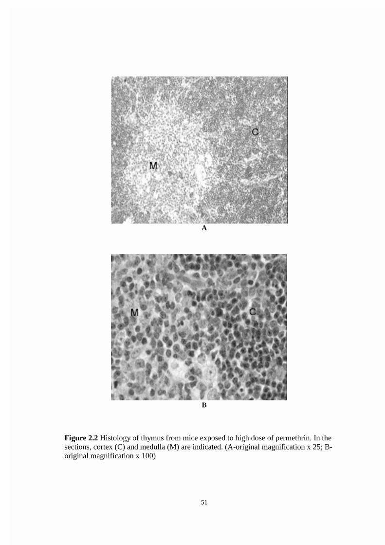
51
A
B
Figure 2.2 Histology of thymus from mice exposed to high dose of permethrin. In thesections, cortex (C) and medulla (M) are indicated. (A-original magnification x 25; B-original magnification x 100)

52
A
B
Figure 2.3 Histology of spleen from control mice. . In the sections, white pulp (W) andred pulp (R) are indicated. (A-original magnification x 25; B-original magnification x100)

53
A
B
Figure 2.4 Histology of spleen from mice exposed to high dose of permethrin. In thesections, white pulp (W) and red pulp (R) are indicated. (A-original magnification x 25;B-original magnification x 100)

54
CHAPTER 3.0: EFFECTS OF TOPICAL PERMETHRIN EXPOSURE
ON MACROPHAGE FUNCTION AND ANTIBODY PRODUCTION
3.1: ABSTRACT
Permethrin was applied to the shaved dorsal interscapular region of C57Bl/6N
mice at doses of 0.5, 1.5, or 5.0 µl/day. The highest of these doses was approximately
equal to 215 µg/kg/day, which is about seven times the estimated daily human exposure
in individuals wearing permethrin treated clothing for insect protection. Mice were thus
exposed to permethrin daily for 10 or 30 consecutive days, or every other day for 7 or 14
exposures. The splenic macrophage chemiluminescent response was affected by the
treatment at 2 days after exposure to permethrin, but phagocytic ability was not affected.
Antibody production as shown by plaque forming cell (PFC) assay decreased
significantly after 10 consecutive day exposure to permethrin. Taken together, these data
indicate that low-level topical permethrin exposure may produce systemic immune
effects.

55
3.2: INTRODUCTION
Since the end of the Persian Gulf War, numerous studies and reports have
described illness of veterans from this war (i.e., Persian Gulf Syndrome) (Doucet, 1994;
Unwin et al., 1999; Klaustermeyer et al., 1998; Proctor et al., 1998). The symptoms
included headache, loss of memory, fatigue, muscle and joint pain, ataxia, skin rash,
respiratory difficulties, and gastrointestinal disturbances (Murphy et al., 1999). It was
suggested that this syndrome may result from multiple assaults upon the body’s immune
system (Doucet, 1994). In support of this possibility, some immune parameters were
found to be significantly different between veterans with syndrome and controls (Zhang
et al., 1999). These alterations included increased total T cells and major
histocompatibility complex (MHC) II-restricted T cells, decreased natural killer (NK)
cells, and increased levels of interleukin-2 (IL-2), IL-10, gamma interferon (IFN-γ), and
tumor necrosis factor alpha (TNF-α).
Permethrin, a photostable class I pyrethroid insecticide, has been used by the US
military to impregnate into clothing along with another chemical, N,N-diethyl-m-
toluamide (DEET), for highly effective prevention of insect-borne disease (Schreck et al.,
1986; Lillie et al., 1988; Schreck and Kline, 1989; Scholdt et al., 1989). These
permethrin-impregnated clothing were also used during the Gulf War, and calculated to
result in topical exposure of permethrin at about 34 µg/kg/day (Snodgrass, 1992).
Pyrethroid insecticides have been reported to cause immunotoxicity in animal
studies. Cypermethrin was reported to decrease humoral responses and alter cellular
immune responses in rats and rabbits (Desi et al., 1985). This compound was also
reported to suppress both cellular and humoral immune responses in mice and goats
(Tamang et al., 1988). Deltamethrin was reported to have immunosuppressive effects on
both humoral and cellular immune responses in mice (Lukowicz-Ratajczak and
Krechniak, 1992), and to cause thymus atrophy in mice (Enan et al., 1996). In the study
by Enan et al. (1996), it was found that deltamethrin was able to induce apoptosis in the
thymus of treated animals via the alteration of Ca/CaM-dependent protein kinase-
phosphatase cascade.

56
Permethrin has been well studied for its neurotoxicity (Vijverberg and van den
Bercken, 1990), but research examining possible immunotoxicity is limited to only a few
reports. Studies in vitro has shown that permethrin inhibited the mitogenic response of
murine lymphocytes to Concanavalin A (Con A), lipopolysaccharide (LPS) (Stelzer and
Gordon, 1984), and phytohaemagglutinine (PHA), and decreased the production of IFN-γ
and IL-4 (Diel et al., 1998). In another study utilizing in vivo exposure (oral gavage),
cellular immune responses were suppressed in mice exposed to permethrin, including a
decreased mixed lymphocyte response (MLR) to allogenic lymphocytes, and inhibited
cytotoxic T lymphocyte (CTL) and NK cell activity (Blaylock et al., 1995). The more
relevant dermal route of permethrin exposure was not evaluated by these authors for
capacity to alter immune responses.
In a previous investigation, we reported decreased thymic and increased splenic
weight in mice dermally exposed to permethrin (unpublished data). The present study
extends these results to include inhibited humoral immune response (antibody
production) and suppressed splenic macrophage chemicluminescence (H2O2 production)
in mice following subacute topical exposure to permethrin.
3.3: MATERIALS AND METHODS
3.3.1: Mice
Female C57Bl/6 mice (Harlan Sprague Dawley, Indianapolis, IN) were used in
these studies. All mice were quarantined for one week prior to initiation of experiments.
Following the quarantine period, mice were numbered by ear-tags, then assigned random
numbers and randomized into separate sequentially numbered cages. Mice were
anesthetized by brief inhalation exposure to methoxyflurane, and the interscapular area
was shaved of hair using electric clippers. Mice were maintained under controlled
conditions of temperature (22 ± 1 °C), humidity (40-60%), and lighting (12/12 hr
light/dark cycle) and provided with food and water ad libitum throughtout the course of
the experiments. The mice were also examined daily for clinical change.

57
3.3.2: Permethrin preparation and treatment protocols
Permethrin (91.6%) was provided by the U.S. Army Center for Health Promotion
and Preventive Medicine (CHPPM) from stock purchased immediately before by the
Army from Coulston Industries (Easton, PA). Mice were treated with permethrin using
three dose solutions defined as low-dose (0.5 ml permethrin in 4.5 ml corn oil), middle-
dose (1.5 ml permethrin in 3.5 ml corn oil), and high-dose (5.0 ml permethrin). Corn oil
was used as the control solution. All dosing solutions were stored in the dark at room
temperature. Mice were dosed by interscapular topical exposure with 5 µl of respective
dosing solutions using an Eppendorf micropipettor. Dosing was daily for 10 or 30
consecutive days, or every other day for 7 or 14 exposures. After dosing termination,
mice were sacrificed and tested for effects of the chemical exposure at days 2, 10, or 30.
3.3.3: Cell preparation
Spleens from each mouse were collected and placed (separately) in 2 ml of
culture medium (RPMI 1640; Mediatech, Cellgro, Herndon, VA) in a 60x15 mm culture
dish (Fisher Scientific, Norcross, GA). Splenic cells were gently dissociated in the culture
medium using a metallic sieve screen (Sigma) and curved forceps. Erythrocytes were
removed from splenic samples by suspending cells in lysing solution (0.015 M NH4Cl,
1.0 mM NaHCO3, 0.1 mM EDTA) for 5 min at room temperature. Cells were then
washed twice in culture medium, resuspended in 2 ml standard buffer (Hank’s balanced
salt solution), and counted using a CASY-1 electronic cell counter (Coulter Electronics,
Hialeah, FL.).
3.3.4: Chemiluminescence assay
The production of H2O2 in PMA (phorbol-12-myristate 13-acetate)-stimulated
splenic macrophages was determined by the method of Bass et al. (1983). Briftly, Splenic
cell suspensions (0.5x10⁶ cells) were incubated with 5 µl dichlorofluorescein-diacetate
(DCF-DA; Molecular Probes, Eugene, OR; 5 mM) for 15 min at room temperature.
Following incubation with DCF-DA, cells were stimulated by the addition of 10 µl PMA
(Sigma, St.Louis, MO; 100 ng/ml) in a subsequent 30 min incubation period. Cells were
then placed on ice to stop the reaction and immediately analyzed by flow cytometry.

58
Background fluorescence, determined using unstained cells from each treatment group,
was subtracted from respective populations incubated with the fluorescent probe.
3.3.5: Phagocytosis of fluorescent microspheres
The phagocytic capacity of splenic macrophages was determined by a
modification of the method of Dunn and Tylor (1981). Briftly, cells were aliquoted as
above in polystyrene round-bottom tubes at 0.5x10⁶ cells in 0.1 ml standard buffer.
Fluoresbrite microspheres (1.16 µm; Polysciences, Inc., Warrington, PA) were washed
twice in standard buffer, and then sonicated on ice (Ultrasonic Cell Disrupter, Misonix,
Inc., Farmingdale, NY) for 30 sec at 35% to disrupt aggregated microspheres. A 10 µl
volume of microspheres was added to tubes to give an initial ratio of 50 beads/cell. Fresh
culture media then was added to each tube to give a final volume of 2.0 ml/tube. This
latter step prevents the development of acidic media conditions resulting from metabolic
activity of the cells during incubation. Cells then were incubated overnight (18 hr) at
37°C and 5% CO2. Following incubation, cells were washed twice with standard buffer to
remove non-phagocytized microspheres, after which cells were resuspended in 0.5 ml
standard buffer and immediately analyzed by flow cytometry. Before analysis the
cytometer was standardized for fluorescence using a sample of cells without beads. Thus,
the peak on the fluorescence histogram resulting from cells containing single
microspheres could be identified. For each sample 10,000 events were collected and the
number of cells ingesting fluorescent particles was expressed as a percentage of 10,000.
3.3.6: Plaque forming cells (PFC) assay
Ability to produce specific antibody was determined by quantitating the plaque-
forming cell (PFC) response to the T-dependent antigen, sheep red blood cells (SRBC).
Mice were immunized by intraperitoneal injection of 2x10⁸ sheep red blood cells (SRBC)
and IgM PFCs were enumerated in spleen cells 4 days later. Splenic cell supensions
(20x106 cells/ml) were prepared in culture medium. Cells of each sample (50 µl, 106
cells) were then mixed with 20 µl guinea pig complement (Sigma), 30 µl of 30% SRBC,
and 0.4 ml of warmed agar (47°C) in a 35x10 mm culture dish (Fisher Scientific,
Norcross, GA), then incubated in 37°C and 5% CO2 incubator for 4 hr. After incubation,

59
the number of PFC was counted under a light microscope. The values were expressed as
the number of PFC per 106 cells.
3.3.7: Statistical analysis
Data were expressed as arithmetical mean ± SEM. Analysis of variance
(ANOVA) was used with Dunnett’s t test to establish significant differences among
groups. Results described as different in this paper indicate significantly different at p <
0.05.
3.4: RESULTS
3.4.1: Chemiluminescent response
A dose-response inhibition of H2O2 production by splenic macrophages was
present in all exposure groups at 2 days after termination of treatment (Table 3.1). A
significant decrease in H2O2 production continued to 10 days after termination of
treatment in Exposures 1 and 4; a trend toward a decrease was present in Exposure 3.
Using this procedure, values can be compared within but not between experiments.
3.4.2: Phagocytosis response
Phagocytic ability of macrophages tended to display rather high variability by this
assay. A significant difference occurred 2 days after dosing ended, in the low dose group
of Exposure 4 (Table 3.2). Values can again be compared within but not between
experiments. Given the lack of a dose-response trend in this experiment, and lack of an
effect in all other day 2 data, it is doubtful this one effect is biologically meaningful.
3.4.3: PFC response
Permethrin produced a non-significant trend toward decreased plaque number in
Exposure 1 mice at 2 days after dosing termination (Table 3.3). This trend became
significant in this exposure group at 10 days post-dosing. Values can again be compared
within but not between experiments. Of possible importance, Exposure 1 produced the
most significant alterations in other immune parameters measured, including decreased
thymic weight and increased splenic weight (unpublished data from a previous study). No
other exposure regimen significantly altered antibody production.

60
3.5: DISCUSSION
A limited but growing database in the literature suggests certain pyrethroid
compounds may produce immunotoxicity. Brieftly, cypermethrin (a type II or cyano-
pyrethroid) was found to inhibit humoral and cellular immune responses in rats, rabbits
(Desi et al., 1985), mice and goats (Tamang et al., 1988). Deltamethrin produced thymic
atrophy and suppressed humoral and cellular responses in mice (Lukowicz-Ratajczak and
Krechniak, 1992; Queiroz, 1993; Madsen et al., 1996). Enan et al. (1996) further reported
that deltamethrin induced apoptosis in the thymus of treated animals by altering activity
of the Ca/CaM-dependent protein kinase-phosphatase cascade. Permethrin inhibited the
mitogenic response of T lymphocytes to Con A and B lymphocytes to LPS (Stelzer and
Gordon, 1984). Diel et al. (1998) recently demonstrated a similar inhibition of T
lymphoproliferation induced by the mitogen PHA, as well as dose-dependent decreases in
IFNγ and IL-4 in mice exposed to permethrin. Blaylock et al. (1995) reported decreased
mixed lymphocyte responses (MLR), and inhibited cytotoxic T lymphocyte (CTL) and
natural killer (NK) cell activity in mice treated by oral gavage with permethrin.
Collectively, the above reports demonstrate immune effects in experimental
animals treated with pyrethroid insecticides. However, none of the above reports
examined the possibility that topical pyrethroid (i.e., the typical route of human exposure)
may alter function of the immune system. Percutaneous absorption of varying levels of
permethrin has been demonstrated in rhesus monkey (Sidon et al., 1988), rat, rabbit, dog,
and human (Taplin and Meinking, 1990). Further Snodgrass (1992) estimated that
humans wearing permethrin-treated clothing received a topical exposure of about 34
µg/kg/day. These reports raise questions about immune effects resulting from absorbed
permethrin in humans wearing treated clothing (i.e., military uniforms; hunting clothing)
or following use of permethrin-based shampoos or creams for body lice or scabies.
Regarding the latter, a 1% permethrin formulation (NIX ) is widely used in children for
treatment of head lice (Brandenburg et al., 1986; Taplin and Meinking, 1990), while a 5%

61
cream (Elimite ) has been found more effective for treating scabies than 10% crotamiton
(Eurax ) (Taplin et al., 1990) or 1% lindane lotion (Schultz et al., 1990).
Results of the present study indicate that topical permethrin may alter both
humoral mediated immunity (antibody production) and non-specific immune function
(ability of splenic macrophages to produce reactive oxygen species) in mice. Specific
mechanisms by which these effects may occur were not investigated in the present
experiments. However, the ability of permethrin to inhibit production of cytokines (e.g.,
IFNγ and IL-4) essential for antibody production (Diel et al., 1998) and to inhibit B
lymphocyte proliferation (Stelzer and Gordon, 1984) may be related to an effect on
antibody production.
An effect of permethrin on the ability of macrophages to produce H2O2 was
clearly evident in this study. The inhibition effect was dose-dependent and persisted up to
10 days after dosing termination. This effect was the only immune effect observed in all
topical chemical exposure groups, in this or a previous study, suggesting that the
macrophage respiratory burst (i.e., chemiluminescent response) may represent a highly
sensitive immune target of permethrin exposure. Reasons for this again remain unclear,
however, Gassner et al. (1997) have demonstrated that permethrin inhibits mitochondrial
complex I, an effect which may interfere with the electron transport chain, and thus the
respiratory metabolic burst of phagocytic cells.
In conclusion, results of the present study support the possibility that topical
permethrin exposure may cause systemic immune effects. The effect of this compound on
antibody production may be particularly noteworthy, in that this endpoint is one of the
strongest predictors of clinically significant immunosuppression (individual predictive
value 0.78) (Luster et al., 1992).
3.6: REFERENCES
Bass, D.A., Parce, J.W., Dechatelet, L.R., Szejda, P., Seeds, M.C., and Thomas, M. 1983
Flow cytometric studies of oxidative product formation by neutrophils: a graded
response to membrane stimulation. J Immunol 130:4 1910-7.

62
Blaylock, B.L., Abdel-Nasser, M., McCarty, S.M., Knesel, J.A., Tolson, K.M., Ferguson,
P.W., and Mehendale, H.M. 1995. Suppression of cellular immune responses in
BALB/c mice following oral exposure to permethrin. Bull Environ Contam
Toxicol. 54:5 768-74.
Brandenburg, K., Deinard, A.S., DiNapoli, J., Englender, S.J., Orthoefer, J., and Wagner,
D. 1986. 1% permethrin cream rinse vs 1% lindane shampoo in treating
pediculosis capitis. Am J Dis Child. 140:9 894-6.
Dési, I., Varga, L., Dobronyi, I., and Szklenarik, G. 1985. Immunotoxicological
investigation of the effects of a pesticide; cypermethrin. Arch Toxicol Suppl 8:
305-9.
Diel, F., Detscher, M., Borck, H., Schrimpf, D., Diel, E., and Hoppe, H.W. 1998. Effects
of permethrin on human basophils and lymphocytes in vitro. Inflamm Res. 47
Suppl 1: S11-2.
Doucet, I. 1994. Desert Storm syndrome: sick soldiers and dead children? Med War. 10:3
183-94.
Dunn, P.A. and Tyrer, H.W. 1981. Quantitation of neutrophil phagocytosis, using
fluorescent latex beads.Correlation of microscopy and flow cytometry. J Lab Clin
Med. 98:3 374-81.
Gassner, B., Wüthrich, A., Scholtysik, G., and Solioz, M. 1997. The pyrethroids
permethrin and cyhalothrin are potent inhibitors of the mitochondrial complex I. J
Pharmacol Exp Ther. 281:2 855-60.
Klaustermeyer, W.B., Kraske, G.K., Lee, K.G., and Kurohara, M.L. 1998. Allergic and
immunologic profile of symptomatic Persian Gulf War veterans. Ann Allergy
Asthma Immunol. 80:3 269-73.
Lillie, T.H., Schreck, C.E., and Rahe, A.J. 1988. Effectiveness of personal protection
against mosquitoes in Alaska. J Med Entomol. 25:6 475-8
Luster, M.I., Portier, C., Pait, D.G., White, K.L. Jr., Gennings, C., Munson, A.E.,
Rosenthal, G.J. 1992. Risk assessment in immunotoxicology. I. Sensitivity and
predictability of immune tests. Fundam Appl Toxicol 18:2 200-10.

63
Madsen, C., Claesson, M.H., and Ropke, C. 1996. Immunotoxicity of the pyrethroid
insecticides deltametrin and alpha- cypermetrin. Toxicology 107:3 219-27.
Murphy, F.M., Kang, H., Dalager, N.A., Lee, K.Y., Allen, R.E., Mather, S.H., and Kizer,
K.W. 1999. The health status of Gulf War veterans: lessons learned from the
Department of Veterans Affairs Health Registry. Mil Med. 164:5 327-31.
Proctor, S.P., Heeren, T., White, R.F., Wolfe, J., Borgos, M.S., Davis, J.D., Pepper, L.,
Clapp, R., Sutker, P.B., Vasterling, J.J., and Ozonoff, D. 1998. Health status of
Persian Gulf War veterans: self-reported symptoms, environmental exposures and
the effect of stress. Int J Epidemiol. 27:6 1000-10.
Queiroz, M. L. 1993. Hematopoietic effects in mice exposed to deltamethrin and
hydrocortisone. International Journal Of Immunopharmacology 15:3 301-7.
Schreck, C.E., and Kline, D.L. 1989. Personal protection afforded by controlled-release
topical repellents and permethrin-treated clothing against natural populations of
Aedes taeniorhynchus. J Am Mosq Control Assoc. 5:1 77-80.
Schreck, C.E., Snoddy, E.L., and Spielman, A. 1986. Pressurized sprays of permethrin or
deet on military clothing for personal protection against Ixodes dammini (Acari:
Ixodidae). J Med Entomol. 28 23:4 396-9.
Schultz, M.W., Gomez, M., Hansen, R.C., Mills, J., Menter, A., Rodgers, H., Judson,
F.N., Mertz, G., and Handsfield, H.H. 1990. Comparative study of 5% permethrin
cream and 1% lindane lotion for the treatment of scabies. Arch Dermatol. 126:2
167-70.
Sholdt, L.L., Rogers, E.J.Jr., Gerberg, E.J., and Schreck, C.E. 1989. Effectiveness of
permethrin-treated military uniform fabric against human body lice. Mil Med.
154:2 90-3.
Sidon, E.W., Moody, R.P., and Franklin, C.A. 1988. Percutaneous absorption of cis- and
trans-permethrin in rhesus monkeys and rats: anatomic site and interspecies
variation. J Toxicol Environ Health 23:2 207-16.
Snodgrass, H.L. 1992. Permethrin transfer from treated cloth to the skin surface: potential
for exposure in humans. J Toxicol Environ Health. 35:2 91-105.

64
Stelzer, K.J. and Gordon, M.A. 1984. Effects of pyrethroids on lymphocyte mitogenic
responsiveness. Res Commun Chem Pathol Pharmacol. 46:1 137-50.
Tamang, R.K., Jha, G.J., Gupta, M.K., Chauhan, H.V., and Tiwary, B.K. 1988. In vivo
immunosuppression by synthetic pyrethroid (cypermethrin) pesticide in mice and
goats. Vet Immunol Immunopathol. 19:3-4 299-305.
Taplin, D. and Meinking, T.L. 1990. Pyrethrins and pyrethroids in dermatology. Arch
Dermatol.126:2 213-21.
Taplin, D., Meinking, T.L., Chen, J.A., and Sanchez, R. 1990. Comparison of crotamiton
10% cream (Eurax) and permethrin 5% cream (Elimite) for the treatment of
scabies in children. Pediatr Dermatol. 7:1 67-73.
Unwin, C., Blatchley, N., Coker, W., Ferry, S., Hotopf, M., Hull, L., Ismail, K., Palmer,
I., David, A., and Wessely, S. 1999. Health of UK servicemen who served in
Persian Gulf War. Lancet. 353:9148 169-78.
Vijverberg, H.P. and van den Bercken, J. 1990. Neurotoxicological effects and the mode
of action of pyrethroid insecticides. Crit Rev Toxicol. 21:2 105-26.
Zhang, Q., Zhou, X.D., Denny, T., Ottenweller, J.E., Lange, G., LaManca, J.J., Lavietes,
M.H., Pollet, C., Gause, W.C., and Natelson, B.H. 1999. Changes in immune
parameters seen in Gulf War veterans but not in civilians with chronic fatigue
syndrome. Clin Diagn Lab Immunol 6:1 6-13.

65
Table 3.1 Chemiluminescent response in C57Bl/6 mice topically exposed topermethrin
Chemiluminescent Response (% positive cells, means ± sem)
Permethrin exposure Days after dosing termination2 day 10 day 30 day
EXPOSURE 1:Control 33.40 ± 3.00 45.60 ± 4.00 9.20 ± 1.000.5 µl/day 25.90 ± 1.70 32.00 ± 1.50 7.90 ± 0.901.5 µl/day 22.50 ± 1.40* 27.50 ± 2.10* 9.90 ± 3.505.0 µl/day 16.50 ± 1.20* 20.40 ± 1.20* 9.60 ± 1.90
EXPOSURE 2:Control 25.60 ± 2.40 19.60 ± 2.60 21.70 ± 1.800.5 µl/day 23.80 ± 2.70 23.60 ± 3.30 23.20 ± 1.601.5 µl/day 21.40 ± 2.00* 28.00 ± 4.10 24.00 ± 1.605.0 µl/day 13.80 ± 1.40* 20.90 ± 4.30 33.60 ± 4.80
EXPOSURE 3:Control 30.18 ± 1.26 20.48 ± 0.96 18.07 ± 1.330.5 µl/day 23.93 ± 2.32 17.77 ± 2.65 17.87 ± 1.671.5 µl/day 23.13 ± 2.48 13.80 ± 2.07 14.65 ± 1.315.0 µl/day 20.15 ± 2.08* 14.62 ± 1.67 14.73 ± 0.63
EXPOSURE 4:Control 23.37 ± 0.87 29.27 ± 2.63 29.96 ± 2.930.5 µl/day 21.92 ± 1.71 21.17 ± 0.59* 22.00 ± 0.981.5 µl/day 18.60 ± 1.91* 24.07 ± 1.91 26.00 ± 4.205.0 µl/day 17.10 ± 0.96* 23.02 ± 0.89* 21.98 ± 4.33
Exposure 1 = every day for 10 consecutive daysExposure 2 = every other day for 7 exposuresExposure 3 = every other day for 14 exposuresExposure 4 = every day for 30 consecutive days
* statistically significant (P < .05)

66
Table 3.2 Macrophage phagocytosis in C57Bl/6 mice topically exposed topermethrin
Macrophage Phagocytic Activity (% positive cells, means ± sem)
Permethrin exposure Days after dosing termination2 day 10 day 30 day
EXPOSURE 1:Control 5.92 ± 0.59 16.40 ± 1.80 20.70 ± 3.000.5 µl/day 5.57 ± 0.35 18.70 ± 4.20 25.00 ± 3.201.5 µl/day 6.69 ± 0.78 16.10 ± 1.90 25.40 ± 2.205.0 µl/day 6.31 ± 0.31 20.60 ± 3.30 31.50 ± 5.90
EXPOSURE 2:Control 9.10 ± 0.80 3.50 ± 0.50 1.65 ± 0.3290.5 µl/day 7.50 ± 0.50 3.60 ± 0.90 2.58 ± 0.8141.5 µl/day 7.80 ± 1.00 3.00 ± 0.40 5.32 ± 1.455*5.0 µl/day 7.30 ± 0.80 6.30 ± 1.30* 3.63 ± 1.102
EXPOSURE 3:Control 3.00 ± 1.580 8.95 ± 6.285 1.52 ± 0.3470.5 µl/day 1.33 ± 0.220 4.10 ± 1.160 1.20 ± 0.3021.5 µl/day 1.02 ± 0.110 2.72 ± 0.697 1.25 ± 0.1805.0 µl/day 0.90 ± 0.060 8.48 ± 3.793 1.23 ± 0.147
EXPOSURE 4:Control 0.917 ± 0.147 0.25 ± 0.034 0.36 ± 0.0510.5 µl/day 0.567 ± 0.056* 0.25 ± 0.043 0.68 ± 0.0951.5 µl/day 0.933 ± 0.033 0.20 ± 0.026 0.60 ± 0.0005.0 µl/day 0.650 ± 0.099 0.18 ± 0.040 0.58 ± 0.092
Exposure 1 = every day for 10 consecutive daysExposure 2 = every other day for 7 exposuresExposure 3 = every other day for 14 exposuresExposure 4 = every day for 30 consecutive days
* statistically significant (P < .05)

67
Table 3.3 Plaque forming cell (PFC) assay in C57Bl/6 mice topically exposed topermethrin
PFC Response (plaque numbers/106 cells, means ± sem)
Permethrin exposure Days after dosing termination2 day 10 day 30 day
EXPOSURE 1:Control 60.9 ± 9.67 19.5 ± 3.76 10.8 ± 4.030.5 µl/day 67.3 ± 10.43 12.4 ± 4.28 8.9 ± 1.391.5 µl/day 47.3 ± 8.33 5.0 ± 1.08* 14.3 ± 2.735.0 µl/day 47.5 ± 6.41 4.3 ± 1.24* 12.2 ± 3.50
EXPOSURE 2:Control 7.5 ± 2.33 35.6 ± 3.41 32.0 ± 6.140.5 µl/day 3.3 ± 0.51 20.1 ± 3.63 22.3 ± 6.531.5 µl/day 4.6 ± 1.27 23.2 ± 3.23 31.6 ± 6.705.0 µl/day 5.0 ± 1.58 35.2 ± 17.05 32.6 ± 8.74
EXPOSURE 3:Control 8.8 ± 1.65 14.3 ± 5.17 12.4 ± 4.420.5 µl/day 14.9 ± 6.12 16.8 ± 7.81 11.8 ± 2.091.5 µl/day 6.7 ± 1.42 16.7 ± 8.49 6.5 ± 1.225.0 µl/day 14.3 ± 7.00 8.8 ± 1.49 13.1 ± 2.86
EXPOSURE 4:Control 16.1 ± 5.38 36.5 ± 7.84 44.2 ± 31.270.5 µl/day 11.9 ± 2.70 20.6 ± 4.82 12.8 ± 3.851.5 µl/day 5.3 ± 0.60 26.3 ± 6.63 9.6 ± 2.335.0 µl/day 14.7 ± 5.49 35.8 ± 3.91 7.1 ± 0.76
Exposure 1 = every day for 10 consecutive daysExposure 2 = every other day for 7 exposuresExposure 3 = every other day for 14 exposuresExposure 4 = every day for 30 consecutive days
* statistically significant (P < .05)

68
CHAPTER 4.0: CONCLUSION AND SIGNIFICANCE OF THE
CURRENT STUDY
A large number of chemicals have been reported to produce immunotoxicity in
humans and animals. The consequences could be immunosuppression, allergic disease, or
autoimmune disease. Permethrin, a pyrethroid insecticide, has been used in agriculture,
human medicine, and veterinary medicine, thus a variety of people ranging from children
to adults have been exposed to this chemical. It was also used by the U.S. Army to
impregnate into military uniforms in order to protect soldiers from insect bites. Since
Persian Gulf Syndrome occurred, much research has been performed to find the cause of
this syndrome. Permethrin, which is usually considered to be a neurotoxic chemical, has
been reported to have immunotoxic potential in vitro and in animal studies. Therefore,
permethrin was evaluated in present study for its potential of producing immunotoxicity
by dermal exposure.
This study was designed to evaluate immunotoxicity of permethrin in mice. The
experiment design mimicked the actual human exposure. Mice were exposed to
permethrin by the topical route daily for 10 or 30 consecutive days, or every other day for
7 or 14 exposures. The highest dosage used was approximately 7 times human exposure.
After dosing termination, mice were examined at 2, 10, and 30 days using selected tests
from the immunotoxicity testing battery approved by the National Toxicology Program.
These tests included thymus/body weight ratio, spleen/body weight ratio, histopathology
of thymus and spleen, cell surface marker analysis of thymocytes, splenocytes, and bone
marrow cells, plaque forming cell (PFC) assay, macrophage chemiluminescent assay, and
macrophage phagocytosis.
In this study, permethrin caused decreased thymus/body weight ratio, increased
spleen/body weight ratio, decreased thymic cellularity, decreased macrophage
chemiluminescent response, and decreased antibody production. The non-affected
immune tests were cell-surface antigen expression, splenic cellularity, macrophage
phagocytic ability, and histopathology of thymus and spleen. Most of the effects occurred
following the 10-consecutive day exposure, except altered chemiluminescent response

69
that occurred in all exposures. The effects occurred mostly 2 days after dosing
termination.
Permethrin may cause thymic atrophy by increasing thymocyte apoptosis or
suppressing thymocyte proliferation. The possibility of permethrin-induced thymocyte
apoptosis was supported by the evidence that permethrin was a potent inhibitor of
calmodulin, a universal calcium binding protein, and may cause alteration in Ca2+
homeostasis. This change in intracellular Ca2+ may lead to the process of thymocyte
apoptosis. The possibility of permethrin-suppressed thymocyte proliferation was
supported by the evidence that permethrin suppressed lymphocyte proliferation
stimulated by mitogen, and decreased the production of cytokines. The inhibition effect
of permethrin on mitochondrial and calmodulin activities could also contribute to the
suppressed-cell growth.
The mechanism of permethrin that caused splenomegaly was unknown. However,
the increased splenic weight may come from splenic congestion.
The specific mechanism, which permethrin inhibited antibody production was not
investigated in this study. However, this effect may relate to the ability of permethrin to
inhibit production of cytokines (IFNγ and IL-4) essential for antibody production, and
inhibit B lymphocyte proliferation.
The inhibition effect on the ability of macrophages to produce H2O2 was dose-
dependent and persisted up to 10 days after dosing termination. This effect was found in
all chemical exposures, suggesting that the macrophage respiratory burst may represent a
highly sensitive immune target of permethrin exposure. Permethrin has been shown to
interrupt mitochondrial activity and this effect may relate to the inhibited respiratory
metabolic burst of macrophages.
The present study has shown that permethrin, considered a safe chemical for
using on human body, was able to alter immune functions in a murine study. This was
also the first evidence showing immune effects of permethrin by topical exposure. Even
though the effects were transient and on only some immune function tests, it offered
significant information to the public.

70
CURRICULUM VITAE
Korawuth Punareewattana
Department of Biomedical Sciences and Pathobiology
Virginia-Maryland Regional College of Veterinary Medicine
Virginia Polytechnic Institute and State University
Blacksburg, VA 24061 (540) 231-7367
email: [email protected], [email protected], [email protected]
Permanet address Department of Pharmacology and Toxicology
Faculty of Veterinary Medicine
Khon Kaen University
Khon Kaen, 40002 Thailand.
(66) 43-241-570
DATE OF BIRTH October 8, 1964
EDUCATION
Masters of Science, Veterinary Medical Science (Immunotoxicology),
November 1999
Virginia Polytechnic Institute and State University, Blacksburg, VA
Thesis: Immune function determination in mice dermally exposed to permethrin
Advisors: Dr. Steven D. Holladay
Q.C.A.: 3.78
Doctor of Veterinary Medicine (D.V.M.), March 1988
Chulalongkorn University, Bangkok, Thailand
Q.C.A.: 2.95; GRE Verbal: 320 GRE Quantitative: 680
TEACHING INTERESTS
Veterinary Pharmacology
Veterinary Toxicology
Veterinary Immunology

71
PRESENTATIONS AT MEETINGS
Punareewattana, K., Holladay, S.D., Smith, B.J., and Blaylock, B.L. Topical permethrin
exposure alters skin immune responses and produces systemic immune effects.
Virginia-Maryland Regional College of Veterinary Medicine 11th Annual
Research Symposium. May 1999.
ABSTRACTS PUBLISHED
Punareewattana, K., Holladay, S.D., Smith, B.J., and Blaylock, B.L. Topical permethrin
exposure alters skin immune responses and produces systemic immune effects.
The Toxicologist (38th Annual Meeting) March 1999 48:1-S 180.
MANUSCRIPTS (to be submitted)
Punareewattana, K., Holladay, S.D., and Smith, B.J. Effects of topical permethrin
exposure on spleen, thymus, and bone marrow: weights, cell counts, cell-surface
antigen expression, and histopathology.
Punareewattana, K., Holladay, S.D., and Smith, B.J. Effects of topical permethrin
exposure on macrophage function and antibody production.


![Lesions of the Rete Testis in Mice Exposed Prenatally to … · [CANCER RESEARCH 45, 5145-5150, October 1985] Lesions of the Rete Testis in Mice Exposed Prenatally to Diethylstilbestrol](https://img.pdfslide.net/doc/110x75/5fc722183cfb0439ef1b1dc9/lesions-of-the-rete-testis-in-mice-exposed-prenatally-to-cancer-research-45-5145-5150.jpg)