Embed Size (px)
Citation preview
ARTICLE IN PRESS
InsectBiochemistry
andMolecularBiology
0965-1748/$ - se
doi:10.1016/j.ib
�CorrespondE-mail addr
Insect Biochemistry and Molecular Biology 37 (2007) 655–666
www.elsevier.com/locate/ibmb
Immune upregulation of novel antibacterial proteins from silkmoths(Lepidoptera) that resemble lysozymes but lack muramidase activity
Archana S. Gandhe, Gude Janardhan, Javaregowda Nagaraju�
Laboratory of Molecular Genetics, Centre for DNA Fingerprinting and Diagnostics, ECIL Road, Nacharam, Hyderabad 500076, India
Received 26 January 2007; received in revised form 19 March 2007; accepted 21 March 2007
Abstract
Study on immune proteins in domesticated and wild silkmoths Bombyx mori and Antheraea mylitta, respectively, led to identification
of a new class of antimicrobial proteins. We designated them as lysozyme-like proteins (LLPs) owing to their partial similarity with
lysozymes. However, lack of characteristic catalytic amino acid residues essential for muramidase activity in LLPs puts them functionally
apart from classical lysozymes. Two LLPs, one from B. mori (BLLP1) and the other from A. mylitta (ALLP1) expressed in a recombinant
system, exhibited a broad-spectrum antibacterial action. Further investigation of the antibacterial mechanism revealed that BLLP1 is
bacteriostatic rather than bactericidal against Escherichia coli and Micrococcus luteus. Substantial increase in hemolymph bacterial load
was observed in B. mori upon RNA interference mediated in vivo knockdown of BLLP1. We demonstrate that the antibacterial
mechanism of this protein depends on peptidoglycan binding unlike peptidoglycan hydrolysis or membrane permeabilization as observed
with lysozymes and most other antimicrobial peptides. To our knowledge, this is the first report on functional analysis of novel, non-
catalytic lysozyme-like family of antibacterial proteins that are quite apart functionally from classical lysozymes. The present analysis
holds promise for functional annotation of similar proteins from other organisms.
r 2007 Elsevier Ltd. All rights reserved.
Keywords: Insect; Immunity; Antimicrobial proteins; Lysozyme; Muramidase
1. Introduction
The immune repertoire in insects comprises mainly of theinnate mechanisms, manifested by both cellular andhumoral defense mechanisms though recent evidencessuggest the presence of adaptive responses as well (Donget al., 2006; Sadd and Schmid-Hempel, 2006; Watson et al.,2005). The highlight of the humoral defense is a rapidrelease of different types of antimicrobial peptides (AMPs)in the insect hemolymph within few hours upon infection.Collectively, these AMPs are known to mount an effectiveimmune defense against invading pathogens (Yamakawaand Tanaka, 1999). Lysozyme is a widespread AMPoccurring in insects, vertebrates, plants and microorgan-isms. Lysozymes are muramidases that hydrolyse the b-1,4-
e front matter r 2007 Elsevier Ltd. All rights reserved.
mb.2007.03.013
ing author. Tel.:+91 40 2715 1344; fax:+9140 2715 5610.
ess: [email protected] (J. Nagaraju).
glycosidic linkage in the N-acetyl glucosamine and N-acetylmuramic acid residues in the peptidoglycan layer of thebacterial cell and cause their lysis. Lysozymes areupregulated upon infection in the lepidopteran insectsunlike their constitutively expressed vertebrate counter-parts (Daffre et al., 1994).A c-type lysozyme has been previously characterized
from the domesticated and wild silkmoths, Bombyx mori
and Antheraea mylitta, respectively (Jain et al., 2001; Leeand Brey, 1995). In the present study, we report novelAMPs from the two silkworm species from which we havepreviously characterized c-type lysozymes. These novelproteins are unique in that they share 50–60% similarity atthe amino acid level with the c-type insect lysozymes butlack characteristic catalytic activity of peptidoglycanhydrolysis exhibited by lysozymes. Hence we chose todesignate them as lysozyme-like proteins (LLPs). Never-theless, we found, these LLPs exhibit profound antibacter-ial activity towards a wide range of gram-positive and
ARTICLE IN PRESSA.S. Gandhe et al. / Insect Biochemistry and Molecular Biology 37 (2007) 655–666656
gram-negative bacteria and are upregulated upon bacterialinfection. An immune related function is thereby suggested.Further, the in vivo RNA interference (RNAi) mediatedknockdown of B. mori lysozyme-like protein 1 (BLLP1)resulted in an enhanced bacterial load in hemolymph. Wefurther demonstrated that BLLP1 exhibits bacteriostaticeffects against Escherichia coli and Micrococcus luteus andthe inhibition was rescued when peptidoglycan was addedexternally. These results tempt us to hypothesize that themechanism of inhibition is related to binding of peptido-glycan by BLLP1, leading to growth inhibition rather thanhydrolysis of peptidoglycan or membrane permeabiliza-tion. To the best of our knowledge this is the first report onnatural occurrence of non-bacteriolytic, antibacterial LLPsthat adds yet another novelty to the already existingdiversity of AMPs.
2. Materials and methods
2.1. Isolation of LLP and lysozyme cDNAs
A. mylitta lysozyme (AL) cDNA was isolated bydesigning primers based on the protein sequence reportedearlier (Jain et al., 2001) and cDNA clone of B. mori
lysozyme (Lee and Brey, 1995) was a kind gift from PaulBrey. Partial sequence of A. mylitta lysozyme-like protein 1(ALLP1) was identified from an E. coli-challenged fat bodyEST library (Gandhe et al., 2006) and subsequently the fulllength sequence was obtained by 50 RACE PCR using the50 RACE kit (Clontech). The 50 end sequence was amplifiedby using an adaptor primer and a reverse gene specificprimer. PCR was performed for 25 cycles on an eppendorfmaster cycler. A 650 bp band was isolated, sequenced andconfirmed to be 50 ALLP1 sequence. BLLP1 was identifiedfrom Silkbase (Mita et al., 2003) by BLAST search analysiswith ALLP1 as a query. BLLP2 and BLLP3 were identifiedby tBLASTx analysis at SilkDB with BLLP1 as a querysequence (Wang et al., 2005). Primers were designed byPrimer-3 software (Rozen and Skaletsky, 2000) and theirsequences were as follows:
(50–30)-AL Forward (F)-AAACGTTTCACCAGATG-CG, AL Reverse (R)-ACAGTCGCTAATATCTGG;
BL F-AAAACGTTCACGAGATGCG, BL R-GCAG-CTGCTAATATCAGG;
ALLP1 R (for 50 RACE)-CCTTCGAACTCTTCGC-GTTA, Adaptor (Forward) primer was supplied byClontech; BLLP1 F-AAGGTCTTCACGAGATGCCA-AC BLLP1 R-GCATCTGGAGATGTCTGGTAGGTT-CTTC.
Following PCR conditions were used—94 1C, 2min—initial denaturation, 35 cycles (94 1C—30 s, 60 1C—30 s,72 1C—2min) and a final elongation at 72 1C for 10min.Actin cDNA was amplified as an endogenous control. PCRreaction components included: 1X buffer, 100 mM dNTPs,1.5mM MgCl2, 0.5 units Taq polymerase (MBI), 0.5 mMprimers.
2.2. Bacterial strains
BL21-CodonPlus strain (Stratagene) was used forprotein expression. For the antibacterial assays followingstrains were procured from Microbial Type CultureCollection (MTCC), Chandigarh, India: Klebsiella pneu-
moniae subsp.pneumoniae (MTCC no. 39), Serratia mar-
cescens (MTCC no. 86), Pseudomonas fluorescens (MTCCno. 103), Bacillus thuringiensis subsp. Kurstaki (MTCC no.868).
2.3. Phylogenetic analysis
Phylogenetic analysis was done by comparison of maturefull-length protein sequences of 35 insect lysozymes withhen lysozyme (HL) as an outgroup. All the sequences werealigned using ClustalX 1.8 and manually edited usingGeneDoc Version 2.6.002. The GenBank accession num-bers for the sequences is provided as supplementary data(Supplementary file 1). A neighbour-joining (NJ) tree wasconstructed using HYPHY (Pond et al., 2005) and wasused as a base tree to test the best-fit model of evolution inHYPHY. Akaike Information Criterion (AIC) was used toselect the best-fit model, which selected WAG (Whelan andGoldman) model of amino acid substitution (Whelan andGoldman, 2001). A maximum likelihood tree was con-structed with the program TREE-PUZZLE 5.2 (Schmidtet al., 2002) that utilized 50,000 quartet-puzzling steps.
2.4. Molecular weight (MW) and isoelectric pH (pI)
The MW and pI of the lysozymes were predicted usingthe Expasy software. The SignalP software was usedto predict the signal peptide (Bendtsen et al., 2004)(www.cbs.dtu.dk/services/SignalP/).
2.5. Real time PCR
A. mylitta and B. mori, 5th instar, day 3 larvae werechallenged with log phase E. coli or M. luteus as describedearlier (Gandhe et al., 2006). A set of larvae, injected withsterile saline (mock infection) and a set of uninjected larvaewere used as control. Fat body total RNA was isolated byTrizol method (Invitrogen) from the differentially chal-lenged larvae and it was treated with RNAse free DNAse(MBI) to remove genomic DNA contamination. Subse-quently, cDNA was synthesized from 1 mg of total RNAwith oligo dT primers and MMLV reverse transcriptase(Invitrogen). For quantitative PCR, the componentsincluded the cDNA template; primers (0.5 mM) and SYBRGreen master mix (Eurogentec). PCR was carried out inABI Prism 7000 real time PCR cycler under followingconditions—50 1C—2min, 95 1C—10min and 40 cycles of95 1C—15 s and 60 1C—1min. The fold increase oflysozyme transcripts over the unchallenged levels wascalculated by the comparative Ct method (AppliedBiosystems). The data obtained were normalized against
ARTICLE IN PRESSA.S. Gandhe et al. / Insect Biochemistry and Molecular Biology 37 (2007) 655–666 657
the endogenous actin control, which was also analyedunder similar conditions. The data presented are anaverage of two independent reactions. Primers weredesigned by Primer Express software (ABI) and theirsequences (50–30) were as follows:
AL F-GAGACAAGGCTTCGACGAGAG, AL R-TT-GTTCACTTTACCAACTTTATCCGTAT;
ALLP1 F-CGATGGAAATTGTGCTCTAAAGG, AL-LP1 R-TGCATCTGGCCACCCATT;
BL F-GGCTCGAAGGACTACGGATTG, BL R-CC-GGACTGGCGCCTTT;
BLLP1 F-AGGGCGGAAATTGCAACAT, BLLP1
R-ATACCCGTTTTGCGCATCTAA;Actin F-GGCATGGGACAGAAGGACT, Actin R-TA-
GTGACGATTCCGTGTTCG.
2.6. Recombinant expression, purification and refolding
AL and BL were cloned into pET-23a (+) and ALLP1
and BLLP1 were cloned in pET-28a (+) E. coli expressionvectors (Novagen). The NdeI and XhoI sites wereutilized to clone ALLP1 and BLLP1 downstream to theN-terminal vector encoded histidine tag containing athrombin cleavage site for removal of histidine tag. NdeIand NotI sites were used to clone AL and BL wherein anN-terminal histidine tag was incorporated in the protein byincluding it in the forward primer. The restriction siteswere incorporated in the primers and then the PCRproduct was digested and ligated to the vector digestedwith identical restriction enzymes. The clones wereconfirmed by sequencing, recombinant plasmid was iso-lated and transformed into BL21-CodonPlus strain (Stra-tagene) of E. coli. The protein expression in BL21-CodonPlus cells was induced by adding 0.8mM IPTG toa log phase culture. The cells were harvested 6 h postinduction and the protein was purified from the inclusionbodies under denaturing conditions. The N-terminalhistidine-tagged recombinant proteins were purified underdenaturing conditions (6M guanidium hydrochloride) byaffinity chromatography with Ni-NTA Agarose (Qiagen)using the manufacturer’s protocol and the purified proteinwas incubated with 5mM DTT at 4 1C overnight. Thedenatured and reduced lysozymes were then refolded toobtain a soluble active protein by dilution with refoldingbuffer containing 1mM GSSG (oxidized glutathione)and 0.1mM GSH (reduced glutathione), 264mM NaCl,11mM KCl, 55mM Tris-HCl, pH 8.2; 1.1mM EDTA(Lopez-Zavala et al., 2004). For ALLP1 and BLLP1,1mM CaCl2 was added and EDTA was omitted fromthe refolding buffer (Koshiba et al., 1999). The resultingprotein was concentrated using centricon columns(Millipore) with a 10KD cut-off membrane, dialysedagainst Milli Q water overnight. For ALLP1 and BLLP1,dialysis was carried out with 1mM CaCl2 and 20mM Tris-HCl, pH 8. The N-terminal histidine tag of ALLP1 andBLLP1 was removed by thrombin (Novagen) digestionaccording to the manufacturer’s protocol and the cleaved
protein was purified subsequently by size exclusionchromatography using Superdex-200 (Amersham) col-umns. Protein estimation was carried out by Bradford’smethod (Bradford, 1976).
2.7. Muramidase assay
Muramidase assay was carried out with M. luteus
substrate (Sigma) in 50mM sodium phosphate buffer, pH6.5 and the absorbance at 450 nm was recorded after every30 s. (Shugar, 1952). One unit of lysozyme is defined as theamount that decreases the absorbance of the substratesolution by 0.001 absorbance units /ml/min.
2.8. Peptidoglycan binding assay
Peptidoglycan binding assay was performed as describedearlier (Kang et al., 1998) with slight modifications. Briefly,E. coli and M. luteus peptidoglycan (gift from Dr. BrunoLemaitre, Centre de Genetique Moleculaire, France) werewashed and resuspended in PBS (40 ml) and 10 mg ofALLP1 and BLLP1 were added to it. After 30-minincubation, the reaction components were pelleted down,the pellet was washed once with PBS and the supernatantand wash fractions were collected. The pellets wereresuspended in 40 ml PBS, boiled for 5min after additionof 2X-SDS/PAGE loading buffer and analysed by westernblot. Appropriate controls were kept for the experiment.Anti-histidine primary antibody (Qiagen) at 1:2000 dilu-tion and anti-mouse IgG secondary antibody conjugated tohorseradish peroxidase (Amersham) at 1:2000 dilution wasused. The detection was done with ECL Plus detectionreagents (Amersham).
2.9. Antibacterial assays
2.9.1. Radial diffusion assay
The antibacterial activity of the purified proteins wastested in a radial diffusion assay (Steinberg and Lehrer,1997). The log phase bacteria were incorporated in anunderlay gel of 1% agarose in citrate phosphate buffer(9mM sodium phosphate, 1mM sodium citrate, pH 6.0)supplemented with 3% tryptic soy broth (TSB). Wells of3mm diameter were bored into this underlay geland lysozymes and LLPs were added to each well andallowed to diffuse for 3 h after which an overlay of 1%agarose in citrate phosphate buffer supplemented with6%TSB was overlaid on the lower gel and incubatedovernight at the ambient temperature. The zone ofbacterial inhibition was measured after incubation. Thebacterial strains used were—gram-negative: E. coli
MG1655, Salmonella typhi, Klebsiella pneumoniae andPseudomonas fluorescens and gram-positive: Staphylococ-
cus aureus, M. luteus, and Bacillus thuringiensis. Theinhibitory concentration (IC) was calculated from theinhibition zone data using a formula (method (a)) valid forinhibition zones smaller than critical diameter as described
ARTICLE IN PRESSA.S. Gandhe et al. / Insect Biochemistry and Molecular Biology 37 (2007) 655–666658
earlier by Hultmark et al. (1982). Method (a) was used inthe present study because a curved plot of log n versus d2
was obtained with the series of concentrations used or incase of a few bacterial species the inhibition zones wereobtained only with the highest concentration used in theassay:
IC ¼0:468 n
ad2;
wherein a is the thickness of agar layer (cm), d is theinhibition zone diameter (cm) and n is the concentration innanomole of the antimicrobial protein utilized for theassay, and IC is expressed in mM.
2.9.2. Kinetics of bacterial inhibition
The kinetics and bacteriostatic or bactericidal nature ofthe antibacterial activity of BL and BLLP1 was determinedagainst E. coli and M. luteus by incubating the cells at aconcentration of approximately 104–105 cells/ml with10 mM of the peptide in 1 X PBS (pH 6.5). Samples werewithdrawn at 1, 2, 4 and 8 h post incubation withthe AMPs and plated onto LB agar to determine theviable colony forming units (cfu/ml). A control in whichcells were incubated with equivalent amount of sterilewater was also kept in parallel. For peptidoglycan(peptidoglycan) competition assay, similar experimentwas carried out but BL and BLLP1 were pre-incubatedfor 15min with 32 mg of M. luteus peptidoglycan (Sigma)and then assayed for kinetics of inhibition against E. coli
and M. luteus as mentioned above.
2.10. Inner membrane (IM) permeabilization assay
IM permeabilization of E. coli strain MG1655 lacY:Tn10dKan which is a lactose permease deficient strain wasestimated by analysing its b-galactosidase activity afterincubation with BL and BLLP1 (Steinberg and Lehrer,1997). The cells were grown in poor LB broth (1%bactotryptone, 0.9% NaCl) in the presence of 0.1mMIPTG to log phase and 100 ml of bacterial suspensions(106 cfu/ml) were incubated at 37 1C with BL and BLLP1,respectively (10 mM). The samples were withdrawn at 1, 4and 8 h post incubation, centrifuged and the supernatantswere stored at �20 1C. The b-galactosidase activity of thesamples was assayed with ONPG as the substrate and theabsorbance was read at 420 nm. As a control, cellsincubated with sterile water were assayed for b-galactosi-dase activity.
2.11. Preparation of dsRNA and RNAi
BLLP1 cDNA (357 bp) was cloned into pCRII-TOPOvector (Invitrogen) and amplified with M13 forward andreverse primers. This template with flanking T7 and SP6promoters was utilized for in vitro transcription reactionand sense and antisense RNA strands were generated withthe T7 and SP6 Megascript kits (Ambion). The DNA
template was removed from the transcripts by DNAsetreatment and the RNA products were subsequentlypurified by Trizol extraction (Invitrogen) followed byisopropanol precipitation. The complementary singlestranded RNAs were dissolved in DEPC treated water,combined in equimolar amounts in 1X insect buffersaline (IBS, composition: NaCl—160mM, KCl—10mM,CaCl2—4mM) and annealed by heating to 95 1C and slowcooling overnight at room temperature. Similarly, dsRNAspecific to green fluorescent protein (GFP) was synthesizedas a non-specific control. The dsRNA formation wasconfirmed by agarose gel electrophoresis and the concen-tration was determined spectrophotometrically. To deter-mine the in vivo role of BLLP1 in immunity, we knockeddown the BLLP1 expression by RNAi and then assessedthe effects on immune function by an experimentmentioned below.
2.12. In vivo bacteria clearance assay
The ability of clearance of bacteria injected in larvaesystemically was assessed and compared with that obtainedfrom RNAi-mediated BLLP1 knockdown larvae. Forbacterial clearance assay, dsRNA specific for BLLP1(1 mg per larva) or 1X IBS (control) or GFP-dsRNA(control) was injected to a set of 5 B. mori larvaefollowed by an E. coli MG1655 injection (approximately106 cells), 6 h later. 18-hour post E. coli-challenge, thehemolymph was aseptically collected in pre-chilled eppen-dorf tubes containing phenylthiourea crystals and thehemocytes were pelleted by centrifugation at 2000 rpm. Thesupernatant plasma of 100 ml was plated on LB agar plates,incubated overnight at 37 1C and colony-forming units(cfu/ml) were estimated. As a control, the clearance ofbacteria in saline-injected and GFP-dsRNA injected larvaewas analysed.
3. Results
3.1. Isolation of LLP and lysozyme cDNAs
A full-length cDNA of a lysozyme-like transcript froman immune challenged fat body library of A. mylitta wasobtained as described in the experimental procedures. Thededuced amino acid sequence revealed 52% similarity atthe amino acid level with A. mylitta c-type lysozyme(AL) but there was a substitution at the catalyticallyessential aspartate-50 residue by a tyrosine residue (Fig. 1).We designated the new protein as A. mylitta lysozyme-likeprotein 1 (ALLP1). The BLAST search of B. mori genomic(Wang et al., 2005) and EST database (Mita et al.,2003) with ALLP1 as a query sequence, led to theidentification of three additional B. mori lysozyme-liketranscripts (BLLPs) which too lacked both the catalyticamino acid residues. We designated them as BLLP1, 2 and3, respectively.
ARTICLE IN PRESS
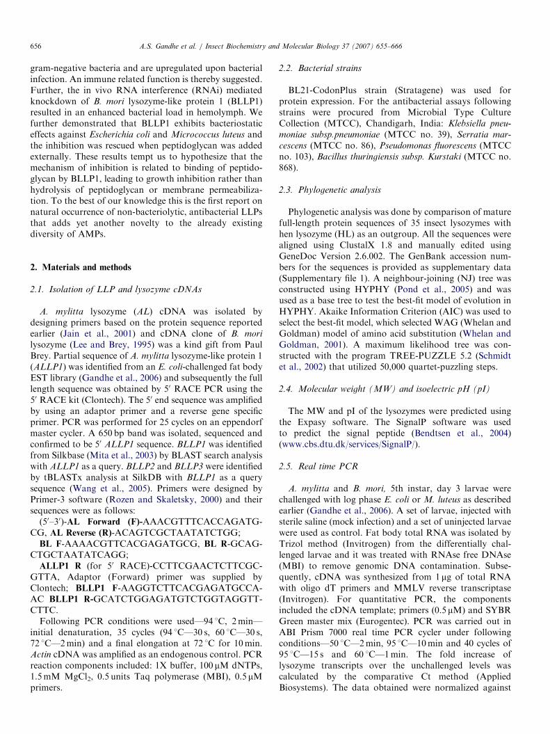
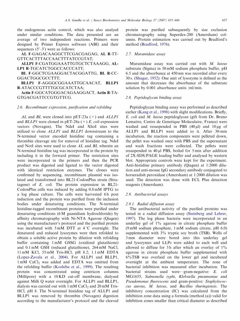
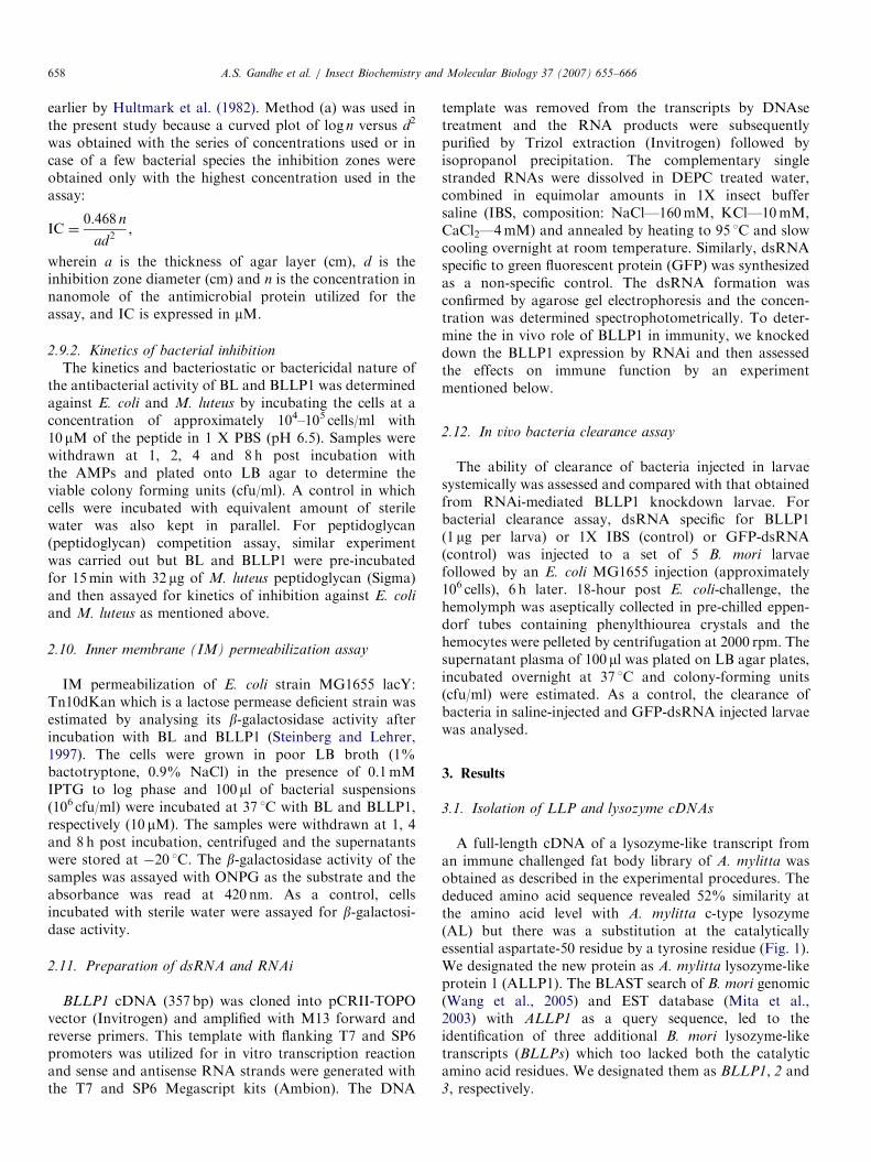
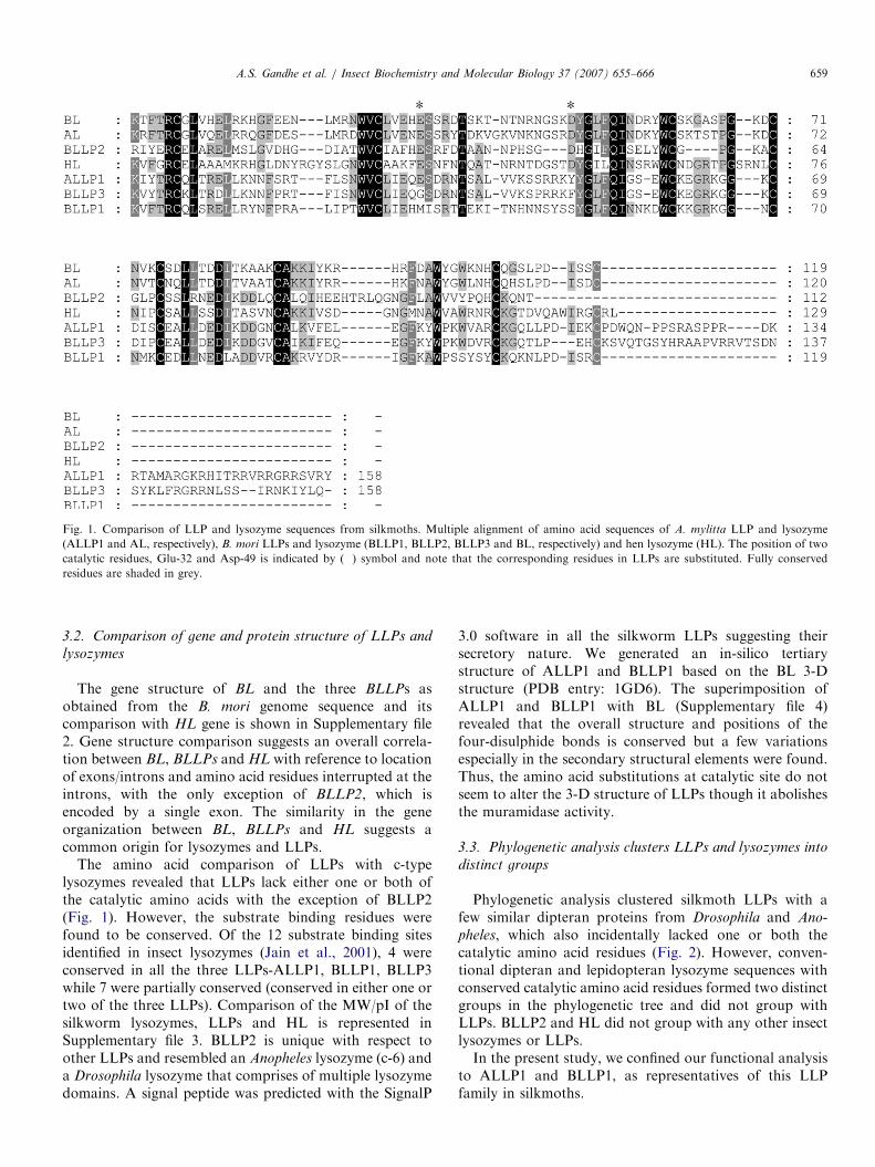
Fig. 1. Comparison of LLP and lysozyme sequences from silkmoths. Multiple alignment of amino acid sequences of A. mylitta LLP and lysozyme
(ALLP1 and AL, respectively), B. mori LLPs and lysozyme (BLLP1, BLLP2, BLLP3 and BL, respectively) and hen lysozyme (HL). The position of two
catalytic residues, Glu-32 and Asp-49 is indicated by (�) symbol and note that the corresponding residues in LLPs are substituted. Fully conserved
residues are shaded in grey.
A.S. Gandhe et al. / Insect Biochemistry and Molecular Biology 37 (2007) 655–666 659
3.2. Comparison of gene and protein structure of LLPs and
lysozymes
The gene structure of BL and the three BLLPs asobtained from the B. mori genome sequence and itscomparison with HL gene is shown in Supplementary file2. Gene structure comparison suggests an overall correla-tion between BL, BLLPs and HL with reference to locationof exons/introns and amino acid residues interrupted at theintrons, with the only exception of BLLP2, which isencoded by a single exon. The similarity in the geneorganization between BL, BLLPs and HL suggests acommon origin for lysozymes and LLPs.
The amino acid comparison of LLPs with c-typelysozymes revealed that LLPs lack either one or both ofthe catalytic amino acids with the exception of BLLP2(Fig. 1). However, the substrate binding residues werefound to be conserved. Of the 12 substrate binding sitesidentified in insect lysozymes (Jain et al., 2001), 4 wereconserved in all the three LLPs-ALLP1, BLLP1, BLLP3while 7 were partially conserved (conserved in either one ortwo of the three LLPs). Comparison of the MW/pI of thesilkworm lysozymes, LLPs and HL is represented inSupplementary file 3. BLLP2 is unique with respect toother LLPs and resembled an Anopheles lysozyme (c-6) anda Drosophila lysozyme that comprises of multiple lysozymedomains. A signal peptide was predicted with the SignalP
3.0 software in all the silkworm LLPs suggesting theirsecretory nature. We generated an in-silico tertiarystructure of ALLP1 and BLLP1 based on the BL 3-Dstructure (PDB entry: 1GD6). The superimposition ofALLP1 and BLLP1 with BL (Supplementary file 4)revealed that the overall structure and positions of thefour-disulphide bonds is conserved but a few variationsespecially in the secondary structural elements were found.Thus, the amino acid substitutions at catalytic site do notseem to alter the 3-D structure of LLPs though it abolishesthe muramidase activity.
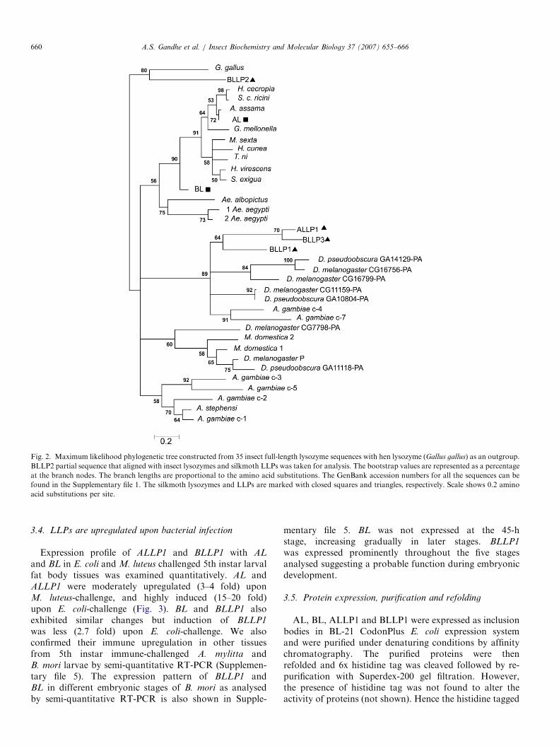
3.3. Phylogenetic analysis clusters LLPs and lysozymes into
distinct groups
Phylogenetic analysis clustered silkmoth LLPs with afew similar dipteran proteins from Drosophila and Ano-
pheles, which also incidentally lacked one or both thecatalytic amino acid residues (Fig. 2). However, conven-tional dipteran and lepidopteran lysozyme sequences withconserved catalytic amino acid residues formed two distinctgroups in the phylogenetic tree and did not group withLLPs. BLLP2 and HL did not group with any other insectlysozymes or LLPs.In the present study, we confined our functional analysis
to ALLP1 and BLLP1, as representatives of this LLPfamily in silkmoths.
ARTICLE IN PRESS
Fig. 2. Maximum likelihood phylogenetic tree constructed from 35 insect full-length lysozyme sequences with hen lysozyme (Gallus gallus) as an outgroup.
BLLP2 partial sequence that aligned with insect lysozymes and silkmoth LLPs was taken for analysis. The bootstrap values are represented as a percentage
at the branch nodes. The branch lengths are proportional to the amino acid substitutions. The GenBank accession numbers for all the sequences can be
found in the Supplementary file 1. The silkmoth lysozymes and LLPs are marked with closed squares and triangles, respectively. Scale shows 0.2 amino
acid substitutions per site.
A.S. Gandhe et al. / Insect Biochemistry and Molecular Biology 37 (2007) 655–666660
3.4. LLPs are upregulated upon bacterial infection
Expression profile of ALLP1 and BLLP1 with AL
and BL in E. coli and M. luteus challenged 5th instar larvalfat body tissues was examined quantitatively. AL andALLP1 were moderately upregulated (3–4 fold) uponM. luteus-challenge, and highly induced (15–20 fold)upon E. coli-challenge (Fig. 3). BL and BLLP1 alsoexhibited similar changes but induction of BLLP1
was less (2.7 fold) upon E. coli-challenge. We alsoconfirmed their immune upregulation in other tissuesfrom 5th instar immune-challenged A. mylitta andB. mori larvae by semi-quantitative RT-PCR (Supplemen-tary file 5). The expression pattern of BLLP1 andBL in different embryonic stages of B. mori as analysedby semi-quantitative RT-PCR is also shown in Supple-
mentary file 5. BL was not expressed at the 45-hstage, increasing gradually in later stages. BLLP1
was expressed prominently throughout the five stagesanalysed suggesting a probable function during embryonicdevelopment.
3.5. Protein expression, purification and refolding
AL, BL, ALLP1 and BLLP1 were expressed as inclusionbodies in BL-21 CodonPlus E. coli expression systemand were purified under denaturing conditions by affinitychromatography. The purified proteins were thenrefolded and 6x histidine tag was cleaved followed by re-purification with Superdex-200 gel filtration. However,the presence of histidine tag was not found to alter theactivity of proteins (not shown). Hence the histidine tagged

ARTICLE IN PRESS
Fig. 3. Real time PCR profile of (a) AL and ALLP1 and (b) BL and
BLLP1 in fat body from differentially challenged 5th instar, day 3 larvae.
The Y-axis represents the fold increase of the transcripts over the
unchallenged levels. The values reported are an average of two
independent experiments and the standard deviation is represented as
the Y error bar. X-axis represents the four differentially challenged larval
groups—1. Unch, unchallenged, 2. Saline; 3. ML-Micrococcus luteus; and
4. EC, E. coli.
Fig. 4. Peptidoglycan binding assay of ALLP1 and BLLP1 with M. luteus
and E. coli insoluble peptidoglycan. The bound (B) and free (F) fractions
were obtained as described in the text. All the samples were run on a 12%
SDS-PAGE gel and ALLP1 and BLLP1 proteins were detected by western
blot using anti-histidine antibodies. A control containing only ALLP1 or
BLLP1 without peptidoglycan was treated in the same manner as the test
samples and bound and free fractions were analysed. EC, E. coli, ML,
M. luteus.
Table 1
Muramidase activity of silkmoth lysozymes (AL, BL) and lysozyme-like
proteins (ALLP1, BLLP1)
Lysozyme/LLP Specific activity (U/mg)
AL 1565871117
BL 115727241
ALLP1 No activity
BLLP1 No activity
A.S. Gandhe et al. / Insect Biochemistry and Molecular Biology 37 (2007) 655–666 661
proteins were used for functional assays. The homogeneityand specificity of recombinant LLPs was confirmed bygel filtration, SDS-PAGE (Supplementary file 6) andwestern blot (not shown). The estimated MW of ALLP1and BLLP1 were 29.47 and 19.61 kDa, respectively,which is slightly higher than the predicted MW of boththe recombinant proteins. This is also true with thesilkworm lysozymes, wherein estimated MW was foundto be higher than the predicted MW. ALLP1 is a largerprotein than BLLP1 and possesses 39 extra amino acids atthe C-terminal end. Refolding was assessed by CDspectroscopy and the estimated secondary structure wasin agreement with the in-silico predicted structure (Supple-mentary file 7).
3.6. LLPs lack muramidase activity but exhibit substrate
binding property
The spectrophotometric assay for muramidase activityutilizes the lyophilized M. luteus as a substrate and theactivity is monitored by a decrease in the turbidity uponaddition of the enzyme. Assay results (Table 1) show thatALLP1 and BLLP1 lack muramidase activity even at 10times higher protein concentration over AL and BL, whichexhibited a specific activity of 15,657 and 11,571U/mg,respectively. This confirmed the ablation of muramidaseactivity in LLPs as predicted from the substitutions at thecatalytic residues (Malcolm et al., 1989).Both ALLP1 and BLLP1 were observed to bind
peptidoglycan, the characteristic lysozyme substrate, whenanalysed by western blot analysis (Fig. 4). This confirmsthat the LLPs bind to peptidoglycan although they areunable to hydrolyse it unlike classical lysozymes.
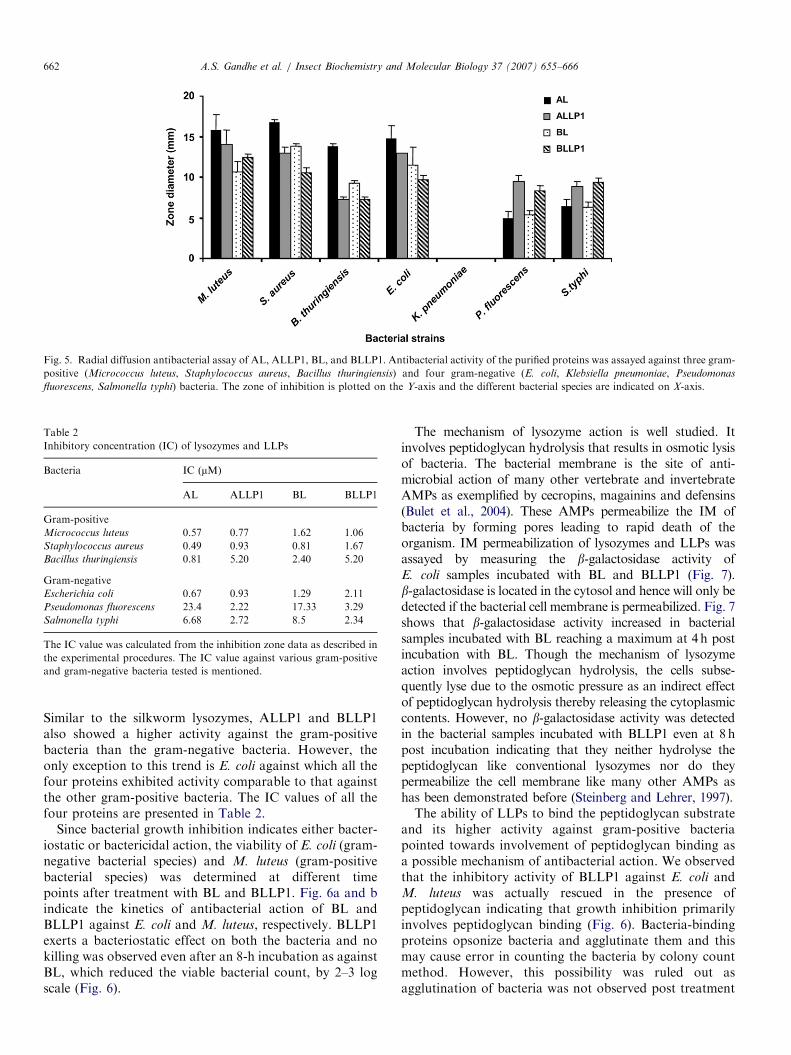
3.7. LLPs exhibit antibacterial activity
Both ALLP1 and BLLP1 inhibited a wide range ofbacterial species including both gram-positive and gram-negative bacteria in the radial diffusion assay (Fig. 5).
ARTICLE IN PRESS
Fig. 5. Radial diffusion antibacterial assay of AL, ALLP1, BL, and BLLP1. Antibacterial activity of the purified proteins was assayed against three gram-
positive (Micrococcus luteus, Staphylococcus aureus, Bacillus thuringiensis) and four gram-negative (E. coli, Klebsiella pneumoniae, Pseudomonas
fluorescens, Salmonella typhi) bacteria. The zone of inhibition is plotted on the Y-axis and the different bacterial species are indicated on X-axis.
Table 2
Inhibitory concentration (IC) of lysozymes and LLPs
Bacteria IC (mM)
AL ALLP1 BL BLLP1
Gram-positive
Micrococcus luteus 0.57 0.77 1.62 1.06
Staphylococcus aureus 0.49 0.93 0.81 1.67
Bacillus thuringiensis 0.81 5.20 2.40 5.20
Gram-negative
Escherichia coli 0.67 0.93 1.29 2.11
Pseudomonas fluorescens 23.4 2.22 17.33 3.29
Salmonella typhi 6.68 2.72 8.5 2.34
The IC value was calculated from the inhibition zone data as described in
the experimental procedures. The IC value against various gram-positive
and gram-negative bacteria tested is mentioned.
A.S. Gandhe et al. / Insect Biochemistry and Molecular Biology 37 (2007) 655–666662
Similar to the silkworm lysozymes, ALLP1 and BLLP1also showed a higher activity against the gram-positivebacteria than the gram-negative bacteria. However, theonly exception to this trend is E. coli against which all thefour proteins exhibited activity comparable to that againstthe other gram-positive bacteria. The IC values of all thefour proteins are presented in Table 2.
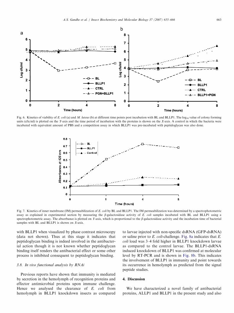
Since bacterial growth inhibition indicates either bacter-iostatic or bactericidal action, the viability of E. coli (gram-negative bacterial species) and M. luteus (gram-positivebacterial species) was determined at different timepoints after treatment with BL and BLLP1. Fig. 6a and bindicate the kinetics of antibacterial action of BL andBLLP1 against E. coli and M. luteus, respectively. BLLP1exerts a bacteriostatic effect on both the bacteria and nokilling was observed even after an 8-h incubation as againstBL, which reduced the viable bacterial count, by 2–3 logscale (Fig. 6).
The mechanism of lysozyme action is well studied. Itinvolves peptidoglycan hydrolysis that results in osmotic lysisof bacteria. The bacterial membrane is the site of anti-microbial action of many other vertebrate and invertebrateAMPs as exemplified by cecropins, magainins and defensins(Bulet et al., 2004). These AMPs permeabilize the IM ofbacteria by forming pores leading to rapid death of theorganism. IM permeabilization of lysozymes and LLPs wasassayed by measuring the b-galactosidase activity ofE. coli samples incubated with BL and BLLP1 (Fig. 7).b-galactosidase is located in the cytosol and hence will only bedetected if the bacterial cell membrane is permeabilized. Fig. 7shows that b-galactosidase activity increased in bacterialsamples incubated with BL reaching a maximum at 4h postincubation with BL. Though the mechanism of lysozymeaction involves peptidoglycan hydrolysis, the cells subse-quently lyse due to the osmotic pressure as an indirect effectof peptidoglycan hydrolysis thereby releasing the cytoplasmiccontents. However, no b-galactosidase activity was detectedin the bacterial samples incubated with BLLP1 even at 8hpost incubation indicating that they neither hydrolyse thepeptidoglycan like conventional lysozymes nor do theypermeabilize the cell membrane like many other AMPs ashas been demonstrated before (Steinberg and Lehrer, 1997).The ability of LLPs to bind the peptidoglycan substrate
and its higher activity against gram-positive bacteriapointed towards involvement of peptidoglycan binding asa possible mechanism of antibacterial action. We observedthat the inhibitory activity of BLLP1 against E. coli andM. luteus was actually rescued in the presence ofpeptidoglycan indicating that growth inhibition primarilyinvolves peptidoglycan binding (Fig. 6). Bacteria-bindingproteins opsonize bacteria and agglutinate them and thismay cause error in counting the bacteria by colony countmethod. However, this possibility was ruled out asagglutination of bacteria was not observed post treatment
ARTICLE IN PRESS
Fig. 6. Kinetics of viability of E. coli (a) and M. luteus (b) at different time points post incubation with BL and BLLP1. The log10 value of colony forming
units (cfu/ml) is plotted on the Y-axis and the time period of incubation with the proteins is shown on the X-axis. A control in which the bacteria were
incubated with equivalent amount of PBS and a competition assay in which BLLP1 was pre-incubated with peptidoglycan was also done.
Fig. 7. Kinetics of inner membrane (IM) permeabilization of E. coli by BL and BLLP1. The IM permeabilization was determined by a spectrophotometric
assay as explained in experimental section by measuring the b-galactosidase activity of E. coli samples incubated with BL and BLLP1 using a
spectrophotometric assay. The absorbance is plotted on Y-axis, which is proportional to the b-galactosidase activity and the incubation time of bacterial
samples with BL and BLLP1 is shown on X-axis.
A.S. Gandhe et al. / Insect Biochemistry and Molecular Biology 37 (2007) 655–666 663
with BLLP1 when visualized by phase contrast microscopy(data not shown). Thus at this stage it indicates thatpeptidoglycan binding is indeed involved in the antibacter-ial action though it is not known whether peptidoglycanbinding itself renders the antibacterial effect or some otherprocess is inhibited consequent to peptidoglycan binding.
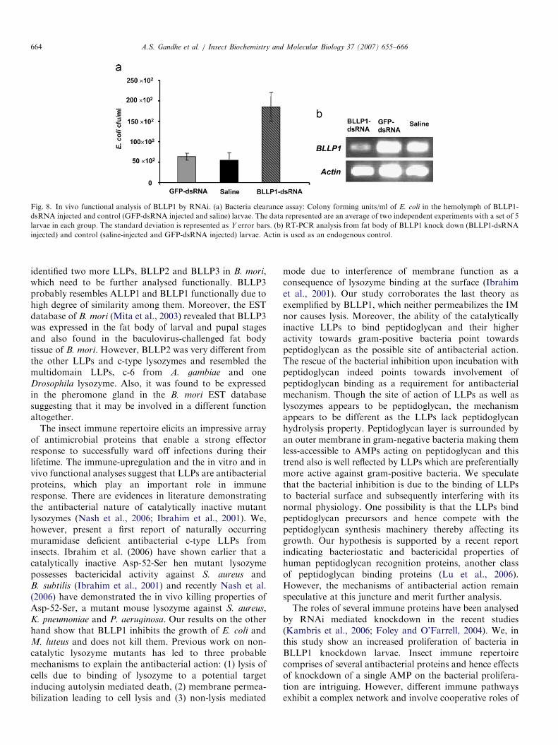
3.8. In vivo functional analysis by RNAi
Previous reports have shown that immunity is mediatedby secretion in the hemolymph of recognition proteins andeffector antimicrobial proteins upon immune challenge.Hence we analysed the clearance of E. coli fromhemolymph in BLLP1 knockdown insects as compared
to larvae injected with non-specific dsRNA (GFP-dsRNA)or saline prior to E. coli-challenge. Fig. 8a indicates that E.
coli load was 3–4 fold higher in BLLP1 knockdown larvaeas compared to the control larvae. The BLLP1-dsRNAinduced knockdown of BLLP1 was confirmed at molecularlevel by RT-PCR and is shown in Fig. 8b. This indicatesthe involvement of BLLP1 in immunity and point towardsits occurrence in hemolymph as predicted from the signalpeptide studies.
4. Discussion
We have characterized a novel family of antibacterialproteins, ALLP1 and BLLP1 in the present study and also
ARTICLE IN PRESS
Fig. 8. In vivo functional analysis of BLLP1 by RNAi. (a) Bacteria clearance assay: Colony forming units/ml of E. coli in the hemolymph of BLLP1-
dsRNA injected and control (GFP-dsRNA injected and saline) larvae. The data represented are an average of two independent experiments with a set of 5
larvae in each group. The standard deviation is represented as Y error bars. (b) RT-PCR analysis from fat body of BLLP1 knock down (BLLP1-dsRNA
injected) and control (saline-injected and GFP-dsRNA injected) larvae. Actin is used as an endogenous control.
A.S. Gandhe et al. / Insect Biochemistry and Molecular Biology 37 (2007) 655–666664
identified two more LLPs, BLLP2 and BLLP3 in B. mori,which need to be further analysed functionally. BLLP3probably resembles ALLP1 and BLLP1 functionally due tohigh degree of similarity among them. Moreover, the ESTdatabase of B. mori (Mita et al., 2003) revealed that BLLP3was expressed in the fat body of larval and pupal stagesand also found in the baculovirus-challenged fat bodytissue of B. mori. However, BLLP2 was very different fromthe other LLPs and c-type lysozymes and resembled themultidomain LLPs, c-6 from A. gambiae and oneDrosophila lysozyme. Also, it was found to be expressedin the pheromone gland in the B. mori EST databasesuggesting that it may be involved in a different functionaltogether.
The insect immune repertoire elicits an impressive arrayof antimicrobial proteins that enable a strong effectorresponse to successfully ward off infections during theirlifetime. The immune-upregulation and the in vitro and invivo functional analyses suggest that LLPs are antibacterialproteins, which play an important role in immuneresponse. There are evidences in literature demonstratingthe antibacterial nature of catalytically inactive mutantlysozymes (Nash et al., 2006; Ibrahim et al., 2001). We,however, present a first report of naturally occurringmuramidase deficient antibacterial c-type LLPs frominsects. Ibrahim et al. (2006) have shown earlier that acatalytically inactive Asp-52-Ser hen mutant lysozymepossesses bactericidal activity against S. aureus andB. subtilis (Ibrahim et al., 2001) and recently Nash et al.(2006) have demonstrated the in vivo killing properties ofAsp-52-Ser, a mutant mouse lysozyme against S. aureus,K. pneumoniae and P. aeruginosa. Our results on the otherhand show that BLLP1 inhibits the growth of E. coli andM. luteus and does not kill them. Previous work on non-catalytic lysozyme mutants has led to three probablemechanisms to explain the antibacterial action: (1) lysis ofcells due to binding of lysozyme to a potential targetinducing autolysin mediated death, (2) membrane permea-bilization leading to cell lysis and (3) non-lysis mediated
mode due to interference of membrane function as aconsequence of lysozyme binding at the surface (Ibrahimet al., 2001). Our study corroborates the last theory asexemplified by BLLP1, which neither permeabilizes the IMnor causes lysis. Moreover, the ability of the catalyticallyinactive LLPs to bind peptidoglycan and their higheractivity towards gram-positive bacteria point towardspeptidoglycan as the possible site of antibacterial action.The rescue of the bacterial inhibition upon incubation withpeptidoglycan indeed points towards involvement ofpeptidoglycan binding as a requirement for antibacterialmechanism. Though the site of action of LLPs as well aslysozymes appears to be peptidoglycan, the mechanismappears to be different as the LLPs lack peptidoglycanhydrolysis property. Peptidoglycan layer is surrounded byan outer membrane in gram-negative bacteria making themless-accessible to AMPs acting on peptidoglycan and thistrend also is well reflected by LLPs which are preferentiallymore active against gram-positive bacteria. We speculatethat the bacterial inhibition is due to the binding of LLPsto bacterial surface and subsequently interfering with itsnormal physiology. One possibility is that the LLPs bindpeptidoglycan precursors and hence compete with thepeptidoglycan synthesis machinery thereby affecting itsgrowth. Our hypothesis is supported by a recent reportindicating bacteriostatic and bactericidal properties ofhuman peptidoglycan recognition proteins, another classof peptidoglycan binding proteins (Lu et al., 2006).However, the mechanisms of antibacterial action remainspeculative at this juncture and merit further analysis.The roles of several immune proteins have been analysed
by RNAi mediated knockdown in the recent studies(Kambris et al., 2006; Foley and O’Farrell, 2004). We, inthis study show an increased proliferation of bacteria inBLLP1 knockdown larvae. Insect immune repertoirecomprises of several antibacterial proteins and hence effectsof knockdown of a single AMP on the bacterial prolifera-tion are intriguing. However, different immune pathwaysexhibit a complex network and involve cooperative roles of
ARTICLE IN PRESSA.S. Gandhe et al. / Insect Biochemistry and Molecular Biology 37 (2007) 655–666 665
different immune factors. It is possible that BLLP1 isinvolved in other functions in addition to the antibacterialeffector function. For example, human peptidoglycanrecognition proteins have recently been shown to functionas both recognition and antibacterial proteins (Lu et al.,2006). The immune role of LLPs is established by theseanalyses but a further study is required to probe its exactrole in the immune system. A strong expression oftranscripts at the embryonic stages also suggests a possiblerole in development. Many immune related proteinmolecules, for example, hemolin has been shown to havea dual role in immunity as well as development (Bettencourtet al., 2002) and hence other functions cannot be ruled out.
The current studies present a first report of a functionalanalysis of a novel class of LLPs lacking muramidaseactivity from insects. The functional annotation of this newgroup of lysozymes likely provides cues for analysis of theirhomologues in other organisms. For example, two humanc-type LLPs lacking catalytic residues have been recentlyreported (Zhang et al., 2005) whose function may beelucidated in the light of the insect LLPs reported in thepresent study.
Acknowledgements
We thank Dr. Paul Brey for providing the BL clone andDr. Bruno Lemaitre for providing M. luteus and E. coli
peptidoglycan. We thank Mr. Pankaj Kumar, Lab ofComputational Biology, CDFD for his valuable assistancein protein homology modeling. We are also thankful to MsSaisri, Centre for Cellular and Molecular biology (CCMB),Hyderabad, India for phase contrast microscopy andDr Manjula, CCMB for providing the E. coli MG1655lacY: Tn10dKan strain. This project is funded by Depart-ment of Biotechnology (DBT), Govt. of India, New Delhito JN under the Centre of Excellence (CoE) programme forGenetics and Genomics of Silkmoths and AG is a recipientof UGC fellowship.
Appendix A. Supplementary data
Supplementary data associated with the article canbe found in the online version at doi:10.1016/j.ibmb.2007.03.013.
References
Bendtsen, J.D., Nielsen, H., von Heijne, G., Brunak, S., 2004. Improved
prediction of signal peptides: SignalP 3.0. J Mol Biol 340, 783–795.
Bettencourt, R., Terenius, O., Faye, I., 2002. Hemolin gene silencing by
ds-RNA injected into Cecropia pupae is lethal to next generation
embryos. Insect Mol Biol 11, 267–271.
Bradford, M.M., 1976. A rapid and sensitive method for the quantitation
of microgram quantities of protein utilizing the principle of protein-
dye binding. Anal Biochem 72, 248–254.
Bulet, P., Stocklin, R., Menin, L., 2004. Anti-microbial peptides: from
invertebrates to vertebrates. Immunol Rev 198, 169–184.
Daffre, S., Kylsten, P., Samakovlis, C., Hultmark, D., 1994. The lysozyme
locus in Drosophila melanogaster: an expanded gene family adapted
for expression in the digestive tract. Mol Gen Genet 242, 152–162.
Dong, Y., Taylor, H.E., Dimopoulos, G., 2006. AgDscam, a Hypervari-
able Immunoglobulin Domain-Containing Receptor of theAnopheles
gambiae Innate Immune System. PLoS Biol 4, e229.
Foley, E., O’Farrell, P.H., 2004. Functional dissection of an
innate immune response by a genome-wide RNAi screen. PLoS Biol
2, E203.
Gandhe, A.S., P, A.K., John, S.H., Nagaraju, J., 2006. Analysis of
bacteria-challenged wild silkmoth, Antheraea mylitta (Lepidoptera)
transcriptome reveals potential immune genes. BMC Genomics 7, 184.
Hultmark, D., Engstrom, A., Bennich, H., Kapur, R., Boman, H.G., 1982.
Insect immunity: isolation and structure of cecropin D and four minor
antibacterial components from Cecropia pupae. Eur J Biochem 127,
207–217.
Ibrahim, H.R., Matsuzaki, T., Aoki, T., 2001. Genetic evidence that
antibacterial activity of lysozyme is independent of its catalytic
function. FEBS Lett 506, 27–32.
Jain, D., Nair, D.T., Swaminathan, G.J., Abraham, E.G., Nagaraju, J.,
Salunke, D.M., 2001. Structure of the induced antibacterial protein
from tasar silkworm, Antheraea mylitta. Implications to molecular
evolution. J Biol Chem 276, 41377–41382.
Kambris, Z., Brun, S., Jang, I.H., Nam, H.J., Romeo, Y., Takahashi, K.,
Lee, W.J., Ueda, R., Lemaitre, B., 2006. Drosophila immunity: a large-
scale in vivo RNAi screen identifies five serine proteases required for
Toll activation. Curr Biol 16, 808–813.
Kang, D., Liu, G., Lundstrom, A., Gelius, E., Steiner, H., 1998. A
peptidoglycan recognition protein in innate immunity conserved from
insects to humans. Proc Natl Acad Sci U S A 95, 10078–10082.
Koshiba, T., Hayashi, T., Miwako, I., Kumagai, I., Ikura, T., Kawano,
K., Nitta, K., Kuwajima, K., 1999. Expression of a synthetic gene
encoding canine milk lysozyme in E. coli and characterization of the
expressed protein. Protein Eng 12, 429–435.
Lee, W.J., Brey, P.T., 1995. Isolation and characterization of the
lysozyme-encoding gene from the silkworm Bombyx mori. Gene 161,
199–203.
Lopez-Zavala, A.A., de-la-Re-Vega, E., Calderon-Arredondo, S.A.,
Garcia-Orozco, K.D., Velazquez, E.F., Islas-Osuna, M.A., Valdez,
M.A., Sotelo-Mundo, R.R., 2004. Biophysical characterization of an
insect lysozyme from Manduca sexta. Protein Pept Lett 11, 85–92.
Lu, X., Wang, M., Qi, J., Wang, H., Li, X., Gupta, D., Dziarski, R., 2006.
Peptidoglycan recognition proteins are a new class of human
bactericidal proteins. J Biol Chem 281, 5895–5907.
Malcolm, B.A., Rosenberg, S., Corey, M.J., Allen, J.S., de Baetselier, A.,
Kirsch, J.F., 1989. Site-directed mutagenesis of the catalytic residues
Asp-52 and Glu-35 of chicken egg white lysozyme. Proc Natl Acad Sci
USA 86, 133–137.
Mita, K., Morimyo, M., Okano, K., Koike, Y., Nohata, J., Kawasaki, H.,
Kadono-Okuda, K., Yamamoto, K., Suzuki, M.G., Shimada, T.,
Goldsmith, M.R., Maeda, S., 2003. The construction of an EST
database for Bombyx mori and its application. Proc Natl Acad Sci
USA 100, 14121–14126.
Nash, J.A., Ballard, T.N., Weaver, T.E., Akinbi, H.T., 2006. The
peptidoglycan-degrading property of lysozyme is not required for
bactericidal activity in vivo. J. Immunol 177, 519–526.
Pond, S.L., Frost, S.D., Muse, S.V., 2005. HyPhy: hypothesis testing using
phylogenies. Bioinformatics 21, 676–679.
Rozen, S., Skaletsky, H., 2000. Primer3 on the WWW for general users
and for biologist programmers. Methods Mol Biol 132, 365–386.
Sadd, B.M., Schmid-Hempel, P., 2006. Insect immunity shows specificity
in protection upon secondary pathogen exposure. Curr. Biol 16,
1206–1210.
Schmidt, H.A., Strimmer, K., Vingron, M., von Haeseler, A., 2002.
TREE-PUZZLE: maximum likelihood phylogenetic analysis using
quartets and parallel computing. Bioinformatics 18, 502–504.
Shugar, D., 1952. The measurement of lysozyme activity and the ultra-
violet inactivation of lysozyme. Biochim. Biophys. Acta. 8, 302–309.

ARTICLE IN PRESSA.S. Gandhe et al. / Insect Biochemistry and Molecular Biology 37 (2007) 655–666666
Steinberg, D.A., Lehrer, R.I., 1997. Designer assays for antimicrobial
peptides. Disputing the ‘‘one-size-fits-all’’ theory. Methods Mol. Biol.
78, 169–186.
Wang, J., Xia, Q., He, X., Dai, M., Ruan, J., Chen, J., Yu, G., Yuan, H., Hu,
Y., Li, R., Feng, T., Ye, C., Lu, C., Wang, J., Li, S., Wong, G.K., Yang,
H., Wang, J., Xiang, Z., Zhou, Z., Yu, J., 2005. SilkDB: a knowledgebase
for silkworm biology and genomics. Nucleic Acids Res 33, D399–D402.
Watson, F.L., Puttmann-Holgado, R., Thomas, F., Lamar, D.L., Hughes,
M., Kondo, M., Rebel, V.I., Schmucker, D., 2005. Extensive diversity
of Ig-superfamily proteins in the immune system of insects. Science
309, 1874–1878.
Whelan, S., Goldman, N., 2001. A general empirical model of protein
evolution derived from multiple protein families using a maximum-
likelihood approach. Mol. Biol. Evol. 18, 691–699.
Yamakawa, M., Tanaka, H., 1999. Immune proteins and their gene
expression in the silkworm, Bombyx mori. Dev Comp Immunol 23,
281–289.
Zhang, K., Gao, R., Zhang, H., Cai, X., Shen, C., Wu, C., Zhao, S., Yu,
L., 2005. Molecular cloning and characterization of three novel
lysozyme-like genes, predominantly expressed in the male reproductive
system of humans, belonging to the c-type lysozyme/alpha-lactalbumin
family. Biol Reprod 73, 1064–1071.