Embed Size (px)
Citation preview

IMMUNITYMakes the bad pathogens die!

THE IMMUNE SYSTEM Involves
The lymphatic system The circulatory system Bone marrow
The lymphatic system can carry the plasma and white blood cells from the circulatory system
Lymph nodes are a common ‘collection’ site

FOREIGN BODIES Any cells not recognised as ‘self’ cells ‘Self’ cells are recognised by the immune
system by MHC (Major Histocompatibility Complex) markers on the surface
MHC markers are specific antigens found on the surfaces of all cells MHC 1 Markers are found on all cells, except RBC MHC 2 Markers are found on immunity cells (B
cells, T cells and some monocytes Cells lacking recognised MHC markers are
deemed ‘non-self’ and attract an immune response
Non-self markers that trigger a response of B cells or T cells are called antigens

SELF AND NON-SELF RECEPTORS

THREE LINES OF DEFENSE
1st line non-specific Blocks foreign bodies from entering tissues
2nd line non-specific Attacks foreign bodies in tissues
3rd line Specific Has a ‘memory’ Involves cell differentiation

1ST LINE
Mucous membranes Secreted by cells to ‘trap’ foreign particles
Secretions Includes tears, mucous, sweat, etc
Cilia Fine hairs lining the trachea Sweep foreign particles back to the oesophagus
Skin Acts a barrier between internal environments
(tissue) and external environments Natural flora
Symbiotic bacteria

2ND LINE
Phagocytes WBC which engulf and destroy foreign cells
Neutrophils (made in bone marrow) Monocytes (made in bone marrow) Monocytes become macrophages when they enter the
blood stream
Natural Killer (NK) cells Acts on viral-infected cells Release perforins (protein) which act to lyse
infected cells

COMPLEMENT PROTEINS
About 20 types Made in the liver Inactive until infection occurs Antibody-antigen complex attracts them to
site of infection Three ways to act
Stick to the bacteria to aide identification Destroy membrane of pathogen Stimulate pahgocytes to become more active

INTERFERON
Secreted by cells infected by virus Acts on local cells to avoid further infection Stimulates antiviral protein production
CYTOKINES Protein messenger
Messages between immunity cells Messages to nervous system
Secreted by many cells (particularly T cells)

INFLAMMATION
Controlled by multiple enzymes and compounds
Mast cells, basophils and platelets all release messengers to encourage inflammation
Inflammation involvse the dilation of blood vessels at the site of infection This brings more white blood cells It also accounts for the symptoms of swelling,
redness and heat The first phagocytes to the area release
histamine, this attracts more phagocytes Pus is generated from dead white blood cells
containing the pathogen

3RD LINE OF DEFENSE
Specific immunity Part of acquired immunity All lymphocytes originate in the bone marrow B cells
mature and differentiate in the bone marrow Involved in humoral responses
T cells Mature and differentiate in the thymus Involved in cell-mediated responses

TYPES OF CELLS
B Cells Plasma cells B memory cells
T Cells Th cells (T helper cells)
Activated Th cells Th memory cells
Tc cells (Cytotoxic T cells) Activated Tc cells Memory Tc cells

B CELLS
Have antibodies (immunoglobulins) on the surface
Antibodies are made of protein chains and possess very specific receptors

TYPES OF IMMUNOGLOBULINS IgA (two molecules)
Present in milk Active against viruses and some bacteria Present in tears and saliva
IgD (single molecule) Role is unknown
IgE (single molecule) Present in allergic reactions
IgG (single molecule) Able to cross placenta and present in milk Active against viruses and some bacteria
IgM (five molecules) Active against some bacteria and viruses

TYPES OF IMMUNOGLOBULINS

B CELLS
Each B cell is specific for one type of antigen (it only has one type of receptor on its surface)
Only a few copies of each B cell are created, so they all have slightly different DNA.
This allows the immune system to respond to millions of antigens
When an antigen enters the body it will quickly come into contact with the corresponding B cell
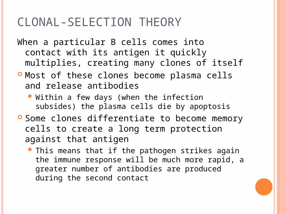
CLONAL-SELECTION THEORY
When a particular B cells comes into contact with its antigen it quickly multiplies, creating many clones of itself
Most of these clones become plasma cells and release antibodies Within a few days (when the infection subsides)
the plasma cells die by apoptosis Some clones differentiate to become memory
cells to create a long term protection against that antigen This means that if the pathogen strikes again the
immune response will be much more rapid, a greater number of antibodies are produced during the second contact

PRIMARY AND SECONDARY RESPONSES

T CELLS
Cell-mediated immunity T helper cells (Th cells) recognise antigens
and stimulate B cells Cytotoxic T cells (Tc cells) destroy infected
‘self’ cells These cells have foreign antigens on the surface
as well as MHC 1 markers The Tc cells recognises the foreign marker and
secretes proteins to lyse the cell Tc cells are not effective against free virus
particles Some Tc cells can also destroy cancerous cells

TYPES OF BLOOD CELLS

IMMUNITY CELLS

VACCINES

TYPES OF IMMUNITY
Active immunity Receiving the
pathogen and creating a normal immune response
Memory cells created Passive immunity
Infant receiving antibodies from the mother
No memory created
Active immunity Receiving
immunisation (aka. vaccines or antigens) to create an immune response
Memory cells created Passive immunity
Receiving antibodies from an injection/serum
No memory created
Natural Induced

VACCINE
Contain dead or inactive micro-organisms’ Inactive organisms can be called attenuated, as
they are still able to reproduce but cannot cause disease
The antigens present in the serum create an immune response without causing disease
This leads to a primary response, with memory cells being produced
If a vaccinated person comes into contact with the live pathogen, they will have an immediate and increased immune response
Toxoids, inactive toxins, can also be used in vaccines

ALLERGIES
Mast cells release histamines creating contraction in smooth muscles This decreases the passage of area in the
trachea and bronchi Mast cells are found in blood vessels and
connective tissue in the gut and respiratory tract Large scale IgE production creates a stronger
response, resulting in anaphylaxis IgE binds to mast cells to create a response IgE is released when ‘sensitive’ antigens are
present Some people are more sensitive to antigens than
others, as they have antibodies against that particular protein

AUTOIMMUNITY
The immune system fails to recognise ‘self’ proteins
B and T cells attack and destroy ‘self’ cells Examples:
Systemic Lupus Erythematosus – major organs (esp. kidney) are not recognised as self
Multiple sclerosis – the myelin sheath around nerves is not recognised as self
Rheumatoid arthritis – joint cartilage is not recognised as self

ORGAN TRANSPLANTS
Donor organs must match a certain number of markers in the recipient
Biological family members often have a high number of matches due to similar DNA being shared
T helper cells may recognise the grafted organ and attack it
Transplant patients must receive drugs to suppress the action of T cells – eg. Cyclosporin suppresses T cells, meaning the immune system is still partially active

HUMAN BLOOD TYPES
Your blood type is determined by the antigens present on your red blood cells
O type people can only receive O type blood, as they have antibodies against A-antigens and B-antigens
AB type people can receive any type of blood, because they have no antibodies against these antigens

COMPARISON OF BLOOD TYPE AND ANTIBODIES

RHESUS FACTOR
The “positive” or “negative” in blood types refers to another protein
The rhesus protein is either present (positive) or absent (negative)
Negative blood types can only receive negative blood
This can affect expectant mothers

RHESUS FACTOR IN PREGNANCY
If the mother is Rh+ then no problems occur If the mother is Rh- problems can arise
The first Rh+ fetus is unaffected, however at birth Rh+ blood cells can enter the mother, creating an immune response (the production of Rh antibodies
If the mother becomes pregnant to a second Rh+ child, antibodies from the mother may enter the fetus causing harm
A Rhesus negative mother can have injections after birth to remove the fetal blood cells from her bloodstream, reducing the immune response.

TESTING FOR BLOOD TYPES
Blood is put in 4 wells Antibodies are added to
the blood sample wells Anti-A is the antibody
for antigen A, Anti-B is for antigen B and Anti-D is for the rhesus factor
Agglutination is observed as clumping (indicating the presence of the antigen)

PLANT IMMUNITY
Mechanical barriers Cuticle or waxy layers Cork cells creating galls Dropping infected leaves
Chemical barriers Release of ‘gum’ to seal off infections Oils, acids and other chemical factors