Embed Size (px)
Citation preview

pubs.acs.org/JAFCPublished on Web 10/27/2010© 2010 American Chemical Society
J. Agric. Food Chem. 2010, 58, 11955–11961 11955
DOI:10.1021/jf102543g
Immunochemical and Mass Spectrometric Analysisof Nε-(Carboxymethyl)lysine Content of AGE-BSA SystemsPrepared with and without Selected Antiglycation Agents
CHOU SREY,*,† SIMON A. HAUGHEY,† LISA CONNOLLY,†
MARIA DOLORES DEL CASTILLO,§ JENNIFER M. AMES,# AND CHRISTOPHER T. ELLIOTT†
†Institute of Agri-Food and Land Use, School of Biological Sciences, Queen’s University,Belfast, Northern Ireland BT9 5AG, §Department of Food Analysis and Bioactivity,
Institute of Food Science Research (CSIC-UAM), C/Nicolas Cabrera 9, 28049 Madrid, Spain, and#School of Life Sciences, Northumbria University at Newcastle, England NE1 8ST
The present study was designed to compare surface plasmon resonance (SPR) biosensor, enzyme-
linked immunosorbent assay (ELISA), and ultraperformance liquid chromatography-tandem mass
spectrometry (UPLC-MS/MS) methods for the analysis of Nε-(carboxymethyl)lysine (CML) in glu-
cose-bovine serum albumin (BSA) model systems and to investigate the possible inhibitory effect of
selected compounds (R-tocopherol, ferulic acid, rutin, thiamin, thiamin monophosphate, and thiamin
pyrophosphate) on CML formation. The reported levels of CML detected were dependent upon the
method of analysis employed. The highest reported concentrations were obtained with the SPR
biosensor, whereas the lowest were found by ELISA. However, a high correlation was observed
between these two immunochemical procedures. CML concentrations were dependent upon the type
and concentration of the candidate CML inhibitor. All inhibitory compounds investigated, with the
exception of R-tocopherol, decreased the level of CML formation in the glucose-BSA system.
KEYWORDS: N ε-(Carboxymethyl)lysine (CML); advanced glycation endproducts (AGEs); CML inhibi-tor; enzyme-linked immunosorbent assay (ELISA); surface plasmon resonance (SPR) biosensor;ultraperformance liquid chromatography-tandem mass spectrometry (UPLC-MS/MS)
INTRODUCTION
Advanced glycation endproducts (AGEs) are a class of Mail-lard reaction (MR) products (MRPs).Chemically, theMRinvolvesa reaction between a free amino group, for example, the ε-aminogroups of lysine residueswithin protein, with the carbonyl group ofa reducing sugar, such as glucose (1). Nε-(Carboxymethyl)lysine(CML), one of the best known AGEs, can be formed througha number of different pathways. The condensation reactionbetween glucose and the ε-amino group of lysine forms fructose-lysine (the Amadori rearrangement product, ARP, of the re-action), which is subsequently oxidized to form CML. Glyoxalcan be formed from the oxidation of glucose, and it can alsoreact directly with the ε-amino group of lysine to form CML (2).CML has been associated with aging and diseases such as renalfailure and diabetes (3, 4). Furthermore, the accumulationof CML in the hearts of patients with diabetes may contributeto the increased risk of heart failure associated with hyperglyce-mia (5). Therefore, the search for inhibitors of CML formationis of significant medical interest. The effectiveness of variouspotentialglycationinhibitorshasbeentested inmodel systems (6,7),in foods (8), and in vivo (9). Mechanisms include reactive
carbonyl trapping (6, 10), antioxidant activity (7, 8), sugarautoxidation inhibition (6), and amino group binding inhibi-tion/competition (9, 11).
Several analytical methods have been reported for the detec-tion and quantification of CML, including LC-MS/MS (12),GC-MS (13), and ELISA (14). Recently, new analytical ap-proaches such as UPLC-MS/MS (15) and surface plasmonresonance imaging (SPRI) biosensor assays (16 ) have beenemployed for the quantitative analysis of CML. Only a smallnumber of reports on inter-/intralaboratory comparisons ofCML data obtained via different analytical approaches havebeen published to date in the scientific literature (13, 16).Biosensor assays have proved to be versatile, robust, and capableof producing rapid and reliable data for the analysis of a widerange of components in complex food matrices with minimalsample preparation (17-20). The main difference between ELI-SA and SPR biosensor assays is that the SPR biosensor approachis a label-free technique which relies for detection upon anincrease in molecular mass due to antibody-analyte interactionson a chip surface.
Research has been conducted on AGE inhibitors (8, 21),but a small number of reports have been focused on CML. Nocomparison of levels of CML determined by SPR, ELISA,and UPLC-MS/MS has been reported. Therefore, the presentstudy aimed to employ three analytical methods in the detectionof CML and to investigate the effect of three antioxidants
*Address correspondence to this author at the Institute of Agri-Food and Land Use, Queen’s University, David Keir Building,Stranmillis Road, Belfast BT9 5AG, Northern Ireland [phone þ 44(0)28 9097 6543; e-mail [email protected] or [email protected]].

11956 J. Agric. Food Chem., Vol. 58, No. 22, 2010 Srey et al.
(R-tocopherol, ferulic acid, and rutin hydrate) and competitors(thiamin, thiamin monophosphate, and thiamin pyrophosphate)on the formation of CML, in AGE-bovine serum albumin(BSA) model systems.
MATERIALS AND METHODS
Reagents andApparatus.All of the reagents used in this studywere ofanalytical grade. R-Tocopherol, bovine serum albumin (BSA, fraction V),ferulic acid, glucose, glyoxylic acid, keyhole limpet hemocyanin (KLH),N-hydroxysuccinimide (NHS), N-(3-dimethylaminopropyl)-N0-ethylcar-bodiimide hydrochloride (EDC), polyethylene glycol sorbitanmonolaurate(Tween 20), rutin hydrate, sodium cyanoborohydride, sodium phosphatemonobasic, sodium phosphate dibasic, sodium pyruvate, thiamin hydro-chloride, thiamin monophosphate, thiamin pyrophosphate, and otherchemicals were purchased from Sigma-Aldrich (Gillingham, U.K.).Dimethyl sulfoxide (DMSO), Hybridoma Feeder Supplement (Doma-Drive), Dulbecco’s modified eagle’s medium (DMEM), heat-inactivatedfetal calf serum (HI-FCS), hypoxanthine aminopterin thymidine (HAT)medium, penicillin streptomycin (pen strep), and polyethylene glycol(PEG) were from Invitrogen (Paisley, U.K.). Gelatin and horseradishperoxidase-linked anti-mouse immunoglobulin were obtained fromDAKO (Cambridge, U.K.). 3,30,5,50-Tetramethylbenzidine (TMB)solution was from Chemicon International (Temecula, CA). Theoptical surface plasmon resonance (SPR) biosensor system (BiacoreQ), Biosensor chip (CM5), ethanolamine hydrochloride (1 M), andHBS-EP buffer (0.01 MHEPES, 0.15 MNaCl, 3 mM EDTA, 0.005%polysorbate 20 (v/v), pH 7.4) were supplied by GE Healthcare(Uppsala, Sweden). Nunc-Immuno 96 microwell plates (NUNCBrand Products) were from Thermo-Scientific, Denmark. The TecanSafire plate reader was from Vector Scientific, Ireland. The Genevacevaporator (EZ-2) was from Ipswich, U.K. A Waters Acquity UPLCtriple-quadrupole MS/MS (Manchester, U.K.) was used for massspectrometric analyses.
Sample Preparation. Glycated Bovine Serum Albumin (GlycatedBSA). BSA (10mg, equivalent to 8.85mM lysine), glucose (90mg, 0.5M),and inhibitors (8.85 and 88.5mMR-tocopherol, ferulic acid, rutin hydrate,thiamin hydrochloride, thiaminmonophosphate, thiamin pyrophosphate)weremixed in sodium phosphate buffer (0.2M, pH 7.2, 1 mL) to bring themolecular ratio of lysine/inhibitor to 1:1 and 1:10. Themixed sampleswereincubated, in a 5mL glass bottle with a screw-tight lid, at 50 �C for 10 daysand vortexed once per day. Glycated BSA, without addition of aninhibitor, was prepared by mixing BSA (10 mg) with glucose (90 mg) inphosphate buffer (0.2 M, pH 7.2, 1 mL) and used as a positive control tocalculate the percentage inhibition of CML formation caused by thetrialed compounds. All of the samples were prepared in triplicate and werestored at -20 �C prior to analysis.
Nε-(Carboxymethyl)lysine-Bovine Serum Albumin/Keyhole LimpetHemocyanin Protein (CML-BSA/KLH). CML-BSA was prepared byincubating BSA (10 mg, equivalent to 8.85 mM lysine) with glyoxylic acid(8.85 mM) in phosphate buffer (pH 7.5, 0.5 M) for 1 h at 37 �C, and thensodium cyanoborohydride (17.70 mM) was added and the incubationcontinued for a further 23 h. The pH was adjusted to 7.4 with NaOH(0.1 M), if required. CML-modified KLH was prepared by incubatingKLH (10 mg) with glyoxylic acid (260 mM) in phosphate buffer (pH 7.5,0.5M) for 1 h at 37 �C, and then sodium cyanoborohydride (520mM)wasadded and the incubation continued for a further 23 h. Blank samples werealso prepared as previously stated, but with the omission of glyoxylic acid.The incubated solutions were dialyzed against phosphate buffer (0.05 M,pH 7.2) containing 0.15 M NaCl and stored at -20 �C. The conjugateswere used to prepare immunogens to raise antibodies and for use in theELISA and SPR assays.
Nε-(Carboxyethyl)lysine-Modified Bovine Serum Albumin (CEL-BSA). CEL-BSA was prepared by incubating BSA (20 mg/mL), sodium
pyruvate (17.14 mM, 17.14 μL), and sodium cyanoborohydride(25.71 mM, 25.7 μL) in phosphate buffer (0.2M, pH 7.4, 1 mL). The solu-tionwas incubated at 37 �C for 24 h.A control was also prepared using thesame conditions but with the omission of sodium pyruvate.
Preparation of a Monoclonal Antibody to CML. The immuniza-tion and fusion procedure described previously by Stewart et al. (17) was
used to prepare anti-CML monoclonal antibody. Briefly, three BALB/cmice were immunized at 3 week intervals with CML-KLH immunogen(20 μg of protein). Primary and secondary booster immunizations wereadministered using Quil A adjuvant by subcutaneous injection. Third(20 μg protein) and fourth (80 μg protein) boosters were administered byintraperitoneal injection with Freund’s complete adjuvant. Tail bleedstaken from mice 10 days after each booster were tested using ELISA andSPR assays. The most responsive mouse, as determined by antibody titer,was selected and, 4 days prior to the fusion being performed, received afinal booster intraperitoneally of the immunogen (100 μg of protein) inphosphate-buffered saline (pH 7.2). The fusion was performed accordingto a modification of the method of Kohler and Milstein (22). A single cellsuspension was collected from the spleen of the immunized mouse andfused with SP2/O-Ag14 myeloma cells using polyethylene glycol. After10-14 days of fusion, the resulting hybrid cells (hybridomas) werescreened using ELISA and SPR assays. Serum from the final heart bleedof the fusion mouse was used as a positive control, and cell culture me-dium buffer was used as a negative control, in the screening assays.The hybridomas that produced antibodies specific for CML but did notbind to CEL were selected for further investigation. All cell lines that gavea strong binding to CML-coated biosensor chips (as detected by SPR)were cloned twice and selected for scale-up antibody production, and theproducts were stored in liquid nitrogen.
Developmentof anEnzyme-Linked ImmunosorbentAssay (ELISA).Ninety-six-well NuncMaxisorp plates were coated with CML-BSA (1 μg/mL, 100 μL) and blocked with PBS/gelatin (1%) blocking buffer (0.1 M,pH 7.2, 200 μL) overnight at room temperature. After the blocking bufferwas discarded, 50 μL of glycated BSA (with or without inhibitors, 300 μg/mL) and 50 μL of anti-CML antibody, 1:15000-fold dilution, were addedto the wells and incubated by shaking at 37 �C for 90min. The supernatantwas discarded and the plate washed three times with wash buffer (1%Tween 20 and 0.9%NaCl). The secondary goat anti-mouseHRPantibodywas added (1:2000 dilution) and incubated at 37 �C for 60 min. Thesupernatant was again discarded, and the plate was washed three timeswith wash buffer. TMB (100 μL) was added to each well and developed indarkness for 5 min. The substrate reaction was stopped using sulfuric acid(2.5 M, 25 μL/well). Absorbance was read at 450 nm usinga microplate reader. A range of CML-BSA standards (50%, 0-100 μgof BSA/mL, equivalent to 0-3500 μg of CML/g BSA) were also added toa number of wells and used to generate a calibration curve. Prism5 software was used to calculate CML concentration in the samples.Buffer (no antibody) was used as a negative control. Unheated glycatedBSA (prepared with and without an inhibitor) was tested for backgroundeffects.
Development of the SPR Biosensor Assay. CML-BSA Immobi-lization onto a Biosensor Chip (CM5). The CM5 sensor chip is composedof a glass slide coated with a thin layer of gold to which a carboxymethyl-ated dextran matrix is covalently attached. The conditions for theimmobilization of the CML-BSA were optimized (e.g., concentration ofCML-BSA, speed of injection, and contact time) to ensure a high surfacecoverage of the protein. The immobilization procedure was performedwithin the biosensor unit using Biacore control software. Briefly, the con-centration, injection speed, and contact time of each solution were carriedout as follows: EDC (0.4M, 50%) andNHS (0.1M, 50%)weremixed andinjected for 7 min (10 μL/min) onto the CM5 chip (flow cell 1), to activatecarboxyl groups on the chip. Jeffamine diluted (1:5) in sodium boratebuffer (pH 8.5, 63mM) was injected (5 μL/min, 7min) to cover the surfaceof the flow cell with amino groups. The flow cell was then deactivatedwithethanolamine-HCl (10 μL/min, 3 min). After deactivation, CML-BSAsolutionswere coated for 10min (1mg/mL). The coated chipwas ready foruse immediately or could be stored at 4 �C for several months under dryconditions. After use, the chip was washed with distilled water and driedunder a gentle stream of nitrogen.
Samples Analysis Using the SPR Biosensor Assay. Glycated BSAsamples (10 mg of BSA/mL, 2 μL) and HBS-EP buffer (pH 7.4, 38 μL)were pipetted and mixed in a 96-well plate to bring the final concentrationto 0.5 mg of BSA/mL. The CML-specific monoclonal antibodies werediluted in HBS-EP buffer (60-fold dilution). The antibody was mixedautomatically with glycated BSA (50:50) in wells of a 96-microtiter plateand then injected over the CML-BSA coated chip at a flow rate of 10 μL/min and a contact time 2 min. The chip surface was regenerated
Article J. Agric. Food Chem., Vol. 58, No. 22, 2010 11957
with 50 mM NaOH (flow rate = 25 μL/min, 1 min contact time).The concentration of CML in glycated samples was calculated against aCML-BSA calibration curve (50%, 0-100 μg of BSA/mL, equivalent to0-3500 μg of CML/g BSA, R2 = 0.997) and using Prism 5 software.
UPLC-MS/MS Analysis. The CML content of glycated BSA wasdetermined byUPLC-MS/MS (15). Samples were prepared for analysis bysodium borohydride reduction, protein isolation using TCA precipitation,protein hydrolysis with 6 M HCl at 110 �C for 24 h, and solid phaseextraction using a C18 cartridge. Protein hydrolysates (equivalent to 7.5 μgof protein, 7.5 μL) were injected into a BEH C18 UPLC column (Waters,2.1 � 50 mm, 1.7 μm) housed in a column oven at 50 �C in gradientelution mode. Solvent A was nonafluoropentanoic acid (NFPA, 5 mM),and solvent Bwas acetonitrile. The injection timewas 7.5min.The analysiswas performed using a Waters Acquity UPLC coupled to a WatersPremier triple-quadruple MS operating in multiple reaction monitoring(MRM) mode. The flow rate was 0.2 mL/min. The MS was operated inelectrospray ionization (ESI) positive mode usingMRMmode. The CMLdata were analyzed usingMassLynx software. CML concentrations in thesamples were quantified by means of reference to the internal standard.Data were reported as the mean ( SD.
Statistical Analysis. Statistical analysis (ANOVA) was performed todetermine differences between three groups of means (P<0.01). Limits ofdetection (LOD) and limits of quantification (LOQ) ofCMLconcentrationanalysis by immunochemical methods (SPR and ELISA) were determinedfrom three independent runs of 20 unheated AGE-BSA samples. TheLOD and LOQ of CML concentration, analysis by UPLC-MS/MSmethod, were 1.62 and 5.41 μg of CML/g of BSA, respectively (15).
RESULTS
Anti-CMLMonoclonalAntibody.Fourteendays after fusion, 447hybridoma supernatants were screened by two methods (ELISAand SPR biosensor). ELISA screening of the 447 hybridomasupernatants gave 23 positive reactors to CML-BSA, whereasscreening by SPR biosensor revealed 5 positive reactors. These 5positive reactors (shown by SPR) were also found to be positivebinders by ELISA and were considered to be true positivebinders. After further testing using CML-BSA and CEL-BSAin assay inhibition studies, 1 of the 5 positive reactors was selectedfor full assay development and designated 2C1. This monoclonalantibodywas chosen because it exhibited the highest specificity ofthe five positive reactors for CML-BSA but not CEL-BSA asobserved in inhibitionbindingELISA. 2C1was applied toELISAand SPR assays for the determination of CML in glycated BSA.
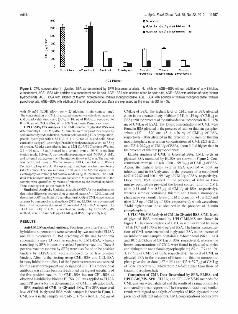
SPR Analysis of CML in Glycated BSA. The SPR-measuredlevel of CML in glycated BSAmodel samples is shown in Figure 1.CML levels in the samples were (45 ( 4.76)-(1603 ( 156) μg of
CML/g of BSA. The highest level of CML was in BSA glycatedeither in the absence of any inhibitor (1543 ( 119 μg of CML/g ofBSA) or in thepresenceof the antioxidantR-tocopherol (1603(156μg of CML/g of BSA). The lowest concentrations of CML werefound in BSA glycated in the presence of rutin or thiamin pyropho-sphate (127 ( 5.20 and 45 ( 4.76 μg of CML/g of BSA,respectively). BSA glycated in the presence of thiamin or thiaminmonophosphate gave similar concentrations of CML (223 ( 20.1and 233( 20.2 μg of CML/g of BSA), about 5-fold higher than inthe presence of thiamin pyrophosphate.
ELISA Analysis of CML in Glycated BSA. CML levels inglycated BSA measured by ELISA are shown in Figure 2. Con-centrations were (6 ( 0.80)-(900 ( 99.0) μg of CML/g of BSA.Again, the highest levels were in BSA glycated without aninhibitor and in BSA glycated in the presence of R-tocopherol(852( 27.82 and 900( 99.0 μg of CML/g of BSA, respectively).Once more, BSA glycated in the presence of rutin or thia-min pyrophosphate provided the lowest concentration of CML(8 ( 0.55 and 6 ( 0.37 μg of CML/g of BSA, respectively),whereas samples containing thiamin and thiamin monophos-phate gave very similar levels of CML formation (41 ( 2.20 and66( 3.43 μg of CML/g of BSA, respectively), which were about7-fold higher than those obtained in the presence of thiaminpyrophosphate.
UPLC-MS/MSAnalysis of CML in Glycated BSA.CML levelsof glycated BSA measured by UPLC-MS/MS are shown inFigure 3. The concentrations of CML in samples varied between194( 19.7 and 1075( 68.6 μg/g of BSA. The highest concentra-tions of CMLwere determined in glycated BSA in the absence ofan inhibitor and samples containing R-tocopherol (960 ( 14.4and 1075( 68.6 μg of CML/g of BSA, respectively), whereas thelowest concentrations of CML were found in glycated samplescontaining rutin and thiamin pyrophosphate (389( 17.7 and 194( 19.7 μg of CML/g of BSA, respectively). The level of CML inglycated BSA in the presence of thiamin or thiamin monophos-phate gave similar data (467( 25.4 and 433( 39.7 μg of CML/gof BSA, respectively), which were 2.4-fold higher than those ofthiamin pyrophosphate.
Comparison of CML Data Determined by SPR, ELISA, and
UPLC-MS/MS. SPR, ELISA, and UPLC-MS/MS methods forCMLanalysis were validated and the results of a range of samplescompared by linear regression. The threemethods showed similartrends with regard to analysis of samples of BSA glycated in thepresence of different inhibitors. CML concentrations obtained by
Figure 1. CML concentration in glycated BSA as determined by SPR biosensor analysis. No inhibitor, AGE-BSA without addition of any inhibitor;R-tocopherol, AGE-BSA with addition of R-tocopherol; ferulic acid, AGE-BSA with addition of ferulic acid; rutin, AGE-BSA with addition of rutin; thiaminhydrochloride, AGE-BSA with addition of thiamin hydrochloride; thiamin monophosphate, AGE-BSA with addition of thiamin monophosphate; thiaminpyrophosphate, AGE-BSA with addition of thiamin pyrophosphate. Data are expressed as the mean( SD (n = 3).

11958 J. Agric. Food Chem., Vol. 58, No. 22, 2010 Srey et al.
SPR,ELISA, andUPLC-MS/MSmethods are shown inFigure 4.Correlations between CML levels determined by SPR versusELISA, UPLC-MS/MS versus SPR, and UPLC-MS/MS versusELISA methods of analysis are shown in panels a, b, and c,respectively, of Figure 5.
The SPRbiosensor analysis ofCMLgave the highest LODandLOQ (1.7 and 17 μg of CML/g of BSA, respectively), whereasELISA analysis gave the lowest (1.51 and 5.20 μg of CML/g ofBSA, respectively). The LOD and LOQ for UPLC-MS/MS were1.62 and 5.41 μg of CML/g of BSA, respectively, very similarto the values for the ELISA. The intra-assay % CV for samplesanalyzed by each method was found to be <10%. The inter-assay % CV was also <10% for the SPR and UPLC-MS/MSmethods, but the variation for the ELISA was up to 24% forsome samples. CML concentrations detected by ELISA [(6 (0.80)-(900 ( 99.0) μg/g of BSA) were significantly lower(P < 0.01) compared to SPR biosensor [(45 ( 4.76)-(1603 (156) μg/g of BSA) and UPLC-MS/MS [(194 ( 19.7)-(1075 (68.6) μg/g of BSA) methods (Figure 4). CML levels detected inglycated BSA using the SPR biosensor were 1.6-fold higher thanthose obtained by ELISA, but there was good linearity betweenthemethods (R2=0.977) (Figure 5a). The concentration ofCMLobtained by UPLC-MS/MS was 0.79-fold lower than by SPRanalysis (Figure 5b), but 1.35-fold higher than by ELISA assay
(Figure 5c). The coefficients of determination of a linear correlationCML values obtained between UPLC-MS/MS versus SPR andUPLC-MS/MS versus ELISA were fairly good (R2 = 0.85 and0.75, respectively).
DISCUSSION
An earlier study compared a SPRI biosensor method and anELISA for the analysis of AGEs in serum from Zucker diabeticfatty (ZDF) rats and Zucker lean (ZL) rats (16). The authorsreported that the two methods gave similar results, although thedata from the two assayswere expressed in different units. It is notpossible to compare those data sets with CML concentrationsdetermined by the three methods used in the current study. Thedifferences in values obtained by the two methods may be due tothe different equipment used. In the present study a SPRbiosensor assay was employed, whereas Kim et al. (16) useda surface plasmon resonance imaging (SPRI) biosensor. A pub-lished study (23) reported the detection levels of progesterone inbovine milk to be higher when analyzed by a biosensor comparedto an ELISA, but, in agreement with our study, a reasonablecorrelation (R2 = 0.75) was observed between both methods ofanalysis. In contrast, Yman et al. (19), measuring tropomyosin incrabmeat (surimi), illustrated that biosensor analysis reportedlower levels than an ELISA. The discrepancies observed between
Figure 2. CML concentration in glycated BSA determined by ELISA assay. No inhibitor, AGE-BSA without addition of any inhibitor; R-tocopherol,AGE-BSA with addition of R-tocopherol; ferulic acid, AGE-BSA with addition of ferulic acid; rutin, AGE-BSA with addition of rutin; thiamin hydrochloride,AGE-BSA with addition of thiamin hydrochloride; thiamin monophosphate, AGE-BSA with addition of thiamin monophosphate; thiamin pyrophosphate,AGE-BSA with addition of thiamin pyrophosphate. Data are expressed as the mean ( SD (n = 3).
Figure 3. CML concentration in glycated BSA determined by UPLC-MS/MS analysis. No inhibitor, AGE-BSAwithout addition of any inhibitor;R-tocopherol,AGE-BSA with addition of R-tocopherol; ferulic acid, AGE-BSA with addition of ferulic acid; rutin, AGE-BSA with addition of rutin; thiamin hydrochloride,AGE-BSA with addition of thiamin hydrochloride; thiamin monophosphate, AGE-BSA with addition of thiamin monophosphate; thiamin pyrophosphate,AGE-BSA with addition of thiamin pyrophosphate. Data are expressed as the mean ( SD (n = 3).

Article J. Agric. Food Chem., Vol. 58, No. 22, 2010 11959
the methods applied in these different papers may be due todifferent instrumentations in the case of biosensor-based assay orinterfering factors that are present in the samples (24).
Compared to UPLC-MS/MS, immunochemical (ELISA andSPR) analysis has advantages such as simpler sample prepara-tion, speed, and cost.However, the advantages ofUPLC-MS/MSinclude the smaller volumes of sample (7.5 μL, 7.5 μg of protein)required compared to ELISA and SPRmethods. The correlationof the CML levels found in glycated samples, with or withoutinhibitors, analyzed by immunochemical methods and UPLC-MS/MS were also well correlated. Charissou et al. (13) reporteda good correlation between ELISA and GC-MS data for CMLlevels in model milk (slope =1.18) and powdered formulas, butsatisfactory linear or nonlinear fitting in liquid formula was notobserved. These authors also reported that data for CML inliquid milk were almost 10-fold higher when analyzed by ELISAcompared toGC-MS (13). This is in contrast to the present studyin which measured concentrations of CML in glycated BSAobtained by the ELISA were lower compared to those obtainedby UPLC-MS/MS.
In the current study, the concentration of CML in glycatedBSA was dependent on the concentration and the nature of theinhibitor used. R-Tocopherol did not affect CML formation,whatever the concentration of R-tocopherol applied. In contrast,ferulic acid, rutin, thiaminhydrochloride, and thiamin derivatives
(thiamin monophosphate and thiamin pyrophosphate) inhibitedCML formation, and the inhibitory effect increased with con-centration. In good agreement with the current study, Yinand Chan (25) published that R-tocopherol did not inhibitCML and pentosidine formation in the glycated BSA modelsystem. This is possibly due to R-tocopherol insolubility in theaqueous media (phosphate buffer, 0.2 M, pH 7.2) used in bothinvestigations.
Ferulic acid is a free radical scavenger (26, 27), and in thecurrent study its inhibitory effect on CML formation was con-centration dependent. The strong free radical scavenging(hydroxyl or superoxide radical) activity of ferulic acid isdue to its phenolic nucleus and extended side-chain conjuga-tion, which allow it to form a resonance-stabilized phenoxyradical (8 , 26 , 27 ). The anti-CML effect of ferulic acid isattributed to the second phase of the glycation reaction, thatis, glyoxal production from sugar or Amadori productoxidation. The data presented here agree with an earlierstudy (8 ) suggesting that ferulic acid (0.25 mg/mL) inhibitsAGE formation as a result of its free radical scavengingcapacities. However, much lower concentrations of ferulicacid appear not to inhibit CML formation (7 ).
Rutin, a powerful antioxidant and antiglycation agent, inhib-ited CML formation in the current study due to its free radicalscavenging capacity (21), which mainly inhibits glyoxal (10)
Figure 4. Comparison of CML concentration detected by threemethods of analysis (SPR, ELISA, and UPLC-MS/MS): (a) amount of AGE inhibitor used wasequimolar with respect to the lysine content of BSA; (b) amount of AGE inhibitor used was 10-fold greater than the lysine content of BSA. No inhibitor,AGE-BSA without addition of any inhibitor; R-tocopherol, AGE-BSA with addition of R-tocopherol; ferulic acid, AGE-BSA with addition of ferulic acid;rutin, AGE-BSA with addition of rutin; thiamin hydrochloride, AGE-BSA with addition of thiamin hydrochloride; thiamin monophosphate, AGE-BSAwith addition of thiamin monophosphate; thiamin pyrophosphate, AGE-BSA with addition of thiamin pyrophosphate. Data are expressed as the mean( SD(n = 3).

11960 J. Agric. Food Chem., Vol. 58, No. 22, 2010 Srey et al.
formation. The data agree with earlier studies (6,10,21) suggest-ing that rutin inhibits all stages of protein glycation formation,that is, autoxidation of glucose, glyoxal formation, retroaldocondensation of Schiff base, and oxidative degradation of Ama-dori products to CML. Furthermore, dietary rutin has beenproven to reduce glycation in tissue protein of streptozotocin-induceddiabeticrats(28).Datafrompreviousstudies (6,10,21,28)and our current investigation would suggest that rutin is a power-ful antioxidant which inhibits CML formation both in vitroand in vivo.
Thiamin and its derivatives, thiamin monophosphate andthiamin pyrophosphate, are not antioxidants, but do inhibitCML formation, dependent on their concentration investigated,in the current study. The inhibitory effect may be througha competitive mechanism between the amino group of thiamin/thiamin derivatives with the amino group of lysine residueswithin protein during glycation, as well as R-oxoaldehyde forma-tion (29). The data agree with CMLplasma levels of diabetic rats,which were reduced by thiamin administered orally (9). Besidesthis evidence, Booth et al. (11) also report a similar effect of
thiamin pyrophosphate on AGE formation (98%), even at lowconcentrations. However, the authors (11) did not observe anyantiglycation capacity for thiamin and thiamin monophosphate.In this investigation we found thiamin pyrophosphate to exhibita greater inhibitory effect on CML formation than thiamin andthiamin monophosphate. Each has a similar chemical structure,and all contain a functional amino group. The proposedmechan-ism may be due to the diphosphate group on thiamin pyrophos-phate interfering in the reaction rate between the amino group ofthiamin pyrophosphate and the carbonyl group of a reducingsugar during the glycation process, thus inhibiting the formationof CML through a competitive reaction with the amino group onthe protein.
In conclusion, the concentration of CML formed was depen-dent on the types and concentrations of AGE inhibitor, and thereported level ofCMLwas found to be dependent on themethodsof analysis (SPR biosensor, ELISA, and UPLC-MS/MS). How-ever, good correlations were observed between those threeanalysis methods of analysis. The concentrations of CMLdetected by SPR and UPLC-MS/MS were closer compared to
Figure 5. Correlation between CML concentrations obtained by SPR, ELISA, and UPLC-MS/MS analysis: (a) correlation of CML levels obtained by SPR andELISA analysis; (b) correlation of CML levels obtained by UPLC-MS/MS and SPR analysis; (c) correlation of CML levels obtained by UPLC-MS/MS andELISA analysis.
Article J. Agric. Food Chem., Vol. 58, No. 22, 2010 11961
an ELISA analysis. The SPR biosensor has a number of advan-tages over ELISA such as the need for smaller volumes ofreagents, no need for a labeled compound, higher repeatability,high automation, and higher precision between runs. However,the biosensor assay required a higher concentration of the anti-body. Immunochemical analysis of CML, in the AGE-BSAmodel system, has advantages over the UPLC-MS/MS methodsuch as reduced sample preparation, reduced analysis time,increased speed, and lower costs. R-Tocopherol had no measur-able effect on CML formation in AGE-BSA model systems. Incontrast, ferulic acid, rutin, thiamin, and thiamin metabolites,thiamin monophosphate and thiamin pyrophosphate, showedvarious degrees of antiglycation capacity on CML formation.These compounds may be used for health therapy.
ABBREVIATIONS USED
CML,Nε-(carboxymethyl)lysine; CEL,Nε-(carboxyethyl)lysine;AGEs, advanced glycation endproducts; BSA, bovine serumalbumin; KLH, keyhole limpet hemocyanin; CML-BSA,Nε-(carboxymethyl)lysine-modified bovine serum albumin; CEL-BSA, Nε-(carboxyethyl)lysine-modified bovine serum albumin;CML-KLH, N ε-(carboxymethyl)lysine-modified keyhole limpethemocyanin; ELISA, enzyme-linked immunosorbent assay; SPR,surface plasmon resonance; UPLC-MS/MS, ultraperformanceliquid chromatography-tandem mass spectrometry.
ACKNOWLEDGMENT
We thank Julie Meneely, Dr. Linda Stewart, and Dr. KatrinaCampbell for providing technical support and guidance.
LITERATURE CITED
(1) Lima, M.; Assar, S. H.; Ames, J. M. Formation of N ε-(carboxymethyl)lysine and loss of lysine in casein glucose-fatty acid modelsystems. J. Agric. Food Chem. 2010, 58, 1954-1958.
(2) Ames, J. M. Determination of Nε-(carboxymethyl)lysine in foodsand related systems. Ann. N.Y. Acad. Sci. 2008, 1126, 20-24.
(3) Semba, R. D.; Fink, J. C.; Sun, K.; Windham, B. G.; Ferrucci, L.Serum carboxymethyl-lysine, a dominant advanced glycation endproduct, is associated with chronic kidney disease: the Baltimorelongitudinal study of aging. J. Renal Nutr. 2010, 20, 74-81.
(4) Matsumoto, T.; Ozawa, Y.; Taguchi, K.; Kobayashi, T.; Kamata, K.Diabetes-associated changes and role ofNε-(carboxymethyl)lysine inbig ET-1-induced coronary vasoconstriction. Peptides 2010, 31,346-353.
(5) Schalkwijk, C. G.; Baidoshvili, A.; Stehouwer, C. D.; vanHinsbergh,V. W.; Niessen, H. W. Increased accumulation of the glycoxidationproduct Nε-(carboxymethyl)lysine in hearts of diabetic patients:generation and characterization of a monoclonal anti-CMLantibody. Biochim. Biophys. Acta 2004, 1636, 82-89.
(6) Cervantes-Laurean, D.; Schramm, D. D.; Jacobson, E. L.;Halaweish, I.; Bruckner, G. G.; Boissonneault, G. A. Inhibition ofadvanced glycation end product formation on collagen by rutin andits metabolites. J. Nutr. Biochem. 2006, 17, 531-540.
(7) Wu, J. W.; Hsieh, C. L.; Wang, H. Y.; Chen, H. Y. Inhibitory effectsof guava (Psidium guajava L.) leaf extracts and its active compoundson the glycation process of protein. Food Chem. 2009, 113, 78-84.
(8) Wang, J.; Sun, B.; Cao, Y.; Tian, Y. Protein glycation inhibitoryactivity of wheat bran feruloyl oligosaccharides. Food Chem. 2009,112, 350-353.
(9) Karachalias, N.; Babaei-Jadidi, R.; Kupich, C.; Ahmed, N.;Thornalley, P. J. High-dose thiamine therapy counters dyslipide-mia andadvanced glycationof plasmaprotein in streptozotocin-induceddiabetic rats. Ann. N.Y. Acad. Sci. 2005, 1043, 777-783.
(10) Pashikanti, S.; de-Alba, D. R.; Boissonneault, G. A.; Cervantes-Laurean, D. Rutin metabolites: Novel inhibitors of nonoxidativeadvanced glycation end products. Free Radical Biol. Med. 2010, 48,656-663.
(11) Booth, A. A.; Khalifah, R. G.; Hudson, B. G. Thiamine pyrophos-phate and pyridoxamine inhibit the formation of antigenic advancedglycation end-products: comparison with amino guanidine. Biochem.Biophys. Res. Commun. 1996, 220, 113-119.
(12) Hegele, J.; Buetler, T.; Delatour, T. Comparative LC-MS/MSprofiling of free and protein-bound early and advanced glycation-induced lysine modifications in dairy products. Anal. Chim. Acta2008, 617, 85-96.
(13) Charissou, A.; Ait-Ameur, L.; Birlouez-Aragon, I. Evaluation ofa gas chromatography/mass spectrometry method for the quantifi-cation of carboxymethyllysine in food samples. J. Chromatogr., A2007, 1140, 189-194.
(14) Kioto,W.; Araki, T.; Horiuchi, S.; Nagai, R. Conventional antibodyagainst Nε-(carboxymethyl)lysine (CML) shows cross-reaction toNε-(carboxyethyl)lysine (CEL): immunochemical quantification ofCML with a specific antibody. J. Biochem. 2004, 136, 331-837.
(15) Assar, S. H.; Moloney, C.; Lima, M.; Magee, R.; Ames, J. M.Determination of Nε-(carboxymethyl)lysine in food systems byultraperformance liquid chromatography-mass spectrometry.Amino Acids 2009, 36, 317-326.
(16) Kim, Y. S.; Yi, S. Y.; Kim, J.; Kim,M.; Kim, C. S.; Chung, B. H.; Kim,J. S.Novel application of surface plasmon resonance biosensor chips formeasurement of advanced glycation end products in serum of Zuckerdiabetic fatty rats. Biosens. Bioelectron. 2009, 25, 248-252.
(17) Stewart, L. D.; Elliott, C. T.; Walker, A. D.; Curran, R. M.;Connolly, L. Development of a monoclonal antibody bindingokadaic acid and dinophysistoxins-1, -2 in proportion to theirtoxicityequivalence factors. Toxicon 2009, 54, 491-498.
(18) Campbell, K.; Huet, A. C.; Charlier, C.; Higgins, C.; Delahaut, P.;Elliott, C. T. Comparison of ELISA and SPR biosensor technologyfor the detection of paralytic shellfish poisoning toxins. J. Chroma-togr., B 2009, 877, 4079-4089.
(19) Yman, I. M.; Eriksson, A.; Johansson, M. A.; Hellenas, K. E. Foodallergen detection with biosensor immunoassays. J. AOAC Int. 2006,89, 856-861.
(20) Dupont, D.; Muller-Renaud, S. Quantification of proteins in dairyproducts using an optical biosensor. J. AOAC Int. 2006, 89, 843-848.
(21) Wu, C. H.; Yen, G. C. Inhibitory effect of naturally occurringflavonoids on the formation of advanced glycation endproductsJ. Agric. Food Chem. 2005, 53, 3167-3173.
(22) Kohler, G.; Milstein, C. Continuous cultures of fused cells secretingantibody of predefined specificity. Nature 1975, 256, 495-497.
(23) Gillis, E. H.; Gosling, J. P.; Sreenan, J. M.; Kane, M. Developmentand validation of a biosensor-based immunoassay for progesteronein bovine milk. J. Immunol. Methods 2002, 267, 131-138.
(24) Dorrian, C. A.; Cathcart, S.; Clausen, J.; Shapiro, D.; Dominiczak,M. H. Factors in human serum interfere with the measurement ofadvanced glycation endproducts. Cell Mol Biol. 1998, 44, 1069-1079.
(25) Yin, M. C.; Chen, K. C. Nonenzymatic antioxidative and antigly-cative effects of oleanolic acid and ursolic acid. J. Agric. Food Chem.2007, 55, 7177-7181.
(26) Srinivasan, M.; Sudheer, A. R.; Menon, V. P. Ferulic acid: ther-apeutic potential its antioxidant property. J. Clin. Biochem. Nutr.2007, 40, 92-100.
(27) Kanski, J.; Aksenova, M.; Stoyanova., A.; Butterfield, D. A. Ferulicacid antioxidant protection against hydroxyl and peroxyl radicaloxidation in synaptosomal and neuronal cell culture systems in vitro:structure-activity studies. J. Nutr. Biochem. 2002, 13, 273-281.
(28) Nagasawa, T.; Tabata, N.; Ito, Y.; Aiba, Y.; Nishizawa, N.; Kitts,D. D. Dietary G-rutin suppresses glycation in tissue proteins ofstreptozotocin-induced diabetic rats. Mol. Cell. Biochem. 2003, 252,141-147.
(29) Babaei-Jadidi, R.; Karachalias, N.; Ahmed, N.; Battah, S.;Thornalley, P. J. Prevention of incipient diabetic nephropathy byhigh-dose thiamine and benfotiamine. Diabetes 2003, 52, 2110-2120.
Received for review July 1, 2010. Revised manuscript received October
7, 2010. Accepted October 11, 2010. We acknowledge The Department
of Agriculture and Rural Development for Northern Ireland for a
studentship (to C.S.).