Embed Size (px)
Citation preview
NEUROSYSTEMS
Immunocytochemical evidence for SNAREprotein-dependent transmitter release from guinea pighorizontal cells
Helen Lee1,2 and Nicholas C. Brecha1,2,3,4,5
1Department of Neurobiology,2Jules Stein Eye Institute,3Department of Medicine, and4CURE-Digestive Diseases Research Center, David Geffen School of Medicine at UCLA, University of California at Los Angeles,10833 Le Conte Avenue, Los Angeles, CA 90095-1763, USA5Veterans Administration Greater Los Angeles Healthcare System, Los Angeles, CA, USA
Keywords: mammalian visual system, retina, synaptic proteins, synaptic vesicle
Abstract
Horizontal cells are lateral interneurons that participate in visual processing in the outer retina but the cellular mechanisms underlyingtransmitter release from these cells are not fully understood. In non-mammalian horizontal cells, GABA release has been shown tooccur by a non-vesicular mechanism. However, recent evidence in mammalian horizontal cells favors a vesicular mechanism as theylack plasmalemmal GABA transporters and some soluble NSF attachment protein receptor (SNARE) core proteins have beenidentified in rodent horizontal cells. Moreover, immunoreactivity for GABA and the molecular machinery to synthesize GABA havebeen found in guinea pig horizontal cells, suggesting that if components of the SNARE complex are expressed they could contributeto the vesicular release of GABA. In this study we investigated whether these vesicular and synaptic proteins are expressed byguinea pig horizontal cells using immunohistochemistry with well-characterized antibodies to evaluate their cellular distribution.Components of synaptic vesicles including vesicular GABA transporter, synapsin I and synaptic vesicle protein 2A were localizedto horizontal cell processes and endings, along with the SNARE core complex proteins, syntaxin-1a, syntaxin-4 and synaptosomal-associated protein 25 (SNAP-25). Complexin I ⁄ II, a cytosolic protein that stabilizes the activated SNARE fusion core, stronglyimmunostained horizontal cell soma and processes. In addition, the vesicular Ca2+-sensor, synaptotagmin-2, which is essential forCa2+-mediated vesicular release, was also localized to horizontal cell processes and somata. These morphological findings fromguinea pig horizontal cells suggest that mammalian horizontal cells have the capacity to utilize a regulated Ca2+-dependent vesicularpathway to release neurotransmitter, and that this mechanism may be shared among many mammalian species.
Introduction
Horizontal cells play an important, although not fully understood, rolein visual information processing by interacting with photoreceptorsand bipolar cells in the outer plexiform layer (OPL). There are twotypes of horizontal cells in the mammalian retina that separately servethe cone and rod pathways. The dendrites of B-type horizontal cellscontact cones and their axon terminal system contacts rods, whereasA-type horizontal cells contact cones exclusively as they have no axonterminals (for review see Peichl et al., 1998); both types areimmunostained by antibodies to calbindin (Uesugi et al., 1992; Peichl &Gonzalez-Soriano, 1994; Raven & Reese, 2002; Hirano et al., 2007).Physiological evidence demonstrates that horizontal cells contribute to
center-surround properties, at least in part, through feedback ontophotoreceptors in some species (Baylor et al., 1971; O’Bryan, 1973;Burkhardt, 1977; Verweij et al., 2003; Babai & Thoreson, 2009) andfeedforward onto bipolar cells in other species (Dowling & Werblin,1969; Yang & Wu, 1991; Billups & Attwell, 2002; Zhang & Wu,2009). Small, clear-core vesicles in horizontal cell tips that invaginatethe synaptic triad have been demonstrated at the ultrastructural level(Dowling et al., 1966; Linberg & Fisher, 1988). The localization ofthe vesicular GABA transporter (VGAT) to horizontal cell endings inmammalian retinas (Haverkamp et al., 2000; Cueva et al., 2002;Jellali et al., 2002; Guo et al., 2009b) supports the view thathorizontal cells can concentrate GABA into vesicles, as VGATmediates the uptake and storage of GABA and glycine in neurons(Burger et al., 1991; Liu & Edwards, 1997; McIntire et al., 1997;Chaudhry et al., 1998; Gasnier, 2004). Mammalian horizontal cellshave also been found to express L-type (Ueda et al., 1992; Lohrke &Hofmann, 1994; Rivera et al., 2001) and N-type (Schubert et al.,
Correspondence: Dr Helen Lee, 1Department of Neurobiology, David Geffen School ofMedicine at UCLA, as above.E-mail: [email protected]
Received 11 November 2009, accepted 15 February 2010
European Journal of Neuroscience, Vol. 31, pp. 1388–1401, 2010 doi:10.1111/j.1460-9568.2010.07181.x
ª The Authors (2010). Journal Compilation ª Federation of European Neuroscience Societies and Blackwell Publishing Ltd
European Journal of Neuroscience
2006; Witkovsky et al., 2006) calcium channels, suggesting thepossibility of a Ca2+-dependent vesicular release mechanism. Immuno-cytochemical studies demonstrate that GABAA or GABAC receptors orboth (Vardi et al., 1992, 1994; Greferath et al., 1993, 1995; Grigorenko& Yeh, 1994; Enz et al., 1996; Wassle et al., 1998) are expressedin mammalian photoreceptors, bipolar cells and horizontal cells,suggesting that they may be potential targets of GABA released fromhorizontal cells.
Although a vesicular mechanism pertaining to the release of GABAfrom horizontal cells has not been established unequivocally, someprotein components of the neuronal exocytotic machinery areexpressed in mammalian horizontal cells. In central neurons, GABArelease relies on Ca2+-dependent vesicular mechanisms (Olsen &Tobin, 1990; Macdonald & Olsen, 1994; Poncer et al., 1997).Moreover, new observations in guinea pig horizontal cells report thelack of plasmalemmal GABA transporter expression (Guo et al.,2009b) but the presence of GABA and the biosynthetic machinery tosynthesize GABA (Guo et al., 2009a). Studies in some mammalianhorizontal cells have identified soluble NSF attachment proteinreceptor (SNARE) proteins that are classically associated withsynaptic vesicles and exocytosis, including synaptosomal-associatedprotein (SNAP-25) (Catsicas et al., 1992; Ullrich & Sudhof, 1994;Grabs et al., 1996; von Kriegstein et al., 1999; Greenlee et al., 2001),syntaxin-1 (Nag & Wadhwa, 2001; Hirano et al., 2005), syntaxin-4(Sherry et al., 2006; Hirano et al., 2007) and complexin I ⁄ II (Hiranoet al., 2005). However, there have not been any studies evaluatingthese proteins comprehensively in a single animal model.
The aim of the present study was to address the hypothesis thatguinea pig horizontal cells could release GABA through a Ca2+-dependent vesicular mechanism. The guinea pig is an emerging animalmodel for retina research and has been used to study the cellularorganization and function of the retina, including ganglion cells (Dembet al., 1999), amacrine cells (Oh et al., 1999; Fujieda et al., 2000; Kao& Sterling, 2006) and Muller cells (Malgorzata Goczalik et al., 2005;Rillich et al., 2009), and is unique because it exhibits robust expressionof proteins related to GABA neurotransmission (Guo et al., 2009a).This is the first study to systematically evaluate vesicular and synaptic-related proteins in this species, which is necessary to validate theguinea pig for future studies evaluating retinal anatomy and synapticfunction. Our findings in a mammalian species other than rodentsprovide a basis for understanding common mechanisms underlyingtransmitter release from mammalian horizontal cells.
Materials and methods
Animals
Adult Hartley guinea pigs (CRL 051) of either sex were purchasedfrom Charles River Laboratories (Wilmington, MA, USA). Allexperiments were performed in accordance with the guidelines forthe welfare of experimental animals issued by the UCLA AnimalResearch Committee and the U.S. Public Health Service Policy on theHumane Care and Use of Laboratory Animals. Guinea pigs used forretinal tissue collection were killed by isoflurane inhalation anesthesia(Novaplus, Lake Forest, IL, USA) and decapitated.
Tissue preparation
Guinea pig eyes were enucleated, the cornea, lens and vitreous wereremoved, and the eyecups were immersion fixed in 4% (w ⁄ v)paraformaldehyde in 0.1 m phosphate buffer (PB) (pH 7.4) for 15–30 min at 4�C. The fixed eyecups were subsequently transferred to a
30% sucrose solution overnight at 4�C for tissue cryoprotection. Theeyecups were then briefly washed in 0.1 m PB, embedded in OCTcompound (Sakura Finetek Inc., Torrance, CA, USA) and rapidlyfrozen with dry ice. Cryostat sections of 10–12 lm were madeperpendicular to the vitreal surface and retinal sections were collectedonto gelatin-coated slides. Sections were then air dried and stored at)20�C.
Immunohistochemistry
Immunohistochemical labeling was performed using an indirectimmunofluorescence method (Hirano et al., 2005, 2007). Retinalfrozen sections were thawed for 15 min at 37�C on a tissue warmingtray, then rinsed three times with 0.1 m PB (pH 7.4) for 10 min perrinse. Retinal sections were then incubated in a blocking solution of10% normal goat serum, 1% bovine serum albumin and 0.5% TritonX-100 in 0.1 m PB for 1 h at room temperature (22�C). The blockingsolution was removed and the primary antibody solution wasimmediately added to the sections. The sections were incubated withthe primary antibody solution for 12–16 h at 4�C in a humidifiedchamber. Primary antibody solution contained 3% normal goat serum,1% bovine serum albumin, 0.05% sodium azide and 0.5% Triton X-100 in 0.1 m PB, pH 7.4. Retinal sections were rinsed three times for10 min per rinse with 0.1 m PB to remove excess primary antibodyand then incubated in secondary antibodies conjugated withAlexa 568 or Alexa 488 (1 : 500; Invitrogen, Carlsbad, CA, USA)for 1 h at room temperature in 0.1 m PB containing 0.5% Triton X-100. For labeling of cone photoreceptors, sections were incubated influorescein isothiocyanate-conjugated peanut agglutinin (1 : 500,Vector Labs, Burlingame, CA, USA) for 1 h, after primary antibodylabeling. To remove the secondary antibody solution, sections werewashed three times in 0.1 m PB for 10 min per rinse, air-dried andmounted using Aqua Poly ⁄ Mount (Polysciences, Inc., Warrington,PA, USA).
Antibodies
The optimal working dilution for each antibody was determinedexperimentally. Mouse monoclonal antibody against calbindin(1 : 2500; Sigma-Aldrich, St Louis, MO, USA; C9848 cloneCB-955) and rabbit polyclonal antibody against calbindin(1 : 10 000; Swant, Bellinzona, Switzerland; CB38) were used asmarkers of type A and B horizontal cells in mammalian retina (Uesugiet al., 1992; Peichl & Gonzalez-Soriano, 1994; Raven & Reese, 2002;Hirano et al., 2007). Antibodies used to identify synaptic vesicleswere as follows: mouse monoclonal antibody against VGAT cyto-plasmic domain (1 : 200; Synaptic Systems, Gottingen, Germany;131 011 clone 117G4) to identify GABA-containing vesicles; mousemonoclonal antibody to synapsin I (1 : 100; Millipore, Billerica, MA,USA; MAB10137 clone 3C5) to identify conventional synapses;mouse monoclonal antibody to adult zebrafish hindbrain protein(Trevarrow et al., 1990), which recognizes synaptotagmin-2 in mouse[1 : 200; Zebrafish International Resource Center, Eugene, OR, USA;Znp-1 (Fox & Sanes, 2007)]; and rabbit polyclonal antibody tosynaptic vesicle protein 2 (SV2)A (1 : 500; Synaptic Systems,119 002) to identify synaptic vesicles. SNARE complex andSNARE-related proteins were identified with the following antibodies:rabbit polyclonal antibodies against complexin I ⁄ II [1 : 15 000;Synaptic Systems; 122 102, which recognizes both complexin I andII (Reim et al., 2001)]; rabbit monoclonal antibody to SNAP-25(1 : 60 000; Sigma-Aldrich; S9684); mouse monoclonal antibody to
Synaptic and vesicular proteins in mammalian horizontal cells 1389
ª The Authors (2010). Journal Compilation ª Federation of European Neuroscience Societies and Blackwell Publishing LtdEuropean Journal of Neuroscience, 31, 1388–1401
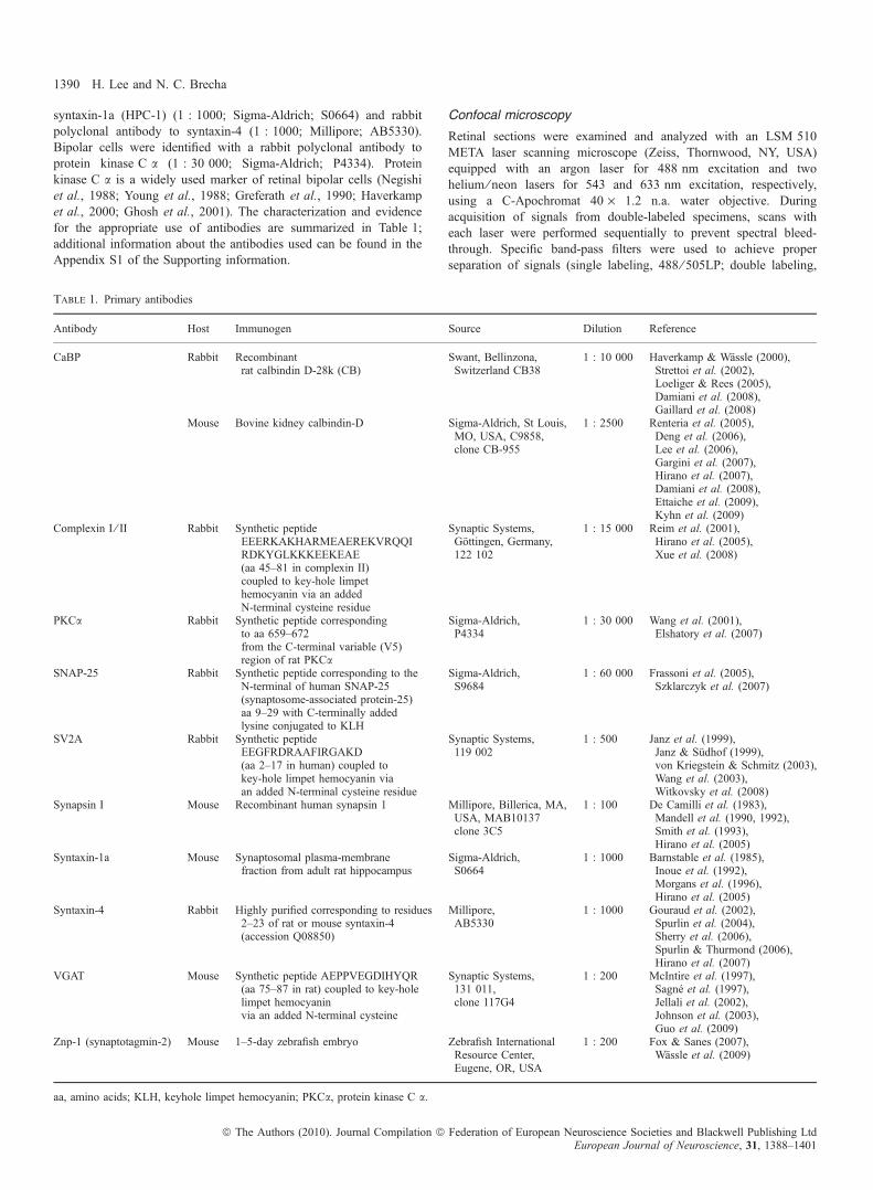
syntaxin-1a (HPC-1) (1 : 1000; Sigma-Aldrich; S0664) and rabbitpolyclonal antibody to syntaxin-4 (1 : 1000; Millipore; AB5330).Bipolar cells were identified with a rabbit polyclonal antibody toprotein kinase C a (1 : 30 000; Sigma-Aldrich; P4334). Proteinkinase C a is a widely used marker of retinal bipolar cells (Negishiet al., 1988; Young et al., 1988; Greferath et al., 1990; Haverkampet al., 2000; Ghosh et al., 2001). The characterization and evidencefor the appropriate use of antibodies are summarized in Table 1;additional information about the antibodies used can be found in theAppendix S1 of the Supporting information.
Confocal microscopy
Retinal sections were examined and analyzed with an LSM 510META laser scanning microscope (Zeiss, Thornwood, NY, USA)equipped with an argon laser for 488 nm excitation and twohelium ⁄ neon lasers for 543 and 633 nm excitation, respectively,using a C-Apochromat 40 · 1.2 n.a. water objective. Duringacquisition of signals from double-labeled specimens, scans witheach laser were performed sequentially to prevent spectral bleed-through. Specific band-pass filters were used to achieve properseparation of signals (single labeling, 488 ⁄ 505LP; double labeling,
Table 1. Primary antibodies
Antibody Host Immunogen Source Dilution Reference
CaBP Rabbit Recombinantrat calbindin D-28k (CB)
Swant, Bellinzona,Switzerland CB38
1 : 10 000 Haverkamp & Wassle (2000),Strettoi et al. (2002),Loeliger & Rees (2005),Damiani et al. (2008),Gaillard et al. (2008)
Mouse Bovine kidney calbindin-D Sigma-Aldrich, St Louis,MO, USA, C9858,clone CB-955
1 : 2500 Renteria et al. (2005),Deng et al. (2006),Lee et al. (2006),Gargini et al. (2007),Hirano et al. (2007),Damiani et al. (2008),Ettaiche et al. (2009),Kyhn et al. (2009)
Complexin I ⁄ II Rabbit Synthetic peptideEEERKAKHARMEAEREKVRQQIRDKYGLKKKEEKEAE(aa 45–81 in complexin II)coupled to key-hole limpethemocyanin via an addedN-terminal cysteine residue
Synaptic Systems,Gottingen, Germany,122 102
1 : 15 000 Reim et al. (2001),Hirano et al. (2005),Xue et al. (2008)
PKCa Rabbit Synthetic peptide correspondingto aa 659–672from the C-terminal variable (V5)region of rat PKCa
Sigma-Aldrich,P4334
1 : 30 000 Wang et al. (2001),Elshatory et al. (2007)
SNAP-25 Rabbit Synthetic peptide corresponding to theN-terminal of human SNAP-25(synaptosome-associated protein-25)aa 9–29 with C-terminally addedlysine conjugated to KLH
Sigma-Aldrich,S9684
1 : 60 000 Frassoni et al. (2005),Szklarczyk et al. (2007)
SV2A Rabbit Synthetic peptideEEGFRDRAAFIRGAKD(aa 2–17 in human) coupled tokey-hole limpet hemocyanin viaan added N-terminal cysteine residue
Synaptic Systems,119 002
1 : 500 Janz et al. (1999),Janz & Sudhof (1999),von Kriegstein & Schmitz (2003),Wang et al. (2003),Witkovsky et al. (2008)
Synapsin I Mouse Recombinant human synapsin 1 Millipore, Billerica, MA,USA, MAB10137clone 3C5
1 : 100 De Camilli et al. (1983),Mandell et al. (1990, 1992),Smith et al. (1993),Hirano et al. (2005)
Syntaxin-1a Mouse Synaptosomal plasma-membranefraction from adult rat hippocampus
Sigma-Aldrich,S0664
1 : 1000 Barnstable et al. (1985),Inoue et al. (1992),Morgans et al. (1996),Hirano et al. (2005)
Syntaxin-4 Rabbit Highly purified corresponding to residues2–23 of rat or mouse syntaxin-4(accession Q08850)
Millipore,AB5330
1 : 1000 Gouraud et al. (2002),Spurlin et al. (2004),Sherry et al. (2006),Spurlin & Thurmond (2006),Hirano et al. (2007)
VGAT Mouse Synthetic peptide AEPPVEGDIHYQR(aa 75–87 in rat) coupled to key-holelimpet hemocyaninvia an added N-terminal cysteine
Synaptic Systems,131 011,clone 117G4
1 : 200 McIntire et al. (1997),Sagne et al. (1997),Jellali et al. (2002),Johnson et al. (2003),Guo et al. (2009)
Znp-1 (synaptotagmin-2) Mouse 1–5-day zebrafish embryo Zebrafish InternationalResource Center,Eugene, OR, USA
1 : 200 Fox & Sanes (2007),Wassle et al. (2009)
aa, amino acids; KLH, keyhole limpet hemocyanin; PKCa, protein kinase C a.
1390 H. Lee and N. C. Brecha
ª The Authors (2010). Journal Compilation ª Federation of European Neuroscience Societies and Blackwell Publishing LtdEuropean Journal of Neuroscience, 31, 1388–1401
488 ⁄ 505–530 and 543 ⁄ 560LP). To increase the signal-to-noise ratio,images were averaged online (e.g. n = 4) and the scan speed andphotomultiplier detector gain were decreased. Digital images wereacquired at a magnification zoom of 1.5 · and a resolution of2048 · 2048 pixels. Confocal images were acquired at an opticalthickness between 0.5 and 0.7 lm and approximately 1.0 Airy Units.The tortuous coursing of horizontal cell processes and spray ofhorizontal cell endings necessitated stacks through the OPL forclearer, more complete images of the localization of signals; however,images of individual scans of a single optical slice are available insupporting Figs S1–S8. For projections, 6–10 optical sections wereacquired with a total thickness ranging from 2.5 to 6.3 lm andcompressed for viewing. Digital confocal images were saved as Zeiss.LSM files and final publication quality images were exported in the.TIFF format at 300 dpi using LSM 510 META software version 4.2(Zeiss). Images were adjusted for contrast and brightness, labeled andformatted using Photoshop CS3 (Adobe Systems, Inc., San Jose, CA,USA), and saved at 300 dpi at their final magnification.
Results
Vesicular GABA transporter expression in the outer retina
The VGAT immunoreactivity has been localized to horizontal cellprocesses and terminals in mouse, rat, monkey and human retinas
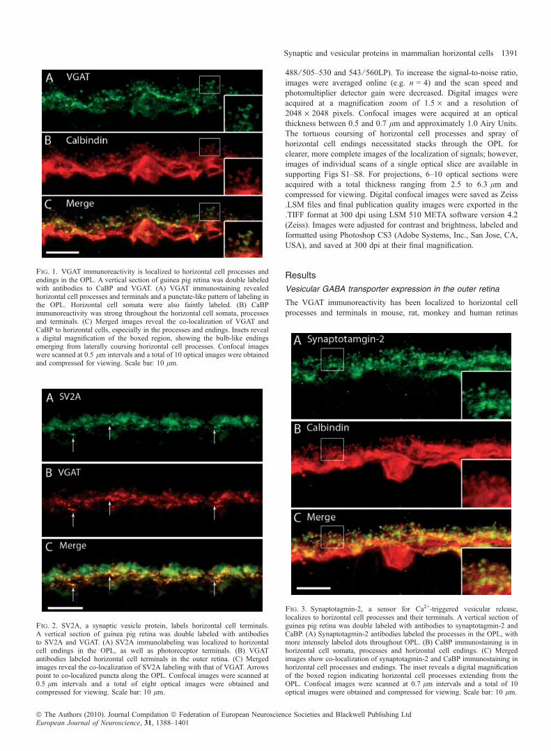
Fig. 1. VGAT immunoreactivity is localized to horizontal cell processes andendings in the OPL. A vertical section of guinea pig retina was double labeledwith antibodies to CaBP and VGAT. (A) VGAT immunostaining revealedhorizontal cell processes and terminals and a punctate-like pattern of labeling inthe OPL. Horizontal cell somata were also faintly labeled. (B) CaBPimmunoreactivity was strong throughout the horizontal cell somata, processesand terminals. (C) Merged images reveal the co-localization of VGAT andCaBP to horizontal cells, especially in the processes and endings. Insets reveala digital magnification of the boxed region, showing the bulb-like endingsemerging from laterally coursing horizontal cell processes. Confocal imageswere scanned at 0.5 lm intervals and a total of 10 optical images were obtainedand compressed for viewing. Scale bar: 10 lm.
Fig. 2. SV2A, a synaptic vesicle protein, labels horizontal cell terminals.A vertical section of guinea pig retina was double labeled with antibodiesto SV2A and VGAT. (A) SV2A immunolabeling was localized to horizontalcell endings in the OPL, as well as photoreceptor terminals. (B) VGATantibodies labeled horizontal cell terminals in the outer retina. (C) Mergedimages reveal the co-localization of SV2A labeling with that of VGAT. Arrowspoint to co-localized puncta along the OPL. Confocal images were scanned at0.5 lm intervals and a total of eight optical images were obtained andcompressed for viewing. Scale bar: 10 lm.
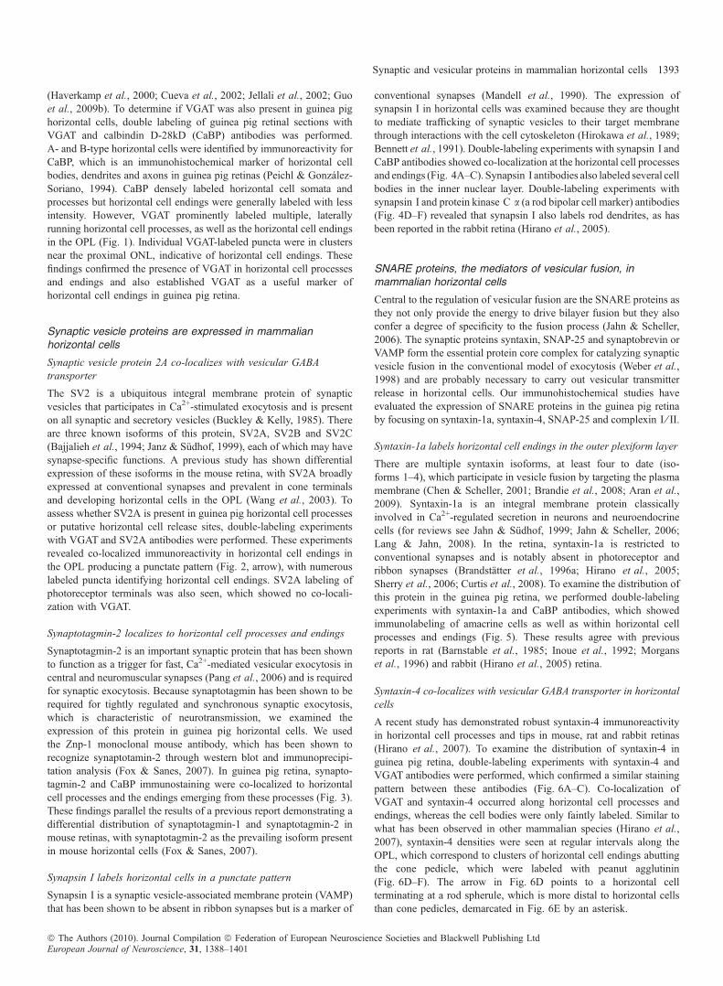
Fig. 3. Synaptotagmin-2, a sensor for Ca2+-triggered vesicular release,localizes to horizontal cell processes and their terminals. A vertical section ofguinea pig retina was double labeled with antibodies to synaptotagmin-2 andCaBP. (A) Synaptotagmin-2 antibodies labeled the processes in the OPL, withmore intensely labeled dots throughout OPL. (B) CaBP immunostaining is inhorizontal cell somata, processes and horizontal cell endings. (C) Mergedimages show co-localization of synaptotagmin-2 and CaBP immunostaining inhorizontal cell processes and endings. The inset reveals a digital magnificationof the boxed region indicating horizontal cell processes extending from theOPL. Confocal images were scanned at 0.7 lm intervals and a total of 10optical images were obtained and compressed for viewing. Scale bar: 10 lm.
Synaptic and vesicular proteins in mammalian horizontal cells 1391
ª The Authors (2010). Journal Compilation ª Federation of European Neuroscience Societies and Blackwell Publishing LtdEuropean Journal of Neuroscience, 31, 1388–1401
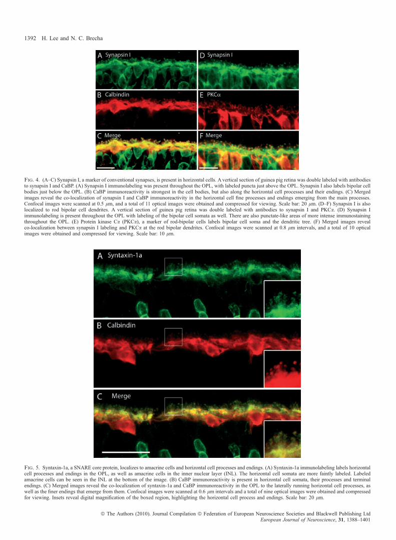
Fig. 4. (A–C) Synapsin I, a marker of conventional synapses, is present in horizontal cells. A vertical section of guinea pig retina was double labeled with antibodiesto synapsin I and CaBP. (A) Synapsin I immunolabeling was present throughout the OPL, with labeled puncta just above the OPL. Synapsin I also labels bipolar cellbodies just below the OPL. (B) CaBP immunoreactivity is strongest in the cell bodies, but also along the horizontal cell processes and their endings. (C) Mergedimages reveal the co-localization of synapsin I and CaBP immunoreactivity in the horizontal cell fine processes and endings emerging from the main processes.Confocal images were scanned at 0.5 lm, and a total of 11 optical images were obtained and compressed for viewing. Scale bar: 20 lm. (D–F) Synapsin I is alsolocalized to rod bipolar cell dendrites. A vertical section of guinea pig retina was double labeled with antibodies to synapsin I and PKCa. (D) Synapsin Iimmunolabeling is present throughout the OPL with labeling of the bipolar cell somata as well. There are also punctate-like areas of more intense immunostainingthroughout the OPL. (E) Protein kinase Ca (PKCa), a marker of rod-bipolar cells labels bipolar cell soma and the dendritic tree. (F) Merged images revealco-localization between synapsin I labeling and PKCa at the rod bipolar dendrites. Confocal images were scanned at 0.8 lm intervals, and a total of 10 opticalimages were obtained and compressed for viewing. Scale bar: 10 lm.
Fig. 5. Syntaxin-1a, a SNARE core protein, localizes to amacrine cells and horizontal cell processes and endings. (A) Syntaxin-1a immunolabeling labels horizontalcell processes and endings in the OPL, as well as amacrine cells in the inner nuclear layer (INL). The horizontal cell somata are more faintly labeled. Labeledamacrine cells can be seen in the INL at the bottom of the image. (B) CaBP immunoreactivity is present in horizontal cell somata, their processes and terminalendings. (C) Merged images reveal the co-localization of syntaxin-1a and CaBP immunoreactivity in the OPL to the laterally running horizontal cell processes, aswell as the finer endings that emerge from them. Confocal images were scanned at 0.6 lm intervals and a total of nine optical images were obtained and compressedfor viewing. Insets reveal digital magnification of the boxed region, highlighting the horizontal cell process and endings. Scale bar: 20 lm.
1392 H. Lee and N. C. Brecha
ª The Authors (2010). Journal Compilation ª Federation of European Neuroscience Societies and Blackwell Publishing LtdEuropean Journal of Neuroscience, 31, 1388–1401
(Haverkamp et al., 2000; Cueva et al., 2002; Jellali et al., 2002; Guoet al., 2009b). To determine if VGAT was also present in guinea pighorizontal cells, double labeling of guinea pig retinal sections withVGAT and calbindin D-28kD (CaBP) antibodies was performed.A- and B-type horizontal cells were identified by immunoreactivity forCaBP, which is an immunohistochemical marker of horizontal cellbodies, dendrites and axons in guinea pig retinas (Peichl & Gonzalez-Soriano, 1994). CaBP densely labeled horizontal cell somata andprocesses but horizontal cell endings were generally labeled with lessintensity. However, VGAT prominently labeled multiple, laterallyrunning horizontal cell processes, as well as the horizontal cell endingsin the OPL (Fig. 1). Individual VGAT-labeled puncta were in clustersnear the proximal ONL, indicative of horizontal cell endings. Thesefindings confirmed the presence of VGAT in horizontal cell processesand endings and also established VGAT as a useful marker ofhorizontal cell endings in guinea pig retina.
Synaptic vesicle proteins are expressed in mammalianhorizontal cells
Synaptic vesicle protein 2A co-localizes with vesicular GABAtransporter
The SV2 is a ubiquitous integral membrane protein of synapticvesicles that participates in Ca2+-stimulated exocytosis and is presenton all synaptic and secretory vesicles (Buckley & Kelly, 1985). Thereare three known isoforms of this protein, SV2A, SV2B and SV2C(Bajjalieh et al., 1994; Janz & Sudhof, 1999), each of which may havesynapse-specific functions. A previous study has shown differentialexpression of these isoforms in the mouse retina, with SV2A broadlyexpressed at conventional synapses and prevalent in cone terminalsand developing horizontal cells in the OPL (Wang et al., 2003). Toassess whether SV2A is present in guinea pig horizontal cell processesor putative horizontal cell release sites, double-labeling experimentswith VGAT and SV2A antibodies were performed. These experimentsrevealed co-localized immunoreactivity in horizontal cell endings inthe OPL producing a punctate pattern (Fig. 2, arrow), with numerouslabeled puncta identifying horizontal cell endings. SV2A labeling ofphotoreceptor terminals was also seen, which showed no co-locali-zation with VGAT.
Synaptotagmin-2 localizes to horizontal cell processes and endings
Synaptotagmin-2 is an important synaptic protein that has been shownto function as a trigger for fast, Ca2+-mediated vesicular exocytosis incentral and neuromuscular synapses (Pang et al., 2006) and is requiredfor synaptic exocytosis. Because synaptotagmin has been shown to berequired for tightly regulated and synchronous synaptic exocytosis,which is characteristic of neurotransmission, we examined theexpression of this protein in guinea pig horizontal cells. We usedthe Znp-1 monoclonal mouse antibody, which has been shown torecognize synaptotamin-2 through western blot and immunoprecipi-tation analysis (Fox & Sanes, 2007). In guinea pig retina, synapto-tagmin-2 and CaBP immunostaining were co-localized to horizontalcell processes and the endings emerging from these processes (Fig. 3).These findings parallel the results of a previous report demonstrating adifferential distribution of synaptotagmin-1 and synaptotagmin-2 inmouse retinas, with synaptotagmin-2 as the prevailing isoform presentin mouse horizontal cells (Fox & Sanes, 2007).
Synapsin I labels horizontal cells in a punctate pattern
Synapsin I is a synaptic vesicle-associated membrane protein (VAMP)that has been shown to be absent in ribbon synapses but is a marker of
conventional synapses (Mandell et al., 1990). The expression ofsynapsin I in horizontal cells was examined because they are thoughtto mediate trafficking of synaptic vesicles to their target membranethrough interactions with the cell cytoskeleton (Hirokawa et al., 1989;Bennett et al., 1991). Double-labeling experiments with synapsin I andCaBP antibodies showed co-localization at the horizontal cell processesand endings (Fig. 4A–C). Synapsin I antibodies also labeled several cellbodies in the inner nuclear layer. Double-labeling experiments withsynapsin I and protein kinase C a (a rod bipolar cell marker) antibodies(Fig. 4D–F) revealed that synapsin I also labels rod dendrites, as hasbeen reported in the rabbit retina (Hirano et al., 2005).
SNARE proteins, the mediators of vesicular fusion, inmammalian horizontal cells
Central to the regulation of vesicular fusion are the SNARE proteins asthey not only provide the energy to drive bilayer fusion but they alsoconfer a degree of specificity to the fusion process (Jahn & Scheller,2006). The synaptic proteins syntaxin, SNAP-25 and synaptobrevin orVAMP form the essential protein core complex for catalyzing synapticvesicle fusion in the conventional model of exocytosis (Weber et al.,1998) and are probably necessary to carry out vesicular transmitterrelease in horizontal cells. Our immunohistochemical studies haveevaluated the expression of SNARE proteins in the guinea pig retinaby focusing on syntaxin-1a, syntaxin-4, SNAP-25 and complexin I ⁄ II.
Syntaxin-1a labels horizontal cell endings in the outer plexiform layer
There are multiple syntaxin isoforms, at least four to date (iso-forms 1–4), which participate in vesicle fusion by targeting the plasmamembrane (Chen & Scheller, 2001; Brandie et al., 2008; Aran et al.,2009). Syntaxin-1a is an integral membrane protein classicallyinvolved in Ca2+-regulated secretion in neurons and neuroendocrinecells (for reviews see Jahn & Sudhof, 1999; Jahn & Scheller, 2006;Lang & Jahn, 2008). In the retina, syntaxin-1a is restricted toconventional synapses and is notably absent in photoreceptor andribbon synapses (Brandstatter et al., 1996a; Hirano et al., 2005;Sherry et al., 2006; Curtis et al., 2008). To examine the distribution ofthis protein in the guinea pig retina, we performed double-labelingexperiments with syntaxin-1a and CaBP antibodies, which showedimmunolabeling of amacrine cells as well as within horizontal cellprocesses and endings (Fig. 5). These results agree with previousreports in rat (Barnstable et al., 1985; Inoue et al., 1992; Morganset al., 1996) and rabbit (Hirano et al., 2005) retina.
Syntaxin-4 co-localizes with vesicular GABA transporter in horizontalcells
A recent study has demonstrated robust syntaxin-4 immunoreactivityin horizontal cell processes and tips in mouse, rat and rabbit retinas(Hirano et al., 2007). To examine the distribution of syntaxin-4 inguinea pig retina, double-labeling experiments with syntaxin-4 andVGAT antibodies were performed, which confirmed a similar stainingpattern between these antibodies (Fig. 6A–C). Co-localization ofVGAT and syntaxin-4 occurred along horizontal cell processes andendings, whereas the cell bodies were only faintly labeled. Similar towhat has been observed in other mammalian species (Hirano et al.,2007), syntaxin-4 densities were seen at regular intervals along theOPL, which correspond to clusters of horizontal cell endings abuttingthe cone pedicle, which were labeled with peanut agglutinin(Fig. 6D–F). The arrow in Fig. 6D points to a horizontal cellterminating at a rod spherule, which is more distal to horizontal cellsthan cone pedicles, demarcated in Fig. 6E by an asterisk.
Synaptic and vesicular proteins in mammalian horizontal cells 1393
ª The Authors (2010). Journal Compilation ª Federation of European Neuroscience Societies and Blackwell Publishing LtdEuropean Journal of Neuroscience, 31, 1388–1401
SNAP-25 strongly labels horizontal cell processes and endings
SNAP-25, a critical component of the neural SNARE complex thatfacilitates membrane fusion between synaptic vesicles and thepresynaptic plasma membrane, has been reported in multiple retinalcell types, including horizontal cells (Catsicas et al., 1992). Recently,it was shown that SNAP-25 not only subserves cholinergic andglutamatergic neurotransmission but is also critical for evoked GABArelease and is expressed by mature GABAergic neurons (Tafoya et al.,2006). We tested whether horizontal cells in guinea pig retina expressSNAP-25 with double-labeling experiments with both CaBP andSNAP-25 antibodies. In guinea pig, immunoreactivity using theSNAP-25 anti-rabbit antibody was robust in horizontal cell endings,identified by co-labeling with CaBP antibody (Fig. 7). Immunostain-ing was very weak or absent in horizontal cell somata (arrow) but thehorizontal cell terminals and lateral processes along the OPL beneathphotoreceptor terminals were intensely labeled. SNAP-25 also labeledthe bipolar cell bodies (asterisk), dendrites and axons, and has beenreported to occur in other retinal cell types (Galli et al., 1995;Brandstatter et al., 1996a; von Kriegstein et al., 1999; Morgans &Brandstatter, 2000; von Kriegstein & Schmitz, 2003).
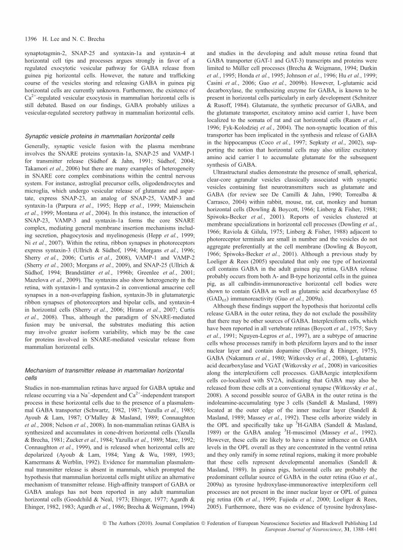
Complexin I ⁄ II labels horizontal cell soma, processes and endings
Fast Ca2+-triggered fusion requires a host of proteins, includingcomplexins. These are soluble SNARE complex-binding proteins thathave been shown to have an essential role in synaptic fusion by
regulating a late step in Ca2+-dependent neurotransmitter release(Reim et al., 2001; Tang et al., 2006). They control the force transferfrom SNARE complexes to membranes in fusion (Maximov et al.,2009) by serving as ‘grappling proteins’ to hold the SNARE complexinto an activated but frozen state (Rizo & Rosenmund, 2008; Sudhof& Rothman, 2009). Given that these are essential proteins involved inCa2+-dependent vesicular fusion, we tested whether they are expressedin guinea pig horizontal cells. Double-labeling experiments withcomplexin I ⁄ II and CaBP showed that complexin I ⁄ II immunoreac-tivity was localized to all parts of the guinea pig horizontal cells,including the soma, processes and endings (Fig. 8). These resultsagree with earlier findings in mouse and rabbit retina, which revealedcomplexin I ⁄ II immunoreactivity in the entire horizontal cell, includ-ing the endings, at both the light and electron microscopy level(Hirano et al., 2005; Reim et al., 2005). Amacrine cells are alsolabeled with the complexin I ⁄ II antibody (Fig. 8, asterisk).
Discussion
This study provides novel insights and morphological evidence forthe mechanism of transmitter release from horizontal cells. It hasbeen shown previously that both GABA and the biosyntheticmachinery to synthesize GABA are present in guinea pig horizontalcells (Guo et al., 2009a). This study extends these findings to revealkey protein components involved in Ca2+-dependent and SNAREprotein-dependent exocytosis. Most notably, the expression of VGAT,
Fig. 6. Syntaxin-4, a SNARE complex protein, localizes to horizontal cell processes and endings. (A–C) A vertical section of guinea pig retina was double labeledwith antibodies to syntaxin-4 and VGAT. (A) Syntaxin-4 immunolabeling was robust along the OPL with a punctate pattern. There were also regions of more intensestaining in the OPL that were located along horizontal cell processes and near the soma. (B) VGAT immunolabeling was localized to the laterally running processesof horizontal cells, as well as the finer processes and endings emerging from them. (C) Merged images reveal the co-localization of syntaxin-4-labeled puncta andVGAT-labeled endings of horizontal cells. The insets are a digital magnification of the boxed region, which demonstrates staining of the delicate processes andendings of horizontal cells by both syntaxin-4 and VGAT antibodies. Confocal images were scanned at 0.5 lm intervals and a total of 10 optical images wereobtained and compressed for viewing. Scale bar: 20 lm. (D–F) Syntaxin-4 does not label cone photoreceptor terminals. (D) Dense spots of syntaxin-4immunoreactivity occur at regular intervals along the OPL. (E) Cone pedicles, labeled with peanut-agglutinin (PNA), produce a similar punctate pattern of densitiesalong the OPL. (F) Merged images reveal that the syntaxin-4-immunoreactive clusters correspond to horizontal cell endings grouped together just underneath conepedicles. The arrow points to a horizontal cell ending that continues up past the cone pedicle (indicated by the asterisk) to terminate at a rod spherule, which is moredistal to horizontal cells than cone pedicles. Confocal images were scanned at 0.5 lm intervals and a total of eight optical images were obtained and compressed forviewing. Scale bar: 10 lm.
1394 H. Lee and N. C. Brecha
ª The Authors (2010). Journal Compilation ª Federation of European Neuroscience Societies and Blackwell Publishing LtdEuropean Journal of Neuroscience, 31, 1388–1401
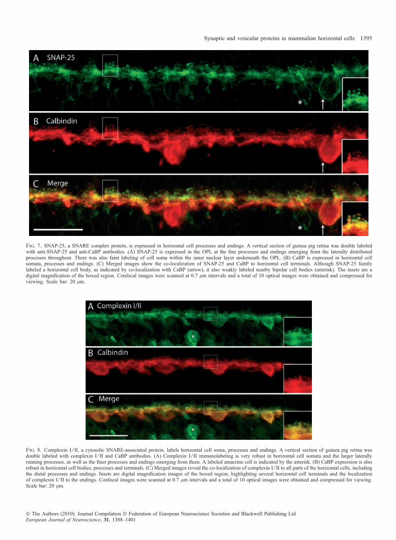
Fig. 7. SNAP-25, a SNARE complex protein, is expressed in horizontal cell processes and endings. A vertical section of guinea pig retina was double labeledwith anti-SNAP-25 and anti-CaBP antibodies. (A) SNAP-25 is expressed in the OPL at the fine processes and endings emerging from the laterally distributedprocesses throughout. There was also faint labeling of cell soma within the inner nuclear layer underneath the OPL. (B) CaBP is expressed in horizontal cellsomata, processes and endings. (C) Merged images show the co-localization of SNAP-25 and CaBP to horizontal cell terminals. Although SNAP-25 faintlylabeled a horizontal cell body, as indicated by co-localization with CaBP (arrow), it also weakly labeled nearby bipolar cell bodies (asterisk). The insets are adigital magnification of the boxed region. Confocal images were scanned at 0.7 lm intervals and a total of 10 optical images were obtained and compressed forviewing. Scale bar: 20 lm.
Fig. 8. Complexin I ⁄ II, a cytosolic SNARE-associated protein, labels horizontal cell soma, processes and endings. A vertical section of guinea pig retina wasdouble labeled with complexin I ⁄ II and CaBP antibodies. (A) Complexin I ⁄ II immunolabeling is very robust in horizontal cell somata and the larger laterallyrunning processes, as well as the finer processes and endings emerging from them. A labeled amacrine cell is indicated by the asterisk. (B) CaBP expression is alsorobust in horizontal cell bodies, processes and terminals. (C) Merged images reveal the co-localization of complexin I ⁄ II to all parts of the horizontal cells, includingthe distal processes and endings. Insets are digital magnification images of the boxed region, highlighting several horizontal cell terminals and the localizationof complexin I ⁄ II to the endings. Confocal images were scanned at 0.7 lm intervals and a total of 10 optical images were obtained and compressed for viewing.Scale bar: 20 lm.
Synaptic and vesicular proteins in mammalian horizontal cells 1395
ª The Authors (2010). Journal Compilation ª Federation of European Neuroscience Societies and Blackwell Publishing LtdEuropean Journal of Neuroscience, 31, 1388–1401
synaptotagmin-2, SNAP-25 and syntaxin-1a and syntaxin-4 athorizontal cell tips and processes argues strongly in favor of aregulated exocytotic vesicular pathway for GABA release fromguinea pig horizontal cells. However, the nature and traffickingcourse of the vesicles storing and releasing GABA in guinea pighorizontal cells are currently unknown. Furthermore, the existence ofCa2+-regulated vesicular exocytosis in mammalian horizontal cells isstill debated. Based on our findings, GABA probably utilizes avesicular-regulated secretory pathway in mammalian horizontal cells.
Synaptic vesicle proteins in mammalian horizontal cells
Generally, synaptic vesicle fusion with the plasma membraneinvolves the SNARE proteins syntaxin-1a, SNAP-25 and VAMP-1for transmitter release (Sudhof & Jahn, 1991; Sudhof, 2004;Takamori et al., 2006) but there are many examples of heterogeneityin SNARE core complex combinations within the central nervoussystem. For instance, astroglial precursor cells, oligodendrocytes andmicroglia, which undergo vesicular release of glutamate and aspar-tate, express SNAP-23, an analog of SNAP-25, VAMP-3 andsyntaxin-1a (Parpura et al., 1995; Hepp et al., 1999; Maienscheinet al., 1999; Montana et al., 2004). In this instance, the interaction ofSNAP-23, VAMP-3 and syntaxin-1a forms the core SNAREcomplex, mediating general membrane insertion mechanisms includ-ing secretion, phagocytosis and myelinogenesis (Hepp et al., 1999;Ni et al., 2007). Within the retina, ribbon synapses in photoreceptorsexpress syntaxin-3 (Ullrich & Sudhof, 1994; Morgans et al., 1996;Sherry et al., 2006; Curtis et al., 2008), VAMP-1 and VAMP-2(Sherry et al., 2003; Morgans et al., 2009), and SNAP-25 (Ullrich &Sudhof, 1994; Brandstatter et al., 1996b; Greenlee et al., 2001;Mazelova et al., 2009). The syntaxins also show heterogeneity in theretina, with syntaxin-1 and syntaxin-2 in conventional amacrine cellsynapses in a non-overlapping fashion, syntaxin-3b in glutamatergicribbon synapses of photoreceptors and bipolar cells, and syntaxin-4in horizontal cells (Sherry et al., 2006; Hirano et al., 2007; Curtiset al., 2008). Thus, although the paradigm of SNARE-mediatedfusion may be universal, the substrates mediating this actionmay involve greater isoform variability, which may be the casefor proteins involved in SNARE-mediated vesicular release frommammalian horizontal cells.
Mechanism of transmitter release in mammalian horizontalcells
Studies in non-mammalian retinas have argued for GABA uptake andrelease occurring via a Na+-dependent and Ca2+-independent transportprocess in these horizontal cells due to the presence of a plasmalem-mal GABA transporter (Schwartz, 1982, 1987; Yazulla et al., 1985;Ayoub & Lam, 1987; O’Malley & Masland, 1989; Connaughtonet al., 2008; Nelson et al., 2008). In non-mammalian retinas GABA issynthesized and accumulates in cone-driven horizontal cells (Yazulla& Brecha, 1981; Zucker et al., 1984; Yazulla et al., 1989; Marc, 1992;Connaughton et al., 1999), and is released when horizontal cells aredepolarized (Ayoub & Lam, 1984; Yang & Wu, 1989, 1993;Kamermans & Werblin, 1992). Evidence for mammalian plasmalem-mal transmitter release is absent in mammals, which prompted thehypothesis that mammalian horizontal cells might utilize an alternativemechanism of transmitter release. High-affinity transport of GABA orGABA analogs has not been reported in any adult mammalianhorizontal cells (Goodchild & Neal, 1973; Ehinger, 1977; Agardh &Ehinger, 1982, 1983; Agardh et al., 1986; Brecha & Weigmann, 1994)
and studies in the developing and adult mouse retina found thatGABA transporter (GAT-1 and GAT-3) transcripts and proteins werelimited to Muller cell processes (Brecha & Weigmann, 1994; Durkinet al., 1995; Honda et al., 1995; Johnson et al., 1996; Hu et al., 1999;Casini et al., 2006; Guo et al., 2009b). However, L-glutamic aciddecarboxylase, the synthesizing enzyme for GABA, is known to bepresent in horizontal cells particularly in early development (Schnitzer& Rusoff, 1984). Glutamate, the synthetic precursor of GABA, andthe glutamate transporter, excitatory amino acid carrier 1, have beenlocalized to the somata of rat and cat horizontal cells (Rauen et al.,1996; Fyk-Kolodziej et al., 2004). The non-synaptic location of thistransporter has been implicated in the synthesis and release of GABAin the hippocampus (Coco et al., 1997; Sepkuty et al., 2002), sup-porting the notion that horizontal cells may also utilize excitatoryamino acid carrier 1 to accumulate glutamate for the subsequentsynthesis of GABA.Ultrastructural studies demonstrate the presence of small, spherical,
clear-core agranular vesicles classically associated with synapticvesicles containing fast neurotransmitters such as glutamate andGABA (for review see De Camilli & Jahn, 1990; Torrealba &Carrasco, 2004) within rabbit, mouse, rat, cat, monkey and humanhorizontal cells (Dowling & Boycott, 1966; Linberg & Fisher, 1988;Spiwoks-Becker et al., 2001). Reports of vesicles clustered atmembrane specializations in horizontal cell processes (Dowling et al.,1966; Raviola & Gilula, 1975; Linberg & Fisher, 1988) adjacent tophotoreceptor terminals are small in number and the vesicles do notaggregate preferentially at the cell membrane (Dowling & Boycott,1966; Spiwoks-Becker et al., 2001). Although a previous study byLoeliger & Rees (2005) speculated that only one type of horizontalcell contains GABA in the adult guinea pig retina, GABA releaseprobably occurs from both A- and B-type horizontal cells in the guineapig, as all calbindin-immunoreactive horizontal cell bodies wereshown to contain GABA as well as glutamic acid decarboxylase 65(GAD65) immunoreactivity (Guo et al., 2009a).Although these findings support the hypothesis that horizontal cells
release GABA in the outer retina, they do not exclude the possibilitythat there may be other sources of GABA. Interplexiform cells, whichhave been reported in all vertebrate retinas (Boycott et al., 1975; Savyet al., 1991; Nguyen-Legros et al., 1997), are a subtype of amacrinecells whose processes ramify in both plexiform layers and to the innernuclear layer and contain dopamine (Dowling & Ehinger, 1975),GABA (Nakamura et al., 1980; Witkovsky et al., 2008), L-glutamicacid decarboxylase and VGAT (Witkovsky et al., 2008) in varicositiesalong the interplexiform cell processes. GABAergic interplexiformcells co-localized with SV2A, indicating that GABA may also bereleased from these cells at a conventional synapse (Witkovsky et al.,2008). A second possible source of GABA in the outer retina is theindoleamine-accumulating type 3 cells (Sandell & Masland, 1989)located at the outer edge of the inner nuclear layer (Sandell &Masland, 1989; Massey et al., 1992). These cells arborize widely inthe OPL and specifically take up 3H-GABA (Sandell & Masland,1989) or the GABA analog 3H-muscimol (Massey et al., 1992).However, these cells are likely to have a minor influence on GABAlevels in the OPL overall as they are concentrated in the ventral retinaand they only ramify in some retinal regions, making it more probablethat these cells represent developmental anomalies (Sandell &Masland, 1989). In guinea pigs, horizontal cells are probably thepredominant cellular source of GABA in the outer retina (Guo et al.,2009a) as tyrosine hydroxylase-immunoreactive interplexiform cellprocesses are not present in the inner nuclear layer or OPL of guineapig retina (Oh et al., 1999; Fujieda et al., 2000; Loeliger & Rees,2005). Furthermore, there was no evidence of tyrosine hydroxylase-
1396 H. Lee and N. C. Brecha
ª The Authors (2010). Journal Compilation ª Federation of European Neuroscience Societies and Blackwell Publishing LtdEuropean Journal of Neuroscience, 31, 1388–1401
immunolabeled amacrine cell processes in the guinea pig ramifying inthe OPL (Oh et al., 1999).
Several studies support the notion that all cells in the photoreceptortriad are end targets of GABA released from horizontal cells.Mammalian horizontal cells exhibit GABA-induced currents (Feigen-span & Weiler, 2004), consistent with GABAA receptor expression onhorizontal cells (Greferath et al., 1994, 1995; Blanco et al., 1996).These findings suggest that GABA acts as an auto-receptor. GABAA,C
receptors have also been detected on bipolar cell dendrites (Greferathet al., 1994; Enz et al., 1996; Vardi et al., 1998; Pattnaik et al., 2000;Delgado et al., 2009), which provides evidence in support ofhorizontal cells mediating feedforward action onto bipolar cells.ON-bipolar cells require that GABAergic input be depolarizing toprovide them with the corrective horizontal cell input analogous to thatreceived by OFF-bipolar cells. Indeed, Duebel et al. (2006) reportsthat ON-bipolar cells employ a somatodendritic [Cl-](i) gradient toinvert GABAergic horizontal cell input, thereby depolarizingON-bipolar cells with a high dendritic [Cl-](i). There are severalstudies showing the presence of GABAA,C receptor immunoreactivityat photoreceptor terminals (Greferath et al., 1995; Picaud et al., 1998;Haverkamp & Wassle, 2000; Pattnaik et al., 2000). However, GABA’saction at photoreceptor terminals remains controversial. GABA-evoked currents are reported for mouse and pig cones (Picaud et al.,1998; Pattnaik et al., 2000) but the predominant finding is a lack ofGABA-evoked currents in mammalian cones (Verweij et al., 2003).
There are still many questions that must be answered regarding themechanism underlying transmitter release from horizontal cells. Theresults of this study argue for regulated vesicular-mediated exocytosisas the underlying mechanism of release and suggest that the guinea pigretina is uniquely suited for functional studies of mammalianhorizontal cells. Understanding the mechanism of release willcontribute to understanding how these cells function and communicatewithin the OPL.
Supporting Information
Additional supporting information may be found in the online versionof this article:Fig. S1. Single confocal image of VGAT and CaBP immunoreactivityin guinea pig vertical section.Fig. S2. Single confocal image of SV2A and VGAT immunoreactivityin guinea pig vertical section.Fig. S3. Single confocal image of synaptotagmin-2 and CaBP immuno-reactivity in guinea pig vertical section.Fig. S4. Single confocal image of synapsin I and CaBP immuno-reactivity in guinea pig vertical section.Fig. S5. Single confocal image of syntaxin-1a and CaBP immuno-reactivity in guinea pig vertical section.Fig. S6. Single confocal image of syntaxin-4 and VGAT immuno-reactivity in guinea pig vertical section.Fig. S7. Single confocal image of SNAP-25 and CaBP immuno-reactivity in guinea pig vertical section.Fig. S8. Single confocal image of complexin I ⁄ II and CaBP immu-noreactivity in guinea pig vertical section.Appendix S1. Antibody characterization.Please note: As a service to our authors and readers, this journalprovides supporting information supplied by the authors. Suchmaterials are peer-reviewed and may be re-organized for onlinedelivery, but are not copy-edited or typeset by Wiley-Blackwell.Technical support issues arising from supporting information (otherthan missing files) should be addressed to the authors.
Acknowledgements
We thank Drs Salvatore Stella Jr, Arlene Hirano, Chenying Guo, Iona Raymondand Steve Barnes for their insightful comments on this manuscript. Supportedby NIH EY 15573 and the Jules Stein Eye Institute EyeSTAR program. N.C.B.is a VA Senior Career Research Scientist.
Abbreviations
CaBP, calbindin D-28kD; OPL, outer plexiform layer; PB, phosphate buffer;SNAP-25, synaptosomal-associated protein; SNARE, soluble NSF attachmentprotein receptor; SV2, synaptic vesicle protein 2; VAMP, vesicle-associatedmembrane protein; VGAT, vesicular GABA transporter.
References
Agardh, E. & Ehinger, B. (1982) (3H)-muscimol, (3H)-nipecotic acid and (3H)-isoguvacine as autoradiographic markers for GABA neurotransmission.J. Neural Transm., 54, 1–18.
Agardh, E. & Ehinger, B. (1983) Retinal GABA neuron labelling with[3H]isoguvacine in different species. Exp. Eye Res., 36, 215–229.
Agardh, E., Bruun, A., Ehinger, B. & Storm-Mathisen, J. (1986) GABAimmunoreactivity in the retina. Invest. Ophthalmol. Vis. Sci., 27, 674–678.
Aran, V., Brandie, F.M., Boyd, A.R., Kantidakis, T., Rideout, E.J., Kelly, S.M.,Gould, G.W. & Bryant, N.J. (2009) Characterization of two distinct bindingmodes between syntaxin 4 and Munc18c. Biochem. J., 419, 655–660.
Ayoub, G.S. & Lam, D.M. (1984) The release of gamma-aminobutyric acidfrom horizontal cells of the goldfish (Carassius auratus) retina. J. Physiol.,355, 191–214.
Ayoub, G.S. & Lam, D.M. (1987) Accumulation of gamma-aminobutyric acidby horizontal cells isolated from the goldfish retina. Vision Res., 27, 2027–2034.
Babai, N. & Thoreson, W.B. (2009) Horizontal cell feedback regulates calciumcurrents and intracellular calcium levels in rod photoreceptors of salamanderand mouse retina. J. Physiol., 587, 2353–2364.
Bajjalieh, S.M., Frantz, G.D., Weimann, J.M., McConnell, S.K. & Scheller,R.H. (1994) Differential expression of synaptic vesicle protein 2 (SV2)isoforms. J. Neurosci., 14, 5223–5235.
Barnstable, C.J., Hofstein, R. & Akagawa, K. (1985) A marker of earlyamacrine cell development in rat retina. Brain Res., 352, 286–290.
Baylor, D.A., Fuortes, M.G. & O’Bryan, P.M. (1971) Receptive fields of conesin the retina of the turtle. J. Physiol., 214, 265–294.
Bennett, A.F., Hayes, N.V. & Baines, A.J. (1991) Site specificity in theinteractions of synapsin 1 with tubulin. Biochem. J., 276(Pt 3), 793–799.
Billups, D. & Attwell, D. (2002) Control of intracellular chloride concentrationand GABA response polarity in rat retinal ON bipolar cells. J. Physiol., 545,183–198.
Blanco, R., Vaquero, C.F. & de la Villa, P. (1996) The effects of GABA andglycine on horizontal cells of the rabbit retina. Vision Res., 36, 3987–3995.
Boycott, B.B., Dowling, J.E., Fisher, S.K., Kolb, H. & Laties, A.M. (1975)Interplexiform cells of the mammalian retina and their comparison withcatecholamine-containing retinal cells. Proc. R. Soc. Lond. B Biol. Sci., 191,353–368.
Brandie, F.M., Aran, V., Verma, A., McNew, J.A., Bryant, N.J. & Gould, G.W.(2008) Negative regulation of syntaxin4 ⁄ SNAP-23 ⁄ VAMP2-mediatedmembrane fusion by Munc18c in vitro. PLoS ONE, 3, e4074.
Brandstatter, J.H., Lohrke, S., Morgans, C.W. & Wassle, H. (1996a)Distributions of two homologous synaptic vesicle proteins, synaptoporinand synaptophysin, in the mammalian retina. J. Comp. Neurol., 370, 1–10.
Brandstatter, J.H., Wassle, H., Betz, H. & Morgans, C.W. (1996b) Theplasma membrane protein SNAP-25, but not syntaxin, is present atphotoreceptor and bipolar cell synapses in the rat retina. Eur. J. Neurosci.,8, 823–828.
Brecha, N.C. & Weigmann, C. (1994) Expression of GAT-1, a high-affinitygamma-aminobutyric acid plasma membrane transporter in the rat retina.J. Comp. Neurol., 345, 602–611.
Buckley, K. & Kelly, R.B. (1985) Identification of a transmembraneglycoprotein specific for secretory vesicles of neural and endocrine cells.J. Cell Biol., 100, 1284–1294.
Burger, P.M., Hell, J., Mehl, E., Krasel, C., Lottspeich, F. & Jahn, R. (1991)GABA and glycine in synaptic vesicles: storage and transport characteristics.Neuron, 7, 287–293.
Burkhardt, D.A. (1977) Responses and receptive-field organization of cones inperch retinas. J. Neurophysiol., 40, 53–62.
Synaptic and vesicular proteins in mammalian horizontal cells 1397
ª The Authors (2010). Journal Compilation ª Federation of European Neuroscience Societies and Blackwell Publishing LtdEuropean Journal of Neuroscience, 31, 1388–1401
Casini, G., Rickman, D.W. & Brecha, N.C. (2006) Expression of the gamma-aminobutyric acid (GABA) plasma membrane transporter-1 in monkey andhuman retina. Invest. Ophthalmol. Vis. Sci., 47, 1682–1690.
Catsicas, S., Catsicas, M., Keyser, K.T., Karten, H.J., Wilson, M.C. & Milner,R.J. (1992) Differential expression of the presynaptic protein SNAP-25 inmammalian retina. J. Neurosci. Res., 33, 1–9.
Chaudhry, F.A., Reimer, R.J., Bellocchio, E.E., Danbolt, N.C., Osen, K.K.,Edwards, R.H. & Storm-Mathisen, J. (1998) The vesicular GABAtransporter, VGAT, localizes to synaptic vesicles in sets of glycinergic aswell as GABAergic neurons. J. Neurosci., 18, 9733–9750.
Chen, Y.A. & Scheller, R.H. (2001) SNARE-mediated membrane fusion. Nat.Rev. Mol. Cell Biol., 2, 98–106.
Coco, S., Verderio, C., Trotti, D., Rothstein, J.D., Volterra, A. & Matteoli, M.(1997) Non-synaptic localization of the glutamate transporter EAAC1 incultured hippocampal neurons. Eur. J. Neurosci., 9, 1902–1910.
Connaughton, V.P., Behar, T.N., Liu, W.L. & Massey, S.C. (1999) Immuno-cytochemical localization of excitatory and inhibitory neurotransmitters inthe zebrafish retina. Vis. Neurosci., 16, 483–490.
Connaughton, V.P., Nelson, R. & Bender, A.M. (2008) Electrophysiologicalevidence of GABAA and GABAC receptors on zebrafish retinal bipolar cells.Vis. Neurosci., 25, 139–153.
Cueva, J.G., Haverkamp, S., Reimer, R.J., Edwards, R., Wassle, H. & Brecha,N.C. (2002) Vesicular gamma-aminobutyric acid transporter expression inamacrine and horizontal cells. J. Comp. Neurol., 445, 227–237.
Curtis, L.B., Doneske, B., Liu, X., Thaller, C., McNew, J.A. & Janz, R. (2008)Syntaxin 3b is a t-SNARE specific for ribbon synapses of the retina.J. Comp. Neurol., 510, 550–559.
Damiani, D., Alexander, J.J., O’Rourke, J.R., McManus, M., Jadhav, A.P.,Cepko, C.L., Hauswirth, W.W., Harfe, B.D. & Strettoi, E. (2008) Dicerinactivation leads to progressive functional and structural degeneration of themouse retina. J. Neurosci., 28, 4878–4887.
De Camilli, P. & Jahn, R. (1990) Pathways to regulated exocytosis in neurons.Annu. Rev. Physiol., 52, 625–645.
De Camilli, P., Harris, S.M. Jr, Huttner, W.B. & Greengard, P. (1983)Synapsin I (Protein I), a nerve terminal-specific phosphoprotein. II. Itsspecific association with synaptic vesicles demonstrated by immunocyto-chemistry in agarose-embedded synaptosomes. J. Cell Biol., 96, 1355–1373.
Delgado, L.M., Vielma, A.H., Kahne, T., Palacios, A.G. & Schmachtenberg, O.(2009) The GABAergic system in the retina of neonate and adult Octodondegus, studied by immunohistochemistry and electroretinography. J. Comp.Neurol., 514, 459–472.
Demb, J.B., Haarsma, L., Freed, M.A. & Sterling, P. (1999) Functional circuitryof the retinal ganglion cell’s nonlinear receptive field. J. Neurosci., 19, 9756–9767.
Deng, Q., Wang, L., Dong, W. & He, S. (2006) Lateral components in the coneterminals of the rabbit retina: horizontal cell origin and glutamate receptorexpression. J. Comp. Neurol., 496, 698–705.
Dowling, J.E. & Boycott, B.B. (1966) Organization of the primate retina:electron microscopy. Proc. R. Soc. Lond. B Biol. Sci., 166, 80–111.
Dowling, J.E. & Ehinger, B. (1975) Synaptic organization of the amine-containing interplexiform cells of the goldfish and Cebus monkey retinas.Science, 188, 270–273.
Dowling, J.E. & Werblin, F.S. (1969) Organization of retina of themudpuppy, Necturus maculosus. I. Synaptic structure. J. Neurophysiol.,32, 315–338.
Dowling, J.E., Brown, J.E. & Major, D. (1966) Synapses of horizontal cells inrabbit and cat retinas. Science, 153, 1639–1641.
Duebel, J., Haverkamp, S., Schleich, W., Feng, G., Augustine, G.J., Kuner, T.& Euler, T. (2006) Two-photon imaging reveals somatodentritic chloridegradient in retinal ON-type bipolar cells expressing the biosensor Clomeleon.Neuron, 49, 81–94.
Durkin, M.M., Smith, K.E., Borden, L.A., Weinshank, R.L., Branchek, T.A. &Gustafson, E.L. (1995) Localization of messenger RNAs encoding threeGABA transporters in rat brain: an in situ hybridization study. Brain Res.Mol. Brain Res., 33, 7–21.
Ehinger, B. (1977) Glial and neuronal uptake of GABA, glutamicacid, glutamine and glutathione in the rabbit retina. Exp. Eye Res., 25,221–234.
Elshatory, Y., Everhart, D., Deng, M., Xie, X., Barlow, R.B. & Gan, L. (2007)Islet-1 controls the differentiation of retinal bipolar and cholinergic amacrinecells. J. Neurosci., 27, 12707–12720.
Enz, R., Brandstatter, J.H., Wassle, H. & Bormann, J. (1996) Immunocyto-chemical localization of the GABAc receptor rho subunits in the mammalianretina. J. Neurosci., 16, 4479–4490.
Ettaiche, M., Deval, E., Pagnotta, S., Lazdunski, M. & Lingueglia, E. (2009)Acid-sensing ion channel 3 in retinal function and survival. Invest.Ophthalmol. Vis. Sci., 50, 2417–2426.
Feigenspan, A. & Weiler, R. (2004) Electrophysiological properties of mousehorizontal cell GABAA receptors. J. Neurophysiol., 92, 2789–2801.
Fox, M.A. & Sanes, J.R. (2007) Synaptotagmin I and II are present in distinctsubsets of central synapses. J. Comp. Neurol., 503, 280–296.
Frassoni, C., Inverardi, F., Coco, S., Ortino, B., Grumelli, C., Pozzi, D.,Verderio, C. & Matteoli, M. (2005) Analysis of SNAP-25 immunoreactivityin hippocampal inhibitory neurons during development in culture and in situ.Neuroscience, 131, 813–823.
Fujieda, H., Scher, J., Hamadanizadeh, S.A., Wankiewicz, E., Pang, S.F. &Brown, G.M. (2000) Dopaminergic and GABAergic amacrine cells are directtargets of melatonin: immunocytochemical study of mt1 melatonin receptorin guinea pig retina. Vis. Neurosci., 17, 63–70.
Fyk-Kolodziej, B., Qin, P., Dzhagaryan, A. & Pourcho, R.G. (2004)Differential cellular and subcellular distribution of glutamate transportersin the cat retina. Vis. Neurosci., 21, 551–565.
Gaillard, F., Bonfield, S., Gilmour, G.S., Kuny, S., Mema, S.C., Martin, B.T.,Smale, L., Crowder, N., Stell, W.K. & Sauve, Y. (2008) Retinal anatomy andvisual performance in a diurnal cone-rich laboratory rodent, the Nile grass rat(Arvicanthis niloticus). J. Comp. Neurol., 510, 525–538.
Galli, T., Garcia, E.P., Mundigl, O., Chilcote, T.J. & De Camilli, P. (1995)v- and t-SNAREs in neuronal exocytosis: a need for additional componentsto define sites of release. Neuropharmacology, 34, 1351–1360.
Gargini, C., Terzibasi, E., Mazzoni, F. & Strettoi, E. (2007) Retinalorganization in the retinal degeneration 10 (rd10) mutant mouse: amorphological and ERG study. J. Comp. Neurol., 500, 222–238.
Gasnier, B. (2004) The SLC32 transporter, a key protein for the synaptic releaseof inhibitory amino acids. Pflugers Arch., 447, 756–759.
Ghosh, K.K., Haverkamp, S. & Wassle, H. (2001) Glutamate receptors in therod pathway of the mammalian retina. J. Neurosci., 21, 8636–8647.
Goodchild, M. & Neal, M.J. (1973) The uptake of 3Hc -aminobutyric acid bythe retina. Br. J. Pharmacol., 47, 529–542.
Gouraud, S., Laera, A., Calamita, G., Carmosino, M., Procino, G., Rossetto, O.,Mannucci, R., Rosenthal, W., Svelto, M. & Valenti, G. (2002) Functionalinvolvement of VAMP ⁄ synaptobrevin-2 in cAMP-stimulated aquaporin 2translocation in renal collecting duct cells. J. Cell Sci., 115, 3667–3674.
Grabs, D., Bergmann, M., Urban, M., Post, A. & Gratzl, M. (1996) Rab3proteins and SNAP-25, essential components of the exocytosis machinery inconventional synapses, are absent from ribbon synapses of the mouse retina.Eur. J. Neurosci., 8, 162–168.
Greenlee, M.H., Roosevelt, C.B. & Sakaguchi, D.S. (2001) Differentiallocalization of SNARE complex proteins SNAP-25, syntaxin, and VAMPduring development of the mammalian retina. J. Comp. Neurol., 430, 306–320.
Greferath, U., Grunert, U. & Wassle, H. (1990) Rod bipolar cells in themammalian retina show protein kinase C-like immunoreactivity. J. Comp.Neurol., 301, 433–442.
Greferath, U., Muller, F., Wassle, H., Shivers, B. & Seeburg, P. (1993) Local-ization of GABAA receptors in the rat retina. Vis. Neurosci., 10, 551–561.
Greferath, U., Grunert, U., Muller, F. & Wassle, H. (1994) Localization ofGABAA receptors in the rabbit retina. Cell Tissue Res., 276, 295–307.
Greferath, U., Grunert, U., Fritschy, J.M., Stephenson, A., Mohler, H. &Wassle, H. (1995) GABAA receptor subunits have differential distributionsin the rat retina: in situ hybridization and immunohistochemistry. J. Comp.Neurol., 353, 553–571.
Grigorenko, E.V. & Yeh, H.H. (1994) Expression profiling of GABAA receptorbeta-subunits in the rat retina. Vis. Neurosci., 11, 379–387.
Guo, C., Hirano, A.A., Stella, S.L. Jr, Bitzer, M. & Brecha, N.C. (2009a)Guinea pig horizontal cells express GABA, the GABA synthesizing enzyme,GAD65 and the GABA vesicular transporter. J. Comp. Neurol., 518, 1647–1669.
Guo, C., Stella, S.L. Jr, Hirano, A.A. & Brecha, N.C. (2009b) Plasmalemmaland vesicular gamma-aminobutyric acid transporter expression in thedeveloping mouse retina. J. Comp. Neurol., 512, 6–26.
Haverkamp, S. & Wassle, H. (2000) Immunocytochemical analysis of themouse retina. J. Comp. Neurol., 424, 1–23.
Haverkamp, S., Grunert, U. & Wassle, H. (2000) The cone pedicle, a complexsynapse in the retina. Neuron, 27, 85–95.
Hepp, R., Perraut, M., Chasserot-Golaz, S., Galli, T., Aunis, D., Langley, K. &Grant, N.J. (1999) Cultured glial cells express the SNAP-25 analogueSNAP-23. Glia, 27, 181–187.
Hirano, A.A., Brandstatter, J.H. & Brecha, N.C. (2005) Cellular distributionand subcellular localization of molecular components of vesicular
1398 H. Lee and N. C. Brecha
ª The Authors (2010). Journal Compilation ª Federation of European Neuroscience Societies and Blackwell Publishing LtdEuropean Journal of Neuroscience, 31, 1388–1401
transmitter release in horizontal cells of rabbit retina. J. Comp. Neurol.,488, 70–81.
Hirano, A.A., Brandstatter, J.H., Vila, A. & Brecha, N.C. (2007) Robustsyntaxin-4 immunoreactivity in mammalian horizontal cell processes. Vis.Neurosci., 24, 489–502.
Hirokawa, N., Sobue, K., Kanda, K., Harada, A. & Yorifuji, H. (1989) Thecytoskeletal architecture of the presynaptic terminal and molecular structureof synapsin 1. J. Cell Biol., 108, 111–126.
Honda, S., Yamamoto, M. & Saito, N. (1995) Immunocytochemical localiza-tion of three subtypes of GABA transporter in rat retina. Brain Res. Mol.Brain Res., 33, 319–325.
Hu, M., Bruun, A. & Ehinger, B. (1999) Expression of GABA transportersubtypes (GAT1, GAT3) in the developing rabbit retina. Acta Ophthalmol.Scand., 77, 261–265.
Inoue, A., Obata, K. & Akagawa, K. (1992) Cloning and sequence analysis ofcDNA for a neuronal cell membrane antigen, HPC-1. J. Biol. Chem., 267,10613–10619.
Jahn, R. & Scheller, R.H. (2006) SNAREs – engines for membrane fusion. Nat.Rev. Mol. Cell Biol., 7, 631–643.
Jahn, R. & Sudhof, T.C. (1999) Membrane fusion and exocytosis. Annu. Rev.Biochem., 68, 863–911.
Janz, R. & Sudhof, T.C. (1999) SV2C is a synaptic vesicle protein with anunusually restricted localization: anatomy of a synaptic vesicle proteinfamily. Neuroscience, 94, 1279–1290.
Janz, R., Goda, Y., Geppert, M., Missler, M. & Sudhof, T.C. (1999) SV2A andSV2B function as redundant Ca2+ regulators in neurotransmitter release.Neuron, 24, 1003–1016.
Jellali, A., Stussi-Garaud, C., Gasnier, B., Rendon, A., Sahel, J.A., Dreyfus, H.& Picaud, S. (2002) Cellular localization of the vesicular inhibitory aminoacid transporter in the mouse and human retina. J. Comp. Neurol., 449, 76–87.
Johnson, J., Chen, T.K., Rickman, D.W., Evans, C. & Brecha, N.C. (1996)Multiple gamma-aminobutyric acid plasma membrane transporters (GAT-1,GAT-2, GAT-3) in the rat retina. J. Comp. Neurol., 375, 212–224.
Johnson, J., Tian, N., Caywood, M.S., Reimer, R.J., Edwards, R.H. &Copenhagen, D.R. (2003) Vesicular neurotransmitter transporter expressionin developing postnatal rodent retina: GABA and glycine precede glutamate.J. Neurosci., 23, 518–529.
Kamermans, M. & Werblin, F. (1992) GABA-mediated positive autofeedbackloop controls horizontal cell kinetics in tiger salamander retina. J. Neurosci.,12, 2451–2463.
Kao, Y.H. & Sterling, P. (2006) Displaced GAD65 amacrine cells of the guineapig retina are morphologically diverse. Vis. Neurosci., 23, 931–939.
von Kriegstein, K. & Schmitz, F. (2003) The expression pattern and assemblyprofile of synaptic membrane proteins in ribbon synapses of the developingmouse retina. Cell Tissue Res., 311, 159–173.
von Kriegstein, K., Schmitz, F., Link, E. & Sudhof, T.C. (1999) Distributionof synaptic vesicle proteins in the mammalian retina identifies obligatoryand facultative components of ribbon synapses. Eur. J. Neurosci., 11, 1335–1348.
Kyhn, M.V., Warfvinge, K., Scherfig, E., Kiilgaard, J.F., Prause, J.U., Klassen,H., Young, M. & la Cour, M. (2009) Acute retinal ischemia caused bycontrolled low ocular perfusion pressure in a porcine model. Electrophys-iological and histological characterisation. Exp. Eye Res., 88, 1100–1106.
Lang, T. & Jahn, R. (2008) Core proteins of the secretory machinery. Handb.Exp. Pharmacol., 184, 107–127.
Lee, E.J., Mann, L.B., Rickman, D.W., Lim, E.J., Chun, M.H. & Grzywacz,N.M. (2006) AII amacrine cells in the distal inner nuclear layer of the mouseretina. J. Comp. Neurol., 494, 651–662.
Linberg, K.A. & Fisher, S.K. (1988) Ultrastructural evidence that horizontalcell axon terminals are presynaptic in the human retina. J. Comp. Neurol.,268, 281–297.
Liu, Y. & Edwards, R.H. (1997) The role of vesicular transport proteins insynaptic transmission and neural degeneration. Annu. Rev. Neurosci., 20,125–156.
Loeliger, M. & Rees, S. (2005) Immunocytochemical development of theguinea pig retina. Exp. Eye Res., 80, 9–21.
Lohrke, S. & Hofmann, H.D. (1994) Voltage-gated currents of rabbit A- and B-type horizontal cells in retinal monolayer cultures. Vis. Neurosci., 11, 369–378.
Macdonald, R.L. & Olsen, R.W. (1994) GABAA receptor channels. Annu. Rev.Neurosci., 17, 569–602.
Maienschein, V., Marxen, M., Volknandt, W. & Zimmermann, H. (1999) Aplethora of presynaptic proteins associated with ATP-storing organelles incultured astrocytes. Glia, 26, 233–244.
Malgorzata Goczalik, I., Raap, M., Weick, M., Milenkovic, I., Heidmann, J.,Enzmann, V., Wiedemann, P., Reichenbach, A. & Francke, M. (2005) Theactivation of IL-8 receptors in cultured guinea pig Muller glial cells ismodified by signals from retinal pigment epithelium. J. Neuroimmunol., 161,49–60.
Mandell, J.W., Townes-Anderson, E., Czernik, A.J., Cameron, R., Greengard,P. & De Camilli, P. (1990) Synapsins in the vertebrate retina: absence fromribbon synapses and heterogeneous distribution among conventionalsynapses. Neuron, 5, 19–33.
Mandell, J.W., Czernik, A.J., De Camilli, P., Greengard, P. & Townes-Anderson, E. (1992) Differential expression of synapsins I and II among ratretinal synapses. J. Neurosci., 12, 1736–1749.
Marc, R.E. (1992) Structural organization of GABAergic circuitry in ectothermretinas. Prog. Brain Res., 90, 61–92.
Massey, S.C., Mills, S.L. & Marc, R.E. (1992) All indoleamine-accumulatingcells in the rabbit retina contain GABA. J. Comp. Neurol., 322, 275–291.
Maximov, A., Tang, J., Yang, X., Pang, Z.P. & Sudhof, T.C. (2009) Complexincontrols the force transfer from SNARE complexes to membranes in fusion.Science, 323, 516–521.
Mazelova, J., Ransom, N., Astuto-Gribble, L., Wilson, M.C. & Deretic, D.(2009) Syntaxin 3 and SNAP-25 pairing, regulated by omega-3 docosa-hexaenoic acid, controls the delivery of rhodopsin for the biogenesis of cilia-derived sensory organelles, the rod outer segments. J. Cell Sci., 122, 2003–2013.
McIntire, S.L., Reimer, R.J., Schuske, K., Edwards, R.H. & Jorgensen, E.M.(1997) Identification and characterization of the vesicular GABA transporter.Nature, 389, 870–876.
Montana, V., Ni, Y., Sunjara, V., Hua, X. & Parpura, V. (2004) Vesicularglutamate transporter-dependent glutamate release from astrocytes.J. Neurosci., 24, 2633–2642.
Morgans, C. & Brandstatter, J.H. (2000) SNAP-25 is present on the Golgiapparatus of retinal neurons. Neuroreport, 11, 85–88.
Morgans, C.W., Brandstatter, J.H., Kellerman, J., Betz, H. & Wassle, H. (1996)A SNARE complex containing syntaxin 3 is present in ribbon synapses ofthe retina. J. Neurosci., 16, 6713–6721.
Morgans, C.W., Kensel-Hammes, P., Hurley, J.B., Burton, K., Idzerda, R.,McKnight, G.S. & Bajjalieh, S.M. (2009) Loss of the Synaptic VesicleProtein SV2B results in reduced neurotransmission and altered synapticvesicle protein expression in the retina. PLoS ONE, 4, e5230.
Nag, T.C. & Wadhwa, S. (2001) Differential expression of syntaxin-1 andsynaptophysin in the developing and adult human retina. J. Biosci., 26, 179–191.
Nakamura, Y., McGuire, B.A. & Sterling, P. (1980) Interplexiform cell in catretina: identification by uptake of gamma-[3H]aminobutyric acid and serialreconstruction. Proc. Natl Acad. Sci. USA, 77, 658–661.
Negishi, K., Kato, S. & Teranishi, T. (1988) Dopamine cells and rod bipolarcells contain protein kinase C-like immunoreactivity in some vertebrateretinas. Neurosci. Lett., 94, 247–252.
Nelson, R., Bender, A.M. & Connaughton, V.P. (2008) Transporter-mediatedGABA responses in horizontal and bipolar cells of zebrafish retina. Vis.Neurosci., 25, 155–165.
Nguyen-Legros, J., Versaux-Botteri, C. & Savy, C. (1997) Dopaminergic andGABAergic retinal cell populations in mammals. Microsc. Res. Tech., 36,26–42.
Ni, Y., Malarkey, E.B. & Parpura, V. (2007) Vesicular release of glutamatemediates bidirectional signaling between astrocytes and neurons. J. Neuro-chem., 103, 1273–1284.
O’Bryan, P.M. (1973) Properties of the depolarizing synaptic potential evokedby peripheral illumination in cones of the turtle retina. J Physiol, 235, 207–223.
Oh, S.J., Kim, I.B., Lee, E.J., Kim, K.Y., Kim, H.I. & Chun, M.H. (1999)Immunocytological localization of dopamine in the guinea pig retina. CellTissue Res., 298, 561–565.
Olsen, R.W. & Tobin, A.J. (1990) Molecular biology of GABAA receptors.FASEB J., 4, 1469–1480.
O’Malley, D.M. & Masland, R.H. (1989) Co-release of acetylcholine andgamma-aminobutyric acid by a retinal neuron. Proc. Natl Acad. Sci. USA,86, 3414–3418.
Pang, Z.P., Melicoff, E., Padgett, D., Liu, Y., Teich, A.F., Dickey, B.F., Lin, W.,Adachi, R. & Sudhof, T.C. (2006) Synaptotagmin-2 is essential for survivaland contributes to Ca2+ triggering of neurotransmitter release in central andneuromuscular synapses. J. Neurosci., 26, 13493–13504.
Parpura, V., Fang, Y., Basarsky, T., Jahn, R. & Haydon, P.G. (1995) Expressionof synaptobrevin II, cellubrevin and syntaxin but not SNAP-25 in culturedastrocytes. FEBS Lett., 377, 489–492.
Synaptic and vesicular proteins in mammalian horizontal cells 1399
ª The Authors (2010). Journal Compilation ª Federation of European Neuroscience Societies and Blackwell Publishing LtdEuropean Journal of Neuroscience, 31, 1388–1401
Pattnaik, B., Jellali, A., Sahel, J., Dreyfus, H. & Picaud, S. (2000) GABAC
receptors are localized with microtubule-associated protein 1B in mammaliancone photoreceptors. J. Neurosci., 20, 6789–6796.
Peichl, L. & Gonzalez-Soriano, J. (1994) Morphological types of horizontalcell in rodent retinae: a comparison of rat, mouse, gerbil, and guinea pig. Vis.Neurosci., 11, 501–517.
Peichl, L., Sandmann, D. & Boycott, B.B. (1998). Comparative anatomy andfunction of mammalian horizontal cells. In Chalupa, L. & Finlay, B.L. (Eds),Development and Organization of the Retina. Plenum Press, New York, pp.147–172.
Picaud, S., Pattnaik, B., Hicks, D., Forster, V., Fontaine, V., Sahel, J. &Dreyfus, H. (1998) GABAA and GABAC receptors in adult porcine cones:evidence from a photoreceptor-glia co-culture model. J. Physiol., 513 (Pt 1),33–42.
Poncer, J.C., McKinney, R.A., Gahwiler, B.H. & Thompson, S.M. (1997)Either N- or P-type calcium channels mediate GABA release at distincthippocampal inhibitory synapses. Neuron, 18, 463–472.
Rauen, T., Rothstein, J.D. & Wassle, H. (1996) Differential expression of threeglutamate transporter subtypes in the rat retina. Cell Tissue Res., 286, 325–336.
Raven, M.A. & Reese, B.E. (2002) Horizontal cell density and mosaic regularityin pigmented and albino mouse retina. J. Comp. Neurol., 454, 168–176.
Raviola, E. & Gilula, N.B. (1975) Intramembrane organization of specializedcontacts in the outer plexiform layer of the retina. A freeze-fracture study inmonkeys and rabbits. J. Cell Biol., 65, 192–222.
Reim, K., Mansour, M., Varoqueaux, F., McMahon, H.T., Sudhof, T.C., Brose,N. & Rosenmund, C. (2001) Complexins regulate a late step in Ca2+-dependent neurotransmitter release. Cell, 104, 71–81.
Reim, K., Wegmeyer, H., Brandstatter, J.H., Xue, M., Rosenmund, C.,Dresbach, T., Hofmann, K. & Brose, N. (2005) Structurally and functionallyunique complexins at retinal ribbon synapses. J. Cell Biol., 169, 669–680.
Renteria, R.C., Strehler, E.E., Copenhagen, D.R. & Krizaj, D. (2005) Ontogenyof plasma membrane Ca2+ ATPase isoforms in the neural retina of thepostnatal rat. Vis. Neurosci., 22, 263–274.
Rillich, K., Gentsch, J., Reichenbach, A., Bringmann, A. & Weick, M. (2009)Light stimulation evokes two different calcium responses in Muller glial cellsof the guinea pig retina. Eur. J. Neurosci., 29, 1165–1176.
Rivera, L., Blanco, R. & de la Villa, P. (2001) Calcium-permeable glutamatereceptors in horizontal cells of the mammalian retina. Vis. Neurosci., 18,995–1002.
Rizo, J. & Rosenmund, C. (2008) Synaptic vesicle fusion. Nat. Struct. Mol.Biol., 15, 665–674.
Sagne, C., El Mestikawy, S., Isambert, M.F., Hamon, M., Henry, J.P., Giros, B.& Gasnier, B. (1997) Cloning of a functional vesicular GABA and glycinetransporter by screening of genome databases. FEBS Lett., 417, 177–183.
Sandell, J.H. & Masland, R.H. (1989) Shape and distribution of an unusualretinal neuron. J. Comp. Neurol., 280, 489–497.
Savy, C., Simon, A. & Nguyen-Legros, J. (1991) Spatial geometry of thedopamine innervation in the avascular area of the human fovea. Vis.Neurosci., 7, 487–498.
Schnitzer, J. & Rusoff, A.C. (1984) Horizontal cells of the mouse retina containglutamic acid decarboxylase-like immunoreactivity during early develop-mental stages. J. Neurosci., 4, 2948–2955.
Schubert, T., Weiler, R. & Feigenspan, A. (2006) Intracellular calciumis regulated by different pathways in horizontal cells of the mouse retina.J. Neurophysiol., 96, 1278–1292.
Schwartz, E.A. (1982) Calcium-independent release of GABA from isolatedhorizontal cells of the toad retina. J. Physiol., 323, 211–227.
Schwartz, E.A. (1987) Depolarization without calcium can release gamma-aminobutyric acid from a retinal neuron. Science, 238, 350–355.
Sepkuty, J.P., Cohen, A.S., Eccles, C., Rafiq, A., Behar, K., Ganel, R., Coulter,D.A. & Rothstein, J.D. (2002) A neuronal glutamate transporter contributesto neurotransmitter GABA synthesis and epilepsy. J. Neurosci., 22,6372–6379.
Sherry, D.M., Wang, M.M. & Frishman, L.J. (2003) Differential distribution ofvesicle associated membrane protein isoforms in the mouse retina. Mol. Vis.,9, 673–688.
Sherry, D.M., Mitchell, R., Standifer, K.M. & du Plessis, B. (2006) Distributionof plasma membrane-associated syntaxins 1 through 4 indicates distincttrafficking functions in the synaptic layers of the mouse retina. BMCNeurosci., 7, 54.
Smith, T.W., Nikulasson, S., De Girolami, U. & De Gennaro, L.J. (1993)Immunohistochemistry of synapsin I and synaptophysin in human nervoussystem and neuroendocrine tumors. Applications in diagnostic neuro-oncology. Clin. Neuropathol., 12, 335–342.
Spiwoks-Becker, I., Vollrath, L., Seeliger, M.W., Jaissle, G., Eshkind, L.G. &Leube, R.E. (2001) Synaptic vesicle alterations in rod photoreceptors ofsynaptophysin-deficient mice. Neuroscience, 107, 127–142.
Spurlin, B.A. & Thurmond, D.C. (2006) Syntaxin 4 facilitates biphasicglucose-stimulated insulin secretion from pancreatic beta-cells. Mol. Endo-crinol., 20, 183–193.
Spurlin, B.A., Park, S.Y., Nevins, A.K., Kim, J.K. & Thurmond, D.C. (2004)Syntaxin 4 transgenic mice exhibit enhanced insulin-mediated glucoseuptake in skeletal muscle. Diabetes, 53, 2223–2231.
Strettoi, E., Porciatti, V., Falsini, B., Pignatelli, V. & Rossi, C. (2002)Morphological and functional abnormalities in the inner retina of the rd ⁄ rdmouse. J. Neurosci., 22, 5492–5504.
Sudhof, T.C. (2004) The synaptic vesicle cycle. Annu. Rev. Neurosci., 27, 509–547.
Sudhof, T.C. & Jahn, R. (1991) Proteins of synaptic vesicles involved inexocytosis and membrane recycling. Neuron, 6, 665–677.
Sudhof, T.C. & Rothman, J.E. (2009) Membrane fusion: grappling withSNARE and SM proteins. Science, 323, 474–477.
Szklarczyk, A., Oyler, G., McKay, R., Gerfen, C. & Conant, K. (2007)Cleavage of neuronal synaptosomal-associated protein of 25 kDa byexogenous matrix metalloproteinase-7. J. Neurochem., 102, 1256–1263.
Tafoya, L.C., Mameli, M., Miyashita, T., Guzowski, J.F., Valenzuela, C.F.& Wilson, M.C. (2006) Expression and function of SNAP-25 as auniversal SNARE component in GABAergic neurons. J. Neurosci., 26,7826–7838.
Takamori, S., Holt, M., Stenius, K., Lemke, E.A., Grønborg, M., Riedel, D.,Urlaub, H., Schenck, S., Brugger, B., Ringler, P., Muller, S.A., Rammner, B.,Grater, F., Hub, J.S., De Groot, B.L., Mieskes, G., Moriyama, Y., Klingauf,J., Grubmuller, H., Heuser, J., Wieland, F. & Jahn, R. (2006) Molecularanatomy of a trafficking organelle. Cell, 127, 831–846.
Tang, J., Maximov, A., Shin, O.H., Dai, H., Rizo, J. & Sudhof, T.C. (2006)A complexin ⁄ synaptotagmin 1 switch controls fast synaptic vesicle exocy-tosis. Cell, 126, 1175–1187.
Torrealba, F. & Carrasco, M.A. (2004) A review on electron microscopy andneurotransmitter systems. Brain Res. Brain Res. Rev., 47, 5–17.
Trevarrow, B., Marks, D.L. & Kimmel, C.B. (1990) Organization of hindbrainsegments in the zebrafish embryo. Neuron, 4, 669–679.
Ueda, Y., Kaneko, A. & Kaneda, M. (1992) Voltage-dependent ionic currents insolitary horizontal cells isolated from cat retina. J. Neurophysiol., 68, 1143–1150.
Uesugi, R., Yamada, M., Mizuguchi, M., Baimbridge, K.G. & Kim, S.U.(1992) Calbindin D-28k and parvalbumin immunohistochemistry in devel-oping rat retina. Exp. Eye Res., 54, 491–499.
Ullrich, B. & Sudhof, T.C. (1994) Distribution of synaptic markers in the retina:implications for synaptic vesicle traffic in ribbon synapses. J. Physiol. Paris,88, 249–257.
Vardi, N., Masarachia, P. & Sterling, P. (1992) Immunoreactivity to GABAA
receptor in the outer plexiform layer of the cat retina. J. Comp. Neurol., 320,394–397.
Vardi, N., Kaufman, D.L. & Sterling, P. (1994) Horizontal cells in cat andmonkey retina express different isoforms of glutamic acid decarboxylase.Vis. Neurosci., 11, 135–142.
Vardi, N., Morigiwa, K., Wang, T.L., Shi, Y.J. & Sterling, P. (1998)Neurochemistry of the mammalian cone ‘synaptic complex’. Vision Res.,38, 1359–1369.
Verweij, J., Hornstein, E.P. & Schnapf, J.L. (2003) Surround antagonism inmacaque cone photoreceptors. J. Neurosci., 23, 10249–10257.
Wang, S.W., Kim, B.S., Ding, K., Wang, H., Sun, D., Johnson, R.L., Klein,W.H. & Gan, L. (2001) Requirement for math5 in the development of retinalganglion cells. Genes Dev., 15, 24–29.
Wang, M.M., Janz, R., Belizaire, R., Frishman, L.J. & Sherry, D.M. (2003)Differential distribution and developmental expression of synaptic vesicleprotein 2 isoforms in the mouse retina. J. Comp. Neurol., 460, 106–122.
Wassle, H., Koulen, P., Brandstatter, J.H., Fletcher, E.L. & Becker, C.M. (1998)Glycine and GABA receptors in the mammalian retina. Vision Res., 38,1411–1430.
Wassle, H., Puller, C., Muller, F. & Haverkamp, S. (2009) Cone contacts,mosaics, and territories of bipolar cells in the mouse retina. J. Neurosci., 29,106–117.
Weber, T., Zemelman, B.V., McNew, J.A., Westermann, B., Gmachl, M.,Parlati, F., Sollner, T.H. & Rothman, J.E. (1998) SNAREpins: minimalmachinery for membrane fusion. Cell, 92, 759–772.
Witkovsky, P., Shen, C. & McRory, J. (2006) Differential distribution ofvoltage-gated calcium channels in dopaminergic neurons of the rat retina.J. Comp. Neurol., 497, 384–396.
1400 H. Lee and N. C. Brecha
ª The Authors (2010). Journal Compilation ª Federation of European Neuroscience Societies and Blackwell Publishing LtdEuropean Journal of Neuroscience, 31, 1388–1401
Witkovsky, P., Gabriel, R. & Krizaj, D. (2008) Anatomical and neurochemicalcharacterization of dopaminergic interplexiform processes in mouse and ratretinas. J. Comp. Neurol., 510, 158–174.
Xue, M., Stradomska, A., Chen, H., Brose, N., Zhang, W., Rosenmund, C. &Reim, K. (2008) Complexins facilitate neurotransmitter release at excitatoryand inhibitory synapses in mammalian central nervous system. Proc. NatlAcad. Sci. USA, 105, 7875–7880.
Yang, X.L. & Wu, S.M. (1989) Effects of prolonged light exposure, GABA,and glycine on horizontal cell responses in tiger salamander retina.J. Neurophysiol., 61, 1025–1035.
Yang, X.L. & Wu, S.M. (1991) Feedforward lateral inhibition in retinal bipolarcells: input-output relation of the horizontal cell-depolarizing bipolar cellsynapse. Proc. Natl Acad. Sci. USA, 88, 3310–3313.
Yang, X.L. & Wu, S.M. (1993) Effects of GABA on horizontal cells in the tigersalamander retina. Vision Res., 33, 1339–1344.
Yazulla, S. & Brecha, N. (1981) Localized binding of [3H]muscimol tosynapses in chicken retina. Proc. Natl Acad. Sci. USA, 78, 643–647.
Yazulla, S., Cunningham, J. &Neal,M. (1985) Stimulated release of endogenousGABA and glycine from the goldfish retina. Brain Res., 345, 384–388.
Yazulla, S., Studholme, K.M., Vitorica, J. & de Blas, A.L. (1989) Immuno-cytochemical localization of GABAA receptors in goldfish and chickenretinas. J. Comp. Neurol., 280, 15–26.
Young, S., Rothbard, J. & Parker, P.J. (1988) A monoclonal antibodyrecognising the site of limited proteolysis of protein kinase C. Inhibition ofdown-regulation in vivo. Eur. J. Biochem., 173, 247–252.
Zhang, A.J. & Wu, S.M. (2009) Receptive fields of retinal bipolar cells aremediated by heterogeneous synaptic circuitry. J. Neurosci., 29, 789–797.
Zucker, C., Yazulla, S. & Wu, J.Y. (1984) Non-correspondence of [3H]GABAuptake and GAD localization in goldfish amacrine cells. Brain Res., 298,154–158.
Synaptic and vesicular proteins in mammalian horizontal cells 1401
ª The Authors (2010). Journal Compilation ª Federation of European Neuroscience Societies and Blackwell Publishing LtdEuropean Journal of Neuroscience, 31, 1388–1401