Embed Size (px)
Citation preview

Histochem Cell Biol (2012) 137:217–233
DOI 10.1007/s00418-011-0889-9ORIGINAL PAPER
Immunodetection of cyclooxygenase-2 (COX-2) is restricted to tissue macrophages in normal rat liver and to recruited mononuclear phagocytes in liver injury and cholangiocarcinoma
Marta Wójcik · Pierluigi Ramadori · Martina Blaschke · Sadaf Sultan · Sajjad Khan · Ihtzaz A. Malik · Naila Naz · Gesa Martius · Giuliano Ramadori · Frank C. Schultze
Accepted: 13 November 2011 / Published online: 1 December 2011© The Author(s) 2011. This article is published with open access at Springerlink.com
Abstract It has been suggested that cyclooxygenase-2(COX-2)-mediated prostaglandin synthesis is associatedwith liver inXammation and carcinogenesis. The aim of thisstudy is to identify the cellular source of COX-2 expressionin diVerent stages, from acute liver injury through liverWbrosis to cholangiocarcinoma (CC). We induced in ratsacute and “chronic” liver injury (thioacetamide (TAA) orcarbon tetrachloride (CCl4)) and CC development (TAA)and assessed COX-2 gene expression in normal and dam-aged liver tissue by RT-PCR of total RNA. The cellularlocalization of COX-2 protein in liver tissue was analyzedby immunohistochemistry as well as in isolated rat livercells by Western blotting. The Wndings were compared withthose obtained in human cirrhotic liver tissue. The speciWc-ity of the antibodies was tested by 2-DE Western blot and
mass spectrometric identiWcation of the positive proteinspots. RT-PCR analysis of total RNA revealed an increaseof hepatic COX-2 gene expression in acutely as well as“chronically” damaged liver. COX-2-protein was detectedin those ED1+/ED2+ cells located in the non-damaged tis-sue (resident tissue macrophages). In addition COX-2 posi-tivity in inXammatory mononuclear phagocytes (ED1+/ED2¡), which were also present within the tumoral tissuewas detected. COX-2 protein was clearly detectable inisolated KupVer cells as well as (at lower level) in iso-lated “inXammatory” macrophages. Similar results wereobtained in human cirrhotic liver. COX-2 protein is consti-tutively detectable in liver tissue macrophages. InXamma-tory mononuclear phagocytes contribute to the increase ofCOX-2 gene expression in acute and chronic liver damageinduced by diVerent toxins and in the CC microenviron-ment.
Keywords COX-2 · Macrophages · KupVer cells · Liver injury · Cholangiocarcinoma
AbbreviationsCC CholangiocarcinomaCOX-2 Cyclooxygenase-2TAA ThioacetamideCK-19 Cytokeratin-19
Introduction
COX-1 and COX-2 are two isoforms of the enzymecyclooxygenase (COX), also known as prostaglandin Hsynthase (PGHS) or prostaglandin endoperoxide synthase(PTGS). Both COX isoforms are associated with innermembranous compartments (Bayly et al. 1999; Picot et al.
Two brief statements Expression of cyclooxygenase-2 (COX-2) is restricted to tissue macrophages and to recruited mononuclear phagocytes in rat liver injury and cholangiocarcinoma. Our results indicate that the targets of COX-2 inhibitors are not the tumor cells themselves, but the tissue macrophages of the tumor microenvironment.
Electronic supplementary material The online version of this article (doi:10.1007/s00418-011-0889-9) contains supplementary material, which is available to authorized users.
M. Wójcik · P. Ramadori · M. Blaschke · S. Sultan · S. Khan · I. A. Malik · N. Naz · G. Martius · G. Ramadori (&) · F. C. SchultzeDepartment of Gastroenterology and Endocrinology, University Medical Center Goettingen, Robert-Koch-Str. 40, 37075 Goettingen, Germanye-mail: [email protected]
M. WójcikChair of Preclinical of Veterinary Sciences, Department of Pathophysiology, Faculty of Veterinary Medicine, University of Life Sciences in Lublin, Lublin, Poland
123

218 Histochem Cell Biol (2012) 137:217–233
1994) and represent key enzymes in the conversion of ara-chidonic acid to prostaglandin (PG). COX-1 is constitu-tively expressed in most cell types and is involved in thehomeostasis of various physiological functions, whileCOX-2 is considered to be a mitogen-inducible form,associated with biologic events such as injury, inXamma-tion, and proliferation (O’Banion et al. 1991, 1992;Kirschenbaum et al. 2000). COX-2 gene expression wasdemonstrated in vitro in diVerent cell types, e.g. in mono-cytes, human umbilical vein endothelial cells, vascularsmooth muscle cells, and Wbroblasts (Hla and Neilson1992), but only a few data are available for tissue macro-phages (Ahmad et al. 2002).
COX-2-derived PGE2 can stimulate angiogenesis byinduction of the vascular endothelial growth factor (Sim-mons et al. 2004; Tsujii et al. 1998). Furthermore, it hasbeen shown that tumor angiogenesis and growth ofexplanted tumors are reduced in COX-2 null mice (Wil-liams et al. 2000). PGE2 also has an eVect on the immunesystem by regulating cytokine production in leukocytes(Betz and Fox 1991; Kunkel et al. 1986a; Kunkel et al.1988; Kunkel et al. 1986b). Bennett et al. (1977) showedthat large amounts of PGs are produced by certain tumorcells and it has been suggested that PGE2 is associated withcancer through depression of the immune system (Simmonset al. 2004). It is therefore believed that COX-2 plays animportant role in carcinogenesis and COX-2 has been stud-ied extensively as a key rate-limiting enzyme for prostanoidbiosynthesis.
COX-2 has been implicated in the carcinogenesis of var-ious human cancers, including colorectal cancer and alsoCC (Endo et al. 2002; Gasparini et al. 2003; Hu 2003; Hanet al. 2004; Han and Wu 2005; Wu 2005; Eisinger et al.2007; Zhang et al. 2005). In many cases, however, the pres-ence of mononuclear phagocytes in the tumor samples hasnot been considered.
Several conXicting reports exist in the literature aboutCOX-2 expression in liver. For example, some publicationsdescribed COX-2 immunodetection in normal or damagedhepatocytes, and it has been suggested that COX-2 expres-sion could be related to the inXammatory phenomena pres-ent in the early phases of diVerent chronic liver diseasesand is probably also related to the induction of hepatocarci-nogenesis (Chariyalertsak et al. 2001; Giannitrapani et al.2009) while others have established that adult hepatocytesfail to express the COX-2 gene (Casado et al. 2007;Mohammed et al. 2004). Furthermore, COX-2 positivityhas been described in CC cells by immunohistology usingimmunoperoxidase staining (Endo et al. 2002).
COX-2 has attracted particular interest because of thecancer preventive and therapeutic potential of its inhibitionand because of its possible role in the early phases of CCdevelopment. Furthermore, in several reports it has been
assumed that malignant cells became able to express theCOX-2 gene and that these cells may be the target of COX-2 inhibitors (Zhang et al. 2004; Thakur and Sanyal 2010).In fact, it has been shown that COX-2 inhibitors reducedthe in vitro growth of 5 human COX-2-expressing CC celllines (Zhang et al. 2004).
We recently established an animal model of CC in therat by administering TAA in drinking water which havemorphological similarities with CC observed in humanpathology (Mansuroglu et al. 2009a,b). We investigated theexpression of COX-2 at various stages of acute and chronicliver injury up to CC development using this model. More-over, we worked at identifying the cell type(s) expressingCOX-2 in the same rat liver. For comparison one other ratmodel of hepatocellular damage and inXammation wasused. In addition, human liver tissue samples were alsostudied. We found that COX-2 protein is constitutivelyexpressed in liver tissue macrophages and that theincreased expression during inXammation is however notonly due to an upregulation of the gene in those cells butnewly recruited inXammatory mononuclear phagocytesalso contribute to increased COX-2 gene expression. Nei-ther hepatocytes nor CC cells seem to be able to expressCOX-2 in this experimental setting.
Materials and methods
Chemicals and antibodies
The majority of the chemicals and solutions used were pur-chased from Sigma (Steinheim, Germany): DL-dithiothreitol(DTT), lipopolysaccharide (LPS), phenylmethylsulfonyl Xuo-ride (PMSF), phorbol 12-myristate 13-acetate (PMA), phos-phatase inhibitor cocktail 1 and 2, TAA, thiourea, urea; fromMerck KGaA (Darmstadt, Germany): glycerine, HCl; fromPAA Laboratories GmbH (Cölbe, Germany) we purchased:RPMI 1640 medium, fetal calf serum (FCS), penicillin/streptomycin, and phosphate-buVered saline (Dulbecco’sPBS); were obtained from Biochrom AG (Berlin, Germany):medium M199, L-glutamine and from Bio-Rad (Munich,Germany): a Bio-Rad protein assay kit and ampholytes (Bio-Lyte® 3/10). Bromphenol blue and Tris came from Carl RothGmbH (Karlsruhe, Germany) and 3-[(3-cholamidopropyl)dimethylammonio]-1-propanesulfonate (CHAPS) fromAppliChem GmbH (Darmstadt, Germany). Bovine serumalbumin (BSA) and sodium dodecyl sulfate were purchasedfrom Serva (Heidelberg, Germany). Primer pairs of COX-2,ED1, ubiquitin C (UBC), protector RNAse inhibitor, and1x RT buVer were supplied by Invitrogen (Darmstadt,Germany). Fast Sybr Green master mix was supplied by AB(Applied Biosystem, USA). 4’,6-diamidino-2-phenylindole(DAPI) was delivered by Molecular Probes (Leiden, The
123
Histochem Cell Biol (2012) 137:217–233 219
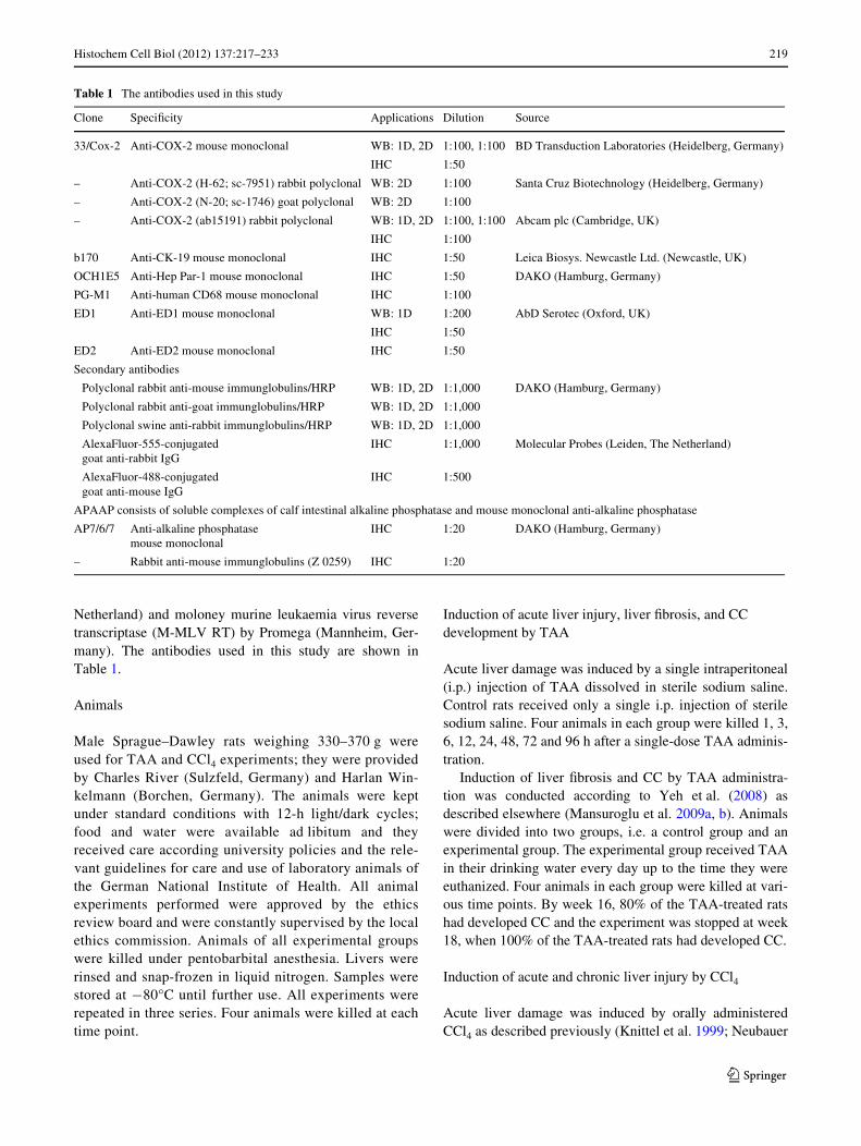
Netherland) and moloney murine leukaemia virus reversetranscriptase (M-MLV RT) by Promega (Mannheim, Ger-many). The antibodies used in this study are shown inTable 1.
Animals
Male Sprague–Dawley rats weighing 330–370 g wereused for TAA and CCl4 experiments; they were providedby Charles River (Sulzfeld, Germany) and Harlan Win-kelmann (Borchen, Germany). The animals were keptunder standard conditions with 12-h light/dark cycles;food and water were available ad libitum and theyreceived care according university policies and the rele-vant guidelines for care and use of laboratory animals ofthe German National Institute of Health. All animalexperiments performed were approved by the ethicsreview board and were constantly supervised by the localethics commission. Animals of all experimental groupswere killed under pentobarbital anesthesia. Livers wererinsed and snap-frozen in liquid nitrogen. Samples werestored at ¡80°C until further use. All experiments wererepeated in three series. Four animals were killed at eachtime point.
Induction of acute liver injury, liver Wbrosis, and CC development by TAA
Acute liver damage was induced by a single intraperitoneal(i.p.) injection of TAA dissolved in sterile sodium saline.Control rats received only a single i.p. injection of sterilesodium saline. Four animals in each group were killed 1, 3,6, 12, 24, 48, 72 and 96 h after a single-dose TAA adminis-tration.
Induction of liver Wbrosis and CC by TAA administra-tion was conducted according to Yeh et al. (2008) asdescribed elsewhere (Mansuroglu et al. 2009a, b). Animalswere divided into two groups, i.e. a control group and anexperimental group. The experimental group received TAAin their drinking water every day up to the time they wereeuthanized. Four animals in each group were killed at vari-ous time points. By week 16, 80% of the TAA-treated ratshad developed CC and the experiment was stopped at week18, when 100% of the TAA-treated rats had developed CC.
Induction of acute and chronic liver injury by CCl4
Acute liver damage was induced by orally administeredCCl4 as described previously (Knittel et al. 1999; Neubauer
Table 1 The antibodies used in this study
Clone SpeciWcity Applications Dilution Source
33/Cox-2 Anti-COX-2 mouse monoclonal WB: 1D, 2D 1:100, 1:100 BD Transduction Laboratories (Heidelberg, Germany)
IHC 1:50
– Anti-COX-2 (H-62; sc-7951) rabbit polyclonal WB: 2D 1:100 Santa Cruz Biotechnology (Heidelberg, Germany)
– Anti-COX-2 (N-20; sc-1746) goat polyclonal WB: 2D 1:100
– Anti-COX-2 (ab15191) rabbit polyclonal WB: 1D, 2D 1:100, 1:100 Abcam plc (Cambridge, UK)
IHC 1:100
b170 Anti-CK-19 mouse monoclonal IHC 1:50 Leica Biosys. Newcastle Ltd. (Newcastle, UK)
OCH1E5 Anti-Hep Par-1 mouse monoclonal IHC 1:50 DAKO (Hamburg, Germany)
PG-M1 Anti-human CD68 mouse monoclonal IHC 1:100
ED1 Anti-ED1 mouse monoclonal WB: 1D 1:200 AbD Serotec (Oxford, UK)
IHC 1:50
ED2 Anti-ED2 mouse monoclonal IHC 1:50
Secondary antibodies
Polyclonal rabbit anti-mouse immunglobulins/HRP WB: 1D, 2D 1:1,000 DAKO (Hamburg, Germany)
Polyclonal rabbit anti-goat immunglobulins/HRP WB: 1D, 2D 1:1,000
Polyclonal swine anti-rabbit immunglobulins/HRP WB: 1D, 2D 1:1,000
AlexaFluor-555-conjugatedgoat anti-rabbit IgG
IHC 1:1,000 Molecular Probes (Leiden, The Netherland)
AlexaFluor-488-conjugatedgoat anti-mouse IgG
IHC 1:500
APAAP consists of soluble complexes of calf intestinal alkaline phosphatase and mouse monoclonal anti-alkaline phosphatase
AP7/6/7 Anti-alkaline phosphatasemouse monoclonal
IHC 1:20 DAKO (Hamburg, Germany)
– Rabbit anti-mouse immunglobulins (Z 0259) IHC 1:20
123
220 Histochem Cell Biol (2012) 137:217–233
et al. 1995). A single dose of 150 �l CCl4/corn oil solution(50%, v/v) per 100 g body weight was given to each rat.Control rats received only corn oil. Rats were killed 3, 6, 9,12, 24, 48, 72, and 96 h after the CCl4 administration.
Chronic liver injury was induced according to the proto-col by Proctor and Chatamra (1982). The experimentalgroup received CCl4 orally once a week. After a period of 8and 13 weeks, the animals were killed and livers wereobtained.
Isolation of hepatocytes, tissue macrophages (KupVer cells), and myoWbroblasts from rat livers
Hepatocytes from male Sprague–Dawley rats were isolatedby in situ perfusion and cultured as reported previously(Neubauer et al. 1995; Knittel et al. 1992). KupVer cellsand myoWbroblasts of normal liver were isolated asdescribed by Knook and Sleyster (1976) with some modiW-cations (Armbrust et al. 1993; Neubauer et al. 2008).
Isolated cells were cultured for 24 h and protein extractswere prepared from KupVer cells, hepatocytes, and myoW-broblasts. For mRNA isolation, KupVer cells (in duplicate)were stimulated with 10 �g/ml LPS, and mRNA wasobtained 2, 4, and 6 h from LPS-treated as well as fromcontrol cultures.
Isolation of mononuclear phagocytes from damaged rat livers
Mononuclear phagocytes from TAA- and CCl4-damagedrat livers were isolated by in situ perfusion. Centrifugal elu-triation was performed to separate small KC from largeKC. The Wrst fraction was collected at Xow rates rangingfrom 19 up to 28 ml/min (Beckmann centrifuge J2-21, J-6Brotor, 2,500 rpm) and the second fraction at Xow rates rang-ing from 28 up to 55 ml/min. Each fraction was sedi-mented, resuspended in culture medium (M199, 15% FCS,100 U penicillin/ml, 100 �g streptomycin/ml), and countedin a Neubauer chamber after Trypan Blue-staining. Cellswere then plated onto 24-well plates (5 £ 105 cells/well).Two hours after plating, the cultures were washed inten-sively to eliminate non-adherent cells. Cultures were keptin a 5% CO2 atmosphere and saturated humidity at 37°C.Total protein extracts and mRNA were obtained.
Human liver tissue
Four specimens of cirrhotic human livers were obtainedfrom patients undergoing transplantation after developingHCC or CC and four specimens of cirrhotic liver withouttumor. Specimens were frozen in liquid nitrogen immedi-ately after surgical removal. Human liver samples wereused for immunohistochemical analysis.
Culture and activation of macrophage cell line
The monocytic/macrophage cell line RAW 264.7 (Lüderet al. 2003; Raschke et al. 1978) was a gift from the Depart-ment of Bacteriology, Georg-August University Goettingen(UMG, Goettingen, Germany). RAW 264.7 cells wereincubated with RPMI 1640 medium containing 10% FCS,100 U/ml penicillin, 100 mg/ml streptomycin, 2 mmol/lL-glutamine, and 2.5 g/l D-glucose at 37°C, 20% O2, and 5%CO2. Cells were used to determine the speciWty of COX-2antibody binding and the eVect of stimulation on COX-2expression. Cells were activated with 10 �g/ml LPS and10 ng/ml PMA. PMA was dissolved in DMSO (0.5 mg/ml)and aliquots were stored at ¡20°C; LPS was dissolved inPBS. Control cells were incubated with the DMSO vehicleonly. For protein or mRNA isolation, cells were seeded at0.2 £ 107 cells in 75 cm2 tissue culture Xasks (Sarstedt AG& Co., Nümbrecht, Germany).
RNA isolation and real-time PCR
RNA was isolated from all control and TAA-treated rat liv-ers. RNA quality was tested by agarose gel electrophoresisand visualized with UV light. RNA concentration wasquantiWed by measuring the absorbance at 260/280 nm. ThecDNA was generated by reverse transcription of 3.0 �g oftotal RNA with 100 nmol/l of dNTPs, 50 pmol/l of primeroligo(dT)15, 200 units of M-MLV RT, 16 units of protectorRNAse inhibitor, 1£ RT buVer, and 2.5 ml of 0.1 mol/lDTT for 1 h at 40°C as described previously (Kondo et al.1999). Gene expression of COX-2 (forward primer 5�-TACCGGACTGGATTCTAG-3�, reverse primer 5�-AAGTTGGTGGGCTGTCAATC-3�), and ED1 (forward primer 5�-ATTGAACCCGAACAAAACCA-3�, reverse primer 5�-GCTTGTGGGAAGGACACATT-3�) were analyzed using aFast Sybr Green master mix. UBC (forward primer 5�-CACCAAGAAGGTCAAACAGGAA-3�, reverse primer 5�-AAGACACCTCCCCATCAAACC-3�) was used as house-keeping gene in the TAA model. The ampliWcation wasperformed through two-step cycling (95–60°C) for 40cycles in a StepOne Plus RT-PCR detection system, fol-lowing the instructions of the supplier (AB, Applied Bio-system, USA). All samples were assayed in duplicate. Theresults were normalized to the controls, and fold change ofthe gene expression was calculated using threshold cycle(Ct) values.
Total protein extraction from rat liver, isolated rat liver cells, and monocytic/macrophage cell line RAW 264.7
Total protein extracts were prepared from all controls,TAA/CCl4-treated rat livers, and dissected CC tissues, aswell as from isolated hepatocytes, myoWbroblasts, KupVer
123
Histochem Cell Biol (2012) 137:217–233 221
cells, and RAW 264.7 cells. Tissues were washed in ice-cold PBS to remove blood and homogenized at room tem-perature in cell lysis buVer containing 7 mol/l urea, 2 mol/lthiourea, 4% (w/v) CHAPS, 2% ampholytes, 1% (w/v) DL-dithiothreitol and 10 mmol/l phenylmethylsulphonyl Xuo-ride, as well as 1% (v/v) phosphatase inhibitor cocktail 1,and 1% (v/v) phosphatase inhibitor cocktail 2 with ahomogenizer (Ultra-Turrax, Jahnke and Kunkel GmbH &CO.KG, Staufen, Germany). The protein concentration wascalculated according to Bradford (1976) using the Bio-Radprotein assay kit. The protein samples were stored at¡80°C until further analysis.
1-DE/2-DE Western blot analysis of total protein lysates from rat liver tissue, isolated rat liver cells, and RAW 264.7cells
For 1-DE Western blot analysis, 50 �g total protein wasloaded on a 4–12% NuPAGE Bis–Tris gel (Invitrogen,Darmstadt, Germany) and separated for 2 h by electro-phoresis at 80 V. For 2-DE Western blot analysis, 140 �gtotal protein lysate from rat liver tissues or from macro-phage cell line RAW 264.7 and a trace of bromphenolblue were loaded on immobilized pH gradient (IPG) stripswith a nonlinear pH range of 3–10. After rehydration, iso-electric focusing was performed in a Protein IEF Cell(Bio-Rad) at 20°C set to 32,000 Vh. The IPG strip wasequilibrated with equilibration buVer (6 mol/l urea, 30%glycerine, 2% sodium dodecyl sulphate, 0.05 mol/l Tris/5 N HCl to pH 8.8 and a trace of bromophenol blue) con-taining 15 mmol/l DTT, followed with equilibrationbuVer containing 40 g/l iodoacetamide. Afterwards, thestrip was loaded onto a vertical 12% polyacrylamideSDS-PAGE for separation by molecular weight for 19 h at4°C and 90 V. After Western blot, Ponceau staining wasperformed as a control for successful protein transfer.Nitrocellulose membranes were blocked in blockingbuVer containing 5% BSA and were incubated overnightat 4°C with diVerent anti-COX-2 antibodies (33/Cox-2,ab15191, sc-1746, sc-7951). The secondary antibodieswere conjugated with horseradish peroxidase. The mem-branes were developed with an ECL chemiluminescencekit (GE Healthcare, Munich, Germany). Antibodies anddilutions used are listed in Table 1.
Silver-staining of proteins
2-DE gels were Wxed and washed and silver-staining wasperformed according to the modiWed silver-staining methodof Blum et al. (1987). Gels were scanned (CanoScan8400F, Canon) and Wnally dried for further storage (GelDryer, Model 583; Bio-Rad).
Protein identiWcation
In-gel digestion was carried out according to a modiWedprotocol of Shevchenko et al. (1996). Spots of interest werethe positive spots from the ECL staining after 2-DE blots.These were excised and after de-staining with potassiumferricyanide and sodium thiosulphate; proteins/peptideswere digested with trypsin. Gel slices were washed andequilibrated with ammonium bicarbonate followed by incu-bation with acetonitrile (ACN). Peptides were extractedusing triXuoroacetic acid and ACN. Solutions with digestedprotein/peptide were dried in a speed vacuum system(UniEquip GmbH, Munich, Germany) and stored at ¡20°C
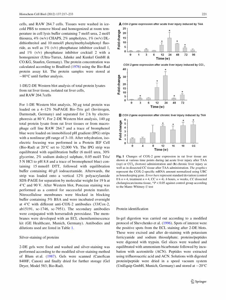
Fig. 1 Changes of COX-2 gene expression in rat liver tissue areshown at various time points during (a) acute liver injury after TAA(top) or CCl4 (bottom) administration and (b) chronic liver injury aswell as in dissected CC tissue after TAA administration. The graphicsrepresent the COX-2-speciWc mRNA amount normalized using UBCas housekeeping gene. Error bars represent standard deviation (control0 h n = 4, treatment n = 4, CC n = 4). h hours, w weeks, CC dissectedcholangiocarcinoma tissue, *P < 0.05 against control group accordingto the Mann–Whitney U test
123

222 Histochem Cell Biol (2012) 137:217–233
until further analysis. Dried samples were diluted in 0.1%formic acid and 1 �l was loaded for chromatographic sepa-ration on a CapLC-System (Waters, Milford, MA, USA).Peptide sequence analysis was carried out on a Q-TOFUltima Global mass spectrometer (Micromass, Manchester,UK) equipped with a nanoXow ESI Z-spray source in posi-tive ion mode, as described previously (Schultze et al.2010). Data were processed using Protein Lynx GlobalServer (2.0, Micromass) and searched against MSDB andSwissProt data bases through the Mascot search engine
with oxidation (M) and carbamidomethyl (C) modiWcation,when appropriate.
Histochemical analysis and immunohistochemical investigations
Tissue sections were Wrst analyzed by hematoxylin andeosin staining (HE). Liver tissue sections (5 �m thick) ofthree representative rats as well as three control rats wereanalyzed at the following time points: 0, 24, 48, 96 h, 8,
Fig. 2 Immunolocalization of COX-2 protein by double immunoXuo-rescence staining (CK-19, Hep Par-1, and ED1) in normal rat liver.ImmunoXuorescence staining of a: COX-2 (red) and CK-19 (green);b: COX-2 (red) and Hep Par-1 (green); c: COX-2 (red) and ED1 (green).
Co-localization of COX-2 and ED1 is detectable. Polyclonal COX-2antibody (ab15191) was used. The blue staining with DAPI representsthe nuclei (£100/£400 original magniWcation). pv portal vein, bd bileduct. Bar 50 �m
123

Histochem Cell Biol (2012) 137:217–233 223
16, and 18 weeks. Furthermore, specimens of four cir-rhotic human livers after developing HCC or CC and fourspecimens of cirrhotic liver without tumor were ana-lyzed. After blocking of non-speciWc binding with 1%BSA and 10% goat serum (DAKO, Hamburg, Germany)containing PBS for 1 h at room temperature, two primaryantibodies were simultaneously incubated on the sectionsovernight at 4°C. We used primary antibodies: rabbitpolyclonal anti-COX-2; mouse monoclonal anti-COX-2(33/Cox-2), mouse monoclonal anti-CK-19; mousemonoclonal anti-Hep Par-1; mouse monoclonal anti-
ED1, mouse monoclonal anti-CD68, and mouse mono-clonal anti-ED2. Rabbit polyclonal antibodies weredetected with AlexaFluor-555-conjugated goat anti-rabbitsecondary antibody and mouse monoclonal antibodieswere visualized with AlexaFluor-488-conjugated goatanti-mouse secondary antibody. Antibody dilutions usedare listed in Table 1.
In the double staining immunohistochemistry, ED1+/COX-2+ positive cells were counted in Wve randomlyselected areas (0.51 mm2/area) in the centrilobular area ofthe hepatic lobule at a magniWcation of 100£.
Fig. 3 Immunolocalization of COX-2, ED1 and ED2 in TAA-inducedacute rat liver injury (48 h). An immunoXuorescence staining of COX-2- (red), ED1- (green, left side), and ED2¡ (green, right side) positive
cells is shown. Polyclonal COX-2 antibody (ab15191) was used. Stain-ing with DAPI represents the nuclei (£100/£200 original magniWca-tion). pv portal vein. Bar 50 �m
123
224 Histochem Cell Biol (2012) 137:217–233
Negative control immunostainings were performed byomission of the primary antibodies, use of non-immuneserum, and by isotype matching control immunoglobulin.Sections were counter-stained with DAPI and observedwith an epiXuorescence microscope (Axiovert 200 M,Zeiss, Jena, Germany).
Alkaline phosphatase anti-alkaline phosphatase (APAAP) technique
We used the mouse monoclonal anti-COX-2 antibody (33/Cox-2) in this experiment. To make the antigen (COX-2)/antibody reaction visible, the APAAP technique was
performed (Cordell et al. 1984). The APAAP complex isstained with neufuchsin, which stains the COX-2-positivecells red. The remaining tissue is made visible by counter-staining with hematoxylin.
Statistical analysis
The data were analyzed with GraphPad Prism 4.0 software(San Diego, USA) and SPSS (V14.0 for Windows; Chi-cago, IL, USA). The results are shown as means § standarddeviation. SigniWcant diVerence was assessed at P < 0.05against the control group and calculated according to theMann–Whitney U test.
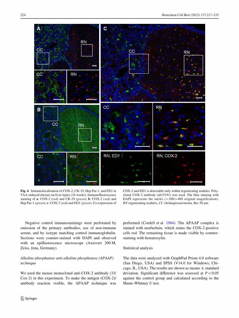
Fig. 4 Immunolocalization of COX-2, CK-19, Hep Par-1, and ED1 inTAA-induced chronic rat liver injury (16 weeks). ImmunoXuorescencestaining of a: COX-2 (red) and CK-19 (green); b: COX-2 (red) andHep Par-1 (green); c: COX-2 (red) and ED1 (green). Co-expression of
COX-2 and ED1 is detectable only within regenerating nodules. Poly-clonal COX-2 antibody (ab15191) was used. The blue staining withDAPI represents the nuclei (£100/£400 original magniWcation).RN regenerating nodules, CC cholangiocarcinoma. Bar 50 �m
123
Histochem Cell Biol (2012) 137:217–233 225
Results
COX-2 gene expression in normal and damaged rat liver
COX-2 speciWc mRNA
COX-2 mRNA was detectable in normal liver tissue andan upregulation up to 15-fold was detected during acute(Fig. 1a) and chronic liver injury (Fig. 1b). SigniWcant
upregulation of COX-2 gene expression (P < 0.05) dur-ing acute liver injury was observed 12 h (7.2 § 2.3-fold),24 h (14.69 § 5.99-fold), 48 h (8.76 § 0.36-fold), and72 h (4.48 § 1.2-fold) after TAA administration. Chronicliver injury revealed signiWcant upregulation (P < 0.05)after 12 weeks (4.31 § 0.65-fold), 16 weeks (3.56 §0.65-fold), and 18 weeks of TAA administration (4.34 §1.94-fold), as well as in dissected CC tissue (4.94 § 1.78-fold).
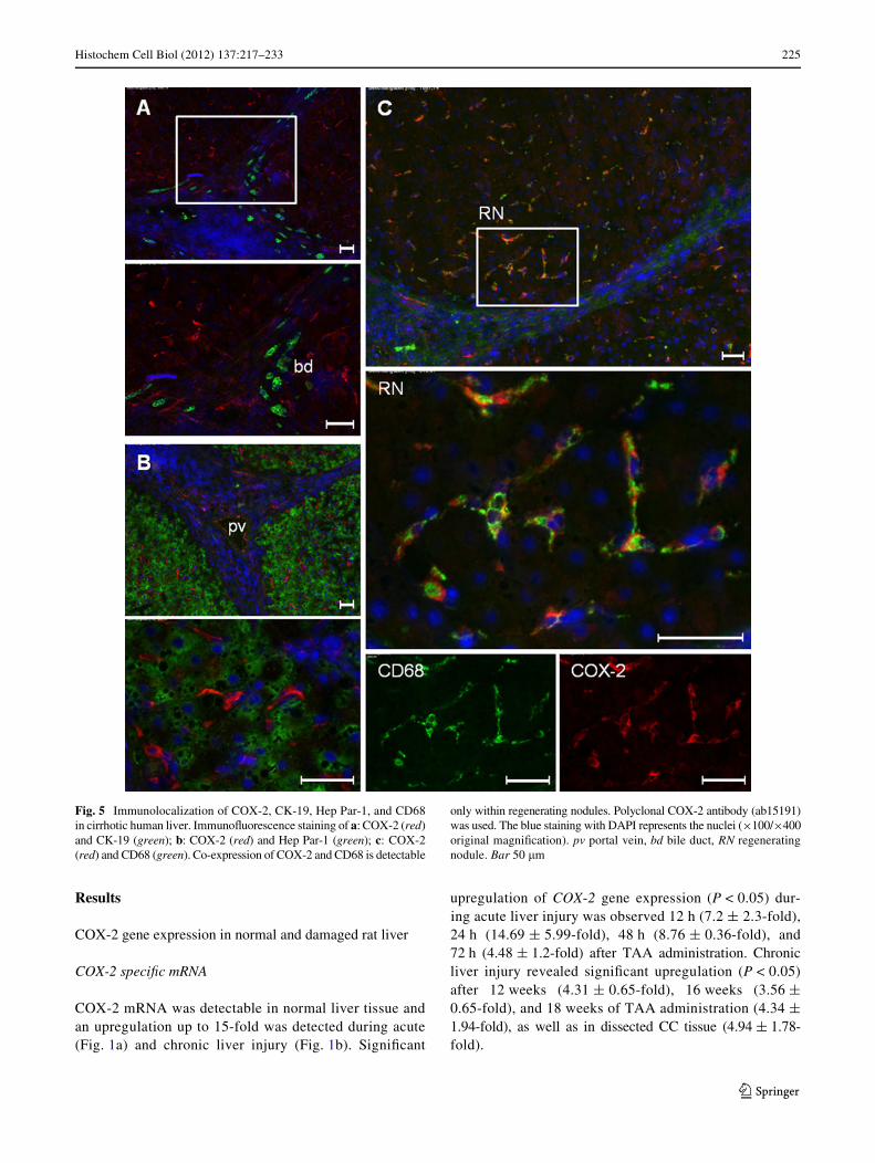
Fig. 5 Immunolocalization of COX-2, CK-19, Hep Par-1, and CD68in cirrhotic human liver. ImmunoXuorescence staining of a: COX-2 (red)and CK-19 (green); b: COX-2 (red) and Hep Par-1 (green); c: COX-2(red) and CD68 (green). Co-expression of COX-2 and CD68 is detectable
only within regenerating nodules. Polyclonal COX-2 antibody (ab15191)was used. The blue staining with DAPI represents the nuclei (£100/£400original magniWcation). pv portal vein, bd bile duct, RN regeneratingnodule. Bar 50 �m
123
226 Histochem Cell Biol (2012) 137:217–233
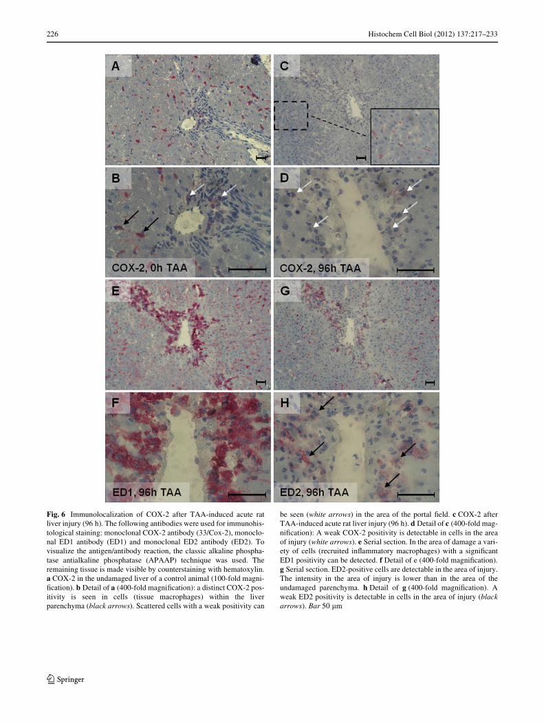
Fig. 6 Immunolocalization of COX-2 after TAA-induced acute ratliver injury (96 h). The following antibodies were used for immunohis-tological staining: monoclonal COX-2 antibody (33/Cox-2), monoclo-nal ED1 antibody (ED1) and monoclonal ED2 antibody (ED2). Tovisualize the antigen/antibody reaction, the classic alkaline phospha-tase antialkaline phosphatase (APAAP) technique was used. Theremaining tissue is made visible by counterstaining with hematoxylin.a COX-2 in the undamaged liver of a control animal (100-fold magni-Wcation). b Detail of a (400-fold magniWcation): a distinct COX-2 pos-itivity is seen in cells (tissue macrophages) within the liverparenchyma (black arrows). Scattered cells with a weak positivity can
be seen (white arrows) in the area of the portal Weld. c COX-2 afterTAA-induced acute rat liver injury (96 h). d Detail of c (400-fold mag-niWcation): A weak COX-2 positivity is detectable in cells in the areaof injury (white arrows). e Serial section. In the area of damage a vari-ety of cells (recruited inXammatory macrophages) with a signiWcantED1 positivity can be detected. f Detail of e (400-fold magniWcation).g Serial section. ED2-positive cells are detectable in the area of injury.The intensity in the area of injury is lower than in the area of theundamaged parenchyma. h Detail of g (400-fold magniWcation). Aweak ED2 positivity is detectable in cells in the area of injury (blackarrows). Bar 50 �m
123

Histochem Cell Biol (2012) 137:217–233 227
COX-2 localization in liver tissue
ImmunoXuorescence staining
Double immunoXuorescence staining was performed usingthe polyclonal (ab15191) and/or monoclonal (33/Cox-2)COX-2 antibody combined with antisera speciWc for CK-19, Hep Par-1, ED1 or ED2.
In healthy adult rat liver, the bile duct epithelial cellswere strongly positive for CK-19, a typical bile duct cellantigen. CK-19+ cells were COX-2¡ as were Hep Par1+
hepatocytes (Fig. 2a, b). Only ED1+/ED2+ cells (tissuemacrophages) in normal liver parenchyma were COX-2+
(Fig. 2c).After administration of the toxins (TAA or CCl4), an
increasing number of ED1+/ED2¡ mononuclear cellsbecame detectable in the areas of damage. These cellsshowed only a weak positivity for COX-2. In contrast, theED1+/ED2+ cells of the non-damaged tissue were clearlyCOX-2+. There was no diVerence in the distributionbetween ED1+ and COX-2+ cells (Fig. 3).
The cell counting performed in both models (TAA andCCl4) of liver damage showed a signiWcant increase of thetotal number of ED1+/COX-2+ cells after 24 h (1.4-fold) aswell as after 48 h (1.86-fold).
ED1+/COX-2+ cells located in the non-damaged areawere ED2+ as well. Their number was not signiWcantly
altered after 24 h (1.08-fold) and 48 h (0.73-fold) in com-parison with the control.
ED1+/COX-2+ cells located in the damaged area werepredominantly ED2¡ and only a few cells with a weakED2+ were detectable. These cells showed a signiWcantincrease after 24 h (4.29-fold) and after 48 h (12-fold).
In chronically injured livers a strong increase of CK-19+
cells is observable but these CK-19+ cells remained COX-2¡
as did those localized within the CC region (Fig. 4a).However, COX-2+ and ED1+/ED2+ cells were observedwithin the “regenerating” nodules. While ED1+/ED2+ cellswithin the regenerating nodules strongly correlated withCOX-2+ cells (ED1 and COX-2 co-localization), ED1+/ED2¡ cells within the CC region were COX-2¡ (Fig. 4c).
In human cirrhotic livers, co-expression of CD68 (tissuemacrophages) and COX-2 was conWrmed in the “regener-ating” nodules, whereas no co-expression of Hep Par-1and COX-2, nor of CK-19 and COX-2 were observed(Fig. 5). There was no diVerence between cirrhotic liversafter developing HCC or CC and cirrhotic livers withouttumor.
Immunohistochemistry by APAAP method
After speciWcity analysis performed by 2-DE Western blotanalysis and characterisation of the identiWed proteins werealized the potentially non-speciWc binding of other proteins.
Fig. 7 Immunolocalization of COX-2 in TAA-induced cholangiocar-cinoma. For immunostaining the monoclonal COX-2 antibody (33/Cox-2) was used. To visualize the antigen/antibody reaction, the clas-sic alkaline phosphatase antialkaline phosphatase (APAAP) techniquewas used. The remaining tissue is made visible by counterstaining with
hematoxylin (£100/£400 original magniWcation). The black arrowsindicate COX-2-positive cells in the liver parenchyma. The whitearrows point to COX-2-positive cells within the tumor microenviron-ment (lower images, 400-fold magniWcation). Tumor cells are COX-2negative. Bar 50 �m
123
228 Histochem Cell Biol (2012) 137:217–233
For immunohistochemistry with the APAAP method, weused the COX-2 monoclonal antibody (33/Cox-2). Theresults of APAAP staining of acute damaged livers con-Wrmed the results of immunoXuorescence staining. Nonspe-ciWc staining, particularly in the area of injury (“inXamedhepatocytes”), did not appear (Fig. 6).
In the chronically damaged liver (tumor region), someweakly COX-2+ cells were detectable within the tumormicroenvironment (Fig. 7).
COX-2 gene expression in a macrophage cell line RAW 264.7 and in isolated rat liver cells
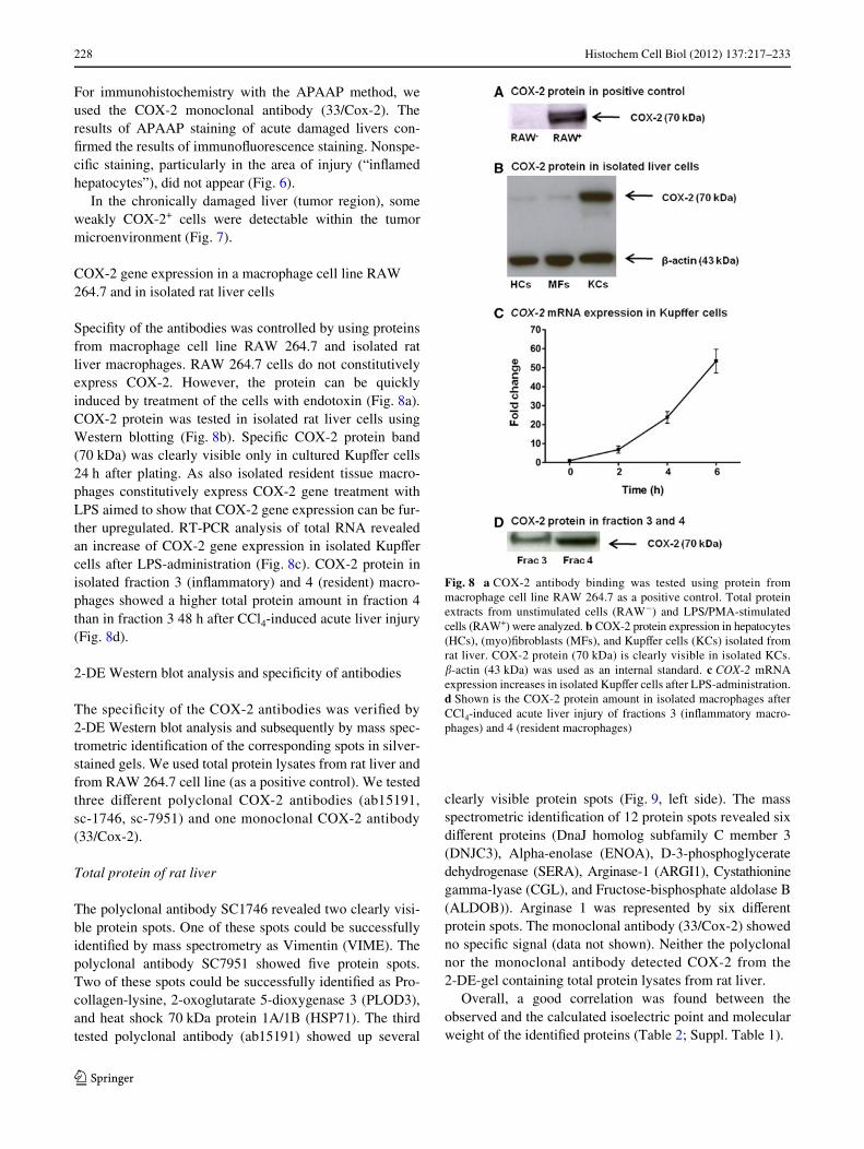
SpeciWty of the antibodies was controlled by using proteinsfrom macrophage cell line RAW 264.7 and isolated ratliver macrophages. RAW 264.7 cells do not constitutivelyexpress COX-2. However, the protein can be quicklyinduced by treatment of the cells with endotoxin (Fig. 8a).COX-2 protein was tested in isolated rat liver cells usingWestern blotting (Fig. 8b). SpeciWc COX-2 protein band(70 kDa) was clearly visible only in cultured KupVer cells24 h after plating. As also isolated resident tissue macro-phages constitutively express COX-2 gene treatment withLPS aimed to show that COX-2 gene expression can be fur-ther upregulated. RT-PCR analysis of total RNA revealedan increase of COX-2 gene expression in isolated KupVercells after LPS-administration (Fig. 8c). COX-2 protein inisolated fraction 3 (inXammatory) and 4 (resident) macro-phages showed a higher total protein amount in fraction 4than in fraction 3 48 h after CCl4-induced acute liver injury(Fig. 8d).
2-DE Western blot analysis and speciWcity of antibodies
The speciWcity of the COX-2 antibodies was veriWed by2-DE Western blot analysis and subsequently by mass spec-trometric identiWcation of the corresponding spots in silver-stained gels. We used total protein lysates from rat liver andfrom RAW 264.7 cell line (as a positive control). We testedthree diVerent polyclonal COX-2 antibodies (ab15191,sc-1746, sc-7951) and one monoclonal COX-2 antibody(33/Cox-2).
Total protein of rat liver
The polyclonal antibody SC1746 revealed two clearly visi-ble protein spots. One of these spots could be successfullyidentiWed by mass spectrometry as Vimentin (VIME). Thepolyclonal antibody SC7951 showed Wve protein spots.Two of these spots could be successfully identiWed as Pro-collagen-lysine, 2-oxoglutarate 5-dioxygenase 3 (PLOD3),and heat shock 70 kDa protein 1A/1B (HSP71). The thirdtested polyclonal antibody (ab15191) showed up several
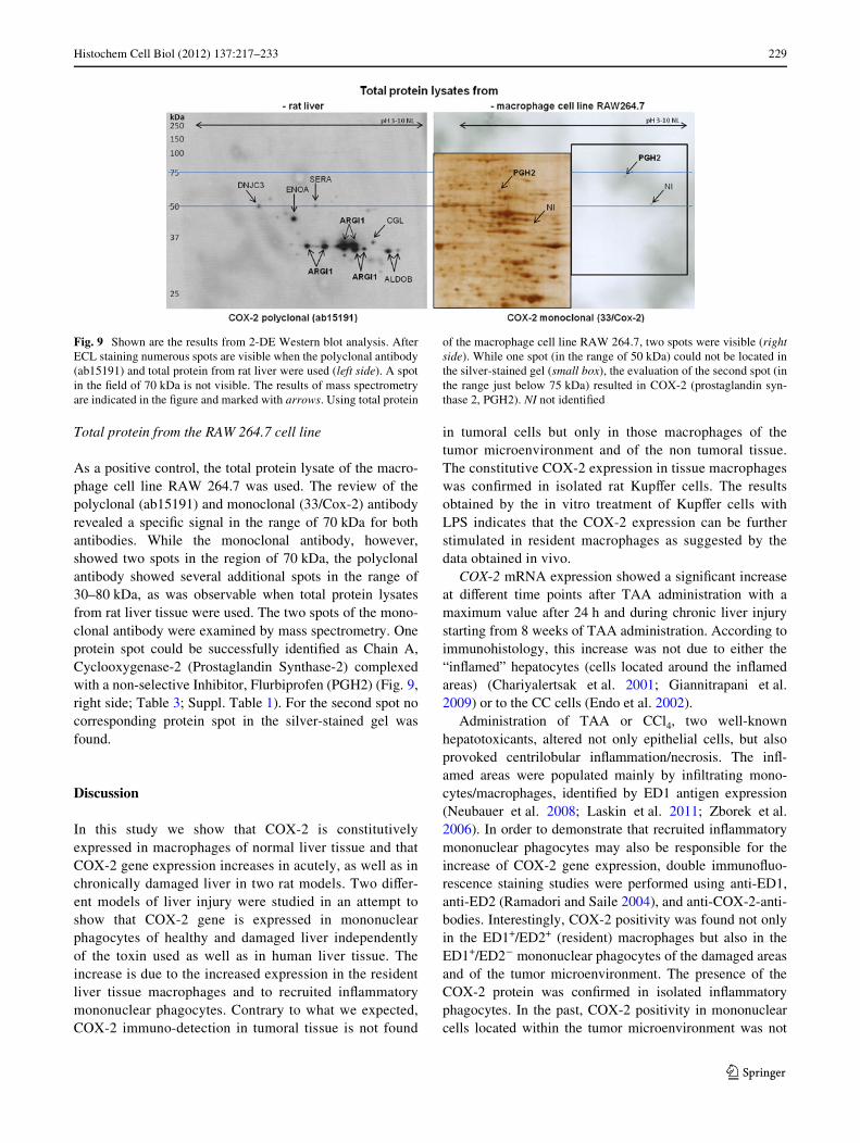
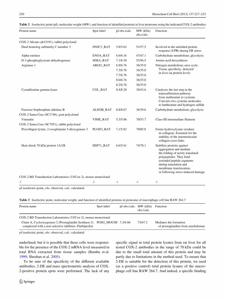
clearly visible protein spots (Fig. 9, left side). The massspectrometric identiWcation of 12 protein spots revealed sixdiVerent proteins (DnaJ homolog subfamily C member 3(DNJC3), Alpha-enolase (ENOA), D-3-phosphoglyceratedehydrogenase (SERA), Arginase-1 (ARGI1), Cystathioninegamma-lyase (CGL), and Fructose-bisphosphate aldolase B(ALDOB)). Arginase 1 was represented by six diVerentprotein spots. The monoclonal antibody (33/Cox-2) showedno speciWc signal (data not shown). Neither the polyclonalnor the monoclonal antibody detected COX-2 from the2-DE-gel containing total protein lysates from rat liver.
Overall, a good correlation was found between theobserved and the calculated isoelectric point and molecularweight of the identiWed proteins (Table 2; Suppl. Table 1).
Fig. 8 a COX-2 antibody binding was tested using protein frommacrophage cell line RAW 264.7 as a positive control. Total proteinextracts from unstimulated cells (RAW¡) and LPS/PMA-stimulatedcells (RAW+) were analyzed. b COX-2 protein expression in hepatocytes(HCs), (myo)Wbroblasts (MFs), and KupVer cells (KCs) isolated fromrat liver. COX-2 protein (70 kDa) is clearly visible in isolated KCs.�-actin (43 kDa) was used as an internal standard. c COX-2 mRNAexpression increases in isolated KupVer cells after LPS-administration.d Shown is the COX-2 protein amount in isolated macrophages afterCCl4-induced acute liver injury of fractions 3 (inXammatory macro-phages) and 4 (resident macrophages)
123
Histochem Cell Biol (2012) 137:217–233 229
Total protein from the RAW 264.7 cell line
As a positive control, the total protein lysate of the macro-phage cell line RAW 264.7 was used. The review of thepolyclonal (ab15191) and monoclonal (33/Cox-2) antibodyrevealed a speciWc signal in the range of 70 kDa for bothantibodies. While the monoclonal antibody, however,showed two spots in the region of 70 kDa, the polyclonalantibody showed several additional spots in the range of30–80 kDa, as was observable when total protein lysatesfrom rat liver tissue were used. The two spots of the mono-clonal antibody were examined by mass spectrometry. Oneprotein spot could be successfully identiWed as Chain A,Cyclooxygenase-2 (Prostaglandin Synthase-2) complexedwith a non-selective Inhibitor, Flurbiprofen (PGH2) (Fig. 9,right side; Table 3; Suppl. Table 1). For the second spot nocorresponding protein spot in the silver-stained gel wasfound.
Discussion
In this study we show that COX-2 is constitutivelyexpressed in macrophages of normal liver tissue and thatCOX-2 gene expression increases in acutely, as well as inchronically damaged liver in two rat models. Two diVer-ent models of liver injury were studied in an attempt toshow that COX-2 gene is expressed in mononuclearphagocytes of healthy and damaged liver independentlyof the toxin used as well as in human liver tissue. Theincrease is due to the increased expression in the residentliver tissue macrophages and to recruited inXammatorymononuclear phagocytes. Contrary to what we expected,COX-2 immuno-detection in tumoral tissue is not found
in tumoral cells but only in those macrophages of thetumor microenvironment and of the non tumoral tissue.The constitutive COX-2 expression in tissue macrophageswas conWrmed in isolated rat KupVer cells. The resultsobtained by the in vitro treatment of KupVer cells withLPS indicates that the COX-2 expression can be furtherstimulated in resident macrophages as suggested by thedata obtained in vivo.
COX-2 mRNA expression showed a signiWcant increaseat diVerent time points after TAA administration with amaximum value after 24 h and during chronic liver injurystarting from 8 weeks of TAA administration. According toimmunohistology, this increase was not due to either the“inXamed” hepatocytes (cells located around the inXamedareas) (Chariyalertsak et al. 2001; Giannitrapani et al.2009) or to the CC cells (Endo et al. 2002).
Administration of TAA or CCl4, two well-knownhepatotoxicants, altered not only epithelial cells, but alsoprovoked centrilobular inXammation/necrosis. The inX-amed areas were populated mainly by inWltrating mono-cytes/macrophages, identiWed by ED1 antigen expression(Neubauer et al. 2008; Laskin et al. 2011; Zborek et al.2006). In order to demonstrate that recruited inXammatorymononuclear phagocytes may also be responsible for theincrease of COX-2 gene expression, double immunoXuo-rescence staining studies were performed using anti-ED1,anti-ED2 (Ramadori and Saile 2004), and anti-COX-2-anti-bodies. Interestingly, COX-2 positivity was found not onlyin the ED1+/ED2+ (resident) macrophages but also in theED1+/ED2¡ mononuclear phagocytes of the damaged areasand of the tumor microenvironment. The presence of theCOX-2 protein was conWrmed in isolated inXammatoryphagocytes. In the past, COX-2 positivity in mononuclearcells located within the tumor microenvironment was not
Fig. 9 Shown are the results from 2-DE Western blot analysis. AfterECL staining numerous spots are visible when the polyclonal antibody(ab15191) and total protein from rat liver were used (left side). A spotin the Weld of 70 kDa is not visible. The results of mass spectrometryare indicated in the Wgure and marked with arrows. Using total protein
of the macrophage cell line RAW 264.7, two spots were visible (rightside). While one spot (in the range of 50 kDa) could not be located inthe silver-stained gel (small box), the evaluation of the second spot (inthe range just below 75 kDa) resulted in COX-2 (prostaglandin syn-thase 2, PGH2). NI not identiWed
123
230 Histochem Cell Biol (2012) 137:217–233
underlined, but it is possible that these cells were responsi-ble for the presence of the COX-2 mRNA level measured intotal RNA extracted from tissue samples (Bamba et al.1999; Sheehan et al. 2005).
To be sure of the speciWcity of the diVerent availableantibodies, 2-DE and mass spectrometric analysis of COX-2-positive protein spots were performed. The lack of any
speciWc signal in total protein lysates from rat liver for alltested COX-2 antibodies in the range of 70 kDa could bedue to the small total amount of this protein and may bepartly due to limitations in the method used. To ensure that2-DE is suitable for the detection of this protein, we used(as a positive control) total protein lysates of the macro-phage cell line RAW 264.7. And indeed, a speciWc binding
Table 2 Isoelectric point (pI), molecular weight (MW), and function of identiWed proteins in liver proteome using the indicated COX-2 antibodies
pI isoelectric point, obs. observed, calc. calculated
Protein name Spot label pI obs./calc. MW (kDa) obs./calc.
Function
COX-2 Abcam (ab15191), rabbit polyclonal
DnaJ homolog subfamily C member 3 DNJC3_RAT 5.8/5.62 51/57.5 Involved in the unfolded protein response (UPR) during ER stress
Alpha-enolase ENOA_RAT 6.6/6.16 47/47.1 Carbohydrate metabolism; glycolysis
D-3-phosphoglycerate dehydrogenase SERA_RAT 7.1/6.30 52/56.5 Amino-acid biosynthesis
Arginase-1 ARGI1_RAT 6.9/6.76 36/35.0 Nitrogen metabolism; urea cycle. Tissue speciWcity: detected in liver (at protein level)
7.3/6.76 36/35.0
7.7/6.76 36/35.0
8.0/6.76 36/35.0
8.2/6.76 36/35.0
Cystathionine gamma-lyase CGL_RAT 8.4/8.20 38/43.6 Catalyzes the last step in the transsulfuration pathway from methionine to cysteine. Converts two cysteine molecules to lanthionine and hydrogen sulWde
Fructose-bisphosphate aldolase B ALDOB_RAT 8.8/8.67 36/39.6 Carbohydrate metabolism; glycolysis
COX-2 Santa Cruz (SC1746), goat polyclonal
Vimentin VIME_RAT 5.3/5.06 70/53.7 Class-III intermediate Wlament
COX-2 Santa Cruz (SC7951), rabbit polyclonal
Procollagen-lysine, 2-oxoglutarate 5-dioxygenase 3 PLOD3_RAT 7.1/5.82 70/85.0 Forms hydroxylysine residues in collagens. Essential for the stability of the intermolecular collagen cross-links
Heat shock 70 kDa protein 1A/1B HSP71_RAT 6.6/5.61 74/70.1 Stabilize proteins against aggregation and mediate the folding of newly translated polypeptides. They bind extended peptide segments during translation and membrane translocation, or following stress-induced damage
COX-2 BD Transduction Laboratories (33/Cox-2), mouse monoclonal
./. ./. ./. ./. ./.
Table 3 Isoelectric point, molecular weight, and function of identiWed proteins in proteome of macrophage cell line RAW 264.7
pI isoelectric point, obs. observed, calc. calculated
Protein name Spot label pI obs./calc. MW (kDa) obs./calc.
Function
COX-2 BD Transduction Laboratories (33/Cox-2), mouse monoclonal
Chain A, Cyclooxygenase-2 (Prostaglandin Synthase-2) complexed with a non-selective inhibitor, Flurbiprofen
PGH2_MOUSE 7,3/6.86 73/67.2 Mediates the formation of prostaglandins from arachidonate
123
Histochem Cell Biol (2012) 137:217–233 231
of the monoclonal antibody (33/Cox-2) to COX-2 proteincould be detected by mass spectrometry. However, theCOX-2 protein spot in the total protein lysate of the macro-phage cell line was a rather small protein spot. This couldbe interpreted as a further indication of an only limited totalamount of this protein. This assumption is supported by theabsence of COX-2 detection in various other studies of pro-teome of the liver conducted to date (Kawase et al. 2009; Liet al. 2004; Sun et al. 2010).
The results of 2-DE were reXected also in immunohisto-chemistry. The monoclonal antibody (33/Cox-2), whosespeciWc binding could be demonstrated, was strictly corre-lated with ED1+ cells. In contrast, the polyclonal antibody(ab15191) showed not only a clear correlation with ED1+
cells, but also a weak positivity in hepatocytes. However,this antibody also binds to other proteins, including ARGI1.ARGI1 is involved in the urea cycle and appears to play arole in liver injury and NO synthesis (Reid et al. 2007). In arecent study, ARGI1 was introduced as a possible newimmunohistochemical marker for hepatocytes and hepato-cellular neoplasms (Yan et al. 2010). This may explain theweak hepatocyte positivity detectable in the present studyusing the polyclonal COX-2 antibody (ab15191).
Similar results are obtained with two other tested poly-clonal COX-2 antibodies (SC1746, SC7951). Infact,vimentin (VIME) was detected by the polyclonal COX-2antibody SC1746 and recently described as a potential bio-marker for HCC (Sun et al. 2010). Furthermore, COX-2expression was described in hepatocytes during liver regen-eration using COX-2 antibody SC7951 (Fernández-Martí-nez et al. 2004). This antibody, however, in our hands alsobinds to HSP71 which is upregulated in hepatocytes in liverregeneration (Brockmann et al. 2005; Shi et al. 2007).These potentially non-speciWc bindings, depending on theantibodies used, may explain the conXicting resultsreported in the literature regarding the hepatic COX-2expression.
Conclusion
This work shows that COX-2 is constitutively expressed intissue macrophages of rat and human liver. Increase ofCOX-2 gene expression in damaged liver tissue is due toincreased expression in resident macrophages and inXam-matory cells which are also present in tumoral tissue. Chol-angiocarcinoma cells of this model are COX-2 negative.The contribution of mononuclear phagocytes should betaken into account when total RNA extracted from tumoraltissue is studied.
Acknowledgments We greatly appreciate the skillful technicalassistance of Mrs. D. Fey and C. Dunaiski. We thank Dr. Lüder from
the Dept. of Bacteriology, Georg-August-University Goettingen, Ger-many, for the RAW 264.7 cell line. We thank Professor H. Urlaub fromthe MPI Goettingen for mass spectrometric identiWcation of the sam-ples. The contribution of Dr. Marta Wójcik in this study was supportedby Grant no. N N308 3169 33 from the Ministry of Science and HigherEducation, Poland.
ConXict of interest The authors of this manuscript have no conXictsof interest to disclose.
Open Access This article is distributed under the terms of the Crea-tive Commons Attribution Noncommercial License which permits anynoncommercial use, distribution, and reproduction in any medium,provided the original author(s) and source are credited.
References
Ahmad N, Chen LC, Gordon MA, Laskin JD, Laskin DL (2002) Reg-ulation of cyclooxygenase-2 by nitric oxide in activated hepaticmacrophages during acute endotoxemia. J Leukoc Biol 71:1005–1011
Armbrust T, Schwogler S, Zohrens G, Ramadori G (1993) C1 esteraseinhibitor gene expression in rat KupVer cells, peritoneal macro-phages and blood monocytes: modulation by interferon gamma.J Exp Med 178:373–380
Bamba H, Ota S, Kato A, Adachi A, Itoyama S, Matsuzaki F (1999)High expression of cyclooxygenase-2 in macrophages of humancolonic adenoma. Int J Cancer 83:470–475
Bayly CI, Black WC, Leger S, Ouimet N, Ouellet M, Percival MD(1999) Structure-based design of COX-2 selectivity into Xurbi-profen. Bioorg Med Chem Lett 9:307–312
Bennett A, Del Tacca M, Stamford IF, Zebro T (1977) Prostaglandinsfrom tumors of human large bowel. Br J Cancer 35:881–884
Betz M, Fox BS (1991) Prostaglandin E2 inhibits the production of Th1lymphokines but not Th2 lymphokines. J Immunol 146:108–113
Blum H, Beier H, Gross HJ (1987) Improved silver staining of plantproteins, RNA and DNA in polyacrylamide gels. Electrophoresis8:93–99
Bradford MM (1976) A rapid and sensitive method for the quantitationof microgram quantities of protein using the principle of protein-dye binding. Anal Biochem 72:248–254
Brockmann JG, August C, Wolters HH, Hömme R, Palmes D, Baba H,Spiegel HU, Dietl KH (2005) Sequence of reperfusion inXuencesischemia/reperfusion injury and primary graft function followingporcine liver transplantation. Liver Transpl 11:1214–1222
Casado M, Molla B, Roy R, Fernández-Martínez A, Cucarella C,Mayoral R, Boscá L, Martín-Sanz P (2007) Protection againstFas-induced liver apoptosis in transgenic mice expressing cyclo-oxygenase 2 in hepatocytes. Hepatology 45:631–637
Chariyalertsak S, Sirikulchayanonta V, Mayer D, Kopp-Schneider A,Fürstenberger G, Marks F, Müller-Decker K (2001) Aberrantcyclooxygenase isozyme expression in human intrahepatic chol-angiocarcinoma. Gut 48:80–86
Cordell JL, Falini B, Erben WN, Gosh AK, MacDonald S, Pulford KA,Stein H, Mason DY (1984) Immunoenzymatic labeling of mono-clonal antibodies using immune complexes of alkanine phospha-tase and monoclonal anti-alkaline phosphatase (APAAPcomplexes). J Histochem Cytochem 32:219–229
Eisinger AL, Prescott SM, Jones DA, StaVorini DM (2007) The role ofcyclooxygenase-2 and prostaglandins in colon cancer. Prosta-glandins Other Lipid Mediat 82:147–154
Endo K, Yoon BI, Pairojkul C, Demetris AJ, Sirica AE (2002) ERBB-2 overexpression and cyclooxygenase-2 up-regulation in humancholangiocarcinoma and risk conditions. Hepatology 36:439–450
123
232 Histochem Cell Biol (2012) 137:217–233
Fernández-Martínez A, Callejas NA, Casado M, Boscá L, Martín-SanzP (2004) Thioacetamide-induced liver regeneration involves theexpression of cyclooxygenase 2 and nitric oxide synthase 2 inhepatocytes. J Hepatol 40:963–970
Gasparini G, Longo R, Sarmiento R, Morabito A (2003) Inhibitorsof cyclo-oxygenase 2: a new class of anticancer agents? LancetOncol 4:605–615
Giannitrapani L, Ingrao S, Soresi M, Florena AM, La Spada E, Sando-nato L, D’Alessandro N, Cervello M, Montalto G (2009) Cyclo-oxygenase-2 expression in chronic liver diseases andhepatocellular carcinoma: an immunohistochemical study. Ann NY Acad Sci 1155:293–299
Han C, Wu T (2005) Cyclooxygenase-2-derived prostaglandin E2 pro-motes human cholangiocarcinoma cell growth and invasionthrough EP1 receptor-mediated activation of the epidermalgrowth factor receptor and Akt. J Biol Chem 280:24053–24063
Han C, Leng J, Demetris AJ, Wu T (2004) Cyclooxygenase-2 pro-motes human cholangiocarcinoma growth: evidence for cycloox-ygenase-2-independent mechanism in celecoxib-mediatedinduction of p21 waf1/cip1 and p27kip1 and cell cycle arrest.Cancer Res 64:1369–1376
Hla T, Neilson K (1992) Human cyclooxygenase-2 cDNA. Proc NatlAcad Sci 89:7384–7388
Hu KQ (2003) Cyclooxygenase 2 (COX-2)-prostanoid pathway andliver diseases. Prostaglandins Leukotrienes Essent Fat Acids69:329–337
Kawase H, Fujii K, Miyamoto M, Kubota KC, Hirano S, Kondo S,Inagaki F (2009) DiVerential LC-MS-based proteomics of surgicalhuman cholangiocarcinoma tissues. J Proteome Res 8:4092–4103
Kirschenbaum A, Liotta DR, Yao S, Liu XH, Klausner AP, Unger P,Shapiro E, Leav I, Levine AC (2000) Immunohistochemicallocalization of cyclooxygenase-1 and cyclooxygenase-2 in thehuman fetal and adult male reproductive tracts. J Clin EndocrMetab 85:3436–3441
Knittel T, Armbrust T, Schwoegler S, Schuppan D, Meyer zum Bues-chenfelde KH, Ramadori G (1992) Distribution and cellular ori-gin of undulin in the rat liver. Lab Invest 67:779–787
Knittel T, Dinter C, Kobold D, Neubauer K, Mehde M, Eichhorst S,Ramadori G (1999) Expression and regulation of cell adhesionmolecules by hepatic stellate cells (HSC) of rat liver: involvementof HSC in recruitment of inXammatory cells during hepatic tissuerepair. Am J Pathol 154:153–167
Knook DL, Sleyster EC (1976) Separation of KupVer and endothelialcells of the rat liver by centrifugal elutriation. Exp Cell Res99:444–449
Kondo M, Yamamoto H, Nagano H, Okami J, Ito Y, Shimizu J, EguchiH, Miyamoto A, Dono K, Umeshita K, Matsuura N, Wakasa K,Nakamori S, Sakon M, Monden M (1999) Increased expression ofCOX-2 in nontumor liver tissue is associated with shorter disease-free survival in patients with hepatocellular carcinoma. Clin Can-cer Res 5:4005–4012
Kunkel SL, Chensue SW, Phan SH (1986a) Prostaglandins as endogenousmediators of interleukin 1 production. J Immunol 136:186–192
Kunkel SL, Wiggins RC, Chensue SW, Larrick J (1986b) Regulationof macrophage tumour necrosis factor production by prostaglan-din E2. Biochem Biophys Res Commun 137:404–410
Kunkel SL, Spengler M, May MA, Spengler R, Larrick J, Remick D(1988) Prostaglandin E2 regulates macrophage-derived tumornecrosis factor gene expression. J Biol Chem 263:5380–5384
Laskin DL, Sunil VR, Gardner CR, Laskin JD (2011) Macrophagesand tissue injury: agents of defense or destruction? Annu RevPharmacol Toxicol 51:267–288
Li C, Hong Y, Tan YX, Zhou H, Ai JH, Li SJ, Zhang L, Xia QC, WuJR, Wang HY, Zeng R (2004) Accurate qualitative and quantita-tive proteomic analysis of clinical hepatocellular carcinoma usinglaser capture microdissection coupled with isotope-coded aYnity
tag and two-dimensional liquid chromatography mass spectrome-try. Mol Cell Proteomics 3:399–409
Lüder CG, Algner M, Lang C, Bleicher N, Gross U (2003) Reducedexpression of the inducible nitric oxide synthase after infectionwith Toxoplasma gondii facilitates parasite replication in acti-vated murine macrophages. Int J Parasitol 33:833–844
Mansuroglu T, Baumhoer D, Dudas J, Haller F, Cameron S, Lorf T,Füzesi L, Ramadori G (2009a) Expression of stem cell factorreceptor c-kit in human nontumoral and tumoral hepatic cells. EurJ Gastroenterol Hepatol 21:1206–1211
Mansuroglu T, Ramadori P, Dudás J, Malik I, Hammerich K, Füzesi L,Ramadori G (2009b) Expression of stem cell factor and its recep-tor c-Kit during the development of intrahepatic cholangiocarci-noma. Lab Invest 89:562–574
Mohammed NA, Abd El-Aleem SA, El-HaWz HA, McMahon RF(2004) Distribution of constitutive (COX-1) and inducible (COX-2) cyclooxygenase in postviral human liver cirrhosis: a possiblerole for COX-2 in the pathogenesis of liver cirrhosis. J Clin Pathol57:350–354
Neubauer K, Knittel T, Armbrust T, Ramadori G (1995) Accumulationand cellular localisation of Wbrinogen/Wbrin during short-term andlongterm rat liver injury. Gastroenterology 108:1124–11135
Neubauer K, Lindhorst A, Tron K, Ramadori G, Saile B (2008)Decrease of PECAM-1-gene-expression induced by proinXam-matory cytokines IFN-� and IFN-� is reversed by TGF-� in sinu-soidal endothelial cells and hepatic mononuclear phagocytes.BMC Physiol 8:9
O’Banion MK, Sadowski HB, Winn V, Young DA (1991) A serum-and glucocorticoid-regulated 4-kilobase mRNA encodes a cyclo-oxygenase-related protein. J Biol Chem 266:23261–23267
O’Banion MK, Winn VD, Young DA (1992) cDNA cloning and func-tional activity of a glucocorticoid-regulated inXammatory cyclo-oxygenase. Proc Natl Acad Sci 89:4888–4892
Picot D, Loll PJ, Garavito RM (1994) The X-ray crystal structure of themembrane protein prostaglanind H2 synthase-1. Nature (Lond)367:243–249
Proctor E, Chatamra K (1982) High yield micronodular cirrhosis in therat. Gastroenterology 83:1183–1190
Ramadori G, Saile B (2004) InXammation, damage repair, immunecells, and liver Wbrosis: speciWc or nonspeciWc, this is the ques-tion. Gastroenterology 127:997–1000
Raschke WC, Baird S, Ralph P, Nakoinz I (1978) Functional macro-phages cell lines transformed by Abelson leukemia virus. Cell15:261–267
Reid KM, Tsung A, Kaizu T, Jeyabalan G, Ikeda A, Shao L, Wu G,Murase N, Geller DA (2007) Liver I/R injury is improved by thearginase inhibitor, N(omega)-hydroxy-nor-L-arginine (nor-NO-HA). Am J Physiol Gastrointest Liver Physiol 292:G512–G517
Schultze FC, Petrova DT, Oellerich M, Armstrong VW, Asif AR(2010) DiVerential proteome and phosphoproteome signatures inhuman T-lymphoblast cells induced by sirolimus. Cell Prolif43:396–404
Sheehan KM, Steele C, Sheahan K, O’Grady A, Leader MB, MurrayFE, Kay EW (2005) Association between cyclooxygenase-2-ex-pressing macrophages, ulceration and microvessel density incolorectal cancer. Histopathology 46:287–295
Shevchenko A, Wilm M, Vorm O, Mann M (1996) Mass spectrometricsequencing of proteins from silver-stained polyacrylamide gels.Anal Chem 68:850–858
Shi Q, Dong Z, Wei H (2007) The involvement of heat shock proteinsin murine liver regeneration. Cell Mol Immunol 4:53–57
Simmons DL, Botting RM, Hla T (2004) Cyclooxygenase isozymes:the biology of prostaglandin synthesis and inhibition. PharmacolRev 56:387–437
Sun S, Poon RT, Lee NP, Yeung C, Chan KL, Ng IO, Day PJ, Luk JM(2010) Proteomics of hepatocellular carcinoma: serum vimentin
123
Histochem Cell Biol (2012) 137:217–233 233
as a surrogate marker for small tumors (<2 cm). J Proteome Res9:1923–1930
Thakur P, Sanyal SN (2010) Chemopreventive action of diclofenac indimethybenzanthracene induced lung cancer in female wistar rat.J Environ Pathol Toxicol Oncol 29:255–265
Tsujii M, Kawano S, Tsuji S, Sawaoka H, Hori M, DuBois RN (1998)Cyclooxygenase regulates angiogenesis induced by colon cancercells. Cell 93:705–716
Williams CS, Tsujii M, Reese J, Dey SK, DuBois RN (2000) Hostcyclooxygenase-2 modulates carcinoma growth. J Clin Investig105:1589–1594
Wu T (2005) Cyclooxygenase-2 and prostaglandin signaling in chol-angiocarcinoma. Biochim Biophys Acta 1755:135–150
Yan BC, Gong C, Song J, Krausz T, Tretiakova M, Hyjek E, Al-Ah-madie H, Alves V, Xiao SY, Anders RA, Hart JA (2010) Argi-nase-1: a new immunohistochemical marker of hepatocytes andhepatocellular neoplasms. Am J Surg Pathol 34:1147–1154
Yeh CN, Lin KJ, Hsiao IT, Yen TC, Chen TW, Jan YY, Chung YH,Lin CF, Chen MF (2008) Animal PET for thioacetamide-inducedrat cholangiocarcnoma: a novel and reliable platform. Mol Imag-ing Biol 10:209–216
Zborek A, Malusecka E, Rusin A, Krzyzowska-Gruca S, Krawczyk Z(2006) InXux of macrophages into livers of rats treated withhepatotoxicans (thioacetamide, allyl alcohol, D-galastosamine)induced expression of HSP25. J Mol Hist 37:381–389
Zhang Z, Lai GH, Sirica AE (2004) Celecoxib-induced apoptosis in ratcholangiocarcinoma cells mediated by Akt inactivation and Baxtranslocation. Hepatology 39:1028–1037
Zhang X, Miao X, Tan W, Ning B, Liu Z, Hong Y, Song W, Guo Y,Zhang X, Shen Y, Qiang B, Kadlubar FF, Lin D (2005) IdentiW-cation of functional genetic variants in cyclooxygenase-2 andtheir association with risk of esophageal cancer. Gastroenterology129:565–576
123