Embed Size (px)
Citation preview

IMMUNOLOGICAL CHARACTERIZATION OF A PUTATIVE SERINE PROTEASE EXPRESSED IN ANTHERS OF LILIUM LONGIFLORUM
Andrew Alexander Taylor
A thesis submitted in conforrnity with the requirements for the degree of Master of Science Gradua te Department of Bo tany
University of Toronto
O Copyright by Andrew Alexander Taylor 1997

National Libmy 1*1 of Canada Bibliothèque nationale du Canada
Acquisitions and Acquisitions et Bibliographic Services services bibliographiques
395 Wellington Street 395. nie Weltington Ottawa ON K1A ON4 Ottawa ON K I A ON4 Canada Canada
The author has granted a non- L'auteur a accordé une licence non exclusive licence allowing the exclusive permettant à la National Library of Canada to Bibliothèque nationale du Canada de reproduce, loan, distribute or sell reproduire, prêter, distribuer ou copies of this thesis in microform, vendre des copies de cette thèse sous paper or electronic formats. la fome de microfiche/nlm, de
reproduction sur papier ou sur format électronique.
The author retains ownership of the L'auteur conserve la propriété du copyright in this thesis. Neither the droit d'auteur qui protège cette thèse. thesis nor substantial extracts fiom it Ni la thèse ni des extraits substantiels may be printed or otheMrise de celle-ci ne doivent être imprimés reproduced without the author's ou autrement reproduits sans son pemiission. autorisation.

Immunological diaracterization of a putative serine protease expressed in anthers of Lilium longiflorum
Andrew Alexander Taylor
Master of Science, 1997 Graduate Department of Botany,
University of Toronto
An antiserum was generated which specifically recognizes the product of
LIM9, an anther-specific gene from Lilium longiflorurn. Immunoblotting and
immunocytochemistry experiments indicate that the LIM9 protein appears
during the late zygotene stage of microsporogenesis and accumulates until tetrad
dissolution. Although it occurs within the microsporocytes and microspores, it
is expressed predominantly within the cells of the surrounding tapetum. The
mature protein is secreted into the locule where it coats the microspore tetrads.
hnmunoblotting experiments suggest that the L M 9 gene encodes an
84kDa preproprotein which is posttranslationally modified to yield an 82kDa
glycoprotein possessing complex glycans. Sequence homologies suggest that the
mature L M 9 glycoprotein is a member of the subtilisin-like family of serine
proteases. Preliminary electrophoretic functional assays provide empirical
evidence suppor ting the LIM9 pro tein's identity as a pro tease.

1 would like to extend my sincere gratitude to my supervisor, Dr. C.D.
Riggs for his guidance, trust and patience. 1 am also indebted to Dr. C.A.
Hasenkampf and Dr. R. Dengler for their assistance with immunocytochemistry
and tissue fixation, the use of their laboratory equipment, and their helpful
suggestions.
The LM9 cDNA and the xylose antiserurn were generously donated by Dr.
Satoshi Tabata and Dr. Arnd Sturm respectively. 1 would also like to
acknowledge two years of financial support provided by a University of Toronto
Open Fellowship.
1 would like to thank my friends and colleagues Michelle Dookheran,
Duane Mendis, Pat Manzerra, Lakshmi Tirupathipanayam, Hania Quraishi, and
Scott Walsh. In particular, 1 wish to thank Annette Rzepuyk for her comments
and, most importantly, for her encouragement and friendship.
1 am es~ecially grateful to my parents, Ronald and Jean Taylor, for their
support through difficult times and their unending patience and understanding.
It is to my parents that I dedicate this thesis.
iii

Table of Contents:
. . ABSTRACT ................................................................................. .....................................II ... ACKNOWLE DGMENTS .............................................................................................. m
TABLE OF CONTENTS ................................................................................................ i v ... LIST OF RGURES .................................................................................................. viii
ABBREVIATIONS .......................................................................................................... -x
INTRODUCTION Meiosis and the al terna tion of genera tions ........................................... 1
Angiosperm reproduction .......................................................................... 2 A . The angiospenn flower ............................................................... 2 B . The angiosperm lifecyde ............................................................... 3
................................................................... An introduction to the anther 6 A . Phylogeny and morphology ......................................................... 6
B . Histology ........................................................................................... 6
An ther developmen t ................................................................................... 8
A . Phase 1: an ther histogenesis and microsporogenesis .............. 8 i) Anther his togenesis ....................................................... 8
........................................................ ii) Microsporogenesis 15 B . Phase II: pollen maturation and dehiscence ........................... 19
......................... i) Maturation of the pollen protoplast 19
ii) Assembly of the pollen wall ....................................... 20
........................................................ iii) Anther dehiscence 21
................................ The role of the tapetum in pollen development 23 A . Nutrition of the sporogenous tissue ........................................ 26 B . Callase syn thesis ............................................................................ 28
C . Sporopollenin synthesis .............................................................. 29
D . Formation of the pollen coat ...................................................... 29
E . Provision of self-incompa tibility proteins .............................. 30 ............................................... Molecular aspects of rnicrosporogenesis 31
.................................................. A . Gametophytic gene expression 32 i) Transcription of mRNAs ............................................ 32

ii) Meiotic purging and the transcription of rRNAs ........................................................................... 33
iii) Protein synthesis in gerrninating pollen grains ............................................................................. 34
B . Sporophytic gene expression ...................................................... 34
i) Overlap of sporophytic and gametophytic gene expression ........................................................... 34
ii) Gene expression in the tapetum ............................... 35 The identification of LIM9. an anther-specific gene from
Liliurn ......................................................................................................... 36 . VIII Research goals ............................................................................................ 3 7
................................................. ............................ MATERIALS AND METHODS ,. 40 1 .
II .
III . IV .
v .
General molecular techniques ................................................................. 40
.............................................. A . Acetone precipi tation of pro teins 40
B . Estimation of protein concentration by the micro- Lowry tedinique .......................................................................... 40
....................................................................................... C SDS-PAGE 41
Purification of a L M 9 polypeptide expressed in E . coli ...................... 42
A . Bacterial expression of a L M 9 polypeptide ............................ 42
B . IMAC purification of the expressed protein ........................... 43 i) Overview ........................................................................ 43 ii) Column prepara tion .................................................... 43
............................................................ iii) Colurnn washes 44
....................................... iv) Elution of the target protein 44
C . Dialysis of the ProBondm column eluate ................................ 44
D . SDSPAGE purification of the expressed pro tein ................... 45 ............................................................. Generation of a LIM9 antiserum 46
Collection and s taging of Lilium anthers .............................................. 47
A . An ther collection .......................................................................... 47
. B Anther squash preparations ....................................................... 47 Localiza tion of the LIM9 pro tein wi thin the developing an ther ......................................................................................................... -48
................................................. A . Preparation of anther sections 48
i) Anther fixation .................... ...,.. ................................ 4 8 ii) Dehydration and paraffin embedding of
anthers .......................................................................... 49
iii) Anther sectioning and section staining ................... 50

B . Immunocy tochemistry ................................................................ 51 VI . Characterization of the LM9 maturation pathway ............................ 54
A . Anther protein prepara tions ..................................................... -54
i) Whole anther pro tein prepara tions ......................... 54
ii) Whole locule protein preparations .......................... 55 iii) lntracellular and extracellular protein
preparations ................................................................. 55 ....................................... iv) Pollen pro tein preparations 55
B . Endo H treatrnent of pro tein prepara tions .............................. 56 C . Immunoblo t thg of protein preparations ................................ 56
VI1 . Electrophore tic assay of LIM9 pro tease activity .................................... 58
RESULTS ........................................................................................................................ 6 0 1 . LIM9 expression in the lily anther .......................................................... 60
A . The morphology and immunoreactivity of anther tissues ........................................................................................... 6 0
B . LIM9 expression in the developing an ther ............................. 61
i) Interphase ....................................................................... 61
ii) Zygotene .............................. .. ..................................... 6 4
iii) Pachytene ........................................................................ 64 iv) Tetrads ............................................................................ .64
V ) Free microspores ........................................................... 65
vi) Microspore/ tetrad transition ...................................... 65
viii) Summary ........................................................................ 68
The LIM9 product is posttranslationally modified ............................ 68 A . L M 9 is predicted to encode a preproprotein with glycan
accep tor sites ................................................................................. 68 B . The maturation of the LIM9 protein: the empirical
evidence ........................................................................................ 71
i ) The LIM9 antibody identifies three polypeptides ................................................................ -71
ii) The LIM9 protein acquires high mannose glycans ........................................................................... 71
iii) The mature LIM9 protein possesses complex gl ycans .......................................................................... -73
iv) The mature LIM9 protein occurs extracellularly ............................................................. -76

C. Summary of LM9 protein maturation ................................. 76 III . 1s the LIM9 protein a protease? .............................................................. 80
DISCUSSION ................................................................................................................ 8 4 1 . Similarities between the LIM9 protein and known serine
proteases ..................................................................................................... 84 A . The LIM9 protein shares homology with the serine
......................... pro tease subtüisin .................................... .......... 84
B . The LIM9 protein is one of several recentiy described .................. ............................... subtilisin-iike proteins .......... 86
C Subtilisin-Iike proteases may function as protein convertases ................................................................................... 90
II . The maturation of preproproteins and pro tein secretion ................. 92 A . Entering the ER lumen: the role of the signal peptide ......... 93
B . The proregion: an intramolecular chaperone? ....................... 94
i) The proregion plays an essential role in ................................................. subtiiisin ma turation 95
ii) The st-ucture of the subtilisin proregion ................ 96 C Protein glycosyla tion .................................................................... 96
........................ i) The assembly of plant glycopro teins 97 ........................................... ii) The role of glycosyiation 101
D . Extracellular pro tein modification: the au tolysis of cucunisin ................................................................................... 101
III . Possible roles of the L M 9 protein ....................................................... 102 IV . Future goals and prospects of LIM9/TMP research ......................... ..iû 7
REFERENCES .............................................................................................................. 1 0 9

List of Fimites:
Figure 1:
Figure 2:
Figure 3:
Figure 4:
Figure 5:
Figure 6:
Figure 7:
Figure 8:
Figure 9:
Figure 10:
Figure 11:
Figure 12:
Figure 13:
Figure 14:
Figure 15:
Figure 16:
Anther maturation in Lilium longiflorum (var . Enchantment) .................... .... ....................................................... 9
Ceil lineages in a generaiized anther ................................................. 12
........................ Anther histogenesis in monocotyledonous plants 14
............. Poilen generation and the development of the tapetum 25
An antiserum to DNA cellulose-binding meiocyte proteins ........................ ..................................... identifies the LIM9 protein ... 38
Silver intensified immunogold labelling of the L M 9 protein in the developing anther .................................................. 62-3
Silver intensified immunogold l a b e b g of the LIM9 protein within the tapetum and locule of a tetrad stage anther section ..................................................................................................... 66
The LIM9 protein is not a readily extracted component of mature pollen ...................................................................................... 69
The predicted amino acid sequence of the LIM9 translation ................................................................................................... product 70
The LIM9 antiserum recognizes three polypeptides from the locules of tetrad stage anthers ........................................................... 72
Endo H treatment reveals the presence of high-mannose glycans on the 92kDa LM9 protein .................................................. 74
A xylose antiserum identifies an 82kDa glycoprotein from te trad stage an thers ....................................................................... 75
The 82kDa L M 9 protein is an extracellular component of the locule .......................................................................................... 77
.................. A putative maturation pathway for the LIM9 pro tein 79
.................... Zymogram of intra- and extracellular locule extracts 81
Zymogram of extracellular locule extracts ....................................... 82
viii

Figure 17: Sequence alignment of the LIM9 protein with other subtilisin-like pro teases ...................................................................... 87
Figure 18: Schematic representation of subtilisin and four subtilisin- like pro teases ........................................................................................ -88
......................... Figure 19: The processing pathway of plant complex glycans 99

Abbreviations:
BSA
ccc
DIW DNA EDTA
Endo H ER EIOH Fuc
l5 Glc GlcNac G-phase GTP
IgGW M A C
kDa M Man
ammonium persulpha te 5-Bromo-4-Moro-3-indoly-
phosphate p-toluidine salt base pair(s) bovine serum albumin 2-mercap toe thanol degrees Celcius central ce11 clus ter complementary DNA denatura tion buffer deionized wa ter deoxyribonucleic aad ethylenediaminetetraacetic acid endoglycosidase H endoplasmic re ticulum ethanol fucose
glucose
gap-phase guanosine triphosphate y-class immunoglobulin(s) imrnobilized metal affinity chroma tography N-hydroxyethylpiperazine-Nt-2
ethane sulfonic acid kilo Da1 ton molari ty mannose microgram microli tre

Pm
mg m l m m m M mRNA M W MWCO NBT n m PEG PBS poly(A) RNA PMC(s) RNA
rpm rRNA
SC SDS SDS-PAGE
SI:
S-phase SRP TBA TEMED
TM tRNA TTBS Tween 20
microme tre milligrams millilitre millime tre millimolar
messenger RNA molecular weight molecular weight cut off nitroblue tetrazolium chloride nanome tre polye thylene glycol phosphate buffered saline poly adenylated RNA
pollen mother cell(s) ribonucleic acid revolutions per minute ribosomal RNA
synap tonemal complex sodium dodecyl sulphate SDSpolyacrylamide gel
electrophoresis self-incompa tibili ty DNA-synthesis phase signal recognition par ticle tertiary butyl alcohol N,N,N1,~', tetra-
me thylenediamine trade mark transfer RNA
Tween/Tris buffered saline polyoxye thylenesorbi tan
monolaura te volts xylose

Introduction:
I Meiosis and the Alternation of Generations: -
Natural selection, the driving force of Darwinian evolution, is fueled by
the diversity of form and behavior inherent to a population. Genetic exchange,
by generating novel recombinations of genes from two parents, may inaease
phenotypic diversity within a population and thereby enhance its ability to adapt
and evolvo. In sexually reproduang organisms, these genetic recombinations
occur during a special type of nudear division known as meiosis. In contrast to
the consemative nature of an asexual mitotic division, the two divisions of
meiosis generate four geneticallv uniuue hadoid cells from a sinele d i ~ l o i d
parent cell. This halving of
of syngamy or fer tiliza tion.
game tes res tores the diploid
4 V A
the chromosome number is a necessary prerequisite
At fertilization the fusion of two haploid sex cells or
state in the zygote, thereby conserving the
chromosome number passed from parent to offspring.
The lifecydes of al1 sexually reproduung organisms consist of an
alternation between the diploid and haploid state. Multicellularity can occur in
one or both of these phases by the addition of mitotic divisions. In animals,
multicellularity occurs only in the diploid phase. The gametes of animals are
formed directly by the meiotic division of specialized diploid cells which are set
aside as a germ line during embryogenesis. In contrast, meiosis in plants
generates haploid spores which, through one to several mitotic divisions,
become multicellular individuals known as game tophytes. Additional mit0 tic
divisions within the gametophyte generate one or more gametes. This cycle of a
diploid spore-produchg (sporophytic) generation followed by a haploid gamete-

producing (gametophytic) generation is appropriately termed "the altemation of
generations".
The life cycle of the seed plants is dominated by the sporophytic
generation (Goldberg, 1988). The gametophytes are not free-living, but instead
are housed and nourished within sporophytic structures temed sporangia (Esau,
1977). Perhaps the best documented evolutionary trend in terrestrial plants, this
suppression of the gametophyte generation is most pronounced within the
angiosperms, or floweiuig plants (Bladanore and Knox, 1990). The product of
only two or three mitotic divisions of the haploid spore, the angiosperm
gametophyte is dependent upon the sporophyte to the extent that it may be
considered a "parasitic plantlet" (Dickinson, 1987). As in al1 seed plants, the
sporophyte not only generates the reduced gametophytes, but is also the site of
fertiiization and embryogenesis. In angiosperms, each of these events occurs
within a highly specialized structure termed the flower. Comprising more than
90% of the 275 000 known plant species (Raven et al., 19861, the angiosperms may
owe their success to this unique reproductive structure.
Angiosgerm Reproduction:
A The Angiosperm Flower:
The angiosperm flower is a modified shoot comprised of whorls of sterile
and reproductive organs (each a modified leaf) borne on a shortened axis termed
the receptacle. A flower is said to be complete if it consistç of four whorls, as
follows (Greyson, 1994). The outermost whorl, or calyx, is a collection of leaf-like
sepals. Inward from this is a whorl of petals collectively termed the corolla.
When indis tinguishable from one ano ther, the sepals and pe tals are termed
tepals). Together these sterile structures make up the perianth of the plant. An

essential component of the angiosperm flower, the perianth of most
angiosperms has evolved elaborate forms and diverse patterns of pigmentation
to attract animal pollinators (Weberkg, 1989)
Endosed within the perianth are the reproductive organs (sporophylls) of
the flower. Angiosperms produce sexually dimorphic spores and are thus said to
be heterosporous. Small microspores ultimately develop into microgameto-
phytes (male gametophytes), while the larger megaspores develop into
megagametophytes (female gametophytes). Hermaphroditic or "perfect" flowers
generate both spore types. Miaospores are produced in a whorl of stamens
(microsporophylls) immediately interior to the petals. Collectively, the stamens
form the androeaum of the flower (Greyson, 1994). Each stamen consists of a
lobed anther comprised of two or four sporangia (microsporangia) which is
connected to the receptacle of the flower by a long thin stalk termed the filament.
Megaspores are produced within the innermost whorl (gynoecium) of the flower
in structures termed carpels (megasporophylls). Each sporangium
(megasporangium) is encased by an integument to form a structure termed an
ovule (Bold et al., 1987). One or more ovules are contained within the base or
ovary of each carpel. The tip of the carpel, termed the stigma, forms a receptive
surface for microgametophytes. A columnar structure termed the style extends
between the stigma and the ovary. In many angiosperms, the carpels fuse along
their entire length to form a single structure terrned a compound pistil or
syncarpous gynoecium (Esau, 1977).
B The Angiospemi Life Cycle:
The megagametophyte or embryo sac of most angiosperms is comprised
of seven cells (Jenson, 1973). The haploid egg ce11 (the female gamete) and two
adjacent synergids form the "egg apparatus" at the micropylar end of the embryo

sac while at the opposite (chalazal) end of the embryo sac are three antipodal
cells. These two groups of cells are separated by a large binucleate cell termed the
central celI. The two "polar nudei" of the central cell may fuse prior to
fertilization, forming a single "secondary endosperm nucleus" (Esau, 1977). In
the majority of angiosperms the megametophyte is monosporic in origin (Bold et
al., 1987). That is, the meiotic division of a megaspore mother ce11 or mega-
sporocyte produces a chah of four haploid megaspores, al1 but one of which
degenerate. The surviving megaspore undergoes three free-nuclear mitotic
divisions to generate eight haploid nudei which are then partitioned into seven
cells (Bold et al., 1987; Esau, 1977). In Liliurn and Fritillaria however, the meiotic
division of the megasporocyte nucleus is not accompanied by cytokinesis. The
four haploid megaspore nuclei, contained within a common cytoplasm,
collectively contribute to the formation of the megasporangium, whidi is thus
said to be tetrasporic in origin. Following the fusion of three of these nuclei,
three rounds of mitosis generate four triploid nuclei and four haploid nudei.
Ce11 wall formation produces an embryo sac comprised of three triploid antipodal
cells, two haploid synergids, a haploid egg cell, and a central cell containhg one
haploid nucleus and one hiploid nucleus (Bold ef RI., 1987; Esau, 1977). [Other
variations also occur, such as the embryo sac of Allium which is bisporic in
origin (Maheshwari, 1950)].
Within the anther, numerous microsporocytes or pollen mother cells
(PMCs) divide meiotically to generate tetrads of haploid microspores. Following
their release from the tetrads, each of the microspores undergoes an asymmehic
mitotic division which yields a generative ce11 completely enclosed within a
vegetative ce11 (Jenson, 1973). The microgametophyte or pollen grain is typically
shed from the anther at this two-celled stage. During its short autonomous
existence, the microgametophyte serves as a vehide for the transport of spenn

cells to the sessile megagametophyte, the site of fertilization and embryogenesis.
The transfer of pollen to the gynoeaum (pollination) is accomplished by wind or
animal pollinators. Contact with the stigma of a compatible flower stimulates
the growth of a pollen tube from an aperture in the pollen wall. The pollen tube,
con tainhg the genera tive cell and vege ta tive cell nucleus, penetrates be tween
cells of the stigma and elongates through the style unül it reaches the embryo sac.
Prior to reaching the embryo sac, the generative cell divides mitotically into two
sperm cells (male gametes). [While this second division usually occurs within
the pollen tube, in approximately 30% of angiosperm species it can occur prior to
the pollen's release from the anther (Brewbaker, 1967)l.
The poilen tube enters the embryo sac, usually through an opening termed
the micropyle (Jenson, 1973), and its contents are discharged. While the
vegetative ce11 nucleus degenerates, the sperm cells participate in a process
unique to the angiosperms. Termed "double fertilization", one of the sperm cells
fuses with the haploid egg cell to produce a diploid zygote while the nucleus of
the second sperm cell fuses with the polar nuclei or secondary endosperm
nucleus of the central ce11 to form a triploid (most speaes) or pentaploid (Lilium
and Fritillaria) prirnary endosperm nucleus (Esau, 1977). The zygote develops
into a histologically complex embryo possessing the axial meristems and
mdimentary body plan of the adult sporophyte (Sussex, 1989). Mitotic divisions
of the primary endospenn nucleus generate a nutritive polyploid tissue, the
endosperm, surrounding the embryo. (The other cells of the megagametophyte
rapidly degenerate). The integument of the ovule develops into a durable seed
coat, while surrounding tissues of the ovary form the fruit. Safely packaged, the
embryo may remain dormant for an extended period of time. When
environmental conditions are favorable, the embryo will develop into an
independent sporophyte and complete the life cycle.

III An Introduction to the Anther: -
Of all floral organs, the anther has attracted the greatest scientifiî interest
and is the best studied component of the Bower (Greyson, 1994). Androgenesis in
the flowering plants has been investigated for more than 30 years by dassical
techniques of cytology, cytochemistry, and (to a lesser extent) biochemistry (Scott
et al., 1991). Beyond its agricultural significance, pollen development within the
anther is a relatively simple system in which many fundamental biological
processes can be studied with relative ease. These include: ceU differentiation;
meiosis; ce11 communication; and the establishment and maintenance of celiular
domains leading to the polarization of cells (Bedinger, 1992). The recent
application of molecular biological techniques to the study of microsporogenesis
is likely to make far-reaching contributions to the field of biology as a whole.
4 Phylogeny and Morphology:
The microsporophyll or stamen is comprised of a fertile anther supported
and nourished by a sterile filament. Evolutionary reduction produced the thin
single-veined stamen from a wider three-veined leaf-like sporophyll still present
in some primitive dicotyledons today (Esau, 1977). The anther evolved through
the fusion of sporangia on the surface of this leaf-like archetype. The anthers of
most angiosperms are tetrasporangiate. They consist of two lobes, or thecae, each
in turn comprised of two pollen sacs (microsporangia). Each pollen sac consists
of several concentric wall layers surrounding a central fluid-filled cavity termed
the locule (Esau, 1977). The locdes contain sporogenous cells which will
ultimately give rise to mature pollen grains.
B Histology:
The anther consists of six functionally distinct sterile tissues (Goldberg et

al, 1993). The sterile tissues protect and nourish the developing microgameto-
phytes and coordinate anther dehiscence (the spontaneous release of the mature
polien). At the centre of the anther is the connective tissue. Comprised of
undifferentiated parenchyma cells, this tissue for- the "intersporangial
septum" between adjacent pollen sacs of the same theca and binds the thecae to
the filament (Bonner and Dickinson, 1989). Within each septum, specialized
cells may form what is termed a circular cell duster (CCC) (Goldberg et al, 1993).
Centered within the connective tissue is a single vascdar bundle. The xylem
and phloem of this bundle are continuous with that of the filament and serve to
transport water and nutrients into the anther.
The epidermis, endo thecium, middle layer(s), and tapetum cons ti tute the
wall of the microsporangium. The epidermis or exothecium is the outermost
wall layer of the anther. The cutide, a waxy covering on the outer surface of
these cells, protects the anther from desiccation. Two columns of small
isodiametric epidermal cells termed stomial cells may run the length of the
anther overlying the intersporangial septae. The stomial cells, the endothecium
(immediately beneath the epidermis), and the CCCs cooperate to form the
stomium, a slit-like opening through which the mature pollen is shed from each
theca (see below). Beneath the endotheaum are one or more seemingly
unspecialized ce11 layers, collectively termed the middle layers. These wall layers
are compressed by adjacent tissues as the anther matures and may completely
degenerate prior to dehiscense.
The tapetum forms the lining of the locule and is the only sterile tissue in
direct contact with the sporogenous cells. This highly speaalized seaetory layer
may take one of three forms. A secretory or glandular tapetum consisting of a
continuous layer of cells lining the locule, is the most common form among
angiosperms. Tapetal cells of this type remain associated with the wall of the
anther throughout its development Alternatively, an amoeboidal tapetum may
occur during sporogenesis with amoeboid tapetal cells migrating into the locule
and penetrating between the developing microspores. A plasmodial tapetum
results if the walls of these amoeboid cells disintegrate (Weberling, 1989).
IV Anther Development: -
The development of the diverse sterile tissues of the anther is intimately
linked with the development of the male gametophyte. Goldberg et nl (1993)
suggest that anther development can be conveniently divided into two phases.
The first phase consists of a "histospecification program" in which tissue
differentia lion is comple ted and meiosis within the sporogenous tissue
generates haploid microspores. A "cell degeneration and dehiscence program"
follows in which microspores undergo mitosis to yield pollen grains which are
shed by the coordinated breakdown or desiccation of sterile anther tissues.
Mitotic divisions accompany the growth of the anther during the first phase of
development, while anther growth during the second phase is simply the result
of ce11 enlargement. The maturation and dehiscence of the anther is depicted in
Figure 1.
a Phase 1: Anther Histogenesis and Microsporogenesis:
il Anther Histogenesis:
In response to environmental or developmental stimuli, flower evocation
initiates a program of "coordinately executed morphological changes" that
generates the highly complex structure of the flower from the simple vegetative
shoot apex (Scott et nl., 1991). The vegetative meristem ceases its indeterminate
reiterative leaf forming activity and, as a floral meristem, generates a limited


Figure 1 Anther maturation in Liliiim longiflorrim (var. Enchantment). The morphology of the anther (i), the histology of the pollen sac (ii), and the cytology of the microsporocytes, microspores, and pollen (iii) are shown in transverse sections of anthers from three developmental stages (stained with Toluidine Blue). Following anther histogenesis, microsporocytes (pollen mother cells) develop from the sporogenous tissue. Prior to the initiation of meiosis, the microsporocytes contain several nucleoli and their metabolically active cytoplasm stains darkly (A). Each microsporocyte undergoes meiosis to yield a tetrad of haploid microspores surrounded by callose (B). At this stage the tapetum and endothecium are prominent components of the anther wall. By dehiscence (C) , the tapetum has degenerated and the microspores have undergone mitosis to yield two-celled pollen grains. Sporopollenin (green) lines the remnants of the tapetum and comprises the exine of the pollen. The exine layer is thin or absent a t germinal apertures, where the pollen tube will emerge upon germination. The cells of the epidermis have expanded between adjacent pollen sacs, but have degenerated elsewhere. Mature pollen grains are shed from each theca through the stomium, an opening produced by the enzymatic degradation of cells within the intersporangial septum and mechanical forces generated within the anther wall. Co, connective; CCC, circular cell cluster; E, epidermis; En, endothecium; G, generative cell; GA, germinal aperture; M, middle wall layers; P, two-celled pollen grain; PMC, 'pollen mother cells (microsporocytes); PS, pollen sac (microsporangium); Gu, guard ce11 of stoma; St, stomium; T, tapetum; Te, tetrad of microspores; Th, theca; V, vegetative ce11 nucleus; VB, vascular bundle.

sequence of morphologically and functionally distinct floral organs (Sussex,
1989). The stamen primordia rnake their appearance as a ârcle of small bumps
initiated by the elongation and peridinal division of subepidermal cells. The
initial longitudinal growth of the stamen primordia proceeds from a subapical
initial cell. Rows of submarginal initial cells then differentiate on the lateral
faces of the primordia, taking over its lateral growth and defining the anther and
füament compartrnents (Weberling, 1989). Although the filament differentiates
early during stamen development, it remains short during anther development
and often does not reach its full length until after the flower has opened
(Weberhg, 1989).
The shoot apical and floral meristems of higher plants are comprised of
three superimposed 'germ' layers: a superficial LI layer; a subsurface L2 layer; and
a deeper L3 layer (Satina et al., 1940) (Figure 2 and Figure 3). The lineage of each
of the anther's various tissues has been traced to these three layers through the
study of genetic chimeras in which one of the initial layers is polyploid or albino
(e.g. Satina and Blakeslee, 1941). The differentiation of cells from these layers
appears to be position dependent and not lineage dependent. Cells displaced
from one 'germ' layer into another layer inherit this second layer's
developmental future (Sussex, 1989). Although cell cornmitment does not occur
until late in development (Sussex, 19891, unique and precisely timed
histodifferentiation events appear to occur within specific regions established
early in anther development (Goldberg et nl., 1993).
Cells from the L1 layer or protoderm corne to form the epidermis of the
anther. In some anthers, stomial cells form between adjacent pollen sacs to
demarcate where the anther will open during dehiscence (Bonner and
Dickinson, 1989). As epidermal divisions are restricted to the anticlinal plane,
the morphology of the anther is the product of differential mitosis and ce11


Anther histogenesis in monocotyledonous plants. The tissues of the anther are derived from three cell layers (LI, L2, and L3) present in the anther prhordia. The imermost portions (towards the vasculature) of the tapetuxn and middle wall layers are derived from the L3 layer while the outermost portions of these tissues are derived from the L2 layer. AU other tissues are derived from a single 'germ' layer. Many of the anther's tissues ultimately degenerate as part of the dehiscence program. The stomial cells define the longitudinal dit (stomium) through which the pollen grains are shed. The middle wall layers may be crushed, or (as in Lilium) remain intact at dehiscence (modified from Goldberg et al., 1993).

P Meiosis
Seconda
Microspore a Wal
Thickenings Develop
I
~ o r n k e s s e d or Crushed
I I I 1 1
1
1 I Rmuf
enlargement within the hypodermal layers (Raghavan, 1988).
W i t h each of the four corners of the anther prirnordium, cells of the L2
layer (hypoderm) differentiate to form a column of archesporial initials
(Raghavan, 1988; Scott, 1993). These initials divide peridinally to produce a
prîmary parietal layer directly beneath the epidermis and a deeper primary
sporogenous layer. The primary parietal layer divides again to form two
secondary parietal layers between the epidermis and the pnmary sporogenous
layer (Esau, 1977). In the dicotyledons, the outermost of these two layers divides
to form the endothecium and one or more middle layers while the inner
secondary parietal layer becomes the outer tapetum. In the monocotyledons, the
outer secondary parietal layer becomes the endothecium directly while the
middle layer(s) and the tapetum are derived from a division of the inner
secondary parietal layer (Davis, 1966). The archesporial cells of the primary
sporogenous layer undergo several asynchronous rounds of mitosis, the
completion of which yields microporocytes (PMCs) (Scott, 1993). Meiosis follows
and generates four microspores from each of these cells (see below).
The L3 layer gives rise to the connective and vascular tissues of the anther
as well as the circular cell clusters. The L3 layer also forms the inner portions
(towards the vasculature) of the middle wall layers and the tapetum. The
differentiation of the middle layers and the tapetum from two separate 'germ'
layers is likely orchestra ted by precise cell- to-ce11 communications (Goldberg et
al., 1993). The cytology of the inner and outer tapetal cells, however, may differ
appreciably as a result of their different histories (Po1owick and Sawhney, 1993).
ii) Microspore genesis:
Sporogenous cells exhibit peculiarities several divisions prior to the onset
of meiosis. A progressive prolongation of mitotic Sphase, for example, has been
observed in Triticum (Bennett et al., 1973) and Lilium (Stern and Hotta, 1967). In

many angiosperms, meiosis is initiated synchronously throughout the
thousands of miaosporocytes within the separate locules and anthers of a floral
bud or floret (Heslop-Harrison, 1966a). This synchrony is imposed by a
developmental hold that accumulates the sporogenous cells at G1 of the
interphase immediately preceding meiosis (Bennett et al., 1973). The factors
responsible for this developmental hold also appear to act on the tapetal cells,
causing them to become synchronized at G1 of (mitotic) interphase within a few
hours of the sporogenous cells (Scott et al., 1991). The tapetal and sporogenous
cells are released from Gi and initiate DNA synthesis simultaneously (Scott et al.,
1991).
Po tential microsporocytes and tape tal cells are in direct con tact with one
another and presumably share programs of gene expression. The controlling
elements determining whether a ce11 will enter meiosis or mitosis must
therefore be extremely precise and rigidly controlled. Experiments in which
miaosporocytes were explanted from Lilium anthers at different stages of
development and cultured in vitro, suggest that by premeiotic G2 the
microsporocytes are irreversibly committed to meiosis (Stern and Hotta, 1967; Ito
and Takegami, 1982). As S-phase commences, both the tapetal cells and
microsporocytes develop enlarged nudei with conspicuous regions of condensed
chroma tin (Dickinson, 1987). However, nuclear vacuoles (caused by the
invagination of the inner membrane of the nuclear envelope) appear only
within the microsporocytes and are the first structural features distinguishing
these cells from the cells of the tapetum (Sheffield et al., 1979).
The chromosomes of the microsporocytes condense dramatically as the
first division of meiosis (meiosis 1) is initiated. Recombination and many of the
other events unique to meiosis occur during the five substages of this division's
prolonged prophase. During leptotene, the first substage of prophase 1, the ends

of the chromosomes, or telomeres, attach to the nudear envelope. The
chromosomes condense further and the telomeres are drawn together into a
single region of the nudear envelope produchg a so-called "bouquet structure"
of chromosomes during the second substage, zygotene. Homologous
chromosomes become tightly bound (synapsed) along their entire length by a
structure termed the synaptonemal complex (SC). The bouquet formation
dissolves at the close of zygotene and the homologue pairs (bivalents) disperse
over the nuclear surface (Dickinson, 1987). During pachytene (substage 3),
recombination occurs as homologues exchange segments and generate new
combinations of alieles. As the cells enter the fourth substage, diplotene, the SC
begins to disintegrate. It remains intact in the regions at which recombination
occurred, however, possibly contributing to chiasmata formation. Through
diplotene and diakinesis (the final substage of prophase 1) the chromatin
condenses further. Diakinesis ends as the nuclear envelope disintegrates.
Metaphase I of meiosis begins as fibers of the spindle invade the region
formerly occupied by the nucleus and bind to the centromeres of the
chromosomes. The bivalents are aligned along the metaphase plates with their
homologous chromosomes attached to spindle fibres originating at opposite
poles. Homologous duornosornes separate and are pulled to opposite poles
during anaphase 1. In some cases, the chromosomes decondense completely and
become temporarily surrounded by new nuclear membranes during telophase.
A cell wall may form resulting in a dyad of two diploid ceiis. A brief interphase
may follow, but DNA replication does not occur. The second division of meiosis
(meiosis II) resembles a mitotic division and generates a tetrahedral or tetragonal
tetrad of haploid microspores (Esau, 1977).
Prior to the onset of meiosis, plasmodesmata connect the compactly
arranged microsporocytes to one another and to the celts of the tapetum. These

connections are severed early in prophase 1 of rneiosis as the miaosporocytes
deposit a wall of callose (a pl,3-glucan polymer) between their plasma
membrane and their original celluiosoic wall. The original cell walls are
degraded and the sporocytes remain encapsula ted by a thidc layer of callose
throughout meiosis. Although direct connections with the tapetum are lost, the
microsporocytes remain connected to one another by cytoplasmic bridges
(cytoplasmic or cytomictic connections) 1 .O to 1.5pm in diameter (Heslop-
Harrison, 1966a,b), large enough to permit the passage of organelles (Polowick
and Sawhney, 1992). Electrophysiological techniques have revealed these
connections to be low-resistance junctions (e.g., Spitzer, 1970). By allowing the
rapid transport of nutrients and growth substances, this cytoplasrnic continuity is
thought to maintain the synchrony of meiosis (Esau, 1977). The cytoplasmic
connections be tween microsporocytes remain until metaphase 1. Cy tokinesis and
the deposition of additional callose then completely isolates these cells from one
another. By the completion of meiosis, the entire tetrad and each of its
individual microspores is enveloped in callose.
Callose deposition occurs in ali angiosperm anthers and appears to be a
critical aspect of microsporogenesis, although its precise role remains unclear
(Scott et al., 1991). Several theories have been advanced, including that the
callose wall may act as a mould for exine formation (see below); callose
production provides a mechanism for generating separate cells following meiosis
(Larson and Lewis, 1962); callose acts as a barrier or 'molecular filter' (Heslop-
Harrison, 1966b); and the hygroscopic nature of callose rnay protect the sporocytes
from drought stress (Bhandari, 1984). The callose wall m a y even act as a barrier
preventing the transmittance of viruses from the sporophytic generation to the
gametophytic genera tion (Heslop-Harrison, 1980).

B Phase II: Pollen Maturation and Anther Dehiscence: - i) Maturation of the Pollen Protoplast:
Following telophase of meiosis II, the microspores are released from the
tetrads by the action of callase (a P-1,3 endo- and exo-glycosidase complex)
secreted by the tapetum (Stieglitz, 1977). The microspores (now termed "free
microspores") grow rapidly at first (in Lilium, the spores increase three-fold in
volume within 24 hours of release; Mascarenhas, 1975). Growth slows as
autophagie vacuoles sigmficantly reduce the volume of cytoplasm in the
microspores during the ensuing 'vacuolate stage'. New cytoplasm is generated
and numerous plastids differentiate into amyloplasts to store newly synthesized
starch. Much of this starch is hydrolysed during the formation of the pollen wall
(see below) (Pacini, 1990). The free microspore stage of pollen development
draws to a dose as the growth of a large central vacuole partitions the majority of
the microspore's organelles into what will become the cytoplasm of the
vegetative cell.
The asymmetric rnitotic division of the microspore to generate a small
generative ce11 and a larger vegetative ce11 is a determinative division as it yields
two cells with very different fates (Horvitz and Herskowitz, 1992). The spindle-
shaped generative cell nucleus is initially displaced toward the pollen wall and,
along with its small volume of cytoplasm, is separated frorn the vegetative ce11 by
the establishment of a hemispherical ce11 wall. The ce11 wall lengthens to
encirde the generative cell which detaches from the pollen wall and becomes
completely surrounded by the cytoplasm of the vegetative cell. The generative
ce11 wail (with plasmodesmata) persists in some speaes but is lost in others. In
species in which the ce11 wall disappears, the generative cell is delimited only by
two plasma membranes (Esau, 1977). As occurred in the microspore, vacuolation
of the vegetative ce11 consumes about 50% of its cytoplasm. New cytoplasm is

again generated and a second amylogenesis occurs. In some species this starch is
entirely consumed during the final maturation or "ripening" of the pollen,
while in others (e.g. Lilium) some of this starch remains at dehiscence (Pa&,
1990). The pollen grain is shed from the anther in a highly dehydrated state and
remains metabolically inactive until rehydrated on the stigma tic surface.
3 Assembly of the Pollen Wall:
Perhaps the most distinctive feature of the pollen grain is its complex and
remarkably durable wail. This unique structure typically consists of two layers,
an inner intine and an outer exine. The intine is largely pectocellusoic (Knox,
1984) while the exine is composed mainly of sporopollenin, a chemically
resistant substance formed by the oxidative polymerization of carotenoids and
carotenoid esters (Shaw, 1971). The exine can be further divided into an inner
nexine and an outer sexine. The sexine may be sculpted into elaborate species-
specific patterns. In localized regions termed germinal apertures, the entire exine
layer is thin or absent. These apertures permit the passage of the pollen tube
during pollen germination and also permit the pollen grain to expand or contract
in response to changes in humidity (Walker, 1974).
The formation of the pollen wall is initiated while the microspores are
sûll endosed by callose within the tetrads. A fibrillar matrix termed the
primexine (glycocalyx) is deposited by the miaosporocyte protoplast on the outer
surface of the plasma membrane (Heslop-Harrison, 1971; Rowley, 1973). The
primexine appears to provide a matrix of receptors for the deposition of the
exine. The distribution of these primexine elements on the plasma membrane
may ultimately coordinate the species-specific surface patterning seen on the
mature pollen grain. During the deposition of the primefine, stadcs of
endoplasmic re ticulum (ER) cis ternae occur immediately beneath the plasma
membrane at the future sites of pore formation. The presence of the cisternae
may inhibit the production of a primexine in these regions and thus prevent the
deposition of an exhe layer (Blackrnore and Barnes, 1990). [These domains may
be established as early as meiosis as they often correlate with the position of the
meiotic spindle poles (Bedinger, 1992)l. When released from the tetrads, the
microspores possess a minimal exine constructed using precursor molecules
derived solely from the microspore protoplast. The majority of exine deposition,
however, occurs during the ensuing free-microspore stage using precursor
molecules contributed mainly by the tapetum (McCormick, 1993) (see below).
Construction of the intine does not commence until deposition of the
exine is well underway. The last wall layer to be completed, the intine may not
attain its final thickness until after the firs t mitotic division of the pollen grain
(Scott et al., 1991). The intine is assembled between the exine and plasma
membrane by the microspore protoplast (likely by the fusion of Golgi-derived
vesicles with the plasma membrane) and consists of microfibrils of cellulose held
together by a matrix of pectic materials and hemiceiluloses (Mascarenhas, 1975).
In addition to structural components, the microspore and tapetum also
contribute proteins to the pollen wall. Aud phosphatases, ribonucleases,
esterases, amylases and proteases are among the hydrolytic enzymes contributed
to the matrix of the intine by the microspore. These enzymes are especially
concentrated in the region of the germinal apertures, where they are easily
solubilized and released during rehydration of the pollen grain (Knox and
Heslop-Harrison, 1970). Proteins contributed by the tapetum occur within
cavities of the sexine or as a component of the "pollen coat" (see below).
iii) Anther Dehiscence:
The "dehiscence program", a sequential destruction of specific anther ce11
types, is initiated after the formation of microspore tetrads and is coordinated
temporally with the process of pollen differentiation (Goldberg et al., 1993). The

coordinated breakdown of the anther ultimately effects the release of the mature
pollen grains. Although some anthers shed their pollen through discrete pores
(poricidal dehiscence), the majority dehisce by splitting lengthwise and are said to
possess a longitudinal mode of dehiscence (Weberling, 1989). Longitudinal
dehiscence is preceded by the degeneration of the intersporangial septae and the
subsequent fusion of adjacent locules within each theca. The contents of each
theca are then emptied through the stoma, a slit-like opening produced by
mechanical forces generated within the epidermis and endothecium.
The fusion of adjacent locules is accomplished by the enzymatic
degradation of the Qrcular cell clusters. In tomato (Bonner and Dickinson, 1989)
and tobacco (Goldberg et al., 1993) the cells of the CCC have each acquired a single
large calaum oxalate (druse) crystal by the cornpletion of microsporogenesis. The
accumulation of calcium oxala te in the cells may trigger the activation of
hydrolytic enzymes (Bonner and Dickinson, 1989). Alternatively, the crystals
may provide an indirect means of osmoreplation (Raven and Smith, 1976) or of
maintaining an optimal pH for enzymatic activity (Bonner and Dickinson, 1989).
However regulated, enzymatic activity dissolves the middle lamellae between
adjacent ce11 walls, causing the cells of the CCC to dissociate from one another
(Keijzer, 1987a). The cells become devoid of cytoplasm and their walls thin
dramatically (Bonner and Dickinson, 1989). The circular ce11 clusters may
completely degenerate to form a large lacuna within each septum or, as in
Lilium, the degraded cells may simply form a line of weakness at which the
septum will eventually rupture (Keijzer, 1987a). If a lacuna forms, the swelling
of the locule contents as the pollen expands rnay be suffident to break the
degenerated tapetum and middle wall layers and cause the fusion of adjacent
pollen sacs. In anthers which do not develop intersporangial lacunae, these ce11

Iayers are ruptured just prior to dehiscence as the anther wall bends inwards
(Keijzer, 1987a; see below).
The progressive breakdown of the intersporangial septae is accompanied
by ciramatic changes in the anther wd. The epidermis may desiccate and
degenerate completely. In Lilium and others, the ceiis of the epidermis expand
immediately surrounding the stomial cells despite degenera tion elsewhere. The
cens of the endothecium develop conspicuous U-shaped secondary wall
thickenings along their inner tangential walls (giving the endothecium its
alternate name of fibrous layer). These large cells may comprise the majority of
the anther wall at maturity. Prior to anthesis (i.e. the opening of the flower), the
cells of the endothecium (and epidermis if present) sweU tangentially (Keijzer,
1987a). The reinforced inner walls of the endotheciurn resist the expansion,
however, causing the anther wall to bend inward. The wall ruphires between
the small stomial cells, forming the stomium. Foliowing anthesis, the wall
dehydrates, causing the cells of the epidermis and endothecium to contract. The
strengthened inner walls of the endothecium contract to a lesser extent than the
outer walls causing the entire anther wall to peel back from the stomium and
expose the mature pollen grains (Keijzer, 1987a).
y The Role of the Tapetum in Pollen Development:
The tapetum is a consistent fecture of the anthers of higher plants (Scott et
al., 1991). Its development and degeneration appear to be predsely coordinated
with many events of microsporogenesis and pollen development (Figure 4).
Mariani et al. (1990) expressed a cytotoxic gene specifically within the tapehim of
transgenic Brassicn anthers and found that the resultant premature destruction of
this layer arrested pollen development. Likewise, several natural mutations

F i m e 4 Pollen generation and the developrnent of the tapetum. Mitotic divisions of the sporogenous ceUs generate diploid miaosporocytes or PMCs (a). The walls of these cells are replaced by a layer of callose (grey) as meiosis commences (b). ~ a l l o s e deposition severs plasmo- desmata between the microsporocytes and the cells of the surrounding tapetal layer. However, plasmodesmata continue to interconnect the tapetal ceils, which may become multinucleate at this stage (i). Meiosis in the microspocytes generates tetrads of haploid microspores invested by a thick layer of callose (c). The tapetal cells become highly vacuolate and large openings develop in their radial walls, tuming the ce11 layer into a syncytium (ii). Callase secreted by the tapetum causes the breakup of the tetrads. The free microspores (d), possess a rudimentary exine rayer (asterisks) and develop autcphagic vacuoles within the& cytoplasm. Orbicules form beneath the inner tangential and radial walls of the tapetal ceil (iii) and are released into the locule by the breakdown of the tapetal ce11 walls. Sporopollenin secreted by the tapetum forms the majority of the mature sculpted exine (black) of the ~ o l l e n wall (e). ~ e r m k a l apertures occur where the exine layer is thin or absent. The complete degeneration of the tapetum (iv) releases stored substances (pollenkitt) into the locule. Pollenkitt and tapetal ce11 debris (tryphine) coat each pollen grain, forming the pollen coat. The mature two-celled pollen grain is released from the anther. Upon landing on a receptive stigma, the pollen grain germinates. A pollen tube, enclosed by an extension of the intine (grey), grows from the germinal aperture. Within the pollen tube, the genera tive ce11 divides to form two sperm cells. This final division generates the mature three-celled male gametophyte (f).


affecting tapetal celi metabolism have been found to cause male-sterility (e.g.
Graybosch and Palmer, 1988; Kaul, 1988). While these observations suggest an
important role for the tapetum in microsporogenesis, very few precise functions
of the tapetum have been identified (Scott et ni., 1991).
During meiosis in the microsporocytes, rnitotic divisions unaccompanied
by cytokinesis may generate tapetal cells which are binudeate or multinucleate.
In some species, endoreplication within the tapetal cells may raise the ploidy
number as high as 32n (Chapman, 1987). The resultant replication of gene sites
lilcely serves to increase the number of transaipts sent to the cytoplasm.
Abundant rough ER, dictyosomes and vesicles suggest that the cytoplasm of the
tapetal cells is highly synthetic or ergastoplasmic (Chapman, 1987). Indeed, the
tapetum is known to synthesize a11 major dasses of organic compounds
(Mascarenhas, 1990a).
The cells of the tapetum are highly polarized, often by the development of
a large vacuole which displaces the cytoplasm and nucleus towards the locule
(e.g. Wang et RI. , 1992a). Secretory vesicles accumulate along the locular wall and
their contents are emptied into the locule. Secretion peaks shortly after the
break-up of the miaospore tetrads (Schrauwen et al., 19961, at which time the
cellulose matrix of the locular walls may loosen or dissolve completely to
facilitate the release of materials (e.g. Polowidc and Sawhney, 1993). The nature
of the compounds secreted into the locule by the tapetum has implicated this ce11
layer in many important aspects of microsporogeneçis.
A Nutrition of the Sporogenous Tissue:
The tapetum is the only sporophytic tissue in direct contact with the
sporogenous tissue. While c o ~ e c t e d to the microsporocytes by plasmodesmata,
the tapetal cells may nourish the meiocytes in a manner analogous to that of

mammalian nurse cells (Bedïnger, 1992). The tapetum may also generate or
process the factors which initiate the synchronous meiosis of the
microsporocytes. Miuosporocytes extracted from the anther after the leptotene-
zygotene phase (about the time that the tapetal-miaosporocyte plasmodesmata
are severed), however, can successfully complete meiosis in vitro (Scott et al.,
1991; Takegami et al., 1981). This suggests that beyond this point the sporophyte
does not contribute unique or essential meiotic factors to the miaosporocyte.
As meiosis proceeds, the microsporocytes separate from one another and
become suspended within a fluid secreted and maintained by the tapetum. The
tapetal cells often become connected to one another by large fenestrae, turning
the entire layer into a syncytium. The resultant synchronization of tapetal
activities around the locule ensures that the locular fluid remains homogeneous
(Rowley, 1993). The tapetum elaborates the locdar fluid with metabolites which
are taken up by the developing microspores. Low molecular weight
polysaccharides, for example, are secreted by tapetal ceUs and absorbed by the
microspores (Pacini and Franchi, 1983). Similarly, Reznickova and Dickinson
(1982) have observed the release of lipids from the tapetum and their subsequent
uptake by young pollen grains, presumably to serve as metabolites for the final
stages of pollen development.
While the tapetum is likely involved in the passage of reducing sugars
and simple metabolites to the developing pollen grains, it remains uncertain
how many of these simply pass through from surrounding tissues in an
unaltered state. Clement et al. (1994; 1996) demonstrated that sugars are stored
within the cells of the outer wall and connective tissue of Liliurn anthers and
mobilized at specific times during pollen maturation. Plasmodesmata allow
these sugars to travel a symplastic pathway from the phloem cells of the vascular
bundle to the cells of the middle wall layers. Plasmodesmata are lacking,

however, between the celis of the innermost middle wall layer and the ceUs of
the tapetum (Clement and Audran, 1995). To enter the tapetal cells, sugars and
metabolites must be actively hansported across the membrane, an event which
could be precisely regulated in respowe to microspore requirements.
B Callase Synthesk
Callase is the only anther-specific enzyme yet identified that is synthesized
by the tapetun and and has been shown to play a direct and essential role in
pollen development (Scott et R I . , 1991). In Lilium, callase activity appears during
meiosis 1, peaks just prior to tetrad dissolution and then declines rapidly
(Stieglitz and Stern, 1973). The prease timing of peak callase activity appears to
be coordinated by changes in the locule pH. For example, in anthers of Pefwnia
hybrida, caiiase is inactive above a pH of 6.3. Callase is activated when the locule
pH drops precipitously from its value at meiosis of 6.8-7.0 to a value of 5.9-6.2 at
the tetrad stage (Izhar and Frankel, 1971).
Male-sterile mutants of P. hybrida have been identified in which a
premature drop in pH appears to cause a premature release of the microspores
from the tetrads (Izhar and Frankel, 1971). Worral et nl (1992) observed that
microspores which were released from tetrads early developed abnormal exine
layers which often ruptured prior to maturation. The callose wall may thus be
required as a framework for early exine deposition. Altematively, microspores
may have to attain a certain developmental stage before their release from the
tetrad. The events responsible for the developmental precision of callase activity
may be coordinated by the early synchronization of the tapetal and sporogenous
cells or by direct communication between these two tissues (Scott et al., 1991).

Ç Sporopollenin Synthesis:
The breakdown of the microspore callose wall coincides dosely with the
synthesis and release of sporopollenin precursors by the tapetum. These
precursors appear to become polymerized soon after crossing the tapetal ceil
membrane (Pacini, 1990). Sporopoiienin-containing orbicules (Ubisch bodies)
0.1-0.8pm in diameter accumulate between the plasma membrane and inner
tangential and radial walls of the tapetal cells during the tetrad stage of pollen
development (Pacini, 1990). The breakdown of the tapetal cell walls at the time
of tetrad dissolution releases the orbicules into the locule where they are
transported to the surface of the microspores (Chapman, 1987; Keijzer, 1987b).
The assembly of sporopoiienin into the mature exine is likely accomplished by
enzymatic madunery contained within the microspore protoplasts (Scott et al.,
1991).
12 Formation of the Pollen Coak
Not until after its disintegration does the tapetum make its final
contribution to pollen maturation. The breakdown of the tapetum releases a
complex mixture of stored compounds and cellular debris into the locule. Stored
compounds are released as pollenkitt, a hydrophobic mixture of lipids,
carotenoids, and a small quantity of protein. In My, these compounds are formed
at the time of tetrad dissolution and stored in the cytoplasm of the tapetum as
highly osmophilic globules. As the pollen grains dehydrate, the pollenkitt fills
recesses in the exine and forms a hydrophobic coating (Heslop-Harrison, 1968).
The cellular debris forms a substance known as tryphine, a mixture of
hydrophobic and hydrophilic substances frequently containing remnants of
degenerating organelles. Tryphine is the final addition to the surface of the
pollen grains.

Pollenkitt and tryphine together form a sticky pigmented coating termed
the pollencoat on the pollen grains of most insect-pollinated angiospems. The
pollencoat is thought to cause pollen to dump together and adhere to insects,
thereby aiding in pollen dispersal. Pigments within the pollencoat may serve as
a visual attractant for insects (Heslop-Harrison, 1968). The assembly of some of
these pigments (e.g. flavanoids and cinnamic a a d derivatives) is catalyzed within
the locule or exine cavities by enzymes synthesized within the tapeturn just prior
to its disinteqation (Scott et al., 1991).
H Provision of Self-Incompatibility Recognition Proteins:
In more than one half of al1 angiosperm species, outbreeding is
encouraged by a mechanism termed self-incompatibility (SI). First discussed by
Darwin (1877), this phenomenon may have been one of the most important
factors leading to the evolutionary success of the angiosperms (Whitehouse,
1951; Newbigin et nf., 1993). The self-incompatibility response occurs within
tissues of the stigma and style and arrests the growth of pollen tubes from
microgametophytes generated by the same individual. This genetically
controlled phenomenon is usually under the control of a single locus (the S-
locus) which possesses several alleles. The SI phenotype of the pollen may be
determined by its own haploid S genotype (gametophytic SI), or by the diploid S
genotype of the pollen produung plant (sporophytic SI).
Proteins responsible for gametophytic SI appear to be contained within the
intine of the pollen wall while those responsible for sporophytic SI are
components of the sexine (Mascarenhas, 1975). As the pollen grain is rehydrated
on the surface of the stigma, the sexine-held recognition proteins are rapidly
released (within seconds), while intine-held proteins are released more slowly
(after approximately 5 minutes) through the germinal pores (Mascarenhas, 1975).

While gametophytic SI typically does not arrest poilen tube growth until the tube
has penetrated deeply into the style, sporophytic SI occurs at the pollen-stigrna
interface (Nasrallah and Nasrauah, 1993).
Pandey (1958) suggested that sporophytic SI is generated by S genes which
are expressed within the microsporocytes prior to meiosis. However, a self-
incompatibility response to compatible pollen has been genera ted in pistils of
iberis with tapetal tissue dissected from anthers of the same flower (Heslop-
Harrison et al., 1974). Sporophytic SI may therefore be the result of S gene-
products expressed in the tapetum and incorporated into the matrix of the pollen
wall during sexine formation (Knox, 19841, or incorporated into sexine cavities as
a component of the pollen coat. Allergens may be among the recognition
proteins contributed by the tapeturn (Greyson, 1994).
a Molecular Aspects of Microsporogenesis and Pollen Maturation:
The alternation of diploid and haploid generations is accomplished by
dramatic changes in gene expression. Although a very simple organism, the
microgametophyte undergoes a series of discrete differentiation events requiring
the expression of a large pool of genes contained both within its own haploid
genome and the diploid genome of the sporophyte. Kamalay and Goldberg (1980;
1984) suggest that in anthers of tobacco, 10 000 of the 25 000 genes being expressed
at the free microspore stage are specific to the anther. The expression of some of
these genes occurs within the co~ec t ive and outer wall layers and appears to be
linked to the dehiscence of the anther [for example, the TA56 gene encodes a
thiol endopeptidase within the CCC of tobacco (Goldberg et al., 1993)]. However,
the majority of the anther-specific genes identified appear to be expressed within
the developing gametophytes and the tapetum, where they likely play a direct

role in rnicrosporogenesis and pollen maturation. As in other organs, the
expression of anther-specific genes is likely regulated by a combination of general
and tissue-specific factors and cis-acting elements.
A Gametcphytic Gene Expression:
i) Transcription of mRNAs:
Approximately 20 000 mRNAs occur in mature pollen. This is 60% as
many as are expressed within the many different cell types of the much more
complex shoot (Mascarenhas, 1990a,b). Evidence from colony hybridization
experiments suggests that 10-20% of these genes are pollen-specific and are not
expressed elsewhere during the angiosperm lifecycle (Stinson et d.,1987;
Mascarenhas, 1989). RNA blot hybridization experiments suggest that the genes
expressed in pollen can be grouped into two distinct sets based upon the timing
of their transcription (Mascarenhas, 1990a,b). The first, or "early", set become
active shortly after meiosis. Their mRNAs accumulate to reach peak levels just
prior to the first pollen mitosis and disappear completely by anthesis, suggesting
a role in pollen mitosis and pollen maturation. Among the products of these
genes are cytoskeletal proteins and proteins required for wail synthesis and starch
deposition. Transcription of genes of the second or, "late", set commences
following the first mitotic division of the microspore and continues until the
dehydration of the pollen just prior to dehiscence. Many of the transcripts are
not simultaneously translated, but instead accumulate within the pollen. At
anthesis, the pollen contains a large store of mRNAs and some protein. The
"late" pollen genes which have been tentatively identified with respect to
function are all involved in the degradation of the rniddle lamella, presumably
to facilitate the elongation of the pollen tube through the tissues of the stigma
and style (Mascarenhas, 1989).

The vegetative cell possesses a highly synthetic cytoplasm and has been
implicated by many in situ hybridization experiments as the transcription site of
the "late" pollen genes (e.g. Hanson et al., 1989; Ursin et al., 1989; Twell, 1992).
The cytology of the vegetative ce11 contrasts markedly with that of the small
generative and sperm cek . The cytoplasm of these greatly reduced cells contains
few mitochondria and little endoplasmic reticulum while their small nudei
possess few nudear pores and contain condensed chromatin (Wagner et RI. ,
1990). Despite these characteristics, the generative and s p e m cells are capable of
expressing a small nurnber of genes (Blomstedt et al., 1996; Zang et RI., 1993). The
majority of the proteins these encode do not occur within the vegetative ce11
(Blomstedt et al., 1996) and are thus likely specific to aspects of fertilization. The
drastic divergence of the generative and vegetative cells appears to occur as a
result of the asymmetry of the division from which they arose. If a symmetrical
rnitotic division is induced in cultured microspores by exposure to cold or
colchicine, the gametophytic pathway of development is abandoned and the two-
celled 'microspore' develops in to a haploid plant (McCormick, 1991; 1993).
ii) Meiotic purging and the transcription of rRNAs:
Ribosome and RNA populations are known to decreaçe dramatically in
the microsporocytes of Liliurn and Trillium during prophase I of meiosis.
Termed "meio tic purging", the active degrada tion of these componenh may
relieve the cytoplasm of the sporophytic translation machinery (and perhaps
RNA viruses) and allow for the rapid differentiation of the gametophyte
(Mackenzie et nl., 1967). Ribosomal RNAs (rRNAs) are actively transcribed from
the haploid genome of the microspore in Liliurn and Trnndescnntin prior to the
first microspore mitosis. After this division, there is a sharp drop in the
transcription of these genes. By anthesis, the rRNA genes have become
transcrip tionally inactive and the si tes of rRNA transcip tion (the nucleoli) are

extremely reduced or absent (Tupy et al., 1983). A similar pattern of transcription
is observed for the transfer RNAs (tRNAs) (Peddada and Mascarenhas, 1975).
Ribosome assembly peaks following microspore mitosis and stops pnor to pollen
maturation. At anthesis the pollen grain thus contains a large store of ribosomes
and tRNAs in addition to its store of mRNAs.
üi) Protein Synthesis in Germinating Pollen Grains
Pollen tubes may grow along and between the cells of the style at a rate of
1 - 2 p / s (Heslop-Harrison, 1987). Growth activities occur at or near the tip and
involve the metabolically active cytoplasm of the vegetative ce11 (Greyson, 1994).
Rapid germination and tube growth is facilitated by the large store of ribosomes,
tRNA, mRNA and protein present in the mature pollen grain (Bedinger, 1992;
Mascarenhas, 1990a and b). Frankis and Mascarenhas (1980) observed a 50%
decrease in the quantity of poly(a) mRNA within pollen grains during the first 30
minutes following their rehydration.
B Sporophytic Gene Expression:
i) Overlap of Sporophytic and Gametophytic Gene Expression:
An extensive overlap exists between genes expressed in the male
gametophyte and the vegetative tissues of the sporophyte. Tanksley et nl. (19811,
for example, found that 60% of the structural genes coding for isozymes in the
sporophyte were also expressed in pollen. In the pollen grain, these genes are
likely linked to germination and the growth of the pollen tube. As pollen grains
m u t compete to reach and fertilize one of a limited number of megagameto-
phytes, alleles which enhance the rapidity of germination and pollen tube
growth will be strongly favoured by natural selection. By simultaneously
enhancing the selective pressures acting on a large pool of sporophytic genes,
selection for pollen traits may have contributed to the rapid rate at which the
angiospems have evolved (Mulcahy and Mulcahy, 1987).
ii ) Gene Expression in the Tapetum:
The recessive nature of most male-sterile mutations sugges ts that the
expression of these genes occurs within the diploid celis of the sporophyte
(McCormidc, 1991). As the only direct link between the sporophyte and its
developing gametophytes, the tapetum participates directly in many processes
which are essential to miaosporogenesis. Not surprisingly, many of the
mutations causing male sterility have been linked to this single layer of cells.
Differential screening of cDNA libraries and subtractive hybridization
experiments have identified nurnerous anther-specific mRNAs which occur
solely within the tapetum. The expression of tapetum-specific genes is tightly
regulated in time, sugges ting that they are not involved in general metabolic
activities within the tapetal cells. Instead, the vast majority appear to encode
products which are seaeted into the locule where they are proposed to participate
directly in microsporogenesis and pollen maturation (for review see Schrauwen
et al., 1996). With the exception of the callases, the precise function of each
product remains uncertain.
Some tapetum-specific genes are transcribed shortly after the
differentiation of the tapetum (e.g. the tapi and tap2 genes of Antirrhinurn
mnjus; Nacken et al., 1991) and may play a role in the regdation of micro-
sporocyte meiosis. Callose genes such as the A6 genes of Brnssicn (Scott et n!.,
1991; Hird et al., 1993) and Arnbidopsis (Hird et RI . , 1993; Roberts et al., 1993) are
expressed during microsporocyte meiosis and accumulate within the locule. The
tapetum may simultaneously express callose inhibitors such as the putative
inhibitor TOMAJB protein of tomato (Smith et al., 1990). Schrauwen et nl. (1996)
suggest that at the completion of meiosis, the microspores may generate a signal
which deactivates callase inhibitors and thereby effects their release from the

tetrads. Numerous tapetum-specific genes continue to be expressed following
meiosis; the majority of these are speculated to be involved in the degeneration
of the tapetum, or the construction of the pollen wail or pollen coat [e.g. the LLA-
15 glycoprotein of Liliurn (Wang et al., 1991b)l. The products of the satap 35 and
satap 44 genes of mustard have been directly implicated by immunocyto-
chemistry in the construction of the pollen exine layer (Staiger and Apel, 1993;
Staiger et al., 1994; see Discussion). Based on their deduced amino aad sequences
and the presence of two hydrophobic regions, other tapetum-specific proteins
have been proposed to carry sporopollenin precursors to the pollen exine where
they may also partiapate in its oxidative polymerization (e-g. the tobacco TA29
gene product, Koltunow et al., 1990).
Despite the large number of tapetum-specific transcripts which have been
studied by in situ RNA hybridization, very few of the proteins these encode have
been subjected to biochemical characterization or immunolocalization. Instead,
functions have been proposed for the majority of these proteins based solely on
their deduced amino aad sequences and the time course of their expression
(Schrauwen et al., 1996). Presented in this thesis is perhaps the only example of a
tapetum-expressed protein other than callase which has been localized
temporally and spatially within the anther and for which sequence homology
and preliminary functional assays strongly indicate a specific biochemical
function.
VI1 The Identification of LIM9, An Anther-Specific Gene from Lilium: -
The angiosperm anther provides a valuable means of investigating
meiosis and sporogenesis. Unlike megagametophytes, microspores are produced
in large numbers and are easily extracted from the flower. Lilium (lily) is an

especially valuable system as its large anthers and naturally synchronous meiosis
allow stage-specific meiotic ceils to be collected in quanti ties suffiaen t for
biochemical studies.
In an attempt to identify meiotic regdatory proteins, Riggs and Horsch
(1995) generated an an tiserum directed agains t putative meio tic DNA-binding
proteins by "subtractive immunoscreening". A total protein exhact was prepared
from Lilium microsporocyte nuclei at meiotic prophase. The extract was
subjected to DNA cellulose chromatography and the bound fraction was used to
generate an antiserum. The rabbit antisenun recognized three proteins expressed
only in the anther and thus likely specific to aspects of microsporogenesis (Figure
5). Among these is an 82kDa protein which first appears during the pachytene
stage of microspore meiosis. The concentration of this protein increases as
microsporogenesis continues and appears to peak at the tetrad stage. A sufficient
amount of the protein was purified to perform N-terminal protein sequencing.
A database search revealed that the N-terminal sequence matched perfectly with
that of a recently desaibed cDNA from Lilium named LIM9 (LiIy messages
Induced at Meiosis, clone #9, Kobayashi et al., 1994; GenBank accession number - D21815). The deduced amino aud sequence of the LM9 product suggests it is a
preproprotein exhibiting homology to several recently described serine proteases.
Research Goals:
The objective of this Master's project was to generate an antiserum specific
to the L M 9 protein and to use this to investigate the protein's intracellular
maturation pathway and its spatial and temporal patterns of expression within
the anther. An appropriate antigen was generated by expressing a portion of the
L M 9 cDNA in the bacteria Escherichin coli. The antiserum generated to this

An antiserum to DNA cellulose-binding microsporocyte proteins identifies the LIM9 protein. A protein extract was made from the nuclei of Lilium microsporocytes in prophase 1 of meiosis and subjected to DNA cellulose chromatography. The bound fraction was used to generate an antiserum. On the above immunoblot of anther and vegetative protein extracts, the antiserum has recognized three proteins (Mth apparent molecular weights of IgOkDa, 82kDa, and 29kDa) which appear to be specific to microsporogenesis. The 82kDa protein was first apparent during pachytene and accumulated until the tetrad stage. A small quantity of this protein was purified, subjected to N-terminal sequencing, and identified as the product of the LIM9 gene isolated by Kobayashi et al., 1994. (Each lane of the immunoblot contains approximately 20pg of protein; Riggs and Horsch, unpublished).

protein consistently identified an 82kDa band, an û4kDa band and a 92kDa band
on immunoblots of Lilium anther protein extracts. These bands were shown to
represent stages in the posttranslational modification of the LIM9 protein
consistent with the hypothesis that L M 9 encodes a preproprotein which acquires
complex-glycans. Imrnunocytochemistry experiments indicate that the protein is
expressed primarily in the tapetum and is secreted into the locule where it
appears to encirde the miaosporocytes. Drawing from the immuno-
cytochemishy findings and the functions of homologous proteins, the LIM9
protein is proposed to be a serine protease which may play a role in the assembly
of the pollen wall or pollen coat, perhaps by acting as a protein convertase.

Materials and Methods
1 General Molecular Techniques: -
A Acetone Precipitation of Proteins:
Protein extracts were mixed with 5 volumes of 100% acetone and proteins
were preapitated out of solution overnight at -20°C. Proteins from small
samples (less than 250p.i) were preapitated in an Eppendorf tube and were
pelieted by a 5 minute, 12 OOOrpm spin in an Eppendorf 5414 centrifuge
(Brinkmann Instruments, Mississauga, ON) at 4OC. Pro teins from larger samples
were precipitated in a 30ml CorexTM tube and pelleted by a 25 minute spin at
10 OOOrpm in a Sorvall HB4 rotor (DuPont, Wilmington, DW) at 4OC. The
supernatant was decanted and salts were washed from the pellet by gently
vortexing it in 90% acetone. The tube was spun as before, the supernatant was
discarded, and the pellet was vacuum-dried for approximately 5 minutes in a
SVC 100 SpeedVacm (Savant, Farmingdale, NY).
B Estimation of Protein Concentration by the Micro-Lowry Technique: -
Protein concentrations were estimated by the method of Lowry et RI.
(1951). A small quantity of the sample was mixed with sufficient deionized
water ( D M ) to bring the volume to 100p1. Proteins were then acetone
precipitated as described above. Subsequent steps were performed at room
temperature (approximately 25OC) with occasional vortexing of the samples. The
dried protein pellet was dissolved in 0.5ml of reagent #1 (2% Na2C03 in 0.1M
NaOH). After 30 minutes, 0.5ml of reagent #2 (2~10% potassium tartarate and
1x10⁻³% CuS04 in reagent #1, prepared 10 minutes prior to use) was added.

Samples were then left 10 minutes prior to the addition of lOOpl of Folin-
Ciocalteu phenol reagent (Sigma, St. Louis, MO). After an additional 30
minutes, optical densities were measured for each sample at a wavelength of
700nm using a UltraspecTM II 4050 spectrophotometer (LKB Bioduom,
Cambridge, England). The spectophotometer was calibrated to a 'blank' sample
consisting of 0.51111 of reagent #1 which was treated identically to the samples.
The protein concentration of each sample was then deduced from a standard
absorbance/concentration cuve for bovine serum albumin (BSA).
ç SDS-PAGE:
SDS-polyacrylamide gel electrophoresis (SDSPAGE) was performed using
a discontinuous Laemmli buffer system (Laemrnli, 1970) consisting of a 1045%
polyacrylamide resolving gel and a 4.5% polyacrylamide stadung gel. The
resolving gel solution [IO-15% acrylamide (ICN Biomedicals, Inc., Aurora, OH),
0.05-0.075% ~, ~ ~ - ~ e t h ~ l e n e - b i s a a ~ ~ a r n i d e (Gibco-BRL, Gathersburg, MD), 0.1%
SDS, 375mM Tris pH 8.81 was vacuum degassed for 15 minutes. To this solution,
5pl/ml of 10% ammonium persulfate (APS) (Gibco-BRL) and 0.5pl/ml of
N,N,N',N', tetramethylethylene-diamine (TEMED) (Schwarz/Mam Biotech,
Cleveland, OH) was added. The solution was poured between glass plates
(spaced lmm apart) and carefully overlaid with DIW using a syringe. After at
least one hour, the water was removed and the stacking gel (4.5% acrylamide,
0.12% N,N1-Methylene-bisa~r~lamide, 0.1% SDS, 125mM Tris pH 6.8, 3pl/ml of
APS, lN/ml of TEMED) was similarly degassed and cast. A teflon comb was
inserted between the plates to form wells and the gel was left to polyrnerize for
one hour.
Prior to loading, samples were adjusted to denaturation buffer [1% SDS,
0.33% ZMercaptoethanol (P-SH) (Kodak, Rochester, NY), 20mM Tris pH 8.6,

8.3% glycerol] and heated at 95OC for 5 minutes. The denatured samples were
mixed with 1 /3 volume of protein dye (10% glycerol, 1.0% bromophenol blue)
and loaded into the wells of the stacking gel with a Hamilton 50pl syringe.
Electrophoresis was performed at lOOV for 1.5 to 2 hours in SDS running buffer
(0.1 % SDS, 25mM Tris, 192m.M glycine) using a Mini Pro teanTM II sys tem (Bio-
Rad, Mississauga, ON). Gels were then transferred to nitrocellulose (see below)
or fixed for approximately 15 minutes in 25% methanol/9% acetic acid. During
this, and dl subsequent steps, the gels were rocked gently at room temperature.
Fixed gels were stained in 0.25% Coomassie Brilliant Blue R250 (Sigma) in 25%
methanol/9% acetic acid for one hour and then destained in 25% methanol/9%
acetic acid overnight. A 60 minute wash in DIW (with 3 changes) followed. The
gels were dehydrated by a 30 minute soak in G e l - D p (Novex, San Diego, CA)
and permanently sealed behveen DryEaseM (Novex) mini-cellophanes.
II Purification of a LM9 Polwevtide Expressed in E. coli: -
A Bacterial Expression of a LIM9 Polypeptide:
The native L M 9 protein could not be purified in quantities sufficient to
serve as an antigen for the production of an antiserum. Instead, the LIM9
cDNA, supplied by Dr. Satoshi Tabata (see Kobayashi et al., 1994), was used to
generate a suitable antigen. A 900bp fragment predicted to encode 300 amino
acids of the carboxyl terminus of the L M 9 protein was excised with HindIII
(Gibco-BRL), inserted in frame into an RSETTM expression vector (Invihogen,
San Diego, CA) and expressed in the E. coli strain BL21(DE3)lysS (Gibco-BRL) by
C. D. Riggs. Cells were sedimented from the LIM9 expressing culture by
centrifugation at 5 000 rpm for 5 minutes in a Sorvall HB-4 rotor and
resuspended in 10 ml of 37°C 6M guanidium buffer (6M Guanidine Hydro-

chloride, 20mM sodium phosphate, 500mM NaCl, pH 7.8). The solution was
rocked slowly at room temperature for 15 minutes to ensure thorough cell lysis.
DNA and RNA were sheared by sonicating the cell lysate with three 5 second
high intensity pulses using a ~ i o s o n i k ~ III sonicator (Bronwill Scientific,
Rochester, NY). The celi lysa te was s tored at -20°C.
B IMAC Purification of the Expressed Pro tein:
i) Ovewiew:
A prelirninary purification of the R S E T ~ ~ expressed protein was achieved
by immobilized metal affinity chroma tography (IMAC) using progondTM resin
(Invitrogen). The R S E T ~ vector possesses a nudeotide sequence that encodes a
polyhistidine metal binding domain expressed as an N-terminal fusion to the
expressed protein. This domain gives the expressed protein a high affinity for
the nickel ions of ProgondTM resin. The expressed protein is preferentially
retained within a column of ~ r o ~ o n d ~ ~ resin and contaminating host ce11
proteins can be removed by a stepwise decrease in pH. A final rinse at a much
lower pH elutes the expressed protein.
ii) Column Preparation:
An Invitrogen spin column was loaded with 5x111 of P r o ~ o n d ~ ~ resin and
centrifuged at 800 rpm for one minute in a Sorvall H400 rotor to gently compact
the resin. The buffer was carefully aspirated with an inverted pipette and
replaced with 7ml of DIW. The resin was gently resuspended by tapping and
repeatedly inverting the closed column. The column was again centrifuged, the
water was aspirated and the wash process was repeated two additional times.
The column was then washed (as just described) three times with denaturing
binding buffer (8M urea, 20mM sodium phosphate, 500mM NaCl, pH 7.8). The
buffer was aspirated and 3-5ml of the expressing ceil lysate was added to the

column. The volume in the column was brought up to 9x111 with denaturing
binding buffer and the resin was resuspended by gently inverting the column.
The column was supported in a vertical position and the resin was allowed to
seHe by gravity for 20-30 minutes.
iii) Column Washes:
The resin was washed 5 times with denaturing binding buffer to remove
contaminating bacterial proteins which possessed a low affinity for the resin.
Bacterial proteins with a stronger affinity for ProBondm resin were removed by
washing the resin 5 times with denaturing wash buffer (8M urea, 20mM sodium
phosphate, 500m.M NaCl, pH 6.0). The bottom of the column was then removed
and 8x111 of denaturing wash buffer was loaded and allowed to flow slowly
through the column by gravity (care was taken not to allow the resin to dry).
iv) Elution of the Target Protein:
The immobilized protein was eluted from the resin by passing I d
fractions of denaturing elution buffer (8M urea, 20mM sodium phosphate,
500mM NaCl, pH 4.0) through the column. The protein concentration of each
fraction was assessed by measuring the optical density of the eluted sample at a
wavelength of 280nm. Bound proteins were typically eluted entirely within the
third, fourth and fifth fractions. These fractions were pooled together and placed
in 3 500 kDa molecular weight cut off (MWCO) Spectra/Porm dialysis tubing
(Spechum Medical Industries, Houston, TX) held closed at both ends with
dialysis clamps. The solution was then dialysed to rernove excess urea and salts.
Dialysis of the proBondTM Column Eluate:
Urea was removed from the eluted protein by dialysing it at 4OC in 2 litres
of 20mM sodium phosphate and 500mM NaCl for a minimum of 32 hours
(solution was changed periodically). A white precipitate typically formed in the

tubing dwing this dialysis. The dialysis was continued in 2 litres of lOmM
sodium phosphate, 250mM NaCl and 2.5% Polyethylene glycol 8 000 (PEG 8 000)
(BDH, Toronto, ON) followed by a final dialysis in 2 liters of 5mM sodium
phosphate, 125rnM NaCl and 5.0% PEG 8 000 pH 7.0. Each of these dialyses was
performed at 4OC for at least 24 hours with periodic changes of the dialysing
solutions. The volume of the elution was further reduced to less than 1 ml by
placing the dialysis tubing in solid PEG 8 000 at 4OC for approximately 2 hours.
The solution (including precipitate) was transferred to a 30ml Corexm tube and
proteins were acetone precipitated. The washed protein pellet was air dried for
approximately one hour and resuspended in approximately 250~1 of
denaturation buffer.
Q SDS-PAGE Purification of the Expressed Protein:
A small volume of the dialysed and denatured solution was run on an
SDS-polyacrylamide gel. In addition to the desired target protein of 35kDa, the
solution contained a small quantity of contaminating bacterial proteins. These
contaminants, however, had molecular weights sufficiently different from that
of the target protein to permit their removal by electrophoresis followed by band
excision and electroelution.
The remainder of the preparation (approximately 235~1) was loaded into
the large slot well of a 15% polyacrylamide gel and electrophoresed as previously
described. Proteins were visualized as white bands by staiiung the gel with
0.25M KC1 at 4OC for approximately 30 minutes. The 35kDa band was excised
with a razor and rinsed in room temperature DIW for 40 minutes with gentle
rocking (DIW was changed after 20 minutes). The gel slice was then equilibrated
to electroelution buffer (2.5mM Tris, 19.2 mM glycine, 0.1% SDS) at room
temperature for 20 minutes with gentle roclung (buffer was changed after 10

minutes). The gel siice was cut inko srnall squares and placed into the porous
polyethylene funnel of a gel elechoehtion apparatus filed with electroelution
buffer. The protein was electroeluted ovemight at a voltage of 350V into a
length of 6-8 OOOkDa MWCO dialysis tubing (Spectra/PorTM) knotted at its far
end. The current was then reversed for 30 seconds to recover protein bound to
the diaiysis membrane. The lower liquid phase containing the protein was
collected with a pasteur pipette and transferred to a 30x111 CorexTM tube. The
protein was acetone precipitated and the washed pellet was air dried for
approxirnately one hour and resuspended in phosphate buffered saline (PBS)
(7mM Na2HP04f 3mM NaH2P04, 130mM NaCl, pH 7.4) and 0.1% SDS.
III Generation of a L W Antiserum: -
A small quantity of blood was collected from the ear of a New Zealand
white rabbit (Oryctolngus cuniculus) for use as a preimmune serum. This senun
did not exhibit cross-reactivity on immunoblots of early and late whole anther
total protein preparations (see below) from lily (Lilium longifZorum) and tomato
(Lycopersicon esculen tum).
Approximately 150pg of the purified 35kDa protein was emulsified with
an equal volume (300~1) of Freund's complete adjuvant (85% paraffin oil, 15%
mannide monocleate, lmg/ml of heat-killed and dried Mycobncterium
tuberculosis) (Sigma) using a double-canula microemulsifying needle. This
preparation was injected subcutaneously at multiple sites on the rabbit's back.
Two booster injections followed at three week intervals. The booster injections
consisted of approximately 150pg of the protein emulsified in an equal volume
(150p1) of Freund's incomplete adjuvant (85% paraffin oil, 15% mannide
monodeate) (Sigma). Blood samples were periodically drawn and tested by

immunoblotting for the presence of a L M 9 antibody. A high titer of L M 9
antibody was attained two months after the initial injection and the rabbit was
sacrificed to obtain a large volume of antiserum. The blood was ailowed to dot
in several glass centrifuge tubes overnight at 4°C. The tubes were then
centrifuged at 1500g (3 000rpm) for 10 minutes in a Somail H400 rotor at room
temperature to pellet the dots. The supernatant (antiserum) was then decanted
from the tubes and adjusted to 0.05% NaN3. Aliquots of the antiserum were
stored at both -20°C and -70°C.
IV Collection and Sta - gins of Liliurn Anthers:
A Anther Collection:
Plants of the Lilium longiflorum cultivar Enchantment, were grown
from bulbs in the Scarborough Campus greenhouse under natural lighting
conditions. Additional plants were field grown within the Highland Creek
valley. Floral buds were carefully measured from stalk to tip. Those suspected
to be of the appropriate developmental stage (based on measurements from
previous hawests) were wrapped in wet paper towels and transported on ice to
the laboratory. The buds were carefully dissected and the 6 anthers were
removed intact (unhealthy or damaged anthers were discarded). One of these
anthers was used to assess the stage of microsporocyte development by the
squash technique (see below). While the squash was performed, the remaining
anthers were kept in an Eppendorf tube on ice (if for use in protein preparations)
or submerged in 2ml of fixative.
B Anther Squash Preparations:
The developmental stage of the microsporocytes within an anther was

determined using the squash technique. As the anthers within a single Lilium
floral bud are synchronized in their development (Heslop-Harrison, 1966b1, the
staging of a single anther provided an accurate estimate of the developmental
stage of the microsporocytes within the entire bud.
The tip of the anther was removed with a lance and the contents of the
four locules were gently squeezed onto a glass slide. From anthers in which
meiosis was still occurring, the microsporocytes were extruded as four thin
white filaments. A single drop of iron acetocarmine [45% acetic acid, 1%
cannine, filtered and saturated with Fe(OH)2] was placed on the slide folIowed by
one drop of 2.2% orcein in glacial acetic aad. The process was repeated and the
pool of solution was gently mixed with a lance. The slide was left at room
temperature with occasional mixing for approximately 15 minutes. A coverslip
was added and tapped gently with a small brass rod to spread the cells into a
monolayer. Excess solution was blotted from the slide and the coverslip was
sealed in place using nail enamel. The slides were examined using a Zeiss
Standard 25 binocular microscope. The developmental stage of the micro-
sporocytes was determined based on ce11 and chromosome morphology.
V Localization of the L M 9 Protein within the Developing Anther: -
A Preparation of Anther Sections:
i) Anther Fixation:
A method of fixation which preserved both the morphology and
immunogenicity of the anther's tissues was required. To achieve this goal, two
fixation procedures were evaluated. In one method, anthers from each bud were
prefiied in 2x111 of a 4% solution of paraformaldehyde in DIW (pH 7.4) for 2
hours. During this period, the vials containing the anthers were placed in a

vacuum of 500-600mm of mercury for 15-30 minutes (unfil anthers sank) to
displace interstitial air from the tissue and facilitate the infiltration of the
fixative. The paraformaldehyde solution was then replaced with modified
Cmoy 's solution (60.2% ethanol, 30% chlorofonn, 6% glacial acetic acid, 1.8%
formaldehyde; Hasenkampf et al., 1992). The solution was dianged after 10
minutes and the vials were again vacuum infiltrated for 30 minutes as before.
After a total of 3 hours in modified Carnoy's solution, the fixative was replaced
with 70% ethanol.
Alternatively, the anthers were placed directly into (unmodified) Camoy's
solution (75% ethanol, 25% glacial acetic acid) and fixed in this solution for 3.5
hours. During this period the Camoy's solution was changed 3 times and the
vials were vacuum aspirated as previously described. The fixative was then
replaced with 70% ethanol.
ii) Dehydration and Paraffin Embedding of Anthers:
Once in 70% ethanol, anthers from both fixation treatments were
dehydrated through the same sequence of alcohol solutions. The anthers were
left in 70% ethanol for 90 minutes (changed 3 times) to completely remove the
fixative. The anthers were then dehydrated through the following sequence of
solutions: 56% ethanol/20% 2-Methylopropan-2-01 (Tertiary Butyl Alcohol or
TBA); 55.25% ethanol/35% TBA; 42.75% ethanol/55% TBA; 25% e thanol/75%
TBA (absolute alcohols were completely dehydrated using molecular sieves).
The anthers were left in each solution for a total of 2.5 hours. Solutions were
changed using pasteur pipettes. To ease the transition between solutions, only
2/3 of the solution was changed initially. After the anthers had been in a
solution for one hou, the solution was changed completely (taking care not to
expose the anthers to the air). Following dehydration, the anthers were placed
in 100% TBA for 3 hours (during which the solution was changed completely

three times). Absolute TBA solutions were kept a t approximately 30°C to
prevent solidification.
The anthers were hansferred, along with approximately 3ml of 100%
TBA, to open shell vials containing about 3ml of solidified Tissue prepTM (Fisher
Saentific, Fair Lawn, NJ). The vials were placed in a Fisher IsotempTM oven at
63OC overnight to slowly melt the paraffin and evaporate the TBA. The paraffin
was replaced with fresh molten paraffin and the vials were left in the oven for
two additional days to ensure complete infiltration of the anther tissues (the
paraffin was replaced at approximately 12 hour intervals). The anthers were
then placed in small ceramic trays of molten Tissue Prepm and cooled on ice.
Anthers were stored at room temperature in these solidified tablets. When
required for sectioning, an anther was 'thawed' by an immersion in molten
Tissue I?repTM. The anther was then embedded in Tissue PrepTM in a small
metal mould mounted on a plastic embedding frame. The mould was cooled
and removed and the block was trimmed to give a trapezoidal cutting surface for
sec tioning.
iii) Anther Sectioning and Section Staining:
Ribbons of 10 pm-thick sections were cut using an A 0 820 miaotome
(Arnerican Optical Instrument Company, Buffalo, NY). A scalpel was used to
cut the ribbons into segments consisting of 2-3 sections which were then floated
on 45OC distilled water for approximately 5 minutes to relieve ueasing. The
sections were lifted ont0 slides which had been deaned (first with detergent and
then with 95% ethanol) and coated with a 0.5% gelatin solution. Excess water
was blotted from the slides and the slides were placed on a 37OC slide warmer for
12 to 36 hours.
Slides were placed into a vertical slide rack and dewaxed through two 10
minute immersions in 100% His to-ClearTM (National Diagnostics, Atlanta, GA).

The slides were then rehydrated through the following sequence (slides were
immersed for 2 minutes in each solution): 50% Histo-ClearTM/50% ethanol;
100% ethanol; 95% ethanol; 80% ethanol; 70% ethanol; 50% ethanol; and 30%
ethanol. Foliowing a three minute immersion in distilled water (water was
changed three times), the sections were completely rehydrated and ready for
either immunostainuig (see next section) or staining with dyes. f o assess anther
morphology and cytology, sections were stained with Ehrlich's hematoxylin
(33.3% glycerol, 32% ethanol, 3.3% acetic acid, 22mM hematoxylin, 22mM
aluminum ammonium sulfate, 6.8m.M sodium iodate, 'ripened' by exposure to
air in a cotton-plugged bottle for several weeks with frequent rnixing) for 30
seconds. Alternatively, sections were stained with a 0.5% solution of Toluidine
Blue (Fisher Scientific) in D W for 60 seconds. Following a 5 minute rime in
distiiled water (with 4 changes), the stained slides were dehydrated through the
following sequence of solutions: 30% ethanol for 30 seconds; 70% ethanol for 30
seconds; 95% ethanol for 120 seconds (changed after 60 seconds); 100% ethanol
for 120 seconds (changed after 60 seconds); 50% ethanol/50% Histo-ClearTM for 60
seconds: and 100% Histo-Clearm for 3 minutes. Excess Histo-C1earTM was
drained from the slides and coverslips were permanently mounted over the
sections using a drop of PermountTM (Fisher Scientific).
B Immunocytochemistry:
Immunocytochemistry experiments were performed using the technique
of Hasenkampf et al., 1992 (with slight modifications). Anthers were selected for
immunocytochemistry based on tissue morphology, particularly the
presemation of the delicate tapetum. Anthers were chosen from six
developmental stages to construct a developmental series. Three slides, each
with two lOpm thick sections, were prepared from consecutive sections for each

anther. One of the three slides was immunostained with the LM9 antiserum
while the other two provided a positive control and a negative control (see
below).
Following rehydration, the slides were removed individually from the
distilled water and dried with blotting paper except for a small droplet of water
covering the sections (extreme care was taken to keep the sections wet at ail
times). The water droplet was enurcled with a ring of molten paraffin
approximately 15mm in diameter. This wax ring would serve as a reservoir for
incubation solutions.
As each wax ring was completed, the slides were immediately placed in a
horizontal slide rack submerged in fresh distilled water. Once all of the wax
rings had been prepared, the slides were transferred to phosphate-buffered saline
(PBS) (130mM NaCl, lOmM sodium phosphate, pH 7.4). Foilowing three 5
minute immersions in PBS, the slides were individually removed from the
slide rack. The wax reservoir was kept filled with PBS while the remainder of
the slide was blotted dry. When this step had been completed for ail the slides,
the PBS within the reservoirs was blotted away and immediately replaced with
150~1 of blocking solution [130mM NaCl, lOmM sodium phosphate, 5% goat
serum (Sigma), 0.16% BSA (Gibco-BRL), 0.4mM NaN3, pH 7.41. After 30
minutes, the blocking solution was replaced with 150pl of a dilution of the
primary antibody in incubation buffer (130mM NaCl, lOmM sodium phosphate,
0.08% BSA, 0.2mM NaN3, pH 7.4 ). The LIM9 antiserum was used at a dilution
of 1:2 000. Negative controls were performed by incubating sections in a 1:2 000
dilution of L M 9 preimmune serurn while positive control slides were prepared
using an anti-histone Hl antiserum (411 serum, see Riggs, 1994) at a dilution of
1:350. Each of the three primary incubations was 1 hour in duration. The
solutions were changed after the first 5 minutes to ensure that the primary

antibody was present a t the desired concentration.
Unbound primary antibodies were removed by gently rinsing the sections
with PBS using a pasteur pipette. The slides were retunied to a horizontal rack
and the sections were washed further by three 10 minute immersions in
approximately 250ml of PBS. The slides were then placed on paper towels and
blotted dry in preparation for the secondary incubation (the wax reservoir was
again kept filled with PBS). For each slide, polydonal goat anti-rabbit antibody
conjugated to lnm colioidal gold (British BioCell, c/o Cedarlane Laboratones,
Homby, ON) was used as a secondary antibody at a dilution of 1:150 in
incubation buffer. After 1 h o u , a pasteur pipette was used to rinse the sections
with water. Rinsing was continued in a horizontal slide rack in running
distdled water for 5 minutes followed by two 5 minute immersions in D M .
The location of the secondary antibody was visualized by silver
intensification using a silver s taining kit (British Biocell). The slides were
removed from the water wash and blotted dry, taking care to keep the wax rings
filled with water. At one minute intervals, the water was blotted from a slide's
reservoir and replaced by one drop of initiator and one drop of enhancer. An
additional drop of each was added and the solution was mixed by gently moving
the slide in a circular motion. The silver enhancement process was monitored
for several of the slides using a Zeiss Standard 25 binocular microscope. When
sufficiently stained, the reaction was terminated by carefully rinsing the slides
under a stream of distilled water. Slides were rinsed in the same order that the
silver enhancement was commenced. Rinses were initiated at one minute
intervals to ensure that each slide was enhanced for an equal period of tirne
(typically about 20 minutes). The wax rings were removed under the flowing
water with a razor and the slides were again placed in a horizontal rack. After
rinsing for an additional 5 minutes under flowing distilled water, the slides were

dehydrated and brought into Histo-ClearTM as previously described. Coverslips
were mounted with PennountTM and the sections were viewed and
photographed using a Zeiss Axiophotm photomiaoscope. Photographs were
taken using 100 ASA 35mm colour film (Quality Film, Toronto, ON).
VI Characterization of the L M 9 Maturation Pathwav: -
A Anther Protein Preparations:
Lilium anthers at specific stages of development (as determined by the
squash technique, see above) were used in the production of four different types
of protein preparations for use in immunoblotting and zymogram experiments.
"Whole anther protein preparations" contained proteins from al1 tissues of the
anther. "Whole locule protein preparations" contained proteins from the entire
contents of the anther locules. From the contents of the locules, "intracellular
protein preparations" and "extracellular protein preparations" were also
prepared. The intracellular extracts should contain only those proteins
contained within the microsporocytes, while extracellular extracts should be
dominated by soluble proteins of the locular fluid. In addition, protein
preparations were made from mature pollen extracted from dehisced anthers.
Prior to their use in experiments, the integrity of each preparation was assessed
by SDS-PAGE. Protein concentrations were determined for each extract by the
micro-Lowry technique (see above).
i) Whole Anther Protein Preparations:
Several whole anthers of the appropriate developmental stage were
cooled in liquid nitrogen and ground to a fine powder with a mortar and pestle.
The powder was transferred to a mini-homogenizer and homogenized in lm1 of
hot denaturation buffer. The sample was then placed in a boiling water bath for

5 minutes to completely denature the protein. Insoluble debris was pelleted by a
spin at 12 OOOrpm for 2 minutes in a Sorvall MC12V centrifuge. The dear
supernatant was extracted with a pipette and stored at -20°C.
ii) Whole Locule Protein Preparations:
A razor was used to cut the tips from anthers of the appropriate
developmental stage and the contents of the locule were extracted by gentle
squeezing. The extract was homogenized in hot denaturation buffer using a
mini-homogenizer. The sample was then placed in a boiling water bath for 5
minutes and then stored at -20°C.
iü) Intracellular and Extracellular Protein Preparations:
Anthers of the appropriate developmental stage were cut and gently
squeezed to extract the contents of their locules. The strings of microsporocytes
were placed into a mini-homogenizer containing 250~1 of 4OC White's
rniaosporocyte culture medium (White, 1963) and gently vortexed. The tube
was then centrifuged at 12 OOOrpm for 2 minutes at 4OC. Intracellular proteins
should have been largely confined to the pellet of intact cells while the
supernatant should have contained extracellular proteins of the locde. The
supernatant was carefully extracted with a pipette and the extracellular proteins
were acetone preupitated. The dried pellet was resuspended in 250~1 of non-
reduang denaturation buffer (1% SDS, 20mM Tris pH 8.6, 8.3% glycerol). The
ce11 pellet was homogenized in lm1 of n o ~ e d u a n g denaturation buffer.
Intracellular and extracellular preparations were not heated during preparation.
The samples were stored at -20°C.
iv) Pollen Protein Preparations:
Several dehisced anthers were collected from opened flowers and gently
vortexed in an Eppendorf tube to extract the pollen. The pollen formed clumps
which were removed from the tube, cooled in liquid nitrogen, and ground to a

fine powder with a mortar and pestle. The powder was then homogenized in
I d of hot denaturation buffer using a mini-homgenizer. The homogenized
sample was placed in a boiling water bath and then spun at 12 OOOrpm for 2
minutes in a Sorvall MC12V centrifuge to pellet insoluble debris. The
supernatant was recovered and stored at -20°C.
B Endo H Treatment of Protein Preparations:
Endoglycosidase H digests were performed on whole locule protein
preparations from anthers possessing microspores at the tetrad stage of
development. For each digest, lpl (7.5pg) of the protein preparation was
combined with 7pl of water and lpl of 10x denahiration buffer in an Eppendorf
tube and heated. After 5 minutes a: 95OC, 1pl of G5 buffer (0.5M sodium citrate,
pH 5.5 ) was added followed by 1.5~1(750 units) of Endo H (New England
BioLabs, Beverly, MA). (The solution was mixed by gentle vortexing and pooled
by a 10 second spin at 12 OOOrpm following the addition of each reagent). The
solution was then incubated at 37OC for 3 hours. Negative control samples were
prepared identicaliy with the exception that 1 .5~1 of DIW was substituted for the
Endo H. Samples were run on a 10% polyacrylamide gel (as previously
described) and immunoblotted (see below).
Ç Immunoblotting of Protein Preparations:
Immunoblots were performed on 7.5-15pg aliquots of anther protein
preparations. Samples and marker proteins were run on a 12.5% polyacrylamide
gel in SDS buffer as previously desaibed. Proteins were transferred to Trans-
BlotW nitrocellulose membrane (Bio-Rad) in cold transfer buffer (25mM Tris pH
8.3, 192mM glycine, 15% methanol). The transfers were performed in a Bio-Rad
mini Proteanm II system at a voltage of lOOV for approximately 60 minutes.

Nonspedic IgG binding was minimized by blocking the membranes in 3%
gelatin in Tween/Tris-buffered saline (TTBS) [20mM Tris pH 7.5, 500mM NaCl,
0.1% Polyoxyethylenesorbitan Monolaurate (Tween 20) (Sigma)] for 60 minutes
at room temperature with gentle rocking (alternatively, the membranes were
rocked at room temperature for 20 minutes and then left in blodcing solution
overnight at 4OC).
All immunoblotting incubations were performed at room temperature
with gentle rocking. Membranes were incubated first in a dilution of the
primary antibody in 1% gelatin in TTBS for 90 minutes. The L M 9 antiserum
was used at a dilution of 1:20000. Xylose residues were identified on complex
glycoproteins using a 1:2 500 dilution of 949 polydonal antiserum (see Lauriere
et al., 1989). Following the primary incubation, unbound immunoglobulins
(IgGs) were removed by rocking the membrane in TTBS for approximately 25
minutes (TTBS was changed 5 times). The membrane was then incubated in a
1:3 000 dilution of affinity purified goat anti-rabbit IgGs conjugated to alkaline
phosphatase (GAR-AP) (Bio-Rad, supplied in 1% BSA, 0.01% NaN3) in 1%
gelatin in TTBS for 60 minutes. The membrane was again rinsed in TTBS as
before. The membrane was then incubated in 20ml of substrate solution
[3.3pl/ml of 5-Bromo-4-chloro-3-indolylphosphate p-Toluidine salt (BCIP)(Gibco-
BRL, supplied @ 50mg/ml), 4.4pl/rnl of Nitroblue Tetrazolium chloride (NBT)
(Gibco-BRL, supplied Q 75mg/ml), 50mM MgC12,O.lM NaCl, 0.1M Tris, pH 9.51.
Removai of a phosphate group from the BCIP by the alkaline phosphatase and
reduction of the NBT produced a purple-blue precipitate marking the position of
the bound secondary antibody. Once the reaction had proceeded sufficiently
(usually less than 15 minutes), the blot was rinsed in D W and the reaction was
terrninated by incubating the blot in 0.1M Ethylenediaminetetraace tic Aad
(EDTA) pH 8.0 for at least 1 hour.

W Electrophoretic Assa of L M 9 Protease Activity:
Protein extracts containing the LIM9 protein were assessed for protease
activity by an electrophoretic protease assay. In this technique, proteins are
mildly denatured and separated by SDSPAGE on a resolving gel containing a
suitable substrate protein. The proteins are then renatured within the gel and
the gel is incubated in a buffer appropriate for the protease activity of interest.
Active proteases should degrade the substrate protein in situ. When stained
with Coomassie Brilliant Blue R250, regions in which protease activity has
occurred (and hence the substrate protein is absent) remain unstained. The
resulting gel is termed a zymogram.
Zymograms were prepared using gelatin as a substrate by a modified
version of the technique of Michaud et nl. (1993). A gelatin-containing
polyaaylamide resolving gel I7.5-10% auylamide, 0.3-0.4% bis acrylamide, 0.10-
0.15% gela tin (from a 0.75% boiled stock solution), 0.1% SDS, 375mM Tris pH8.81
and a 4.5% polyacrylamide stacking gel were polymerized and cast as previously
described. Samples to be run on a zymogram were prepared in nomeduchg
denaturation buffer (Le. without P-SH) and were not heated. Aliquots of
intracellular and extracellular locule extracts containing 10pg of protein were
brought up to a volume of 12pl with nonreducing denaturation buffer, mixed
with 1 /3 volume of protein dye and loaded into the wells of the stacking gel as
described previously. Electrophoresis was performed at a voltage of lOOV for
a p p r o h a t e l y 3.5 hours. To prevent overheating of the SDS running buffer, the
process was performed at 4°C.
Following electrophoresis, SDS was removed from the gel by rocking it
gently in lOOml of 2.5% Triton X-100 for 30 minutes at room temperature
(solution was changed after 15 minutes). The gel was then placed in lOOml of

activation buffer [1% Triton X-100,SOmM ~ - ~ ~ d r o x ~ e thylpiperazine-d-2-
Ethane Sulfonic Aad (HEPES) buffer (USB, Cleveland, OH), 5mM L-Cysteine
(Sigma), pH 8.01 and allowed to equilibrate at room temperature for 15 minutes
with gentle rocking. The activation buffer was replaced and the gel was
incubated at 37OC overnight (approximateely 15 hours). The following day, the
gel was fixed for 15 minutes in 25% methanol/9% acetic acid and stained in 0.5%
Coomassie Brilliant Blue R250 in 25% methanol/9% acetic acid. Next, the gel
was destained in 25% methanol/9% acetic acid for several hours. Completed
zymograms were washed and dried as described previously for SDS-
polyaaylamide gels.

1 LIM9 Expression in the Lilv Anther: -
A The Morphology and lmmunoreactivity of Anther Tissues:
The microsporocytes of Lilium longifIorurn undergo meiosis syndiro-
nously throughout the six tetrasporangiate anthers of a flower. The five
thousand or more microsporocytes of a single locule (Taylor and McMaster, 1954)
are surrounded by a prominent glandular tapetum and 3-4 middle wall layers.
The middle wall layers remain intact at anther dehiscence despite being
compressed by surroundhg tissues. Outward from the middle wall layers are the
endo thecium and epidermis. Although initially the same width, the endo-
thecium expands to form the majority of the anther wall at dehiscence while the
epidermis largely degenerates.
Paraffin embedded thin sections were prepared from Lilium anthers fixed
by two protocols. The large vacuolate cells of the tapetum were particularly
susceptible to damage during fixation. Tape ta1 morphology was preserved mos t
faithfully in anthers fixed with 4% paraformaldehyde followed by modified
Carnoy's solution. In these anthers, the tapetal cells remained intact and in
contact with the middle wall layers. In contrast, anthers fixed only with
unmodified Carnoy's often possessed premahuely disrupted tapetal layers which
had detached from the remainder of the anther wdl.
Despite contrasting preservation of the tapetum, anthers prepared by both
fixation treatments dispiayed identical temporal and spatial patterns of
immunoreactivity. An anti-histone Hl antibody identified ce11 nuclei from each
tissue, independent of the anther's fixation or developmental stage (see Figure 6).

The fixation protocois thus appear to have preserved the accessibility and
immunoreactivity of proteins throughout the anther. Although each of the
anther tissues reacted to the histone Hl antiserum, L M 9 immunostaining
appeared to be restricted to the tapetum and locule. With the exception of
dehiscent anthers (see below), immunostaining was completely absent from
negative conhol sections in which LIM9 preimmune serum was used as the
primary antibody. These observations indicate that neither the L M 9 antiserum
nor the secondary antibody generate nonspecific signals.
H LIM9 Expression in the Developing Anther:
The spatial and temporal patterns of L M 9 immunostaining are presented
in a composite photograph from a developmental series of anther sections
(Figure 6). For each of these sections, the bound prirnary antibody was labelled
using a secondary antibody conjugated to colloidal gold. The presence of the
secondary antibody was visualized by a silver intensification process which
generates a grey to brown to black gradation of colour. For each anther,
immunoblotting experiments (comple te with positive and nega tive controls)
were repeated a minimum of five times; typical results are shown.
i) Interphase:
An thers containing premeio tic (in terphase) microsporocytes (PMCs) were
the youngest examined by immunocytochemistry. The dense cytoplasm of these
closely packed cells was intensely stained by Toluidine Blue. Anther histogenesis
had been completed by this stage. The tapetal cells had enlarged and elongated
radialiy, making them easily discernable from the adjacent microsporocytes and
cells of the middle wail layers. The LIM9 antiserum consistently failed to
generate immunos taining within these early an thers.
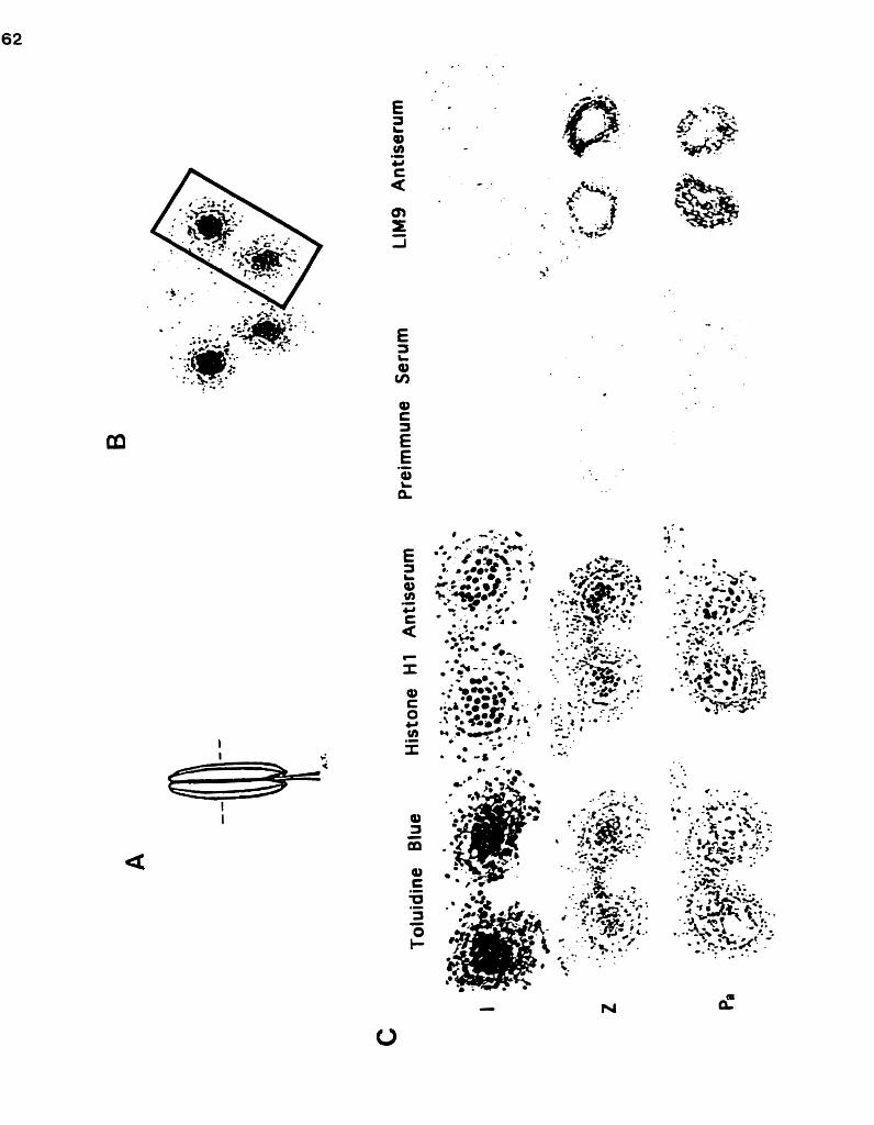
-2 -- - . . 'y.. . : - . .; ;..- - 5 % -- $ ...:;@& - -
.\ ' :-..%&

i f - c,

ii) Zygotene:
The miuosporocytes maintained dose contact with one another as meiosis
commenced. The tapetal cells continued to enlarge and by the zygotene phase of
rnicrosporogenesis had become highly vacuolate. In late zygotene sections
immunostained for the LIM9 protein, the tapetal cytoplasm was dark brown to
bladc while the cytoplasm of the microsporocytes was grey to light brown. Each
miaosporocyte was encirded by a ring of more darkly stained material.
iii) Pachytene:
Intercellular spaces fomed within the mass of microsporocytes as these
cells progressed through pachytene. (The growth and fusion of these spaces
completely isola ted the microsporocytes from one another prior to their firs t
division). The tapetal cells continued to elongate into the locule. A single
vacuole occupied the majority of the volume of each tapetal cell, causing the
cytoplasm to rnigrate towards the innermost (locular) walls.
Within the locule, amorphous dumps of extracellular material were
intensely stained by the LM9 antiserum at the late pachytene stage. Immuno-
staining was also generated within the cytoplasm of the microsporocytes. The
strongest signal, however, was generated within the cytoplasm of the tapetal
cells, adjacent to the locular walls (presumably due to the accumulation of
secre tory vesicles).
iv) Tetrads:
Following prophase 1, the microsporocytes separated completely from one
another and underwent two rounds of cytokinesis to yield tetrads of microspores
encased within callose. The tapetal ceUs attained their maximum volume at this
stage. Continued growth of the tapetal ce11 vacuoles had pressed the entire
cytoplasm of each ce11 tightly against the interior of the locular waUs.

LIM9 immunostaining of sections from tehad stage anthers generated a
strong signal within the cytoplasm of the tapetal cells and revealed a large
quantity of darkly staining extracellular material scattered throughout the locule.
Stained extracellular material also coated the surface of each tetrad. Higher
magrufication (Figure 7) dearly shows that, although the cytoplasm of the
microspores was stained by the LIM9 antisenun, the nudei and the interior of
the cailose walis were not.
V) Free Microspores:
The period of pollen development following tetrad dissolution but prior
to microspore mitosis is termed the free microspore stage. The degeneration of
specific anther wall layers was apparent by this stage. The cells of the inter-
sporangial septum had shrunken significantly as had most of the cells of the
epidermis. In contrast, the endotheaum had developed its characteristic wall
thickenings and had expanded in width to become the prominent layer of the
anther wall. The tapetal cells were highly active and their cytoplasm was darkly
stained by Toluidine Blue. The innermost tangential walls of the tapetal cells
were thin or absent, presumably to facilitate secretion.
Staining of the microspores by the LIM9 antiserum peaked immediately
following their release from the tetrads. The single nucleus of each microspore
appeared as an unstained region (data not shown), suggesting that the LA49
protein within the protoplast was restricted to the cytoplasm. Intensely stained
material occurred abundantly in the locular fluid. The cytoplasm of the tapetal
ceils continued to be stained by the LIM9 antiserum, but this staining was weaker
and more diffuse than it had been during the tetrad stage.
vi) Microspore/Pollen Transition:
Although the walls of the tapetal cells were extremely thin or absent by
this stage, the tapetal layer remained intact. A layer of yellow sporopollenin

Figure 7 Silver Intensified Immunogold labelling of the HM9 protein within the tapehim and locule of a tetrad stage anther section. Immunocyto- chemistry using the LIM9 antiserum stained the cytoplasm of the large vacuolate tapetal cells but failed to stain the surrounding wall layers. Extracellular material was identified within the locule where it formed amorphous deposits and coated each microspore tetrad. Staining of the microspore pro toplas t was res tricted to the cytoplasm: the nuclei and calloçe walls remained unstained. c, callose; E, epidermis; En, endothecium; L, locule; n, microspore nucleus; M, middle wall layers; T, tapetun (Magnified approxima tely 600X).

(rendered green by Toluidine Blue) Lined the locule and formed a nearly
completed sculpted exine on the surface of the microspores. The shrunken
microspores had coilapsed inwardly along a longitudinal groove on their surface
and become U-shaped in cross-section. As the pollen contained very little
cytoplasm, it could not be ascertained whether a mitotic division had occurred.
The small volume of cytoplasm was pressed tightly against the pollen w d and
was darkly stained by the LIM9 antiserum. Surprisingly, the cytoplasm of the
tapetal cells also bound the histone Hl antiserum at this stage. Nonspecific
binding does not appear to have occurred, however, as the cytoplasm was not
immunostained by the L M preimmune serum or secondary antibody. Instead,
histone Hl may have been released into the cytoplasm of the tapetal cells by the
breakdown of their nuclear envelopes. Degeneration of the tapetal ceil nuclei
has been observed during the later stages of microsporogenesis in Liliurn by
several researchers (e.g. Herich and Lux, 1984).
vii) Pollen:
Dehiscent anthers contained large circular pollen grains in which the
vegetative and generative cells could be easily discerned (see Figure 1). The
tapehun had degenerated completely in these anthers, leaving only a thin layer
of cellular debris and sporopollenin-containing orbicules. When sections from
these anthers were subjected to immunocy tochemis try the mature pollen grains
bound an tibodies indiscriminately. Sections s tained with the L M 9 antibody
could not be distinguished from the nega tive control sections for the same
anther (data not shown). Perhaps, among its large store of diverse molecules, the
mature pollen grain contains compounds with a high (nonspeùfic) affinity for
immunoglo bulins.
From the immunocytochemistry experiments it was unclear whether the
LIM9 protein was present within mature pollen grains (and thus might play a

role in pollen germination or pollen tube growth). Total protein exhacts were
therefore prepared from the mature poilen of dehisced anthers and immuno-
blotted using the LIM9 antiserum (Figure 8). Despite being several fold more
sensitive than immunogold localiza tion (Balsamo et al., 19951, immunoblo tting
failed to identify the L M 9 protein in these extracts.
viii) Summ ary:
The L M 9 protein first appears in the anther at the late zygotene stage of
miaosporogenesis and reaches peak levels during tetrad dissolution. The LM9
protein appears to be expressed predominantly by the tapetum and is secreted
into the locule where it coats the callose walls of h e microsporocytes and
microspore tetrads. The protein also o c m within the cytoplasm of the micro-
sporocytes and microspores. Immunoblo tting experiments sugges t tha t the LIM9
protein is not present as an easily extracted component of mature pollen.
II The L M 9 Product is Posttranslationallv Modified: -
A LIM9 is Predicted to Encode a Preproprotein with Glycan Acceptor Sites:
The predicted translation product of the L M 9 cDNA (Figure 9) is
comprised of 787 amino a à d residues and is estimated to have a molecular
weight of 84kDa. The mature amino terminus (as determined by N-terminal
sequencing of the native LIA49 protein) does not immediately follow the putative
initiator methionine, but instead is preceded by 149 amino acid residues. The
first 22 of these residues (approximately 2.3kDa) are predominantly hydrophobic
and likely constitute a signal peptide (preregion) lost during the protein's entry
into the endomembrane system. The following 127 residues are thought to form
a proregion which is cleaved from the mature amino acid terminus within the
endomembrane system. The remaining 638 amino acid residues (approximately

Figure 8 The LIM9 protein is not a readily extracted component of mature pollen. Total protein extracts were prepared from whole tetrad-stage anthers and mature pollen, subjected to SDS-PAGE, and immuno- blotted with LIM9 antiserurn as described in Materials and Methods. Despite a prolonged incubation in the substrate solution and an intense signal in the tetrads lane, signal was not generated within the pollen samples. T, 15pg of tetrad stage anther extract; P i , l5pg of pollen extract; PL 30pg of pollen extract.

e 9 The predicted amino acid sequence of the LIM9 translation product. The LIM9 gene is predicted to encode an 84kDa preproprotein. Following the initiator methionine (outlined at position 1) is a putative 2.3kDa signal peptide of 22 amino acid residues (underlined) and a lOkDa proregion of 93 residues. The mature amino terminus was identified by N-terminal sequencing as the outlined sequence TTHPDYLG. The mature 678 amino aad (72kDa) sequence contains six consensus sites for the addition of glycans (doubly underlined) and four residues which are highly consewed among subtilisin-like serine proteases (bold and underlined).

72kDa of sequence) constitute the mature LM9 protein. The presence of six
consensus sites for the addition of glycans indicate that the mature product of the
LIM9 gene may be a glycoprotein.
B The Maturation of the LIM9 Protein: The Empirical Evidence:
i) The LIM9 Antibody Identifies Three Polypeptides:
Immunoblotting experiments presented a temporal pattern of LIM9
expression similar to that observed within the intact anther by immunocyto-
chemistry. The L M 9 antiserum failed to generate a signal on immunoblots
prepared usùig total protein extracts from the locuies of premeiotic anthers and
anthers at the early zygotene stage of miaosporogenesis. However, when locule
protein extracts from anthers at the tetrad stage of microsporogenesis were
imrnunoblotted, the L M 9 antiserum consistently identified three distinct bands:
a faint 92kDa band; a faint 84kDa band and an intense 82kDa band (Figure 10).
Additional immunoblotting experiments (see below), support the hypothesis
that the 84kDa band contains the initial LIM9 gene product, the 92kDa band
contains a modified transitional polypeptide, while the 82kDa band contains the
mature form of the LIM9 protein.
ii) The LIM9 Protein Acquires High-Mannose Glycans:
The glycosylation of proteins begins within the lumen of the ER with the
acquisition of high-mannose glycans (see Figure 19). The presence of these
glycans on a precursor of the mature L M 9 protein was demonstrated using
Endoglycosidase H (Endo H), an enzyme which specifically deaves the diitobiose
core of high-mannose glycans but is incapable of deaving complex glycans
(Tarentino et RI., 1974). Total protein extracts prepared from the locules of tetrad
stage anthers were incubated with Endo H. On imrnunoblots of these digested

S Z T 2 0 0 -
The LIM9 antisemm recognizes three polypeptides from the locules of tetrad stage anthers. Total protein extracts were prepared from the locule contents of Lilium anthers at the premeiotic S-phase (S), early zygo tene (Z), and tetrad (T) stages of microsporogenesis. 10pg aliquots of each were subjected to SDS-PAGE and immunoblotted with LIM9 antiserum. The antiserum failed to generate signal within the S- phase and zygotene lanes. Within the tetrads lane, however, three immunoreactive bands are visible: a dark 82kDa band, and much fainter 84kDa and 92kDa bands.

samples (Figure Il), the LIM9 antiserum identified the 82kDa and 84kDa bands
and a new 72kDa band but failed to recognize the 92kDa band.
The susceptibility of the 92kDa polypeptide to Endo H digestion suggests
that it may result from the 82kDa proprotein's acquisition of lOkDa of high-
mannose glycans. The loss of the proregion from this polypeptide would result
in an 82kDa glycoprotein, the Endo H digestion of which rnay have yielded the
faint 72kDa immunoreactive band. The 82kDa band, however, remained after
Endo H digestion indicating that it is not comprised solely of a high-mannose
glycoprotein. Evidence is presented below that the majority of the 82kDa
immunoreactive band is comprised of the mature form of the LIM9 protein
which possesses Endo H resistant complex glycans.
iii) The Mature LIM9 Protein Possesses Complex Glycans:
High-mannose glycans can be modified to complex glycans only within the
Golgi apparatus. In plants, this occurs by the addition of xylose and/or other
sugars to the chitobiose core and renders the glycans resistant to Endo H. To
determine if any of the L1M9 polypeptides possessed complex glycans, total
protein extracts from the locules of premeiotic, early zygotene stage, and tehad
stage anthers were immunoblotted with a xylose antiserum (949 antiserum; see
Lauriere et al., 1989). The antiserum failed to identify the 84kDa and 92kDa
polypeptides in each of the samples but did recognize an 82kDa glycoprotein in
the tehad stage sample (Figure 12).
That the 84kDa polypeptide is unaffected by Endo H and is not recognized
by the 949 antiserum suggests that it lacks both high-mannose and complex
glycans. Together with its apparent molecular weight, these observations
strongly implicate it as the initial translation product of the LIM9 gene. The
82kDa polypeptide identified by the xylose antiserum possessed a temporal
pattern of expression similar to that of the L M 9 protein. The 82kDa LIM9

F i w e 11 Endo H treatment reveals the presence of high-mannose glycans on the 92kDa LIM9 protein. A 7.5pg aliquot of a total protein extract from the locules of tetrad stage anthers was incubated with Endoglycosidase H (see Materials and Methods). Additional tetrad stage 7.5pg aliquots were incubated in buffer solution alone (buffer control) or remained untreated (control). Samples were subjected to SDS-Page and immunoblotted with LIM9 antiserum. The antiserum identified an 82kDa band, an 84kDa band and a 92kDa band in both control samples. Following Endo H treatment, the 92kDa band was no longer identified and a new 72kDa immunoreactive band (arrow) was present. C, conhol (untreated); B, buffer control; E, Endo H-treated.

F i a r e 12 A xylose antisenim identifies an 82 kDa glycoprotein from tetrad stage anthers. Total protein extracts were prepared from the Iocule contents of anthers at the premeiotic S-phase (S ) , early zygotene (Z), and tetrad (T) stages of microsporogenesis. 10pg aliquots of each were subjected to SDS-PAGE and immunoblotted with 949 anti-serum. An 82kDa band (arrow) is identified only in the tetrads lane.

protein thus appears to possess Endo H resistant complex glycans. As complex
glycans are generated only within the Golgi apparatus, this 82kDa glycoprotein is
likely the mature modified product of the LIM9 gene. An additional immuno-
blotting experiment (see below and Figure 13) strengthens this conclusion by
indicating that it is the 82kDa protein which is secreted into the anther locule.
iv) The Mature LIM9 Protein Occurs Extracellularly:
Immunocytochemistry experiments provided evidence that the mature
LIM9 protein is seaeted into the anther locule. To further test this finding, the
locule contents of late anthers (zygotene to tetrad stage) were separated into
intracellular and extracellular fractions by centrifugation and subjected to
immunoblotting with the LIM9 antiserum. Although the LIM9 antiserum
identified al1 three protein bands in the intracellular fraction, only the 82kDa
band was identified in the extracellular fraction (Figure 13). Together with the
results of the immunoblotting experiments described above, this observation
provides strong evidence that the 82kDa polypeptide is the mature secreted form
of the LIM9 protein while the 84kDa and 92kDa polypeptides are intracellular
precursors.
6 Summary of LIM9 Protein Maturation:
The LM9 protein is thought to be synthesized in the cytoplasm as an
84kDa precursor (Figure 14). A 2.3kDa signal peptide allows the protein to enter
the endomembrane system. The signal peptide is cleaved off and high-mannose
glycans are added in the endoplasrnic reticulum to yield a 92kDa glycoprotein. A
proregion of approximately lOkDa is then cleaved off and the protein is
transferred to the Golgi apparatus where some or al1 of the high-mannose
glycans are modified to complex glycans. The resulting 82kDa mature
glycoprotein exits the Golgi apparatus and is seaeted into the anther locule.

F i m e - 13 The 82kDa LIM9 protein is an extracellular component of the locule. Locule contents were collected from late anthers (zygotene to tetrad stages). Whole cells were pelleted and homogenized to generate an intracellular total protein extract. An extracellular total protein extract was obtained by precipitating soluble proteins from the locular fluid. 10pg aliquots of each extract were subjected to SDS-PAGE and immunoblotted with LIM9 antiserurn. The antiserum recognized 82kDa, 84kDa, and 92kDa bands within the intracellular lane but identified only the 82kDa band within the extracelldar lane. 1, intracellular extract; E, extracellular extract .

F i m e 14 A putative maturation pathway for the LIM9 protein. The L M 9 protein is translated within the cytosol as an 84 kDa preproprotein (a). Cleavage of the 2.3kDa signal peptide as the protein enters the ER results in an 82kDa polypeptide (b). Within the ER, the proprotein acquires lOkDa of high-mannose glycans (c). The lOkDa proregion is deaved off and the glycoprotein is transferred to the Golgi apparatus where its high mannose glycans are converted to complex glycans (dl. The resulting 82kDa mature glycoprotein is secreted into the anther locule. (a glycan core, mannose sugar residues, ô xylose and other sugar residues).
Translation product
Clleavage of signal peptide
w p l a s m i c Re ticulum
? Acquisition of high- mannose glycans
Ueavage of Proregion ..........................................................................................
Modification of glycans
...- ..
Seaetion of mature protein

III 1s the LM9 Protein a Protease?: -
The predicted sequence of the LM9 protein contains four regions (see
Figure 9 and Figure 17) which are highly conserved among the subtilisin-like
family of serine proteases (see Discussion). To provide empirical evidence that
the L M 9 protein exhibits proteolytic activity, an electrophoretic protease assay
was performed using Lilium anther protein extracts. Proteases, separated by size
through a gelatin-containing polyacrylamide gel, were renatured and activated in
situ. Staining the gel yielded a zymogram on whidi the locations of proteases
were revealed as unstained regions where the gelatin had been degraded.
Zymograms were generated using intracellular and extracellular protein
extracts from Lilium anther locules containing microsporocytes at the
premeiotic, zygotene, and tetrad stages of development (Figure 15 and Figure 16).
Each of the lanes containing intracellular protein preparations contained a single
weak band which migrated with an apparent molecular weight greater than
2OOkDa (Figure 15). As the protease responsible for this activity is not observed
within the extracellular fraction and its expression is stage-independent, it likely
performs a 'housekeeping' role essential to the general metabolism of the
miuosporocytes and microspores. Extensive proteolytic activity occurred only
within the extracellular samples where it peaked in intensity at the tetrads stage.
The majority of this activity occurred within a diffuse band with an apparent
molecular weight of approximately 60kDa. Narrower bands of higher molecular
weight were also present; one of which migrated with an apparent molecular
weight of 82kDa (Figure 16). The occurrence of the 82kDa band only within the
lane containing extracellular proteins of the tetrad stage provides strong evidence
that it was generated by the mature LIM9 protein.

F i m e - 15 Zymogram of intra- and extracellular locule extracts. Iniracellular (i) and extracellular (e) samples were prepared from the locule contents of Lilium anthers at the premeiotic S-phase (S), zygotene (Z), and tetrad (T) stages of microsporogenesis. Electrophoresis was performed with lOpg aliquots of each sample using a 7% polyacrylamide gel containhg 0.10% gelatin. Froteins were renatured in situ as described in Materials and Methods. Proteolytic activity was observed in each of the intracellular extracts at a molecular weight greater than 200kDa (arrow). The extracellular fraction exhibited intense proteolytic activity at the tetrad stage (at approximately 60kDa, bottom of lane).

F i m e 16 Zymogram of extracellular locule extracts. Extracellular samples were prepared from the locule contents of anthers at the premeiotic S- phase (S), zygotene (Z), and tetrad (T) stages of microsporogenesis. Electrophoresis was performed with 10pg aliquots of each sample using a 10% polyacrylamide gel containing 0.15% gelatin. Proteins were renatured in situ as described in Materials and Methods. Low levels of proteolytic activity were observed within the S-phase and zygotene lanes. Intense proteolytic activity was observed within the tetrad lane (the majority of which occurred at approximately 60kDa). A well-defined 82kDa band of protease activity (arrow) was present only within the tetrad lane.

The LIM9 protein might also be responsible for some of the protease
activity observed at 6OkDa. Within the anther loct.de, a portion of the C-
terminus may be cleaved from the putative 82kDa protease by autoproteolysis.
Such a deavage occurs in the closely related serine protease cucumisin, resulting
in a sigmficantly shortened enzyme which retains its proteolytic activity (see
Discussion).

Discussion
1 Similarities Between the LIM9 Protein and Known Serine Proteases: -
The predicted product of the LIM9 gene possesses three amino acid
residues (aspartic acid, histidine, and serine) which are highly conserved among
serine proteases where they form a "catalytic triad" or "charge relay system"
essential for endopeptidase activity (Polgar, 1989; Fersht, 1985). The most
extensively studied of the proteolytic enzymes (Polgar, 1989), serine proteases
take their name from the uniquely reactive serine residue which, acting as an
electron acceptor, participates directly in the hydrolysis of a peptide bond. This
residue also reacts irreversibly with organophosphates such as the inhibitor
diisopropyl fluorophosphate (DFP) (Walsh and Wilcox, 1970; Fersht, 1985). The
reactivity of the serine residue is enhanced by the imidazole ring of the histidine
which is held in position by a hydrogen bond to the aspartic acid residue. The
mature protease is folded such that these three residues are brought together
within a hydrophobic pocket, forming the active site of the enzyme (Fersht, 1985).
A The LIM9 Protein Shares Homology with the Serine Protease Subtilisin: - Although comprised of many distinct families, the (chyrno)trypsin and
subtilisin families are the most thoroughly studied of the serine proteases
(Siezen et al., 1991). The members o f these two families are widespread and serve
diverse functions in both prokaryotes and eukaryotes. Despite operating by an
analogous catalytic mechanism, the (chymo)trypsins and subtilisins lack
sequence homology and even differ in their secondary structure (Siezen et RI.,
1991) and the linear arrangement of their reactive residues (see Figure 17 and

Figure 18). In addition to the catalytic residues common to both families, the
subtilisins also possess a highiy conserved asparagine residue which appears to be
an essential stabilizing component of the substrate binding site (Robertuç et al.,
1972). These striking ciifferences have prompted several researchers to suggest
that the two families arose through convergent evolution (e.g. Julius et al., 1984).
It is to the subtilisins that the L M 9 protein exhibits greatest homology.
Subtilisins are alkaline endoproteases produced by species of the genus Bncillus,
where they are linked with the onset of spordation (Wells et RI., 1983). The
subtilisins have considerable industrial importance (Siezen et RI., 1991) and,
through extensive enzymatic studies and x-ray crystallographic studies, have
become one of the most thoroughly characterized families of bacterial proteases
(Ikemura et al., 1987; Wells et al., 1983). Aii subtilisins appear to be generated as
preproproteins. Subtilisin BPN' from B. nrnyloliquefnciens, for example, is
originally translated as a 377 amino aad polypeptide containing a putative signal
peptide (preregion) of 27 amino aad residues and a proregion of 75 residues
(Wells et al., 1983). The mature 275 amino acid (27.5kDa) protease is a roughly
spherical molecule comprised of a single polypeptide folded into three
structurally distinct parts, with its active site at their conjunction (Wright et al.,
1969). Mature subtilisins are secreted into the extracellular medium where they
likely scavenge extracellular proteins and rnay assist ceil lysis and subsequent
spore release (Wells et al., 1983; Power et RI. , 1986). These proteolytic enzymes
possess a broad substrate-specificity and have been shown to attack the bonds
between a variety of amino acids (Walsh and Wilcox, 1970).

B The LIM9 Protein is One of Several Recently Described Subülisin-Like - Proteins:
Although once thought to be restricted to the prokaryotes, subülisin-like
proteases have recently been identified in a number of eukaryotic organisms,
induding: fungi (e.g. St. Leger et dl., 1992; Joshi, 1995); yeast (e-g. Mizuno et al.,
1988); nematodes (eg. Yang and Kramer, 1994; Thacker et al., 1995); insects (e.g.
Roebroek et al., 1991; Chen and Raikhel, 1996); mammals (e-g. Wise et nl.. 1990);
and higher plants (see below). In total, more than 70 subtilisin-like proteases or
"subfdases" are currently known (Siezen, 1996; Siezen et al., 1991). Eadi of these
share sequence homology to the subtiüsins within the vianity of their conserved
residues (Figure 17). Nearly al1 subtilisin-like proteases are generated as
preproproteins (Siezen et al., 1991) and, although larger than the subtilisins, each
shares a similar arrangement of the four characteristic residues of the subtilisin
active site (Figure 18). An increased linear displacement of the stabilizing
asparagine residue and reactive serine residue, however, appears to be
characteristic of the plant members (Tornero et al., 1996). In addition to the LIM9
protein and its tomato cognate TMP (tomato meiotic proteinase; Riggs and
Horsch, 1995), these plant proteases include the PR-P69 protein from tomato
(Tornero et al., 1996), cucumisin from melon fruits (Yamagata et nl., 1994), and
the products of the ag12 gene in Alnus glutinosn and the ara12 gene in
Arabidopsis (Ribeiro et al., 1995).
Tomato (Lycopersicon esculen tum) PR-P69 is a pa thogenous rela ted (PR)
protein expressed within the leaves and stems of viroid-infected plants (Vera and
Conejero, 1988; Tornero et al., 1996). Immunocytochemistry experiments suggest
that the protease is seaeted into extracellular spaceç and is also sequestered
within vacuoles where it associates with inclusion bodies (Vera et al., 1989). The
745 amino acid product of the PR-P69 gene contains a putative 22 amino acid

1) mf.f9 . . (u9)DGW IGMIDTGI (150) . . . (2mDADG 2) WP . . (14o)AGWIGMIDTGI (lsu . . . (223)DADG 3) PR-P69 . . (i3aKGVI I G V I D T G I (1491 . . . (iss)DDDG 4) n i d s i n . . (132)SNIWGVLDTGI (143) . . . (2oo)DTNG 5) ml2 . . ( I ~ E D V I I G V I D S G V ( ~ ) . . . (212)DTLG 6) Araî2 . . cim SDVVVGVLDTGV (133) . . . (199)DDDG 7) çubtilisin BEN' . . (o24)SWKVAVIDSGI (035). . . (o6o)DNNS 8) . . ( I ~ A G W A A I V D D G L cm) . . . (2os)YDDY 9) Furin/PACE . . (i4s)HGI WS ILDDGI ( i s a . . . (ISIOINDNR 10) W i n (082) FNGNY LDND IML ( 0 9 3 (o42)VSAA
JGSHTASTAAG(23i). . BGSHTASTAAG (237) . . 3GTHTASTAAG (213) . . JGTHTASTAAG (214) . . JGTHTASTAAG (226) . . ZGTHTSSTAAG (213) . . BGTHVAGTVAA (074) . . CIGTRCAGE IAA (223) . . JGTRCAGEVM(204) . . JCYKSRIQVRL ( o s a
F i w e 17 Sequence alignment of the LIM9 protein with other subtilisin-like proteases. Sequences are shown surrounding the conserved aspartic acid (D), histidine (H), asparagine (N), and serine (S) residues of the active site. The LIM9 protein (1) exhibits high sequence homology with the tomato cognate TMP (2) and other plant proteases (3-6). To a lesser extent, homology is exhibited to subtilisin BPN' from Bacillus n~nyloliquefnciens (71, the yeast KEX2 gene product (8), and human furin/PACE (9). Trypsin (101, an unrelated serine protease, shares very little sequence hornology with the LIM9 protein, its catalytic residues are arranged in the order his-asp-serf and it lacks the conserved asparagine residue of the sub tilisins and subtilisin-like proteases. [References: (1): Kobayashi et al, 1994; (2): Riggs and Horsch, 1995; (3): Tornero et ni., 1996; (4): Yamagata et al., 1994; (5,6): Ribeiro et al., 1995; (7): Wells et al., 1983; (8) Mizuno et al., 1988; (9): Wise et al., 1990; (10): Hermodson et al ., 19731.

1) Trypsin
2) Subtillsin BPN' N b OOH
3) Furin/PACE ~y OOH
- Slg. Pro. CP
D ii N 1 1 * + s
*1. 6) UM9 N b 7
* WOOH
19. Pro.
m r e 18 Schematic representations of subtilisin and four subtilisin-like proteases. The positions of the conserved aspartic acid (O), histidine (H), asparagine (N), and senne (S) residues are shown. Signal peptides (Sig.), proregions (Pro.), and potential gljcan acceptor sites (*) are also included. Trypsin (11, differs from the subtilisins in the linear arrangement of its catalytic residues and lacks a conserved asparagine residue. The linear displacement of the serine residue from the asparagine residue is characteristically large in the plant subtilisin-like proteases (3-6). Furin/PACE (3) possesses a transmembrane domain (TMD) which likely anchors it within the Golgi apparatus. Cucumisin (4) possesses a 14kDa carboxyl peptide (CP) which is cleaved from the mature protease following its secretion from the celf. [References: (1 ): Hermodson e t al., 1973; (2): Wells et al., 1 983; (3): Wise e t al., 1990; (4): Yamagata et al., 1994; (5): Tornero e t al., 1996; (6): Kobayashi et al., 19941.

signal peptide and a 92 amino aud proregion (Figure 18). The remaining 631
amino acid sequence of the mature 69kDa proteinase contains seven potential N-
linked glycosylation sites (Tomero et al., 1996). Through in vitro assays, Vera
and Conejero (1988; 1990), demonstrated that the PR4359 protease is capable of
degrading fibrinogen and fluorescein thiocarbanoyl derivative (FI'C)-casein (a t an
optimum pH of 8.5-9.0) and can also cleave the large subunit of ribulose 1,5-
bisphosphate carboxylase/oxygenase (Rubisco) into several smaller polypeptides.
Despite its broad substrate-specificity, Tomero et al. (1996) speculate that PR-P69
does not degrade the proteins of invading pathogens directly, but instead
participates in the pos t-transla tional modification of addi tional pro teins
involved in the defense response (see below).
Cucumisin accumulates within the extracellular juices of melon fruits
(Yamagata ef al., 1989). Originally isolated from the fruit of Cucumis me10 (musk
melon: Kaneda and Tominaga, 1975), similar enzymes have since been
discovered in the fruits of Benincirsn cerifera (white gourd: Kaneda and
Tominaga, 1977) and Trichosnnthes cucumeroides (snake gourd: Kaneda et al.,
1986). Mature cucumisin is a glycoprotein with a carbohydrate content of
appro"mate1y 1.4% (w/w) (Yamagata et al., 1994). The prepro-form of cucumisin
has a molecular weight of nearly 79kDa and is predicted to contain 731 amino
acid residues. The 22 amino aad residues of the putative signal peptide are
followed by a prosequence of 88 residues (see Figure 18). The remaining 621
residues comprise a 67kDa extracellular protein which exhibits pproteolytic
activity in vitro when supplied with a broad range of substrates (Yamagata et al.,
1994). The 67kDa protease is processed further by an autolytic cleavage which
removes 11 6 non-essential arnino acids from the carboxyl terminus, generating a
54kDa form of cucumisin in which proteolytic activity is undiminished
(Yamagata et al., 1989; see below). Cucumisin is exceptionally stable and remains

active over a wide range of pH (4-11) and temperatures. Even after 20 minutes at
a temperature of 60°C and a pH of 11.1, cucumisin retains 80% of its optimum
proteolytic activity (Kaneda et al., 1995).
The ag12 gene was identified in Alnus glutinosn root nodules where it was
shown by RNA blot hybridization and in situ hybridization to be expressed
predominantly within actinorhizal-infected cells during early nodule
development (Ribeiro et n1.,1995). Apart from very low levels of expression
within the shoot tips, the gene does not appear to be expressed within other
organs of A. glutinosn. Using the amino acid sequence of the predicted
translation product of ag12 to perform a database search, Ribeiro et 01. (1995)
identified a highly homologous Arnbidopsis cDNA done which they designated
aralî. Two additional Arnbidopsis sequences with 65% homology to ara12 were
also identified. RNA blot hybridization suggests that the ara12 gene is transcribed
in all organs of Arnbidopsis with the highest levels of transcription occurring
during early silique development (Ribeiro et al., 1995).
Ç Subtilisin-Like Proteases May Function as Protein Convertases:
The KEX2 gene product of the yeast S~ccharomyces cerevisine is perhaps
the best characterized member of the eukaryotic subtilisin-like proteases. Unlike
the plant subtilisin-like pro teases, the 135kDa enzyme is no t secre ted but ins tead
appears to be retained within the Golgi apparatus where it is uivolved in the
posttranscriptional modification of prohormones (Fuller et al., 1989a). The
protease is likely anchored within the membrane by a transmembrane domain
(TMD) with its proteolytic NHrterminal domain extending into the lumen of
the Golgi apparatus and its carboxyl terminus extending into the cytosol (Fuller et
al., 1989a). The Ca2+-dependent (Fuller et al., 1989b) activity of the protease is
responsible for the cleavage of proregions from pro-a-factor and pro-killer toxin

(Julius et al., 1984). The deavages occur following pairs of basic amino acid
residues (most commonly lysine-arginine and arginine-arginine: Wise et al.,
1990). These simple dibasic sites have been conserved during evolution, making
it possible for the KEX2 protease to process mammalian proproteins such as
proalbumin (Bathurst et al., 1987) and proinsulin (Thim et d., 1986).
The proregion is assumed to maintain hormone precursors in an inactive
state during transport; its removal then allows the protein to attain its mature
conformation. The enzymes responsible for the precise cleavage of proregions
from the prohormones of higher animals have long eluded researchers (Barr,
1991). Subtilisin-like proteases, however, have recently been implicated in the
proregion cleavage of several hormone and pheromone precursors in nema-
todes, insects, and mammals (Denault and Leduc, 1996). (This has prompted
their alternate name of sub tilisin-rela ted propro tein convertases or SPCs). Seven
such proteases have been discovered in mammals alone (Denault and Leduc,
1996). For example, the mammalian subtilisin-like proteases furin (or PACE:
p i r e d basic gmino acid deaving enzyme) and PC2/PC3 (protein convertase 2 and
3) have been s h o w to cleave the proregions from pro-von Wilebrand factor
(Wise et al., 1990), and proglucagon (Rouille et al., 1995) respectively. With the
exception of furin/PACE, mammalian subtilisin-like proteases appear to be
secreted (Steiner et al., 1992). Perhaps they function at specific extracellular sites
in a manner similar to that of urokinase-type plasminogen activator (U-PA), a
serine protease which cleaves the proregion from inactive blood plasminogen to
yield active plasmin at wounds and other sites of tissue remodeling (Alberts et
al., 1994). Many of the mammalian subtilisin-like proteases have been
implicated in the processing of Wal glycoproteins, including the human
immunodeficiency virus (HIV-1) envelop glycoprotein gp160 (e.g. Vey, 1995;
Vollenweider et al., 1996; Miranda et al., 1996). As the production of infectious

HIV-1 M o n s is dependent upon the processing of gp160 by these host ce11
proteases, marnmalian subtilisin-like proteases have rapidly become an area of
intense s tudy . Al1 plant subtilisin-like proteases appear to be secreted and, while not
involved in intracellular proprotein processing, may be responsible for the
precise modification of extracellular proteins or ceil-surface proteins. The
expression of ag12 and ara12 peaks during the early stages of root nodule
development and silique development respectively. It therefore seems likely
that these proteases play a role in protein processing rather than protein
degradation (Ribeiro et al., 1995). Tornero et al. (1996) have recently implicated
the tomato PR-P69 protein in the cleavage of an extracellular matrix protein.
These researchers propose that such an interaction may mediate signalling
processes and modulate interactions of the ce11 surface with the extracellular
environment.
II The Maturation of Preproproteins and Protein Secretion: -
In eukaryotic cells, the endoplasmic reticulum, Golgi apparatus, and the
vesicles, vacuoles and other organelles derived from them form a structiirally
and functionally connected system termed the endomembrane system. Within
this system, proteins are posttranslationally modified and sorted to non-
cytoplasmic compartments of the ce11 or the ce11 exterior (for review see Harris,
1986). Proteins enter the endomembrane system through the endoplasmic
reticulum and are shuttled by vesicles to the Golgi apparatus and between its
numerous cis ternae. As pro teins move unidirectionally through the sys tem
many are sequentially modified, each modification catalyzed by an enzyme
localized to a cornpartment or sub-cornpartment. Specific sequences target some

proteins to be retained within a particular cornpartment. Proteins lacking such
sequences flow passively through the system and are ultimately packaged into
vesicles which fuse with the ce11 surface. Seaetion is thus the "bulk flow" or
"default" pathway through the endomembrane system (Jones and Robinson,
1989).
A Entering the ER Lumen: the Role of the Signal Peptide:
The translation of mRNAs encoded by nudear genes occurs within the
cytosol of the eukaryotic cell. Proteins destined for the lumina of eukaryotic
organelles or for secretion must fiist cross the barrier imposed by the ER
membrane. Similarly in prokaryotes, proteins destined for secretion must cross
the ce11 membrane. In a l l ceus, proteins appear to be targeted for translocation
across lipid bilayers by a hydrophobie amino-terminal sequence termed the signal
peptide (Briggs and Gierasch, 1986). A putative signal peptide of 22 amino acids is
thought to mediate the LIM9 protein's entrance into the endomembrane system
and ih subsequent secretion into the anther locule.
The translation of a secretory protein is initiated within the cytosol as its
mRNA is bound by a ribosome. Once the signal peptide protrudes sufficiently
from the ribosome, it is recognized and bound by a ribonucleoprotein termed the
signal recognition partide (SRP). The SRP also binds to the ribosome,
temporarily arresting translation (Rapoport, 1991). The SRP then binds to an
integral ER membrane protein, the SRP recep tor or "docking protein".
Additional proteins within the membrane may anchor the ribosome and form a
hydrophilic protein-conducting channel through the membrane (Simon and
Blobel, 1991).
Translation cornpetence is restored as the binding of a guanosine
triphosphate (GTP) fo the SRP causes it to disassociate from the signal peptide

and ribosome (Rapoport, 1991). Hydrolysis of the GTP actuates the SRP's release
from its receptor. Freed from the SRP, the signal peptide is bound by the iipid
bilayer, perhaps directly (von Heijne, 1986; see below), or through interactions
with integral membrane proteins (Rapoport, 1991). As the translation product
elongates, it protmdes as a loop through the newly-formed membrane pore and
into the lumen of the ER. During or after translation, signal peptidase catalyses
the deavage and digestion of the signal sequence, freeing the amino terminus of
the new protein and making the translocation across the membrane irreversible
(Rapoport, 1991).
With the exception of mitochondrial and chloroplast proteins, signal
sequences range from 13 to 36 amino aud residues in length (von Heijne, 1986).
Despite a la& of sequence homology, signal peptides function properly when
interchanged between different proteins, even when these proteins are from
evolutionarily distant organisms. One of the few characteristic features of signal
peptides is a central region of uncharged hydrophobic residues known as the
hydrophobic core (Walter and Lingappa, 1986). (Of the 22 amino acids of the
LIM9 protein's putative signal peptide, 17 are hydrophobic and likely constitute
this region). The hydrophobic core may take the form of an a-helix or B-strand
capable of partitioning into and spanning the lipid bilayer, thereby andioring the
nascent protein to the membrane (Bedouelle and Hofnug, 1981). The core region
may also interact with a hydrophobic pocket of the signal recognition particle
(Finklestein et al., 1983).
B The Proregion: an Intramolecular Chaperone?:
Proregions appear to be common to the precursors of al1 plant proteases
(Yamagata et R I . , 1994). In mammals, proregions are known to maintain
enzymes in an inactive state untii they are secreted from the expressing cell or

reach their target location. Several serine proteases, for example, are stored
within the mammalian pancreas as inactive proproteins termed zymogens.
These protease precursors are unable to attain a mature conformation until the
proregion is removed (Fersht, 1985). While an inhibitory role also seems likely
for the proregions of the subtilisins and subtilisin-like proteases, these sequences
also appear to participate directly in protein maturation and are a prerequisite for
the generation of active proteases.
i) The proregion plays an essential role in subtilisin maturation:
Several experiments suggest that the proregions of subtilisins act as
"intramolecular chaperones" which guide the proper folding of the proteases
during their maturation (Ohta et RI. , 1991). Ikemura et al. (1987) ligated portions
of the subtilisin E gene into an E. coli expression vector containing the E. coli
OmpA signal peptide and expressed them in B. subtilus. When the entire coding
region for prosubtilisin was expressed, proteolytic subtilisin was detected in the
periplasmic space. However, if the proregion sequence was omitted, the mature
subtilisin which was secreted was incapable of proteolytic activity. Although
both mature polypeptides were shown to possess the same primary structure,
subtilisin expressed without the prosequence probably ladced the secondary
structure required for enzymatic activity. This hypothesis is further supported by
the findings of Ikemura and Inouye (1988). These researchers observed that
prosubtiiisin E expressed in E. coli and denatured with 6M guanidine-HCL was
self-processed into mature active subtilisin when dialysed in a Na-phosphate
renaturing solution. However, when the mature form of subtilisin (i.e. lacking
the proregion) was similarly expressed and denatured, renaturation was
wuccessful. The exogenous addition of a synthetic subtilisin E propeptide in
vitro permitted the successful renaturation of mature subtilisin E and other

mature subtilisins expressed without their proregions (Ohta et al. 1991; Zhu et nL,
1989).
Autoproteolysis appears to effect the final step in the maturation of
subtilisin, the removal of the proregion. Power et al. (1986) found that
preprosubtilisin expressed in oioo did not undergo proregion cleavage if it
possessed a mutation of one i h catalytic residues. The proregion of this inactive
protease was, however, deaved in vitro by the addition of functional subtilisin.
ii) The structure of the subtilisin proregion:
Although sequence homology is not observed among the proregions of
subtilisins and subtilisin-like proteases, the proregions may share a common
overall structure which facilitates their functioning. Using localized polymerase
chah reaction (PCR) random mutagenesis, Lerner et al. (1990) identified several
single residue mutations within the prosequence of subtilisin E which effected
the protease's activity. The majority of these mutations were found to occur
within clusters of hydrophobic residues. (A similar clustering of hydrophobic
residues occurs within the proregion of the LIM9 protein). Kobayashi and Inouye
(1992) hypothesize that the proregion may interact directly with the mature
portion of the protease through these hydrophobic regions. Alternatively, the
folding may be facilitated by electrostatic interactions between the proregion and
the mature polypeptide. The proregion of subtilisin E has an abundance of
charged residues: of the 77 residues comprising the proregion, 23 (30%) are
charged (Ikemura et R I . , 1987). Similarly, nearly 31% (21 out of 77) of the residues
in the putative proregion of the LIM9 protein are charged.
Ç Protein Glycosylation
The majority of secreted eukaryotic enzymes contain asparagine (NI-
linked oligosaccharides or glycans (Jones and Robinson, 1989). N-linked glycans

are grouped into two major categories. High-mannose glycans possess 6-9
mannose (Man) residues and two N-acetylglucosamine (GlcNAc) residues.
Complex or modified glycans are derived from high-mannose glycans with
which they share a common pentasaccharide core structure (Kornfeld and
Kornfeld, 1985):
Man N GlcNAc-GlcNAc-Man
\ Man
In addition to this core structure, complex glycans contain one or more other
sugar residues (fucose, xylose and arabinose in plants).
i) The assembly of plant glycoproteins:
The assembly of high-mannose glycans and their modification to complex
glycans is achieved through a cascade of enzyme-catalyzed events (Figure 19).
Immunocytochemical techniques have allowed most of the enzymes responsible
for glycosylation to be localized to compartments or even sub-compartments of
the endomembrane system (for review see Jones and Robinson, 1989). The
glycosylation process begins in the ER where the glycan precursor Glc3Man9
(GlcNAc)* is assembled by the sequential addition of N-acetylglucosamine,
mannose and glucose (Glc) to the integral ER membrane protein dolichol-
phosphate (Elbein, 1988). Within the ER lumen, oligosaccharyl-transferase
catalyses the cotranslational transfer of this precursor to an asparagine residue
within an acceptor site on the nascent protein (Kornfeld and Kornfeld, 1985).
Acceptor sites consist of the amino acid sequence Asparagine-X-
Serine/Threonine, where X is any amino acid except proline or aspartic acid
(Marshall, 1972). The rate at which the nascent protein assumes it proper folding
and the acceptor sites become inaccessible to oligosaccharyltransferase may
determine how many of these sites are actually glycosylated (Kornfeld and

The Processing pathway of plant complex glycans. The transfer of ( G l ~ ) ~ M a n ~ ( G l c N A c ) ~ from dolichyi-phosphate (Dol-P-P) to the asparagine residue (N) of a nascent polypeptide is catalyzed by oligosaccharyl-transferase (a). Glucose residues @) are removed by a- glucosidases 1 and II (b, c). The glycoprotein is hansported to the Golgi apparatus where three mannose residues (n) are cleaved from the high-mannose glycan by a-mannosidase 1 (d). A GicNAc residue (O) is added by GlcNAc-transferase (e) and two additional mannose residues are cleaved by a-mannosidase II (f). A fucose residue (F) is added by fucosyl-transferase (g) and an additional GlcNAc is added by GlcNAc-transferase (h). Xylose (X) is added by xylosyl-transferase (i) and the two GlcNAc residues added in the Golgi apparatus are removed by N-acetylglucosaminidase (j) to yield a mature complex glycan [Xyl(Man)3Fuc(GlcNAc)2] (modified from Chrispeels and Tague, 1991; Komfeld and Kornfeld, 1985).

-
a) oligosaccharyl-transferase
a-glucosidase I
a-glucosidase II
a-mannosidase I
GlcNAc-transferase I
a-mannosidase II
fucosyl-transferase
GlcNAc-transferase II
xylosyl-transferase

Kornfeld, 1985). Immediately following the transfer of the oligosaccharide
precursor to the nascent polypeptide, glucosidases 1 and II deave the three
glucose residues, generating a typical high-mannose glycan [Mang(Gl~NAc)~].
Glycoproteins not targeted to remain within the ER are transported within
vesicles to the cis face of the Golgi apparatus. wthin the as- and mediai-
cistemae, mannose residues are trimmed from the glycans by a-mannosidases 1
and II, leaving only the pentasaccharide core [M~II~(G~CNAC)~]. In plants,
transferases within the medial- and trans-cisternae then add one or more of the
following sugar residues: N-acetylglucosamine, fucose, galactose, arabinose, and
xylose (Xyl) (Chrispeels and Tague, 1991; Gleeson and Clarke, 1980). These
additions typicdy yield plant complex glycans with the formda Xyl(Man)3
FUC(G~CNAC)~ (Driouich et d., 1989; Lauriere et RI., 1989). [The majority of
antibodies raised to these glycans (e.g. the 949 antiserum) recognize the xylose
residues; as this sugar does not occur in mammals it is probably highly
immunogenic (Driouich et al., 1989)]. Glycoproteins targeted for seaetion are
then packaged into vesicles which fuse with the plasma membrane and empty
their contents through exocytosis.
High-mannose glycans can pass through the Golgi apparatus without
being modified to complex glycans. Vitale et al. (1984), for example, identified
glycans of both types on mature phytohemagglutinin from Phnçeolus oulgnris.
The complex glycans of this plant glycoprotein were also found to Vary in
composition, with some modified by the addition of a single fucose residue and
others by the addition of a single xylose residue. As in this study of the LM9
protein, the nature of the glycans on phytohemagglutinin was revealed by their
susceptibility to the enzyme endoglycosidase H (Vitale et R I . , 1984). As previously
desaibed, Endo H cleaves high-mannose glycans between the GlcNAc residues of

the core structure (Tarentino et RI., 1974). In complex glycans, the binding of
additional sugar residues to the core likely prevents the binding of Endo H.
ii) The role of glycosylation:
Glycans do not appear to play a specXc role in protein sorting or targeting
for seaetion as was once thought (Driouich et nl., 1989); many secreted proteins
occur which are not glycosylated (e.g. barley aleurone a-amylase: Jones et al.,
1987). GlycosyIation, however, may affect the efficiency of a protein's transport
within the endomembrane system. For example, the glycosylation inhibitor
tunacamyan greatly diminishes the transport of proconcanavalin A (pro-Con A)
to protein bodies (Faye and Chrispeels, 1987). Perhaps glycoproteins alter the
physiochemical properties of the protein, thereby making it more soluble or
allowing it to fold correctly (Gibson et ni., 1981). Incorrectly folded proteins
appear to be inefficiently transported to the Golgi apparatus and may be
selectively retained within the endoplasmic reticulum (Lodish, 1988).
Altematively, glycosylation may make proteins more hydrophilic and prevent
interactions with hydrophobie domains of the ER which would retard their
transport (Jones and Robinson, 1989). The glycans of glycoproteins may also
provide protection against proteolytic degradation (Driouich et al., 1989). Finally,
glycans may play a role in post-translational proteolytic processing (Faye and
Chrispeels, 1987) or the biological activity of the mature protein (Tsaftari et al.,
1980).
Extraceilular Protein Modification: The Autolysis of Cucumisin:
As the fruit of Cucumis me10 matures, the concentration of 67kDa
cucumisin decreases with a concurrent increase in the concentration of the
mature 54kDa form of cucumisin. Similarly, the concentration of 67kDa
cucumisin diminishes in vitro with a parallel inaease in the concentrations of

54kDa cucumisin and a 14kDa polypeptide (Yamagata et al, 1989). If, however,
serine protease inhibitors are included in the incubations, this transition does
not occur. It therefore seems likely that the 54kDa and l4kDa proteins &se from
the limited autolysis of 67kDa cucumisin. The conversion of cucumisin from its
67kDa form to its 54kDa form in vitro or in vivo is not accompanied by a
decrease in proteolytic activity. The 54kDa cucumisin, however, is less stable
than 67kDa cucumisin at aadic pH and high temperatures (Yamagata et al., 1989).
The LM9 protein is dosely related to cucumisin and thus may be modified
in a similar fashion. When zymograms were prepared using samples containing
the LIM9 protein, a major band of proteolytic activity was observed at approxi-
mately 6OkDa. Perhaps a 6OkDa protease is generated by an extracellular autolytic
cleavage of the LIM9 protein in a manner similar to that observed for cucumisin.
III Possible Functions of the LIM9 Protein: -
The LIM9 protein occurs within the tapetum, locular fluid and, to a lesser
extent, within the microsporocytes and microspores. Immunoblotting
experiments indicate that the 82kDa LIM9 protein and its 84kDa and 92kDa
precursors occur within the microsporocytes (i.e. the intracellular fraction of the
locule) after their cytoplasmic connections to the tapetal cells have been severed
(see Figure 13). This strongly suggests that, rather than being transferred
symplastically or apoplas tically from the tape tum to the microspores, the LIM9
protein is simultaneously expressed in both ce11 types. If verified by in sih4
hybridization experiments, LIM9 would becorne one of very few cloned anther-
specific genes known to be expressed by both the sporophyte and the gametophyte
(Roberts et al., 1993).

Miaosporogenesis and the onset of dehiscence appear to require the
transcriptional activation of numerous tissue-specific gene sets that are inactive
during anther his togenesis (Goldberg et dl., 1993). Although the L M 9 protein
was recognized by an antiserum to proteins which bound DNA cellulose (Riggs
and Horsch, 19951, it certainïy does not appear to be among the transcription
factors involved in this transformation. (More likely, it merely shares epitopes
with one or more DNA-binding pro teins). Immunolocalization s tudies failed to
identify any association between the LIM9 protein and the nudei of either the
tapetal cells or the Ncrosporocytes and microspores. Instead, the protein is
modified within the endomembrane system of the cytoplasm and secreted
the anther locule. A strong sequence homology to subtilisin-like
the prelirninary results of SDS-PAGE functional assays implicate
protein as an extracellular serine protease.
enzymes
the LM9
Serine proteases serve several functions in biological systems, from
precise cleavage and modification of protein precursors to the complete
in to
and
the
proteolytic degradation of proteins into their constituent amino acids. While in
vivo studies have indicated that some subtilisin-like serine proteases act as
pro tein conver tases, in oitro s tudies sugges t tha t O thers (e. g. cucumisin and PR-
P69) are capable of extensive non-specific proteolysis. At present, neither activity
can be ruled out For the LIM9 protein. Microsporogenesis and dehiscence are
cornplex processes requiring the coordinated turnover of a large population of
enzymes, metabolites and structural proteins. As the LIM9 protein is present
within the locule for an extensive period of rnicrosporogenesis and pollen
development, it could be responsible for the modification or degradation of any
of a large nurnber oi proteins. Perhaps the timing of the LIM9 protein's activity
is more precisely conshained by the pH of the locule (as has been obsenred for

callases; Izhar and Frankel, 1971) or by Caf2 concentration (as has been observed
for other plant subtilisin-iike proteases; e-g. Vera and Conejero, 1988).
The apparent restriction of the LIM9 protein to the tapetum and locule
suggests that it plays a role in pollen development and not dehiscence. Extensive
enzymatic activity occurs within the cytoplasm of the microsporocytes and
microspores. During meiosis, numerous enzyme-catalyzed events coordinate
chromosome segregation and ce11 division, while proteases and RNAses likely
purge the microspore of the sporophytic transcription and translation machinery.
Additional enzymes must catalyze two major episodes of amlyogenesis and
amlyolysis and the recycling of the microspore cytoplasm (Pacini, 1990). It seems
unlikely, however, that the LJM9 protein plays a direct role in these cytoplasmic
events. The presence of a putative signal peptide and the results of immuno-
blotting and immunocytochernis try experiments indicate tha t the mature LIM9
protein is secreted; zymogram experiments suggest that it remains inactive
within the cell. The LIM9 protein could, however, coordinate events within the
microspore indirectly through the degradation or modification of receptors on its
surface, thereby providing a means of communication between sporophyte and
gametophyte. This role, however, is also unlikely due to the apparent
impermeability of the callose wall to polypeptides (Heslop-Harrison, 1966a).
It has been suggested that the callose wall of the developing microspores
and the sporopollenin lining of the tapetum serve to prevent the transfer of
pathogens from the sporophyte to the gametophyte. Perhaps the accumulation
of the LIM9 protein in the locule plays a similar role. The highly homologous
plant subtilisin-like protease PR-P69 plays a role in the pathogen defense
response of tomato (Tornero et al., 1996). Likewise the Ag12 gene product of A.
glutinosn likely moderates the activities of infectious actinormycetes (Ribeiro e t
al., 1995). By activating PR proteins, or by degrading foreign proteins directly, the

L M 9 protein might constitute an early defence against the establishment of
pathogens within the locule.
The locular fluid in which the microsporocytes are suspended is a complex
solution of metabolites, enzymes and precursors of the poilen wall and pollen
coat Perhaps the LIM9 protein maintains the composition of the locular fluid by
acting as an extracellular scavenger in a manner similar to that suggested for the
bacterial subtilisin proteases. The putative protease may facilitate the turnover of
enzymes or degrade other complex molecules within the locule, making the
simple subunits (metabolites) available to the developing microspores.
Alternatively, the LIM9 protein may play a role in the breakdown of the tapetum
and the subsequent release of pollenkitt and tryphine. The temporal patterns of
the pro tein's expression and the spatial pa ttems of i ts accumulation, however,
suggest that the protein is most likely involved in the processing of enzymes and
precursor molecules required for the assembly of the pollen wall.
The poilen wall is unique to the male gametophyte and its construction is
likely catalyzed by several enzymes that are encoded by microspore- and tapetum-
speufic genes. several anther-specific genes possessing a temporal pattern of
expression similar to that of LIM9 have been implicated in the construction of
the pollen wall. Only the products of the satap35 and satap44 genes from
mustard, however, have been shown by immunocytochemistry to înteract
directly with pollen wall components (Staiger and Apel, 1993; Staiger et al., 1994).
The expression of these genes appears to occur exclusively within the tapetum,
beginning during prophase 1 of microsporocyte meiosis. The proteins are
secreted into the locule where they reach peak levels as the microspores are
released from the tetrads. The proteins are initially associated with globules
presumed to contain sporopollenin and may act as carriers, directing precursor
molecules from the tapetal cells to the pollen wall. The satap proteins appear to

be ultimately incorporated into the exine of the developing pollen grains,
although it is uncertain whether the satap proteins form a structural component
of the pollen wall or are involved in the in situ polymerization of exine
precursors (S taiger et al., l994).
Immunocytochemisty experiments revealed that the LIM9 protein
accumulates around the microspores while they are encased within callose. In
this location, the LIM9 protein might modify wall precursor molecules or cleave
them from the carrier molecules that directed them to the microspore. As a
protein convertase, the LIM9 protein might activate callase, or enzymes involved
in the polymerization of wall precursors. As the callose wall appears to act as a
barrier to pro teins, the LIM9 protein coa ting its outer surface likely originates
solely within the tapetum. This protein could assist in the elaboration of the
exine only after the dissolution of the callose wall. The primexine would thus be
constructed with the assistance of L M 9 protein expressed within the miaospore.
The distribution of this protein on the surface of the microspore plasma
membrane migh t ultimately effect the species-specific pa tterning of the pollen
wall.
hmunoblotting experiments failed to identify the LIM9 protein in pollen
extracts. It is therefore unlikely that this protein is a component of the protoplast
or an easily extracted component of the germinal apertures or sexine cavities
which could be solubilized upon germination. Thus, although the L M 9 protein
may be present within mature pollen as an inextractable component of the wall
mahix, it does not appear to play a role in incompatibility responses, pollen
germination, or the conductance of the pollen b b e through the tissues of the
style.

IV Future Goals and Prospects of LIM9/TMP Research: -
Definitive proof that the LIM9 protein is capable of proteolysis requires
that functional assays be performed using an extract free of contaminating
proteins. Attempts to purify the L M 9 protein from anther extracts by immuno-
preapitation have proved unsuccessful. (As the LIM9 antiserum was raised to a
denatured polypeptide, it may not recognize the L M 9 protein in its native state).
Work is currently undenvay to express the entire LIM9 coding region in E. coli.
This strategy should provide a functional protease, providing that glycosylation
is not a prerequisite for the protein's biological activity. If an active protease is
obtained, its subs h a te specifiaty, optimal pH range, suscep tibility to class-specific
protease inhibitors, and capacity for self-processing could be assessed. Functional
assays could also be designed to determine if the protease is Ca2+-dependent,
whiie cross-linking assays might also be performed to detemine if the protease
interacts with (and modifies) other anther proteins.
Zymogram experiments with the LIM9 protein suggest that it may incur
an additional cleavage to generate a 6OkDa polypeptide. Such a protein has not
been identified by immunoblotting experiments. The LIM9 antiserum used in
this study was generated toward the carboxyl terminus of the recombinant
protein. The deavage (and degradation) of the C-terminus from the LIM9
protein (as occurs in cucumisin) would thus remove most or al1 of the antigenic
determinants (epitopes) recognized by the L M 9 antibody. Work is currently
underway to generate an antiserum to the N+terminus of the protein. If this
new antiserum identifies an additional LIM9 product, immunocytochemistry
experiments should be repeated to determine if this protein is incorporated into
the mature pollen or is transported outside of the locule.

Mutagenesis and hansgenic technologies offer a means by which the
function of genes can be assessed in vivo. Unfortunately, due to its large genome
and the la& of an appropriate transformation/regeneration çystem, Lilium is not
amenable to this sort of study. A tomato cognate to the L M 9 gene (TMP) has,
however, been isolated and shown by DNA gel blot hybridization to exist at a
single locus within the tomato genome (Riggs and Horsch, 1995). Tomato is
amenable to transgenic studies and work is currently underway to generate
transgenic tomato plants expressing antisense TMP. The function of the TMP
protein could then be inferred cytologically from developmental abnormalties
observed within the microspores and anther tissues. (For example, the absence
of the TMP protein might result in a thin or malformed pollen exine layer). The
evolutionary conservation of the L M 9 and TMP proteins in two distantly related
plants (a monocotyledon and a dicotyledon respectively) suggests that these
proteins perform a role which is essential to the process of rnicrosporogenesis.

Ref erences:
Alberts, B., Bray, D., Lewis, J., Raff, M., Roberts, K., and Watson, J. D. 1994. Molecular biology of the ceII, hird edition. Garland Publishing, Inc., New York.
Balsamo, R. A., Wang, J. -L., Ekard, K. J., Wang, C. -S., and Lord, E. M. 1995. Immunogold locaiization of a developmentally regulated, tapetal specific, 15kDa lily anther protein. Protoplasma, 189: 17-25.
Barr, P. J. 1991. Mammalian subtilisins: the long-sought dibasic processing endoproteases. Ceil, 66: 1-3.
Bathurst, 1. C., Brennan, O. S., Carrell, R. W., Cousens, L. S., Brake, A. J., and Barr, P. J. 1987. Yeast KEX2 protease has the properties of a human proalbumin converhg enzyme. Science, 235: 348-350.
Bedinger, P. 1992. The remarkable biology of pollen. Plant Cell, 4: 879-887.
Bedouelle, H. and Hofnug, M. 1981. In Membrane transport and neuroreceptors. Edited by D. Oxender. Man R. Liss Incorporated, New York, pp. 399-403.
Bennett, M. D., Rao, M. K., Smith, J. B., and Bayliss, M. W. 1973. Ce11 development in the anther, the ovule, and the young seed of Triticum aestiaum L. var. Chinese Spring. Phil. Trans. Roy. Soc. London Ser. B, 226: 39-81.
Bhandari, N. N. 1984. The microsporangium. In Embryology of angiosperms. Edited by B. M. Johri. Springer-Verlag, London, pp. 53-121.
Blackmore, S. and Barnes, S. H. 1990. Pollen wall development in angiosperms. In Microspores: evolution and ontogeny. Edited by S. Bladcmore and R. B. Knox. Academic Press, London, pp. 173-192.
Blackmore, S. and Knox, R. B. 1990. Microsporogenesis: the male programme of development. In Microspores: development and ontogeny. Edited by S. Bladunore and R. B. Knox. Academic Press, New York, pp. 1-10.
Blomstedt, C. K., Knox, R. B., and Singh, M. B. 1996. Generative cells of Lilium longiflorum possess translatable mRNA and functional protein synthesis machinery. Plant Mol. Biol., 31: 1083-1086.

Bold, H. C., Alexopoulos, C. J., and Delevoryas, T. 1987. Morphology of plants and fungi: fifth edition. Harper and Row, Publishers, New York.
Bonner, L. J. and Dickinson, H. G. 1989. Anther dehiscence in Lycopersicon esculenturn Mill. 1. structural aspects. New Phytol., 113: 97-115.
Brewbaker, J. L. 1967. The distribution and phjjlogenetic sigdcance of binucleate and trinucleate pollen grains in the angiosperms. Am. J. Bot., 54: 1069-1083.
Briggs, M. S. and Gierasch, L. M. 1986. Molecular mechanisms of protein secretion: the role of the signal sequence. Advan. Protein Chem., 38: 109-179.
Chapman, G. P. 1987. The tapetum. Internat. Rev. Cytol., 107: 111-125.
Chen, J. -S. and Raikhel, A. S. 1996. Subunit cleavage of mosquito pro- vitellogenin by a subtilisin-like convertase. Proc. Nat. Acad. Sci. USA, 93(12): 6186-6190.
Chrispeels, M. J. and Tague, B. W. 1991. Protein sorting in the secretory system of plant cells. Internat Rev. Cytol., 125: 1-45.
Clement, C . and Audran, J. C. 1995. Anther wall layers control sugar nutrition in Lilium. Protoplasma, 187: 172-181.
Clement, C., Burrus, M., and Audran, J. C. 1996. Floral organ growth and carbohydrate content dunng pollen development in Lilium. Am. J. Bot., 83(4): 459-469.
Clement, C., Chavant, L., Burrus, M., and Audran, J. C. 1994. Anther starch variations in Li1 ium during pollen developmen t. Sex. Plant Reprod., 7: 347-356.
Darwin, C. 1877. The different forms of flowers on plants of the same species. John Murray, London.
Davis, G. L. 1966. Systematic embryology of the angiosperms. John Wiley & Sons, New York.
Denault, J. B. and Leduc, R. 1996. Furin/PACE/SPCl: A convertase involved in exocytic and endocytic processing of precursor proteins. FEBS Letters, 379(2): 113-116.
Dickinson, H. G. 1987. The physiology and biochemistry of meiosis in the anther. Int. Rev. Cytol., 107: 79-109.
Driouich, A., Gonnet, P., Makkie, M., Laine, A., and Faye, L. 1989. The role of high-mannose and complex asparagine-linked glycans in the secretion and stability of glycopro teins. Plan ta, 180: 96-104.
Elbein, A. D. 1988. Glycoprotein processing and glycoprotein processing inhibitors. Plant Physiol., 87: 291-295.
Esau, K. 1977. Anatomy of seed plants. John Wiley & Sons, New York.
Faye, L. and Chrispeels, M. J. 1987. Transport and processing of the glycosylated precursor of concanavalin A in jack-bean. Planta, 170: 217-224.
Fersht, A. 1985. Enzyme structure and mechanism: second edition. W. H. Freeman and Company, New York.
Finkelstein, A. V., Bendzko, P., and Rapoport, T. A. 1983. Recognition of signal sequences. FEBS Le tt., 161: 176-179.
Frankis, R. C. and Mascarenhas, J. P. 1980. Messenger RNA in the ungerminated pollen grain: a direct demonstration of its presence. Ann. Bot., 45: 595-599.
Fuller, R. S., Brake, A., and Thorner, J. 1989a. Intracellular targeting and structural conservation of a prohormone-processing endopeptide. Science, 246: 482-486.
Fuller, R. S., Brake, A., and Thorner, J. 198913. Yeast prohormone processing enzyme (KEXZ gene product) is a Ca2+-dependent serine protease. Proc. Nat. Acad. Sci. USA, 86: 14341438.
Gibson, R., Kornfeld, S., and Schlesinger, S. 1981. The effect of oligosaccharide chains of different sizes on the maturation and physical properties of vesicular stomatitis virus. J. Biol. Chem., 256: 456-462.
Gleeson, P. A. and Clarke, A. E. 1980. Antigenic determinants of a plant proteoglycan, the Gladiolus style arabinogalactan-pro tein. Biochem. J., 191: 437-447.
Goldberg, R. B. 1988. Plants: novel developmental processes. Saence, 240: 1460- 1467.

Goldberg, R B., Beals, T. P., and Sanders, P. M. 1993. Anther development basic principles and practical applications. Plant Cell, 5: 1217-1229.
Graybosch R. A. and Palmer, R. G. 1988. Male sterility in soybean: an overview. Am. J. Bot., 75: 144-156.
Greyson, R 1. 1994. The development of flowers. Oxford University Press. Oxford.
Hanson, D. D., Hamilton, D. A., Travis, J. L., Bashe, D. M., and Mascarenhas, J. P. 1989. Characterization of a pollen-specific cDNA from Zem mnys and its expression. Plant Cell, 1: 173-179.
Harris, N. 1986. Organization of the endomembrane system. Ann. Rev. Plant Physiol., 37: 73-92.
Hasenkampf, C., Qureshi, M., Horsch, A., and Riggs, C. D. 1992. Temporal and spatial dis tribution of meio th-1 in anthers of Lilium longi'orum. Dev. Gen., 13: 425-434.
Herich, R. and Lux, A. 1984. Electron miaoscope study of tapetal ce11 nudei degradation of Lilium henryi. Physiol. Plant, 20: 1-5.
Hermodson, M. A., Ericsson, L. H., Neurath, H., and Walsh, K. A. 1973. Determination of the amino acid sequence of porcine trypsin by sequenator analysis. Biochem., 12(17): 3146.3153.
Heslop-Harrison, J. 1966a. Cytoplasmic continuities during spore formation in flowering plants. Endeavor, 25: 65-72.
Heslop-Harrison, J. 1966b. Cytoplasmic connections between angiosperrn meiocytes. Am. Bot., 30: 221-230.
Heslop-Harrison, J. 1968. Tapetal origin of pollen-coat substances in Lilium. New Phytol., 67: 7ï9-786.
Heslop-Harrison, J. 1971. The pollen wall: structure and development. In Pollen: development and physiology. Edited by J. Heslop-Harrison. Butterworth, London, pp. 75-98.
Heslop-Harrison, J. 1980. Compartmentation in anther developmrnt and pollen wall morphogenesis. In Ce11 compartmentation and metabolic channelling.

Edited by L. Nover, F. Lynen, and K. Mothes. Elsevier/North/Holland Biomedical Press., New York, pp 471-493.
Heslop-Harrison, J. 1987. Pollen germination and pollen-tube growth. In Pollen: cytology and development. Edited by K. L. Giles and J. Prakash. Int. Rev. Cytol., 107: 'i-78.
Heslop-Harrison, J., Knox, R. B., and Heslop-Harrison, Y. 1974. Pollen wall proteins: exine-held fraction assoaa ted with the incompa tibility response in Cruciferae. Theoret. Appl. Genet., 44: 133-137.
Hird, D. L., Worrall, D., Hodge, R., Smartt, S., Paul, W., and Scott, R. 1993. The anther-specific pro tein encoded by the Brassica napus and Arnbidopsis thnl in nn A6 gene displays similarity to P-1,3-glucanases. Plant J., 4(6): 1023-1033.
Horvitz, H. R. and Herskowitz, 1. 1992. Mechanisms of asymmetric ce11 division: two Bs or not two Bs, that is the question. Cell, 68: 237-255.
Ikemura, H. and Inouye, M. 1988. In vitro processing of pro-subtilisin produced in Escherichin coli. J. Biol. Chem., 263(26): 12959-12963.
Ikemura, H., Takagi, H., and Inouye, M. 1987. Requirement of pro-sequence for the production of active subtilisin E in Escherichin coli. J . Biol. Chem., 262(16): 7859-7864.
Ito, M. and Takegami, M. H. 1982. Cornmitment of mitotic cells to meiosis during the G2 phase of premeiosis. Plant & Ce11 Physiol., 23(6): 943-952.
Izhar, S. and Frankel, R. 1971. Mechanism of male sterility in Petunin: the relationship between pH, callase activity in the anthers, and the breakdown of microsporogenesis. Theor. App. Genet., 41: 104-108.
Jensen, W. A. 1973. Fertilization in flowering plants. BioScience, 23(1): 21-27.
Jones, R. L., Bush, D. S., Sticher, L., Simon, P., and Jacobsen, J. V. 1987. Intracellular transport and secretion of barley aleurone a-amylase. In Plant membranes: structure, function, biogenesis. Edited by Leaver, C. and Sze, H. Liss, New York, pp. 325-340.
Jones, R. L. and Robinson, D. G. 1989. Transley review no. 17 protein secretion in plants. New Phytol., 111: 567-597.

Joshi, L. 1995. Cloning of a cutidedegrading protease from the entomopathogenic fungus, Beauverin bassinna. FEMS Miaobiol. Lett., 125: 211-218.
Julius, D., Brake, A., Blair, L., Kunisawa, R., and Thorner, J. 1984. Isolation of the putative structural gene for the lysine-arginine-deaving endopep tidase required for processing of yeast prepro-a-factor. Cell, 37: 1075-1089.
Kamalay, J. C. and Goldberg, R. B. 1980. Regulation of structural gene expression in tobacco. Cell, 19: 935-946.
Kamalay, J. C. and Goldberg, R. B. 1984.Organ-specific nudear RNAs in tobacco. Proc. Nat. Acad. Sci. USA, 81: 2801-2805.
Kaneda, M., Sobue, A., and Tominaga, N. 1986. Isolation and characterization of proteinases from the sarcocarp of snake gourd (Trichosanthes cucumeroides) fruit. J. Biochem., 99(2): 569-578.
Kaneda, M. and Tominaga, N. 1975. Isolation and characterization of a protease from the sarcocarp of melon fruit. J. Biochem., 78: 1287-1296.
Kaneda, M. and Tominaga, N. 1977. Isolation and characterization of a proteinase from white gourd. Phytochem., 16: 345-346.
Kaneda, M., Yonezawa, H., and Uchikoba, T. 1995. Improved isolation, stability and substrate specificity of cucumisin, a plant serine protease. Biotechnol. Appl. Biochem., 22: 215-222.
Kaul, M. L. K. 1988. Male sterility in higher plants. In Monographs on theoretical and applied genetics, Vol. 10. Edited by R. Frankel, M. Grossman, H. Linskens, P. Maliga, and R. Ripley. Springer Verlag, Berlin. pp. 15-95.
Keijzer, C. J. 1987a. The process of anther dehiscence and pollen dispersal. 1. the opening mechanism of longitudinally dehiscing anthers. New Phytol., 105: 487-498.
Keijzer, C. J. 1987%. The process of anther dehiscence and pollen dispersal. II. the formation and the transfer mechanism of pollenkitt, cell-wall development of the loculus tissues and a function of orbicules in pollen dispersal. New Phytol., 105: 499-507.
Knox, R. B. 1984. The pollen grain. In Embryology of angiosperms. Edited by B. M. Johri. Springer-Verlag, New York, pp. 197-271.

Knox, R. B. and Heslop-Harrison, J. 1970. Pollen wall proteins: localization and enzymatic activity. J. Ce11 Sci., 6: 1-27.
Kobayashi, T. and Inouye, M. 1992. Functional analysis of the intramolecular chaperone: mutational hot spots in the subtilisin pro-peptide and a second-site suppressor mutation within the subtilisin molecule. J. Mol. Biol., 226: 931-933.
Kobayashi, T., Kobayashi, E., Sato, S., Hotta, Y., Miyajima, N., Tanaka, A., and Tabata, S. 1994. Characterization of cDNAs induced in meiotic prophase in lily microsporocytes. DNA Res., 1: 15-26.
Koltunow, A. M., Truettner, J., Cox, K. H., Wallroth, M., and Goldberg, R. B. 1990. Different temporal and spatial gene expression patterns occur during anther development. Plant Ceil, 2: 1201-1224.
Komfeld, R. and Kornfeld, S. 1985. Assembly of asparagine-linked oligosac- charides. Ann. Rev. Biochem., 54: 631-664.
Laemmli, U. K. 1970. Cleavage of structural proteins during the assembly of the head of bacteriophage T4. Nature, 227: 680-685.
Larson, A., Lewis, C. W. 1962. Pollen wall development in Parkinsonin nculentn. Grana Palynol., 3: 21-27.
Lauriere, M., Lauriere, C., Chrispeels, M. J., Johnson, K. D., and Sturm, A. 1989. Characterization of a xylose-specific antiserum that reacts with the complex asparagine-linked glycans of extracellular and vacuolar glycoproteins. Plant Physiol., 90: 1182-1188.
Lerner, C. G., Kobayashi, T., and Inouye, M. 1990. Isolation of subtilisin pro-sequence mutations that affect formation of active protease by localized random polymerase chain reaction mutagenesis. J. Biol. Chem., 265(33): 20085- 20086.
Lodish, H. F. 1988. Transport of secretory and membrane glycoproteins from the rough endoplasmic reticulum to the Golgi. J. Biol. Chem., 263(5): 2107-21 10.
Lowry, O. H., Rosebrough, N. J., Farr, A. L., and Randall, R. J. 1951. Protein measurement with the folin phenol reagent. J. Biol. Chem., 193: 265-275.
Mackenzie, A., Heslop-Harrison, J., and Dickinson, H. G. 1967. Elimination of ribosomes during meiotic prophase. Nature, 215: 997-999.

Maheshwari, P. 1950. An introduction to the angiosperms. McGraw-Hill, New York.
Manani, C., DeBeuckeleer, M., Tmettner, J., Leemaw, J., and Goldberg, R. B. 1990. Induction of male sterility in plants by a chimaenc ribonuclease gene. Nature, 347: 737-741.
Marshall, R. D. 1972. Glycoproteins. AM. Rev. Biochem., 41: 673-702.
Mascarenhas, J. P. 1975. The biochemistry of angiosperm pollen development. Bot. Rev., 41(3): 259-314.
Mascarenhas, J. P. 1989. The male gametophyte of flowering plants. Plant Cell, 1: 657-664.
Mascarenhas, J. P. 1990a. Gene activity during pollen development. Ann. Rev. Plant Physiol. Plant Mol. Biol., 41: 317-338.
Mascarenhas, J. P. 1990b. Gene expression in the angiosperm male gametophyte. In Microspores: development and ontogeny. Edited by S. Blackmore and R. B. Knox. Academic Press, London, pp. 267-280.
McCormick, S. 1991. Molecular analysis of male game togenesis in plants. Trends Genet., 7(9): 298-303.
McCormidc, S. 1993. Male gametophyte development. Plant Cell, 5: 1265-1275.
Michaud, D., Faye, L., and Yelle, S. 1993. Electrophoretic analysis of plant cysteine and serine proteinases using gelatin-containing polyacrylamide gels and class specific pro teinase inhibitors. Electrophoresis, 14: 94-98.
Miranda, L., Wolf, J., Pichuantes, S., Duke, R., and Franzusoff, A. 1996. Isolation of the human PC6 gene encoding the putative host protease for HIV-1 gp160 processing in CD4+ T lymphocytes. Proc. Nat. Acad. Sci. USA, 93(15): 7695-7700.
Mizuno, K., Nakamura, T., Oshima, T., Tanaka, S., and Matsuo, H. 1988. Yeast KEW gene encodes an endopeptidase homologous to sub tilisin-like serine proteases. Biochem. Biophys. Res. Commun., 156: 264-254.
Mulcahy, D. L. and Mulcahy, G. B. 1987. The effects of pollen cornpetition. Am. Scientis t, 75: 44-50.

Nacken, W. K. F., Huijzer, P., Beltram, N. -P., Saedles, H., and Sommer, H. 1991. Molecular characterization of two stamen-specific genes, tapl and f i l l , that are expressed in the wild type but not in the deficiens mutant of Antirrhinum majus. Mol. Gen. Genet., 229: 129-136.
Nasrallah, J. B. and Nasrallah, M. E. 1993. Polien-stigma signalling in the sporophytic self-incompatibility response. Plant Cell, 5: 1325-1335.
Newbigin, E., Anderson, M. A., and Clarke, A. E. 1993. Gametophytic self- incompatibility systems. Plant Ceii, 5: 1315-1324.
Ohta, Y., Hojo, H., Airnoto, S., Kobayashi, T., Zhu, X., Jordan, F., and Inouye, M. 1991. Pro-pep tide as an intramolecular chaperone: renatura tion of dena tured subtilisin E with a synthetic pro-peptide. Mol. Microbiol., 5: 1507-1510.
Olden, K., Parent, J. B., and White, S. L. 1982. Carbohydrate moieties of glycoproteins. a re-evalua tion of their function. Biochim. Biophysic. Acta, 650: 209-232.
Pacini, E. 1990. Tapetum and microspore function. In Microspores: ontogeny and development. Edited by S. Blackmore and R. B. Knox. Academic Press, London, pp. 213-237.
Pacini, E. and Franchi, G. G. 1983. Pollen grain development in Smilnx asperça L. and possible functions of the loculus. In Pollen: biology and implications for plant breeding. Edited by D. L. Mulcahy and E. Ottavino. Elsevier, New York, pp. 183-190.
Pandey, K. K. 1958. Time of S-allele action. Nature, 181: 1220-1221.
Peddada, L. and Mascarenhas, J. P. 1975.5s ribosomal RNA synthesis during pollen development. Dev. Growth. Diff., 17: 1-8.
Polgar, L. 1989. Mechanisms of protease action. CRC Press, Boca Raton, Florida.
Polowick, P. L., and Sawhney, V. K. 1992. Ultrastructural changes in the ce11 wall, nucleus and cytoplasm of pollen mother cells during meiotic prophase 1 in Lycopmicon esculentum (Mill.). Protoplasma, 169: 139-147.
Polowick, P. L. and Sawhney, V. K. 1993. Differentiation of the tapetum during microsporogenesis in toma to (Lycopersicon esculent u m Mill.), wi th special reference to the tapetal ce11 wall. Ann. Bot., 72: 595-605.

Power, S. D., Adams, R M., and Wells, J. A. 1986. Seaetion and autoproteolytic maturation of subtilisin. Proc. Nat. Acad. Sci. USA, 83: 3096-3100.
Raghavan, V. 1988. Anther and pollen development in rice (Oryzn sntivu). Amer. J. Bot., 75(2): 183-196.
Rapoport, T. A. 1991. Protein transport across the endoplasmic reticulum membrane: facts, models, mysteries. FASEB j., 5: 2792-2798.
Raven, P. H., Evert, R. F., and Eichorn, S. E. 1986. Biology of Plants. Worth, New York.
Raven, J. A. and Smith F. A., 1976. Nitrogen assimilation and transport in vascular land plants in relation to intracellular pH regulation. New Phytol., 76: 415-431.
Remidcova, S. A. and Dickinson, H. G. 1982. Ultrastructural aspects of storage lipid mobilization in the tapetum of Liliuni hybrida var. enchantment. Planta, 155: 400-408.
Ribeiro, A., Akkermans, A. D. L., van Kammen, A., Bisseling, T., and Pawlowski, K. 1995. A nodule-specific gene encoding a subtilisin-like protease is expressed in early stages of actinorhizal nodule development. Plant Cell, 7: 785-794.
Riggs, C. D. 1994. Purification and characterization of histone Hl from meiotic cells of a monocotyledonous plant, Lilium longiflorum. Genome, 37(5): 736-742.
Riggs, C. D. and Horsch, A. P. 1995. Molecular doning of an anther specific gene from tomato. Plant Physiol [Suppl.], 108: 117.
Roberts, M. R., Foster, G. D., Blundell, R. P., Robinson, S. W., Kumar, A., Draper, J., and Scott, R. 1993. Gametophytic and sporophytic expression of an anther specific Arizbidopsis thalinnn gene. Plant J., 3(l): 111-120.
Robertus, J. D., Kraut, J., Alden, R. A., and Birktoft, J. 1972. Subtilisin; a s tereochemical mechanism involving transi tion-s ta te stabiliza tion. Biochem., ll(23): 4293-4303.
Roebroek, A. J. M., Pauli, 1. G. L., Zhang, Y., and van de Ven, W. J. M. 1991. cDNA sequence of a Drosophiln melanogaster gene, Dfurl, encoding a protein

s t ruchvdy related to the subtilisin-like proprotein processing enzyme furin. FEBS, 289 (2): 133-137.
Rouille, Y., Martin, S., and Steiner, D. F. 1995. Differential processing of proglucagon by the subtilisin-like prohormone convertases PC2 and PC3 to generate either glucagon or a glucagon-like peptide. J. Biol. Chem., 270(44): 26488926496.
Rowley, J. R. 1973. F~rmation of pollen exine bacules and microchamek on a glycocalyx. Grana, 13: 129-138.
Rowhy, J. R. 1993. Cycle of hyperactivity in tapetal cells. Plant Syst. Evol. [Suppl.], 7: 23-37.
Satina, S. and Blakeslee, A. F. 1941. Periclinal chimeras in Dnfura strnmonium in relation to the development of leaf and flower. Am. J. Bot., 28: 862-871.
Satina, S., Blakeslee, A. F., and Avery, A. G. 1940. Demonstration of the three germ layers in the shoot apex of Dnturn by means of induced polyploidy in peridinal chimeras. Am. J. Bot., 44: 311-317.
Schrauwen, J. A., Mettenmeyer, T., Croes, A. F., and Wullems, G. J. 1996. Tapetum-specific genes: what role do they play in male gametophyte development? Acta. Bot. Neerl., 45(1): 1-15.
Scott, R. 1993. Anther development: a molecular perspective. In The molecular biology of flowering. CAB International, Wallingford, U. K., pp. 141-184.
Scott, R., Hodge, R., Paul, W., and Draper, J. 1991. The molecular biology of anther differentiation. Plant Science, 80: 167-191.
Shaw, G. 1971. The chemistry of sporopollenin. In Sporopollenin. Edifed 6y J. Grant, P. R. Muir, M. Gijzel, P. Van, and G. Shaw. Academic Press, London, pp. 305-350.
Sheffield, E., Cawood, A., Bell, P. R., and Dickinson, H. G. 1979. The development of nuclear vacuoles duricg meiosis in plants. Planta, 146: 597-601
Siezen, R. J. 1996. Modelling and engineering of enzyme/substrate interactions in subtilisin-like enzymes of unknown 3-dimensional structure. In Subtilisin enzymes, practical protein engineering. Ed ited by R. Bott and C. Betzel. Plenum Press, New York/London, pp. 63-93.

Siezen, R. J., de Vos, W. M., Leunissen, J. A. M., and Dijkstra, B. W. 1991. Homology m o d e h g and protein engineering strategy of subtilases, the family of subtilisin-like serine pro teases. Pro tein Eng., 4(7): 719-737.
Simon, S. M. and Blobel, G. 1991. A protein-conducting Channel in the endoplasrnic reticulum. Cell, 65: 371-380.
Smith, A. G., Gasser, C. S., Budelier, K. A., and Fraley, R. T. 1990. Identification and characterization of stamen- and anther-specific genes from tomato. Mol. Gen. Genet., 222: 9-16.
Spitzer, N. C. 1970. Low resistance connections between cells in the developing anther of the LLy. J. CeU Biol., 45: 565-575.
Staiger, D. and Apel, K. 1993. Molecular characterization of two cDNAs from Sinapis nlbn L. expressed specifically at an early stage of tapetum development. Plant J., 4(4): 697-703.
Staiger, D., Krappeler, S., Muller, M., and Apel, K. 1994. The proteins encoded by the two tapetum-specific transaipts Satap35 and Satap44, from Sinapis alba L., are localized in the exine ce11 wall layer of developing microspores. Planta, 192: 221-231.
Steiglitz, H. 1977. Role of P-1,3-glucanase in postmeiotic microspore release. Dev. Biol., 57: 87-97.
Steiglitz, H. and Stern, X. 1973. Regdation of P(1,3)-glucanase activity in developing anthers of Liliurn. Dev. Biol., 34: 169-173.
Steiner, D. F., Smeekens, S. P., Ohagi, S., and Chan, S. J. 1992. The new enzymology of precursor processing endoproteases. J. Biol. Chem., 267(33): 23435-23438.
Stem, H. and Hotta, Y. 1967. Chromosome behavior during development of meiotic tissue. In The control of nuclear activity. Edited by L. Golstein. Prentice Hall, Inc., Englewood Cliffs, New Jersey, pp. 47-76.
Stinson, J. R., Eisenberg, A. J., Willing, R. P., Pe, M. E., Hanson, D. D., and Mascarenhas, J. P. 1987. Genes expressed in the male gametophyte of flowering plants and their isolation. Plant Physiol., 83: 442-447.

St. Leger, R. J. 1995. The role of cutide-degrading proteases in fungal pathogenesis of insects. Can. J. Bot. [Suppl.], 73: SI 1 19-SI 125.
Sussex, 1. M. 1989. Developmental programming of the shoot meristem. Cell, 56.225-229.
Takegami, M. H., Yoshioka, M., Tanaka, L, and Ito, M. 1981. Characteristics of isolated microsporocytes from liliaceous plants for studies of the meiotic ceil cycle in vitro. Plant Cell Physiol., 22: 1-10.
Tanksley, S. D., Zamir, D., and Rick, C. M. 1981. Evidence for extensive overlap of sporophytic and gametophytic gene expression in Lycopersicon esculen tum. Science, 213: 453-455.
Tarentino, A. L., Plummer, T. H. and Maley, F. 1974. The release of intact oligosaccharides from specific glycoproteins by Endo-P-N-acetyl- glucosaminidase H. J. Biol. Chem., 249(3): 818-824.
Taylor, J. H. and McMaster, R. 1954. Autoradiographic and microphotometric studies of deoxyribose nucleic acid during microgametogenesis in Lilium !ongiflorurn. Chromosoma, 6: 489-521.
Thadcer, C., Peters, K., Srako, M., and Rose, A. M. 1995. The bli-4 locus of Caenorhnbd itis elegnns encodes s tructurally distinct KEX2 /sub tilisin-like endoproteases essential for early developrnent and adult morphology. Genes Dev., 9(8): 956-971.
Thim, L., Hansen, M. T., Norris, K., Hoegh, I., Boel, E., Forstrom, J., Ammerer, G., and Fiil, N. P. 1986. Secretion and processing of insulin precursors in yeast. Proc. Nat. Acad. Sci. USA, 83: 6766-6770.
Tomero, P., Conejero, V., and Vera, P. 1996. Primary structure and expression of a pathogen-induced pro tease (PR-P69) in tornato plants: similari ty of functional domains to subtilisin-like endoproteases. Proc. Nat. Acad. Sci. USA, 93(13): 6332-6337.
Tsaftari, A. S., Sorenson, J. C., and Scandalios, J. C. 1980. Glycosylation of catalase inhibitor is necessary for activity. Biochem. Biophys. es., ~omrnun., 92: 889- 895.
Tupy, J., Suss, J., Hrabetova, E., and Rihova, L. 1983. Developmental changes in gene expresion during pollen differentia tion and maturation in Nicot innn tobncum L. Biologia Plantarum, 25: 231-237.

Twell, D. 1992. Use of a nuclear-targeted P-glucuronidase fusion protein to demons tra te vegeta tive cell-specific gene expression in developing pollen. Plant J., 2(6): 887-892.
Ursin, V. M., Yamaguchi, J., and McCormick, S. 1989. Gametophytic and sporophytic expression of anther-specific genes in developing tomato anthers. Plant Cell, 1: 727-736.
Vera, P. and Conejero, V. 1988. Pathogenesis-related proteins of tomato. Plant Physiol., 87: 58-63.
Vera, P. and Conejero, V. 1990. Effect of ethephon on protein degradation and the accumulation of 'pathogenesis-related' (PR) proteins in tomato leaf discs. Plant Physiol., 92: 227-233.
Vera, P., Hernandez, J., and Conejero, V. 1989. immunogold localization of the citrus exocortis viroid-induced pathogenesis-related proteinase P69 in tomato leaves. Plant Physiol., 91: 119-123.
Vey, M. 1995. Proteolytic processing of human cytomegalo virus glycoprotein B (gpUL55) is mediated by the human endoprotease furin. Virology, 206(1): 746-749.
Vitale, A., Warner, T. G., and Chrispeels, M. J. 1984. Phnseolus oulgnris phytohemagglutinin contains high-mannose and modified oligosaccharide chains. Planta, 160: 256-263.
Vollenweider, F., Benjamet, S., Decroly, E., Savaria, D., Lazure, C., Thomas, G., Chretien, M., and Seidah, N. G. 1996. Comparative cellular processing of the human immunodeficiency virus (HIV-1) envelope glycoprotein gp160 by the mammalian subtilisin/Kexin-like convertases. Biochem. J., 314(2): 521-532.
von Heijne, G. 1986. A new method for predicting signal sequence deavage sites. Nudeic Acids Res., l4(ll): 4683-4690.
Wagner, V. T., Cresti, M., Salvatici, P., and Tiezzi, A. 1990. Changes in volume, surface area, and frequency of nuclear pores on the vegetative nucleus of tobacco pollen in fresh, hydrated, and activated conditions. Planta, 181: 304-309.
Walker, J. W. 1974. Aperture evolution in the pollen of primitive angiosperms. Am. J. Bot., 61: 1112-1137.

Walsh, K. A. and Wücox, P. E. 1970. Serine proteases. Methods Enzymol., 19: 31-41.
Walter, P. and Lingappa, V. R. 1986. Medianism of protein translocation across the endoplasmic reticulum membrane. AM. Rev. Cell Biol., 2: 499-516.
Wang, C. -S., Walling, L. L., Edcard, K. J., and Lord, E. M. 1992a. Patterns of protein accumulation in developing anthers of Lilium longiflorum correlate with his tological events. Am. J. Bo t., 79(2): 118-127.
Wang, C. -S., Walling, L. L., Eckard, K. J., and Lord, E. M. 1992b. Immunological characteriza tion of a tape ta1 pro tein in developing anthers of Lilium longiflorum. Plant Physiol., 99: 822-829.
Weberling, F. 1989. Morphology of Flowers and Inflorescences. Cambridge University Press, Cambridge, U. K.
Wells, J. A., Ferrari, E., Hemer, D., Estell, D. A., and Chen, E. Y. 1983. Cloning, sequencing, and secretion of Bacillus arnyloliquefuciens subtilisin in Bacillus subtil&. Nucleic Acids Res., 22(11): 7911-7925.
White, P. R. 1963. The cultivation of animal and plant cells, second edition. Ronald Press, New York.
Whitehouse, H. L. K. 1951. Multiple-allelornorph incompatibility of pollen and style in the evolution of the angiosperms. Ann. Bot. New Series, 14: 198-216.
Willing, R. P. and Mascarenhas, J. P. 1984. Analysis of the complexity and diversity of mRNAs from pollen and shoots of Trndescantia. Plant Physiol., 75: 865-868.
Wise, R. J., Barr, P. J., Wong, P. A., Kiefer, M. C., Brake, A. JeI and Kaufman, R. J. 1990. Expression of a human proprotein processing enzyme: correct cleavage of the von Willebrand factor precursor at a paired basic amino acid site. Proc. Natl. Acad. Sci. USA, 87: 9378-9382.
Worral, D., Hird, D., Hodge, R., Wyatt, P., Draper, J., and Scott, R. 1992. kemature dissolution of the microsporocyte callose wall causes male sterility in transgenic tobacco. Plant Cell, 4: 759-771.
Wright, C. S., Alden, R. A., and Kraut, J. 1969. Structure of subtilisin BPN' at 2.5 A resolution. Nature, 221: 235-242.

Yamagata, H., Masuzawa, T., Nagaoka, Y., Ohnishi, T., and Iwasaki, T. 1994. Cucumisin, a serine protease from melon fruits, shares structural homology with subtilisin and is generated frorn a large precursor. J. Biol. Chem., 269(52): 32725-32731.
Yamagata, H., Ueno, S., and Iwasaki, T. 1989. Isolation and characterization of a possible native cucumisin from developing melon fruits and its limited autolysis to cucumisin. Agric. Biol. Chem., 53(4): 1009-1017.
Yang, J. and Kramer, J. M. 1994. In vitro mutagenesis of Cnenorhnbditis elegnns eu-tide collagens identifies a potential subtilisin-like protease deavage site and demonstrates that carboxyl domain disulfide bonding is required for normal function but not assembly. Mol. Cell Biol., 14: 2722-2730.
Zang, G., Gifford, D. J., and Cass, D. D. 1993. RNA and protein synthesis in sperm cells isolated from Zen mnys L. pollen. Sex. Plant Reprod., 6: 239-243.
Zhu, X., Ohta, Y., Jordan, F., and Inouye, M. 1989. Pro-sequence of subtilisin can guide the refolding of denatured subtilisin in an intermolecular process. Nature, 339: 483-484.

TEST TARGET (QA-3)
"II e & &
O 3.A
APPLIEO - 2 IMAGE. lnc = 1653 East Main Street - -* - Rochester. NY 14609 USA =-s Phone: i l 6/48Z-O3OO -- -- - - Fax: 71 W88-5989


![JSMC Biochemistry and Molecular Researchin the production of a variety of industrially important enzymes. The fibrinolytic enzymes from Bacillus sp. such as subtilisin NAT [5], subtilisin](https://img.pdfslide.net/doc/110x75/5ff502a94d4e2f21331b5b73/jsmc-biochemistry-and-molecular-research-in-the-production-of-a-variety-of-industrially.jpg)