Embed Size (px)
Citation preview
Immunological Evidence that Non-carboxymethyllysineAdvanced Glycation End-products Are Produced fromShort Chain Sugars and Dicarbonyl Compounds in vivo
Masayoshi Takeuchi,1 Zenji Makita,2 Richard Bucala,3 Takako Suzuki,1
Takao Koike,2 and Yukihiko Kameda1
1Department of Biochemistry, Faculty of Pharmaceutical Science, HokurikuUniversity, Kanazawa, Japan2Department of Internal Medicine II, Hokkaido University School of Medicine,Sapporo, Japan3Laboratory of Medical Biochemistry, The Picower Institute for Medical Research,Manhasset, New York, U.S.A.
Communicated by R. Bucala. Accepted November 15, 1999.
Abstract
Background: The Maillard reaction that leads to theformation of advanced glycation end-products (AGE)plays an important role in the pathogenesis of an-giopathy in diabetic patients and in the aging pro-cess. Recently, it was proposed that AGE were notonly created by glucose, but also by dicarbonyl com-pounds derived from the Maillard reaction, autoxi-dation of sugars and other metabolic pathways of glucose. In this study, we developed four types ofnon- carboxymethyllysine (CML) anti-AGE antibod-ies that recognized proteins modified by incubationwith short chain sugars and dicarbonyl compounds.Materials and Methods: AGE-modified serum al-bumins were prepared by incubation of rabbitserum albumin with glyceraldehyde, glycolalde-hyde, methylglyoxal or glyoxal. After immunizationof rabbits, four types of AGE-specific antisera wereobtained that were specific for the AGE modifica-tion. To separate non-CML AGE antibodies (Ab)(non-CML AGE-Ab-2, -3, -4, and -5), these anti-AGE antisera were subjected to affinity chromatog-raphy on a matrix coupled with four kinds of AGEbovine serum albumin (BSA) or CML-BSA. These
non-CML AGE antibodies were used to investigatethe AGE content of serum obtained from diabeticpatients on hemodialysis.Results: Characterization of the four types of non-CML AGE antibodies obtained by immunoaffinitychromatography was performed by competitive ELISAand immunoblot analysis. Non-CML AGE-Ab-2 cross-reacted with the protein modified by glyceraldehydeor glycolaldehyde. Non-CML AGE-Ab-3 and -Ab-4specifically cross-reacted with protein modified byglycolaldehyde and methylglyoxal, respectively. Non-CML AGE-Ab-5 cross-reacted with protein modifiedwith glyoxal as well as methylglyoxal and glycolalde-hyde. Three kinds of non-CML AGE (AGE-2, -4, and -5) were detected in diabetic serum as three peakswith apparent molecular weights of 200, 1.15, and 0.85 kD; whereas, AGE-3 was detected as two peakswith apparent molecular weights of 200 and 0.85 kD.Conclusion: We propose that various types of non-CML AGE are formed by the Maillard reaction,sugar autoxidation and sugar metabolism. These an-tibodies enable us to identify such compounds cre-ated by the Maillard reaction in vivo.
Address correspondence and reprint requests to: Dr.Zenji Makita, Division of Endocrinology & Metabolism,Kurume University School of Medicine, 67 Asahi-machi,Kurume-city, Fukuoka 830-0011, Japan. Phone: �81-942-35-3311 (ext. 3758); Fax: �81-942-35-8943; E-mail: [email protected]
Molecular Medicine 6(2): 114–125, 2000
Molecular Medicine© 2000 The Picower Institute Press
IntroductionThe Maillard reaction, a nonenzymatic reac-tion between ketones or aldehydes and amino
groups of macromolecules, contributes to the ag-ing of proteins and to complications associatedwith diabetes (1–5). In hyperglycemia associ-ated with diabetes, glucose reacts with the freeamino groups of amino acids, proteins, phospho-lipids and nucleic acids. This process begins withthe conversion of reversible Schiff base adducts to more stable, covalently bound Amadori re-arrangement products. Over the course of weeks
M. Takeuchi et al.: Immunological Evidence for Non-CML AGEs in vivo 115
to months, the Amadori products undergo fur-ther rearrangement reactions to form irreversiblybound moieties called advanced glycation end-products (AGE). Many AGE have fluorescentand covalent cross-linking properties.
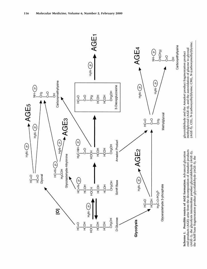
Recently, some studies have suggested thatAGE are produced not only from sugars such asglucose, but also dicarbonyl compounds derivedfrom Maillard reactions, autoxidation of sugarsand other metabolic pathways (Scheme 1). Inmodel systems, AGE have been shown to arisefrom both metalcatalyzed autoxidation of glu-cose with the dicarbonyl glyoxal and arabinoseas intermediates (6), and from the decomposi-tion of Amadori products to the reactive dicar-bonyl 3-deoxyglucosone (7). The dicarbonylmethyglyoxal, produced by nonenzymatic frag-mentation of triose phosphates (glyceraldehyde-3-phosphate, etc.) of the glycolytic intermedi-ates, also forms AGE in vitro and may be a majorsource of intracellular and plasma AGE (8).
Methylglyoxal and glyoxal have beenshown to modify proteins through the Maillardreaction. Several tissues and the plasma of dia-betic individuals exhibit increased levels ofmethylglyoxal (9–11) and even the normal hu-man lens has relatively high levels (12). Al-though formation of cross-linking structures byglyoxal and glycolaldehyde occurs in vitro(6,13), there is still no evidence for such cross-linking in vivo.
To better understand the role of short chainsugars (glyceraldehyde and glycolaldehyde) anddicarbonyl compounds (methylglyoxal andglyoxal) in the modification of proteins in dia-betes, we developed non-carboxymethyllysine(CML) anti-AGE antibodies that recognizeserum proteins modified by these compounds.These antibodies have enabled us to betteridentify the compounds involved in the Mail-lard reaction in vivo.
Materials and Methods
Materials
Bovine serum albumin (BSA), rabbit serum albumin (RSA), and methylglyoxal were pur-chased from Sigma Chemical Co. (St. Louis,MO). D-glyceraldehyde and glycolaldehydewere purchased from Nacalai Tesque (Kyoto,Japan). Glyoxal, glucose, glyoxylic acid, py-ruvic acid and sodium cyanoborohydride(NaCNBH3) were purchased from Wako Pure Chemical Industries, Ltd. (Osaka, Japan). De-
hydrated skim milk was purchased from DifcoLab. (Detroit, MI). A PD-10 column, cyano-genbromide (CNBr)-activated Sepharose 4B,Sephacryl S-200, and Sephadex G-15 werepurchased from Pharmacia Biotech AB (Upp-sala, Sweden). Alkaline phosphatase (AP)-conjugated sheep anti-rabbit immunoglobu-lin G (IgG) antibody was purchased fromBoehringer Mannheim (Mannheim, Germany).Super Block Blocking Buffer in phosphate-buffered saline (PBS) and alkaline phos-phatase substrate kits were purchased fromPierce Chemical Co. (Rockford, IL). Microtitra-tion plates (96-well; High binding E.I.A./R.I.A. plates) were purchased from CorningCostar (Cambridge, MA). Centriprep-10 andPM-10 ultrafiltration membranes were pur-chased from Amicon, Inc. (Beverly, MA), whilea polyvinylidene fluoride (PVDF) membranewas purchased from Atto Co. (Tokyo, Japan).All other chemicals were of the highest gradeavailable from commercial sources.
Preparation of AGE Proteins, CML and CEL Protein
AGE-BSA and AGE-RSA were prepared as described previously (14). Briefly, each pro-tein was incubated under sterile conditionswith 0.1 M D-glyceraldehyde, glycolaldehyde,methylglyoxal or glyoxal and 5 mM DTPA in0.2 M phosphate buffer (pH 7.4) at 37�C for 7 dand, then, low molecular weight reactants andaldehydes were removed using a PD-10 col-umn and dialysis against PBS (pH 7.4). CML-BSA was prepared as described elsewhere (15).Briefly, 50 mg/ml protein was incubated at 37�Cfor 24 hr with 45 mM glyoxylic acid and 150mM NaCNBH3 in 2 ml of 0.2 M phosphatebuffer (pH 7.4), followed by PD-10 columnchromatography and dialysis against PBS. Car-boxyethyllysine (CEL)-BSA was prepared as de-scribed elsewhere (16). Briefly, 50 mg/ml BSAwas incubated at 37�C for 24 hr with 45 mMpyruvic acid and 150 mM NaCNBH3 in 2 ml of0.2 M phosphate buffer (pH 7.4), followed byPD-10 column chromatography and dialysisagainst PBS. Protein concentrations were deter-mined with Dc protein assay reagent (Bio-RadLaboratories, Richmond, CA) using BSA as astandard.
Preparation of Polyclonal Anti-AGE Antibodies
4 mg of various AGE-RSAs (incubated with D-glyceraldehyde, glycolaldehyde, methylgly-
116 Molecular Medicine, Volume 6, Number 2, February 2000
Sch
eme
1.P
oss
ible
ro
ute
s o
f A
GE
fo
rmat
ion
.Ad
van
ced
gly
cati
onen
d-p
rod
uct
s (A
GE
) ar
ise
from
th
e d
ecom
pos
itio
n o
f A
mad
ori
pro
du
cts
(AG
E-1
), t
he
glyc
olys
is i
nte
rmed
iate
pro
du
ct g
lyce
rald
ehyd
e (A
GE
-2),
the
Sch
iff
bas
e fr
agm
enta
tion
pro
du
ct g
lyco
lald
ehyd
e (A
GE
-3),
th
e
glyc
eral
deh
yde
and
th
e A
mad
ori
pro
du
ct f
ragm
enta
tion
pro
du
ctm
eth
ylgl
yoxa
l (A
GE
-4),
an
d t
he
auto
xid
atio
n o
f gl
uco
se t
o gl
yoxa
l(A
GE
-5).
CE
L, N
-(ca
rbox
yeth
yl)l
ysin
e; C
ML
, N-(
carb
oxym
eth
yl)l
ysin
e.
M. Takeuchi et al.: Immunological Evidence for Non-CML AGEs in vivo 117
oxal or glyoxal) were emulsified in 50% Fre-und’s complete adjuvant and injected intra-dermally into rabbits. This procedure was repeated at weekly intervals for 6 weeks. Aftera 2-week pause, the rabbits were given abooster injection of 4 mg of each antigen. Ani-mals were bled on the tenth day after this injection and serum was obtained for puri-fication.
Purification of Non-CML AGE Antibodies from Polyclonal AGE Antibodies by Affinity Chromatography
Antibodies specific for non-CML AGE wereisolated from the antiserum by affinity chro-matography. Four kinds of AGE-BSA or CML-BSA (100 mg of each protein) were coupled to20 ml of CNBr-activated Sepharose 4B accord-ing to the manufacture’s instructions. 20 ml ofrabbit serum was applied to a column (1.5 �5.5 cm) of Sepharose 4B coupled with AGE-BSAs (incubated with D-glyceraldehyde, gly-colaldehyde, methylglyoxal or glyoxal). Afterextensive washing with PBS, the adsorbed frac-tions were eluted with 20 mM sodium phos-phate buffer containing 3 M potassium thio-cyanate (pH 7.4). The AGE antibody fractions,were pooled, concentrated using Centriprep-10(Amicon, Inc.), and passed through a PD-10column (Pharmacia Biotech AB) equilibratedwith PBS. The AGE antibodies thus obtainedwere then loaded onto a CML-BSA-Sepharose4B column (1.5 � 5.5 cm), which was washedwith 30 ml of PBS to obtain the unadsorbedfraction (non-CML AGE antibodies). The ad-sorbed fraction (CML antibody) was theneluted with 20 ml of 20 mM sodium phosphatebuffer containing 3 M potassium thiocyanate(pH 7.4). Fractions (1.0 ml) were monitored forabsorbance at 280 nm. The unadsorbed fractionswere pooled, concentrated with Centriprep-10, and passed through a PD-10 column equil-ibrated with PBS for use in our study.
Enzyme-Linked Immunosorbent Assay (ELISA)
Measurement of AGE was performed with acompetitive ELISA, as described previously(14). Briefly, test samples (50 �l) were added toeach well as a competitor for 50 �l of non-CMLAGE antibodies (1: 250 - 1: 1000), followed byincubation for 2 hr at room temperature withgentle shaking on a horizontal rotary shaker.Results were expressed as:
B�B0, calculated as (experimental OD�back-ground OD)/(total OD�background OD).
The immunoreactivity of each fraction was read from the calibration curve (four kinds of AGE-BSAs) and was expressed as AGEunits (U) per ml, with one unit correspondingto the amount of antibody reactive materialfound in AGE-BSA at a protein concentrationof 1 �g/ml.
Size Distribution of Non-CML AGE in Serum fromDiabetic Patients on Hemodialysis
50 ml of serum from 10 type 2 diabetic patientswith end-stage renal disease on hemodialysiswas subjected to ultrafiltration (PM-10 mem-brane, cut-off molecular weight (MW) 10 kDa,Amicon, Inc.) to separate the high molecularweight (HMW � 10 kDa) fraction from the lowmolecular weight (LMW � 10 kDa) fraction.The HMW fraction was applied to a SephacrylS-200 column (1.5 � 110 cm), which was equi-librated with PBS (pH 7.4) and eluted with thesame buffer (fraction size: 1.5 ml, flow rate: 10 ml/hr) in a cold room. The molecular weightmarkers used were aldolase (MW 160,000),BSA (MW 67,000), chymotrypsinogen A (MW25,000), and vitamin B12 (MW 1,355). TheLMW fraction was pooled, concentrated bylyophilization and dissolved in a small volumeof distilled water. The precipitate was removedby centrifugation at 10,000 rpm for 10 min andthe supernatant was applied to a Sephadex G-15 column (1.5 � 110 cm), which was equili-brated with 50 mM ammonium acetate buf-fer (pH 7.4) and eluted with the same buffer(fraction size: 1.5 ml, flow rate: 8 ml/hr) in acold room. The molecular weight markers usedwere cytochrome c (MW 12,500), vitamin B12
(MW 1,355), and cytidine (MW 243). Each frac-tion was monitored for absorbence at 280 nm andthe AGE activity of each fraction was measuredby both AGE-ELISA and characteristic AGE-specific fluorescence (excitation maxima (Ex) 360 nm/emission maxima (Em) 440 nm). Theimmunoreactivity of each fraction was readfrom the calibration curve (four kinds of AGE-BSAs) and was expressed as AGE units (U) perml, as described above.
Immunoblot Analysis
AGE-proteins and human serum protein sam-ples were electrophoresed on a 7.5% SDS-gel
or 5-20% gradient SDS-gel. The proteins weretransferred electrophoretically to PVDF mem-brane (Atto Co., Tokyo) for 45 min at 2.5 mAper cm2. The membrane was blocked with 4% skim milk (Difco, MI) in PBS for 1 hr at room temperature, reacted for 2 hr with theimmunoaffinity-purified antibodies (1 : 250-1 : 1000 diluted in 4% skim milk in PBS),washed three times for 5 min each with PBS-Tween 20 buffer, incubated for 1 hr in 4% skim milk in PBS with 1 : 2000 diluted anti-rabbit IgG coupled to alkaline phosphatase,washed five times for 5 min each with PBS-Tween 20, and finally, incubated with 5-bromo-4-chloro-3-indoyl phosphate-nitrobluetetrazolium.
Results
Characterization of the Immunogen
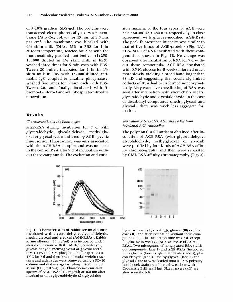
AGE-RSA during incubation for 7 d with glyceraldehyde, glycolaldehyde, methylgly-oxal or glyoxal was monitored by AGE-specificfluorescence. Fluorescence was only associatedwith the AGE-RSA complex and was not seenin the control RSA after 7 d of incubation with-out these compounds. The excitation and emis-
sion maxima of the four types of AGE were360–380 and 430–450 nm, respectively, in closeagreement with glucose-modified AGE-RSA.The peak fluorescence intensity was similar tothat of five kinds of AGE-proteins (Fig. 1A).SDS-PAGE of RSA incubated with these com-pounds is shown in Fig. 1B. No change wasobserved after incubation of RSA for 7 d with-out these compounds. AGE-RSA incubatedwith 0.5 M glucose for 8 weeks migrated muchmore slowly, yielding a broad band larger than68 kD and suggesting that covalently linkedadducts of RSA had been formed nonenzymat-ically. Very extensive crosslinking of RSA wasseen after incubation with short chain sugars,glyceraldehyde and glycolaldehyde. In the caseof dicarbonyl compounds (methylglyoxal andglyoxal), there was much less aggregate for-mation.
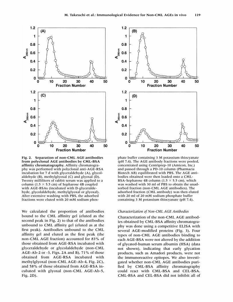
Separation of Non-CML AGE Antibodies fromPolyclonal AGE Antibodies
The polyclonal AGE antisera obtained after in-cubation of AGE-RSA (with glyceraldehyde,glycolaldehyde, methylglyoxal, or glyoxal)were purified by four kinds of AGE-BSA affin-ity chromatography and then were separatedby CML-BSA affinity chromatography (Fig. 2).
118 Molecular Medicine, Volume 6, Number 2, February 2000
Fig. 1. Characteristics of rabbit serum albuminincubated with glyceraldehyde, glycolaldehyde,methylglyoxal and glyoxal (AGE-RSAs). Rabbitserum albumin (20 mg/ml) was incubated understerile conditions with 0.1 M D-glyceraldehyde,glycolaldehyde, methylglyoxal or glyoxal and 5mM DTPA in 0.2 M phosphate buffer (pH 7.4) at37�C for 7 d and then low molecular weight reac-tants and aldehydes were removed using a PD-10column and dialysis against phosphate-bufferedsaline (PBS, pH 7.4). (A) Fluorescence emissionspectra of AGE-RSAs (1.0 mg/ml) at 360 nm afterincubation with glyceraldehyde (), glycolalde-
hyde (�), methylglyoxal (�), glyoxal (�) or glu-cose (�), and after incubation without these com-pounds (�). The incubation time was 7 d, exceptfor glucose (8 weeks). (B) SDS-PAGE of AGE-RSAs. Two micrograms of nonglycated RSA (with-out compounds, lane 1) and AGE-RSAs (incubatedwith glucose (lane 2), glyceraldehyde (lane 3), gly-colaldehyde (lane 4), methylglyoxal (lane 5) andglyoxal (lane 6) were loaded onto a 7.5% polyacry-lamide gel. Staining of the gel was done withCoomassie Brilliant Blue. Size markers (kD) areshown on the left.
M. Takeuchi et al.: Immunological Evidence for Non-CML AGEs in vivo 119
We calculated the proportion of antibodiesbound to the CML affinity gel (eluted as thesecond peak in Fig. 2) to that of the antibodiesunbound to CML affinity gel (eluted as at thefirst peak). Antibodies unbound to the CMLaffinity gel and eluted as the first peak (thenon-CML AGE fraction) accounted for 83% ofthose obtained from AGE-RSA incubated withglyceraldehyde or glycolaldehyde (non-CMLAGE-Ab-2 or -3, Figs. 2A and B), 71% of thoseobtained from AGE-RSA incubated withmethylglyoxal (non-CML AGE-Ab-4, Fig. 2C),and 58% of those obtained from AGE-RSA in-cubated with glyoxal (non-CML AGE-Ab-5,Fig. 2D).
Characterization of Non-CML AGE Antibodies
Characterization of the non-CML AGE antibod-ies obtained by CML-BSA affinity chromatogra-phy was done using a competitive ELISA withseveral AGE-modified proteins (Fig. 3). Fourtypes of non-CML AGE antibodies binding toeach AGE-BSA were not altered by the additionof glycated-human serum albumin (HSA) (datanot shown), indicating that early glycationproducts, such as Amadori products, were notthe immunoreactive epitopes. We also investi-gated whether non-CML AGE antibodies puri-fied by CML-BSA affinity chromatographycould react with CML-BSA and CEL-BSA.CML-BSA and CEL-BSA did not inhibit all of
Fig. 2. Separation of non-CML AGE antibodiesfrom polyclonal AGE antibodies by CML-BSAaffinity chromatography. Affinity chromatogra-phy was performed with polyclonal anti-AGE-RSAincubation for 7 d with glyceraldehyde (A), glycol-aldehyde (B), methylglyoxal (C) and glyoxal (D).Twenty milliliters of rabbit serum was applied to acolumn (1.5 � 5.5 cm) of Sepharose 4B coupledwith AGE-BSAs (incubated with D-glyceralde-hyde, glycolaldehyde, methylglyoxal or glyoxal).After extensive washing with PBS, the adsorbedfractions were eluted with 20 mM sodium phos-
phate buffer containing 3 M potassium thiocyanate(pH 7.4). The AGE antibody fractions were pooled,concentrated using Centriprep-10 (Amicon, Inc.)and passed through a PD-10 column (PharmaciaBiotech AB) equilibrated with PBS. The AGE anti-bodies obtained were then loaded onto a CML-BSA-Sepharose 4B column (1.5 � 5.5 cm), whichwas washed with 30 ml of PBS to obtain the unad-sorbed fraction (non-CML AGE antibodies). Theadsorbed fraction (CML antibody) was then elutedwith 20 ml of 20 mM sodium phosphate buffercontaining 3 M potassium thiocyanate (pH 7.4).
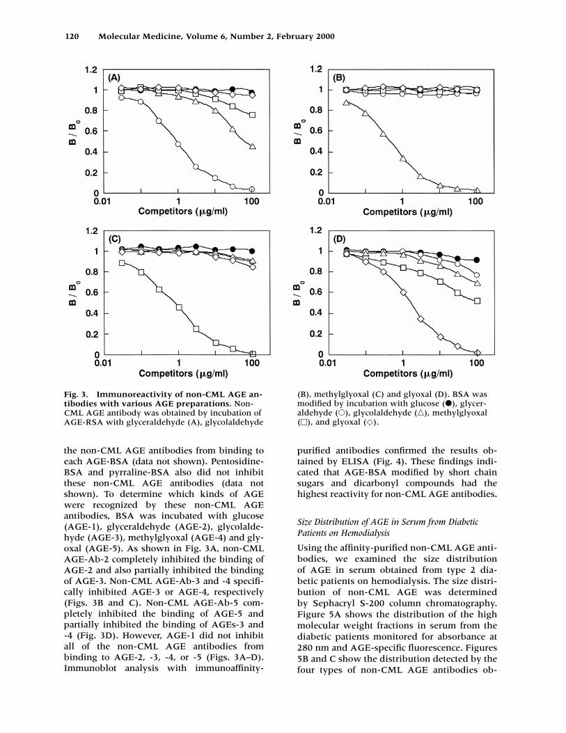
the non-CML AGE antibodies from binding toeach AGE-BSA (data not shown). Pentosidine-BSA and pyrraline-BSA also did not inhibitthese non-CML AGE antibodies (data notshown). To determine which kinds of AGEwere recognized by these non-CML AGE antibodies, BSA was incubated with glucose(AGE-1), glyceraldehyde (AGE-2), glycolalde-hyde (AGE-3), methylglyoxal (AGE-4) and gly-oxal (AGE-5). As shown in Fig. 3A, non-CMLAGE-Ab-2 completely inhibited the binding ofAGE-2 and also partially inhibited the bindingof AGE-3. Non-CML AGE-Ab-3 and -4 specifi-cally inhibited AGE-3 or AGE-4, respectively(Figs. 3B and C). Non-CML AGE-Ab-5 com-pletely inhibited the binding of AGE-5 and partially inhibited the binding of AGEs-3 and -4 (Fig. 3D). However, AGE-1 did not inhibit all of the non-CML AGE antibodies from binding to AGE-2, -3, -4, or -5 (Figs. 3A–D). Immunoblot analysis with immunoaffinity-
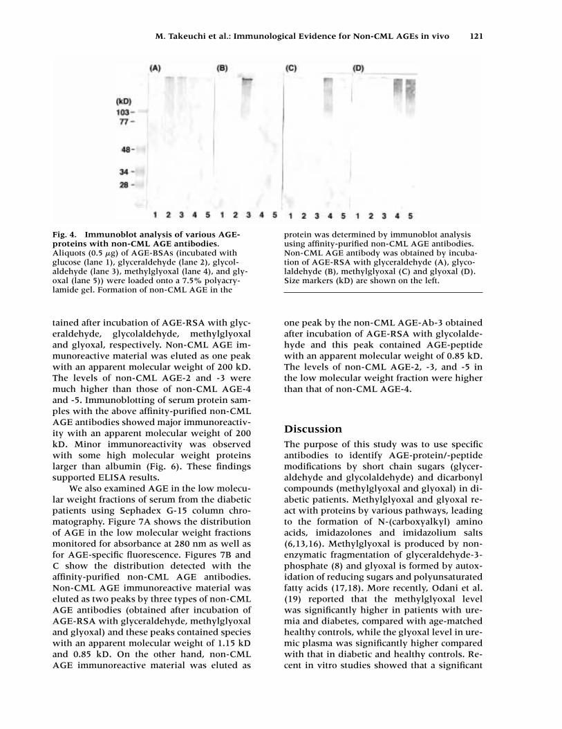
purified antibodies confirmed the results ob-tained by ELISA (Fig. 4). These findings indi-cated that AGE-BSA modified by short chainsugars and dicarbonyl compounds had the highest reactivity for non-CML AGE antibodies.
Size Distribution of AGE in Serum from DiabeticPatients on Hemodialysis
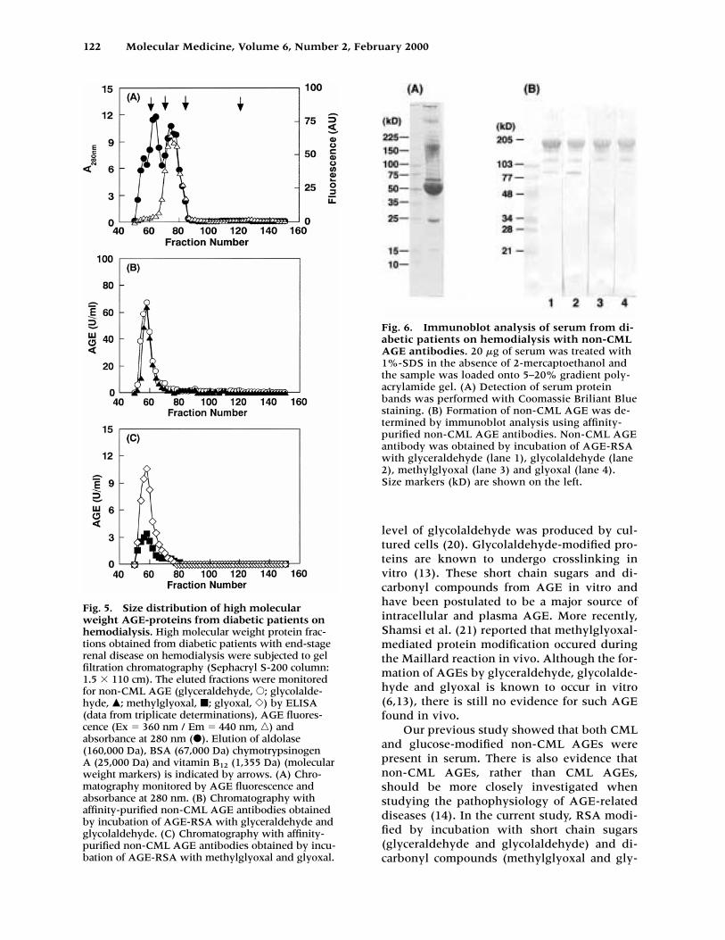
Using the affinity-purified non-CML AGE anti-bodies, we examined the size distribution of AGE in serum obtained from type 2 dia-betic patients on hemodialysis. The size distri-bution of non-CML AGE was determined by Sephacryl S-200 column chromatography. Figure 5A shows the distribution of the highmolecular weight fractions in serum from thediabetic patients monitored for absorbance at 280 nm and AGE-specific fluorescence. Figures5B and C show the distribution detected by thefour types of non-CML AGE antibodies ob-
120 Molecular Medicine, Volume 6, Number 2, February 2000
Fig. 3. Immunoreactivity of non-CML AGE an-tibodies with various AGE preparations. Non-CML AGE antibody was obtained by incubation ofAGE-RSA with glyceraldehyde (A), glycolaldehyde
(B), methylglyoxal (C) and glyoxal (D). BSA wasmodified by incubation with glucose (�), glycer-aldehyde (�), glycolaldehyde (�), methylglyoxal(�), and glyoxal (�).
M. Takeuchi et al.: Immunological Evidence for Non-CML AGEs in vivo 121
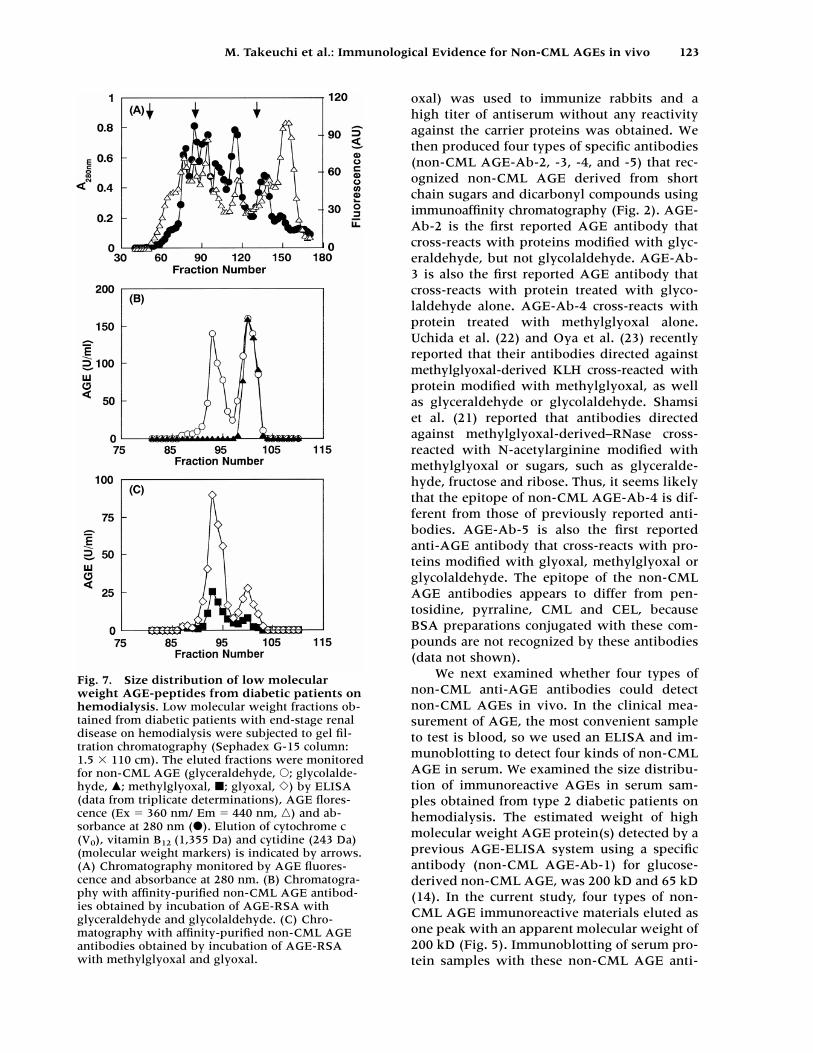
tained after incubation of AGE-RSA with glyc-eraldehyde, glycolaldehyde, methylglyoxaland glyoxal, respectively. Non-CML AGE im-munoreactive material was eluted as one peakwith an apparent molecular weight of 200 kD.The levels of non-CML AGE-2 and -3 weremuch higher than those of non-CML AGE-4and -5. Immunoblotting of serum protein sam-ples with the above affinity-purified non-CMLAGE antibodies showed major immunoreactiv-ity with an apparent molecular weight of 200kD. Minor immunoreactivity was observedwith some high molecular weight proteinslarger than albumin (Fig. 6). These findingssupported ELISA results.
We also examined AGE in the low molecu-lar weight fractions of serum from the diabeticpatients using Sephadex G-15 column chro-matography. Figure 7A shows the distributionof AGE in the low molecular weight fractionsmonitored for absorbance at 280 nm as well asfor AGE-specific fluorescence. Figures 7B andC show the distribution detected with theaffinity-purified non-CML AGE antibodies.Non-CML AGE immunoreactive material waseluted as two peaks by three types of non-CMLAGE antibodies (obtained after incubation ofAGE-RSA with glyceraldehyde, methylglyoxaland glyoxal) and these peaks contained specieswith an apparent molecular weight of 1.15 kDand 0.85 kD. On the other hand, non-CMLAGE immunoreactive material was eluted as
one peak by the non-CML AGE-Ab-3 obtainedafter incubation of AGE-RSA with glycolalde-hyde and this peak contained AGE-peptidewith an apparent molecular weight of 0.85 kD.The levels of non-CML AGE-2, -3, and -5 inthe low molecular weight fraction were higherthan that of non-CML AGE-4.
DiscussionThe purpose of this study was to use specificantibodies to identify AGE-protein/-peptidemodifications by short chain sugars (glycer-aldehyde and glycolaldehyde) and dicarbonylcompounds (methylglyoxal and glyoxal) in di-abetic patients. Methylglyoxal and glyoxal re-act with proteins by various pathways, leadingto the formation of N-(carboxyalkyl) aminoacids, imidazolones and imidazolium salts(6,13,16). Methylglyoxal is produced by non-enzymatic fragmentation of glyceraldehyde-3-phosphate (8) and glyoxal is formed by autox-idation of reducing sugars and polyunsaturatedfatty acids (17,18). More recently, Odani et al.(19) reported that the methylglyoxal level was significantly higher in patients with ure-mia and diabetes, compared with age-matchedhealthy controls, while the glyoxal level in ure-mic plasma was significantly higher comparedwith that in diabetic and healthy controls. Re-cent in vitro studies showed that a significant
Fig. 4. Immunoblot analysis of various AGE-proteins with non-CML AGE antibodies.Aliquots (0.5 �g) of AGE-BSAs (incubated withglucose (lane 1), glyceraldehyde (lane 2), glycol-aldehyde (lane 3), methylglyoxal (lane 4), and gly-oxal (lane 5)) were loaded onto a 7.5% polyacry-lamide gel. Formation of non-CML AGE in the
protein was determined by immunoblot analysisusing affinity-purified non-CML AGE antibodies.Non-CML AGE antibody was obtained by incuba-tion of AGE-RSA with glyceraldehyde (A), glyco-laldehyde (B), methylglyoxal (C) and glyoxal (D).Size markers (kD) are shown on the left.
122 Molecular Medicine, Volume 6, Number 2, February 2000
Fig. 5. Size distribution of high molecularweight AGE-proteins from diabetic patients onhemodialysis. High molecular weight protein frac-tions obtained from diabetic patients with end-stagerenal disease on hemodialysis were subjected to gelfiltration chromatography (Sephacryl S-200 column:1.5 � 110 cm). The eluted fractions were monitoredfor non-CML AGE (glyceraldehyde, �; glycolalde-hyde, �; methylglyoxal, �; glyoxal, �) by ELISA(data from triplicate determinations), AGE fluores-cence (Ex 360 nm / Em 440 nm, �) and absorbance at 280 nm (�). Elution of aldolase(160,000 Da), BSA (67,000 Da) chymotrypsinogen A (25,000 Da) and vitamin B12 (1,355 Da) (molecularweight markers) is indicated by arrows. (A) Chro-matography monitored by AGE fluorescence and absorbance at 280 nm. (B) Chromatography withaffinity-purified non-CML AGE antibodies obtainedby incubation of AGE-RSA with glyceraldehyde andglycolaldehyde. (C) Chromatography with affinity-purified non-CML AGE antibodies obtained by incu-bation of AGE-RSA with methylglyoxal and glyoxal.
Fig. 6. Immunoblot analysis of serum from di-abetic patients on hemodialysis with non-CMLAGE antibodies. 20 �g of serum was treated with1%-SDS in the absence of 2-mercaptoethanol andthe sample was loaded onto 5–20% gradient poly-acrylamide gel. (A) Detection of serum proteinbands was performed with Coomassie Briliant Bluestaining. (B) Formation of non-CML AGE was de-termined by immunoblot analysis using affinity-purified non-CML AGE antibodies. Non-CML AGEantibody was obtained by incubation of AGE-RSAwith glyceraldehyde (lane 1), glycolaldehyde (lane2), methylglyoxal (lane 3) and glyoxal (lane 4).Size markers (kD) are shown on the left.
level of glycolaldehyde was produced by cul-tured cells (20). Glycolaldehyde-modified pro-teins are known to undergo crosslinking invitro (13). These short chain sugars and di-carbonyl compounds from AGE in vitro andhave been postulated to be a major source ofintracellular and plasma AGE. More recently,Shamsi et al. (21) reported that methylglyoxal-mediated protein modification occured duringthe Maillard reaction in vivo. Although the for-mation of AGEs by glyceraldehyde, glycolalde-hyde and glyoxal is known to occur in vitro(6,13), there is still no evidence for such AGEfound in vivo.
Our previous study showed that both CMLand glucose-modified non-CML AGEs werepresent in serum. There is also evidence thatnon-CML AGEs, rather than CML AGEs,should be more closely investigated whenstudying the pathophysiology of AGE-relateddiseases (14). In the current study, RSA modi-fied by incubation with short chain sugars(glyceraldehyde and glycolaldehyde) and di-carbonyl compounds (methylglyoxal and gly-
M. Takeuchi et al.: Immunological Evidence for Non-CML AGEs in vivo 123
oxal) was used to immunize rabbits and a high titer of antiserum without any reactivityagainst the carrier proteins was obtained. Wethen produced four types of specific antibodies(non-CML AGE-Ab-2, -3, -4, and -5) that rec-ognized non-CML AGE derived from shortchain sugars and dicarbonyl compounds usingimmunoaffinity chromatography (Fig. 2). AGE-Ab-2 is the first reported AGE antibody thatcross-reacts with proteins modified with glyc-eraldehyde, but not glycolaldehyde. AGE-Ab-3 is also the first reported AGE antibody thatcross-reacts with protein treated with glyco-laldehyde alone. AGE-Ab-4 cross-reacts withprotein treated with methylglyoxal alone.Uchida et al. (22) and Oya et al. (23) recentlyreported that their antibodies directed againstmethylglyoxal-derived KLH cross-reacted withprotein modified with methylglyoxal, as wellas glyceraldehyde or glycolaldehyde. Shamsi et al. (21) reported that antibodies directedagainst methylglyoxal-derived–RNase cross-reacted with N-acetylarginine modified withmethylglyoxal or sugars, such as glyceralde-hyde, fructose and ribose. Thus, it seems likelythat the epitope of non-CML AGE-Ab-4 is dif-ferent from those of previously reported anti-bodies. AGE-Ab-5 is also the first reportedanti-AGE antibody that cross-reacts with pro-teins modified with glyoxal, methylglyoxal orglycolaldehyde. The epitope of the non-CMLAGE antibodies appears to differ from pen-tosidine, pyrraline, CML and CEL, becauseBSA preparations conjugated with these com-pounds are not recognized by these antibodies(data not shown).
We next examined whether four types ofnon-CML anti-AGE antibodies could detectnon-CML AGEs in vivo. In the clinical mea-surement of AGE, the most convenient sampleto test is blood, so we used an ELISA and im-munoblotting to detect four kinds of non-CMLAGE in serum. We examined the size distribu-tion of immunoreactive AGEs in serum sam-ples obtained from type 2 diabetic patients onhemodialysis. The estimated weight of highmolecular weight AGE protein(s) detected by aprevious AGE-ELISA system using a specificantibody (non-CML AGE-Ab-1) for glucose-derived non-CML AGE, was 200 kD and 65 kD(14). In the current study, four types of non-CML AGE immunoreactive materials eluted asone peak with an apparent molecular weight of200 kD (Fig. 5). Immunoblotting of serum pro-tein samples with these non-CML AGE anti-
Fig. 7. Size distribution of low molecularweight AGE-peptides from diabetic patients onhemodialysis. Low molecular weight fractions ob-tained from diabetic patients with end-stage renaldisease on hemodialysis were subjected to gel fil-tration chromatography (Sephadex G-15 column:1.5 � 110 cm). The eluted fractions were monitoredfor non-CML AGE (glyceraldehyde, �; glycolalde-hyde, �; methylglyoxal, �; glyoxal, �) by ELISA(data from triplicate determinations), AGE flores-cence (Ex 360 nm/ Em 440 nm, �) and ab-sorbance at 280 nm (�). Elution of cytochrome c(V0), vitamin B12 (1,355 Da) and cytidine (243 Da)(molecular weight markers) is indicated by arrows.(A) Chromatography monitored by AGE fluores-cence and absorbance at 280 nm. (B) Chromatogra-phy with affinity-purified non-CML AGE antibod-ies obtained by incubation of AGE-RSA withglyceraldehyde and glycolaldehyde. (C) Chro-matography with affinity-purified non-CML AGEantibodies obtained by incubation of AGE-RSAwith methylglyoxal and glyoxal.
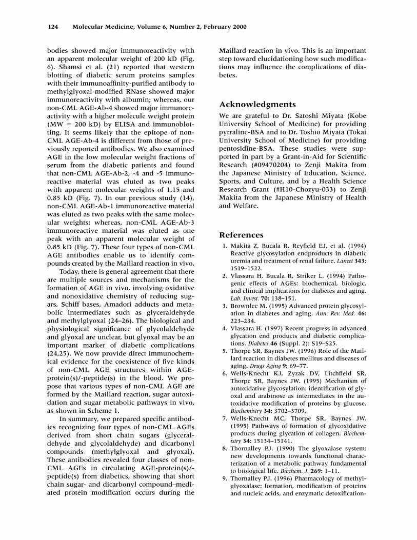
bodies showed major immunoreactivity withan apparent molecular weight of 200 kD (Fig. 6). Shamsi et al. (21) reported that westernblotting of diabetic serum proteins sampleswith their immunoaffinity-purified antibody tomethylglyoxal-modified RNase showed majorimmunoreactivity with albumin; whereas, ournon-CML AGE-Ab-4 showed major immunore-activity with a higher molecule weight protein(MW 200 kD) by ELISA and immunoblot-ting. It seems likely that the epitope of non-CML AGE-Ab-4 is different from those of pre-viously reported antibodies. We also examinedAGE in the low molecular weight fractions ofserum from the diabetic patients and foundthat non-CML AGE-Ab-2, -4 and -5 immuno-reactive material was eluted as two peaks with apparent molecular weights of 1.15 and0.85 kD (Fig. 7). In our previous study (14),non-CML AGE-Ab-1 immunoreactive materialwas eluted as two peaks with the same molec-ular weights; whereas, non-CML AGE-Ab-3immunoreactive material was eluted as onepeak with an apparent molecular weight of0.85 kD (Fig. 7). These four types of non-CMLAGE antibodies enable us to identify com-pounds created by the Maillard reaction in vivo.
Today, there is general agreement that thereare multiple sources and mechanisms for theformation of AGE in vivo, involving oxidativeand nonoxidative chemistry of reducing sug-ars, Schiff bases, Amadori adducts and meta-bolic intermediates such as glyceraldehyde and methylglyoxal (24–26). The biological andphysiological significance of glycolaldehydeand glyoxal are unclear, but glyoxal may be animportant marker of diabetic complications(24,25). We now provide direct immunochem-ical evidence for the coexistence of five kinds of non-CML AGE structures within AGE-protein(s)/-peptide(s) in the blood. We pro-pose that various types of non-CML AGE areformed by the Maillard reaction, sugar autoxi-dation and sugar metabolic pathways in vivo,as shown in Scheme 1.
In summary, we prepared specific antibod-ies recognizing four types of non-CML AGEsderived from short chain sugars (glyceral-dehyde and glycolaldehyde) and dicarbonylcompounds (methylglyoxal and glyoxal).These antibodies revealed four classes of non-CML AGEs in circulating AGE-protein(s)/-peptide(s) from diabetics, showing that shortchain sugar- and dicarbonyl compound–medi-ated protein modification occurs during the
Maillard reaction in vivo. This is an importantstep toward elucidationing how such modifica-tions may influence the complications of dia-betes.
AcknowledgmentsWe are grateful to Dr. Satoshi Miyata (KobeUniversity School of Medicine) for providingpyrraline-BSA and to Dr. Toshio Miyata (TokaiUniversity School of Medicine) for providingpentosidine-BSA. These studies were sup-ported in part by a Grant-in-Aid for ScientificResearch (#09470204) to Zenji Makita from the Japanese Ministry of Education, Science,Sports, and Culture, and by a Health ScienceResearch Grant (#H10-Chozyu-033) to ZenjiMakita from the Japanese Ministry of Healthand Welfare.
References1. Makita Z, Bucala R, Reyfield EJ, et al. (1994)
Reactive glycosylation endproducts in diabeticuremia and treatment of renal failure. Lancet 343:1519–1522.
2. Vlassara H, Bucala R, Striker L. (1994) Patho-genic effects of AGEs: biochemical, biologic,and clinical implications for diabetes and aging.Lab. Invest. 70: 138–151.
3. Brownlee M. (1995) Advanced protein glycosyl-ation in diabetes and aging. Ann. Rev. Med. 46:223–234.
4. Vlassara H. (1997) Recent progress in advancedglycation end products and diabetic complica-tions. Diabetes 46 (Suppl. 2): S19–S25.
5. Thorpe SR, Baynes JW. (1996) Role of the Mail-lard reaction in diabetes mellitus and diseases ofaging. Drugs Aging 9: 69–77.
6. Wells-Knecht KJ, Zyzak DV, Litchfield SR,Thorpe SR, Baynes JW. (1995) Mechanism ofautoxidative glycosylation: identification of gly-oxal and arabinose as intermediates in the au-toxidative modification of proteins by glucose.Biochemistry 34: 3702–3709.
7. Wells-Knecht MC, Thorpe SR, Baynes JW.(1995) Pathways of formation of glycoxidativeproducts during glycation of collagen. Biochem-istry 34: 15134–15141.
8. Thornalley PJ. (1990) The glyoxalase system:new developments towards functional charac-terization of a metabolic pathway fundamentalto biological life. Biochem. J. 269: 1–11.
9. Thornalley PJ. (1996) Pharmacology of methyl-glyoxalase: formation, modification of proteinsand nucleic acids, and enzymatic detoxification-
124 Molecular Medicine, Volume 6, Number 2, February 2000
M. Takeuchi et al.: Immunological Evidence for Non-CML AGEs in vivo 125
a role in pathogenesis and antiproliferativechemotherapys. Gen. Pharmacol. 27: 565–573.
10. McLellan AC, Thornalley PJ, Ben J, SonksenPH. (1994) Glyoxalase system in clinical dia-betes mellitus and correlation with diabetic com-plication Clin. Sci. 87: 21–29.
11. McLellan AC, Phillips SA, Thornalley PJ.(1992) The assay of methylglyoxal in biologicalsystems by derivatization with 1,2-diamino-4,5-dimethoxybenzene. Anal. Biochem. 206: 17–23.
12. Haik GM, Lo TW, Thornalley PJ (1994) Methyl-glyoxal concentration and glyoxalase activitiesin the human lens. Exp. Eye Res. 59: 497–500.
13. Glomb MA, Monnier VM. (1995) Mechanism ofprotein modification by glyoxal and glycolalde-hyde, reactive intermediates of the Maillard re-action. J. Biol. Chem. 270: 10017–10026.
14. Takeuchi M, Makita Z, Yanagisawa K, KamedaY, Koike T. (1999) Detection of noncarboxy-methyllysine and carboxymethyllysine advancedglycation end products (AGE) in serum of dia-betic patients. Mol. Med. 5: 393–405.
15. Ikeda K, Higashi T, Sano H, et al. (1996) N-(carboxymethyl)lysine protein adduct is a ma-jor immunological epitope in proteins modifiedwith advanced glycation end products of theMaillard reaction. Biochemistry 35: 8075–8083.
16. Ahmed MU, Frye EB, Degenhardt TP, ThorpeSR, Baynes JW. (1997) N-(Carboxyethyl)ly-sine, a product of the chemical modification ofproteins by methylglyoxal, increases with agein human lens proteins. Biochem. J. 324: 565–570.
17. Wells-Knecht KJ, Brinkmann E, Wells-KnechtMC, et al. (1996) New biomarkers of Maillardreaction damage to proteins. Nephrol. Dial. Trans-plant. 11 (Suppl 5): 41–47.
18. Requena JR, Fu M-X, Ahmed MU, Jenkins AJ,Lyons TJ, Thorpe SR. (1996) Lipoxidation prod-ucts as biomarkers of oxidative damage to proteins during lipid peroxidation reactions.Nephrol. Dial. Transplant. 11 (Suppl 5): 48–53.
19. Odani H, Shinzato T, Matsumoto Y, Usami J,Maeda K. (1999) Increase in three �, �-dicar-bonyl compound levels in human uremicplasma: specific in vivo determination of inter-mediates in advanced Maillard reaction.Biochem. Biophys. Res. Commun. 256: 89–93.
20. Anderson MM, Hazen SL, Hsu FF, Heinecke JW. (1997) Human neutrophils employ themyeloperoxidase-hydrogen peroxide-chloridesystem to convert hydroxy-amino acids into gly-colaldehyde, 2-hydroxypropanal, and acrolein: amechanism for the generation of highly reactive�-hydroxy and �, �-unsaturated aldehydes byphagocytes at sites of inflammation. J. Clin. In-vest. 99: 424–432.
21. Shamsi FA, Partal A, Sady C, Glomb MA, Na-garaj RH. (1998) Immunological evidence formethylglyoxal-derived modifications in vivo. J. Biol. Chem. 273: 6928–6936.
22. Uchida K, Khor OT, Oya T, Osawa T, Yasuda Y,Miyata T. (1997) Protein modification by a Maillard reaction intermediate methylglyoxalimmunochemical detection of fluorescent 5-methylimidazolone derivatives in vivo. FEBSLett. 410: 313–318.
23. Oya T, Hattori N, Mizuno Y, et al. (1999)Methylglyoxal modification of protein: chem-ical and immunochemical characterization ofmethylglyoxal-arginine adducts. J. Biol. Chem.274: 18492–18502.
24. Brinkmann Frye E, Degenhardt TP, Thorpe SR,Baynes JW. (1998) Role of the Maillard reactionin aging of tissue proteins: advanced glycationend product-dependent increase in imidazoliumcrosslinks in human lens proteins. J. Biol. Chem.273: 18714–18719.
25. Degenhardt TP, Thorpe SR, Baynes JW. (1998)Chemical modification of proteins by methyl-glyoxal. Cell Mol. Biol. 44: 1139–1145.
26. Baynes JW, Thorpe SR. (1999) Role of oxidativestress in diabetic complications: a new perspec-tive on an old paradigm. Diabetes 48: 1–9.