Embed Size (px)
Citation preview
RESEARCH ARTICLE Open Access
Immunotherapy of hepatocellular carcinoma withsmall double-stranded RNATatyana O Kabilova1, Larisa V Kovtonyuk1, Evgeniy V Zonov1, Elena I Ryabchikova1, Nelly A Popova2,Valeriy P Nikolin2, Vasiliy I Kaledin2, Marina A Zenkova1, Valentin V Vlassov1 and Elena L Chernolovskaya1*
Abstract
Background: Hepatocellular carcinoma (HCC) is one of the most common malignancies worldwide with limitedtherapeutic options. Since HCC has been shown to be immunogenic, immunotherapy is considered a promisingtherapeutic approach. Small interfering RNAs (siRNAs), depending on their structure and sequence, can triggerthe innate immune system, which can potentially enhance the adaptive anticancer immune response in thetumor-bearing subjects. Immunostimulatory properties of nucleic acids can be applied to develop adjuvants forHCC treatment.
Methods: The transplantable HCC G-29 tumor in male CBA/LacSto (CBA) mice was used to study the effects ofimmunostimulatory RNA on tumor growth. Tumor size, metastases area in different organs of mice and mousesurvival rate were analyzed. Furthermore the mouse serum IFN-α levels were measured using ELISA.
Results: In the present study, we found that a 19-bp RNA duplex (ImmunoStimulattory RNA or isRNA) with 3-ntoverhangs at the 3′-ends of specific sequence displays immunostimulatory, antitumor, and antimetastatic activitiesin mice bearing HCC G-29. Our results demonstrate that isRNA strongly increases the level of interferon-α (IFN-α) byup to 25-fold relative to the level in mice injected with Lipofectamine alone (Mock), and to a lesser extent increasesthe level of proinflammatory cytokine interleukin-6 (IL-6) (by up to 5.5-fold relative to the Mock level), in mice bloodserum. We showed that isRNA reliably (P < 0.05) inhibits primary tumor growth in mice compared to the mockgroup. Furthermore, injections of isRNA significantly enhanced necrotic processes in the center of the primarytumor, and decreased by twofold the width of the undifferentiated peripheral zone and the number of mitotic cellsin this zone. The results showed that isRNA efficiently reduces the area of metastases in the liver, kidneys, and heartof CBA/LacSto mice with HCC.
Conclusions: The obtained results clearly demonstrate immunostimulatory and antimetastatic properties of theisRNAs in mice with HCC. Consequently, this short double-stranded RNA can be considered as a potential adjuvantfor the therapy of HCC.
Keywords: Immunostimulatory RNA, Hepatocellular carcinoma, Interferon inducer
BackgroundHepatocellular carcinoma (HCC) is one of the mostcommon cancers and the third leading cause of cancer-related deaths worldwide [1]. The standard methods ofhepatocarcinoma treatment include surgical resection,chemical therapy, radiotherapy, ultrasound ablation, orliver transplantation [2-5]. However, the high rate of
recurrence after these therapeutic procedures remains abig issue in patients with HCC. In view of these facts,the necessity to use adjuvant therapy becomes urgent.Tumor pathogenesis is accompanied by disorders of theimmune system, leading to the escape of the tumor fromthe immune response. At the same time, the immunestatus of an organism is impaired, and immunodeficiencyis developed. Immunotherapy aims to provide a moreefficient targeting of tumor cells by inducing or boos-ting the existing tumor-specific immune response.Immunostimulatory agents are widely used as a part of
* Correspondence: [email protected] of Chemical Biology and Fundamental Medicine SB RAS, 8,Lavrentiev Avenue, Novosibirsk 630090, RussiaFull list of author information is available at the end of the article
© 2014 Kabilova et al.; licensee BioMed Central Ltd. This is an Open Access article distributed under the terms of the CreativeCommons Attribution License (http://creativecommons.org/licenses/by/2.0), which permits unrestricted use, distribution, andreproduction in any medium, provided the original work is properly credited. The Creative Commons Public DomainDedication waiver (http://creativecommons.org/publicdomain/zero/1.0/) applies to the data made available in this article,unless otherwise stated.
Kabilova et al. BMC Cancer 2014, 14:338http://www.biomedcentral.com/1471-2407/14/338
the combined therapy or as a monotherapy in the treat-ment of different cancers [6-9]. Moreover, HCC patientswith tumors containing infiltrated tumor-specific effectorT cells have a reduced risk of tumor recurrence followingliver transplantation [10]. The following methods of adju-vant therapy were proposed for the treatment of HCCpatients: cellular immunotherapy [11], interferon (IFN)therapy [12], and therapy using endogenous IFN inducers[13]. It should be noted that in many cases, HCC de-velops on a background of chronic inflammatory liverdisease such as hepatitis B and C or cirrhosis. Manystudies have documented that IFN therapy significantlysuppresses the onset of HCC from chronic hepatitis andliver cirrhosis [14-16].Natural and synthetic immunostimulators cause a wide
variety of effects at the levels of the immunocompetentcells and an entire organism including activation ofdifferent types of immune cells (macrophages, T- andB-lymphocytes, NK-cells), induction of type I interferonsand/or inflammatory cytokines synthesis by immune cells,and also cause proliferation blockage, and differentiationor apoptosis of tumor cells. The innate immune systemcan be activated by exogenous nucleic acids (e.g., bacterial,viral, and fungal nucleic acids) via several families of pat-tern recognition receptors, including endosomal Toll-likereceptors (TLRs) 3/7/8/9 or cytosolic receptors: retinoicacid inducible gene-I (RIG-I)-like helicases includingRIG1 and melanoma differentiation-associated gene 5(MDA5); Interferon-induced, double-stranded RNA-acti-vated protein kinase (PKR); 2′-5′-oligoadenylate synthase1 and 2 (OAS); and less studied NOD-like receptors(NLRP3, NOD2) [17,18]. These receptors are responsiblefor the activation of the complement system, coagulation,opsonization, phagocytosis, and the induction of apoptosisand cytokine production. The role of TLRs in antitumorprotection of an organism is confirmed by the fact thatmutations in the genes encoding TLRs increase both theincidence of infectious diseases and the frequency of can-cer [19]. The antitumor activity of TLR 3/7/8/9 agonistshas been demonstrated in several tumor types includingmelanoma, breast cancer, renal cell carcinoma, glioblast-oma, cutaneous T-cell and non-Hodgkin’s lymphomas,and basal cell carcinoma [6]. Unlike other cancers, thetreatment of HCC involves not only the treatment of thecarcinoma itself, but also the treatment of underlyingchronic hepatitis. TLR7 and −9 agonists have great the-rapeutic potential against HCC because they induce thesynthesis of the mediators of anti-viral and anticancerimmunity such as IFN-α and IFN-γ-inducible protein 10(IP-10), and induce a sustained increase in 2′5′-oligoade-nylate synthesis [20]. In a recent phase 1 trial in patientswith chronic HCV infection, TLR7 agonist isatoribine(Anadys Pharmaceuticals, Inc.) and TLR9 ligand CPG10101 (Actilon; Coley Pharmaceutical Group, Inc.) induced
an effective decrease in HCV RNA [13,21-23] and theanti-viral effect was similar to that reported for pegy-lated interferon monotherapy [23].Recently, we designed a series of short double-stranded
RNAs that possess pronounced antiproliferative activity incancer cells [24,25]. The study of their sequence/activityrelationships showed [25] that the introduction of substi-tutions in the middle part of isRNA sequence does notalter the antiproliferative activity. Disruption of GUGUmotif at the 5′-end of strand 1, which is similar to the re-duced version of immunostimulatory sequence accordingto [26], caused a minor decrease of the antiproliferativeactivity compared with the parent isRNA. While substitu-tions in the 3′-end regions of isRNA substantially reduceits biological activity. Thus, introduction of only one sub-stitution, disrupting oligoU6 motif at the 3′-end of strand1 entirely abolishes the antiproliferative activity. Thesequence of these molecules did not have substantialhomology to any human or mouse mRNAs, and thesecompounds were shown to have the immunostimulatoryeffects in culture of human adherent peripheral bloodmononuclear cells (PBMCs) and in C57BL mice.In this study we investigated the antitumor and anti-
metastatic properties of the selected most effective im-munostimulatory RNA (isRNA) in CBA/LacSto micewith hepatocellular carcinoma G-29. We show that theisRNA induces interferon-α production in mice, inhibitstumor growth, and efficiently reduces the metastasesarea in the liver, kidneys, and heart of mice with HCCG-29.
MethodsRNAOligoribonucleotides (strand 1: 5′-AAAUCUGAAAGCCUGACACUUA-3′ and strand 2: 5′-GUGUCAGGCUUUCAGAUUUUUU-3′) were synthesized on an auto-matic ASM-800 DNA/RNA synthesizer (Biosset) usingribo-β-cyanoethyl phosphoramidites (Glen Research). Afterstandard deprotection, oligoribonucleotides were puri-fied using denaturing polyacrylamide gel electrophoresis(PAGE) and were then isolated as sodium salts. Oligori-bonucleotides were characterized by MALDI-TOF massspectra on REFLEX III (Bruker Daltonics, Germany).isRNAs were annealed at a concentration of 50 μM in abuffer containing 30 mM HEPES-KOH (pH 7.4), 100 mMsodium acetate, and 2 mM magnesium acetate.High purity polyinosinic-polycytidylic acid (poly(I:C))
(less than 1% free nucleotides) was purchased fromSigma-Aldrich.
Mice and injection of isRNAsAll animal procedures were carried out in accordance withthe protocols approved by the Bio-ethics committee of theSiberian Branch of the Russian Academy of Sciences and
Kabilova et al. BMC Cancer 2014, 14:338 Page 2 of 11http://www.biomedcentral.com/1471-2407/14/338
recommendations for proper use and care of laboratoryanimals (European Communities Council Directive 86/609/CEE). We used male CBA/LacSto (CBA) mice fromvivarium of the Institute of Cytology and Genetics SBRAS. Mice were 10–14 weeks old at the beginning of theexperiments with an average weight of 23–27 g. Micewere housed in groups of 8–10 individuals in plastic cages,and had free access to food and water; daylight conditionswere normal.isRNA and poly(I:C) were complexed with Lipofecta-
mine 2000 (Invitrogene) according to the manufacturer’sprotocol. Briefly, 10 μg per mouse of isRNA or poly(I:C)was mixed with 35 μl of Lipofectamine in 200 μl ofOptiMEM medium and incubated at room temperaturefor 20 min, and then the preparation was injected intra-peritoneally or intravenously into mice.
Analysis of IFN-α and IL-6 levels in mouse blood serum10–14-week-old male CBA mice were injected intraperito-neally with 200 μl of isRNA/Lipofectamine complexes insterile OptiMEM medium and the blood was collected 1,6, 10 or 16 hours after the injection via head-clipping. Theserum was prepared from the whole blood by coagulationfor 30 min at 37°C and subsequent centrifugation, andthe levels of IFN-α and IL-6 were measured using sand-wich ELISA kits (BD Biosciences) in accordance with themanufacturer’s instructions. Samples were measured intriplicate.
Tumor treatmentTransplantable Hepatocarcinoma-29 (G-29) tumor wasused throughout these experiments. It is maintained inthe vivarium of ICG SB RAS by regular passages in CBAmice. The solid form of the tumor was used in theseexperiments. 2 × 10⁵ tumor cells in a volume of 100 μlwere transplanted into the right thigh muscles of 40male CBA mice. After tumor transplantation, the ani-mals were divided into experimental and control groupsand then were kept in their compartments until the endof the experiment. For tumor treatment, on days 2, 5, 8,and 12 after tumor transplantation, mice received intra-peritoneal injections of isRNA/Lipofectamine complexesin sterile OptiMEM. Control mice were injected withthe same volumes of vehicle. Tumor volume was mea-sured with calipers and calculated using the formulaπ/6 × (length × width × height). On the 24th day aftertumor inoculation, three mice from each group weretaken out of the experiment by dislocation of cervicalvertebrae. The organs of mice and the primary tumorswere fixed in 4% formaldehyde solution in DMEM, andprocessed for paraffin sectioning by standard protocols.The remaining tumor-bearing mice were kept for thestudy of the survival rate.
MicroscopyParaffin cross-sections of the primary tumor werestained with hematoxylin/eosin and with the Mallorystaining according to standard protocols. Images wereobtained using a microscope DM2500 equipped with adigital camera DFC420 (Leica, Germany). The percent-age of the metastases area was determined relative tothe total area of sections using Adobe Photoshop Soft-ware from five random fields of view (magnification oflens 10×), and the confidence interval with parameterα = 0.05. Metastases in the heart had a strongly elon-gated shape that did not allow applying this method forthe evaluation of their relative area. The area of metasta-ses in the heart was determined using the following for-mula: X = (20 × l × n/106) × 100, where the average widthof the metastasis in mice hearts from all groups was ob-tained from direct measurements and set as 20 μm. land n were the average length and number of metastasesper mm2 of cross section of the mice heart in eachgroup, respectively.
StatisticsThe statistical significance of the differences in IFN-α pro-duction was determined using the two-tailed Student’st-test (data are expressed as means ± SD). The nonpara-metric Mann–Whitney U test was used for the analysisof the tumor size and metastases area to compare themean values between two groups (data are expressed asmeans ± SEM). Differences were considered statisticallysignificant for p < 0.05.
ResultsCytokine induction by isRNAs in miceIn the present study we used the most effective amongpreviously tested [25] immunostimulatory isRNA mole-cules of 19-bp long with 3-nt overhangs at the 3′-ends.As the positive control, poly(I:C), which is known to in-duce an interferon response, was used [27].The levels of IFN-α and IL-6 in mouse blood serum
after intraperitoneal administration of isRNA or poly(I:C)in CBA/LacSto mice were assayed to determine theimmunostimulatory potential of isRNA. Groups of micewere injected with isRNA or poly(I:C) (10 μg per mouse)complexed with Lipofectamine, or with Lipofectamineonly. The blood samples were collected 1, 6, 10, and16 hours post-injection to measure the levels of IFN-αand IL-6. The results reveal (Figure 1) that both isRNAand poly(I:C), induce IFN-α and IL-6 secretion in mouseblood serum. isRNA induces 15-fold increase of IFN-αlevel in serum 10 hours after injection (Figure 1A), andthe maximum level of IFN-α synthesis was observed16 hours after injection, up to a 25-fold increase relativeto the level in the mice injected with Lipofectamine(“Mock”). A different time-course was observed for
Kabilova et al. BMC Cancer 2014, 14:338 Page 3 of 11http://www.biomedcentral.com/1471-2407/14/338
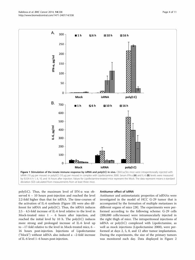
poly(I:C). Thus, the maximum level of IFN-α was ob-served 6 – 10 hours post-injection and reached the level2.2-fold higher than that for isRNA. The time-courses ofthe activation of IL-6 synthesis (Figure 1B) were also dif-ferent for isRNA and poly(I:C). Thus, the isRNA induces2.5 - 4.5-fold increase of IL-6 level relative to the level inMock-treated mice 1 – 6 hours after injection, andreached the initial level by 10 h. The poly(I:C) inducesmore strong and prolonged increase of IL-6 level upto ~17-fold relative to the level in Mock-treated mice, 6 –16 hours post-injection. Injections of Lipofectamine(“Mock”) without isRNA also induced a ~2-fold increaseof IL-6 level 1–6 hours post-injection.
Antitumor effect of isRNAAntitumor and antimetastatic properties of isRNAs wereinvestigated in the model of HCC G-29 tumor that isaccompanied by the formation of multiple metastases indifferent organs of mice [28]. The experiments were per-formed according to the following scheme: G-29 cells(200,000 cells/mouse) were intramuscularly injected inthe right thigh of mice. The intraperitoneal injections ofisRNA or poly(I:C) complexed with Lipofectamine, aswell as mock injections (Lipofectamine 2000), were per-formed at days 2, 5, 8, and 12 after tumor implantation.During the experiments, the size of the primary tumorswas monitored each day. Data displayed in Figure 2
Figure 1 Stimulation of the innate immune response by isRNA and poly(I:C) in vivo. СBA/LacSto mice were intraperitoneally injected withisRNA (10 μg per mouse) or poly(I:C) (10 μg per mouse) in complex with Lipofectamine 2000. Serum IFN-a (A) and IL-6 (B) levels were measuredby ELISA 6 h 1, 6, 10, and 16 hours after injection. Values for Lipofectamine-treated mice represent the Mock. The data represent means ± standarddeviation (SD) calculated from measurements from at least three mice.
Kabilova et al. BMC Cancer 2014, 14:338 Page 4 of 11http://www.biomedcentral.com/1471-2407/14/338
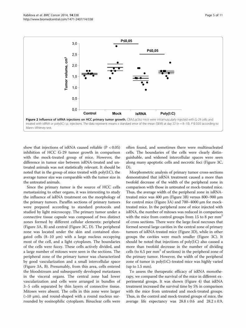
show that injections of isRNA caused reliable (Р < 0.05)inhibition of HCC G-29 tumor growth in comparisonwith the mock-treated group of mice. However, thedifference in tumor size between isRNA-treated and un-treated animals was not statistically relevant. It should benoted that in the group of mice treated with poly(I:C), theaverage tumor size was comparable with the tumor size inthe untreated animals.Since the primary tumor is the source of HCC cells
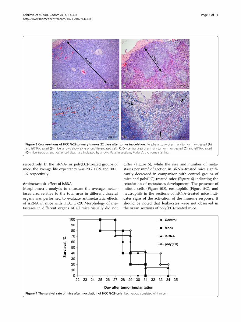
metastasizing to other organs, it was interesting to studythe influence of isRNA treatment on the morphology ofthe primary tumors. Paraffin sections of primary tumorswere prepared according to standard protocols andstudied by light microscopy. The primary tumor under aconnective tissue capsule was composed of two distinctzones formed by different cellular elements: peripheral(Figure 3A, B) and central (Figure 3C, D). The peripheralzone was located under the skin and contained elon-gated cells (8–10 μm) with a large nucleus occupyingmost of the cell, and a light cytoplasm. The boundariesof the cells were fuzzy. These cells actively divided, anda large number of mitoses were seen in the sections. Theperipheral zone of the primary tumor was characterizedby good vascularization and a small intercellular space(Figure 3A, B). Presumably, from this area, cells enteredthe bloodstream and subsequently developed metastasesin the visceral organs. The central zone had lowervascularization and cells were arranged in bundles of3–5 cells separated by thin layers of connective tissue.Mitoses were absent. The cells in this zone were larger(>10 μm), and round-shaped with a round nucleus sur-rounded by eosinophilic cytoplasm. Binuclear cells were
often found, and sometimes there were multinucleatedcells. The boundaries of the cells were clearly distin-guishable, and widened intercellular spaces were seenalong many apoptotic cells and necrotic foci (Figure 3C,D).Morphometric analysis of primary tumor cross-sections
demonstrated that isRNA treatment caused a more thantwofold decrease of the width of the peripheral zone incomparison with those in untreated or mock-treated mice.Thus, the average width of the peripheral zone in isRNA-treated mice was 400 μm (Figure 3B) versus 800–900 μmfor control mice (Figure 3A) and 700–4000 μm for mock-treated mice. In the peripheral zone of mice injected withisRNA, the number of mitoses was reduced in comparisonwith the mice from control groups from 15 to 8 per mm2
of cross sections. There were the large focal necroses thatformed several large cavities in the central zone of primarytumors of isRNA-treated mice (Figure 3D), while in othergroups the cavities were much smaller (Figure 3C). Itshould be noted that injections of poly(I:C) also caused amore than twofold decrease in the number of dividingcells (to 6.5 per mm2 of sections) in the peripheral zone ofthe primary tumor. However, the width of the peripheralzone of tumor in poly(I:C)-treated mice was highly varied(up to 1.5 mm).To assess the therapeutic efficacy of isRNA monothe-
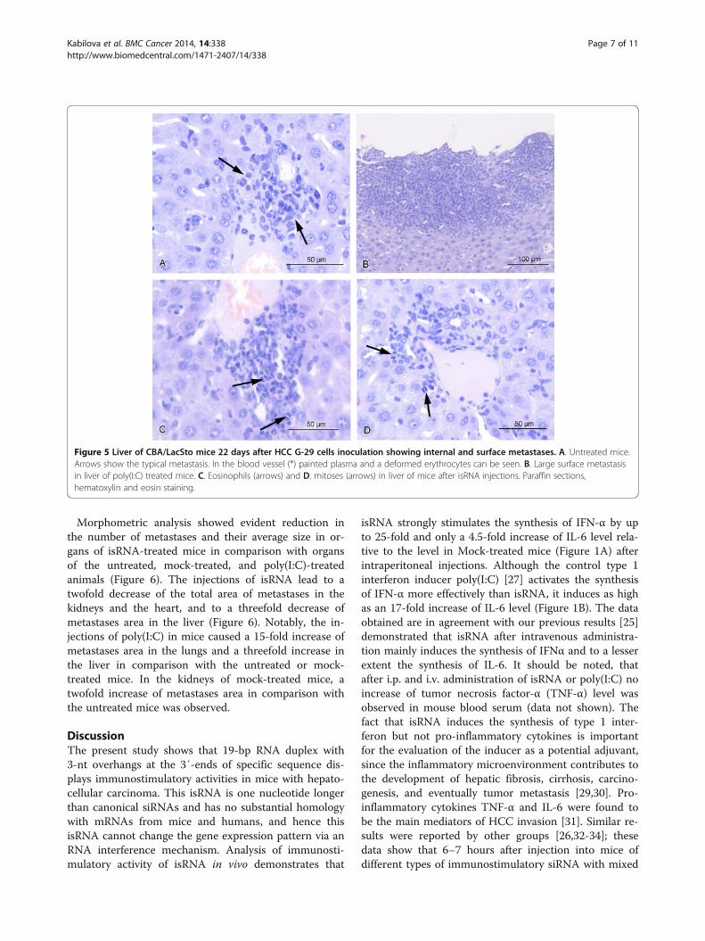
rapy, we compared the survival of the mice in different ex-perimental groups. It was shown (Figure 4) that isRNAtreatment increased the survival time by 5% in comparisonwith the mice from untreated and mock-treated groups.Thus, in the control and mock-treated groups of mice, theaverage life expectancy was 28.8 ± 0.6 and 28.2 ± 0.9,
Figure 2 Influence of isRNA injections on HCC primary tumor growth. CBA/LacSto mice were intramuscularly injected with G-29 cells andtreated with isRNA or poly(I:C) i.p. injections. The data represent means ± standard error of the mean (SEM) at day 22 (n = 8–10). P≤ 0.05 according toMann–Whitney test.
Kabilova et al. BMC Cancer 2014, 14:338 Page 5 of 11http://www.biomedcentral.com/1471-2407/14/338
respectively. In the isRNA- or poly(I:C)-treated groups ofmice, the average life expectancy was 29.7 ± 0.9 and 30 ±1.6, respectively.
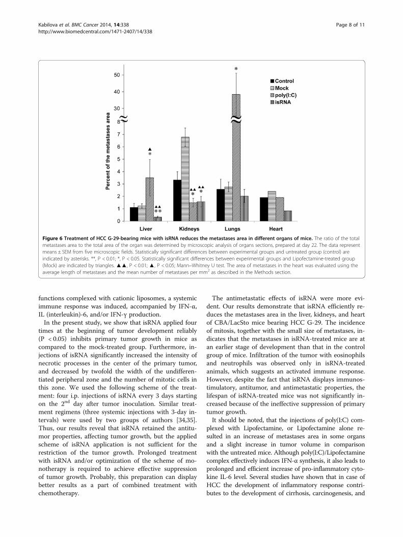
Antimetastatic effect of isRNAMorphometric analysis to measure the average metas-tases area relative to the total area in different visceralorgans was performed to evaluate antimetastatic effectsof isRNA in mice with HCC G-29. Morphology of me-tastases in different organs of all mice visually did not
differ (Figure 5), while the size and number of meta-stases per mm2 of section in isRNA-treated mice signifi-cantly decreased in comparison with control groups ofmice and poly(I:C)-treated mice (Figure 6) indicating theretardation of metastases development. The presence ofmitotic cells (Figure 5D), eosinophils (Figure 5C), andneutrophils in the sections of isRNA-treated mice indi-cates signs of the activation of the immune response. Itshould be noted that leukocytes were not observed inthe organ sections of poly(I:C)-treated mice.
Figure 3 Cross-sections of HCC G-29 primary tumors 22 days after tumor inoculation. Peripheral zone of primary tumor in untreated (A)and isRNA-treated (B) mice: arrows show zone of undifferentiated cells. C, D - central area of primary tumor in untreated (C) and isRNA-treated(D) mice: necrosis and foci of cell death are indicated by arrows. Paraffin sections, Mallory’s trichrome staining.
Figure 4 The survival rate of mice after inoculation of HCC G-29 cells. Each group consisted of 7 mice.
Kabilova et al. BMC Cancer 2014, 14:338 Page 6 of 11http://www.biomedcentral.com/1471-2407/14/338
Morphometric analysis showed evident reduction inthe number of metastases and their average size in or-gans of isRNA-treated mice in comparison with organsof the untreated, mock-treated, and poly(I:C)-treatedanimals (Figure 6). The injections of isRNA lead to atwofold decrease of the total area of metastases in thekidneys and the heart, and to a threefold decrease ofmetastases area in the liver (Figure 6). Notably, the in-jections of poly(I:C) in mice caused a 15-fold increase ofmetastases area in the lungs and a threefold increase inthe liver in comparison with the untreated or mock-treated mice. In the kidneys of mock-treated mice, atwofold increase of metastases area in comparison withthe untreated mice was observed.
DiscussionThe present study shows that 19-bp RNA duplex with3-nt overhangs at the 3′-ends of specific sequence dis-plays immunostimulatory activities in mice with hepato-cellular carcinoma. This isRNA is one nucleotide longerthan canonical siRNAs and has no substantial homologywith mRNAs from mice and humans, and hence thisisRNA cannot change the gene expression pattern via anRNA interference mechanism. Analysis of immunosti-mulatory activity of isRNA in vivo demonstrates that
isRNA strongly stimulates the synthesis of IFN-α by upto 25-fold and only a 4.5-fold increase of IL-6 level rela-tive to the level in Mock-treated mice (Figure 1A) afterintraperitoneal injections. Although the control type 1interferon inducer poly(I:C) [27] activates the synthesisof IFN-α more effectively than isRNA, it induces as highas an 17-fold increase of IL-6 level (Figure 1B). The dataobtained are in agreement with our previous results [25]demonstrated that isRNA after intravenous administra-tion mainly induces the synthesis of IFNα and to a lesserextent the synthesis of IL-6. It should be noted, thatafter i.p. and i.v. administration of isRNA or poly(I:C) noincrease of tumor necrosis factor-α (TNF-α) level wasobserved in mouse blood serum (data not shown). Thefact that isRNA induces the synthesis of type 1 inter-feron but not pro-inflammatory cytokines is importantfor the evaluation of the inducer as a potential adjuvant,since the inflammatory microenvironment contributes tothe development of hepatic fibrosis, cirrhosis, carcino-genesis, and eventually tumor metastasis [29,30]. Pro-inflammatory cytokines TNF-α and IL-6 were found tobe the main mediators of HCC invasion [31]. Similar re-sults were reported by other groups [26,32-34]; thesedata show that 6–7 hours after injection into mice ofdifferent types of immunostimulatory siRNA with mixed
Figure 5 Liver of CBA/LacSto mice 22 days after HCC G-29 cells inoculation showing internal and surface metastases. A. Untreated mice.Arrows show the typical metastasis. In the blood vessel (*) painted plasma and a deformed erythrocytes can be seen. B. Large surface metastasisin liver of poly(I:C) treated mice. C. Eosinophils (arrows) and D. mitoses (arrows) in liver of mice after isRNA injections. Paraffin sections,hematoxylin and eosin staining.
Kabilova et al. BMC Cancer 2014, 14:338 Page 7 of 11http://www.biomedcentral.com/1471-2407/14/338
functions complexed with cationic liposomes, a systemicimmune response was induced, accompanied by IFN-α,IL (interleukin)-6, and/or IFN-γ production.In the present study, we show that isRNA applied four
times at the beginning of tumor development reliably(P < 0.05) inhibits primary tumor growth in mice ascompared to the mock-treated group. Furthermore, in-jections of isRNA significantly increased the intensity ofnecrotic processes in the center of the primary tumor,and decreased by twofold the width of the undifferen-tiated peripheral zone and the number of mitotic cells inthis zone. We used the following scheme of the treat-ment: four i.p. injections of isRNA every 3 days startingon the 2nd day after tumor inoculation. Similar treat-ment regimens (three systemic injections with 3-day in-tervals) were used by two groups of authors [34,35].Thus, our results reveal that isRNA retained the antitu-mor properties, affecting tumor growth, but the appliedscheme of isRNA application is not sufficient for therestriction of the tumor growth. Prolonged treatmentwith isRNA and/or optimization of the scheme of mo-notherapy is required to achieve effective suppressionof tumor growth. Probably, this preparation can displaybetter results as a part of combined treatment withchemotherapy.
The antimetastatic effects of isRNA were more evi-dent. Our results demonstrate that isRNA efficiently re-duces the metastases area in the liver, kidneys, and heartof CBA/LacSto mice bearing HCC G-29. The incidenceof mitosis, together with the small size of metastases, in-dicates that the metastases in isRNA-treated mice are atan earlier stage of development than that in the controlgroup of mice. Infiltration of the tumor with eosinophilsand neutrophils was observed only in isRNA-treatedanimals, which suggests an activated immune response.However, despite the fact that isRNA displays immunos-timulatory, antitumor, and antimetastatic properties, thelifespan of isRNA-treated mice was not significantly in-creased because of the ineffective suppression of primarytumor growth.It should be noted, that the injections of poly(I:C) com-
plexed with Lipofectamine, or Lipofectamine alone re-sulted in an increase of metastases area in some organsand a slight increase in tumor volume in comparisonwith the untreated mice. Although poly(I:C)/Lipofectaminecomplex effectively induces IFN-α synthesis, it also leads toprolonged and efficient increase of pro-inflammatory cyto-kine IL-6 level. Several studies have shown that in case ofHCC the development of inflammatory response contri-butes to the development of cirrhosis, carcinogenesis, and
Figure 6 Treatment of HCC G-29-bearing mice with isRNA reduces the metastases area in different organs of mice. The ratio of the totalmetastases area to the total area of the organ was determined by microscopic analysis of organs sections, prepared at day 22. The data representmeans ± SEM from five microscopic fields. Statistically significant differences between experimental groups and untreated group (control) areindicated by asterisks. **, P < 0.01; *, P < 0.05. Statistically significant differences between experimental groups and Lipofectamine-treated group(Mock) are indicated by triangles. ▲▲, P < 0.01; ▲, P < 0.05; Mann–Whitney U test. The area of metastases in the heart was evaluated using theaverage length of metastases and the mean number of metastases per mm2 as described in the Methods section.
Kabilova et al. BMC Cancer 2014, 14:338 Page 8 of 11http://www.biomedcentral.com/1471-2407/14/338
eventually tumor metastasis [29,30]. Lipofectamine is alipid-based carrier and itself may also induce inflammation[36], that is far less toxic than the poly(I:C)/ Lipofectaminecomplexes, but they are not without overt toxicity.It has been shown in several studies that activation of
innate immunity by the addition of RIG-I agonist triphos-phate to the 5′-ends of oncogene-specific siRNAs [34], orby conjugation of siRNA with CpG oligonucleotide, a po-tent TLR9 agonist [37,38], can potentiate siRNA antitumoreffects. Antitumor activity is employing via the inductionof type I IFNs, the activation of tumor-resident macro-phages, and the restoration of normal immune function.In a recent study, it has been shown by Khairuddin et al.that activation of the innate immune response by siRNAshas significant antitumor effects against HPV-driven tu-mors, even in the absence of a specific gene target [35].These findings imply the potential prophylactic and thera-peutic use of immunostimulatory siRNAs as adjuvants.In the present study, we applied 19-bp RNA duplex with
3-nt overhangs at the 3′-ends in complex with Lipofecta-min for the therapy of HCC G-29 bearing mice. Based onpublished data, at least 4 signaling pathways may re-cognize small RNA molecules and induce production oftype I IFNs and pro-inflammatory cytokines, including theRIG-I/MDA5 pathway, the TLR3 pathway, the TLR7/8pathway, and the PKR pathway. RIG1 recognizes blunt-ended dsRNA molecules with a 5′-triphosphate that areover 20 bp long [39], and MDA5 is activated by longdsRNA [40]. Since our isRNA is 19 bp duplex with 3-ntoverhangs and does not contain blunt ends or triphos-phate residues, it was supposed that it activate innate im-munity trough RIG1/MDA5-independent pathway. TheisRNA under study may, on the one hand, activate PKR,leading to PKR auto phosphorylation, and on the otherhand, enter the endosome by Lipofectamine and recog-nized by TLR3, 7/8. Both the PKR activation and TLR3,7/8 signaling will stimulate production of type I IFNs andpro-inflammatory cytokines. Several studies have shownthat small RNAs complexed with cationic delivery vehiclescan stimulate the innate immune response by activatingTLR3 [41,42], TLR7/8 [26,32], and PKR [43,44], or by in-volving some of these cellular sensors [45,46].Thus, our results and results of different studies
[27,34,36,38] indicate that activation of innate immunityby immunostimulatory nucleic acids and their analogspossess antitumor and antimetastatic properties in tumor-bearing mice. These properties were proved to depend onthe type I IFN signaling pathway, since anti-metastaticproperties of the drugs were significantly decreased inmice deficient for the IFN-α/β receptor [47].
ConclusionsOur results show that 19-bp-long isRNAs with 3-ntoverhangs at the 3′-ends induce IFN-α (Figure 1), but
not proinflammatory cytokines IL-6 and TNF-α secre-tion in CBA/LacSto mice [25]. It is very important forHCC treatment to use type I IFN inducers, because inmany cases HCC develops on a background of chronicinflammatory liver disease (hepatitis B and C, cirrhosis)and secretion of pro-inflammatory cytokines could ad-ditionally complicate the course of disease. IFN therapy,however, significantly suppresses development of HCCson the background of chronic hepatitis and liver cir-rhosis [14-16]. Our data revealed that isRNA displays ef-ficient immunostimulatory and antimetastatic propertiesin mice with HCC. Moreover, isRNA treatment leads toa decrease in the pool of undifferentiated cells in HCCG-29 primary tumor nodes of mice. Thus, the studiedisRNA can be considered as a potential adjuvant for thetherapy of HCC and other immunosuppressive onco-logical diseases.
AbbreviationsHCC: Hepatocellular carcinoma; siRNAs: Small interfering RNAs;isRNA: ImmunoStimulatory RNA; IFN: Interferon; RIG-I: Retinoic acid induciblegene-I; PKR: Interferon-induced, double-stranded RNA-activated protein kinase;IP-10: IFN-γ-inducible protein 10; PBMCs: Peripheral blood mononuclear cells.
Competing interestsThe authors declare that they have no competing interests.
Authors’ contributionsConceived and designed the experiments: TK, EC. Performed theexperiments: TK, LK, NP, VN, VK. Analyzed the data: TK, EZ, ER, EC. Performedmicroscopic analysis: EZ, ER. Wrote the paper: TK, EC. Revised the manuscript:EC, MZ. VV participated in discussion and data interpretation. All authors readand approved the final manuscript.
AcknowledgmentsThe authors thank Dr. Alya G. Venyaminova and Dr. Mariya I. Meschaninova(Institute of Chemical Biology and Fundamental Medicine SB RAS) for thesynthesis of oligoribonucleotides used in the work, and Mrs. Albina V.Vladimirova (Institute of Chemical Biology and Fundamental Medicine SBRAS) for the cell maintenance.This research was supported by the Russian Academy of Science under theprogram “Molecular and Cell Biology” (no. 22–1), and “Science to Medicine”(no. 44); Russian Foundation for Basic Research (no. 14-04-00869-a, 13-04-40181); the Ministry of Science and Education of RF (no. 14.740.11.1058); SBRAS (no. 85), and President’s scholarship for young scientists (RF).
Author details1Institute of Chemical Biology and Fundamental Medicine SB RAS, 8,Lavrentiev Avenue, Novosibirsk 630090, Russia. 2Institute of Cytology andGenetics SB RAS, 10, Lavrentiev Avenue, Novosibirsk 630090, Russia.
Received: 3 December 2013 Accepted: 12 May 2014Published: 18 May 2014
References1. Schütte K, Bornschein J, Malfertheiner P: Hepatocellular carcinoma–
epidemiological trends and risk factors. Dig Dis 2009, 27:80–92.2. Makuuchi M, Kosuge T, Takayama T, Yamazaki S, Kakazu T, Miyagawa S,
Kawasaki S: Surgery for small liver cancers. Semin Surg Oncol 1993,9:298–304.
3. Livragh T, Lazzaroni S, Pellicano S, Ravasi S, Torzilli G, Vettori C:Percutaneous ethanol injection of hepatic tumours: single-sessiontherapy with general anesthesia. AJR Am J Roentgenol 1993,161:1065–1069.
Kabilova et al. BMC Cancer 2014, 14:338 Page 9 of 11http://www.biomedcentral.com/1471-2407/14/338
4. Allgaier HP, Deibert P, Zuber I, Olschewski M, Blum HE: Percutaneousradiofrequency interstitial thermal ablation of small hepatocellularcarcinoma. Lancet 1999, 353:1676–1677.
5. Arii S, Yamaoka Y, Futagawa S, Inoue K, Kobayashi K, Kojiro M, Makuuchi M,Nakamura Y, Okita K, Yamada R: Results of surgical and nonsurgicaltreatment for small-sized hepatocellular carcinomas: a retrospective andnationwide survey in Japan. Hepatology 2000, 32:1224–1229.
6. Adams S: Toll-like receptor agonists in cancer therapy. Immunotherapy2009, 1:949–964.
7. Gold JE, Masters TR, Osband ME: Autolymphocyte therapy III effectiveadjuvant adoptive cellular therapy with in vivo anti-tumor specificityagainst murine melanoma and carcinoma using ex-vivo-activatedmemory T-lymphocytes. J Surg Res 1995, 59:279–286.
8. Kantoff PW, Higano CS, Shore ND, Berger ER, Small EJ, Penson DF, RedfernCH, Ferrari AC, Dreicer R, Sims RB, Xu Y, Frohlich MW, Schellhammer PF:Sipuleucel-T immunotherapy for castration-resistant prostate cancer.N Engl J Med 2010, 363:411–422.
9. Waldmann TA: Immunotherapy: past, present and future. Nat Med 2003,9:269–277.
10. Unitt E, Marshall A, Gelson W, Rushbrook SM, Davies S, Vowler SL, Morris LS,Coleman N, Alexander GJ: Tumour lymphocytic infiltrate and recurrenceof hepatocellular carcinoma following liver transplantation. J Hepatol2006, 45:246–253.
11. Cui J, Wang N, Zhao H, Jin H, Wang G, Niu C, Terunuma H, He H, Li W:Combination of radiofrequency ablation and sequential cellularimmunotherapy improves progression-free survival for patients withhepatocellular carcinoma. Int J Cancer 2014, 134:342–351.
12. Ishikawa T: Secondary prevention of recurrence by interferon therapyafter ablation therapy for hepatocellular carcinoma in chronic hepatitis Cpatients. World J Gastroenterol 2008, 14:6140–6144.
13. Mencin A, Kluwe J, Schwabe RF: Toll-like receptors as targets in chronicliver diseases. Gut 2009, 58:704–720.
14. Ishikawa T, Ichida T: Features of hepatitis B virus-related hepatocellularcarcinoma. Nihon Rinsho 2001, 59:435–439.
15. Nishiguchi S, Kuroki T, Nakatani S, Morimoto H, Takeda T, Nakajima S,Shiomi S, Seki S, Kobayashi K, Otani S: Randomised trial of effects ofinterferon-alpha on incidence of hepatocellular carcinoma in chronicactive hepatitis C with cirrhosis. Lancet 1995, 346:1051–1055.
16. Nishiguchi S, Shiomi S, Nakatani S, Takeda T, Fukuda K, Tamori A, Habu D,Tanaka T: Prevention of hepatocellular carcinoma in patients withchronic active hepatitis C and cirrhosis. Lancet 2001, 357:196–197.
17. Olejniczak M, Galka-Marciniak P, Polak K, Fligier A, Krzyzosiak WJ: RNAimmuno:a database of the nonspecific immunological effects of RNA interferenceand microRNA reagents. RNA 2012, 18:930–935.
18. Desmet CJ, Ishii KJ: Nucleic acid sensing at the interface betweeninnate and adaptive immunity in vaccination. Nat Rev Immunol 2012,12:479–491.
19. El-Omar EM, Ng MT, Hold GL: Polymorphisms in Toll-like receptor genesand risk of cancer. Oncogene 2008, 27:244–252.
20. McHutchison JG, Bacon BR, Gordon SC, Lawitz E, Shiffman M, Afdhal NH,Jacobson IM, Muir A, Al-Adhami M, Morris ML, Lekstrom-himes JA, Efler SM,Davis HL: Phase 1B, randomized, double-blind, dose-escalation trial ofCPG 10101 in patients with chronic hepatitis C virus. Hepatology 2007,46:1341–1349.
21. Horsmans Y, Berg T, Desager JP, Mueller T, Schott E, Fletcher SP, Steffy KR,Bauman LA, Kerr BM, Averett DR: Isatoribine, an agonist of TLR7, reducesplasma virus concentration in chronic hepatitis C infection. Hepatology2005, 42:724–731.
22. Xiang AX, Webber SE, Kerr BM, Rueden EJ, Lennox JR, Haley GJ, Wang T,Ng JS, Herbert MR, Clark DL, Banh VN, Li W, Fletcher SP, Steffy KR,Bartkowski DM, Kirovsky LI, Bauman LA, Averett DR: Discovery of ANA975:an oral prodrug of the TLR-7 agonist isatoribine. Nucleosides NucleotidesNucleic Acids 2007, 26:635–640.
23. Pappas SC: Good science behind hepatitis C virus antiviral drugdevelopment: necessary but not sufficient. Hepatology 2010,51:1897–1903.
24. Kabilova TO, Vladimirova AV, Zenkova MA, Chernolovskaya EL, Vlassov VV:Antiproliferative and interferon-inducing activities of unique shortdouble-stranded RNA. Dokl Biochem Biophys 2011, 436:8–11.
25. Kabilova TO, Meschaninova MI, Venyaminova AG, Nikolin VP, Zenkova MA,Vlassov VV, Chernolovskaya EL: Short double-stranded RNA with
immunostimulatory activity: sequence dependence. Nucleic Acid Ther2012, 22:196–204.
26. Judge AD, Sood V, Shaw JR, Fang D, McClintock K, Maclachlan I: Sequence-dependent stimulation of the mammalian innate immune response bysynthetic siRNA. Nat Biotechnol 2005, 23:457–462.
27. Currie AJ, van der Most RG, Broomfield SA, Prosser AC, Tovey MG, RobinsonBW: Targeting the effector site with IFN-alphabeta-inducing TLR ligandsreactivates tumor-resident CD8 T cell responses to eradicate establishedsolid tumors. J Immunol 2008, 180:1535–1544.
28. Kaledin VI, Zhukova NA, Nikolin VP, Popova NA, Beliaev MD, Baginskaya NV,Litvinova EA, Tolstikova TG, Lushnikova EL, Semenov DE: Hepatocarcinoma-29, a metastasizing transplantable mouse tumor inducing cachexia.Bull Exp Biol Med 2009, 148:903–908.
29. Wang H, Chen L: Tumor microenviroment and hepatocellular carcinomametastasis. J Gastroenterol Hepatol 2013, 28:43–48.
30. Seki S, Nakashima H, Nakashima M, Kinoshita M: Antitumor immunityproduced by the Liver Kupffer Cells, NK Cells, NKT Cells, and CD8+CD122+ TCells. Clin Dev Immunol 2011, 2011:868345.
31. Wang YC, Xu GL, Jia WD, Han SJ, Ren WH, Wang W, Liu WB, Zhang CH,Chen H: Estrogen suppresses metastasis in rat hepatocellular carcinomathrough decreasing interleukin-6 and hepatocyte growth factorexpression. Inflammation 2012, 35:143–149.
32. Hornung V, Guenthner-Biller M, Bourquin C, Ablasser A, Schlee M, Uematsu S,Noronha A, Manoharan M, Akira S, De FA, Endres S, Hartmann G: Sequence-specific potent induction of IFN-alpha by short interfering RNA inplasmacytoid dendritic cells through TLR7. Nat Med 2005, 11:263–270.
33. Shin D, Kim SI, Park M, Kim M: Immunostimulatory properties and antiviralactivity of modified HBV-specific siRNAs. Biochem Biophys Res Commun2007, 364:436–442.
34. Poeck H, Besch R, Maihoefer C, Renn M, Tormo D, Morskaya SS, Kirschnek S,Gaffal E, Landsberg J, Hellmuth J, Schmidt A, Anz D, Bscheider M, SchwerdT, Berking C, Bourquin C, Kalinke U, Kremmer E, Kato H, Akira S, Meyers R,Hacker G, Neuenhahn M, Busch D, Ruland J, Rothenfusser S, Prinz M,Hornung V, Endres S, Tuting T, et al: 5′-Triphosphate-siRNA: turning genesilencing and Rig-I activation against melanoma. Nat Med 2008,14:1256–1263.
35. Khairuddin N, Gantier MP, Blake SJ, Wu SY, Behlke MA, Williams BR, McMillanNA: siRNA-induced immunostimulation through TLR7 promotesantitumoral activity against HPV-driven tumors in vivo. Immunol Cell Biol2012, 90:187–196.
36. Tseng YC, Mozumdar S, Huang L: Lipid-based systemic delivery of siRNA.Adv Drug Deliv Rev 2009, 61:721–731.
37. Kortylewski M, Swiderski P, Herrmann A, Wang L, Kowolik C, Kujawski M, LeeH, Scuto A, Liu Y, Yang C, Deng J, Soifer HS, Raubitschek A, Forman S, RossiJJ, Pardoll DM, Jove R, Yu H: In vivo delivery of siRNA to immune cells byconjugation to a TLR9 agonist enhances antitumor immune responses.Nat Biotechnol 2009, 27:925–932.
38. Zhang Q, Hossain DM, Nechaev S, Kozlowska A, Zhang W, Liu Y, KowolikCM, Swiderski P, Rossi JJ, Forman S, Pal S, Bhatia R, Raubitschek A, Yu H,Kortylewski M: TLR9-mediated siRNA delivery for targeting of normal andmalignant human hematopoietic cells in vivo. Blood 2013, 121:1304–1315.
39. Schlee M, Hartmann G: The chase for the RIG-I ligand–recent advances.Mol Ther 2010, 18:1254–1262.
40. Kang DC, Gopalkrishnan RV, Wu Q, Jankowsky E, Pyle AM, Fisher PB: mda-5:An interferon-inducible putative RNA helicase with double-strandedRNA-dependent ATPase activity and melanoma growth-suppressiveproperties. Proc Natl Acad Sci U S A 2002, 99:637–642.
41. Karikó K, Bhuyan P, Capodici J, Ni H, Lubinski J, Friedman H, Weissman D:Exogenous siRNA mediates sequence-independent gene suppression bysignaling through toll-like receptor 3. Cells Tissues Organs 2004,177:132–138.
42. Reynolds A, Anderson EM, Vermeulen A, Fedorov Y, Robinson K, Leake D,Karpilow J, Marshall WS, Khvorova A: Induction of the interferonresponse by siRNA is cell type- and duplex length-dependent. RNA 2006,12:988–993.
43. Puthenveetil S, Whitby L, Ren J, Kelnar K, Krebs JF, Beal PA: Controllingactivation of the RNA-dependent protein kinase by siRNAs usingsite-specific chemical modification. Nucleic Acids Res 2006, 34:4900–4911.
44. Zhang Z, Weinschenk T, Guo K, Schluesener HJ: siRNA binding proteins ofmicroglial cells: PKR is an unanticipated ligand. J Cell Biochem 2006,97:1217–1229.
Kabilova et al. BMC Cancer 2014, 14:338 Page 10 of 11http://www.biomedcentral.com/1471-2407/14/338
45. Yang C, Li L, Xue Y, Zhao Z, Zhao T, Jia Y, Rong R, Xu M, Nicholson ML,Zhu T, Yang B: Innate immunity activation involved in unprotectedporcine auto-transplant kidneys preserved by naked caspase-3 siRNA.J Transl Med 2013, 11:210.
46. Meng Z, Zhang X, Wu J, Pei R, Xu Y, Yang D, Roggendorf M, Lu M:RNAi induces innate immunity through multiple cellular signalingpathways. PLoS One 2013, 8:e64708.
47. Hafner M, Zawatzky R, Hirtreiter C, Buurman WA, Echtenacher B, Hehlgans T,Mannel DN: Antimetastatic effect of CpG DNA Mediated by Type I IFN.Cancer Res 2001, 61:5523–5528.
doi:10.1186/1471-2407-14-338Cite this article as: Kabilova et al.: Immunotherapy of hepatocellularcarcinoma with small double-stranded RNA. BMC Cancer 2014 14:338.
Submit your next manuscript to BioMed Centraland take full advantage of:
• Convenient online submission
• Thorough peer review
• No space constraints or color figure charges
• Immediate publication on acceptance
• Inclusion in PubMed, CAS, Scopus and Google Scholar
• Research which is freely available for redistribution
Submit your manuscript at www.biomedcentral.com/submit
Kabilova et al. BMC Cancer 2014, 14:338 Page 11 of 11http://www.biomedcentral.com/1471-2407/14/338