Embed Size (px)
Citation preview
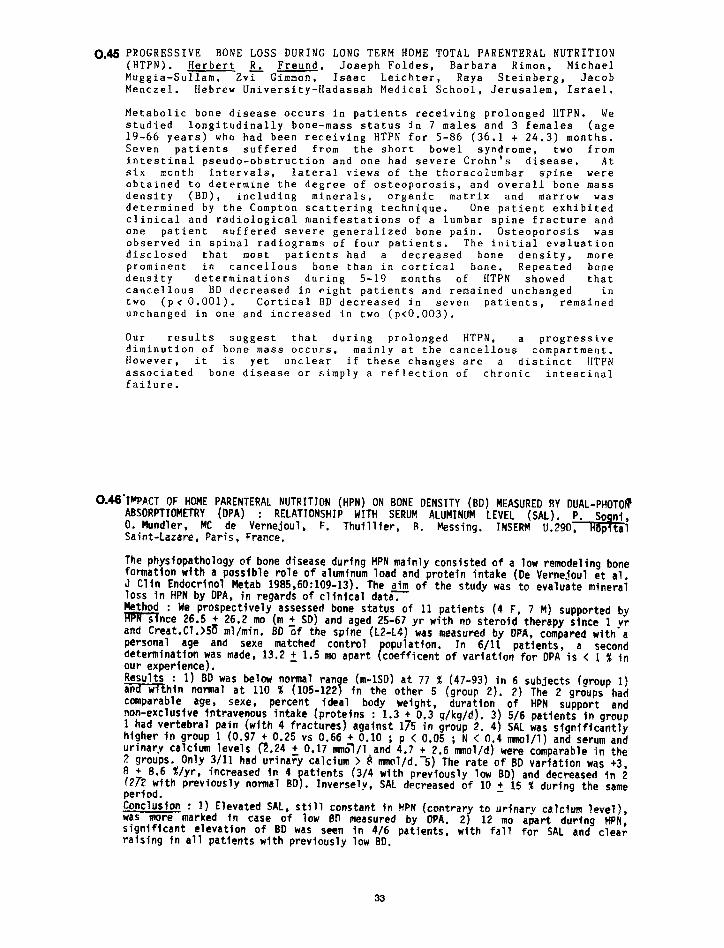
0.46 PROGRESSIVE BONE LOSS DURING LONG TERM HOME TOTAL PARENTERAL NUTRITION (HTPN). Herbert R_ Freund, Joseph Foldes, Barbara Rimon, Michael
Muggia-Sullam, Zvi Gimmon, Isaac Leichter, Raya Steinberg+ Jacob
Menczel. Hebrew University-Hadassah Medical School, Jerusalem, Israel.
Metabolic bone disease occurs in patients receiving prolonged HTPN. We studied longitudinally bone-mass status in 7 males and 3 females (age 19-66 years) who had been receiving HTPN for 5-86 (36.1 t 24.3) months. Seven patients suffered from the short bowel syndrome, two from intestinal pseudo-obstruction and one had severe Crohn’s disease. At six month intervals, lateral views of the thoracolumbar spine were obtained to determine the degree of osteoporosis, and overall bone mass density (BD), including minerals, organic matrix and marrow was determined by the Compton scattering technique. One patient exhibited clinical and radiological manifestations of a lumbar spine fracture and one patient suffered severe generalized bone pain. Osteoporosis was observed in spinal radiograms of four patients. The initial evaluation disclosed that most patients had a decreased bone density, more prominent in cancellous bone than in cortical bone. Repeated bone density determinations during 5-19 months of HTPN showed that cancellous BD decreased in eight patients and remained unchanged in two (pcO.001). Cortical BD decreased in seven patients, remained unchanged in one and increased in two (ptO.003).
Our results suggest that during diminution of bone-mass occurs,
prolonged HTPN. a mainly at the cancellous
oronressive . I compartment.
Elowever, it is yet unclear if these changes are a associated bone disease or simply a reflection of
distinct HTPN chronic intestinal
failure.
0.46’w~~cT OF HOME PARENTEML MJTRITICTN (HPN) ON BONE DENSITY (BD) MEASURED BY DUAL-PHOTOS ABSORPTIOMETRY (DPA) 0. Mundler,
: RELATIONSHIP WITH SERUM ALUMINUM LEVEL (SAL), MC de Vernejoul, F. Thuillier, R. Messing. INSERM U.290,
Saint-Lazare, Paris, France.
The physiopathology of bone disease during HPN mainly consisted of a low remodeling bone formation with a possible role of aluminum load and protein intake (De Vernejoul et al J Clin Endocrinol Hetab 1985,60:109-13). The aim of the study was to evaluate mineral loss in HPN by DPA. in regards of clinical data. Method : We prospectively assessed bone status of 11 patients (4 F, 7 M) supported by mnce 26.5 + 26.2 ma (m + SD) and aged 25-67 yr with no steroid therapy stnce 1 vr and Creat.C?.>Su ml/min. BD zf the spine (L2-L4) was measured by DPA, compared with-a personal age and sexe matched control
P opulation. In 6/11 patients, a second
determination was made, 13.2 + 1.5 mo apart coefficent of variation for DPA is < 1 % in our experience). Results : 1) BD was below normal andwithin normal at 110 %
ran e (105-122 s
(m-1SD) at 77 % (47-93) in 6 subjects (aroup 1)
comparable age, sexe, percent ideal in the other 5 (group 2). 2) The 2 groups had
body weight, non-exclusive intravenous intake (proteins
duration of HPN support and : 1.3 + 0.3 g/kg/d). 3) 5/6 patients in group
1 had vertebral pain (with 4 fractures) against 175 in group 2. 4) SAL was significantly higher in group 1 (0.97 + 0.25 vs 0.66 + 0.10 ; p < 0.05 ; N < 0.4 mnol/l) and serum and urinary calcium levels &.24 + 0.17 mnGl/l and 4.7 t 2.6 mnol/d) were comparable in the 2 groups. Only 3/I1 had urinary calcium > B ml/d.>) The rate of BD variation was t3 8 + 8.6 Xlyr, increased in 4 (Zh with previously normal
atfents BD 9
(3/4 with previously low BD) and decreased in i .
period. Inversely, SAL decreased of 10 + 15 % during the same
Conclusion : 1) Elevated SAL, still constant in HPN (contrar was more marked fn case of low BD measured by DPA. 2 r
to urinarv calcium level),
significant elevation of 8D was seen in 4/6 patients, 12 mo apart during HPN,
raising in all patients with previously low BD. with fall for SAL and clear
33