Embed Size (px)
Citation preview

Impact of meditation training on the defaultmode network during a restful stateVeronique A. Taylor,1 Veronique Daneault,1 Joshua Grant,1,2 Genevieve Scavone,1 Estelle Breton,1
Sebastien Roffe-Vidal,1 Jerome Courtemanche,1 Anaıs S. Lavarenne,1 Guillaume Marrelec,5,6,7
Habib Benali,5,6,7 and Mario Beauregard1,2,3,4
1Departement de Psychologie, Centre de Recherche en Neuropsychologie et Cognition (CERNEC), 2 Departement de Physiologie, Centre de
Recherche en Sciences Neurologiques, 3Departement de Radiologie, Universite de Montreal, 4Centre de recherche du Centre hospitalier de
l’Universite de Montreal (CRCHUM), Montreal, Quebec, Canada, 5Inserm, U678, Laboratoire d’Imagerie Fonctionnelle, F-75013 Paris,
France, 6UPMC Univ Paris 06, UMR-S U678, Paris, F-75013, France-Inserm, and 7Universite de Montreal, LINeM, Montreal, Quebec,
Canada
Mindfulness meditation has been shown to promote emotional stability. Moreover, during the processing of aversive andself-referential stimuli, mindful awareness is associated with reduced medial prefrontal cortex (MPFC) activity, a central defaultmode network (DMN) component. However, it remains unclear whether mindfulness practice influences functional connectivitybetween DMN regions and, if so, whether such impact persists beyond a state of meditation. Consequently, this study examinedthe effect of extensive mindfulness training on functional connectivity within the DMN during a restful state. Resting-state datawere collected from 13 experienced meditators (with over 1000 h of training) and 11 beginner meditators (with no prior experi-ence, trained for 1 week before the study) using functional magnetic resonance imaging (fMRI). Pairwise correlations and partialcorrelations were computed between DMN seed regions� time courses and were compared between groups utilizing a Bayesiansampling scheme. Relative to beginners, experienced meditators had weaker functional connectivity between DMN regionsinvolved in self-referential processing and emotional appraisal. In addition, experienced meditators had increased connectivitybetween certain DMN regions (e.g. dorso-medial PFC and right inferior parietal lobule), compared to beginner meditators. Thesefindings suggest that meditation training leads to functional connectivity changes between core DMN regions possibly reflectingstrengthened present-moment awareness.
Keywords: mindfulness meditation; functional connectivity; default mode network; prefrontal cortex; resting state
INTRODUCTIONOriginating from Ancient Eastern traditions, meditation has
become increasingly studied with brain mapping methods.
Particularly, there is evidence that mindfulness meditation is
beneficial for the treatment of psychological disorders invol-
ving emotional dysregulation, such as major depressive
disorder (MDD) and anxiety disorders (Baer, 2003).
Mindfulness promotes an objective manner of interpreting
thoughts, events and emotions, without elaborating or
‘ruminating’ on their potential implications for the self
(Bishop, 2004).
Mindfulness has been shown to diminish activity in the
medial prefrontal cortex (MPFC; Farb et al., 2007), a cortical
area playing a pivotal role in self-referential processing and
the ‘default mode network’ (DMN) (Gusnard et al., 2001).
Indeed, Farb et al. (2007) recently showed that an
experiential focus condition, involving the mindful monitor-
ing of present-moment circumstances, was associated with
decreased MPFC function (dorso-medial PFC [DMPFC],
ventro-medial PFC [VMPFC]), compared with a narrative
focus condition (i.e. monitoring self-descriptive traits).
These researchers also found that the MPFC deactivations
associated with experiential focus were more pronounced in
participants having received an 8-week mindfulness-based
stress-reduction (MBSR) program, relative to a wait-listed
control group.
There is also evidence supporting the view that mindful-
ness training leads to MPFC deactivations during the pro-
cessing of aversive stimuli, such as painful stimulations
(Grant et al., 2011). Moreover, individuals with MDD fail
to deactivate the MPFC (and other cerebral structures in the
DMN) while passively looking at negative pictures or trying
to reappraise them (Sheline et al., 2009). These findings may
reflect a failure to down-regulate DMN regions involved in
monitoring of internal emotional states, self-referential pro-
cessing and rumination.
Other core regions in the DMN�which are consistently
deactivated during goal-directed tasks and activated during a
restful state�include the inferior parietal lobule (IPL), the
Received 15 June 2011; Accepted 13 November 2011
Advance Access publication 24 March 2012
The authors thank the staff of the Unite de Neuroimagerie Fonctionnelle (UNF), Institut universitaire de
geriatrie de Montreal (IUGM), for their skilful technical assistance. This study was supported by a grant from
the Natural Sciences and Engineering Research Council of Canada (NSERC) (M.B.).
Correspondence should be addressed to Mario Beauregard, PhD, Departement de Psychologie,
Mind/Brain Research Lab (MBRL), Centre de Recherche en Neuropsychologie et Cognition (CERNEC),
Universite de Montreal, C.P. 6128, succursale Centre-Ville, Montreal, Quebec, Canada, H3C 3J7.
E-mail: [email protected]
doi:10.1093/scan/nsr087 SCAN (2013) 8, 4^14
� The Author (2012). Published by Oxford University Press. For Permissions, please email: [email protected]
at MPI C
ognitive and Brain Science on January 28, 2013
http://scan.oxfordjournals.org/D
ownloaded from

precuneus (PC), the posterior cingulate cortex (PCC), as
well as the inferolateral temporal cortex (ITC) (Gusnard
et al., 2001; Raichle et al., 2001; Greicius et al., 2003;
Buckner et al., 2008). These brain regions are thought to
operate as a coherent network, as they exhibit synchronized
low-frequency blood-oxygen-level-dependent (BOLD) signal
fluctuations during ‘resting states’, i.e. when participants are
scanned for several minutes and are instructed to rest with-
out engaging in any specific mental activity or task (Biswal
et al., 1995; Beckmann and Smith, 2004; Damoiseaux et al.,
2006; De Luca et al., 2006; Fransson and Marrelec, 2008).
DMN activity has been proposed to be associated with cog-
nitive processes such as envisioning future scenarios, theory
of mind, autobiographical memory, moral decision making
and self-referential processing (Gusnard et al., 2001;
Northoff et al., 2006; Buckner et al., 2008). It therefore
appears that this network may underlie adaptive planning
and reflection mechanisms when not engaged in any external
activity (Buckner et al., 2008).
Examining the relationship between meditation training
and functional connectivity within the DMN during rest is
important to determine whether the effects of such training
extend beyond a meditative state. Functional connectivity
within the DMN during a state of rest has been examined
between an inexperienced control group and experienced
meditation practitioners of a specific form of meditation
called ‘Brain-wave vibration meditation’ (a kind of moving
meditation that is designed to help quiet the thinking mind
and to release negative emotions through performing natural
rhythmic movements and focusing on bodily sensations
(Jang et al., 2011)) and an inexperienced control group.
Their results revealed that meditation practitioners had
increased functional connectivity within the DMN in the
MPFC relative to controls (Jang et al., 2011). Nevertheless,
the results from Jang et al. (2011) were obtained using
seed-based functional connectivity analyses with anatomic-
ally pre-determined seed locations. To date, however,
the relationship between mindfulness meditation training
and connectivity within DMN regions has not been exam-
ined using data-driven independent component analysis
techniques to identify intrinsic connectivity network maps
at the group level, and then examining group differences
in pairwise connections between regions of the identified
network. In this context, the aim of this functional magnet-
ic resonance imaging (fMRI) study was to investigate the
impact of extensive mindfulness training on functional
connectivity between regions of the DMN during a restful
state. Spatial independent component analysis was used to
identify a DMN map at the group level and determine seed
regions. Pairwise correlations and partial correlations were
then computed between the time courses of these DMN seed
regions. The various correlations between all pairs of
nodes were then compared between individuals highly
experienced in meditation (with over 1000 h of experience
in mindfulness-based meditation) relative to beginner
meditators (with no prior exposure to mindfulness medita-
tion, trained for one week before the completion of the
study). Predicated on evidence that mindfulness is associated
with deactivation of MPFC during self-referential processing
(Farb et al., 2007), it was hypothesized that functional con-
nectivity between medial prefrontal cortical areas and other
DMN regions would be weaker in experienced relative to
beginner meditators.
MATERIALS AND METHODSParticipantsThe sample consisted of two groups. The first group was
composed of 13 (six males) experienced meditators
(age: M¼ 46 years, s.d.¼ 11) with over 1000 h of meditation
experience (M¼ 6519, s.d.¼ 14 445). They were recruited
from Zen meditation centres in the Montreal metropolitan
area. One participant deviated from the group in terms of
the number of hours of meditation (45 000 h of practice),
and hence constituted an outlier from the rest of the group.
Without the inclusion of this participant, the group of
experienced meditators had on average 1709 h of meditation
practice (s.d.¼ 694). Consequently, the analyses reported in
the present study were also conducted with the exclusion of
this participant. Since the results remained essentially un-
changed, this participant was kept in the analyses to avoid
losing any statistical power by decreasing the sample size.
All experienced meditators reported that the core approach
of their regular meditation practice consisted in the mind-
fulness practice technique.
The second group was composed of 11 beginner medita-
tors (seven males), with an average of 37 years of age
(s.d.¼ 13) recruited with the use of advertizement posters
placed at the Universite de Montreal and the Centre de
Recherche de l’Institut Geriatrique de Montreal
(CRIUGM). These individuals had no prior exposure to
meditation (or other practices such as yoga) and were
trained for one week before the completion of the study.
Implemented by the experimenters, the mindfulness training
was documented from several sources (Kabat-Zinn, 1994;
Thich Nhat Hanh, 1994; Ricard, 2008) and consisted of a
guided meditation session recorded on a compact disc
(a written record was also provided). Participants were in-
structed to meditate for 20 min each day for 7 days. The
experimenters followed-up throughout the training week
to ensure that participants completed their practice, and
they all confirmed to have understood and successfully com-
pleted their training (except for one participant who had
2 days of training due to scheduling constraints, but con-
firmed having practiced the compensating number of
hours). There were no significant differences in the
male-to-female ratio or age between the group of experi-
enced meditators and the group of beginners. Participant
characteristics are presented in Table 1.
Before being selected to participate in the study, all par-
ticipants underwent telephone screening, and were excluded
Mindfulness and defaultmode network connectivity SCAN (2013) 5
at MPI C
ognitive and Brain Science on January 28, 2013
http://scan.oxfordjournals.org/D
ownloaded from

if they had a current or past mental health illness, took any
psychotropic drugs, were suffering from major physical
health problems or were not eligible to undergo an MRI
exam (pregnancy, metal parts in body, pacemaker, etc.).
After the completion of the study, participants were com-
pensated 50$ for their time. This research project was
approved by the Ethics Research Committee of the
CRIUGM.
fMRI data acquisitionWhole-brain T2*-weighted functional images were acquired
from a 3 T scanner (Siemens, Erlanger, Germany) using a
two-dimensional echo-planar imaging pulse sequence
(TR¼ 2500 ms, TE¼ 40 ms, voxel size¼ 3� 3� 3.5 mm, 35
contiguous axial slices, no gap between slice acquisition,
matrix size¼ 64� 64, flip angle¼ 908). A high-resolution
T1-weighted anatomical scan was also acquired for each sub-
ject (three-dimensional, spoiled gradient echo sequence,
TR¼ 19 ms, TE¼ 4.92 ms, flip angle¼ 258, matrix
size¼ 256� 256 voxels voxel size¼ 1� 1� 1 mm, 176 con-
tiguous axial slices).
Experimental protocolParticipants first gave informed consent and underwent
screening and questionnaires related to MRI safety and eli-
gibility. Then, to obtain sufficient power for the functional
connectivity analyses given the relatively small sample size,
each participant completed two functional 6-min runs
(144 volumes) in a state of rest (except for two participants
from each group who completed only one run, due to time
or testing constraints). Throughout these runs, participants
observed a cross fixated centrally on the screen inside the
scanner, and were instructed to rest, without engaging in any
specific task or mental activity. In order not to induce bore-
dom or other carry-over effects by performing both resting
state sessions consecutively, and given that connectivity
within the default mode network has been shown to be con-
sistently reproducible across time (Shehzad et al., 2009;
Meindl et al., 2010; Zuo et al., 2010), resting state sessions
were performed at the beginning and at the end of the
experimental paradigm. In between these two sessions, par-
ticipants also completed sessions consisting of different
experimental paradigms; these data are not reported here.
At the end of the experiment, participants were compensated
for their time.
Data analysisThe fMRI data were pre-processed using SPM8 (Wellcome
Department of Cognitive Neurology, London, UK).
For each subject, functional images were slice-time cor-
rected, realigned and spatially smoothed using a Gaussian
kernel (8 mm at full-width at half-maximum). Next, func-
tional connectivity analyses were conducted using the
NetBrainWork software (https://sites.google.com/site/net-
brainwork/, Laboratoire d’Imagerie Fonctionnelle, Paris,
France). To identify functional network maps across partici-
pants, the NEDICA (for Network Detection using
Independent Component Analysis; Perlbarg et al., 2008)
approach was employed. First, as previously validated
(Esposito et al., 2005) the data for each run were reduced
to 40 temporal dimensions using principal component ana-
lysis (PCA). Next, 40 spatially independent components were
extracted from each run using the infomax algorithm (Bell
and Sejnowski, 1995). Independent component (IC) maps
were then converted into z-maps and normalized into MNI
standard stereotaxic space.
After the ICs were extracted at the individual level for each
run, a hierarchical clustering algorithm (Marrelec et al.,
2008) was used to gather all ICs from all runs across both
groups of participants into clusters or ‘classes’ based on their
spatial similarity, the distance between two ICs being taken
as their spatial correlation. The number of classes was calcu-
lated automatically by NetBrainWork as a way to optimize
both the degree of representativity (DR; number of runs
contributing to the class divided by the total number of
runs) and the degree of Unicity (DU; number of runs con-
tributing to class with only one IC, divided by the total
number of runs). As these scores should ideally be equal to
1, only classes with DR > 0.5 and DU > 0.75 were retained
(Perlbarg et al., 2008).
Then, fixed effect analyses were conducted to compute t-
maps for each class: at each voxel, the mean value of each IC
contributing to the class was divided by the variance of each
IC contributing to the class. The resulting t-maps were
thresholded at P < 0.05 corrected at the false discovery rate
(FDR). Finally, a bootstrap procedure was conducted to
assess the confidence interval of each class retained. To do
this, NEDICA was reapplied on half of the runs (randomly
selected), yielding new group maps. The spatial correlation
between each initial group map and new group map was
calculated. The initial map with the highest correlation co-
efficient with its new bootstrap map was retained (except if
the correlation value was below 0.30). As a result of this
bootstrap procedure, which was repeated 100 times, a
number of t-maps for the classes of interest were retained.
The remaining classes represented functionally coherent
brain networks across the entire sample.
Table 1 Participant characteristics
Experienced meditators Beginners
Age (years) 46� 11 years 37� 13 yearsGender 6 M; 7 F 7 M; 4 FEducation 12 undergraduate university
degree or higher; 1 high schooleducation
9 undergraduate universitydegree or higher; 2 high schooleducation
Ethnicity 11 Caucasian; 1 Asian;1 multi-racial (African andEuropean descent)
9 Caucasian; 2 multi-racial(African and European descent)
6 SCAN (2013) V.Taylor et al.
at MPI C
ognitive and Brain Science on January 28, 2013
http://scan.oxfordjournals.org/D
ownloaded from

Next, each network map was manually inspected. Based
on previous reports (Buckner et al., 2008; Perlbarg et al.,
2008), the map which best corresponded to the DMN was
selected for the functional connectivity analyses. Regions of
interest (ROIs) were selected based on the peak voxels iden-
tified in the DMN t-map. Each region selected was com-
posed of 10 voxels, delimited by a region-growing
algorithm (Bellec et al., 2006) from the given peak and was
located at least 30 mm apart from another ROI. Similarly to
previous studies (Fransson and Marrelec, 2008; Perlbarg
et al., 2008), the network comprised nine nodes: the PC /
PCC, the VMPFC, the DMPFC, the left and right IPL, the
left and right ITC and the left and right parahippocampal
gyrus (PHG). Regions of the DMN selected for the analyses
are shown in Figure 1 and the coordinates are shown in
Table 1.
The procedure to identify group maps using NEDICA was
performed within each group, and the peak locations of all
seed regions were very similarly located. As participants were
all healthy individuals and did not significantly differ with
respect to age, it seemed more appropriate to identify DMN
regions based on the group map computed across the entire
sample (for both experienced and beginner meditators). This
approach was also considered best suited for the present
study given that our main interest was to examine functional
connectivity between seed regions of the DMN.
After DMN seed regions were identified, CORrection of
Structured noise using spatial Independent Component
Analysis (CORSICA; Perlbarg et al., 2007) was applied to
remove components related to physiological noise. Then,
pairwise correlations between the time-course of each seed
regions were calculated within the group of experienced
meditators, and within the group of beginner meditators,
using the correlation coefficient cc (Biswal et al., 1995).
Significant correlations between groups were evaluated
based on a Bayesian sampling scheme (Marrelec et al.,
2006, 2008). The probability threshold was set at P¼ 0.95.
Finally, in the same manner, partial correlations were also
computed, using the partial coefficient denoted by �, as
previously described by Marrelec et al. (2006). The partial
correlation between two regions has the advantage of reflect-
ing the covariation between the time series of these two re-
gions after removal of the part of variance explained by any
other seed region. In this sense, the relationship between two
regions as measured by partial correlation cannot be ex-
plained by the contribution of a third seed region.
RESULTSGroup differences in correlations between DMNregionsThe results of the analyses revealed that correlations between
nodes of the DMN were significantly increased for beginners,
Fig. 1 Default mode network t-map identified at the group level using NEDICA across all participants. Peaks revealed in the t-map were chosen as seed regions for the functionalconnectivity analyses, to compare connectivity between regions of the network for experienced relative to beginner meditators. PCC¼ posterior cingulate cortex; PC¼ precuneus;DMPFC¼ dorso-medial prefrontal cortex; VMPFC¼ ventro-medial prefrontal cortex; IPL¼ inferior parietal lobule; ITC¼ inferolateral temporal cortex; PHG¼ parahippocampalgyrus; R¼ right; L¼ left.
Mindfulness and defaultmode network connectivity SCAN (2013) 7
at MPI C
ognitive and Brain Science on January 28, 2013
http://scan.oxfordjournals.org/D
ownloaded from

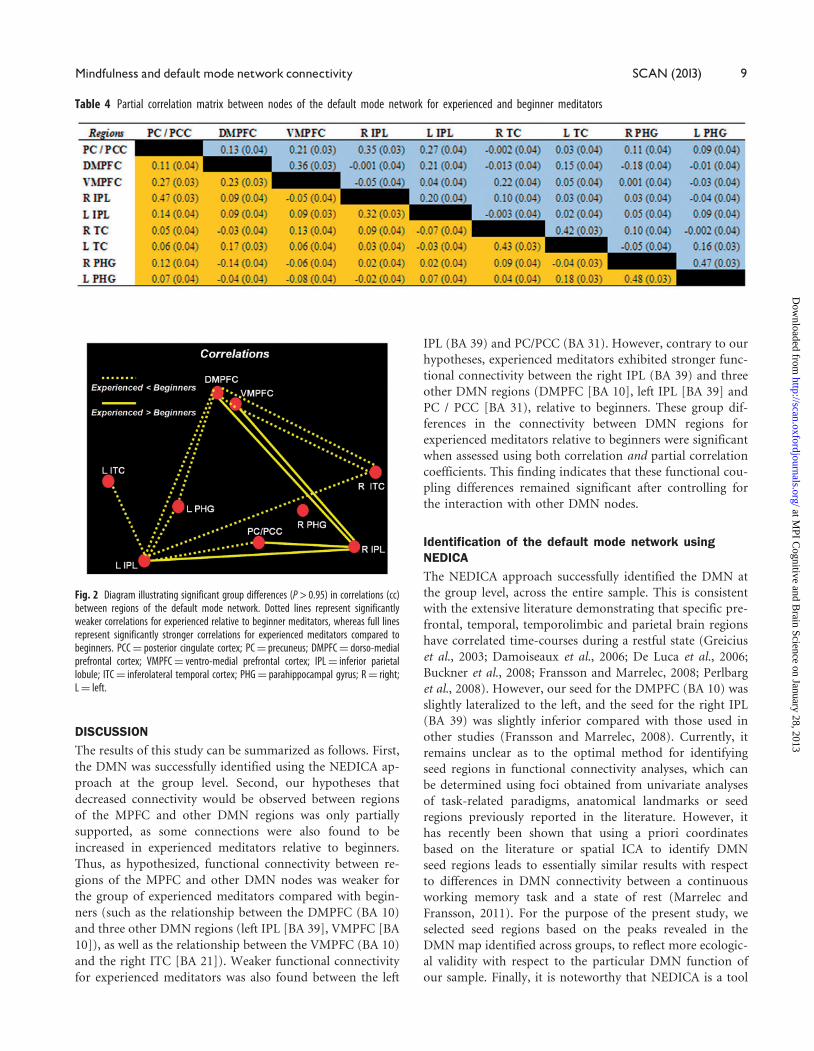
relative to experienced meditators (P > 0.95), between
the DMPFC (BA 10) and the following regions: left
IPL (BA 39), right ITC (BA 21) and left PHG. Weaker cor-
relations were also found, for experienced compared to be-
ginner meditators, between the VMPFC (BA 10) and
the following regions: DMPFC (BA 10), right ITC (BA 21)
and left PHG (BA 36). Finally, other stronger correlations
for beginners vs. experienced meditators were measured be-
tween the left IPL and the following regions: PC/PCC
(BA 39), right PHG (BA 36), left ITC (BA 21) and left
PHG (BA 36).
The analyses also revealed stronger correlations for experi-
enced meditators, relative to beginners (P > 0.95), between
the right IPL and the following regions: PC/PCC (BA 31),
DMPFC (BA 10) and left IPL (BA 39) (Tables 2 and 3,
Figures 2 and 3).
Group differences in partial correlations between DMNregionsThe stronger relationships between DMN regions observed
in beginners vs. experienced meditators, which remained sig-
nificant (P > 0.95) using the partial correlations measure,
were found for the following pairs of regions: PC/PCC (BA
31) and left IPL (BA 39), DMPFC (BA 10) and left IPL (BA
39), DMPFC (BA 10) and VMPFC (BA 10), as well as
VMPFC (BA 10) and right ITC (BA 21).
In addition, all of the stronger correlations for experienced
meditators relative to beginners, which involved the right
IPL (BA 39), remained significant using the partial correl-
ation measure (P > 0.95), yielding the following coefficients
with respect to these regions: PC/PCC (BA 31), DMPFC
(BA 10) and PC / PCC (BA 31) (Figures 4 and 5).
Correlations with hours of meditation practice inexperienced meditatorsCorrelational analyses were conducted to examine whether
the results obtained in the functional connectivity analyses
were also related to the number of hours of meditation prac-
tice in the group of experienced meditators. Pearson r cor-
relation coefficients were computed between the number of
hours of meditation practice and the seven partial correl-
ations which were significant in the between-group compari-
sons given the exploratory nature of these analyses, and
therefore to avoid the occurrence of false positive results.
The only significant correlation with the number of medita-
tion hours was the DMPFC–VMPFC partial correlation,
which was negatively related to the extent of meditation ex-
perience without the inclusion of the outlier with the most
hours of meditation practice (r¼�0.54, P¼ 0.008).
Nonetheless, when including the outlier case and transform-
ing the number of meditation practice hours into a rank
ordered variable, the correlation remained significant
(r¼�0.50, P¼ 0.011). In sum, the partial correlation be-
tween the DMPFC and VMPFC was the only between-region
connection to be significantly related to the number of hours
of meditation practice.
Table 3 Correlation matrix between nodes of the default mode network for experienced and beginner meditators
Table 2 Coordinates for seed regions within the default mode network
Region BA x y z
PC/PCC 31 8 �53 27DMPFC 10 �10 57 19VMPFC 10 �2 47 �10R IPL 39 48 �56 27L IPL 39 �40 �67 34R ITC 21 56 �4 �22L ITC 21 �56 �10 �17R PHG 36 26 �32 �15L PHG 36 �26 �29 �18
Notes: Stereotaxic coordinates are derived from the human atlas of Talairach andTournoux (1988), referring to the medial–lateral position (x) relative to the midline(positive¼ right), and anterior–posterior position (z) relative to the commissural line(positive¼ superior). Designations of Brodmann position (y) relative to the anteriorcommissure (positive¼ anterior), and superior–inferior areas for cortical areas arealso based on this atlas. BA¼ Brodmann area; PCC¼ posterior cingulate cortex;PC¼ precuneus; DMPFC¼ dorso-medial prefrontal cortex; VMPFC¼ ventro-medialprefrontal cortex; IPL¼ inferior parietal lobule; ITC¼ inferolateral temporal cortex;PHG¼ parahippocampal gyrus; R¼ right; L¼ left.
8 SCAN (2013) V.Taylor et al.
at MPI C
ognitive and Brain Science on January 28, 2013
http://scan.oxfordjournals.org/D
ownloaded from
DISCUSSIONThe results of this study can be summarized as follows. First,
the DMN was successfully identified using the NEDICA ap-
proach at the group level. Second, our hypotheses that
decreased connectivity would be observed between regions
of the MPFC and other DMN regions was only partially
supported, as some connections were also found to be
increased in experienced meditators relative to beginners.
Thus, as hypothesized, functional connectivity between re-
gions of the MPFC and other DMN nodes was weaker for
the group of experienced meditators compared with begin-
ners (such as the relationship between the DMPFC (BA 10)
and three other DMN regions (left IPL [BA 39], VMPFC [BA
10]), as well as the relationship between the VMPFC (BA 10)
and the right ITC [BA 21]). Weaker functional connectivity
for experienced meditators was also found between the left
IPL (BA 39) and PC/PCC (BA 31). However, contrary to our
hypotheses, experienced meditators exhibited stronger func-
tional connectivity between the right IPL (BA 39) and three
other DMN regions (DMPFC [BA 10], left IPL [BA 39] and
PC / PCC [BA 31), relative to beginners. These group dif-
ferences in the connectivity between DMN regions for
experienced meditators relative to beginners were significant
when assessed using both correlation and partial correlation
coefficients. This finding indicates that these functional cou-
pling differences remained significant after controlling for
the interaction with other DMN nodes.
Identification of the default mode network usingNEDICAThe NEDICA approach successfully identified the DMN at
the group level, across the entire sample. This is consistent
with the extensive literature demonstrating that specific pre-
frontal, temporal, temporolimbic and parietal brain regions
have correlated time-courses during a restful state (Greicius
et al., 2003; Damoiseaux et al., 2006; De Luca et al., 2006;
Buckner et al., 2008; Fransson and Marrelec, 2008; Perlbarg
et al., 2008). However, our seed for the DMPFC (BA 10) was
slightly lateralized to the left, and the seed for the right IPL
(BA 39) was slightly inferior compared with those used in
other studies (Fransson and Marrelec, 2008). Currently, it
remains unclear as to the optimal method for identifying
seed regions in functional connectivity analyses, which can
be determined using foci obtained from univariate analyses
of task-related paradigms, anatomical landmarks or seed
regions previously reported in the literature. However, it
has recently been shown that using a priori coordinates
based on the literature or spatial ICA to identify DMN
seed regions leads to essentially similar results with respect
to differences in DMN connectivity between a continuous
working memory task and a state of rest (Marrelec and
Fransson, 2011). For the purpose of the present study, we
selected seed regions based on the peaks revealed in the
DMN map identified across groups, to reflect more ecologic-
al validity with respect to the particular DMN function of
our sample. Finally, it is noteworthy that NEDICA is a tool
Table 4 Partial correlation matrix between nodes of the default mode network for experienced and beginner meditators
Fig. 2 Diagram illustrating significant group differences (P > 0.95) in correlations (cc)between regions of the default mode network. Dotted lines represent significantlyweaker correlations for experienced relative to beginner meditators, whereas full linesrepresent significantly stronger correlations for experienced meditators compared tobeginners. PCC¼ posterior cingulate cortex; PC¼ precuneus; DMPFC¼ dorso-medialprefrontal cortex; VMPFC¼ ventro-medial prefrontal cortex; IPL¼ inferior parietallobule; ITC¼ inferolateral temporal cortex; PHG¼ parahippocampal gyrus; R¼ right;L¼ left.
Mindfulness and defaultmode network connectivity SCAN (2013) 9
at MPI C
ognitive and Brain Science on January 28, 2013
http://scan.oxfordjournals.org/D
ownloaded from

designed to detect multiple networks across sessions and sub-
jects, and the analyses conducted in the present study
revealed the presence of several other networks across resting
state runs (visual, motor and auditory, for example).
We chose, however, to examine functional connectivity
only within regions of the DMN for the sake of conciseness,
in addition to our clear a priori hypotheses with respect to
this network and its relationship with meditation training.
Increased connectivity for experienced meditatorscompared to beginnersFirst, our findings are in contrast with those from a recent
study (Kilpatrick et al., 2011) in which differences between
intrinsic connectivity resting state networks were examined
between a group of participants having completed an 8-week
MBSR training program and a wait-listed control group.
Though Kilpatrick et al. (2011) did not observe any differ-
ences between the two groups in functional connectivity
with respect to the DMN, the discrepancy with our findings
may arise in part from the different statistical analytic pro-
cedures employed as well as from the extent of experience
from the group of meditators. As such, it is possible that
differences in DMN connectivity emerge after more than 8
weeks of training.
Nonetheless, the stronger functional connectivity observed
in the present study between the right IPL (BA 39) and
DMPFC (BA 10) in experienced meditators, relative to be-
ginners, is consistent with previous studies (Lutz et al., 2004;
Fell et al., 2010). For instance, it has been reported that,
compared to control subjects with no meditative experience,
Buddhist meditators (with 10 000–40 000 h of meditation
experience) exhibit greater gamma synchrony between
medial prefrontal and parietal areas during a resting state
(Lutz et al., 2004). Interestingly, increased gamma wave syn-
chrony between frontal and parietal lobes has been inter-
preted as reflecting enhanced conscious awareness of the
present moment (Tononi and Edelman, 1998; Engel et al.,
1999), a central characteristic of the mindful state.
The strengthened correlation between the right IPL
(BA 39) and the DMPFC (BA 10) for experienced meditators
(relative to beginner meditators) might reflect adaptive con-
sequences of mindfulness training, as this connection
Fig. 3 Correlation values (cc; y-axis) for all pairwise relationships between default network regions (x-axis). Blue triangles represent values for experienced meditators, andorange squares represent values for beginner meditators. PCC¼ posterior cingulate cortex; PC¼ precuneus; DMPFC¼ dorso-medial prefrontal cortex; VMPFC¼ ventro-medialprefrontal cortex; IPL¼ inferior parietal lobule; ITC¼ inferolateral temporal cortex; PHG¼ parahippocampal gyrus; R¼ right; L¼ left. *Significant group differences (P > 0.95).
Fig. 4 Diagram illustrating significant group differences (P > 0.95) in partial correl-ations (�) between default mode network regions. Dotted lines represent signifi-cantly weaker partial correlation coefficients for experienced compared to beginnermeditators, and full lines represent significantly stronger partial correlation values forexperienced meditators relative to beginners. PCC¼ posterior cingulate cortex;PC¼ precuneus; DMPFC¼ dorso-medial prefrontal cortex; VMPFC¼ ventro-medialprefrontal cortex; IPL¼ inferior parietal lobule; ITC¼ inferolateral temporal cortex;PHG¼ parahippocampal gyrus; R¼ right; L¼ left.
10 SCAN (2013) V.Taylor et al.
at MPI C
ognitive and Brain Science on January 28, 2013
http://scan.oxfordjournals.org/D
ownloaded from

has been shown to be hypofunctional in individuals with
high basal cortisol levels (Schutter et al., 2002). Indeed, it
has been found that elevated baseline levels of the steroid
hormone cortisol, previously associated with depression
(Holsboer et al., 2000), are correlated with reduced function-
al connectivity between the left PFC and right parietal cortex.
These findings are consistent with evidence showing that
transcranial magnetic stimulation (TMS) applied locally to
the left prefrontal or to the right parietal cortex (to increase
activity in these regions) reduces depressive symptoms
(George et al., 1995; 1996; 2010; van Honk et al., 2003).
Given this, the increased connectivity between the DMPFC
and the right IPL found here in experienced meditators
may reflect a beneficial impact of mindfulness training in
terms of emotional resources and conscious awareness of
the present moment. This hypothesis is consistent with evi-
dence that mindfulness is accompanied by increased mood
and well-being, enhanced attention and cognitive perform-
ance, as well as reduced stress, depressive symptoms,
anger and cortisol levels (Baer, 2003; Tang et al., 2007;
Jung et al., 2010).
Increased connectivity was also noted for experienced
meditators, compared to beginners, between right IPL (BA
39) and two other DMN nodes (PC/PCC [BA 31] and left
IPL [BA 39]). The heightened connectivity between the right
IPL and the PC/PCC is in accordance with a recent study
(van Buuren et al., 2010), which demonstrated that
self-referential processing is associated with reduced cou-
pling between the right parietal cortex and the precuneus.
This finding supports the view that mindfulness training in-
duces brain function changes that are accompanied by a re-
duction of self-referential thoughts during rest. Alternatively,
as the parietal cortex is involved in working memory
and visuo-spatial attention (Culham and Kanwisher, 2001),
this finding may reflect greater global attention and
moment-to-moment awareness in experienced meditators
during a restful state. Nonetheless, these interpretations
remain speculative, at best, until being tested using behav-
ioural paradigms assessing these specific processes.
Individuals with MDD and a concomitant anxiety dis-
order display greater right vs. left parietal alpha activity
(Culham and Kanwisher, 2001). In contrast, depressed indi-
viduals without a comorbid anxiety disorder exhibit greater
left vs. right alpha activity over parietal areas. Consequently,
the increased connectivity between the left and right parietal
regions measured in experienced meditators, relative to be-
ginners, may reflect the greater emotional stability that re-
sults from long-term practice of mindfulness (Bruder et al.,
1997; Broderick, 2005; Sheline et al., 2009).
Decreased connectivity for experienced meditatorsrelative to beginnersGreater coherence in the theta range between the PFC and
the left parietal cortex has been measured during working
memory tasks involving verbally related content, whereas
theta coherence enhancement between the PFC and the
right parietal cortex is seen in working memory tasks impli-
cating spatial features (Sarnthein et al., 1998). Therefore, the
decreased connectivity between the DMPFC (BA 10) and the
left IPL (BA 39) in experienced meditators, compared to
beginners, may be related to a diminution of analytic
self-referent processes (Northoff et al., 2006).
For experienced meditators relative to beginners, reduced
connectivity was also measured between the VMPFC (BA 10)
and DMPFC (BA 10). Since these medial prefrontal areas are
adjacent to each other, it is difficult to tease apart their distinct
contributions to DMN functioning. Both areas play a role in
self-relatedness (Schneider et al., 2008), self-referential pro-
cessing (van Buuren et al., 2010), emotional judgements
(Northoff et al., 2004) and in the appraisal of stimuli relative
Fig. 5 Partial correlation coefficients (�; y-axis) for all pairwise relationships between default mode network regions (x-axis). Triangles represent the group of experiencedmeditators, and squares represent the group of beginner meditators. PCC¼ posterior cingulate cortex; PC¼ precuneus; DMPFC¼ dorso-medial prefrontal cortex;VMPFC¼ ventro-medial prefrontal cortex; IPL¼ inferior parietal lobule; ITC¼ inferolateral temporal cortex; PHG¼ parahippocampal gyrus; R¼ right; L¼ left. *Significantgroup differences (P > 0.95).
Mindfulness and defaultmode network connectivity SCAN (2013) 11
at MPI C
ognitive and Brain Science on January 28, 2013
http://scan.oxfordjournals.org/D
ownloaded from

to the self (Ochsner et al., 2004; Ochsner and Gross, 2005).
The VMPFC (BA 10), which has dense projections to the
amygdala (Amaral et al., 1992), is also thought to be impli-
cated in the extinction of conditioned fear, as well as the
down-regulation of emotional responses (LaBar et al., 1998;
Davidson, 2002; Phelps et al., 2004). It thus appears plausible
that the decreased coupling between the DMPFC (BA 10) and
the VMPFC (BA 10) noted in experienced meditators may
reflect a reduction in emotional appraisal during self-referent
processes, consistent with the view that mindfulness is in-
tended to promote acceptance of thoughts, perceptions and
feelings (Bishop, 2004). In addition, since the connectivity
between these regions was also negatively correlated to the
number meditation practice hours in the group of experi-
enced meditators, the decreased DMPFC–VMPFC connectiv-
ity found for experienced meditators relative to beginners may
specifically be related to the extent of meditation training.
Finally, experienced meditators had reduced connectivity
between the VMPFC and the right TC, which may reflect
reduced retrieval or encoding of self-referent memories. As
such, the anterior temporal cortex has been associated with
retrieval of autobiographical emotional memories (com-
pared with neutral autobiographical memories) (Dolan
et al., 2000). Thus, the reduced functional coupling between
the right TC and the VMPFC in experienced vs. beginner
meditators may reflect reduced emotional autobiographical
retrieval during rest for the experienced meditators. Future
studies are needed to investigate the extent and nature of
autobiographical memory retrieval during rest as a result of
meditation training.
Correlations and partial correlations betweenDMN regionsDistinct patterns of functional connectivity within the
DMN regions were revealed, in experienced vs. beginner
meditators, by both correlations and partial correlations.
Several correlations, however, involving the VMPFC
(BA 10), the right IPL (BA 39), the PHG (BA 36) and the
left ITC (BA 21) were no longer different, between the two
groups, when analyzed using partial correlations. This find-
ing is consistent with the notion that the DMN is segregated
into functional subsystems (Perlbarg et al., 2007; Buckner
et al., 2008), and that the power of low-frequency BOLD
fluctuations in different regions of the DMN is rank-ordered,
with the ITC exhibiting the lowest power, and the PC/PCC
the highest power, followed by the VMPFC and the DMPFC
(Jiao et al., 2011).
LIMITATIONS AND FUTURE DIRECTIONSThis study is nonetheless limited in some respects. First, the
two groups of participants may have differed in other aspects
(personality traits, lifestyle, etc.); therefore, longitudinal stu-
dies examining the relationship between meditation training
and functional connectivity are needed to rule out these po-
tential confounds. Second, the partial correlation method
used in this study does not allow for causal inferences to
be made about the relationships between different regions.
Thus, further studies using effective connectivity methods,
such as dynamic causal modelling, are required to investigate
causal relationship between DMN regions as a result of
meditation training. Third, this study assessed differences
regarding functional connectivity between DMN regions at
rest, and did not examine any behavioural processes;
hence, interpretations relating brain connectivity differences
between the two groups remain speculative, at best, until
being replicated in studies using paradigms specifically as-
sessing the behavioural mechanisms involved. Moreover,
though age differences between the two groups did not
attain statistical significance, there was a slight difference
between the mean ages of the two groups but it was not
possible to include a covariate in the independent compo-
nent analyses using the NEDICA software. In addition, given
our relatively small sample size for an fMRI experiment, the
results of the present study should be interpreted with cau-
tion until being replicated in larger samples precisely
matched with respect to age. Finally, it is possible that the
state of rest may have differed qualitatively between the two
groups; therefore, future studies examining the relationship
between meditation training and resting state connectivity
should acquire qualitative measures of thought content and
conscious processes occurring during the rest period to
aid in interpretation of brain imaging functional connectiv-
ity data.
CONCLUSIONIn conclusion, this study demonstrates that individuals with
extensive mindfulness training exhibit significant differences
in functional connectivity between regions of the DMN. Our
findings suggest that mindfulness training leads to changes
in the functional dynamics of the DMN that extend beyond a
state of meditation per se.
Conflict of Interest
None declared.
REFERENCESAmaral, D.G., Price, J.L., Pitkanen, A., Carmichael, S.T. (1992). Wiley-Liss,
1–66.
Baer, R.A. (2003). Mindfulness training as a clinical intervention: A con-
ceptual and empirical review. Clinical Psychology: Science and Practice, 10,
125–43.
Beckmann, C.F., Smith, S.M. (2004). Probabilistic independent component
analysis for functional magnetic resonance imaging. IEEE Transactions on
Medical Imaging, 23, 137–52.
Bell, A.J., Sejnowski, T.J. (1995). An information-maximization approach to
blind separation and blind deconvolution. Neural Computation, 7,
1129–59.
Bellec, P., Perlbarg, V., Jbabdi, S., et al. (2006). Identification of large-scale
networks in the brain using fMRI. Neuroimage, 29, 1231–43.
Bishop, R.S. (2004). Mindfulness: a proposed operational definition.
Clinical Psychology: Science and Practice, 11, 230–41.
12 SCAN (2013) V.Taylor et al.
at MPI C
ognitive and Brain Science on January 28, 2013
http://scan.oxfordjournals.org/D
ownloaded from

Biswal, B., Yetkin, F.Z., Haughton, V.M., Hyde, J.S. (1995). Functional
connectivity in the motor cortex of resting human brain using
echo-planar MRI. Magnetic Resonance in Medicine, 34, 537–41.
Broderick, P.C. (2005). Mindfulness and coping with dysphoric mood:
Contrasts with rumination and distraction. Cognitive Therapy Research,
29, 501–10.
Bruder, G.E., Fong, R., Tenke, C.E., et al. (1997). Regional brain asymme-
tries in major depression with or without an anxiety disorder: a quanti-
tative electroencephalographic study. Biological Psychiatry, 41, 939–48.
Buckner, R.L., Andrews-Hanna, J.R., Schacter, D.L. (2008). The brain’s
default network: anatomy, function, and relevance to disease. Annals of
the New York Academy of Sciences, 1124, 1–38.
Culham, J.C., Kanwisher, N.G. (2001). Neuroimaging of cognitive func-
tions in human parietal cortex. Current Opinion in Neurobiology, 11,
157–63.
Damoiseaux, J.S., Rombouts, S.A., Barkhof, F., et al. (2006). Consistent
resting-state networks across healthy subjects. Proceedings of the
National Academy of Sciences of the United States of America, 103,
13848–53.
Davidson, R.J. (2002). Anxiety and affective style: role of prefrontal cortex
and amygdala. Biological Psychiatry, 51, 68–80.
De Luca, M., Beckmann, C.F., De Stefano, N., Matthews, P.M., Smith, S.M.
(2006). fMRI resting state networks define distinct modes of
long-distance interactions in the human brain. Neuroimage, 29, 1359–67.
Dolan, R.J., Lane, R., Chua, P., Fletcher, P. (2000). Dissociable temporal
lobe activations during emotional episodic memory retrieval.
Neuroimage, 11, 203–9.
Engel, A.K., Fries, P., Konig, P., Brecht, M., Singer, W. (1999). Temporal
binding, binocular rivalry, and consciousness. Conscious Cognition, 8,
128–51.
Esposito, F., Scarabino, T., Hyvarinen, A., et al. (2005). Independent com-
ponent analysis of fMRI group studies by self-organizing clustering.
Neuroimage, 25, 193–205.
Farb, N.A., Segal, Z.V., Mayberg, H., et al. (2007). Attending to the present:
mindfulness meditation reveals distinct neural modes of self-reference.
Social Cognitive and Affective Neuroscience, 2, 313–22.
Fell, J., Axmacher, N., Haupt, S. (2010). From alpha to gamma: electro-
physiological correlates of meditation-related states of consciousness.
Medical Hypotheses, 75, 218–24.
Fransson, P., Marrelec, G. (2008). The precuneus/posterior cingulate cortex
plays a pivotal role in the default mode network: evidence from a partial
correlation network analysis. Neuroimage, 42, 1178–84.
George, M.S., Lisanby, S.H., Avery, D., et al. (2010). Daily left prefront-
al transcranial magnetic stimulation therapy for major depressive dis-
order: a sham-controlled randomized trial. Archives of General
Psychiatry, 67, 507–16.
George, M.S., Wassermann, E.M., Williams, W.A., et al. (1995). Daily re-
petitive transcranial magnetic stimulation (rTMS) improves mood in
depression. Neuroreport, 6, 1853–6.
George, M.S., Wassermann, E.M., Williams, W.A., et al. (1996). Changes in
mood and hormone levels after rapid-rate transcranial magnetic stimu-
lation (rTMS) of the prefrontal cortex. Journal of Neuropsychiatry and
Clinical Neuroscience, 8, 172–80.
Grant, J.A., Courtemanche, J., Rainville, P. (2011). A non-elaborative
mental stance and decoupling of executive and pain-related cortices pre-
dicts low pain sensitivity in Zen meditators. Pain, 152, 150–6.
Greicius, M.D., Krasnow, B., Reiss, A.L., Menon, V. (2003). Functional
connectivity in the resting brain: a network analysis of the default
mode hypothesis. Proceedings of the National Academy of Sciences of the
United States of America, 100, 253–8.
Gusnard, D.A., Akbudak, E., Shulman, G.L., Raichle, M.E. (2001). Medial
prefrontal cortex and self-referential mental activity: relation to a default
mode of brain function. Proceedings of the National Academy of Sciences of
the United States of America, 98, 4259–64.
Holsboer, F. (2000). The corticosteroid receptor hypothesis of depression.
Neuropsychopharmacology, 23, 477–501.
Jang, J.H., Jung, W.H., Kang, D.H., et al. (2011). Increased default mode
network connectivity associated with meditation. Neuroscience Letters,
487(3), 358–62.
Jiao, Q., Lu, G., Zhang, Z., et al. (2011). Granger causal influence predicts
BOLD activity levels in the default mode network. Human Brain
Mapping, 32, 154–61.
Jung, Y.H., Kang, D.H., Jang, J.H., et al. (2010). The effects of mind-body
training on stress reduction, positive affect, and plasma catecholamines.
Neuroscience Letters, 479, 138–42.
Kabat-Zinn, J. (1994). Wherever you go there you are. New York, USA:
Hyperion Books.
Kilpatrick, L.A., Suyenobu, B.Y., Smith, S.R., et al. (2011). Impact of
mindfulness-based stress reduction training on intrinsic brain connectiv-
ity. Neuroimage, 56(1), 290–8.
LaBar, K.S., Gatenby, J.C., Gore, J.C., LeDoux, J.E., Phelps, E.A. (1998).
Human amygdala activation during conditioned fear acquisition and ex-
tinction: a mixed-trial fMRI study. Neuron, 20, 937–45.
Lutz, A., Greischar, L.L., Rawlings, N.B., Ricard, M., Davidson, R.J. (2004).
Long-term meditators self-induce high-amplitude gamma synchrony
during mental practice. Proceedings of the National Academy of Sciences
of the United States of America, 101, 16369–73.
Marrelec, G., Bellec, P., Krainik, A., et al. (2008). Regions, systems, and the
brain: hierarchical measures of functional integration in fMRI. Medical
Image Analysis, 12, 484–96.
Marrelec, G., Fransson, P. (2011). Assessing the influence of different ROI
selection strategies on functional connectivity analyses of fMRI data
acquired during steady-state conditions. Plos One, 6(4), 1–14.
Marrelec, G., Krainik, A., Duffau, H., et al. (2006). Partial correlation for
functional brain interactivity investigation in functional MRI.
Neuroimage, 32, 228–37.
Meindl, T., Teipel, S., Elmouden, R., et al. (2010). Test-retest reproducibility
of the default-mode network in healthy individuals. Human Brain
Mapping, 31(2), 237–46.
Northoff, G., Heinzel, A., Bermpohl, F., et al. (2004). Reciprocal modulation
and attenuation in the prefrontal cortex: an fMRI study on
emotional-cognitive interaction. Human Brain Mapping, 21, 202–12.
Northoff, G., Heinzel, A., de Greck, M., Bermpohl, F., Dobrowolny, H.,
Panksepp, J. (2006). Self-referential processing in our brain�a
meta-analysis of imaging studies on the self. Neuroimage, 31, 440–57.
Ochsner, K.N., Gross, J.J. (2005). The cognitive control of emotion. Trends
in Cognitive Science, 9, 242–9.
Ochsner, K.N., Knierim, K., Ludlow, D.H., et al. (2004). Reflecting
upon feelings: an fMRI study of neural systems supporting the attribution
of emotion to self and other. Journal of Cognitive Neuroscience, 16,
1746–72.
Phelps, E.A., Delgado, M.R., Nearing, K.I., LeDoux, J.E. (2004). Extinction
learning in humans: role of the amygdala and vmPFC. Neuron, 43,
897–905.
Perlbarg, V., Bellec, P., Anton, J.L., Pelegrini-Issac, M., Doyon, J., Benali, H.
(2007). CORSICA: correction of structured noise in fMRI by automatic
identification of ICA components. Magnetic Resonance Imaging, 25,
35–46.
Perlbarg, V., Marrelec, G., Doyon, J., Pelegrini-Issac, M., Lehericy, S.,
Benali, H. (2008). NEDICA: detection of group functional networks in
fMRI using spatial independent component analysis. 5th IEEE
International Symposium on Biomedical Imaging: From Nano to Macro,
1247–50.
Raichle, M.E., MacLeod, A.M., Snyder, A.Z., Powers, W.J., Gusnard, D.A.,
Shulman, G.L. (2001). A default mode of brain function. Proceedings of
the National Academy of Sciences of the United States of America, 98,
676–82.
Ricard, M. (2008). L’art de la meditation, (Nil editions): Paris, France.
Sarnthein, J., Petsche, H., Rappelsberger, P., Shaw, G.L., von Stein, A.
(1998). Synchronization between prefrontal and posterior association
cortex during human working memory. Proceedings of the National
Academy of Sciences of the United States of America, 95, 7092–6.
Mindfulness and defaultmode network connectivity SCAN (2013) 13
at MPI C
ognitive and Brain Science on January 28, 2013
http://scan.oxfordjournals.org/D
ownloaded from

Schneider, F., Bermpohl, F., Heinzel, A., et al. (2008). The resting brain and
our self: self-relatedness modulates resting state neural activity in cortical
midline structures. Neuroscience, 157, 120–31.
Schutter, D.J., Van Honk, J., Koppeschaar, H., Kahn, R. (2002). Cortisol and
reduced interhemispheric coupling between the left prefrontal and the
right parietal cortex. Journal of Neuropsychiatry and Clinical Neuroscience,
14, 89–90.
Shehzad, Z., Kelly, A.M., Reiss, P.T., et al. (2009). The resting brain: un-
constrained yet reliable. Cerebral Cortex, 19(10), 2209–29.
Sheline, Y.I., Barch, D.M., Price, J.L., et al. (2009). The default mode net-
work and self-referential processes in depression. Proceedings of the
National Academy of Sciences of the United States of America, 106, 1942–7.
Tang, Y.Y., Ma, Y., Wang, J., et al. (2007). Short-term meditation training
improves attention and self-regulation. Proceedings of the National
Academy of Sciences of the United States of America, 104, 17152–6.
Thich Nhat Hanh. (1994). Le miracle de la pleine conscience, (L’espace bleu).
Tononi, G., Edelman, G.M. (1998). Consciousness and complexity. Science,
282, 1846–51.
van Buuren, M., Gladwin, T.E., Zandbelt, B.B., Kahn, R.S., Vink, M. (2010).
Reduced functional coupling in the default-mode network during
self-referential processing. Human Brain Mapping, 31, 1117–27.
van Honk, J., Schutter, D.J., Putman, P., de Haan, E.H.,
d’Alfonso, A.A. (2003). Reductions in phenomenological, physiologic-
al and attentional indices of depressive mood after 2 Hz rTMS over
the right parietal cortex in healthy human subjects. Psychiatry
Research, 120, 95–101.
Zuo, X.N., Kelly, C., Adelstein, J.S., Klein, D.F., Castellanos, F.X.,
Milham, M.P. (2010). Reliable intrinsic connectivity networks: test-retest
evaluation using ICA and dual regression approach. Neuroimage, 49(3),
2163–77.
14 SCAN (2013) V.Taylor et al.
at MPI C
ognitive and Brain Science on January 28, 2013
http://scan.oxfordjournals.org/D
ownloaded from


![TheGreenBow VPN · PDF file120349 Default (SA CNXVPN1-P1) RECV phase 1 Main Mode [SA][VID] 120349 Default (SA CNXVPN1-P1) SEND phase 1 Main Mode [KEY][NONCE] 120351 Default (SA](https://img.pdfslide.net/doc/110x75/5a7a7f6a7f8b9a2d788c04c2/thegreenbow-vpn-default-sa-cnxvpn1-p1-recv-phase-1-main-mode-savid-120349.jpg)


![TheGreenBow VPN Client · 115915 Default (SA CNXVPN1-P1) RECV phase 1 Main Mode [KEY][NONCE] 115915 Default (SA CNXVPN1-P1) SEND phase 1 Main Mode [ID][HASH][NOTIFY] 115915 Default](https://img.pdfslide.net/doc/110x75/5f7564d6a29d0c29d968762c/thegreenbow-vpn-115915-default-sa-cnxvpn1-p1-recv-phase-1-main-mode-keynonce.jpg)