Embed Size (px)
Citation preview
Spectrum Denturism – Vol. 4 No. 1 – March 20106
Axial attachments in large mandibular completedentures help make removable prostheses morecomfortable for the patient. Designed to be fixed on
the natural root, their longevity has often beenunpredictable in the medium term due to root fractures,support root caps working loose, and the removableprostheses fracturing. The prognosis improved whenosteointegrated implants were introduced.1 The 2002McGill conference established that the current therapeuticstandard for treatment using mandibular completedentures is the implant supported removable prosthesiswith two osteointegrated implants. The removableprosthesis must also respond to the usual factors affectingequilibrium with the support surface, have correctlymodeled and adjusted edges, and no unstable orunbalanced guidances.
Mandibular Deformation
Although the literature documents the criteria ofadaptability for prostheses, the number of implantsrequired, and the type of attachment quite well, less useis made of other data. The idea that the lateralpterygoidian muscles may, as a result of their obliquity,exercise a compressive action on the mandible wasdiscovered by Gr unewald in 1923. It is no longercontested that the mandible deforms elastically or flexesin both free movement and during mastication. Thehorizontal branches of the mandible move nearer orfurther away from each other during protr usion,retrusion, and opening.
Movement of 0.0 to 1.5 mm occurs during opening,and of 0.1 to 1.5 mm during protrusion.
Implant Supported Removable ProsthesesDr. Bruno Clunet-Coste, Dental Surgeon, Sandrine Garampon, Dental Prosthetist, Sandrine Lagouge, Dental Prosthetist,Yannick Le Guay, Material Engineer
Axial attachments in complete dentures are very effective in stabilizing removable prostheses, buttheir excessive rigidity makes it difficult for patients to integrate them perfectly. Using Fiber Forcereinforced fiber mesh results in considerable resistance to alternating stress.
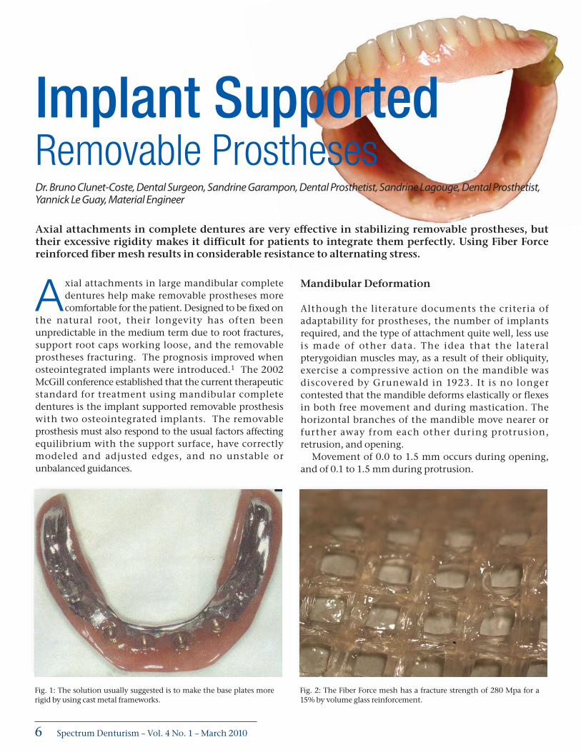
Fig. 1: The solution usually suggested is to make the base plates morerigid by using cast metal frameworks.
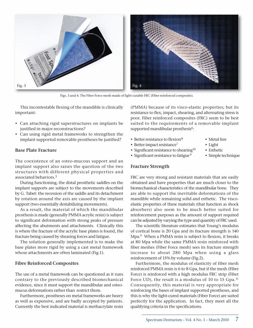
Fig. 2: The Fiber Force mesh has a fracture strength of 280 Mpa for a15% by volume glass reinforcement.
SDen_MAR10:Spectrum 3/3/2010 4:51 PM Page 6
7Spectrum Denturism – Vol. 4 No. 1 – March 2010
This incontestable flexing of the mandible is clinicallyimportant:
• Can attaching rigid superstructures on implants bejustified in major reconstructions?
• Can using rigid metal frameworks to strengthen theimplant supported removable prostheses be justified?
Base Plate Fracture
The coexistence of an osteo-mucous support and animplant support also raises the question of the twostr uctures with different physical properties andassociated behaviors.3
During functioning, the distal prosthetic saddles on theimplant supports are subject to the movements describedby G. Tabet: the recession of the saddle and its detachmentby rotation around the axis are caused by the implantsupport (two essentially destabilizing movements).
As a result, the material of which the mandibularprosthesis is made (generally PMMA acrylic resin) is subjectto significant deformation with strong peaks of pressureaffecting the abutments and attachments. Clinically thisis where the fracture of the acrylic base plates is found, thefracture being caused by shearing forces and fatigue.
The solution generally implemented is to make thebase plates more rigid by using a cast metal frameworkwhose attachments are often laminated (Fig.1).
Fibre Reinforced Composites
The use of a metal framework can be questioned as it runscontrary to the previously described biomechanicalevidence, since it must support the mandibular and osteo-mucus deformations rather than restrict them.
Furthermore, prostheses on metal frameworks are heavyas well as expensive, and are badly accepted by patients.Currently the best indicated material is methacrylate resin
(PMMA) because of its visco-elastic properties; but itsresistance to flex, impact, shearing, and alternating stress ispoor. Fiber reinforced composites (FRC) seem to be bestsuited to the requirements of a removable implantsupported mandibular prosthesis6:
Fracture Strength
FRC are very strong and resistant materials that are easilyobtained and have properties that are much closer to thebiomechanical characteristics of the mandibular bone. Theyare able to support the inevitable deformations of themandible while remaining solid and esthetic. The visco-elastic properties of these materials (that function as shockabsorbers) also seem to be much better suited forreinforcement purposes as the amount of support requiredcan be adjusted by varying the type and quantity of FRC used.
The scientific literature estimates that Young’s modulusof cortical bone is 20 Gpa and its fracture strength is 140Mpa.9 When a PMMA resin is subject to flexion, it breaksat 80 Mpa while the same PMMA resin reinforced withfiber meshes (Fiber Force mesh) sees its fracture strengthincrease to about 280 Mpa when using a glassreinforcement of 15% by volume (Fig.2).
Furthermore, the modulus of elasticity of fiber meshreinforced PMMA resin is 6 to 8 Gpa, but if the mesh (FiberForce) is reinforced with a high modulus FRC strip (FiberForce UD), the result is a modulus of 10 to 15 Gpa.11
Consequently, this material is very appropriate forreinforcing the bases of implant supported prostheses, andthis is why the light-cured materials (Fiber Force) are suitedperfectly for the application. In fact, they meet all thequalifying criteria in the specifications.
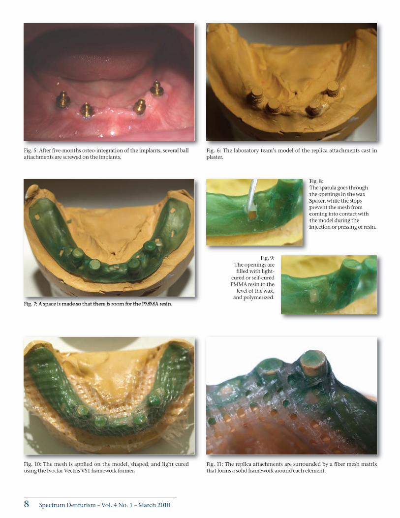
Figs. 3 and 4: The Fiber Force mesh made of light curable FRC (Fiber reinforced composite).
• Better resistance to flexion8
• Better impact resistance7
• Significant resistance to shearing10
• Significant resistance to fatigue9
• Metal free• Light• Esthetic• Simple technique
Fig. 4Fig. 3
SDen_MAR10:Spectrum 3/3/2010 4:51 PM Page 7
Spectrum Denturism – Vol. 4 No. 1 – March 20108
Fig. 5: After five-months osteo-integration of the implants, several ballattachments are screwed on the implants.
Fig. 6: The laboratory team’s model of the replica attachments cast inplaster.
Fig. 7: A space is made so that there is room for the PMMA resin.
Fig. 8: The spatula goes throughthe openings in the waxSpacer, while the stopsprevent the mesh fromcoming into contact withthe model during theinjection or pressing of resin.
Fig. 9: The openings arefilled with light-
cured or self-curedPMMA resin to the
level of the wax,and polymerized.
Fig. 10: The mesh is applied on the model, shaped, and light curedusing the Ivoclar Vectris VS1 framework former.
Fig. 11: The replica attachments are surrounded by a fiber mesh matrixthat forms a solid framework around each element.
SDen_MAR10:Spectrum 3/3/2010 4:51 PM Page 8
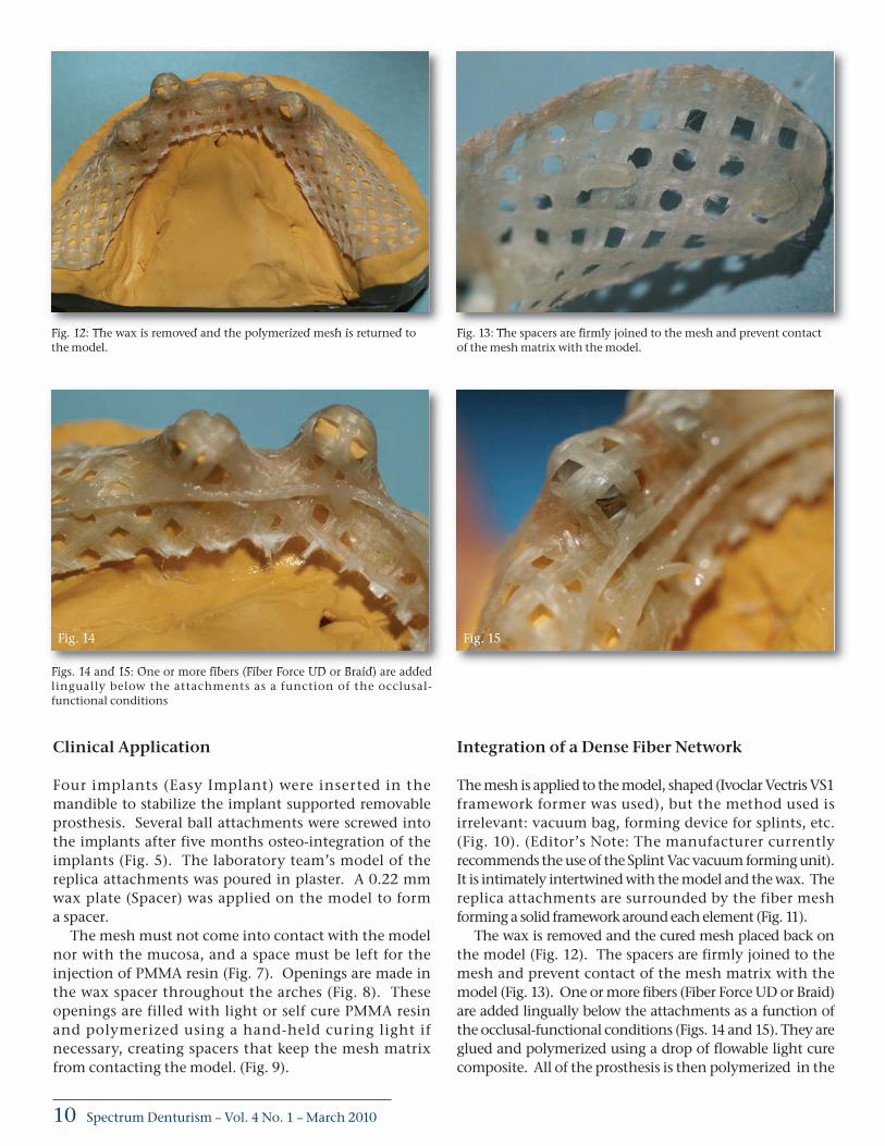
Fig. 12: The wax is removed and the polymerized mesh is returned tothe model.
Fig. 13: The spacers are firmly joined to the mesh and prevent contactof the mesh matrix with the model.
Figs. 14 and 15: One or more fibers (Fiber Force UD or Braid) are addedlingually below the attachments as a function of the occlusal-functional conditions
Clinical Application
Four implants (Easy Implant) were inserted in themandible to stabilize the implant supported removableprosthesis. Several ball attachments were screwed intothe implants after five months osteo-integration of theimplants (Fig. 5). The laboratory team’s model of thereplica attachments was poured in plaster. A 0.22 mmwax plate (Spacer) was applied on the model to forma spacer.
The mesh must not come into contact with the modelnor with the mucosa, and a space must be left for theinjection of PMMA resin (Fig. 7). Openings are made inthe wax spacer throughout the arches (Fig. 8). Theseopenings are filled with light or self cure PMMA resinand polymerized using a hand-held curing light ifnecessary, creating spacers that keep the mesh matrixfrom contacting the model. (Fig. 9).
Integration of a Dense Fiber Network
The mesh is applied to the model, shaped (Ivoclar Vectris VS1framework former was used), but the method used isirrelevant: vacuum bag, forming device for splints, etc.(Fig. 10). (Editor’s Note: The manufacturer currentlyrecommends the use of the Splint Vac vacuum forming unit).It is intimately intertwined with the model and the wax. Thereplica attachments are surrounded by the fiber meshforming a solid framework around each element (Fig. 11).
The wax is removed and the cured mesh placed back onthe model (Fig. 12). The spacers are firmly joined to themesh and prevent contact of the mesh matrix with themodel (Fig. 13). One or more fibers (Fiber Force UD or Braid)are added lingually below the attachments as a function ofthe occlusal-functional conditions (Figs. 14 and 15). They areglued and polymerized using a drop of flowable light curecomposite. All of the prosthesis is then polymerized in the
Fig. 14 Fig. 15
Spectrum Denturism – Vol. 4 No. 1 – March 201010
SDen_MAR10:Spectrum 3/3/2010 4:51 PM Page 10
Spectrum Denturism – Vol. 4 No. 1 – March 201012
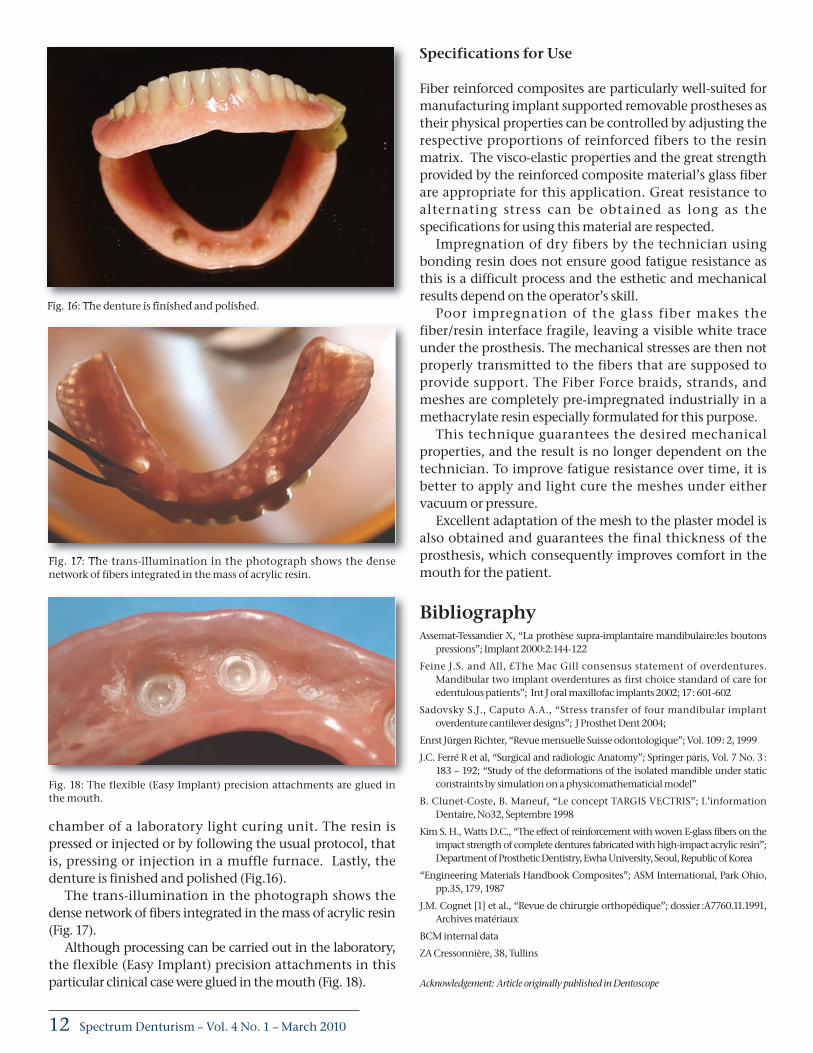
chamber of a laboratory light curing unit. The resin ispressed or injected or by following the usual protocol, thatis, pressing or injection in a muffle furnace. Lastly, thedenture is finished and polished (Fig.16).
The trans-illumination in the photograph shows thedense network of fibers integrated in the mass of acrylic resin(Fig. 17).
Although processing can be carried out in the laboratory,the flexible (Easy Implant) precision attachments in thisparticular clinical case were glued in the mouth (Fig. 18).
Specifications for Use
Fiber reinforced composites are particularly well-suited formanufacturing implant supported removable prostheses astheir physical properties can be controlled by adjusting therespective proportions of reinforced fibers to the resinmatrix. The visco-elastic properties and the great strengthprovided by the reinforced composite material’s glass fiberare appropriate for this application. Great resistance toalternating stress can be obtained as long as thespecifications for using this material are respected.
Impregnation of dry fibers by the technician usingbonding resin does not ensure good fatigue resistance asthis is a difficult process and the esthetic and mechanicalresults depend on the operator’s skill.
Poor impregnation of the glass fiber makes thefiber/resin interface fragile, leaving a visible white traceunder the prosthesis. The mechanical stresses are then notproperly transmitted to the fibers that are supposed toprovide support. The Fiber Force braids, strands, andmeshes are completely pre-impregnated industrially in amethacrylate resin especially formulated for this purpose.
This technique guarantees the desired mechanicalproperties, and the result is no longer dependent on thetechnician. To improve fatigue resistance over time, it isbetter to apply and light cure the meshes under eithervacuum or pressure.
Excellent adaptation of the mesh to the plaster model isalso obtained and guarantees the final thickness of theprosthesis, which consequently improves comfort in themouth for the patient.
BibliographyAssemat-Tessandier X, “La prothèse supra-implantaire mandibulaire:les boutons
pressions”; Implant 2000:2:144-122
Feine J.S. and All, £The Mac Gill consensus statement of overdentures.Mandibular two implant overdentures as first choice standard of care foredentulous patients”; Int J oral maxillofac implants 2002; 17 : 601-602
Sadovsky S.J., Caputo A.A., “Stress transfer of four mandibular implantoverdenture cantilever designs”; J Prosthet Dent 2004;
Enrst Jürgen Richter, “Revue mensuelle Suisse odontologique”; Vol. 109 : 2, 1999
J.C. Ferré R et al, “Surgical and radiologic Anatomy”; Springer paris, Vol. 7 No. 3 :183 – 192; “Study of the deformations of the isolated mandible under staticconstraints by simulation on a physicomathematicial model”
B. Clunet-Coste, B. Maneuf, “Le concept TARGIS VECTRIS”; L’informationDentaire, No32, Septembre 1998
Kim S. H., Watts D.C., “The effect of reinforcement with woven E-glass fibers on theimpact strength of complete dentures fabricated with high-impact acrylic resin”;Department of Prosthetic Dentistry, Ewha University, Seoul, Republic of Korea
“Engineering Materials Handbook Composites”; ASM International, Park Ohio,pp.35, 179, 1987
J.M. Cognet [1] et al., “Revue de chirurgie orthopédique”; dossier :A7760.11.1991,Archives matériaux
BCM internal data
ZA Cressonnière, 38, Tullins
Acknowledgement: Article originally published in Dentoscope
Fig. 16: The denture is finished and polished.
Fig. 17: The trans-illumination in the photograph shows the densenetwork of fibers integrated in the mass of acrylic resin.
Fig. 18: The flexible (Easy Implant) precision attachments are glued inthe mouth.
SDen_MAR10:Spectrum 3/3/2010 4:51 PM Page 12