Embed Size (px)
Citation preview
1
Implication of mast cells, nerve growth factor and splanchnic
nerves in postoperative ileus. Study in patients
undergoing abdominal surgery and in a rat experimental
model
Presented by
Sergio Berdún Marin
A dissertation submitted to obtain the degree of Doctor in Philosophy
PhD program in Neurosciences. Institut de Neurociències
Department of Cell Biology, Physiology and Immunology
Barcelona Veterinary School, Universitat Autònoma de Barcelona
Advisors:
Patrocinio Vergara Esteras
Jakub Rychter
Bellaterra, Barcelona 2015

2
3
Patrocinio Vergara Esteras,
Full professor of Physiology at the Department of Cell Biology, Physiology and
Immunology; Barcelona Veterinary School; Universitat Autònoma de Barcelona
and
Jackub Rychter,
Post-doctoral Researcher at the Department of Cell Biology, Physiology and
Immunology; Barcelona Veterinary School; Universitat Autònoma de Barcelona
We hereby certify that the thesis entitled ‘‘Implication of mast cells, nerve growth
factor and splanchnic nerves in postoperative ileus. Study in patients
undergoing abdominal surgery and in a rat experimental model’’ submitted by SERGIO
BERDÚN MARIN in partial fulfillment of the requirements for the degree of Doctor of
Philosophy was carried out under our supervision and we authorize it submission for oral
defense.
Bellatera, April 2015
Patrocinio Vergara Esteras, PhD Jakub Rychter, PhD

4

5
Cover image: Tryptase-immunostained mast cells in the outer muscular layer of human
colon collected after colorectal surgery. Immunofluorescence technique. 100x.

6
7
‘’Knowledge is the death of research’’
Walther Hermann Nernst
German chemist and physicist

8
9
AGRADECIMIENTOS
Parece mentira que esté sentado delante del ordenador escribiendo estas palabras después de estos
cinco años. Ahora me acuerdo de aquellos momentos en los que pensé en tirar la toalla y no
puedo evitar hacer mención a todas aquellas personas que directa o indirectamente (aunque no
seáis consciente de ello), fuera o dentro de la facultad, me habéis ayudado a recorrer este largo
camino.
Patri, en primer lugar darte las gracias por el simple hecho de permitirme haber llegado hasta
aquí. Gracias también por introducirme en el mundo de la investigación (de eso hace ya 10 años,
contaba yo con veinte añitos…), por motivarme a seguir con ello y por darme la oportunidad de
dar mis primeros pasos como investigador hasta el día de hoy. Por escuchar mis ideas y ponerlas
en práctica, por las horas de discusiones, por confiar en mi criterio y por darme el último
empujoncito que me hacía falta para acabar de presentar esta tesis.
Jakub, tu llegada al departamento me abrió todo un sinfín de nuevas ideas para mi tesis. Gracias
por tu paciencia, por tu ayuda y por todo el tiempo que has invertido. ¡Siento el ajetreo de estos
últimos días!
Pere, indudablemente también formas parte de todo esto. Ha sido un placer conocerte. No podría
estar más agradecido por la ayuda brindada y por tener siempre abiertas las puertas del hospital de
Mataró. Sin duda, esta colaboración ha sido crucial y ha aportado un valor añadido a esta tesis.
Muchas gracias.
También quiero dar las gracias a Ernest Bombuy por la ayuda ofrecida en un momento clave
como fue mi primera andadura por el Hospital de Mataró y por todas las veces que has estado al
otro lado del teléfono siempre dispuesto a echar una mano y a explicarme los clinical composite
endpoints. Gracias también a Òscar Estrada por introducirme en el fascinante mundo de las
colectomías y a Esther Mans por las ideas, comentarios y ánimos aportados a la hora de enviar la
primera publicación. A los tres infinitas gracias por hacer que todo fuera más fácil a la hora de
trabajar la parte clínica del primer capítulo.
También tengo que destacar la ayuda por parte del personal del Hospital de Mataró que
participaron en la recogida y el procesamiento de las muestras: el equipo de cirujanos y
enfermeras y el personal de los laboratorios de microbiología, hematología y patología.
Gracias también a Esther, Marcel y Maite, profesores del departamento, por los momentos
compartidos dentro y fuera del laboratorio.
10
También mencionar al equipo de técnicos y administrativos de la unidad de Fisiología Animal.
Antonio, gracias por hacer que todo esté en su sitio y a punto y por aguantarme los días previos a
las prácticas del curso. Como siempre dices, ¡al final todo sale bien! Pepe y David gracias por
todas las gestiones. Emma, ¿Quieeeen, abrirá la pueeeerta hooooy…? A pesar de haber tenido
nuestros momentos no puedo evitar acordarme de las carcajadas, de las risas, de los bailoteos y
chistes en el 127 y sobre todo por la ayuda prestada para mis experimentos. ¡Muchas gracias!
Por supuesto no puedo olvidarme del resto de becarias, becarios y residentes con los que he
compartido despacho, laboratorio, cubiertos, penas y alegrías. Vosotros sabéis tan bien como yo
lo que es pasar por todo esto. Una tesis no es fácil pero se hace más llevadera si compartes tu
tiempo con gente como vosotros. Mònica, ha sido un placer haber compartido los momentos
máster, las charlas en el despacho acerca de lo incierto que es ( era) nuestro futuro, el hotelucho
de Ámsterdam y tantas otras cosas más. Eres una profesional y no me cabe duda que en Irlanda
los pondrás a todos en su sitio mientras te labras una merecida carrera como investigadora.
Javier, eres el chico que cae bien a todo el mundo, cumplidor, que nunca se mete en líos y con el
que te puedes echar unas buenas risas (ahora me viene a la cabeza el momento ‘pelito sexy’…).
¡¡No cambies nunca!! Joan B, trabajar en el laboratorio 127 no es lo mismo si no se te oye graznar
tus canciones desde la sala de cultivos. Gracias por los chistes, las risas, las preguntas absurdas
sobre la vida que no vienen al caso, por tener siempre (y recalco siempre) tiempo para resolver
mis dudas y por compartir conmigo la idea de que ‘si no te quedas hasta las ocho de la tarde
encerrado en el laboratorio, no estás haciendo una tesis’. Eres un claro ejemplo de trabajo. Estoy
seguro que pronto te llegará la oportunidad que te mereces. Sandra Barbosa; Responsable,
trabajadora y encima del Baix Llobregat! ¡No se puede pedir más! Disfruta la nueva etapa que
está por llegar. ¡Un beso! Marina, estoy muy contento de haber conectado aún más contigo estos
últimos meses. Eres una compañera del SIAL excepcional y que sepas que éxitos como
‘Troublemaker’ ‘We are never getting back together’ o ‘She walks like Rihanna’ no sonarán igual
si no los canto contigo en el laboratorio. Eva, gracias por tu sentido del humor en el laboratorio y
por ser mi compañera de comer galletas a cualquier hora del día. Joan Antoni, por aportar grandes
dosis de ironía y sensatez al grupo y por ser un ejemplo de trabajo. Elena Tapia, ahora me acuerdo
de los momentos con mortero en mano haciendo tránsitos intestinales y del sin vivir a la espera de
que llegará la nueva botella de oxígeno. ¡Qué tiempos aquellos! Un placer haber coincidido
contigo y mil gracias por enseñarme la técnica del centro geométrico y compartir conmigo la zona
de cirugía aséptica. Noe, es genial trabajar con alguien que siempre tiene una sonrisa dibujada en
la cara. Miriam, eres todo un ejemplo de tirar hacia adelante pase lo que pase. Asun, tu sentido del
humor me alegra el día y me quita todos los males. Gracias por las risas y por ser la voz de la
experiencia. También tengo palabras para los nuevos fichajes; Roberto, heredero del legado de los
mastocitos peritoneales, aunque te tire muchas pullitas a lo largo del día, sabes que en el fondo te
11
tengo cariño. Sergio, gracias por recordarme constantemente que pertenezco al SIAL. Bromas
aparte, creo que el preguntarse constantemente el porqué de las cosas es una virtud que hace a un
buen investigador. ¡Suerte a los dos con vuestras tesis! También agradecer al resto de compis con
los que también he compartido despacho/laboratorio: Elena Eyre, Ferran, Victor, Diana,
Claudia…
No puedo olvidarme tampoco de los súper-veteranos. Javier Benito, a pesar de que no
coincidimos nunca mientras yo realizaba la tesis, muchas gracias por motivarme a emprender mi
andadura por el mundo del animal de laboratorio. Esther Jorge y Mónica Porras: Anda que me
avisasteis que esto iba a ser así de complicado jeje…¡¡Un abrazo a los tres!!
Laia, Omar e Irene: Debo confesar que ir a Mataró los jueves representaba una auténtica pesadilla
de autobuses, tráfico, metros, trenes…Sin embargo, llegar al Hospital y encontrar gente como
vosotros hacía que el viaje mereciera la pena. Irene, ¡Infinitas gracias por el soporte técnico y por
tu simpatía!
También quiero aprovechar y dar las gracias a mis compañeros de Farmacología: Rosa, Judith,
Pere y Fernando de Mora. Gracias por dejarme ‘citospinear’ mis muestras y por los consejos
durante los primerísimos días. Rosa y Judith, sabéis que siempre nos quedará Udine. Gracias
también a Blanca y Aída por ayudarme con los bloques de colon y a Mar y Núria del Institut por
ayudarme con las fotos y los criobloques!
¡Y finalmente toca dar las gracias a mi gente! Los que me hacéis desconectar de la tesis en el
momento adecuado: Yoli gracias por las interminables tardes tomando algo en la plaza de la
iglesia al salir de la uni. Mary y David por hacerme reír a carcajadas. Miguel, por ser un apoyo
incondicional, por compartir conmigo esa ‘particular forma de ver la vida’, por preocuparte por
cada experimento, reunión o congreso y por estar aquí después de 10 años. José y Dani por todas
las salidas y fiestas vividas en nuestros años mozos y no tan mozos. Parece que fue ayer (también
incluyo a Miguel) cuando nos conocimos cuando apenas teníamos veinte añitos. Laura, siempre
es un placer salir de la uni y hacer unas bravas y unos vinitos contigo. Fuiste un gran
descubrimiento y lo sabes. Victoria y Arantxa, para nosotros tres la calle Bonavista siempre será
ese lugar especial. Gracias por los momentos vividos, por B&B, por los grititos en la cocina, por
las risas, por las cenas, por ojos de huevo, por la fiesta de halloween, por mortimer, por la fiesta
de los 80, por mis dinosaurios, por celebrar mi 29 y 30 cumple en la azotea….en fin, Gracias.
También gracias a mis vets Esther, Carlos, Mary, Judith, Anna, Silvia y Lidia por las quedadas,
cenas y barbacoas. Esther, especialmente gracias por todas las veces que hemos sacado tiempo
para tomar un café (sobre todo cuando éramos vecinos de barrio. Ahora vives fuera de Barcelona
y ya no molas jejeje).
12
Y lo más importante para el final: mi familia. Al principio había muchas dudas. ‘¿Qué es un
doctorado?’, ‘¿Qué hace un veterinario estudiando enfermedades humanas’?, ‘¿Pero te pagan,
no?’. Pero a pesar de ello, vuestro apoyo incondicional ha traspasado fronteras y me ha hecho
capaz de llegar a cumplir esta meta. Papas, gracias por haber hecho todo lo posible para que
llegue hasta aquí, por haberme dado una educación, por vuestra dedicación, paciencia y
comprensión conmigo. Estoy muy orgulloso de tener unos padres como vosotros que han
trabajado duro toda su vida para que no nos faltara de nada. Por todo eso y más, ¡Muchas gracias!
Eva, gracias por cuidarme y estar siempre pendiente de mí. Supongo que aunque pasen los años
siempre seguiré siendo tu niño pequeño. Eres una luchadora y lo has demostrado durante estos
últimos meses. Estoy muy orgulloso de ti. Te quiero. Os quiero. Y finalmente mencionar a esas
dos personitas, Carla y Gerard, que me hacen ver las cosas de otra manera, que son la alegría de la
casa y que no dejan de sorprenderme ni un solo día.
13
TABLE OF CONTENTS
Abbreviations……………………………………………………………………………………...1
Summary…………………………………………………………………………………………..3
Summary (Spanish)……………………………………………………………………………….5
Introduction……………………………………………………………………………………….7
1. The gastrointestinal tract………………………………………………………………...9
1.1. Anatomy of the gastrointestinal tract………………………………………………...9
1.2. Histological organization of the intestinal wall………………………………………9
2. Control of gastrointestinal motility…………………………………………………….11
2.1. The enteric nervous system…………………………………………………………11
2.2. Extrinsic innervations of the gastrointestinal tract.....................................................12
2.2.1. Efferent fibers: parasympathetic and sympathetic innervations........................13
2.2.2. Afferent innervations………………………………………………………….14
2.3. Other mechanisms of gastrointestinal motility control..............................................15
3. Gastrointestinal motility: patterns and assessment…………………………………..16
3.1. Motility patterns…………………………………………………………………….16
3.1.1. Gastric motility………………………………………………………………..16
3.1.2. Small intestine motility………………………………………………………..17
3.1.3. Large intestine motility……………………………………………………..…17
3.2. Gastrointestinal motility measurement……………………………………………...18
4. Mast cells………………………………………………………………………………...20
4.1. Activation and degranulation……………………………………………………….20
4.2. Types of mast cells in the gastrointestinal tract……………………………………..21
4.3. Mast cell-nerve interactions: Nerve endings and Nerve Growth Factor……………24
14
5. Postoperative ileus………………………………………………………………………26
5.1. Definition and clinical symptoms…………………………………………………...26
5.2. Pathophysiology of postoperative ileus……………………………………………..27
5.2.1. Neural mechanisms……………………………………………………………27
5.2.2. Inflammatory mechanisms……………………………………………………28
5.2.3. Pharmacological mechanisms…………………………………………………28
5.3. Strategies for prevention…………………………………………………………….29
5.4. Experimental models of postoperative ileus………………………………………...29
5.5. Postoperative ileus and mast cells…………………………………………………..30
Hypothesis and objectives……………………………………………………………………….33
Chapter 1…………………………………………………………………………………………37
Chapter 2…………………………………………………………………………………………67
Chapter 3…………………………………………………………………………………………93
Discussion……………………………………………………………………………………….123
Conclusions……………………………………………………………………………………..129
References………………………………………………………………………………………133
Publications and participations in scientific meetings derived from this thesis……………151
1
ABBREVIATIONS
5-HT 5 hydroxytryptamine (Serotonin)
ACh Acetylcholine
ANS Autonomous nervous system
BMMC Bone marrow-derived mast cells
CCL5 Chemokine ligand 5
CCK Cholecystokinin
CGRP Calcitonin gene-related peptide
CNS Central nervous system
EEC Entero-encodrine cells
ENS Enteric nervous system
FcƐRI High-affinity receptor for immunoglobulin E
GI Gastrointestinal
GLP-1 Glucagon like peptide-1
GM CSF Granulocyte macrophage colony stimulating factor
H2 Hydrogen
IBD Inflammatory bowel disease
IBS Irritable bowel syndrome
IL Interleukin
IPANs Intrinsic primary afferent neurons
MC Mast cell
MCCT Chymase-tryptase subtype mast cells
MCT Tryptase subtype mast cells
MCP-1 Monocyte chemotactic protein 1
MIP Macrophage inflammatory protein
mMCP Mouse mast cell protease
MMC Migrating motor complex
2
NANC Non-adrenergic non-cholinergic
NGF Nerve growth factor
NTS Nucleus tractus solitary
PMC Peritoneal mast cells
PYY Peptide YY
POI Postoperative ileus
RMCP Rat mast cell protease
SP Substance P
SMC Smooth muscle cell
TNFα Tumor necrosis factor α
TGFβ Transforming growth factor β
TrkA Tropomyosin kynase receptor A
VEGF Vascular endothelial growth factor
VIP Vasoactive intestinal peptide
3
SUMMARY
Postoperative ileus (POI) is defined as a temporal cessation of propulsive gastrointestinal (GI)
motility in patients undergoing abdominal surgery, especially those subjected to intestinal
resection. Mast cell (MC) degranulation in peritoneal lavage has been reported in patients
undergoing abdominal surgery and in experimental models of POI in rodents. Preventive
treatments with MC stabilizers or the use of MC-deficient mutant models point towards a role for
MCs in POI, but the exact mechanisms involved remains unclear. Interactions between MCs and
nerve endings or inflammatory mediators such as the nerve growth factor (NGF) may represent a
key factor in POI pathogenesis. The aim of this work was to study POI in human patients by
characterizing the activation of peritoneal mast cells (PMCs) in colorectal surgery. A part from
that, we also aimed to explore the role of NGF antagonists on MCs and MC-nerve interactions in
a rat model of POI.
We firstly studied human POI. We evaluated MC protease release in peritoneal lavage (by means
of ELISA) collected from patients undergoing laparoscopic and open colorectal surgery. In these
patients we also studied MC density in colonic samples (by immunohistochemistry) and
postoperative clinical recovery. Secondly, we set up a model of POI in rat to further explore the
role of MCs. We evaluated the effect of pre-treatment with NGF receptor antagonist, K252a. In
vitro effects of K252a were also evaluated on rat PMCs. Activation of dorsal root ganglia (DRG)
in POI rat model was also characterized in a separate experiment in which the role of MCs was
investigated using ketotifen (MC stabilizer) and compound 48/80 (C48/80, MC degranulator).
Our study demonstrated release of MC proteases into peritoneal cavity after colorectal surgery for
both laparoscopic and open techniques. This protease release was observed only in patients with a
subsequent delay of clinical recovery (those who developed POI) regardless of the surgical
technique employed. MC density in colonic samples was unaltered by abdominal surgery. In our
animal study we demonstrated that induction of POI by intestinal manipulation immediately
evokes release of MC protease RMCP-6 in the peritoneal cavity. At 24h after intestinal
manipulation, our model also presented delayed GI transit and increased expression of
interleukin-6 (IL-6, RT-qPCR) and myeloperoxidase activity (MPO) in ileum samples. In
contrast, density of intestinal MCs (immunohistochemistry and toluidine blue staining) and
RMCP-2 and 6 gene expressions (RT-qPCR) in the ileum were unaltered in the animal POI
model. K252a prevented PMC degranulation in vitro (β-hexosaminidase assay) and the release of
RMCP-6 in the POI model. In addition, K252a attenuated IL-6 expression after intestinal
manipulation and decreased basal peritoneal release of RMCP-2 and TrkA (NGF receptor) gene
expression However, GI transit was not ameliorated after K252a treatment. Intestinal
manipulation in the POI model also increased gene expression of calcitonin gene-related peptide,
NGF, TrkA and protease-activated receptor-2 in somas of DRGs but these changes were not
modulated by ketotifen or C48/80. In contrast, C48/80 did delay GI transit and induced up-
regulation of IL-6 and MPO activity. Ketotifen also prevented delayed gastric emptying and the
postoperative decrease of fecal output.
To sum up, our results indicate that intestinal manipulation is associated with a local response of
MCs in the peritoneal cavity. Intestinal manipulation delayed GI motility in vivo, induced
intestinal inflammation and activated DRGs. K252a stabilized MCs and down-regulated IL-6
expression in the inflammatory response leading to POI. Our data also showed that MCs are
4
involved in GI motility alteration and inflammation after intestinal manipulation. In contrast,
activation of DRGs seems to be independent of MC activation based on our assessment using a
pharmacological approach. We conclude that MCs participate in POI and that interactions
between NGF, TrkA and PMCs may represent a target for treatment of POI or other MC-mediated
diseases.
5
SUMMARY (SPANISH)
El íleo postoperatorio (IPO) se define como el cese temporal de la motilidad propulsiva
gastrointestinal (GI) en pacientes sometidos a cirugía intestinal. En pacientes y en modelos
experimentales de IPO, se ha observado degranulación de mastocitos (MCs) en lavado peritoneal.
Estudios con fármacos estabilizadores de MCs o en modelos en roedores carentes de mastocitos
apoyan la participación de los MCs en el IPO. No obstante, los mecanismos involucrados aún
están por definir. La interacción nervio-mastocito, así como la interacción con el factor de
crecimiento nervioso (NGF), podrían representar un factor clave en el desarrollo del IPO. El
objetivo del presente trabajo ha sido estudiar el IPO humano caracterizando la activación de los
mastocitos peritoneales (PMCs) durante la cirugía colorectal. A parte, también nos propusimos
explorar el papel del NGF sobre los MCs y la interacción nervio-mastocito en un modelo
experimental de IPO en rata.
Primero se estudió el IPO humano. Evaluamos la liberación de proteasas mastocitarias (mediante
la técnica de ELISA) en el lavado peritoneal de pacientes sometidos a cirugía colorectal por
laparoscopia y laparotomía, así como también la densidad de MCs (por inmunohistoquímica) y la
recuperación clínica postoperatoria. Seguidamente se puso a punto un modelo de IPO en rata para
profundizar en el papel de los MCs. En él, evaluamos el efecto del antagonista del receptor del
NGF, K252a. Previamente habíamos demostrado el efecto in vitro del K252a sobre PMCs.
Asimismo, hemos evaluado la participación de los ganglios de la raíz dorsal (DRG) en el IPO
además de evaluar las consecuencias funcionales de la activación mastocitaria en animales
tratados con el estabilizador de mastocitos ketotifeno o mediante la exposición del intestino al
degranulador de mastocitos compuesto 48/80.
Los resultados de este trabajo demuestran que la cirugía colorectal induce la liberación de
proteasas mastocitarias en la cavidad peritoneal en aquellos pacientes que desarrollaron un IPO.
Además este hecho apareció independientemente de la técnica de resección empleada. La
densidad de MCs en la pared del intestino no se vio alterada tras la cirugía. En la rata,
inmediatamente tras la inducción del modelo, se produjo un aumento en la liberación de la
proteasa mastocitaria RMCP-6 (inmunoensayo) en la cavidad peritoneal. A las 24h, se observó un
retraso en el tránsito GI y un aumento en la expresión de la interleuquina-6 (IL-6, RT-qPCR) y en
la actividad mieloperoxidasa (MPO) en las muestras de íleon. En cambio, la inducción del modelo
no conllevó cambios ni en la densidad de MCs (visualizados mediante inmunohistoquímica y
tinción con azul de toluidina) ni en la expresión de las proteasas mastocitarias RMCP-2 y 6. El
K252a inhibió la degranulación in vitro de los PMCs (valorada mediante el ensayo de la β-
hexosaminidasa) así como también atenuó la liberación de RMCP-6 y la expresión génica tanto de
IL-6, como de RMCP-2 y TrkA (receptor del NGF) en el modelo. Sin embargo, este antagonista
no mejoró la motilidad GI después de manipulación del intestino. Por otro lado, la expresión de
mediadores calcitonin gene-related peptide (CGRP), NGF, TrkA and protease-activated receptor-
2 en los DRGs aumentó tras la inducción del modelo. No obstante, estos cambios no se vieron
afectados tras el tratamiento con ketotifeno o la exposición a C48/80. En cambio, el C48/80 si
produjo un retraso en el vaciamiento gástrico/tránsito intestinal así como también aumentó la
expresión de IL-6 y actividad MPO. El uso de ketotifeno, previno el retraso en el vaciamiento
gástrico y la disminución post-operatoria del output fecal.
6
En resumen, nuestros resultados indican que la manipulación del intestino está asociada a una
respuesta local mastocitaria en la cavidad peritoneal. La manipulación del intestino, induce retraso
en la motilidad GI in vivo, inflamación intestinal y activación de los DRGs. El K252a estabilizó
los MCs y disminuyó la expresión de IL-6 de la respuesta inflamatoria que lleva al IPO. Nuestros
datos además demuestran que los MCs están involucrados en la alteración de la motilidad y la
inflamación después de la manipulación del intestino. No obstante, la activación de los DRGs
parece ser independiente de la activación mastocitaria, al menos con nuestra aproximación
farmacológica.
Este trabajo no permite concluir que los MCs participan en el IPO y que la interacción entre el
NGF, TrkA y PMCs puede representar una diana para el tratamiento del IPO u otras alteraciones
mediadas por mastocitos.

7
INTRODUCTION

8
9
1. The gastrointestinal tract
The gastrointestinal (GI) tract allows us to consume and digest food, absorb nutrients and finally
to excrete the remaining waste matter. In order to do this, the intestine integrates diverse functions
such as motility, secretion, absorption and barrier function. These functions do not only permit the
absorption of food to maintain the body but also protects the organism from external antigens and
pathogenic agents.
1.1. Anatomy of the gastrointestinal tract
Major anatomical components of the GI tract are the stomach and the small and large intestine.
The stomach connects with the small intestine through the pylorus, and both constitute the upper
GI tract. The ileocecal valve or sphincter separates the small intestine from the large intestine,
which composes the lower GI tract. The small intestine is divided, from oral to aboral, into the
duodenum, jejunum and ileum, while the large intestine is divided into the cecum, colon and the
rectum.
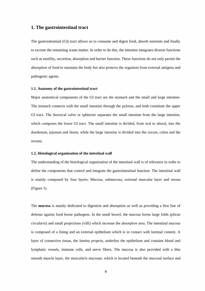
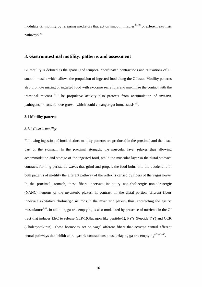
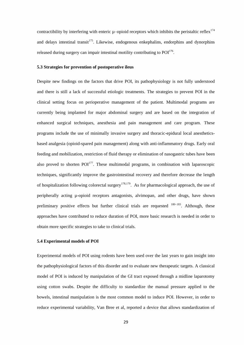
1.2. Histological organization of the intestinal wall
The understanding of the histological organization of the intestinal wall is of relevance in order to
define the components that control and integrate the gastrointestinal function. The intestinal wall
is mainly composed by four layers: Mucosa, submucosa, external muscular layer and serosa
(Figure 1).
The mucosa is mainly dedicated to digestion and absorption as well as providing a first line of
defense against food borne pathogens. In the small bowel, the mucosa forms large folds (plicae
circularis) and small projections (villi) which increase the absorptive area. The intestinal mucosa
is composed of a lining and an external epithelium which is in contact with luminal content. A
layer of connective tissue, the lamina propria, underlies the epithelium and contains blood and
lymphatic vessels, immune cells, and nerve fibers. The mucosa is also provided with a thin
smooth muscle layer, the muscularis mucosae, which is located beneath the mucosal surface and
10
which regulates movements of the mucosa. Underlying the mucosa, we find the submucosa, a
dense connective tissue layer that gives structural support to the mucosa. It contains the
submucous or Meissner’s plexus which mainly controls mucosal blood flow and secretomotor
functions1. The external muscular layer (o muscularis externa) is composed by two layers of
smooth muscle: the inner muscular layer (or circular layer) and the outer muscular layer (or
longitudinal layer). Both are responsible for the contractile movements of the intestinal wall.
Between these two layers, we find the myenteric or Aurbach’s plexus which consists of a
neuronal network and which controls the contraction of both muscular layers. The most external
intestinal layer is the serosa which is composed of connective tissue covered by a monolayer of
flat epithelial cells.
Figure 1. Organization of intestinal wall. Modified from http://philschatz.com/anatomy-book/contents/m46506.html.
11
2. Control of gastrointestinal motility
GI motility is finely regulated by the enteric nervous system (ENS) and the autonomous nervous
system (ANS). While the ENS represents the intrinsic component within the intestinal wall, the
ANS is referred as to the extrinsic component of GI regulation and a link to the central nervous
system (CNS) 2. In most of the cases, the ENS can initiate a motor program independently of the
central neural connections. For instance, the peristaltic reflex, is a motor pattern exclusively
elicited by ENS activity 3. In contrast, ENS and ANS work together to regulate gastric
emptying4,5
.
2.1 The enteric nervous system.
The ENS is composed of a neuronal network embedded within the intestinal wall. This so-called
"brain of the gut” contains more than 108 neurons organised in two major ganglionated plexuses;
the submucous or Meissner’s plexus and the myenteric or Aurbach plexus6. Enteric neurons can
be classified according to their function in motor neurons7,8
, interneurons8,9
and sensory
neurons6,10
, also called intrinsic primary afferent neurons (IPAN’s).
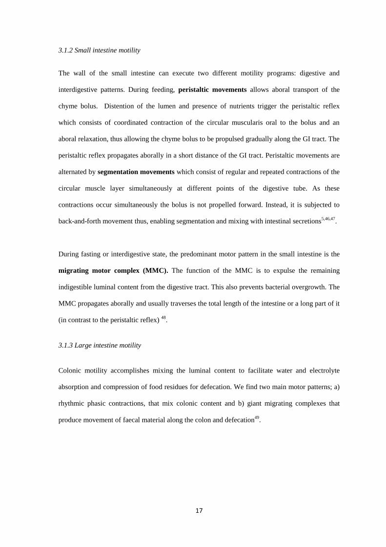
The peristaltic reflex is an example of how enteric neurons work co-ordinately to allow
contraction of the circular muscular layer oral to the point of stimulation while producing an
aboral relaxation (Figure 2). The peristaltic reflex is initiated by mechanical stimulation of the
mucosa and by presence of luminal nutrients which induces release of 5-hydroxytryptamine (5-
HT) from mucosal enter endocrine cells. Subsequently, 5-HT activates IPANs which release
calcitonin gene-related peptide (CGRP), to activate two types of interneurons 1) Ascending
interneurons which activate excitatory, cholinergic and tachykininergic motor-neurons. This
subsequent release of acetylcholine (ACh) and substance P (SP) induces contractions of circular
muscle orally to the stimulation. 2) Descending interneurons which activate inhibitory motor-
neurons that, through release of nitric oxide (NO), vasoactive intestinal peptide (VIP), and other
potential neurotransmitters, such as ATP, induce the relaxation of the circular muscle aboral11,12
.
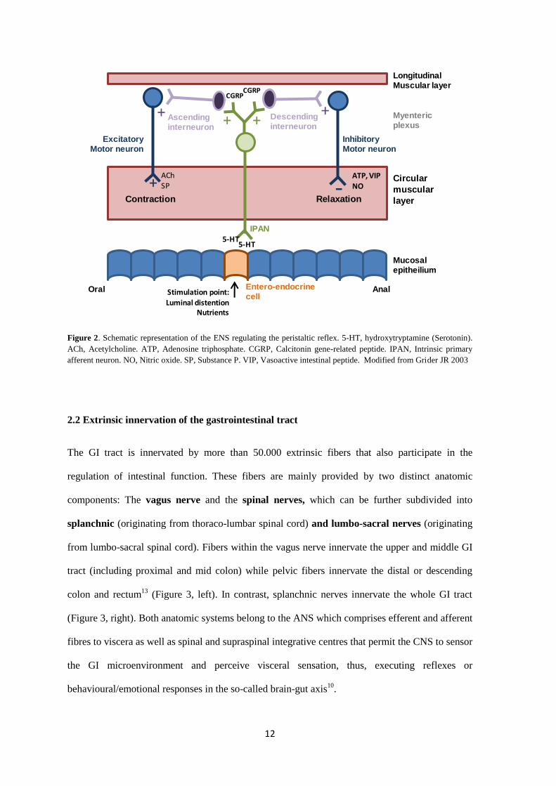
12
Figure 2. Schematic representation of the ENS regulating the peristaltic reflex. 5-HT, hydroxytryptamine (Serotonin).
ACh, Acetylcholine. ATP, Adenosine triphosphate. CGRP, Calcitonin gene-related peptide. IPAN, Intrinsic primary
afferent neuron. NO, Nitric oxide. SP, Substance P. VIP, Vasoactive intestinal peptide. Modified from Grider JR 2003
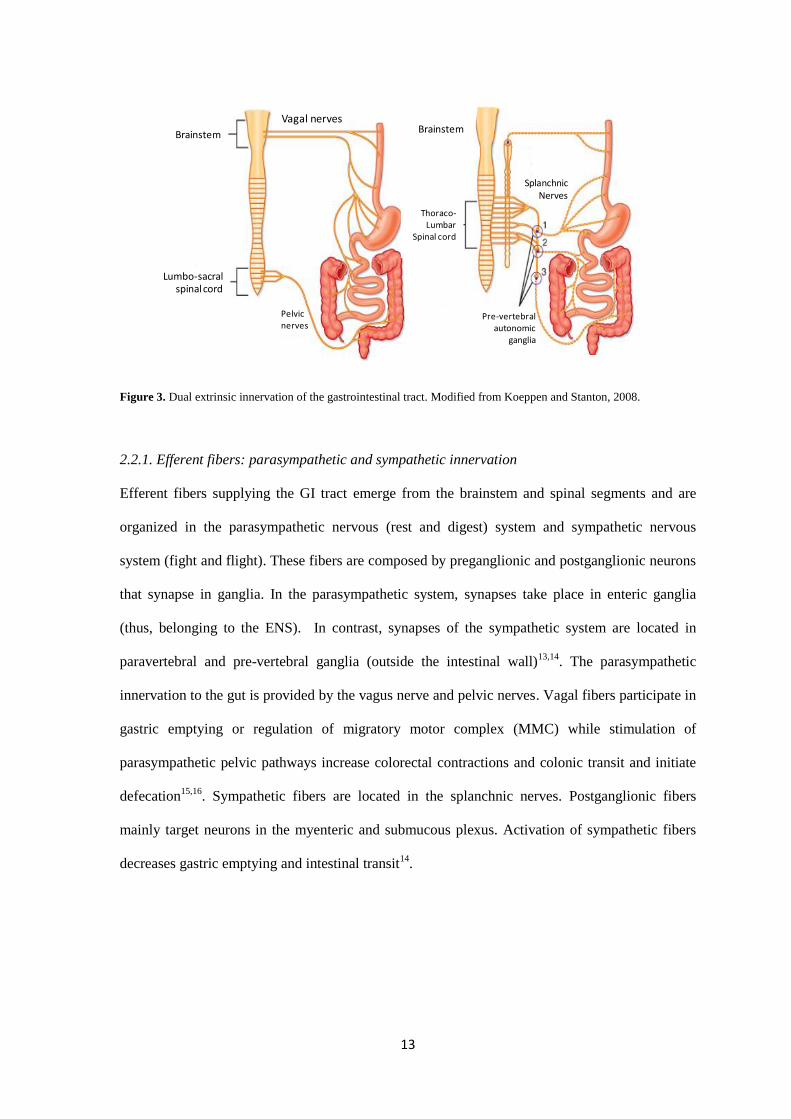
2.2 Extrinsic innervation of the gastrointestinal tract
The GI tract is innervated by more than 50.000 extrinsic fibers that also participate in the
regulation of intestinal function. These fibers are mainly provided by two distinct anatomic
components: The vagus nerve and the spinal nerves, which can be further subdivided into
splanchnic (originating from thoraco-lumbar spinal cord) and lumbo-sacral nerves (originating
from lumbo-sacral spinal cord). Fibers within the vagus nerve innervate the upper and middle GI
tract (including proximal and mid colon) while pelvic fibers innervate the distal or descending
colon and rectum13
(Figure 3, left). In contrast, splanchnic nerves innervate the whole GI tract
(Figure 3, right). Both anatomic systems belong to the ANS which comprises efferent and afferent
fibres to viscera as well as spinal and supraspinal integrative centres that permit the CNS to sensor
the GI microenvironment and perceive visceral sensation, thus, executing reflexes or
behavioural/emotional responses in the so-called brain-gut axis10
.
Contraction Relaxation
5-HT
Entero-endocrinecell
5-HT
IPAN
CGRP
Ascendinginterneuron
Descendinginterneuron
ExcitatoryMotor neuron
InhibitoryMotor neuron
ATP, VIP
NO
AChSP
CGRP
Stimulation point:Luminal distention
Nutrients
Mucosalepitheilium
Circular
muscular
layer
LongitudinalMuscular layer
Myentericplexus
Oral Anal
+ ++ +
+ -
13
Figure 3. Dual extrinsic innervation of the gastrointestinal tract. Modified from Koeppen and Stanton, 2008.
2.2.1. Efferent fibers: parasympathetic and sympathetic innervation
Efferent fibers supplying the GI tract emerge from the brainstem and spinal segments and are
organized in the parasympathetic nervous (rest and digest) system and sympathetic nervous
system (fight and flight). These fibers are composed by preganglionic and postganglionic neurons
that synapse in ganglia. In the parasympathetic system, synapses take place in enteric ganglia
(thus, belonging to the ENS). In contrast, synapses of the sympathetic system are located in
paravertebral and pre-vertebral ganglia (outside the intestinal wall)13,14
. The parasympathetic
innervation to the gut is provided by the vagus nerve and pelvic nerves. Vagal fibers participate in
gastric emptying or regulation of migratory motor complex (MMC) while stimulation of
parasympathetic pelvic pathways increase colorectal contractions and colonic transit and initiate
defecation15,16
. Sympathetic fibers are located in the splanchnic nerves. Postganglionic fibers
mainly target neurons in the myenteric and submucous plexus. Activation of sympathetic fibers
decreases gastric emptying and intestinal transit14
.
Lumbo-sacralspinal cord
Vagal nerves
Thoraco-Lumbar
Spinal cord
Brainstem Brainstem
SplanchnicNerves
Pelvicnerves
Pre-vertebral autonomic
ganglia
Lumbo-sacralspinalcord
Vagal nerves
Thoraco-Lumbar
Spinal cord
BrainstemBrainstem
SplanchnicNerves
Pelvicnerves
Pre-vertebral autonomic
ganglia
14
2.2.2. Afferent innervation
Afferent neurons within the extrinsic nerves provide an anatomical connection between the GI
tract and the CNS. Therefore, these neurons convey sensorial information to the brain allowing
individuals to have visceral perception (discomfort, pain perception, satiety, etc.).
The GI tract receives dual afferent innervation (Figure 3): the vagal afferent system (vagus nerve)
and the spinal afferent system that divides into splanchnic nerves and pelvic nerves. Afferent
extrinsic neurons have their cell bodies outside of the intestinal wall as described below and
mainly, axons are small-diameter-sized and unmyelinated C fibers that conduce at a slow velocity
(2m/s) 17,18
. Anatomical and functional differences between both systems are well characterized
17,19–23 and are summarized in table 1.
Table 1. Distinctive features of visceral afferent systems.
Vagal afferent Splanchnic
afferents
Pelvic afferents
Location
of cell bodies
Nodose and jugular
ganglia
Thoraco-lumbar
DRG
Lumbo-sacral
DRG
CNS integration NTS Spinal dorsal horn Spinal dorsal horn
CNS projections Amygdale or
hypothalamus from
DMN/nucleus
ambiguous
Thalamic nuclei through spinothalamic,
spinoreticular or dorsal column pathways
Stimuli -Physiological
-Non-painful
-Low-threshold
mechanical stimuli
(10-20mmHg)
-5-HT
-Non-physiological
-Painful/ inflamm.
-High threshold
mechanical stimuli
(>30mmHg)
-Bradykinin
-Physiological
-Non-painful
-Low-threshold
mechanical stimuli
(10-20mmHg)
% afferent
representation
90% 10-20% 30-40%
CGRP/SP content
in nerve endings
Low High -
5-HT hydroxytryptamine (Serotonin), CGRP Calcitonin gene-related peptide, CNS Central nervous system,
DMN Dorsal motor nucleus, DRG Dorsal root ganglia, NTS Nucleus tractus solitary, SP Substance P
15
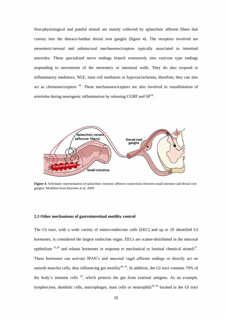
Non-physiological and painful stimuli are mainly collected by splanchnic afferent fibers that
convey into the thoraco-lumbar dorsal root ganglia (figure 4). The receptors involved are
mesenteric/serosal and submucosal mechanonociceptors typically associated to intestinal
arterioles. These specialized nerve endings branch extensively into varicose type endings
responding to movements of the mesentery or intestinal walls. They do also respond to
inflammatory mediators, NGF, mast cell mediators or hypoxia/ischemia, therefore, they can also
act as chemonociceptors 18
. These mechanonociceptors are also involved in vasodilatation of
arterioles during neurogenic inflammation by releasing CGRP and SP24
.
Figure 4. Schematic representation of splanchnic extrinsic afferent connections between small intestine and dorsal root
ganglia. Modified from Knowles et al. 2009
2.3 Other mechanisms of gastrointestinal motility control
The GI tract, with a wide variety of entero-endocrine cells (EEC) and up to 20 identified GI
hormones, is considered the largest endocrine organ. EECs are scatter-distributed in the mucosal
epithelium 25,26
and release hormones in response to mechanical or luminal chemical stimuli27
.
These hormones can activate IPAN’s and mucosal vagal afferent endings or directly act on
smooth muscles cells, thus influencing gut motility28–33
. In addition, the GI tract contains 70% of
the body’s immune cells 10
, which protects the gut from external antigens. As an example,
lymphocytes, dendritic cells, macrophages, mast cells or neutrophils34–36
located in the GI tract
Splanchnic nerves
Dorsal root
Small intestine
ganglia(afferent fibers)
16
modulate GI motility by releasing mediators that act on smooth muscles37–39
or afferent extrinsic
pathways 40
.
3. Gastrointestinal motility: patterns and assessment
GI motility is defined as the spatial and temporal coordinated contractions and relaxations of GI
smooth muscle which allows the propulsion of ingested food along the GI tract. Motility patterns
also promote mixing of ingested food with exocrine secretions and maximize the contact with the
intestinal mucosa 5. The propulsive activity also protects from accumulation of invasive
pathogens or bacterial overgrowth which could endanger gut homeostasis 41
.
3.1 Motility patterns
3.1.1 Gastric motility
Following ingestion of food, distinct motility patterns are produced in the proximal and the distal
part of the stomach. In the proximal stomach, the muscular layer relaxes thus allowing
accommodation and storage of the ingested food, while the muscular layer in the distal stomach
contracts forming peristaltic waves that grind and propels the food bolus into the duodenum. In
both patterns of motility the efferent pathway of the reflex is carried by fibers of the vagus nerve.
In the proximal stomach, these fibers innervate inhibitory non-cholinergic non-adrenergic
(NANC) neurons of the myenteric plexus. In contrast, in the distal portion, efferent fibers
innervate excitatory cholinergic neurons in the myenteric plexus, thus, contracting the gastric
musculature5,42
. In addition, gastric emptying is also modulated by presence of nutrients in the GI
tract that induces EEC to release GLP-1(Glucagon like peptide-1), PYY (Peptide YY) and CCK
(Cholecystokinin). These hormones act on vagal afferent fibers that activate central efferent
neural pathways that inhibit antral gastric contractions, thus, delaying gastric emptying4,29,43–45
.
17
3.1.2 Small intestine motility
The wall of the small intestine can execute two different motility programs: digestive and
interdigestive patterns. During feeding, peristaltic movements allows aboral transport of the
chyme bolus. Distention of the lumen and presence of nutrients trigger the peristaltic reflex
which consists of coordinated contraction of the circular muscularis oral to the bolus and an
aboral relaxation, thus allowing the chyme bolus to be propulsed gradually along the GI tract. The
peristaltic reflex propagates aborally in a short distance of the GI tract. Peristaltic movements are
alternated by segmentation movements which consist of regular and repeated contractions of the
circular muscle layer simultaneously at different points of the digestive tube. As these
contractions occur simultaneously the bolus is not propelled forward. Instead, it is subjected to
back-and-forth movement thus, enabling segmentation and mixing with intestinal secretions5,46,47
.
During fasting or interdigestive state, the predominant motor pattern in the small intestine is the
migrating motor complex (MMC). The function of the MMC is to expulse the remaining
indigestible luminal content from the digestive tract. This also prevents bacterial overgrowth. The
MMC propagates aborally and usually traverses the total length of the intestine or a long part of it
(in contrast to the peristaltic reflex) 48
.
3.1.3 Large intestine motility
Colonic motility accomplishes mixing the luminal content to facilitate water and electrolyte
absorption and compression of food residues for defecation. We find two main motor patterns; a)
rhythmic phasic contractions, that mix colonic content and b) giant migrating complexes that
produce movement of faecal material along the colon and defecation49
.
18
3.2 Gastrointestinal motility measurement
The measurement of GI motility is of major importance to obtain information on physiological
processes and pathologies related to the GI tract. The most common techniques used in the GI
research and clinical field are summarized in table 2.
Table 2. Techniques to assess gastro-intestinal motility
Parameter
In vivo techniques
Distribution of oral markers % GE
Geometric centre (intestinal
transit)
Mice50
Mice51
Scintography Time to half GE
Intestinal transit time
Mice52
Human53
Rat54
Ultrasonography Time to half GE Human55
Wireless motility capsule GE and intestinal transit
time
Human56
Charcoal head Intestinal transit Rat57
Lactulose breath test Intestinal transit time Human53
Radiopaque markers Intestinal transit time Human58
Cat59
Strain gauge Isometric tension
(muscle contractibility)
Rat60
Radio-telemetry Muscle electrical activity Rat61
Manometry Intraluminal pressure Human62
Food intake
Defecation
Estimation of GE
and colonic transit
Rat63
Human64
In vitro techniques
Organ bath Isometric tension (muscle contractibility)
Rat40,65
Spatio-temporal motility
mapping Luminal diameter Mice66
GE Gastric emptying; GC Geometric center
For the development of this thesis, in vivo small intestine transit has been used as a primary
endpoint for motility assessment in rodents. Intestinal transit can be defined as the passage of
ingested bolus through the GI time in a defined time. Because intestinal transit is subjected to
various regulatory neural and hormonal mechanisms, it reflects a complete physiological process,
19
including feedback from other GI areas or the CNS. This provides a much more complete and
integrated view of the GI transport function compared to the measurement of, for instance,
isolated smooth muscle cells or muscular strips ex-vivo, which provide information on only
intrinsic neuromuscular function 67
. In this work, the Geometric center (GC) technique has been
used for the evaluation of intestinal transit in rodents for being more accurate than other methods.
Other methods commonly used include the charcoal head technique. In this method transit is
measured by analyzing the time required for a suspension of non-absorbable charcoal to travel
throughout the length of the GI tract. Intestinal transit is expressed as a percentage of the total
intestinal length. While this method does not require much time it does not provide an accurate
transit estimation since it only focuses on the head of the charcoal bolus 57
. On the other hand, the
CG technique used in this work involves the analysis of the distribution of an oral-ingested
marker along the GI tract over a period of time. Mostly, marker distribution is assessed post-
mortem after dividing the GI tract in equally-sized segments. The calculated CG value is based on
the percentage of the marker in the individual segments and indicates in which segment the
marker has accumulated predominantly. Therefore, CG technique, compared to charcoal, results
in a more accurate estimation of intestinal transit as it takes into consideration the distribution of
the whole ingested bolus, not only the head of it, which may lead to intestinal transit
overestimation. Higher CG values indicate a quicker intestinal transit68
. Non-absorbable and inert
colorimetric (phenol red), fluorescent (fluorescein-labelled dextran 70kDa) or radiolabeled
markers (54
Cr) are routinely used 69–71
.
For patients undergoing surgery, intestinal transit in this work was evaluated using the lactulose
breath test. This technique measures oro-cecal transit time by means of ingestion of a
carbohydrate which is only metabolized by cecal bacteria. Hydrogen (H2) which is produced
exclusively by fermentation of lactulose in the cecum passes from the blood to lungs and is finally
exhaled. The exhaled H2 is monitored by sampling the breath and the arrival of the lactulose into
20
the cecum can be registered as a sharp rise in breath H2. The orocecal transit time is then given as
the time between ingestion of lactulose and a specified threshold concentration of breath H253,72
.
Finally, intestinal motility was indirectly assessed in animal and patients studies using clinical
parameters. For instance, fecal output and food intake have been previosly used as endopoints to
define gastric emptying and colonic propulsion activity in animal models. Likewise, solid oral
intake and defecation has been demonstrated to correlate with colonic transit in patients after
colorectal surgery 64,73–75
.
4. Mast cells
4.1 Activation and degranulation
In general terms, mast cells (MCs) are heterogeneous inflammatory cells widely distributed in the
organism carrying out defense and surveillance functions. MC are predominantly located in
strategic positions in tissues exposed to the external environment such as the as the respiratory
epithelium, the mucosa and submucosa of digestive and urinary tract, and the skin 76,77
. This
location allows MCs to be a first line of defense against noxious stimuli and pathogens and to
orchestrate the innate and the adaptive immune response76,77
. MCs are also involved in many
inflammatory conditions such as asthma, atopic dermatitis, arthritis, atherosclerosis or interstitial
cystitis 78–82
. Likewise, MCs are involved in diseases of the gastrointestinal tract (GI) such as
inflammatory bowel disease (IBD), irritable bowel syndrome (IBS) or postoperative ileus (POI)
83,84.
First reported by Paul Ehrlich over a century ago, MCs are easily recognized under light
microscopy by cytoplasmatic granules that stain purple with basic aniline dyes such as toluidine
blue, a phenomenon known as metachromasia85
. These granules contain mediators that are
released when the MC is stimulated. These mediators can be found already synthesized (and
21
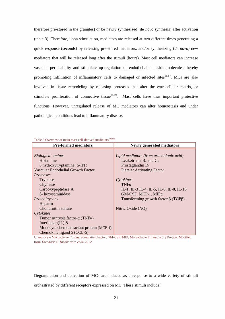
therefore pre-stored in the granules) or be newly synthesized (de novo synthesis) after activation
(table 3). Therefore, upon stimulation, mediators are released at two different times generating a
quick response (seconds) by releasing pre-stored mediators, and/or synthesizing (de novo) new
mediators that will be released long after the stimuli (hours). Mast cell mediators can increase
vascular permeability and stimulate up-regulation of endothelial adhesion molecules thereby
promoting infiltration of inflammatory cells to damaged or infected sites86,87
. MCs are also
involved in tissue remodeling by releasing proteases that alter the extracellular matrix, or
stimulate proliferation of connective tissue88,89
. Mast cells have thus important protective
functions. However, unregulated release of MC mediators can alter homeostasis and under
pathological conditions lead to inflammatory disease.
Table 3 Overview of main mast cell-derived mediators 91,92
Pre-formed mediators Newly generated mediators
Biological amines
Histamine
5 hydroxytryptamine (5-HT)
Vascular Endothelial Growth Factor
Proteases
Tryptase
Chymase
Carboxypeptidase A
β- hexosaminidase
Proteolgycans
Heparin
Chondroitin sulfate
Cytokines
Tumor necrosis factor-α (TNFα)
Interleukin(IL)-8
Monocyte chemoattractant protein (MCP-1)
Chemokine ligand 5 (CCL-5)
Lipid mediators (from arachidonic acid)
Leukotriene B4 and C4
Prostaglandin D2
Platelet Activating Factor
Cytokines
TNFα
IL-1, IL-3 IL-4, IL-5, IL-6, IL-8, IL-1β
GM-CSF, MCP-1, MIPα
Transforming growth factor β (TGFβ)
Nitric Oxide (NO)
Granulocyte Macrophage Colony Stimulating Factor, GM-CSF; MIP, Macrophage Inflammatory Protein. Modified
from Theoharis C Theoharides et al. 2012
Degranulation and activation of MCs are induced as a response to a wide variety of stimuli
orchestrated by different receptors expressed on MC. These stimuli include:
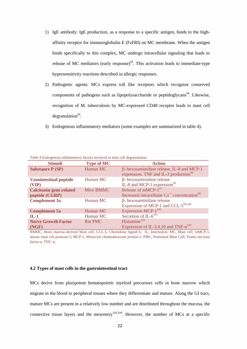
22
1) IgE antibody: IgE production, as a response to a specific antigen, binds to the high-
affinity receptor for immunoglobulin E (FcƐRI) on MC membrane. When the antigen
binds specifically to this complex, MC undergo intracellular signaling that leads to
release of MC mediators (early response)93
. This activation leads to immediate-type
hypersensitivity reactions described in allergic responses.
2) Pathogenic agents: MCs express toll like receptors which recognize conserved
components of pathogens such as lipopolysaccharide or peptidoglycans94
. Likewise,
recognition of M. tuberculosis by MC-expressed CD48 receptor leads to mast cell
degranulation95
.
3) Endogenous inflammatory mediators (some examples are summarized in table 4).
Table 4 Endogenous inflammatory factors involved in mast cell degranulation
Stimuli Type of MC Action
Substance P (SP) Human MC β- hexosaminidase release, IL-8 and MCP-1
expression. TNF and IL-3 production96
Vasointestinal peptide
(VIP)
Human MC β- hexosaminidase release
IL-8 and MCP-1 expression96
Calcitonin gene related
peptide (CGRP)
Mice BMMC Release of mMCP-197
Increased intracellular Ca++
concentration98
Complement 3a Human MC β- hexosaminidase release
Expression of MCP-1 and CCL-599,100
Complement 5a Human MC Expression MCP-1100
IL-1 Human MC Secretion of IL-6101
Nerve Growth Factor
(NGF)
Rat PMC
Histamine102
Expression of IL-3,4,10 and TNF-α103
BMMC, Bone marrow-derived Mast cell; CCL-5, Chemokine ligand-5; IL, Interleukin; MC, Mast cell; mMCP-1,
mouse mast cell protease-1; MCP-1, Monocyte chemoattractant protein-1; PMC, Peritoneal Mast Cell; Tumor necrosis
factor-α, TNF- α.
4.2 Types of mast cells in the gastrointestinal tract
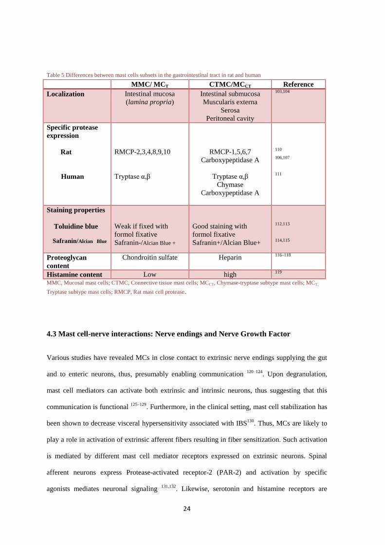
MCs derive from pluripotent hematopoietic myeloid precursors cells in bone marrow which
migrate in the blood to peripheral tissues where they differentiate and mature. Along the GI tract,
mature MCs are present in a relatively low number and are distributed throughout the mucosa, the
connective tissue layers and the mesentery103,104
. However, the number of MCs at a specific

23
location can increase dramatically as result of inflammatory response. Local environmental
factors (Stem cell factor, IL-3, Th2 inflammation) drive the differentiation of MC precursors and
ultimately lead to different phenotypes of MC104,105
. Mature MC classification generally falls into
two different subtypes. In the GI tract, we find mucosal mast cells (MMC) and Connective tissue
mast cells (CTMC).
The most predominant feature that differentiates both subtypes of MC is the localization and the
differential expression of MC specific serine-proteases pre-stored in cytoplasmatic granules.
In the rat, while MMC reside in the mucosa’s lamina propria, CTMC can be found in the
submucosa, muscular layers, serosa and peritoneum. Likewise rat MMC mainly express Rat Mast
Cell Protease-2 (RMCP-2), a soluble chymase106
. While rat CTMC mainly express the tryptase
called RMCP-6107
.
In humans, the nomenclature differs from that on the rat. MCs expressing only tryptase are
referred as tryptase subtype mast cells (MCT) and reside predominantly in the mucosa. On the
other hand, chymase-tryptase subtype mast cells (MCCT) are those expressing both tryptase and
chymase. MCCT are confined to the submucosa, muscular layers, serosa and peritoneum108,109
.
Other morphological and functional differences between both subtypes of MCs are detailed in
table 5. Each subtype has different roles in disease. Therefore, characterization and understanding
of the differences of MC subtypes is of major relevance. In addition, it allows to specifically study
the subtype of interest. For the development of this thesis RMCP-2, 6 and human tryptase and
chymase have been selected as specific markers for identification of the different subtypes of
MCs.
24
Table 5 Differences between mast cells subsets in the gastrointestinal tract in rat and human
MMC/ MCT CTMC/MCCT Reference
Localization Intestinal mucosa
(lamina propria)
Intestinal submucosa
Muscularis externa
Serosa
Peritoneal cavity
103,104
Specific protease
expression
Rat
Human
RMCP-2,3,4,8,9,10
Tryptase α,β
RMCP-1,5,6,7
Carboxypeptidase A
Tryptase α,β
Chymase
Carboxypeptidase A
110
106,107
111
Staining properties
Toluidine blue
Safranin/Alcian Blue
Weak if fixed with
formol fixative
Safranin-/Alcian Blue +
Good staining with
formol fixative
Safranin+/Alcian Blue+
112,113
114,115
Proteoglycan
content
Chondroitin sulfate Heparin 116–118
Histamine content Low high 119
MMC, Mucosal mast cells; CTMC, Connective tissue mast cells; MCCT, Chymase-tryptase subtype mast cells; MCT,
Tryptase subtype mast cells; RMCP, Rat mast cell protease.
4.3 Mast cell-nerve interactions: Nerve endings and Nerve Growth Factor
Various studies have revealed MCs in close contact to extrinsic nerve endings supplying the gut
and to enteric neurons, thus, presumably enabling communication 120–124
. Upon degranulation,
mast cell mediators can activate both extrinsic and intrinsic neurons, thus suggesting that this
communication is functional 125–129
. Furthermore, in the clinical setting, mast cell stabilization has
been shown to decrease visceral hypersensitivity associated with IBS130
. Thus, MCs are likely to
play a role in activation of extrinsic afferent fibers resulting in fiber sensitization. Such activation
is mediated by different mast cell mediator receptors expressed on extrinsic neurons. Spinal
afferent neurons express Protease-activated receptor-2 (PAR-2) and activation by specific
agonists mediates neuronal signaling 131,132
. Likewise, serotonin and histamine receptors are
25
expressed by sensory spinal neurons and their activation induces neuronal sensitization and
enhances visceral hypersensitivity in vivo 133–136
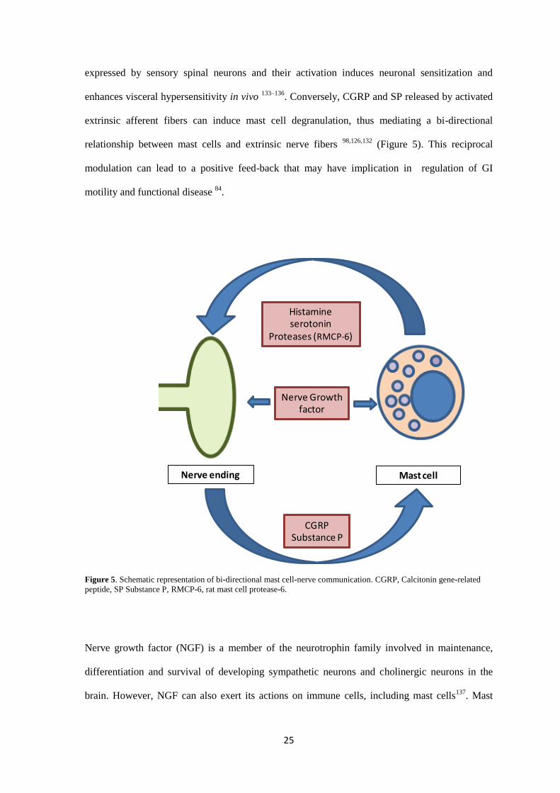
. Conversely, CGRP and SP released by activated
extrinsic afferent fibers can induce mast cell degranulation, thus mediating a bi-directional
relationship between mast cells and extrinsic nerve fibers 98,126,132
(Figure 5). This reciprocal
modulation can lead to a positive feed-back that may have implication in regulation of GI
motility and functional disease 84
.
Figure 5. Schematic representation of bi-directional mast cell-nerve communication. CGRP, Calcitonin gene-related
peptide, SP Substance P, RMCP-6, rat mast cell protease-6.
Nerve growth factor (NGF) is a member of the neurotrophin family involved in maintenance,
differentiation and survival of developing sympathetic neurons and cholinergic neurons in the
brain. However, NGF can also exert its actions on immune cells, including mast cells137
. Mast
Histamineserotonin
Proteases (RMCP-6)
CGRPSubstance P
Nerve Growthfactor
Nerve ending Mast cell
26
cells express the high affinity tropomyosin kynase receptor A (TrkA) through which NGF-
induced responses are mediated 138–140
. NGF acts as an mast cell-chemoattractant in vitro, plays a
role in differentiation and proliferation of MCs and serves as a trigger for mast cell degranulation
and mast cell hyperplasia 98,102,126,141–145
. Furthermore, NGF contributes to the sensitization of
spinal afferent neurons by up-regulating expression of neuropeptides CGRP and SP in dorsal root
ganglia. This could lead to a NGF-dependent visceral hypersensitivity observed in a rat model of
colitis146–149
. Various studies reported a role for NGF in the IBS model, in which MC activation
and impaired intestinal motility are prevented by using anti-NGF or NGF-blocking
treatment138,150
. Therefore, NGF-MC-nerve interactions (Figure 5) could provide a new
therapeutic target to treat functional GI-disorders.
5. Postoperative ileus
5.1 Definition and clinical symptoms
Postoperative ileus (POI) is defined as a temporal cessation of bowel motility after major
abdominal surgery leading to impaired propulsive activity of the GI tract151
. POI occurs as
consequence of opening of the peritoneum and handling or resection of the viscera especially the
intestines151
. The most common surgical procedures leading to POI are the resection of the small
intestine (19.2% of small intestine resection patients) and large (14.9%) intestine 152
. Clinical
symptoms of POI include abdominal pain and distention, nausea and vomiting, delayed passage
of stool and the inability to ingest fluids and solids. Although POI is not life-threatening, it
contributes to prolonged discomfort, morbidity and hospitalization of patients, thus increasing
health care expenses. The economic burden is estimated to be 600 million € per year153
. POI is
therefore considered an important health care issue.
27
5.2 Pathophysiology of POI
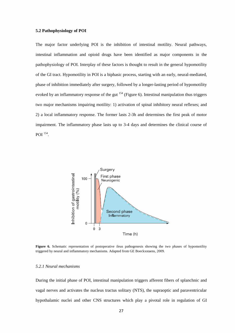
The major factor underlying POI is the inhibition of intestinal motility. Neural pathways,
intestinal inflammation and opioid drugs have been identified as major components in the
pathophysiology of POI. Interplay of these factors is thought to result in the general hypomotility
of the GI tract. Hypomotility in POI is a biphasic process, starting with an early, neural-mediated,
phase of inhibition immediately after surgery, followed by a longer-lasting period of hypomotility
evoked by an inflammatory response of the gut 154
(Figure 6). Intestinal manipulation thus triggers
two major mechanisms impairing motility: 1) activation of spinal inhibitory neural reflexes; and
2) a local inflammatory response. The former lasts 2-3h and determines the first peak of motor
impairment. The inflammatory phase lasts up to 3-4 days and determines the clinical course of
POI 154
.
Figure 6. Schematic representation of postoperative ileus pathogenesis showing the two phases of hypomotility
triggered by neural and inflammatory mechanisms. Adapted from GE Boeckxstaens, 2009.
5.2.1 Neural mechanisms
During the initial phase of POI, intestinal manipulation triggers afferent fibers of splanchnic and
vagal nerves and activates the nucleus tractus solitary (NTS), the supraoptic and paraventricular
hypothalamic nuclei and other CNS structures which play a pivotal role in regulation of GI
28
motility 155,156
. Adrenergic sympathetic pathways have been postulated as the efferent arm which
inhibits intestinal motility following surgical handling of the bowel. These inhibitory pathways
may arise from spinal ventral horn closing an entero-enteric inhibitory reflex or from descending
pathways travelling back from the activated structures in the CNS157
. A part from adrenergic
pathways, inhibition of the intestine is also mediated by non-adrenergic non-cholinergic (NANC)
efferent pathways158,159
which could be mediated by the vagus nerve160
.
During the late phase of POI, inhibition of intestinal motility is also mediated by a spinal reflex
through activation of splanchnic afferent and sympathetic fibers. This reflex depends on
inflammatory leukocyte infiltrate in the muscular layer 155,157,161,162
. Simultaneously, the
inflammatory infiltrate of the muscular layer, mobilized by the intestinal manipulation and tissue
damage, activates the afferent and efferent arm of the vagus nerve thereby activating the anti-
inflammatory vago-vagal reflex 163
.
5.2.2 Inflammatory mechanisms
Intestinal manipulation of the bowel results in an inflammatory response of the muscular layer
which leads to the dysfunction of smooth muscle contractibility. This was demonstrated by Kalff
et al. who identified an inflammatory infiltrate composed of macrophages, neutrophils, natural
killer cells, T-lymphocytes and mast cells in the manipulated jejunum which correlated with
impairment of smooth muscle contractibility in vitro in rodents and human 164–167
. The infiltrating
leucocytes, mostly macrophages and TH1 lymphocytes, release inflammatory mediators such as
IL-1 168
, IL-6 169
, IL-8167
, IL-12, IFN-γ 170
, prostanoids and NO 39,171
which are up-regulated upon
gut manipulation and contribute to dysmotility in POI. Intercellular adhesion molecule 1 (ICAM-
1) and lymphocyte function-associated antigen 1 (LFA-1) participate in leukocyte recruitment and
also play a role in the development of inflammatory POI172,173
.
5.2.3 Pharmacological mechanisms
Opiates given in the perioperative period and endogenous-opioids released during surgery also
contribute to POI. Opioid-based pain management impairs intestinal smooth muscle
29
contractibility by interfering with enteric µ–opioid receptors which inhibits the peristaltic reflex174
and delays intestinal transit175
. Likewise, endogenous enkephalins, endorphins and dynorphins
released during surgery can impair intestinal motility contributing to POI176
.
5.3 Strategies for prevention of postoperative ileus
Despite new findings on the factors that drive POI, its pathophysiology is not fully understood
and there is still a lack of successful etiologic treatments. The strategies to prevent POI in the
clinical setting focus on perioperative management of the patient. Multimodal programs are
currently being implanted for major abdominal surgery and are based on the integration of
enhanced surgical techniques, anesthesia and pain management and care program. These
programs include the use of minimally invasive surgery and thoracic-epidural local anesthetics-
based analgesia (opioid-spared pain management) along with anti-inflammatory drugs. Early oral
feeding and mobilization, restriction of fluid therapy or elimination of nasogastric tubes have been
also proved to shorten POI177
. These multimodal programs, in combination with laparoscopic
techniques, significantly improve the gastrointestinal recovery and therefore decrease the length
of hospitalization following colorectal surgery178,179
. As for pharmacological approach, the use of
peripherally acting µ-opioid receptors antagonists, alvimopan, and other drugs, have shown
preliminary positive effects but further clinical trials are requested 180–183
. Although, these
approaches have contributed to reduce duration of POI, more basic research is needed in order to
obtain more specific strategies to take to clinical trials.
5.4 Experimental models of POI
Experimental models of POI using rodents have been used over the last years to gain insight into
the pathophysiological factors of this disorder and to evaluate new therapeutic targets. A classical
model of POI is induced by manipulation of the GI tract exposed through a midline laparotomy
using cotton swabs. Despite the difficulty to standardize the manual pressure applied to the
bowels, intestinal manipulation is the most common model to induce POI. However, in order to
reduce experimental variability, Van Bree et al, reported a device that allows standardization of
30
applied pressure onto the intestine184
. Although these models are intended to resemble the surgical
handling of the intestine during colorectal surgery, they do not always reflect the clinical setting.
Such is the case with the intestinal anastomotic model, which although better reflecting the
condition in patients, is not used routinely as a model of POI. This may be due to post-surgical
complications – stenosis, dehiscence or ischemia - invalidating animals to remain in the
experiment or interfering with functional outcome 185
. Finally, another model recently reported in
POI research is the use of minimally invasive surgery in which intestinal manipulation is
performed by laparoscopy, thus allowing comparison with open procedure186
.
5.5 Postoperative ileus and mast cells
Over the last ten years many studies have focused on the role of mast cells (MCs) in the
pathogenesis of POI. The first evidence of MCs involvement in POI was the observation of mast
cell protease release in peritoneal lavage soon after intestinal manipulation187
. In addition, the use
of mast cell stabilizer ketotifen improved symptoms of POI in animal models. Mast cell-deficient
KO-mice also suffered from a less severe POI than wild type controls187
. These observations
suggested that MCs may be involved in the pathophysiology52
. This was supported by the
observation that degranulation of human peritoneal MCs (PMC) was related to the intensity of gut
manipulation in patients undergoing abdominal surgery167
. In addition, a clinical trial using
ketotifen as pre-treatment demonstrated improvement of gastric emptying in patients undergoing
gynecological surgery188
. These data suggest an important role for MC in POI and has provided
new potential therapeutic options for treatment.
In addition it has been proposed that locally released MC mediators, increase intestinal
permeability189
. This facilitates entrance of luminal, bacteria-derived products which additionally
activates intestinal macrophages154
following intestinal manipulation164
. Upon activation, these
macrophages secrete pro-inflammatory cytokines and chemokines that induce leukocyte
recruitment and prostanoids and nitric oxide production that impair enteric muscular function154
.
Therefore, MCs have been considered as major contributors of the inflammatory cascade leading
31
to POI. However the exact mechanisms involved in the activation of MCs following abdominal
surgery are not known. There is also a lack of knowledge on the mediators released by MCs
during abdominal surgery and their effector mechanisms contributing to POI. Thus the specific
role of MCs in the pathogenesis of POI remains unclear and requires further investigation.

32
33
HYPOTHESIS AND
OBJECTIVES

34
35
Previous studies referred in the introduction suggest that MCs activation and impaired GI motility
must be considered as potential effectors for POI. In addition, intestinal manipulation leads to
activation of MCs located in the peritoneum and inhibition of such events prevents POI. On the
other hand, MCs can interact with intestinal nerve endings. Such interactions are mainly mediated
by neuropeptides or NGF, and may suppose one of the potential mechanisms leading to POI.
On this basis, our working hypothesis was that MC degranulation would be mediated by NGF and
would trigger extrinsic neural activation in POI. MCs mediators (including or not NGF) would
act on splanchnic afferents that would activate inhibitory mechanisms on GI motility. The
ultimate aim of this thesis was to gain insight on mechanisms by which MCs contribute to POI.
For this purpose, we first studied patients undergoing colorectal surgery for being the most
susceptible to POI development. And secondly, we set up a POI experimental model in the rat to
further study and characterize GI motor function and morphological and molecular changes for
the protocols proposed.
In order to verify our working hypothesis, the objectives for this work were the following:
1. To characterize and associate mast cell activation and clinical recovery in colorectal
surgery.
2. To characterize intestinal motility, inflammation and mast cell activation in POI using a
rat model.
3. To study if mast cells contribute to POI rat experimental model.
4. To analyze gene expression of nociceptors in thoraco-lumbar dorsal root ganglia with the
aim to determine splanchnic activation in experimental POI.
5. To determine the involvement of mast cells in dorsal root ganglia activation during POI
using both mast cell degranulators and stabilizing drugs.
6. To determine if the NGF-antagonist, K252a, inhibits mast cell degranulation in vitro.
36
7. To study the effect of K252a in vivo on mast cell degranulation and in an experimental
model of POI.

37
CHAPTER 1

38
39
PERITONEAL MAST CELL DEGRANULATION AND
GASTROINTESTINAL RECOVERY IN PATIENTS UNDERGOING
COLORECTAL SURGERY
SERGIO BERDUN1; ERNEST BOMBUY M.D., Ph.D.
2; OSCAR ESTRADA M.D.
2; ESTHER
MANS M.D.2; JAKUB RYCHTER Ph.D.
1,3; PERE CLAVÉ M.D., Ph.D.
2,3; PATRI VERGARA
Ph.D. 1,3
1Department of Cell Biology, Physiology and Immunology, Universitat Autònoma de Barcelona,
Barcelona, Spain
2Department of Surgery. Consorci Sanitari del Maresme (CSdM) - Hospital de Mataró,
Universitat Autònoma de Barcelona, Barcelona, Spain
3Centro de Investigación Biomédica en Red de Enfermedades Hepáticas y Digestivas
(CIBERehd), Instituto de Salud Carlos III, Barcelona, Spain
Key words: Clinical recovery, intestinal surgery, mast cells, postoperative ileus
Neurogastroenterol Motil. 2015 Feb 9. doi: 10.1111/nmo.12525. [Epub ahead of print]

40
41
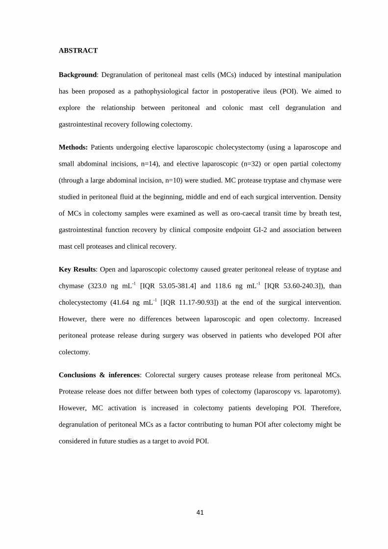
ABSTRACT
Background: Degranulation of peritoneal mast cells (MCs) induced by intestinal manipulation
has been proposed as a pathophysiological factor in postoperative ileus (POI). We aimed to
explore the relationship between peritoneal and colonic mast cell degranulation and
gastrointestinal recovery following colectomy.
Methods: Patients undergoing elective laparoscopic cholecystectomy (using a laparoscope and
small abdominal incisions, n=14), and elective laparoscopic (n=32) or open partial colectomy
(through a large abdominal incision, n=10) were studied. MC protease tryptase and chymase were
studied in peritoneal fluid at the beginning, middle and end of each surgical intervention. Density
of MCs in colectomy samples were examined as well as oro-caecal transit time by breath test,
gastrointestinal function recovery by clinical composite endpoint GI-2 and association between
mast cell proteases and clinical recovery.
Key Results: Open and laparoscopic colectomy caused greater peritoneal release of tryptase and
chymase (323.0 ng mL-1
[IQR 53.05-381.4] and 118.6 ng mL-1
[IQR 53.60-240.3]), than
cholecystectomy (41.64 ng mL-1
[IQR 11.17-90.93]) at the end of the surgical intervention.
However, there were no differences between laparoscopic and open colectomy. Increased
peritoneal protease release during surgery was observed in patients who developed POI after
colectomy.
Conclusions & inferences: Colorectal surgery causes protease release from peritoneal MCs.
Protease release does not differ between both types of colectomy (laparoscopy vs. laparotomy).
However, MC activation is increased in colectomy patients developing POI. Therefore,
degranulation of peritoneal MCs as a factor contributing to human POI after colectomy might be
considered in future studies as a target to avoid POI.
42
ABBREVIATIONS
ASA-PS American Society of Anaesthesiologists Physical-Health Status
GI Gastrointestinal
GI-2 Gastrointestinal index recovery 2 composite endpoint
GI-3 Gastrointestinal index recovery 3 composite endpoint
LHBT Lactulose hydrogen breath test
MC Mast cells
MCT Tryptase subtype of mast cell
MCCT Chymase-tryptase subtype of mast cells
OCTT Oro-caecal transit time
POI Postoperative ileus
43
KEY MESSAGES
-Peritoneal mast cell activation and postoperative clinical recovery were evaluated in patients
undergoing cholecystectomy and laparoscopic and open colectomies in a prospective and non-
randomized study.
- We demonstrate for the first time an increased release of mast cell mediators into the peritoneal
cavity during intestinal surgery.
- Colectomy patients with prolonged gastrointestinal clinical recovery presented higher levels of
peritoneal mast cell mediators.
44
INTRODUCTION
Postoperative ileus (POI) is a prevalent motility disorder defined as the transient cessation
of coordinated bowel motility after surgical intervention, which prevents effective transit of
intestinal contents and/or tolerance of oral intake 1. Primary POI occurs in almost all patients
recovering from gastrointestinal surgery and is usually resolved spontaneously by the second or
third day after laparoscopic surgery and by the fourth or fifth day after open laparotomy2–6
. POI
increases postoperative morbidity, prolongs hospitalization and increases healthcare costs7.
Although postsurgical recovery of gastrointestinal (GI) functions has improved with minimally
invasive surgery and multimodal perioperative care8,9
, the mechanisms underlying POI are still
not fully understood and specific and effective treatments are needed10
.
Over the last decade, mast cells (MC) have been thought to have a major role in the
pathophysiology of POI and could provide a mechanical connection between severity of intestinal
manipulation during abdominal surgery and clinical duration of POI. Human MCs are classified
according to their specific protease content. The tryptase subtype of mast cells, MCT, are located
within the intestinal mucosa while the tryptase-chymase subtype, MCCT, are mainly confined to
the submucosa, inner and outer muscularis and serosa/peritoneal cavity11
. MCCT located in the
peritoneum release a wide range of inflammatory mediators when activated by intestinal
manipulation12–14
, and this activation has been related to the intensity of bowel manipulation and
POI15
after major gynaecological surgery. In addition, De Jonge et al showed that stabilization of
peritoneal mast cells prevented POI in rodents14
. Studies with mast-cell-deficient mice, kitW/Wv
and kitW-sh/W-sh
, further corroborate mast cell implication in POI dysmotility12,14
. The et al reported
that the MC stabilizer, ketotifen, improved gastric emptying after hysterectomy in humans16
.
However, results with a new model of mutant mast-cell-deficient mice17
questioned the role of
MC in POI.
The aim of the present study was to explore MC activation and release of MC mediators
after colorectal surgery and to assess whether the intensity of this release was related to delay in
45
patients’ intestinal motility and clinical recovery. To our knowledge this the first study to
compare mast cell activation between laparoscopic and open colectomy. We focused on colorectal
surgery as it is the most common type of surgery to be followed by POI6,18
and so the one likely to
benefit the most from future pharmacological treatment.
MATERIAL AND METHODS
Study participants
The study included 56 non-consecutive patients undergoing elective laparoscopic
cholecystectomy or segmental colectomy at Mataró Hospital (Barcelona, Spain), from October
2010 to February 2012 (Table 1). Exclusion criteria were: 1) previous major abdominal surgery;
2) intra-abdominal infection or inflammation; 3) the use of non-steroidal anti-inflammatory drugs,
corticosteroids or mast cell stabilizers within 7 days prior to surgery; 4) treatment with prokinetics
within 14 days prior to surgery; 5) colostomies/ileostomies; and 6) intra-abdominal radiotherapy
within 30 days prior to surgery. Physical status was assessed before surgery according to the
American Society of Anaesthesiologists Physical-Health Status (ASA-PS).
Study design
This is a prospective and non-randomized study. Patients were divided into three groups for each
surgical procedure: 1) laparoscopic cholecystectomy (N=14), 2) laparoscopic segmental
colectomy (N=32), and 3) open segmental colectomy (N=10). Laparoscopic or open colectomy
procedures were assigned according to standard medical indications at our institution19
: Patients
with 1) anaesthetic contraindication, 2) severe cardiorespiratory insufficiency or 3) chronic liver
disease underwent open colectomy. Cholecystectomy patients were included as it is a
laparoscopic procedure that avoids manipulation of the GI tract and could therefore serve as
control group to obtain basal/normal values of proteases without intestinal manipulation.
Regarding clinical output, cholecystectomy patients were a reference for normal clinical recovery
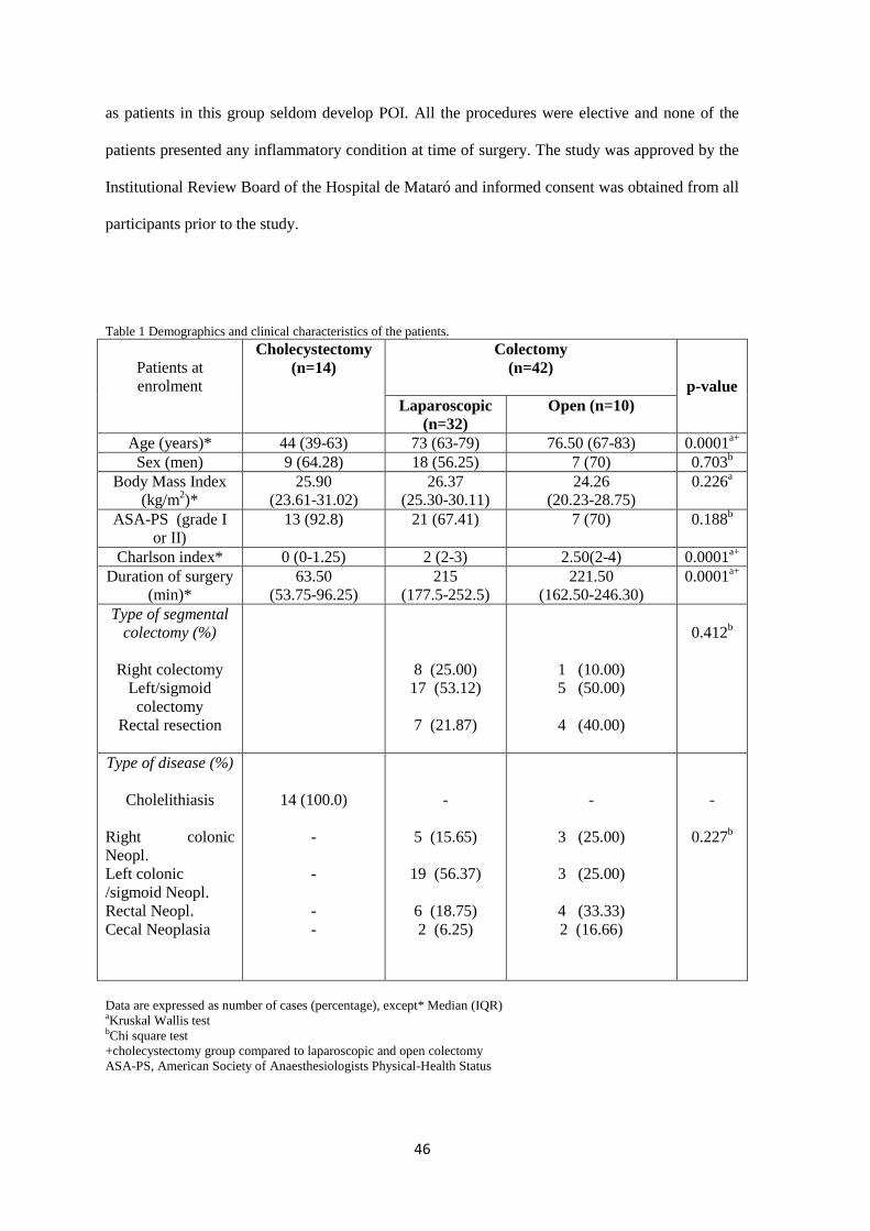
46
as patients in this group seldom develop POI. All the procedures were elective and none of the
patients presented any inflammatory condition at time of surgery. The study was approved by the
Institutional Review Board of the Hospital de Mataró and informed consent was obtained from all
participants prior to the study.
Table 1 Demographics and clinical characteristics of the patients.
Patients at
enrolment
Cholecystectomy
(n=14)
Colectomy
(n=42)
p-value
Laparoscopic
(n=32)
Open (n=10)
Age (years)* 44 (39-63) 73 (63-79) 76.50 (67-83) 0.0001a+
Sex (men) 9 (64.28) 18 (56.25) 7 (70) 0.703b
Body Mass Index
(kg/m2)*
25.90
(23.61-31.02)
26.37
(25.30-30.11)
24.26
(20.23-28.75)
0.226a
ASA-PS (grade I
or II)
13 (92.8) 21 (67.41) 7 (70) 0.188b
Charlson index* 0 (0-1.25) 2 (2-3) 2.50(2-4) 0.0001a+
Duration of surgery
(min)*
63.50
(53.75-96.25)
215
(177.5-252.5)
221.50
(162.50-246.30)
0.0001a+
Type of segmental
colectomy (%)
Right colectomy
Left/sigmoid
colectomy
Rectal resection
8 (25.00)
17 (53.12)
7 (21.87)
1 (10.00)
5 (50.00)
4 (40.00)
0.412b
Type of disease (%)
Cholelithiasis
Right colonic
Neopl.
Left colonic
/sigmoid Neopl.
Rectal Neopl.
Cecal Neoplasia
14 (100.0)
-
-
-
-
-
5 (15.65)
19 (56.37)
6 (18.75)
2 (6.25)
-
3 (25.00)
3 (25.00)
4 (33.33)
2 (16.66)
-
0.227b
Data are expressed as number of cases (percentage), except* Median (IQR) aKruskal Wallis test bChi square test
+cholecystectomy group compared to laparoscopic and open colectomy
ASA-PS, American Society of Anaesthesiologists Physical-Health Status
47
Perioperative care
Surgical interventions were performed by four expert colorectal surgeons exclusively dedicated to
colorectal surgery. They have completed the learning curve meaning they have performed at least
100 procedures. The four surgeons were assigned randomly and performed both laparoscopic and
open colectomies. A multidisciplinary case discussion took place for each clinical case (except for
cholecystectomy patients). All patients were informed about the procedure, were scheduled for
operation and underwent a pre-operative risk assessment. Only colectomy patients were put on a
low-fibre diet during preoperative days. Patients were fasted for 6h and premedicated with 5mg of
diazepam (po) the evening before surgery. Nasogastric tube insertion was not routinely
performed. Early feeding and deambulation were started 4-6h after surgery for cholecystectomy
patients and 24h for colectomy patients. Perioperative management is detailed in Table 2.
Assessment of mast cell activation during surgery
Peritoneal lavage samples were collected from each patient at three consecutive time points: 1)
immediately after opening the peritoneal cavity, 2) after initial inspection and the first handling of
the bowel (15 min after the first sample), and 3) at the end of the surgical procedure. Collection
was performed according to a modified method by The et al15
. Briefly, the peritoneal cavity was
gently flushed with 40ml of warm, sterile salt water, 0.9% NaCl. After 30s exposure, 15-20 ml of
the lavage fluid was collected, centrifuged at 240 RCF for 10 min at 4ºC and the resulting
supernatants were stored at -80ºC. A cocktail of protease inhibitors (P8340, Sigma-Aldrich, St
Louis, MO, US) was added to samples (1:100) immediately after collection to prevent protein
degradation by endogenous proteases.
Tryptase and chymase release assay
Tryptase and chymase content of peritoneal lavage and was measured by ELISA (E91070 and
E96515, Uscn, Life Science Inc, Wuhan, China) following manufacturer’s instructions.
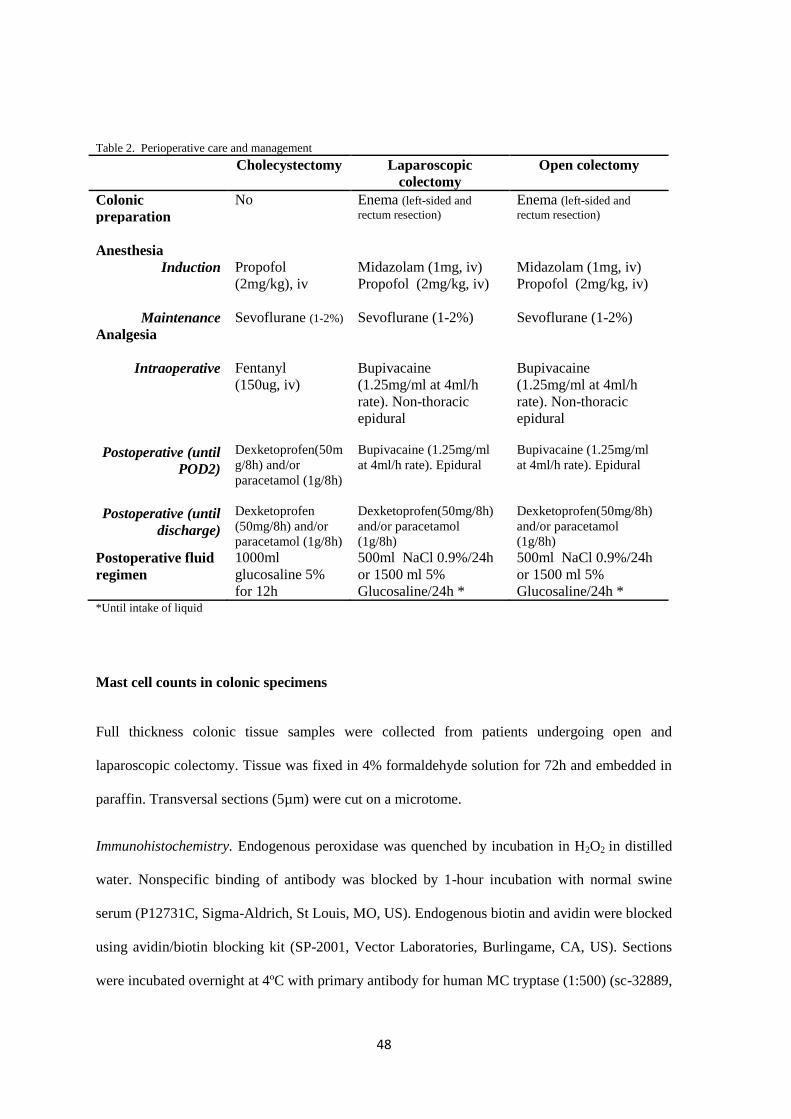
48
Table 2. Perioperative care and management
Cholecystectomy Laparoscopic
colectomy
Open colectomy
Colonic
preparation
No Enema (left-sided and
rectum resection) Enema (left-sided and
rectum resection)
Anesthesia
Induction
Propofol
(2mg/kg), iv
Midazolam (1mg, iv)
Propofol (2mg/kg, iv)
Midazolam (1mg, iv)
Propofol (2mg/kg, iv)
Maintenance
Sevoflurane (1-2%)
Sevoflurane (1-2%)
Sevoflurane (1-2%)
Analgesia
Intraoperative
Fentanyl
(150ug, iv)
Bupivacaine
(1.25mg/ml at 4ml/h
rate). Non-thoracic
epidural
Bupivacaine
(1.25mg/ml at 4ml/h
rate). Non-thoracic
epidural
Postoperative (until
POD2)
Dexketoprofen(50m
g/8h) and/or
paracetamol (1g/8h)
Bupivacaine (1.25mg/ml
at 4ml/h rate). Epidural
Bupivacaine (1.25mg/ml
at 4ml/h rate). Epidural
Postoperative (until
discharge)
Dexketoprofen
(50mg/8h) and/or
paracetamol (1g/8h)
Dexketoprofen(50mg/8h)
and/or paracetamol
(1g/8h)
Dexketoprofen(50mg/8h)
and/or paracetamol
(1g/8h)
Postoperative fluid
regimen
1000ml
glucosaline 5%
for 12h
500ml NaCl 0.9%/24h
or 1500 ml 5%
Glucosaline/24h *
500ml NaCl 0.9%/24h
or 1500 ml 5%
Glucosaline/24h * *Until intake of liquid
Mast cell counts in colonic specimens
Full thickness colonic tissue samples were collected from patients undergoing open and
laparoscopic colectomy. Tissue was fixed in 4% formaldehyde solution for 72h and embedded in
paraffin. Transversal sections (5µm) were cut on a microtome.
Immunohistochemistry. Endogenous peroxidase was quenched by incubation in H2O2 in distilled
water. Nonspecific binding of antibody was blocked by 1-hour incubation with normal swine
serum (P12731C, Sigma-Aldrich, St Louis, MO, US). Endogenous biotin and avidin were blocked
using avidin/biotin blocking kit (SP-2001, Vector Laboratories, Burlingame, CA, US). Sections
were incubated overnight at 4ºC with primary antibody for human MC tryptase (1:500) (sc-32889,
49
Santa Cruz Biotechnology, CA, US). Biotinylated polyclonal swine anti-rabbit IgG (1:200, 1h at
RT, E 0353; (Dakocytomation, Glostrup, Denmark)) was used as a secondary antibody followed
by incubation with Avidin/peroxidase kit (Vectastatin ABC kit, Vector Laboratories, Burlingame,
CA, US). Specific staining was developed with 3,3’-diaminobenzidine (peroxidase substrate kit,
SK-4100, Vector Laboratories Inc, Burlingame, CA, US) and sections were counterstained with
haematoxylin. Specificity of the staining was confirmed by omission of the primary antibody.
Toluidine blue staining. Transversal sections of the colon were also stained with a 0.5% toluidine
blue solution (Scharlau, CI 52040, Sentmenat, Spain) for 5 minutes.
Quantification of tissue MC. Tryptase-positive cells and metachromatic toluidine blue stained
cells were quantified under a light microscope at 40x magnification (Olympus CH 30, Tokyo,
Japan). The number of positive cells was expressed as the mean of 15 non-overlapping high-
power fields. For evaluation of percentage of degranulation, MC were categorized as ‘intact mast
cells’ (those with intact membrane outline, with no granules outside of the cell and intense
staining) or ‘degranulated mast cells’ (disrupted cell membrane and extrusion of granules)20,21
.
The number of degranulated mast cells was expressed as mean of percentage of degranulation of
10 non-overlapping high-power fields (60x).
Clinical assessment of POI and gastrointestinal recovery
Clinical assessment of gastrointestinal function. Recovery of GI function was monitored daily
until discharge by medical staff using the following endpoints: Gastrointestinal index recovery 2
and 3 (GI-2 and GI-3) composite endpoint, time to tolerance of solid and liquid intake, time to
first flatulence and time to be ready for discharge. GI-2 was defined as the maximum time
between first tolerance of solid food and first defecation and was the primary endpoint for
recovery and to define POI22
. GI-3 was defined as the maximum time to first tolerance of solid
50
food and minimum time between first flatus or first bowel movement, the most recent event being
the score counted 22
. Patients were considered to be ready for discharge when they met the
following criteria: tolerance to solid and liquid diet, satisfactory pain control with oral analgesics
and competent mobilization and physical autonomy8.
Colectomy patients were classified as either POI-developers or non-POI developers according to
GI-2 because this parameter has been correlated with colonic transit 23
. As mentioned above, GI-2
is defined by the later of these two events to occur: tolerance of solid food in absence of nausea or
vomiting or first defecation monitored by expert nursing staff. Patients with GI-2 >3 days were
considered to develop POI according to a previously validated definition of POI 2,24
Gastrointestinal transit time. The oro-caecal transit time (OCTT) was determined 24 hours after
surgery by lactulose hydrogen breath test (LHBT) as described elsewhere25
. Patients fasted for 12
hours and then consumed 10g of lactulose (Duphalac, Solvaypharma S.A, Parets del Vallès,
Spain) dissolved in 100ml of a liquid breakfast preparation of 200 Kcal (20.2 g of proteins, 15.6 g
of fats and 43.4 g of carbohydrates, T-diet 20/2 from Vegenat, Badajoz, Spain). Breath samples
were taken every 15 min for 4h. Breath H2 concentration was recorded with Gastrolyzer (Bedfont
Scientific Ltd. Rochester, Kent, England). A baseline breath H2 concentration was measured for
each patient before the ingestion of lactulose. OCTT was defined as the time between lactulose
ingestion and a rise to 20 ppm above baseline in breath H2. The 20ppm cut-off threshold was used
to exclude false positives25,26
and patients with high baseline values above 16 ppm (7 out of 39
patients) or with no detectable exhaled H2 (6 out of 39 patients) were excluded from the
analysis25
. Additionally, 14 healthy volunteers were included in the study to explore normal
OCTT using this methodology. A group of healthy volunteers was included, to have a reference
value of intestinal transit time in non-operated healthy subjects.
51
Statistical analysis
Categorical parameters (sex, ASA and type of surgery) were expressed as percentages of cases
and compared using Chi-Square test. Quantitative variables were considered non-parametrically
distributed and expressed as median and interquartile range (IQR). Protease concentration in
lavages was analysed by Friedman test for repeated measures followed by Dunn’s post-test. MC
counts were compared by Mann-Whitney U test for independent samples. OCTT and clinical
parameters were analysed by Kruskall-Wallis test and Dunn’s post-test for independent samples.
Colectomy patients were also divided regarding diagnosis of clinic POI and data re-analysed:
Protease concentration in lavages, number of mast cells, percentage of degranulation and duration
of surgery were analysed by Mann-Whitney U test (independent samples). The relationship
between MC and clinical parameters was assessed by Pearson (bivariate) correlation test and
partial correlation test.
All statistical analyses were performed using GraphPad Prism 6.0 except for correlation test for
which IBM SPPS v22 software was used. A p value <0.05 was considered significant.
RESULTS
Demographics and clinical characteristics of the sample and surgical procedures
Table 1 summarizes the demographic characteristics of the patients undergoing surgical
procedures. Sex and ASA-PS distribution and body mass index did not differ between
cholecystectomy and colectomy patients. As expected, colectomy patients were older than
cholecystectomy patients and Charlson Comorbidity Index was also higher. Duration of surgery
was shorter for cholecystectomies than for both types of colectomy procedures without any
significant difference between laparoscopic or laparotomic approaches. One laparoscopic
colectomy and one cholecystectomy had to be converted to open procedures for technical/surgical
reasons. Five patients from the colectomy group (with wound dehiscence, sepsis or pneumonia)
52
and 1 patient from cholecystectomy group (with intra-abdominal haemorrhage and wound
infection) were excluded from the analysis of gastrointestinal transit time and GI clinical
recovery.
Protease content in peritoneal lavage
Cholecystectomy did not trigger any detectable release of tryptase at any of the three time points.
In contrast, chymase was released at the end of cholecystectomy (41.64 ng mL-1
(IQR 11.17-
90.93)), P<0.010) (Figure 1). Laparoscopic colectomy did not trigger any detectable release of
tryptase at the beginning (0.12 ng mL-1
(IQR 0.03-0.24)) or middle of surgery but did trigger a
significant release of tryptase at the end (2.02 ng mL-1
(IQR 0.48-31.54)), P<0.0001). Chymase
concentration was significantly higher at the middle (39.10 ng mL-1
(IQR 12.25-106.40)),
P<0.050) and further increased at the end of laparoscopic colectomy (118.60 ng mL-1
(IQR 53.60-
240.30)), P<0.0001). Open colectomy was associated with a release of both proteases at the end
of surgery (tryptase: 4.99 ng mL-1
(IQR 0.417-13.60), P<0.05; chymase: 323 ng mL-1
(IQR 53.05-
381.40), P<0.010)). Protease release did not differ significantly between laparoscopic and open
colectomy at any point in the surgery although the latter tended towards an increased release at
the end of the surgical procedure (Figure 1).
Mast cell count and distribution in colonic wall
Toluidine blue and tryptase immunohistochemistry stained cells displayed MC-specific features
(Figure 2E, 2F and 2G). No metachromatic MCs were found in the mucosa, indicating that
toluidine selectively stains connective tissue MC (MCCT). Number of MC significantly decreased
from the luminal to the serosal side of the colonic wall (Figure 2A,B) and the density and
distribution of MC in colonic wall did not differ between laparoscopic and open surgery groups
(Figure 2A,B). MC were frequently associated with blood vessels in the serosa and submucosa
and only rarely associated with myenteric ganglia (Figure 2F).
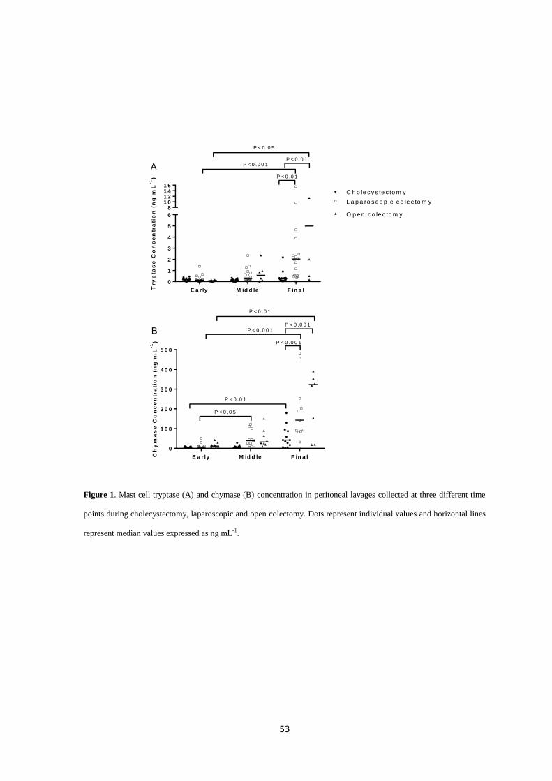
53
Figure 1. Mast cell tryptase (A) and chymase (B) concentration in peritoneal lavages collected at three different time
points during cholecystectomy, laparoscopic and open colectomy. Dots represent individual values and horizontal lines
represent median values expressed as ng mL-1.
E a r ly M id d le F in a l
0
1
2
3
4
5
6
81 01 21 41 6
P < 0 .0 1
P < 0 .0 5
P < 0 .0 0 1A
Try
pta
se
Co
nc
en
tra
tio
n (
ng
mL
-1) P < 0 .0 1
C h o le c y s te c to m y
L a p a ro s c o p ic c o le c to m y
O p e n c o le c to m y
E a r ly M id d le F in a l
0
1 0 0
2 0 0
3 0 0
4 0 0
5 0 0
P < 0 .0 0 1
P < 0 .0 0 1
P < 0 .0 1
P < 0 .0 0 1
P < 0 .0 1
P < 0 .0 5
B
Ch
ym
as
e C
on
ce
ntr
ati
on
(n
g m
L-1
)
Figure 1. Mast cell tryptase (A) and chymase (B) concentration in peritoneal lavages collected at three
different time points during cholecistectomy, laparoscopic and open colectomy . Bars and vertical lines
represents median with IQR expressed as ng/ml. +,++,+++ P<0.050, P<0.010, P<0.0001 vs basal values
of the same group of surgery. **,*** P<0.010, P<0.001 vs cholecistectomy group.
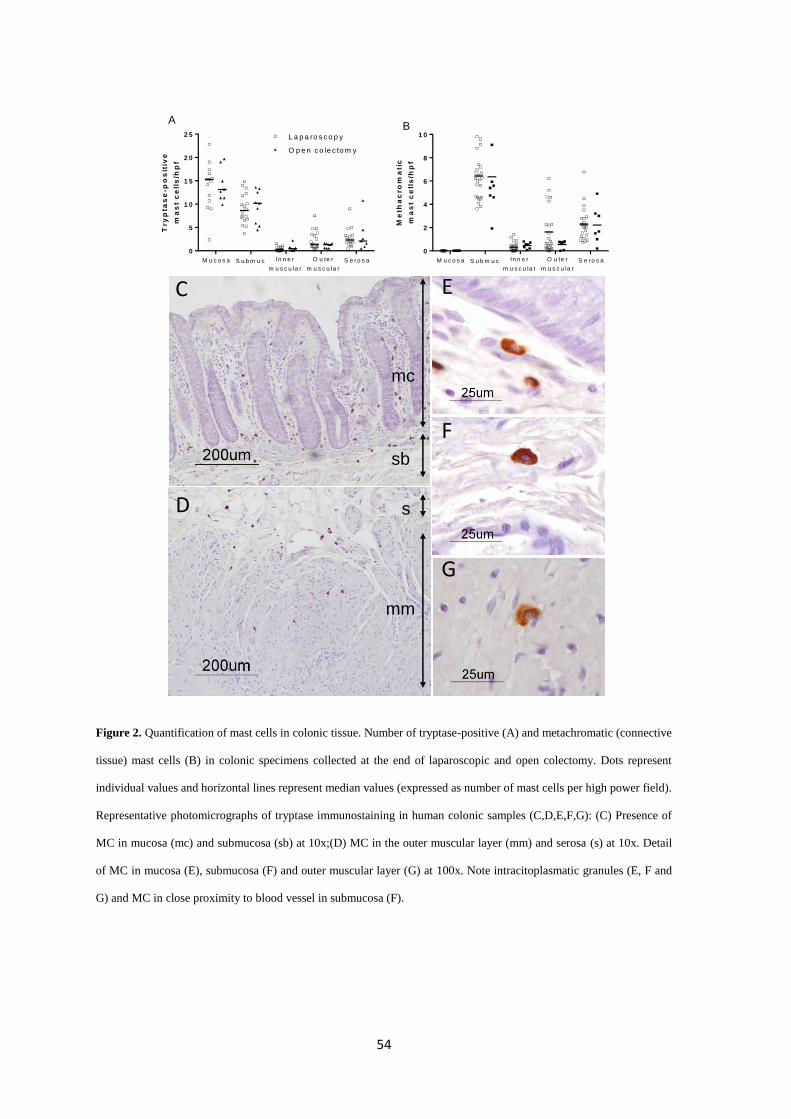
54
Figure 2. Quantification of mast cells in colonic tissue. Number of tryptase-positive (A) and metachromatic (connective
tissue) mast cells (B) in colonic specimens collected at the end of laparoscopic and open colectomy. Dots represent
individual values and horizontal lines represent median values (expressed as number of mast cells per high power field).
Representative photomicrographs of tryptase immunostaining in human colonic samples (C,D,E,F,G): (C) Presence of
MC in mucosa (mc) and submucosa (sb) at 10x;(D) MC in the outer muscular layer (mm) and serosa (s) at 10x. Detail
of MC in mucosa (E), submucosa (F) and outer muscular layer (G) at 100x. Note intracitoplasmatic granules (E, F and
G) and MC in close proximity to blood vessel in submucosa (F).
C
D
E
F
G
mc
sb
mm
s
0
5
1 0
1 5
2 0
2 5
M u c o s a S u b m u c In n e r
m u s c u la r
O u te r
m u s c u la r
S e ro s a
A
Try
pta
se
-po
sit
ive
ma
st
ce
lls
/hp
f
0
2
4
6
8
1 0
M u c o s a S u b m u c In n e r
m u s c u la r
O u te r
m u s c u la r
S e ro s a
BO p e n c o le c to m y
L a p a ro s c o p ic c o le c to m y
Me
tha
cro
ma
tic
ma
st
ce
lls
/hp
f
0
5
1 0
1 5
2 0
2 5
M u c o s a S u b m u c In n e r
m u s c u la r
O u te r
m u s c u la r
S e ro s a
A
Try
pta
se
-po
sit
ive
ma
st
ce
lls
/hp
f
O p e n c o le c to m y
L a p a ro s c o p y
0
2
4
6
8
1 0
M u c o s a S u b m u c In n e r
m u s c u la r
O u te r
m u s c u la r
S e ro s a
B
Me
tha
cro
ma
tic
ma
st
ce
lls
/hp
f
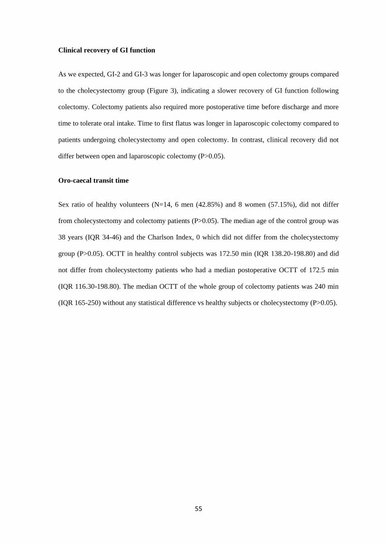
55
Clinical recovery of GI function
As we expected, GI-2 and GI-3 was longer for laparoscopic and open colectomy groups compared
to the cholecystectomy group (Figure 3), indicating a slower recovery of GI function following
colectomy. Colectomy patients also required more postoperative time before discharge and more
time to tolerate oral intake. Time to first flatus was longer in laparoscopic colectomy compared to
patients undergoing cholecystectomy and open colectomy. In contrast, clinical recovery did not
differ between open and laparoscopic colectomy (P>0.05).
Oro-caecal transit time
Sex ratio of healthy volunteers (N=14, 6 men (42.85%) and 8 women (57.15%), did not differ
from cholecystectomy and colectomy patients (P>0.05). The median age of the control group was
38 years (IQR 34-46) and the Charlson Index, 0 which did not differ from the cholecystectomy
group (P>0.05). OCTT in healthy control subjects was 172.50 min (IQR 138.20-198.80) and did
not differ from cholecystectomy patients who had a median postoperative OCTT of 172.5 min
(IQR 116.30-198.80). The median OCTT of the whole group of colectomy patients was 240 min
(IQR 165-250) without any statistical difference vs healthy subjects or cholecystectomy (P>0.05).
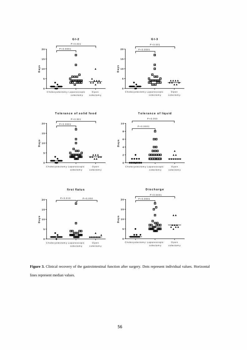
56
Figure 3. Clinical recovery of the gastrointestinal function after surgery. Dots represent individual values. Horizontal
lines represent median values.
G I-2
0
5
1 0
1 5
2 0 P < 0 .0 0 0 1
P < 0 .0 0 1
C h o le c y s te c to m y L a p a ro s c o p ic
c o le c to m y
O p e n
c o le c to m y
Da
ys
G I-3
0
5
1 0
1 5
2 0 P < 0 .0 0 0 1
P < 0 .0 0 1
C h o le c y s te c to m y L a p a ro s c o p ic
c o le c to m y
O p e n
c o le c to m y
Da
ys
T o le ra n c e o f s o lid fo o d
0
5
1 0
1 5
2 0 P < 0 .0 0 0 1
P < 0 .0 0 1
C h o le c y s te c to m y L a p a ro s c o p ic
c o le c to m y
O p e n
c o le c to m y
Da
ys
T o le r a n c e o f l iq u id
0
2
4
6
8
1 0
P < 0 .0 0 0 1
C h o le c y s te c to m y L a p a ro s c o p ic
c o le c to m y
O p e n
c o le c to m y
P < 0 .0 5 0D
ay
s
f irs t f la tu s
0
5
1 0
1 5
2 0P < 0 .0 1 0
C h o le c y s te c to m y L a p a ro s c o p ic
c o le c to m y
O p e n
c o le c to m y
P < 0 .0 5 0
Da
ys
D is c h a r g e
0
5
1 0
1 5
2 0 P < 0 .0 0 0 1
P < 0 .0 0 0 1
C h o le c y s te c to m y L a p a ro s c o p ic
c o le c to m y
O p e n
c o le c to m y
Da
ys
57
Evaluation of mast cells according to diagnosis of POI in colectomy patients
Colectomy patients were divided according to clinic diagnosis of POI and data were re-analysed
in order to see how mast cell-related parameters were distributed in relation to POI incidence
(Figure 4).
Non-POI patients and POI patients did not differ in age, sex, body mass index, Charlson index,
duration of surgery and percentage of type of colectomy and disease (data not shown). In contrast,
a higher proportion of ASA>3 were observed in patients developing POI (47.36% vs 6.6% in non-
POI patients).
According to our definition of POI, 57% (15/27) of laparoscopic colectomies and 44% (4/9) of
open colectomies developed prolonged POI compared to only 15% (2/13) of cholecystectomies.
Moreover, colectomy patients diagnosed with POI had increased peritoneal tryptase (3170 ng mL-
1 [IQR 1764-10971]) vs. non-POI patients (515.3ng mL
-1 [IQR 365.9-1842.0]) P=0.002 and
increased chymase (318.4ng mL-1
[IQR 94.59-456.3]) vs. non-POI patients (31.65ng mL-1
[IQR
18.6-142.6]) P=0.03.
In contrast, number and distribution of MCs and percentage of degranulation (non-POI patients:
mucosa 16.23% [5.91-39.37]; submucosa 4.87% [0.0-7.08]; inner muscular 0.0% [0.0-11.67];
outer muscular 3.03% [0.0-24.04] and serosa 9.52% [3.75-17.36] vs. POI patients: mucosa 5.19%
[1.33-21.72]; submucosa 7.14% [0.0-10.24]; inner muscular layer 0.0% [0.0-6.25]; outer muscular
layer 0.0% [0.0-23.96] and serosa 0.0% [0.0-1.493]) did not differ among groups (P>0.05, Figure
4).
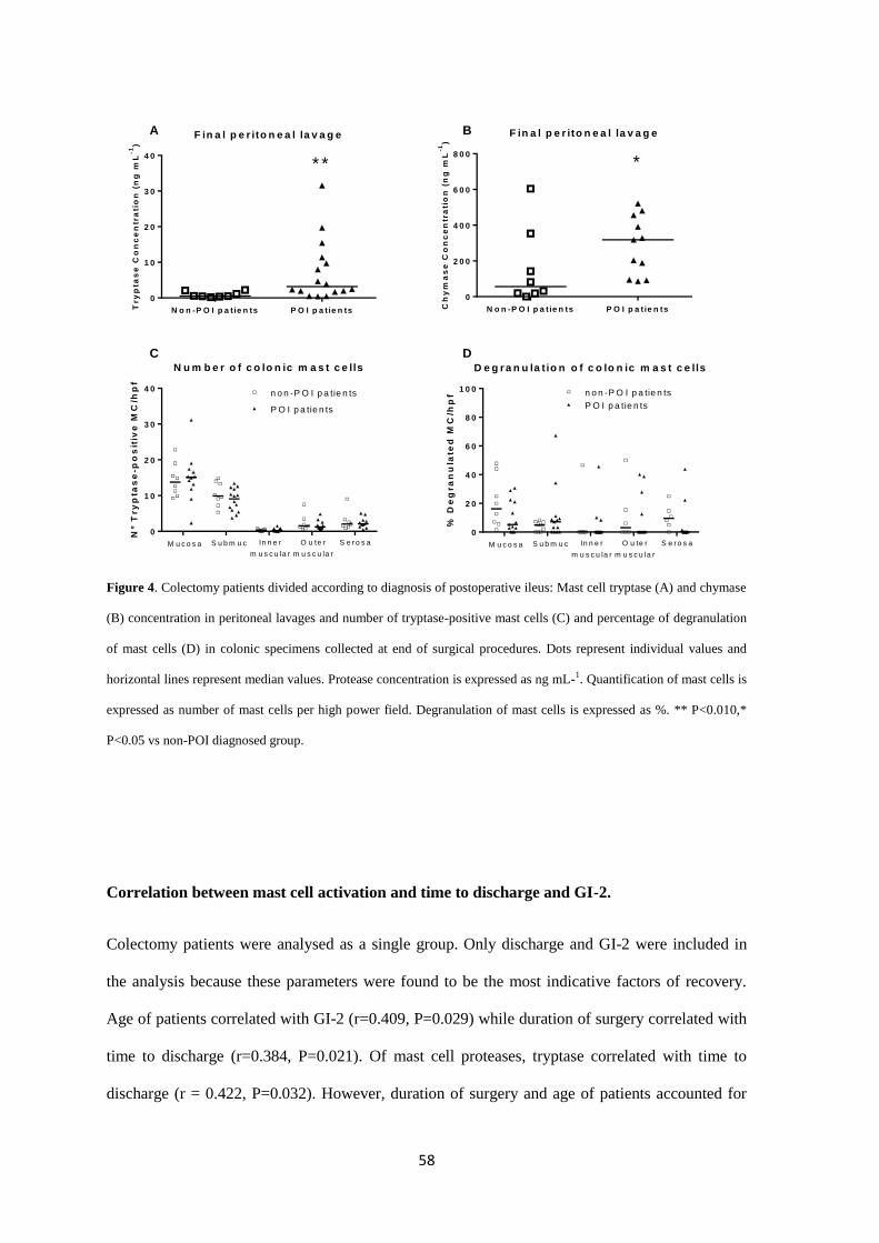
58
Figure 4. Colectomy patients divided according to diagnosis of postoperative ileus: Mast cell tryptase (A) and chymase
(B) concentration in peritoneal lavages and number of tryptase-positive mast cells (C) and percentage of degranulation
of mast cells (D) in colonic specimens collected at end of surgical procedures. Dots represent individual values and
horizontal lines represent median values. Protease concentration is expressed as ng mL-1. Quantification of mast cells is
expressed as number of mast cells per high power field. Degranulation of mast cells is expressed as %. ** P<0.010,*
P<0.05 vs non-POI diagnosed group.
Correlation between mast cell activation and time to discharge and GI-2.
Colectomy patients were analysed as a single group. Only discharge and GI-2 were included in
the analysis because these parameters were found to be the most indicative factors of recovery.
Age of patients correlated with GI-2 (r=0.409, P=0.029) while duration of surgery correlated with
time to discharge (r=0.384, P=0.021). Of mast cell proteases, tryptase correlated with time to
discharge (r = 0.422, P=0.032). However, duration of surgery and age of patients accounted for
F in a l p e r ito n e a l la v a g e
N o n -P O I p a t ie n ts P O I p a t ie n ts
0
1 0
2 0
3 0
4 0
* *
Try
pta
se
Co
nc
en
tra
tio
n (
ng
mL
-1)
F in a l p e r ito n e a l la v a g e
N o n -P O I p a t ie n ts P O I p a t ie n ts
0
2 0 0
4 0 0
6 0 0
8 0 0
*
Ch
ym
as
e C
on
ce
ntr
ati
on
(n
g m
L-1
)
N u m b e r o f c o lo n ic m a s t c e lls
Nº T
ry
pta
se
-po
sit
ive
MC
/hp
f
0
1 0
2 0
3 0
4 0
M u c o s a S u b m u c In n e r
m u s c u la r
O u te r
m u s c u la r
S e ro s a
P O I p a tie n ts
n o n -P O I p a tie n ts
D e g r a n u la tio n o f c o lo n ic m a s t c e lls
% D
eg
ra
nu
late
d M
C/h
pf
0
2 0
4 0
6 0
8 0
1 0 0
M u c o s a S u b m u c In n e r
m u s c u la r
O u te r
m u s c u la r
S e ro s a
n o n -P O I p a tie n ts
P O I p a tie n ts
A B
C D
59
this correlation as confirmed by partial correlation test. In fact, when we included these two
parameters in the analysis –by means of partial correlation test- correlation for tryptase-discharge
turned into r=0.34, P=0.092 after correction for the variable ‘duration’ and r = 0.392, P=0.052
after correction for the variable ‘age of patients’.
DISCUSSION
Our exploratory study shows for the first time that colorectal surgery is associated with MC
activation and the release of chymase and tryptase into the peritoneal cavity, irrespective of
whether the colonic resection is performed by laparotomy or laparoscopy. Furthermore, protease
release was greater in patients with POI. However, we could not establish a cause-effect
relationship between MC activation and postoperative ileus because other factors such as duration
of the surgery or age of the patients might have also influenced POI.
Our results agree with a previous study15
that has shown that intestinal manipulation
during abdominal surgery cause MC activation. In our study, however, MC activation was
evaluated for three different surgical protocols all requiring intraperitoneal approach. Our findings
show that colorectal surgery caused higher release of MC chymase and tryptase into the peritoneal
cavity compared to cholecystectomy where only a moderate release of MC proteases was
observed. This difference in protease release could also be influenced by the duration of the
surgery. However, we believe this is not the case because protease concentration at 15 minutes
(middle point for all groups) showed that cholecystectomy chymase (but not tryptase) release
remained unaltered during cholecystectomies while in colectomies it was already elevated.
Furthermore, when analysing non-POI colectomy patients (median duration of surgery 210 min),
we found similar values of proteases as in cholecystectomy patients (median duration of 63 min).
Consequently, our results strongly suggest that intestinal manipulation is the key factor in MC
activation. Moreover, an unexpected finding was that both laparoscopic and open colectomies
induced a similar release of MC proteases. Colectomies, independently of the surgical approach,
60
require intensive manipulation of the colon. Therefore, our results suggest that degranulation of
MC does not differ much between laparoscopic and open approaches when part of the colon is
removed. This is the first study to compare MC activation in intestinal surgery and the divergence
of our results with other studies in non-intestinal surgeries 15
indicates, that in colorectal surgery,
the handling and the resection of the colon are the main triggers of mast cell degranulation rather
than the surgical approach (laparotomy vs. laparoscopy). This agrees with The’s results that found
mast cell activation in laparotomic hysterectomy but not in the laparoscopic and transvaginal
approach15
.
Our results indicate a local response of MCs but we also aimed to evaluate whether
systemic activation occurs upon intestinal manipulation. We observed that MC tryptase was not
increased in plasma after colorectal surgery (data not shown). However the reduced number of
patients in that part of the study limited the strength of our findings and further research is needed
to confirm whether MC activation is only confined to the peritoneum. Moreover, in our study,
open colectomy did not prolong clinical recovery compared to laparoscopic colectomy, in
agreement with other studies 27–29
. Nonetheless, laparoscopic colectomy has been shown to
decrease surgical trauma30
, reduce POI and improve recovery following abdominal surgery3,8,9,31
.
Perioperative care management also influences patient recovery. Vlug MS 8 and Van Bree SHW
9
clearly showed that open colectomy patients also benefited from fast-track, multimodal programs
and have similar values for clinical recovery as laparoscopic patients receiving the same program.
In our study, all colectomy patients received a standardized, similar and multimodal treatment.
The lack of differences in our study could therefore result from similar perioperative care applied
to all patients from which laparotomy patients benefited. Our results indicate that under a
multimodal care program, the intensity of the manipulation of the colon is a key factor for
postoperative motility recovery rather than the surgical approach itself. This is supported by other
randomized studies that found no postoperative differences between both colectomy techniques
27,28. Furthermore, our findings agree with a previous study in patients undergoing hysterectomy
(where the colon is not removed) in which the surgical approach (laparotomy vs. transvaginal)
61
determined clinical recovery 15
. Criteria selection for open procedures in the colectomy group
does not seem to entail a bias that could explain our finding. In any case, one would expect a bias
towards a more delayed recovery in open colectomy patients according to their basal features.
Likewise, duration of surgery and age of patients correlated with some clinical parameters but as
we compared two similar surgery protocols that did not differ in duration or age of patients, and
where perioperative protocol were similar, we can confirm these factors did not determine
postoperative recovery in our patients.
Other studies have reported that the colon is the last part of GI tract to recover after
surgery32,33
which matches our findings. Our study demonstrated that the speed of clinical
recovery was not associated to major changes in small intestine motility when assessed by oro-
caecal transit time. It should be noted that lactulose by itself can slightly accelerate small
intestinal motility 34
. It is therefore possible that such effect would mask the effect of POI in our
measurement. Furthermore, the number of colectomy patients included in the analysis was limited
due to inclusion criteria.
Our study also showed that peritoneal MC activation was related to occurrence of POI.
Patients that were diagnosed with clinical POI according to a previously validated definition 2,24
,
had a significantly higher release of chymase and tryptase compared to patients that recovered
earlier. MC activation is associated to increased visceral sensitivity35,36
, intestinal permeability 37
and altered motor functions 38
that could fit into the multifactorial scenario leading to POI.
Nonetheless, we cannot conclude that MC degranulation alone is responsible for POI in our
colectomy patients, as there are other variables that potentially affect clinical recovery. In the
correlation analysis, duration of surgery and age of patients also correlated with time to discharge
and GI-2 and so should be considered as possible influences on GI clinical recovery after
colectomy. However, independently of other factors correlating with POI and the fact that our
correlation analysis did not reveal a clear correlation between protease and GI-2/time to
discharge, we report for the first time that POI patients after intestinal surgery presented a higher
MC protease release than those who did not suffer POI. A therapeutic study using MC blockers
62
such as ketotifen or sodium cromoglycate should be conducted in order to find out whether
preventing protease release could prevent POI.
A recent study using a new MC-deficient mouse model showed that there was not a role
for MC in POI, casting doubt on the putative role of MC in inducing POI 17
. Despite this
controversy, former models of murine POI and patients undergoing abdominal surgery 12–15
show
degranulation of peritoneal MC and this is now further supported by the present study in a
population of patients especially affected by POI. This study also describes MC distribution along
the human colon wall, something scarcely reported in the literature20,21
. However, we found no
evidence that surgical handling of the colon was followed by recruitment of MC in the
manipulated tissue, as reported in animal models of POI39
. Furthermore, POI patients did not
present changes in number of colonic MCs or morphological traits of degranulation that could
suggest activation of intestinal-wall-resident MCs. However, MC have been shown to have
piecemeal activation rather than full degranulation particularly in the gastrointestinal tract which
could explain these results37
. A more in-depth study using electron microscopy would be useful to
address this question. In addition, as MCs in the serosa are peritoneal, and peritoneal MCs are
difficult to stain or immunolabel (own observation and data not published), we think that this
could have masked changes in the colonic wall, at least in the serosa.
To sum up, our study shows, for the first time, that colorectal surgery causes significant
MC degranulation. In addition, colonic manipulation associated to resection seems to be the
predominant factor determining peritoneal MC degranulation rather than colectomy surgical
approach. Finally, higher levels of MC proteases in peritoneal lavage were found in patients with
delayed GI recovery and POI. We suggest, therefore, that MC may play an important role in POI
and efforts should be made to block this response regardless of the type of colectomy technique.
Further studies are needed to verify MC involvement in POI and to explore whether MC
stabilizers play a role in the prevention of POI following colorectal surgery.
63
ACKNOWLEDGMENTS, FUNDING AND DISCLOSURES
The authors thank E. Martínez, I. López, and O. Ortega for their assistance. Surgery fellows (Dr
M Del Bas, Dr M García, Dr A Heredia), the nursing staff and pathologic and laboratory clinic
diagnosis staff at Mataró Hospital are also thanked for their inestimable and enthusiastic support.
The authors thank Mrs Jane Lewis for editorial revision.
S.B designed and performed the studies related to mast cell activation, analysed data and wrote
the paper. E.B, O.E and E.M designed and performed the study, analysed clinical and functional
data and also contributed to writing the paper. J.R wrote the paper and contributed to analysing
data. P.C and P.V designed the study and wrote the paper.
This study was supported by grants BFU2010-15401 from Ministerio de Ciencia e Innovación,
2009SGR708 from Direcció General de Recerca de la Generalitat de Catalunya and a grant from
the filial of Maresme of the Academia de Ciencies Médiques i de la Salut de Catalunya y Balears.
S.B. personal support: FPU program (AP2010-1692) from Ministerio de Educación, Cultura y
Deporte.
Competing interests: the authors have no competing interests.
64
REFERENCES
1. Delaney C, Kehlet H, S. A. On behalf of the Postoperative Ileus Management Council
(PIMC). Postoperative ileus: profiles, Risk factors and Definitions. A framework for
Optimizing Surgical Outcomes in Patients Undergoing Major Abdominal and Colorectal
Surgery. Clinical Consensus Upd.
http://www.clinicalwebcasts.com/pdfs/GenSurg_WEB.pdf. 2006.
2. Chen, H. H. et al. Laparoscopic colectomy compares favorably with colectomy by
laparotomy for reduction of postoperative ileus. Dis. Colon Rectum 43, 61–5 (2000).
3. Schwenk, W., Böhm, B., Haase, O., Junghans, T. & Müller, J. M. Laparoscopic versus
conventional colorectal resection: a prospective randomised study of postoperative ileus
and early postoperative feeding. Langenbecks. Arch. Surg. 383, 49–55 (1998).
4. Lacy, A. M. et al. Laparoscopy-assisted colectomy versus open colectomy for treatment of
non-metastatic colon cancer: a randomised trial. Lancet 359, 2224–9 (2002).
5. Delaney, C. P. et al. Postoperative upper and lower gastrointestinal recovery and
gastrointestinal morbidity in patients undergoing bowel resection: pooled analysis of
placebo data from 3 randomized controlled trials. Am. J. Surg. 191, 315–9 (2006).
6. Wolff, B. G., Viscusi, E. R., Delaney, C. P., Du, W. & Techner, L. Patterns of
gastrointestinal recovery after bowel resection and total abdominal hysterectomy: pooled
results from the placebo arms of alvimopan phase III North American clinical trials. J. Am.
Coll. Surg. 205, 43–51 (2007).
7. Senagore, A. J. Pathogenesis and clinical and economic consequences of postoperative
ileus. Clin. Exp. Gastroenterol. 3, 87–9 (2010).
8. Vlug, M. S. et al. Laparoscopy in combination with fast track multimodal management is
the best perioperative strategy in patients undergoing colonic surgery: a randomized
clinical trial (LAFA-study). Ann. Surg. 254, 868–75 (2011).
9. Van Bree, S. H. W. et al. Faster recovery of gastrointestinal transit after laparoscopy and
fast-track care in patients undergoing colonic surgery. Gastroenterology 141, 872–880.e1–
4 (2011).
10. Rychter, J. & Clavé, P. Intestinal inflammation in postoperative ileus: pathogenesis and
therapeutic targets. Gut 62, 1534–5 (2013).
11. Irani, A. M., Bradford, T. R., Kepley, C. L., Schechter, N. M. & Schwartz, L. B. Detection
of MCT and MCTC types of human mast cells by immunohistochemistry using new
monoclonal anti-tryptase and anti-chymase antibodies. J. Histochem. Cytochem. 37, 1509–
15 (1989).
12. Snoek, S. A. et al. Mast cells trigger epithelial barrier dysfunction, bacterial translocation
and postoperative ileus in a mouse model. Neurogastroenterol. Motil. 24, 172–84, e91
(2012).
13. Van Bree, S. H. W. et al. Inhibition of spleen tyrosine kinase as treatment of postoperative
ileus. Gut 62, 1581–90 (2013).
65
14. De Jonge, W. J. et al. Mast cell degranulation during abdominal surgery initiates
postoperative ileus in mice. Gastroenterology 127, 535–45 (2004).
15. The, F. O. et al. Intestinal handling-induced mast cell activation and inflammation in
human postoperative ileus. Gut 57, 33–40 (2008).
16. The, F. O. et al. The role of mast cell stabilization in treatment of postoperative ileus: a
pilot study. Am. J. Gastroenterol. 104, 2257–66 (2009).
17. Gomez-Pinilla, P. J. et al. Mast cells play no role in the pathogenesis of postoperative ileus
induced by intestinal manipulation. PLoS One 9, e85304 (2014).
18. Federal Register. Health Care Financing Admnistration, 1999-2000. 2010.
Http://www.gpoaccess.gov/fr
19. Suñol, X. et al. [Planning, management and results of the implantation of laparoscopic
surgery of the colon and rectum in a general hospital]. Cirugía española 82, 99–104
(2007).
20. Maurer, C. A. & Cathomas, G. Colonic mast cells in controls and slow transit constipation
patients. Aliment. Pharmacol. Ther. 34, 92–9 (2011).
21. L, F. & Salerni, B. Increase of colonic mast cells in obstructed defecation and their
relationship with enteric glia. Dig. Dis. Sci. 57, 65–71 (2012).
22. Delaney, C. P. et al. Alvimopan, for postoperative ileus following bowel resection: a
pooled analysis of phase III studies. Ann. Surg. 245, 355–63 (2007).
23. Van Bree, S. H. W. et al. Identification of clinical outcome measures for recovery of
gastrointestinal motility in postoperative ileus. Ann. Surg. 259, 708–14 (2014).
24. Artinyan, A. et al. Prolonged postoperative ileus-definition, risk factors, and predictors
after surgery. World J. Surg. 32, 1495–500 (2008).
25. Yu, D., Cheeseman, F. & Vanner, S. Combined oro-caecal scintigraphy and lactulose
hydrogen breath testing demonstrate that breath testing detects oro-caecal transit, not small
intestinal bacterial overgrowth in patients with IBS. Gut 60, 334–40 (2011).
26. Ghoshal, U. C. How to interpret hydrogen breath tests. J. Neurogastroenterol. Motil. 17,
312–7 (2011).
27. Basse, L. et al. Functional recovery after open versus laparoscopic colonic resection: a
randomized, blinded study. Ann. Surg. 241, 416–23 (2005).
28. Basse, L., Madsen, J. L., Billesbølle, P., Bardram, L. & Kehlet, H. Gastrointestinal transit
after laparoscopic versus open colonic resection. Surg. Endosc. 17, 1919–22 (2003).
29. MacKay, G. et al. Laparoscopic colonic resection in fast-track patients does not enhance
short-term recovery after elective surgery. Colorectal Dis. 9, 368–72 (2007).
30. Hildebrandt, U. et al. Comparison of surgical stress between laparoscopic and open
colonic resections. Surg. Endosc. 17, 242–6 (2003).
66
31. Veldkamp, R. et al. Laparoscopic surgery versus open surgery for colon cancer: short-term
outcomes of a randomised trial. Lancet Oncol. 6, 477–84 (2005).
32. Waldhausen, J. H., Shaffrey, M. E., Skenderis, B. S., Jones, R. S. & Schirmer, B. D.
Gastrointestinal myoelectric and clinical patterns of recovery after laparotomy. Ann. Surg.
211, 777–84; discussion 785 (1990).
33. Condon, R. E. et al. Resolution of postoperative ileus in humans. Ann. Surg. 203, 574–81
(1986).
34. Miller, M. A. et al. Comparison of scintigraphy and lactulose breath hydrogen test for
assessment of orocecal transit: lactulose accelerates small bowel transit. Dig. Dis. Sci. 42,
10–8 (1997).
35. Barbara, G. et al. Mast cell-dependent excitation of visceral-nociceptive sensory neurons
in irritable bowel syndrome. Gastroenterology 132, 26–37 (2007).
36. Barbara, G. et al. Activated mast cells in proximity to colonic nerves correlate with
abdominal pain in irritable bowel syndrome. Gastroenterology 126, 693–702 (2004).
37. Santos, J., Yang, P. C., Söderholm, J. D., Benjamin, M. & Perdue, M. H. Role of mast
cells in chronic stress induced colonic epithelial barrier dysfunction in the rat. Gut 48,
630–6 (2001).
38. Balestra, B. et al. Colonic mucosal mediators from patients with irritable bowel syndrome
excite enteric cholinergic motor neurons. Neurogastroenterol. Motil. 24, 1118–e570
(2012).
39. Kalff, J. C. et al. Biphasic response to gut manipulation and temporal correlation of
cellular infiltrates and muscle dysfunction in rat. Surgery 126, 498–509 (1999).

67
CHAPTER 2

68
69
EFFECTS OF NERVE GROWTH FACTOR ANTAGONIST K252a
ON PERITONEAL MAST CELL DEGRANULATION:
IMPLICATIONS FOR RAT POSTOPERATIVE ILEUS.
SERGIO BERDUN, 1,2
; JAKUB RYCHTER, Ph.D.1,3
; PATRI VERGARA, Ph.D. 1,2,3
1Department of Cell Biology, Physiology and Immunology, Veterinary School, Autonomous
University of Barcelona, Barcelona, Spain
2Neuroscience Institute, Autonomous University of Barcelona, Barcelona, Spain
3Centro de Investigación Biomédica en Red de Enfermedades Hepáticas y Digestivas
(CIBERehd), Instituto de Salud Carlos III, Barcelona, Spain
Key words: mast cells, NGF receptor, postoperative ileus
Submitted to Journal of Pharmacology and Experimental Therapeutics

70
71
ABSTRACT
Background: Stabilization of mast cell (MC) degranulation has been proposed to prevent
postoperative ileus (POI). Nerve growth factor (NGF) mediates MC degranulation. The aim of the
study was to evaluate whether NGF receptor antagonist K252a acts as a MC stabilizer in vitro and
in vivo model of POI.
Methods: Peritoneal mast cells (PMCs) were obtained from Sprague-Dawley rats and were
incubated with K252a and exposed to NGF or Compound 48/80 (C48/80). MC degranulation was
assessed by β-hexosaminidase assay. POI was induced in rats by intestinal manipulation (IM).
Rats were pre-treated with K252a (100µg/kg, sc) 20 min prior POI induction. 20 min after IM, rat
mast cell protease 6 (RMCP-6) release was evaluated in peritoneal lavage. At 24h, intestinal
transit (IT) and gastric emptying (GE) were evaluated. Ileal inflammation was assessed by
myeloperoxidase (MPO) activity, expression of IL-6, NGF, TrkA, RMCP-2 and 6 and MC
density within the full thickness ileum.
Key Results: C48/80 and NGF evoked degranulation of PMCs in a dose dependent manner.
K252a prevented NGF-evoked, but not C48/80-evoked MC degranulation. IM evoked the release
of peritoneal RMCP-6 and subsequently delayed IT and GE. IM increased MPO activity and
expression of IL-6. In IM rats, K252a prevented up-regulation of IL-6 expression and reduced
TrkA and RMCP-2 expression. IT, GE and inflammation were not affected by K252a.
Conclusions & Inferences: K252a inhibited NGF-evoked degranulation of PMCs in vitro. In
vivo, K252a decreased IL-6 and RMCP-2 expression and PMC degranulation. This may be of
relevance for the development of new therapeutic targets for POI.
72
ABBREVIATIONS
C48/80 Compound 48/80
CTMC Connective tissue mast cells
GI Gastrointestinal
GC Geometric centre
IL 6 Interleukin 6
MMC Mucosal mast cells
MC Mast cell
MPO Myeloperoxidase
NGF Nerve growth factor
PMC Peritoneal mast cells
POI Postoperative ileus
RMCP Rat mast cell protease
TrkA Tropomyosin receptor kinase A
73
KEY MESSAGES
- Intestinal manipulation increased release of mast cell mediators into the peritoneal cavity in a
model of rat postoperative ileus.
-NGF antagonist K252a inhibited degranulation of rat peritoneal mast cells in vitro.
-In vivo, K252a attenuated release of mast cell proteases (RMCP-6) from rat peritoneal mats cells
and diminished expression of RMCP-2 within the intestinal wall in a model of postoperative ileus.
74
INTRODUCTION
Postoperative ileus (POI) is a motility disorder characterized by general hypomotility of the
gastrointestinal (GI) tract evoked by intestinal manipulation during major abdominal surgery. POI
leads to postoperative discomfort and prolonged hospitalization. Although many advances have
been made in the perioperative management of patients, such as minimal invasive surgery or
epidural local anaesthetics, there is a lack of etiologic treatments and therapeutic targets 1.
A major event evoked by intestinal manipulation during abdominal surgery is the
activation of peritoneal mast cells (PMCs), observed in mice and humans 2–6
, suggesting a role of
mast cells (MCs) in the pathogenesis of POI. In addition, mast-cell deficient KO-mice suffer from
a less severe POI, and MC stabilizing drugs such as ketotifen and doxantrazole prevent POI in
mice 7. Moreover, ketotifen improves delayed gastric emptying in patients undergoing abdominal
surgery, although it does not successfully ameliorate POI 8. Based on these studies, MC activation
appears to play an important role in the pathophysiology of POI. Therefore, new strategies to
block MC activity might become useful to treat or prevent POI.
Nerve growth factor (NGF) is a member of neurotrophin family exerting its actions
mainly through the high-affinity NGF receptor, tropomyosin receptor kinase A (TrkA). TrkA
activation by NGF leads to MC migration, activation or degranulation which can be antagonized
by the specific TrkA receptor antagonist K252a 9–12
. Furthermore, inhibition of the NGF-TrkA
pathways has been shown to prevent alterations of motility in models of IBS 13,14
. Thus, the TrkA
receptor presents a potential target to modulate MC mediated inflammation and to normalize
intestinal motility under inflammatory conditions.
We therefore investigated the effect of K252a as a potential antagonist of MC
degranulation both in vitro and in an in vivo rat model of POI. The effect of treatment with K252a
was evaluated by the following criteria: (a) intestinal transit and gastric emptying; (b)
myeloperoxidase activity and interleukin-6 expression to determine intestinal inflammation in the
manipulated intestine; (c) release of rat mast cell protease-6 (RMCP-6) in peritoneal lavage; (d)
75
number and protease expression of intestinal, wall-resident MCs, and (d) gene expression of NGF
and TrkA.
MATERIAL AND METHODS
Animals
7-8 week-old (285-350g) male Sprague-Dawley rats (UAB Animal House facility) were kept
under conventional conditions in an environmentally controlled room (temperature: 20-22ºC;
photoperiod: 12h/12h light/dark cycle). Animals had free access to tap water and standard
commercial pellet diet (2014 Harlan Maintenance). Animals were caged in groups of 2 to 3 upon
arrival and allowed to acclimatize to the new environment for 5 days. All experimental
procedures were approved by the Ethical Committee of the Universitat Autònoma de Barcelona
(code 2478) and the Generalitat de Catalunya (code 7974).
Collection of peritoneal mast cells and in vitro activation with NGF
To obtain PMCs, animals were sacrificed by CO2 inhalation, and 15 ml of Phosphate-buffered
saline (PBS) was injected into the peritoneal cavity through a small incision in the abdominal
wall. After 2 min of gentle abdominal massage, 10ml of the peritoneal lavage was extracted with
a syringe. The peritoneal lavage was centrifuged at 240 RCF for 10 min at 4ºC. The pellet was
resuspended in 1ml of Hank's Balanced Salt Solution (H9394, Sigma-Aldrich, St Louis, MO, US)
supplemented with 11,25 µl of heparin (654754.0, Hospira, Madrid, Spain), pipetted on top of a
1-ml histopaque layer (11191, Sigma Aldrich) and centrifuged at 240RCF for 10min. The
resulting pellet was resuspended in 2ml DMEM F-12 medium (12634-010, Invitrogen, Madrid,
Spain) to obtain an enriched suspension of PMCs. To measure degranulation, MC suspensions
were additionally washed twice with DMEM F-12 medium. Approx, 3-4×10⁴ cells/well were then
aliquoted in a 96-well plate in duplicate in a total volume of 100 µl DMEM F-12/well and set to
rest for 45 minutes. Non-adherent cells were removed by replacing the medium twice using a
76
pipette. Cells were then incubated for 90min with NGF (1ng to 10µg/ml, N=3) or Compound
48/80 (C48/80; 0.1 to 100µg/ml, N=3) dissolved in DMEM F-12. To determine whether K252a
affects the degranulatory response of MCs, cells were pre-incubated during 40 min with K252a
(1.0nM to 200nM) or carrier (DMSO, final concentration 0.01%) added as 10 l to each well.
Cells were subsequently incubated with NGF (N=4, 10µg/ml) or C48/80 (N=4, 100µg/ml) for 90
min. Total cellular β-hexosaminidase content was released by lysing cells in designated control
wells with 1% Triton-X 100 (Sigma-Aldrich, St Louis, MO, US). All incubations were performed
in a humidified, 10% CO2 incubator at 37ºC. Following incubation, plates were centrifuged (150g,
4°C, 5 min) and 60 µl of supernatant was collected from each well and stored at -80°C. NGF
(N2513) and C48/80 (C2313) were purchased from Sigma-Aldrich. K252a was purchased from
Tocris bioscience (1683, Ellisville, MO, US).
Assessment of mast cell degranulation in-vitro by β-Hexosaminidase assay
For determination of β-hexosaminidase, 60 µl of supernatant was added in duplicate to a fresh 96-
well plate and mixed with an equal volume of 7.5 mM p-nitrophenyl-N-acetyl-β-D-glucosaminide
(N9376, Sigma-Aldrich) diluted in 0.2 M citrate buffer (pH 4.5). The plate was then incubated for
60 min at 37°C and the reaction was stopped by addition of 120 µl of 0.2 M glycine (pH 10.7) to
each well. Absorbance at 360 nm and 460 nm was determined using a Victor-2 plate reader
(PerkinElmer; Waltham MA, USA). The percentage of β-hexosaminidase was calculated as:
{(a−b)/ (t−b)} ×100, where ‘a’ is the amount of release from stimulated cells, b is that released
from non-stimulated cells and t is the total cellular content.
Prophylactic treatment of rats with k252a
Rats received an injection of K252a (100µg/kg, 1ml, sc) or vehicle (8% Ethanol, 1ml, sc) 20 min
prior induction of the POI model: SHAM-vehicle (N=5), IM-vehicle (N=5), SHAM-k252a (N=5),
IM-k252a (N=5). Peritoneal lavage was collected 20 min after intestinal manipulation while GI
motility test, euthanasia and sampling were performed 24h after induction of the model.
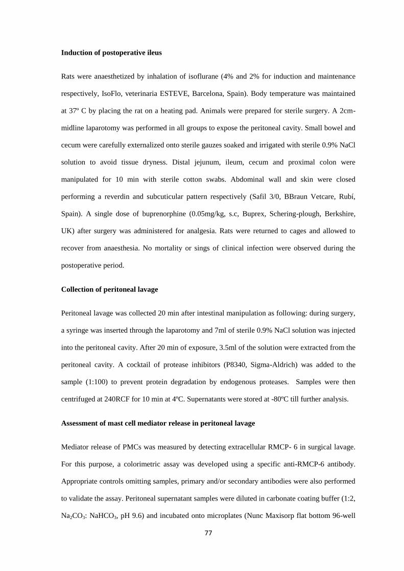
77
Induction of postoperative ileus
Rats were anaesthetized by inhalation of isoflurane (4% and 2% for induction and maintenance
respectively, IsoFlo, veterinaria ESTEVE, Barcelona, Spain). Body temperature was maintained
at 37º C by placing the rat on a heating pad. Animals were prepared for sterile surgery. A 2cm-
midline laparotomy was performed in all groups to expose the peritoneal cavity. Small bowel and
cecum were carefully externalized onto sterile gauzes soaked and irrigated with sterile 0.9% NaCl
solution to avoid tissue dryness. Distal jejunum, ileum, cecum and proximal colon were
manipulated for 10 min with sterile cotton swabs. Abdominal wall and skin were closed
performing a reverdin and subcuticular pattern respectively (Safil 3/0, BBraun Vetcare, Rubí,
Spain). A single dose of buprenorphine (0.05mg/kg, s.c, Buprex, Schering-plough, Berkshire,
UK) after surgery was administered for analgesia. Rats were returned to cages and allowed to
recover from anaesthesia. No mortality or sings of clinical infection were observed during the
postoperative period.
Collection of peritoneal lavage
Peritoneal lavage was collected 20 min after intestinal manipulation as following: during surgery,
a syringe was inserted through the laparotomy and 7ml of sterile 0.9% NaCl solution was injected
into the peritoneal cavity. After 20 min of exposure, 3.5ml of the solution were extracted from the
peritoneal cavity. A cocktail of protease inhibitors (P8340, Sigma-Aldrich) was added to the
sample (1:100) to prevent protein degradation by endogenous proteases. Samples were then
centrifuged at 240RCF for 10 min at 4ºC. Supernatants were stored at -80ºC till further analysis.
Assessment of mast cell mediator release in peritoneal lavage
Mediator release of PMCs was measured by detecting extracellular RMCP- 6 in surgical lavage.
For this purpose, a colorimetric assay was developed using a specific anti-RMCP-6 antibody.
Appropriate controls omitting samples, primary and/or secondary antibodies were also performed
to validate the assay. Peritoneal supernatant samples were diluted in carbonate coating buffer (1:2,
Na2CO3: NaHCO3, pH 9.6) and incubated onto microplates (Nunc Maxisorp flat bottom 96-well
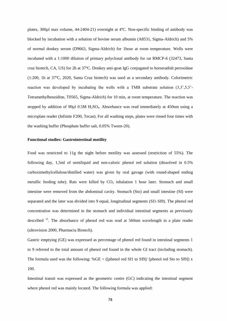
78
plates, 300µl max volume, 44-2404-21) overnight at 4ºC. Non-specific binding of antibody was
blocked by incubation with a solution of bovine serum albumin (A8531, Sigma-Aldrich) and 5%
of normal donkey serum (D9663, Sigma-Aldrich) for 1hour at room temperature. Wells were
incubated with a 1:1000 dilution of primary polyclonal antibody for rat RMCP-6 (32473, Santa
cruz biotech, CA, US) for 2h at 37ºC. Donkey anti-goat IgG conjugated to horseradish peroxidase
(1:200, 1h at 37ºC, 2020, Santa Cruz biotech) was used as a secondary antibody. Colorimetric
reaction was developed by incubating the wells with a TMB substrate solution (3,3’,5,5’-
Tetramethylbenzidine, T0565, Sigma-Aldrich) for 10 min, at room temperature. The reaction was
stopped by addition of 90µl 0.5M H2SO4. Absorbance was read immediately at 450nm using a
microplate reader (Infinite F200, Tecan). For all washing steps, plates were rinsed four times with
the washing buffer (Phosphate buffer salt, 0.05% Tween-20).
Functional studies: Gastrointestinal motility
Food was restricted to 11g the night before motility was assessed (restriction of 55%). The
following day, 1,5ml of semiliquid and non-caloric phenol red solution (dissolved in 0.5%
carboximethylcellulose/distilled water) was given by oral gavage (with round-shaped ending
metallic feeding tube). Rats were killed by CO2 inhalation 1 hour later. Stomach and small
intestine were removed from the abdominal cavity. Stomach (Sto) and small intestine (SI) were
separated and the later was divided into 9 equal, longitudinal segments (SI1-SI9). The phenol red
concentration was determined in the stomach and individual intestinal segments as previously
described 15
. The absorbance of phenol red was read at 560nm wavelength in a plate reader
(ultrovision 2000, Pharmacia Biotech).
Gastric emptying (GE) was expressed as percentage of phenol red found in intestinal segments 1
to 9 referred to the total amount of phenol red found in the whole GI tract (including stomach).
The formula used was the following: %GE = ([phenol red SI1 to SI9]/ [phenol red Sto to SI9]) x
100.
Intestinal transit was expressed as the geometric centre (GC) indicating the intestinal segment
where phenol red was mainly located. The following formula was applied:
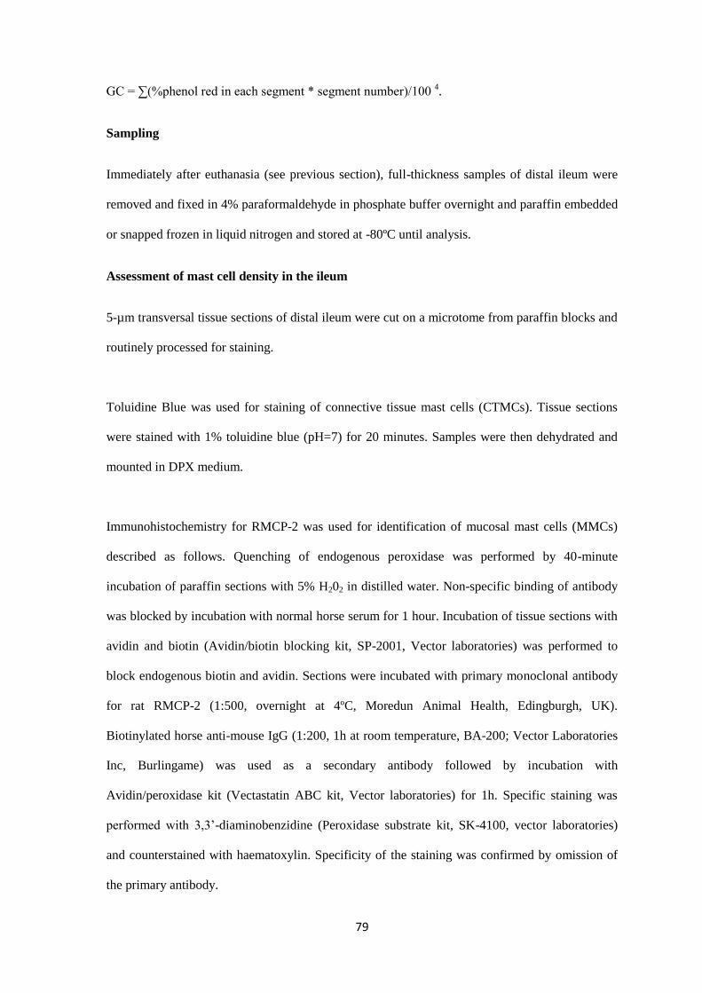
79
GC = ∑(%phenol red in each segment * segment number)/100 4.
Sampling
Immediately after euthanasia (see previous section), full-thickness samples of distal ileum were
removed and fixed in 4% paraformaldehyde in phosphate buffer overnight and paraffin embedded
or snapped frozen in liquid nitrogen and stored at -80ºC until analysis.
Assessment of mast cell density in the ileum
5-µm transversal tissue sections of distal ileum were cut on a microtome from paraffin blocks and
routinely processed for staining.
Toluidine Blue was used for staining of connective tissue mast cells (CTMCs). Tissue sections
were stained with 1% toluidine blue (pH=7) for 20 minutes. Samples were then dehydrated and
mounted in DPX medium.
Immunohistochemistry for RMCP-2 was used for identification of mucosal mast cells (MMCs)
described as follows. Quenching of endogenous peroxidase was performed by 40-minute
incubation of paraffin sections with 5% H202 in distilled water. Non-specific binding of antibody
was blocked by incubation with normal horse serum for 1 hour. Incubation of tissue sections with
avidin and biotin (Avidin/biotin blocking kit, SP-2001, Vector laboratories) was performed to
block endogenous biotin and avidin. Sections were incubated with primary monoclonal antibody
for rat RMCP-2 (1:500, overnight at 4ºC, Moredun Animal Health, Edingburgh, UK).
Biotinylated horse anti-mouse IgG (1:200, 1h at room temperature, BA-200; Vector Laboratories
Inc, Burlingame) was used as a secondary antibody followed by incubation with
Avidin/peroxidase kit (Vectastatin ABC kit, Vector laboratories) for 1h. Specific staining was
performed with 3,3’-diaminobenzidine (Peroxidase substrate kit, SK-4100, vector laboratories)
and counterstained with haematoxylin. Specificity of the staining was confirmed by omission of
the primary antibody.
80
RMCP-2 positive and toluidine blue-stained cells were quantified in a blinded fashion under light
microscope at 40x magnification (Olympus CH 30, Tokyo, Japan). The number of positive cells
was expressed as the mean of counted cells on 15 non-overlapping high power fields.
Myeloperoxidase activity (MPO) assay
Ileum samples were weighted and homogenized in hexadecyltrimethylammonium bromide buffer
(1ml/50mg tissue, 5g HTAB in 1L phosphate buffer 50mM, pH= 6.0) using GentleMACS (130-
093-235, Miltenyi biotec). Samples were centrifuged at 1200rpm for 10 min at 4ºC and
supernatants aliquoted until analysis. A standard was prepared with serial dilutions of purified
MPO standard (M6908, Sigma) in HTAB buffer. 7µl of the samples at the appropriate dilution,
standards and blank (HTAB buffer) were added to microplates (NUNC 96-well microplate,
12565210). O-dianisidine substrate solution was prepared by adding 16.8mg of O-dianisidine to
90ml of distilled water and 10 ml of phosphated buffered saline pH6.0. Before starting the
reaction, 50µl of 1 % H2O2 were added to the substrate solution. To start the reaction, 200µl of
mixture substrate solution were added to samples and absorbance of the reaction were read at
3min intervals during 15 minutes at 450 nm in a plate reader (Infinite F200, Tecan). Samples,
blank and standard were assayed in triplicates. MPO activity was expressed as units of activity per
mg of tissue (U mg-1
).
Reverse transcription–quantitative real-time PCR (RT-qPCR)
For extraction of total RNA, full-thickness ileum samples were homogenized in TRI reagent
(Ambion, AM9738) using a homogenizer (IKA T10 basic ultra-turrax). Sample homogenates
were mixed with chloroform for RNA separation and then with isopropyl alcohol for RNA
precipitation. After washing steps, RNA was quantified by nanodrop (Nanodrop Technologies,
Rockland, DE, USA) and converted into 20µl reaction volume of cDNA using a High Capacity
cDNA Reverse Transcription Kit according to manufacturer’s instructions (Applied Biosystems,
US). RT-qPCR was performed using validated TaqMan gene expressions assays (Applied
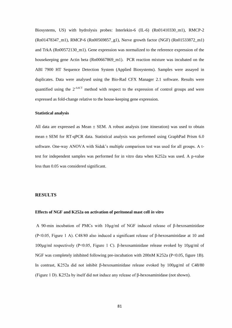
81
Biosystems, US) with hydrolysis probes: Interlekin-6 (IL-6) (Rn01410330_m1), RMCP-2
(Rn01478347_m1), RMCP-6 (Rn00569857_g1), Nerve growth factor (NGF) (Rn01533872_m1)
and TrkA (Rn00572130_m1). Gene expression was normalized to the reference expression of the
housekeeping gene Actin beta (Rn00667869_m1). PCR reaction mixture was incubated on the
ABI 7900 HT Sequence Detection System (Applied Biosystems). Samples were assayed in
duplicates. Data were analysed using the Bio-Rad CFX Manager 2.1 software. Results were
quantified using the 2-ΔΔCT
method with respect to the expression of control groups and were
expressed as fold-change relative to the house-keeping gene expression.
Statistical analysis
All data are expressed as Mean ± SEM. A robust analysis (one itineration) was used to obtain
mean ± SEM for RT-qPCR data. Statistical analysis was performed using GraphPad Prism 6.0
software. One-way ANOVA with Sidak’s multiple comparison test was used for all groups. A t-
test for independent samples was performed for in vitro data when K252a was used. A p-value
less than 0.05 was considered significant.
RESULTS
Effects of NGF and K252a on activation of peritoneal mast cell in vitro
A 90-min incubation of PMCs with 10µg/ml of NGF induced release of β-hexosaminidase
(P<0.05, Figure 1 A). C48/80 also induced a significant release of β-hexosaminidase at 10 and
100µg/ml respectively (P<0.05, Figure 1 C). β-hexosaminidase release evoked by 10µg/ml of
NGF was completely inhibited following pre-incubation with 200nM K252a (P<0.05, figure 1B).
In contrast, K252a did not inhibit β-hexosaminidase release evoked by 100µg/ml of C48/80
(Figure 1 D). K252a by itself did not induce any release of β-hexosaminidase (not shown).
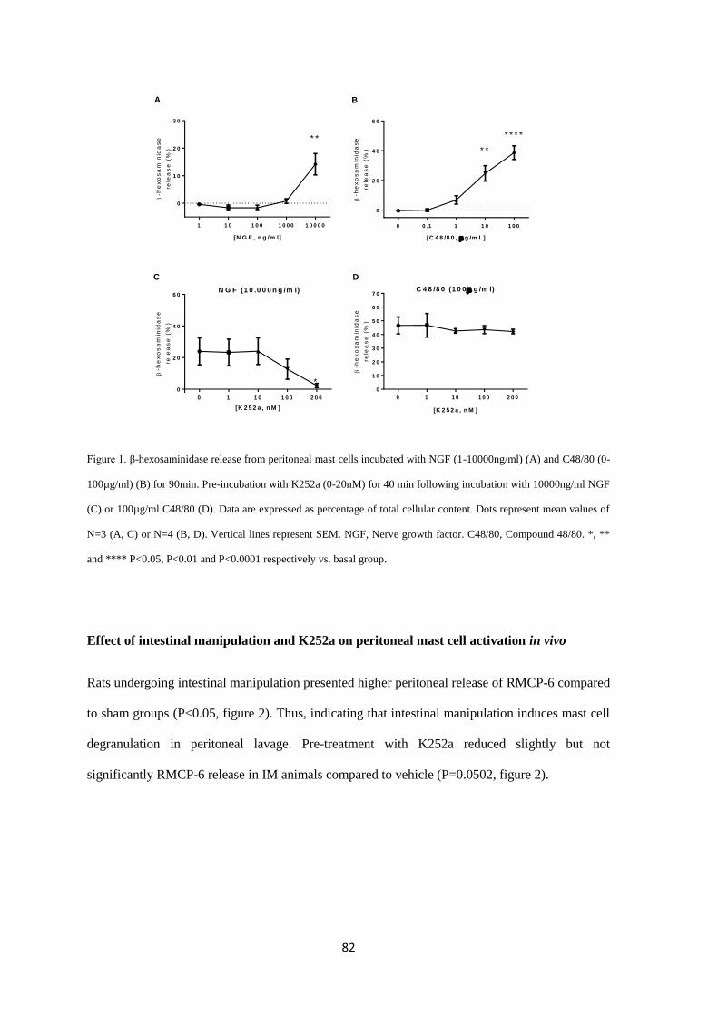
82
Figure 1. β-hexosaminidase release from peritoneal mast cells incubated with NGF (1-10000ng/ml) (A) and C48/80 (0-
100µg/ml) (B) for 90min. Pre-incubation with K252a (0-20nM) for 40 min following incubation with 10000ng/ml NGF
(C) or 100µg/ml C48/80 (D). Data are expressed as percentage of total cellular content. Dots represent mean values of
N=3 (A, C) or N=4 (B, D). Vertical lines represent SEM. NGF, Nerve growth factor. C48/80, Compound 48/80. *, **
and **** P<0.05, P<0.01 and P<0.0001 respectively vs. basal group.
Effect of intestinal manipulation and K252a on peritoneal mast cell activation in vivo
Rats undergoing intestinal manipulation presented higher peritoneal release of RMCP-6 compared
to sham groups (P<0.05, figure 2). Thus, indicating that intestinal manipulation induces mast cell
degranulation in peritoneal lavage. Pre-treatment with K252a reduced slightly but not
significantly RMCP-6 release in IM animals compared to vehicle (P=0.0502, figure 2).
1 1 0 1 0 0 1 0 0 0 1 0 0 0 0
0
1 0
2 0
3 0
* *
[N G F , n g /m l]
-h
ex
os
am
inid
as
e
rele
as
e (
%)
N G F (1 0 .0 0 0 n g /m l)
0 1 1 0 1 0 0 2 0 0
0
2 0
4 0
6 0
*
[K 2 5 2 a , n M ]
-h
ex
os
am
inid
as
e
rele
as
e (
%)
C 4 8 /8 0 (1 0 0 g /m l)
0 1 1 0 1 0 0 2 0 0
0
1 0
2 0
3 0
4 0
5 0
6 0
7 0
[K 2 5 2 a , n M ]
-h
ex
os
am
inid
as
e
rele
as
e (
%)
A
C D
0 0 .1 1 1 0 1 0 0
0
2 0
4 0
6 0
* *
* * * *
[C 4 8 /8 0 , g /m l ]
-h
ex
os
am
inid
as
e
rele
as
e (
%)
B
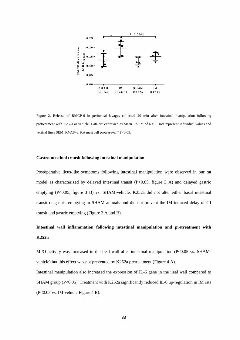
83
Figure 2. Release of RMCP-6 in peritoneal lavages collected 20 min after intestinal manipulation following
pretreatment with K252a or vehicle. Data are expressed as Mean ± SEM of N=5. Dots represent individual values and
vertical lines SEM. RMCP-6, Rat mast cell protease-6. * P<0.05.
Gastrointestinal transit following intestinal manipulation
Postoperative ileus-like symptoms following intestinal manipulation were observed in our rat
model as characterized by delayed intestinal transit (P<0.05, figure 3 A) and delayed gastric
emptying (P<0.05, figure 3 B) vs. SHAM-vehicle. K252a did not alter either basal intestinal
transit or gastric emptying in SHAM animals and did not prevent the IM induced delay of GI
transit and gastric emptying (Figure 3 A and B).
Intestinal wall inflammation following intestinal manipulation and pretreatment with
K252a
MPO activity was increased in the ileal wall after intestinal manipulation (P<0.05 vs. SHAM-
vehicle) but this effect was not prevented by K252a pretreatment (Figure 4 A).
Intestinal manipulation also increased the expression of IL-6 gene in the ileal wall compared to
SHAM group (P<0.05). Treatment with K252a significantly reduced IL-6 up-regulation in IM rats
(P<0.05 vs. IM-vehicle Figure 4 B).
RM
CP
-6 r
ele
as
e
(AB
S4
50
nm
)
S H AM
c o n tr o l
IM
c o n tr o l
S H AM
K 2 5 2 a
IM
K 2 5 2 a
0 .0 0
0 .0 5
0 .1 0
0 .1 5
0 .2 0
0 .2 5*
P = 0 .0 5 0 2
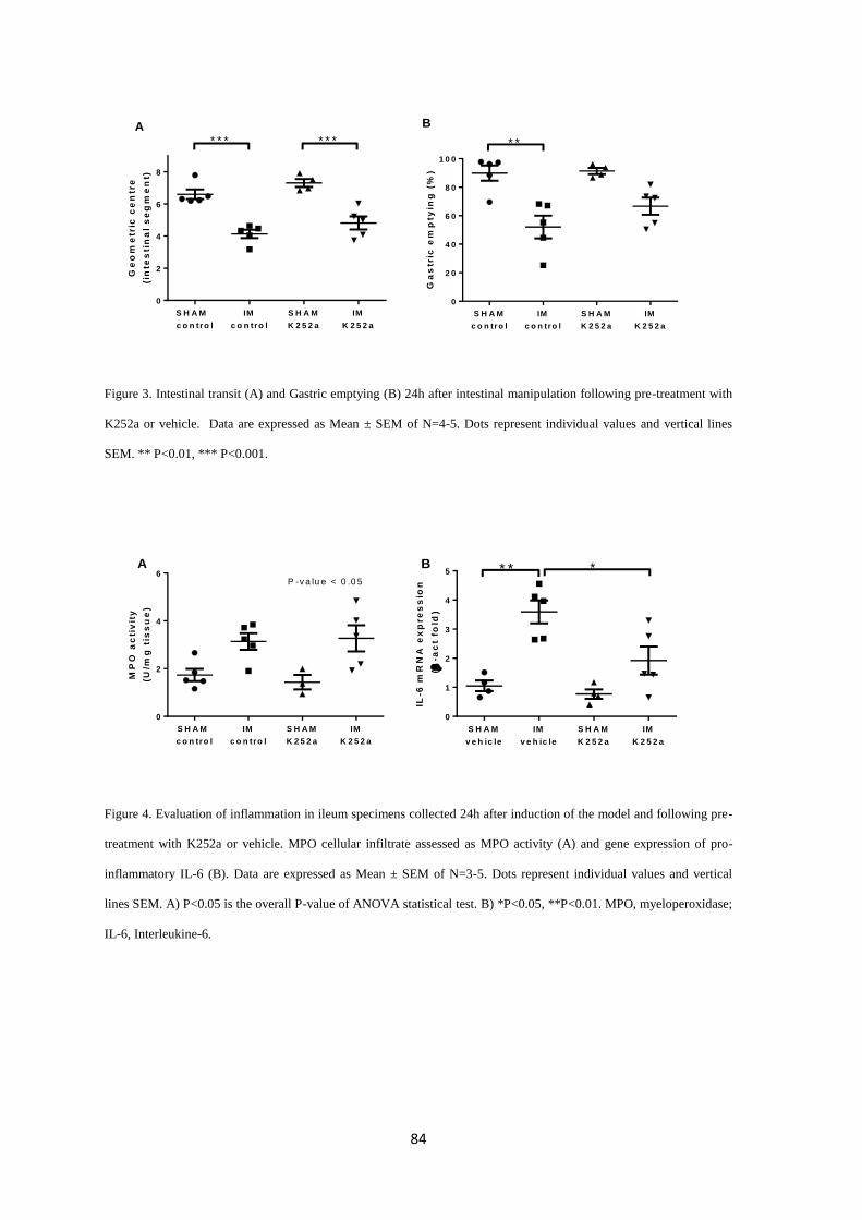
84
Figure 3. Intestinal transit (A) and Gastric emptying (B) 24h after intestinal manipulation following pre-treatment with
K252a or vehicle. Data are expressed as Mean ± SEM of N=4-5. Dots represent individual values and vertical lines
SEM. ** P<0.01, *** P<0.001.
Figure 4. Evaluation of inflammation in ileum specimens collected 24h after induction of the model and following pre-
treatment with K252a or vehicle. MPO cellular infiltrate assessed as MPO activity (A) and gene expression of pro-
inflammatory IL-6 (B). Data are expressed as Mean ± SEM of N=3-5. Dots represent individual values and vertical
lines SEM. A) P<0.05 is the overall P-value of ANOVA statistical test. B) *P<0.05, **P<0.01. MPO, myeloperoxidase;
IL-6, Interleukine-6.
S H A M
c o n tro l
IM
c o n tro l
S H A M
K 2 5 2 a
IM
K 2 5 2 a
0
2
4
6
8
* * * * * *G
eo
me
tric
ce
ntr
e
(in
tes
tin
al
se
gm
en
t)
S H A M
c o n tro l
IM
c o n tro l
S H A M
K 2 5 2 a
IM
K 2 5 2 a
0
2 0
4 0
6 0
8 0
1 0 0
* *
Ga
str
ic e
mp
tyin
g (
%)
A B
IL-6
mR
NA
ex
pre
ss
ion
(
-ac
t fo
ld)
S H A M
v e h ic le
IM
v e h ic le
S H A M
K 2 5 2 a
IM
K 2 5 2 a
0
1
2
3
4
5 * * *
MP
O a
cti
vit
y
(U/m
g t
iss
ue
)
S H A M
c o n tro l
IM
c o n tro l
S H A M
K 2 5 2 a
IM
K 2 5 2 a
0
2
4
6P -v a lu e < 0 .0 5
A B
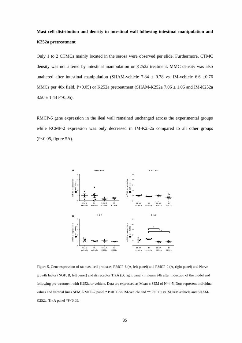
85
Mast cell distribution and density in intestinal wall following intestinal manipulation and
K252a pretreatment
Only 1 to 2 CTMCs mainly located in the serosa were observed per slide. Furthermore, CTMC
density was not altered by intestinal manipulation or K252a treatment. MMC density was also
unaltered after intestinal manipulation (SHAM-vehicle 7.84 ± 0.78 vs. IM-vehicle 6.6 ±0.76
MMCs per 40x field, P>0.05) or K252a pretreatment (SHAM-K252a 7.06 ± 1.06 and IM-K252a
8.50 ± 1.44 P>0.05).
RMCP-6 gene expression in the ileal wall remained unchanged across the experimental groups
while RCMP-2 expression was only decreased in IM-K252a compared to all other groups
(P<0.05, figure 5A).
Figure 5. Gene expression of rat mast cell proteases RMCP-6 (A, left panel) and RMCP-2 (A, right panel) and Nerve
growth factor (NGF, B, left panel) and its receptor TrkA (B, right panel) in ileum 24h after induction of the model and
following pre-treatment with K252a or vehicle. Data are expressed as Mean ± SEM of N=4-5. Dots represent individual
values and vertical lines SEM. RMCP-2 panel * P<0.05 vs IM-vehicle and ** P<0.01 vs. SHAM-vehicle and SHAM-
K252a. TrkA panel *P<0.05.
R M C P -6
mR
NA
ex
pre
ss
ion
(
-ac
t fo
ld)
S H A M
v e h ic le
IM
v e h ic le
S H A M
K 2 5 2 a
IM
K 2 5 2 a
0
1
2
3
4
5
R M C P -2
mR
NA
ex
pre
ss
ion
(
-ac
t fo
ld)
S H A M
v e h ic le
IM
v e h ic le
S H A M
K 2 5 2 a
IM
K 2 5 2 a
0
1
2
3
4
5
* **
N G F
mR
NA
ex
pre
ss
ion
(
-ac
t fo
ld)
S H A M
v e h ic le
IM
v e h ic le
S H A M
K 2 5 2 a
IM
K 2 5 2 a
0
1
2
3
4
5
T r k A
mR
NA
ex
pre
ss
ion
(
-ac
t fo
ld)
S H A M
v e h ic le
IM
v e h ic le
S H A M
K 2 5 2 a
IM
K 2 5 2 a
0
1
2
3
4
5
*
*
A
B
86
NGF and TrkA gene expression
Intestinal manipulation or K252a treatment did not affect the expression of NGF within the wall
of the ileum. However, K252a treatment reduced the expression of TrkA in both SHAM- and IM-
treated animals compared to IM-vehicle treated rats (P<0.05, figure 5B).
DISCUSSION
In this study we demonstrate the ability of the tropomyosin-related kinase receptor
antagonist K252a to inhibit NGF-evoked degranulation of PMCs in vitro. In addition, we
demonstrate that K252a is able to block some of the functional consequences of intestinal
manipulation, such as inflammation and mast cell degranulation, but it does not affect the motor
inhibition observed in the POI model.
Previous studies demonstrated that NGF stimulates degranulation of PMCs 11,16
possibly
via the NGF high-affinity receptor TrkA 13,17,18,9,19
. Our study confirms this by showing that
stimulation of isolated PMCs with NGF evokes a degranulatory response. In addition, K252a
prevented the NGF-evoked degranulation, without affecting the degranulation evoked by the MC
degranulator C48/80 11
. This suggests a specificity of K252a for the TrkA receptor in MCs.
We next evaluated the MC stabilizing properties of K252a in vivo, in the rat model of POI
on the intestinal inflammation and transit. In line with previous studies, intestinal manipulation in
our model resulted in a rapid degranulation of PMCs, as judged by the peritoneal increase of the
CTMC specific product RMCP-6 7,2,4,5
. Intestinal manipulation also resulted in a delay of the GI
transit and produced inflammation of manipulated intestinal segments as previously reported 20,21
.
K252a pretreatment attenuated the release of RMCP-6 in vivo following intestinal
manipulation and expression of RMCP-2 within the intestinal wall, thus, further demonstrating
that K252a has MC stabilizing properties. Jardí et al. demonstrated that MC express TrkA
87
receptor 13
. Taken together, these observations suggest that TrkA modulates MC activity and
furthermore, that K252a could have a role for MC-mediated disorders.
Similarly, we also observed that K252a decreases IL-6 expression after intestinal
manipulation. However, K252a did not prevent neutrophil activation after intestinal manipulation,
as judged by tissue MPO activity. These findings suggest that, in our model, K252a is involved
specifically in the inactivation of IL-6 secreted from intestinal resident cells including MCs or
macrophages 22,23
, cell types with TrKA receptors, rather than newly recruited neutrophils.
Therefore, TrKA receptors seem not to interfere with other mechanisms leading to migration of
circulating neutrophils as observed in other models 24,25
. In fact, only mononuclear cells have been
identified as the most prominent cellular source for IL-6 in the intestine after intestinal
manipulation 26
.
Our study also demonstrates that K252a effectively blocks NGF receptor as TrkA mRNA
levels were decreased in treated rats. NGF-TrkA interactions promote overexpression of TrkA on
cell surface and this may explain our finding in rats receiving K252a 27
. This finding further
corroborates that K252a inhibits MC activation as MCs express TrkA 13
. In fact, we reported
attenuation of RMCP-6 release into the peritoneal cavity and the reduction of gene expression of
RMCP-2 in the ileum after K252a pre-treatment.
However, GI motility was not improved by K252a. This is in contrast to a previous study
in a rat model of food allergy reporting modulation of colonic motility in animals treated with
K252a on a daily basis for 4 weeks 13
. We only observed a moderate effect of intestinal
manipulation on delaying gastric emptying on K252a-treated animals compared to vehicle
animals. So, we cannot confirm or discard an effect of K252a. We suggest that the lack of effect
of K252a on motility in our study could be related to the fact that K252a was given only 20 min
prior to intestinal manipulation, which is possibly not enough time to completely prevent
intestinal manipulation-induced hypomotility.
In a similar way we cannot discard that K252a also acts on other cells expressing the
TrKA receptor28
. However, the fact that K252a specifically stabilizes mast cells allows us to
88
conclude that blocking of TrKA receptors can be a target for MC stabilization and therefore be
useful in those diseases where MCs are involved.
In conclusion, K252a attenuated PMC degranulation, mucosal MC protease expression
and up-regulation of IL-6 induced after intestinal manipulation in a POI model in the rat. Thus,
showing 1) a potential use for treatment of MC-mediated disorders and 2) an anti-inflammatory
effect. Therefore, we suggest that K252a and TrkA receptor should be further explored in
intestinal inflammatory disorders with concomitant activation of MCs.
89
ACKNOWLEDGMENTS, FUNDING AND DISCLOSURES
We thank A. Acosta and E. Martínez for their technical assistance.
S.B and J.R designed and performed the study, analysed data and wrote the paper. P.V designed
the study and wrote the paper.
This study was supported by grants BFU2010-15401 from Ministerio de Ciencia e Innovación,
2009SGR708 and 2014SGR789 from Direcció General de Recerca de la Generalitat de
Catalunya. S.B. personal support: FPU program (AP2010-1692) from Ministerio de Educación,
Cultura y Deporte.
No competing interests declared.
90
REFERENCES
1. Kehlet, H. Postoperative ileus--an update on preventive techniques. Nat. Clin. Pract.
Gastroenterol. Hepatol. 5, 552–8 (2008).
2. The, F. O. et al. Intestinal handling-induced mast cell activation and inflammation in
human postoperative ileus. Gut 57, 33–40 (2008).
3. De Jonge, W. J. et al. Mast cell degranulation during abdominal surgery initiates
postoperative ileus in mice. Gastroenterology 127, 535–545 (2004).
4. Snoek, S. A. et al. Mast cells trigger epithelial barrier dysfunction, bacterial translocation
and postoperative ileus in a mouse model. Neurogastroenterol. Motil. 24, 172–84, e91
(2012).
5. Gomez-Pinilla, P. J. et al. Mast cells play no role in the pathogenesis of postoperative ileus
induced by intestinal manipulation. PLoS One 9, e85304 (2014).
6. Berdún, S. et al. Peritoneal mast cell degranulation and gastrointestinal recovery in
patients undergoing colorectal surgery. Neurogastroenterol. Motil. n/a–n/a (2015).
doi:10.1111/nmo.12525
7. De Jonge, W. J. et al. Mast cell degranulation during abdominal surgery initiates
postoperative ileus in mice. Gastroenterology 127, 535–45 (2004).
8. The, F. O. et al. The role of mast cell stabilization in treatment of postoperative ileus: a
pilot study. Am. J. Gastroenterol. 104, 2257–66 (2009).
9. Sawada, J., Itakura, A., Tanaka, A., Furusaka, T. & Matsuda, H. Nerve growth factor
functions as a chemoattractant for mast cells through both mitogen-activated protein
kinase and phosphatidylinositol 3-kinase signaling pathways. Blood 95, 2052–8 (2000).
10. Stempelj, M. & Ferjan, I. Signaling pathway in nerve growth factor induced histamine
release from rat mast cells. Inflamm. Res. 54, 344–9 (2005).
11. Kawamoto, K. et al. Nerve growth factor activates mast cells through the collaborative
interaction with lysophosphatidylserine expressed on the membrane surface of activated
platelets. J. Immunol. 168, 6412–9 (2002).
12. Marshall, J. S., Stead, R. H., McSharry, C., Nielsen, L. & Bienenstock, J. The role of mast
cell degranulation products in mast cell hyperplasia. I. Mechanism of action of nerve
growth factor. J. Immunol. 144, 1886–92 (1990).
13. Jardí, F., Martínez, V. & Vergara, P. NGF is involved in oral ovalbumin-induced altered
colonic contractility in rats: evidence from the blockade of TrkA receptors with K252a.
Neurogastroenterol. Motil. 24, e580–90 (2012).
14. Torrents, D., Torres, R., De Mora, F. & Vergara, P. Antinerve growth factor treatment
prevents intestinal dysmotility in Trichinella spiralis-infected rats. J. Pharmacol. Exp.
Ther. 302, 659–65 (2002).
91
15. Liu, S. & Chen, J. D. Z. Colonic electrical stimulation regulates colonic transit via the
nitrergic pathway in rats. Dig. Dis. Sci. 51, 502–5 (2006).
16. Pearce, F. L. & Thompson, H. L. Some characteristics of histamine secretion from rat
peritoneal mast cells stimulated with nerve growth factor. J. Physiol. 372, 379–93 (1986).
17. Di Mola, F. F. et al. Nerve growth factor and Trk high affinity receptor (TrkA) gene
expression in inflammatory bowel disease. Gut 46, 670–9 (2000).
18. Tam, S. Y. et al. Expression of functional TrkA receptor tyrosine kinase in the HMC-1
human mast cell line and in human mast cells. Blood 90, 1807–20 (1997).
19. Welker, P., Grabbe, J., Gibbs, B., Zuberbier, T. & Henz, B. M. Nerve growth factor-beta
induces mast-cell marker expression during in vitro culture of human umbilical cord blood
cells. Immunology 99, 418–26 (2000).
20. Kalff, J. C., Schraut, W. H., Simmons, R. L. & Bauer, A. J. Surgical manipulation of the
gut elicits an intestinal muscularis inflammatory response resulting in postsurgical ileus.
Ann. Surg. 228, 652–63 (1998).
21. Kalff, J. C. et al. Biphasic response to gut manipulation and temporal correlation of
cellular infiltrates and muscle dysfunction in rat. Surgery 126, 498–509 (1999).
22. Hsu, C.-L., Neilsen, C. V & Bryce, P. J. IL-33 is produced by mast cells and regulates
IgE-dependent inflammation. PLoS One 5, e11944 (2010).
23. Williams, J. A., Pontzer, C. H. & Shacter, E. Regulation of macrophage interleukin-6 (IL-
6) and IL-10 expression by prostaglandin E2: the role of p38 mitogen-activated protein
kinase. J. Interferon Cytokine Res. 20, 291–8 (2000).
24. Wilfong, E. R. & Dey, R. D. Nerve growth factor and substance P regulation in nasal
sensory neurons after toluene diisocyanate exposure. Am. J. Respir. Cell Mol. Biol. 30,
793–800 (2004).
25. Mohtasham, L., Auais, A. & Piedimonte, G. Nerve growth factor mediates steroid-
resistant inflammation in respiratory syncytial virus infection. Pediatr. Pulmonol. 42, 496–
504 (2007).
26. Kalff, J. C. et al. Intra-abdominal activation of a local inflammatory response within the
human muscularis externa during laparotomy. Ann. Surg. 237, 301–15 (2003).
27. Freund-Michel, V. & Frossard, N. Overexpression of functional TrkA receptors after
internalisation in human airway smooth muscle cells. Biochim. Biophys. Acta 1783, 1964–
71 (2008).
28. Di Mola, F. F. Nerve growth factor and Trk high affinity receptor (TrkA) gene expression
in inflammatory bowel disease. Gut 46, 670–679 (2000).

92

93
CHAPTER 3

94
95
INTESTINAL MANIPULATION INCREASES GENE EXPRESSION
OF VISCERAL-AFFERENT MEDIATORS IN DORSAL ROOT
GANGLIA: ROLE FOR PERITONEAL MAST CELLS
SERGIO BERDUN1,2
; JAKUB RYCHTER Ph.D.1,3
; PATRI VERGARA Ph.D. 1,2,3
1Department of Cell Biology, Physiology and Immunology, Veterinary School, Universitat
Autònoma de Barcelona, Barcelona, Spain
2Neuroscience Institute, Universitat Autònoma de Barcelona, Barcelona, Spain
3Centro de Investigación Biomédica en Red de Enfermedades Hepáticas y Digestivas
(CIBERehd), Instituto de Salud Carlos III, Barcelona, Spain
Key words: Afferent neural pathways, gastrointestinal motility, mast cells, postoperative ileus
Submitted to Neurogastroenterology and motility.

96
97
ABSTRACT
Background: Surgical handling of the bowel evokes degranulation of peritoneal mast cells
(PMC) which has been demonstrated to play a role in postoperative ileus (POI). We aimed to
investigate if MCs contribute to POI pathophysiology by activating intestinal afferent neural
pathways.
Methods: POI was induced by intestinal manipulation in Sprague-Dawley rats. Additionally,
compound 48/80 (C48/80) and ketotifen were used to modulate MC activity. Rat mast cell
protease 6 (RMCP-6, ELISA) release was determined in peritoneal lavage 20 min after intestinal
manipulation. At 24h, gastrointestinal transit was determined. Activation of dorsal root ganglia
(DRG) was determined (PCR) for calcitonin gene-related peptide (CGRP), Protease-activated
receptor-2 (PAR-2), Nerve growth factor (NGF) and TrkA receptor. Ileal wall inflammation was
assessed by myeloperoxidase (MPO) activity, number of MCs, and PCR of IL-6, RMCP-2 and 6.
Key Results: Intestinal manipulation and exposure to C48/80 induced degranulation of PMCs,
delayed gastrointestinal transit and up-regulated IL-6 and MPO activity. Intestinal manipulation,
but not C48/80 up-regulated CGRP, PAR-2 and NGF/TrkA in DRGs. Ketotifen only improved
gastric emptying and faecal output. Up-regulation of CGRP and TrkA expression in DRG was not
prevented by ketotifen.
Conclusions and inferences: POI is accompanied by activation of DRGs thus confirming
activation of splanchnic afferents. MCs are involved in POI dysmotility but, under our
pharmacological approach, we were not able to prove that MCs are involved in DRG activation.
Our results suggest that MCs should be targeted for treatment of dysmotility and that additional
treatments should be employed to prevent DRG-mediated symptoms to ameliorate POI.
98
ABBREVIATIONS
CGRP Calcitonin gene-related peptide
C48/80 Compound 48/80
CTMC Connective tissue mast cell
GI Gastrointestinal
IL 6 Interleukin 6
MMC Mucosal mast cell
MC Mast cell
MPO Myeloperoxidase
NGF Nerve growth factor
PAR-2 Protease-activated receptor 2
PMC Peritoneal mast cell
POI Postoperative ileus
RMCP Rat mast cell protease
TrkA Tropomyosin receptor kinase A
99
KEY MESSAGES
- Intestinal manipulation up-regulates gene expression of calcitonin gene-related peptide,
nerve growth factor/TrkA and protease-activated receptor 2 in thoraco-lumbar dorsal root
ganglia
- Intestinal manipulation induces activation of rat peritoneal mast cells and RMCP-6
release into the peritoneal cavity.
- Degranulation of peritoneal mast cells contributes to delayed gastrointestinal transit and
inflammation during postoperative ileus.
- Modulation of mast cells with ketotifen and C48/80 does not modify gene expression of
dorsal root ganglia.
100
INTRODUCTION
Postoperative ileus (POI) is a functional disorder following abdominal surgery characterised by
hypomotility of the gastrointestinal (GI) tract. Although not life-threatening, POI prolongs
hospitalization of patients and thus increases health care expenses 1.
In the rat GI tract, we find two different mast cell (MC) populations: mucosal mast cells
(MMC) residing in the mucosa and expressing rat mast cell protease-2 (RMCP-2)2,3
and
connective tissue mast cells (CTMC) residing in the submucosa, muscular layer, serosa and
peritoneal cavity and expressing RMCP-6 2,4,5
. Degranulation of peritoneal mast cells (PMCs)
following intestinal manipulation has been demonstrated, and has been proposed to play an
important role in the pathogenesis of POI 6–8
.
Extrinsic nerve endings supplying the GI tract are in close proximity to MCs 9,10
and a
functional interaction between both has been described 11,12
. Activation of afferent systems evoked
by intestinal manipulation has been documented during POI 13–15
and constitutes a first step to
hypomotility 16–18
. Since MCs degranulate upon intestinal manipulation it is likely that a crosstalk
exists between MCs and afferent nerves following intestinal manipulation.
This work aimed to elucidate interactions between MCs and afferent fibers of splanchnic
nerves in an experimental model of POI in the rat. For this purpose we evaluated a) gene
expression of selected genes in thoraco-lumbar dorsal root ganglia (DRG) and b) the effect of MC
degranulator, Compound 48/80, and MC stabilizer, ketotifen on DRG gene expression.
MATERIAL AND METHODS
Animals
8 week-old (275-325g) male Sprague-Dawley rats (UAB Animal House facility colony stock)
were used. Rats were kept under conventional conditions in an environmentally controlled room
(temperature: 20-22ºC; photoperiod; 12h/12h light/dark cycle). Animals had free access to tap
101
water and standard commercial pellet diet (2014 Harlan Maintenance). Animals were caged in
groups of 2 to 3 upon arrival and were allowed to acclimatize to the new environment for 5 days.
All experimental procedures were approved by the Ethical Committee of the Universitat
Autònoma de Barcelona (code 1398 and 2478) and the Generalitat de Catalunya (code 6309 and
7974).
Induction of postoperative ileus
Rats underwent only a laparotomy (SHAM group, N=7-8) or a laparotomy with intestinal
manipulation (IM group, N=11-12) to induce POI. 20 min after induction, peritoneal lavage
samples were collected. 24h post-surgery, animals were euthanized and intestinal transit and
gastric emptying was measured followed by sampling of ileum and DRGs. POI was inducted by
IM as follows. Rats were anaesthetized by inhalation of isoflurane (4% and 2% for induction and
maintenance respectively, IsoFlo, veterinaria ESTEVE, Barcelona, Spain). Body temperature was
maintained at 37º C by placing the rat on a heating pad. Animals were prepared for sterile surgery.
A 2cm-midline laparotomy was performed in all groups to expose the peritoneal cavity. Small
bowel and cecum were carefully externalized onto sterile soaked gauzes and irrigated with sterile
0.9% NaCl solution to avoid tissue dryness. Distal jejunum, ileum, cecum and proximal colon
were manipulated for 10 min with sterile cotton swabs. Abdominal wall and skin were closed
performing a reverdin and subcuticular pattern respectively (Safil 3/0, BBraunVetcare, Rubí,
Spain). A single buprenorphine dose (0.05mg kg-1
, s.c, Buprex, Schering-plough, Berkshire, UK)
was administered after surgery for analgesia. Rats were returned to cages and allowed to recover
from anaesthesia. No mortality or sings of clinical infection were observed during the
postoperative period
Pharmacological activation and stabilization of peritoneal mast cells
Pharmacological activation of MCs: rats were anaesthetized and prepared for sterile surgery as
described above. After 2cm-midline laparotomy, a 8cm-segment of ileum was carefully
externalized and place into an sterile Petri dish containing 6 ml of a 0.3mg mL-1
solution of
102
C48/80 (N=12) or sterile saline (N=12) for 1 min. After incubation, ileum was placed back into
the peritoneal cavity. The surgical wound was closed as described above and animals were let to
recover for 24h. No mortality or sings of clinical infection/anaphylaxis were observed during the
postoperative period.
MC stabilization: Rats received an injection of ketotifen (10mg kg-1
in 2ml, ip) or vehicle (2ml of
NaCl 0.9%, ip) 60 min prior induction of the POI model: SHAM vehicle (N=5), IM-vehicle
(N=5) , SHAM-ketotifen (N=5), IM-ketotifen (N=5).
For both experimental protocols, 20 min after exposure/administration of drugs, peritoneal
lavages samples were collected. 24h post-surgery, animals were euthanized and intestinal transit,
and gastric emptying was determined following by sampling of ileum and DRGs.
Ketotifen fumarate salt (K2628) and Compound 48/80 (C2313) were purchased from Sigma-
Aldrich, St Louis, MO, US.
Collection of peritoneal lavage
Peritoneal lavage was collected 20 min after intestinal manipulation or exposure to C48/80 as
following: during surgery, a syringe was inserted through the laparotomy and 7ml of sterile 0.9%
NaCl solution was injected. After 20 min of exposure, 3.5ml of the solution were extracted and a
cocktail of protease inhibitors (P8340, Sigma-Aldrich, St Louis, MO, US) was added to samples
(1:100) to prevent protein degradation by endogenous proteases. Samples were centrifuged at
240RCF for 10 min at 4ºC. The pellet was resuspended in 1ml of calcium- and magnesium-free
Hanks Balanced Salt Solution (H9394, Sigma-Aldrich) containing 10% of fetal calf serum and
1000UI/ml heparin. The resulting solution was pipeted on top of a 1-ml histopaque layer
(histopaque-1119, 11191, Sigma Aldrich) and centrifuged at 240RCF for 10min at 4ºC. The pellet
was resuspended in 1ml of Hanks solution to obtain an enriched suspension of PMCs for
morphological analysis (see below). Supernatants were kept frozen at -80ºC for further analysis.
103
For the ketotifen experimental protocol, PMC suspensions were further processed as described
below.
Assessment of mast cell mediator release in peritoneal lavage
Mediator release of PMC was measured by detecting extracellular RMCP- 6 in surgical lavage.
For this purpose, a colorimetric assay was developed using a specific anti-RMCP-6 antibody.
Appropriate controls omitting samples, primary and/or secondary antibodies were also performed
to validate the assay. Peritoneal supernatants samples were diluted in carbonate coating buffer
(Na2CO3/NaHCO3) and incubated onto microplates (NuncMaxisorp flat bottom 96-well plates,
300µl max volume, 44-2404-21) overnight at 4ºC. Non-specific binding of antibody was blocked
by incubation with a solution of bovine serum albumin (A8531, Sigma) and 5% of normal donkey
serum (D9663, Sigma) for 1hour at room temperature. Wells were incubated with a 1:1000
dilution of primary polyclonal antibody for rat RMCP-6 (32473, Santa cruz biotech, CA, US) for
2h at 37ºC. Donkey anti-goat IgG conjugated to horseradish peroxidase (1;200, 1h at 37ºC, 2020,
Santa Cruz biotech, CA, US) was used as a secondary antibody. A colorimetric reaction was
developed by incubating the wells with a TMB substrate solution (3,3’,5,5’-
Tetramethylbenzidine, T0565, Sigma) for 10 min at room temperature. The reaction was stopped
by addition of 90µl 0.5M H2SO4. Absorbance was read immediately at 450nm using a microplate
reader (Infinite F200, Tecan). For all washing steps, plates were rinsed four times with the
washing buffer (Phosphate buffer salt, 0.05% Tween-20).
Assessment of mast cell morphology in peritoneal lavage
Cellular suspensions obtained from the surgical lavage were cytospined onto a slide
(shandon 4 cytospin, 600rpm, 6 min, RT) and air-dried for 15 min, then fixed in Carnoy’s fixative
solution (60% pure ethanol, 30% Chloroform, 10% Acetic acid) for 15 minutes and stained with a
0.5% toluidine blue solution (Scharlau, CI 52040) at pH=7, for 5 min. Samples were dehydrated
and mounted in DPX medium. MC counts and morphological assessment of degranulation was
performed under light microscope at 40x magnification (Olympus CH 30, Tokyo, Japan) by an
104
observer blinded to the experimental groups. MCs were classified as either ‘intact mast cells’
(those with intact membrane outline, with no granules outside of the cell and intense staining) or
‘degranulated mast cells’ (disrupted cell membrane, extrusion of granules, poor staining) 19
.
Degranulated MCs were expressed as percentage with respect total mast cells observed.
Postoperative fecal output
Fecal pellets were collected during the dark period (from 18.00 PM to 9.00 AM) before and after
the surgery. For this purpose each animal was caged individually and put onto a metallic bottom
grid. Fecal output was expressed as the percentage of fecal pellets produced by each animal in
respect to its pre-surgical fecal pellet count.
Postsurgical assessment of gastric emptying and intestinal transit.
Food was restricted to 11g the night before motility was assessed (restriction of 55%). 1,5ml of
semi-liquid and non-caloric phenol red solution (dissolved in 0.5%
carboximethylcellulose/distilled water) was given by oral gavage (with round-shaped ending
metallic feeding tube). Rats were euthanized by CO2 inhalation 1 hour later. Stomach and small
intestine were removed from the abdominal cavity. Stomach (Sto) and small intestine (SI) were
separated and the later was divided into 9 longitudinal segments of equal length (SI1-SI9). Phenol
red concentration was determined in the stomach and individual intestinal segments as previously
described 20
. Absorbance for phenol red was read at 560nm wavelength in a plate reader
(ultrovision 2000, Pharmacia Biotech).
Gastric emptying (GE) was expressed as percentage of phenol red found in intestinal segments 1
to 9 relative to the total amount of phenol red found in the whole GI tract (including stomach).
The following formula was used for this calculation: %GE = ([phenol red SI1-SI9]/ [phenol red
Sto to SI9]) x 100.
105
Intestinal transit was expressed as the geometric centre (GC) indicating the intestinal segment
where phenol red is mainly located. The following formula was applied:
GC = ∑(%phenol red in each segment * segment number)/100 21
.
Sampling of ileum and DRG
Animals were euthanized 24h after intestinal manipulation. Full-thickness samples of distal ileum
were removed and fixed in 4% paraformaldehyde in phosphate buffer overnight and subsequently
embedded in paraffin. Thoraco-lumbar (T10-L2) DRGs and ileum were removed, snap-frozen in
liquid nitrogen, and stored at -80º until analysis.
Assessment of mast cell density in the ileum
5-µm transversal tissue sections of distal ileum were obtained from paraffin blocks and routinely
processed for staining.
Toluidine Blue was used for staining of CTMCs. For this purpose, tissue sections were stained
with 1% toluidine blue (pH=7) for 20 minutes. Samples were dehydrated and mounted in DPX
medium.
Immunohistochemistry for RMCP-2 was used for identification of MMCs as following.
Quenching of endogenous peroxidase was performed by a 40-minute incubation of tissue sections
with 5% H202 in distilled water. Non-specific binding of antibody was blocked by incubation with
normal horse serum for 1 hour. Incubation of tissue sections with avidin and biotin (Avidin/biotin
blocking kit, SP-2001, Vector laboratories) was performed to block endogenous biotin and avidin.
Sections were incubated with primary monoclonal antibody for rat RMCP-2 (1:500, overnight at
4ºC, Moredun Animal Health, Edingburgh, UK). Biotinylated horse anti-mouse IgG (1:200, 1h at
room temperature, BA-200; Vector Laboratories Inc, Burlingame) was used as a secondary
antibody followed by incubation with Avidin/peroxidase kit (Vectastatin ABC kit, Vector
laboratories) for 1h. Specific staining was performed with 3,3’-diaminobenzidine (Peroxidase
106
substrate kit, SK-4100, vector laboratories) and counterstained with haematoxylin. Specificity of
the staining was confirmed by omission of the primary antibody.
RMCP-2 positive and toluidine blue-stained cells were quantified in a blinded fashion under a
light microscope at 40x magnification (Olympus CH 30, Tokyo, Japan). The number of RMCP-2
positive cells or toluidine blue-stained cells was expressed as the mean of counted cells on 15
non-overlapping high power fields.
Myeloperoxidase activity (MPO) assay
Ileum samples were weighted and homogenized in hexadecyltrimethylammonium bromide buffer
(1ml/50mg tissue, 5g HTAB in 1L phosphate buffer 50mM, pH= 6.0) using GentleMACS (130-
093-235, Miltenyibiotec). Samples were centrifuged at 1200rpm for 10 min at 4ºC and
supernatants were obtained. A standard was prepared with serial dilutions of purified MPO
standard (M6908, Sigma) in HTAB buffer. 7µl of the samples at the appropriate dilution,
standards and blank (HTAB buffer) were added to microplates (NUNC 96-well microplate,
12565210). An O-dianisidine substrate solution was prepared adding 16.8mg of O-dianisidine to
90ml of distilled water and 10 ml of phosphate buffer pH6.0. Before starting the reaction, 50µl of
1 % H2O2 were added to the substrate solution. To start the reaction, 200µl of mixture substrate
solution were added to samples and kinetics of the reaction were read at 3 min intervals during 15
minutes on a plate reader (450 nm, Infinite F200, Tecan). Samples, blank and standard were
assayed in triplicates. MPO activity was expressed as units of activity per mg of tissue (U mg-1
).
Reverse transcription–quantitative real-time (RT-qPCR)
Total RNA was extracted from full-thickness ileal samples and DRG neurons (6 DRGs per
animal,T10 to L2). Samples were homogenized (IKA T10 basic ultra-turrax) in TRI reagent
(Ambion, AM9738). Homogenates were mixed with chloroform for RNA separation and then
with isopropyl alcohol for RNA precipitation. After washing steps, RNA was quantified by
Nanodrop (Nanodrop Technologies, Rockland, DE, USA) and converted into 20µl reaction
107
volume of cDNA using a High Capacity cDNA Reverse Transcription Kit according to
manufacturer’s instructions (Applied Biosystems). RT-qPCR was performed using validated
TaqMan gene expressions assays with hydrolysis probes: Interlekin-6 (IL-6) (Rn01410330_m1),
RMCP-2 (Rn01478347_m1), RMCP-6 (Rn00569857_g1), Calcitonin gene-related peptide
(CGRP, Rn01511354_m1), CGRP receptor (Rn00688728_m1), Nerve growth factor (NGF,
Rn01533872_m1), NGF-receptor TrkA (Rn00572130_m1) and protease-activated receptor 2
(PAR-2, Rn00588089_m1). Gene’s expression was normalized to the reference expression of
housekeeping genes Actine B (ileum, Rn00667869_m1) and Ribosomal protein S18 (Rps18)
(DRG, Rn01428913_gH). The PCR reaction was performed on an ABI 7900 HT Sequence
Detection System (Applied Biosystems). Samples were assayed in duplicates. Data were analysed
using the Bio-Rad CFX Manager 2.1 software. Results were quantified using the 2-ΔΔCT
method
with respect to the expression of control groups and expressed as fold-change relative to house-
keeping genes expression.
Statistical analysis
All data were expressed as Mean ± SEM. Statistical analysis was performed using GraphPad
Prism 6.0 software (Graph Prism Software, Inc). For induction of POI and C48/80 experiments,
all the parameters were compared using a t-test for independent samples. For ketotifen
experiment, One-way ANOVA test with Sidak’s multiple comparison test was used for all the
parameters, except for % of phenol red distribution along the GI tract, for which a two-way
ANOVA (factors being analysed were the intestinal segment and experimental treatment) with
Turkey’s multiple comparison test were run. A p-value less than 0.05 was considered significant.
RESULTS
Activation of dorsal root ganglia after intestinal manipulation
At 24h, intestinal manipulation increased mRNA expression of mediators related to viscero-
sensorial function: CGRP, CGRP receptor, PAR-2 and NGF in thoraco-lumbar DRGs (P<0.05,
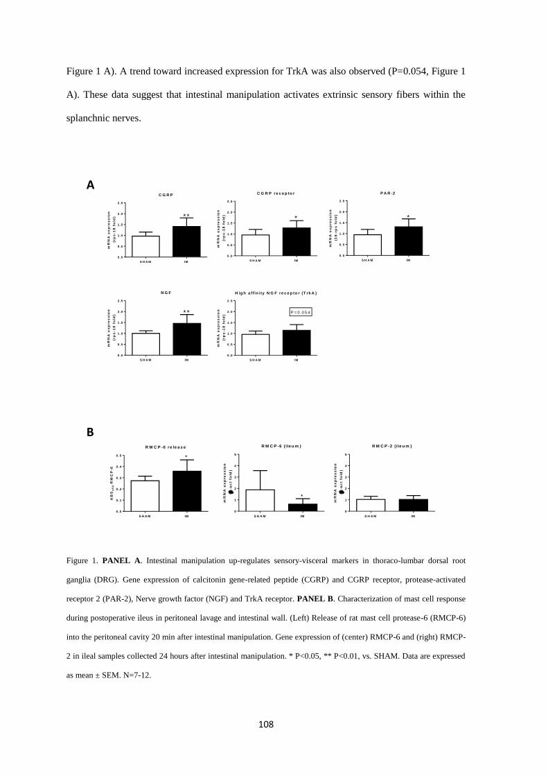
108
Figure 1 A). A trend toward increased expression for TrkA was also observed (P=0.054, Figure 1
A). These data suggest that intestinal manipulation activates extrinsic sensory fibers within the
splanchnic nerves.
Figure 1. PANEL A. Intestinal manipulation up-regulates sensory-visceral markers in thoraco-lumbar dorsal root
ganglia (DRG). Gene expression of calcitonin gene-related peptide (CGRP) and CGRP receptor, protease-activated
receptor 2 (PAR-2), Nerve growth factor (NGF) and TrkA receptor. PANEL B. Characterization of mast cell response
during postoperative ileus in peritoneal lavage and intestinal wall. (Left) Release of rat mast cell protease-6 (RMCP-6)
into the peritoneal cavity 20 min after intestinal manipulation. Gene expression of (center) RMCP-6 and (right) RMCP-
2 in ileal samples collected 24 hours after intestinal manipulation. * P<0.05, ** P<0.01, vs. SHAM. Data are expressed
as mean ± SEM. N=7-12.
C G R P
S H A M IM
0 .0
0 .5
1 .0
1 .5
2 .0
2 .5
* *
mR
NA
ex
pre
ss
ion
(rp
s-1
8 f
old
)
P A R -2
S H A M IM
0 .0
0 .5
1 .0
1 .5
2 .0
2 .5
*
mR
NA
ex
pre
ss
ion
(1
8-r
ps
fo
ld)
N G F
S H A M IM
0 .0
0 .5
1 .0
1 .5
2 .0
2 .5
* *
mR
NA
ex
pre
ss
ion
(rp
s-1
8 f
old
)
H ig h a ff in ity N G F re c e p to r (T rk A )
S H A M IM
0 .0
0 .5
1 .0
1 .5
2 .0
2 .5
P = 0 .0 5 4
mR
NA
ex
pre
ss
ion
(rp
s-1
8 f
old
)
C G R P re c e p to r
S H A M IM
0 .0
0 .5
1 .0
1 .5
2 .0
2 .5
*m
RN
A e
xp
re
ss
ion
(rp
s-1
8 f
old
)
R M C P -6 ( ile u m )
S H A M IM
0
1
2
3
4
5
*
mR
NA
ex
pre
ss
ion
(
-ac
t fo
ld)
R M C P -2 ( ile u m )
S H A M IM
0
1
2
3
4
5
mR
NA
ex
pre
ss
ion
(
-ac
t fo
ld)
R M C P -6 re le a s e
S H A M IM
0 .0
0 .1
0 .2
0 .3
0 .4
0 .5
AB
S4
50
RM
CP
-6
*
A
B
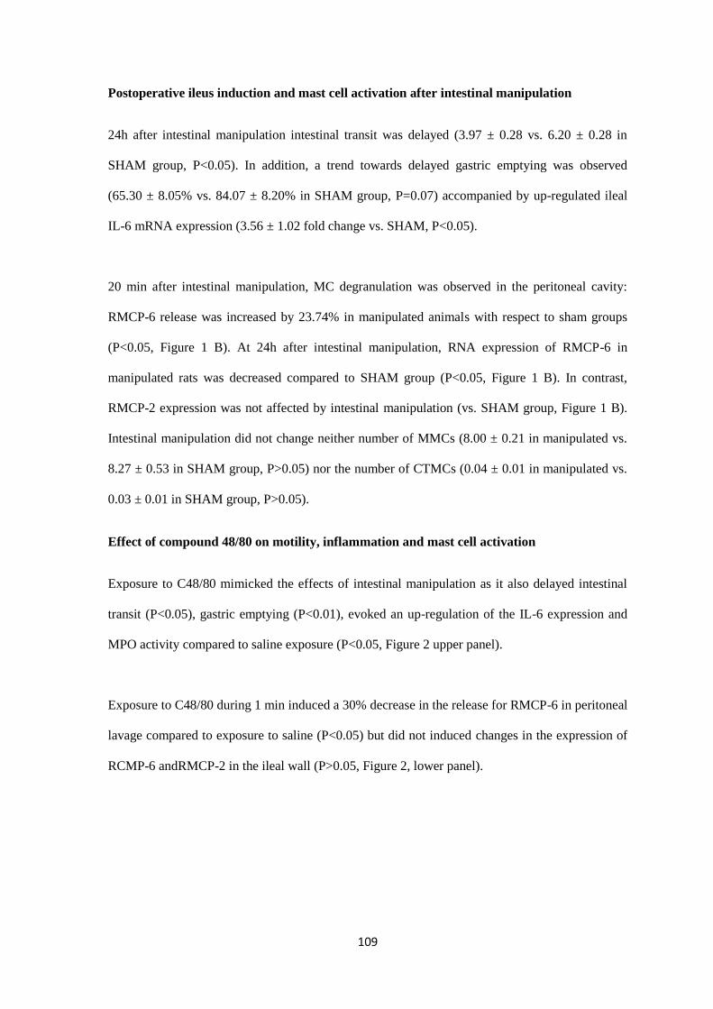
109
Postoperative ileus induction and mast cell activation after intestinal manipulation
24h after intestinal manipulation intestinal transit was delayed (3.97 ± 0.28 vs. 6.20 ± 0.28 in
SHAM group, P<0.05). In addition, a trend towards delayed gastric emptying was observed
(65.30 ± 8.05% vs. 84.07 ± 8.20% in SHAM group, P=0.07) accompanied by up-regulated ileal
IL-6 mRNA expression (3.56 ± 1.02 fold change vs. SHAM, P<0.05).
20 min after intestinal manipulation, MC degranulation was observed in the peritoneal cavity:
RMCP-6 release was increased by 23.74% in manipulated animals with respect to sham groups
(P<0.05, Figure 1 B). At 24h after intestinal manipulation, RNA expression of RMCP-6 in
manipulated rats was decreased compared to SHAM group (P<0.05, Figure 1 B). In contrast,
RMCP-2 expression was not affected by intestinal manipulation (vs. SHAM group, Figure 1 B).
Intestinal manipulation did not change neither number of MMCs (8.00 ± 0.21 in manipulated vs.
8.27 ± 0.53 in SHAM group, P>0.05) nor the number of CTMCs (0.04 ± 0.01 in manipulated vs.
0.03 ± 0.01 in SHAM group, P>0.05).
Effect of compound 48/80 on motility, inflammation and mast cell activation
Exposure to C48/80 mimicked the effects of intestinal manipulation as it also delayed intestinal
transit (P<0.05), gastric emptying (P<0.01), evoked an up-regulation of the IL-6 expression and
MPO activity compared to saline exposure (P<0.05, Figure 2 upper panel).
Exposure to C48/80 during 1 min induced a 30% decrease in the release for RMCP-6 in peritoneal
lavage compared to exposure to saline (P<0.05) but did not induced changes in the expression of
RCMP-6 andRMCP-2 in the ileal wall (P>0.05, Figure 2, lower panel).
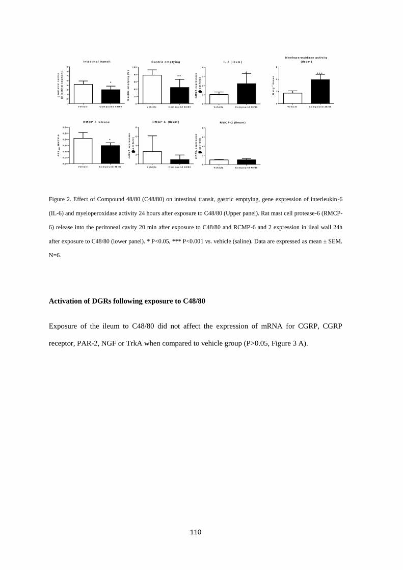
110
Figure 2. Effect of Compound 48/80 (C48/80) on intestinal transit, gastric emptying, gene expression of interleukin-6
(IL-6) and myeloperoxidase activity 24 hours after exposure to C48/80 (Upper panel). Rat mast cell protease-6 (RMCP-
6) release into the peritoneal cavity 20 min after exposure to C48/80 and RCMP-6 and 2 expression in ileal wall 24h
after exposure to C48/80 (lower panel). * P<0.05, *** P<0.001 vs. vehicle (saline). Data are expressed as mean ± SEM.
N=6.
Activation of DGRs following exposure to C48/80
Exposure of the ileum to C48/80 did not affect the expression of mRNA for CGRP, CGRP
receptor, PAR-2, NGF or TrkA when compared to vehicle group (P>0.05, Figure 3 A).
R M C P -6 re le a s e
V e h ic le C o m p o u n d 4 8 /8 0
0 .0 0
0 .0 5
0 .1 0
0 .1 5
0 .2 0
0 .2 5
0 .3 0
*
AB
S4
50
RM
CP
-6
*
In te s tin a l tra n s it
V e h ic le C o m p o u n d 4 8 /8 0
1
2
3
4
5
6
7
8
9
*
ge
om
etr
ic c
en
tre
(in
tes
tin
al
se
gm
en
t)G a s tr ic e m p ty in g
V e h ic le C o m p o u n d 4 8 /8 0
0
2 0
4 0
6 0
8 0
1 0 0
* *
Ga
str
ic e
mp
tyin
g (
%)
IL -6 (i le u m )
V e h ic le C o m p o u n d 4 8 /8 0
0
1
2
3
4
*
mR
NA
ex
pre
ss
ion
(
-ac
t fo
ld)
M y e lo p e r o x id a s e a c tiv ity
( ile u m )
U m
g-1
tis
su
e
V e h ic le C o m p o u n d 4 8 /8 0
0
2
4
6
* * *
R M C P -2 ( Ile u m )
V e h ic le C o m p o u n d 4 8 /8 0
0
2
4
6
8
mR
NA
ex
pre
ss
ion
(
-ac
t fo
ld)
R M C P -6 ( Ile u m )
V e h ic le C o m p o u n d 4 8 /8 0
0
2
4
6
8m
RN
A e
xp
re
ss
ion
(
-ac
t fo
ld)
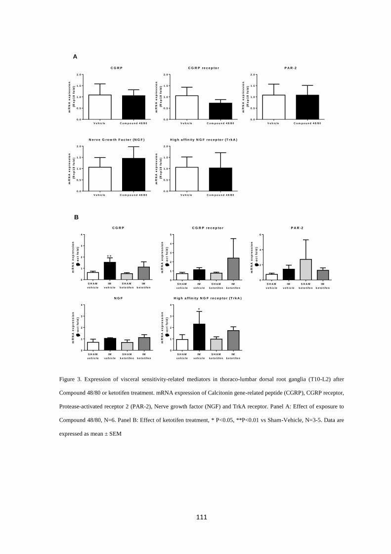
111
Figure 3. Expression of visceral sensitivity-related mediators in thoraco-lumbar dorsal root ganglia (T10-L2) after
Compound 48/80 or ketotifen treatment. mRNA expression of Calcitonin gene-related peptide (CGRP), CGRP receptor,
Protease-activated receptor 2 (PAR-2), Nerve growth factor (NGF) and TrkA receptor. Panel A: Effect of exposure to
Compound 48/80, N=6. Panel B: Effect of ketotifen treatment, * P<0.05, **P<0.01 vs Sham-Vehicle, N=3-5. Data are
expressed as mean ± SEM
C G R P
V e h ic le C o m p o u n d 4 8 /8 0
0 .0
0 .5
1 .0
1 .5
2 .0
mR
NA
ex
pre
ss
ion
(R
sp
18
fo
ld)
C G R P re c e p to r
V e h ic le C o m p o u n d 4 8 /8 0
0 .0
0 .5
1 .0
1 .5
2 .0
mR
NA
ex
pre
ss
ion
(R
sp
18
fo
ld)
P A R -2
V e h ic le C o m p o u n d 4 8 /8 0
0 .0
0 .5
1 .0
1 .5
2 .0
mR
NA
ex
pre
ss
ion
(R
sp
18
fo
ld)
N e rv e G ro w th F a c to r (N G F )
V e h ic le C o m p o u n d 4 8 /8 0
0 .0
0 .5
1 .0
1 .5
2 .0
mR
NA
ex
pre
ss
ion
(R
sp
18
fo
ld)
H ig h a ff in ity N G F re c e p to r (T rk A )
V e h ic le C o m p o u n d 4 8 /8 0
0 .0
0 .5
1 .0
1 .5
2 .0
mR
NA
ex
pre
ss
ion
(R
sp
18
fo
ld)
A
C G R P
mR
NA
ex
pre
ss
ion
(
-ac
t fo
ld)
S H A M
v e h ic le
IM
v e h ic le
S H A M
k e to t ife n
IM
k e to t ife n
0
1
2
3
4
* *
C G R P re c e p to r
mR
NA
ex
pre
ss
ion
(
-ac
t fo
ld)
S H A M
v e h ic le
IM
v e h ic le
S H A M
k e to t ife n
IM
k e to t ife n
0
1
2
3
4
5
P A R -2
mR
NA
ex
pre
ss
ion
(
-ac
t fo
ld)
S H A M
v e h ic le
IM
v e h ic le
S H A M
k e to t ife n
IM
k e to t ife n
0
2
4
6
N G F
mR
NA
ex
pre
ss
ion
(
-ac
t fo
ld)
S H A M
v e h ic le
IM
v e h ic le
S H A M
k e to t ife n
IM
k e to t ife n
0
1
2
3
4
H ig h a ff in ity N G F re c e p to r (T rk A )
mR
NA
ex
pre
ss
ion
(
-ac
t fo
ld)
S H A M
v e h ic le
IM
v e h ic le
S H A M
k e to t ife n
IM
k e to t ife n
0
1
2
3
4
*
B
112
Effect of ketotifen pretreatment on POI model
Intestinal manipulation delayed the intestinal transit and gastric emptying and reduced
postoperative fecal output (IM-vehicle vs. SHAM-vehicle, P<0.05, Figure 4 A,B,C). In sham
groups, ketotifen did not alter GI transit (SHAM-vehicle vs. SHAM-ketotifen, P>0.05, Figure 4
A,B,C). Nonetheless, in IM-ketotifen rats, CG was a 20% higher to that in IM-vehicle group
(P>0.05). Delayed gastric emptying in IM-vehicle group (23.77 ± 11.53%) was improved after
treatment with ketotifen but did not reach statistical significance (56.3 ±15.05%, P>0.05, Figure 4
B). Nonetheless, when plotting the amount of phenol red against each GI segment in the
gastrointestinal histogram, phenol red in gastric content decreased after treatment with ketotifen
(vs. IM-vehicle, P<0.05) indicating a partial effect of ketotifen in the prevention of POI by
intestinal manipulation (Figure 4 D). In addition, ketotifen improved post-surgical fecal output
suggesting improvement of large intestine propulsion (vs. IM-vehicle, P<0.01, Figure 4 C).
Pretreatment with ketotifen did not prevent the increase in mRNA expression of IL-6 induced by
intestinal manipulation (P>0.05, Figure 4 E). Intestinal manipulation increased activity of MPO
within the intestinal wall (IM-vehicle vs. SHAM-vehicle, P<0.05, Figure 4 F). Interestingly,
ketotifen did not prevent the up-regulation of MPO activity but further increased MPO activity
after intestinal manipulation (IM-ketotifen vs. IM-vehicle, Figure 4 F, P<0.05).
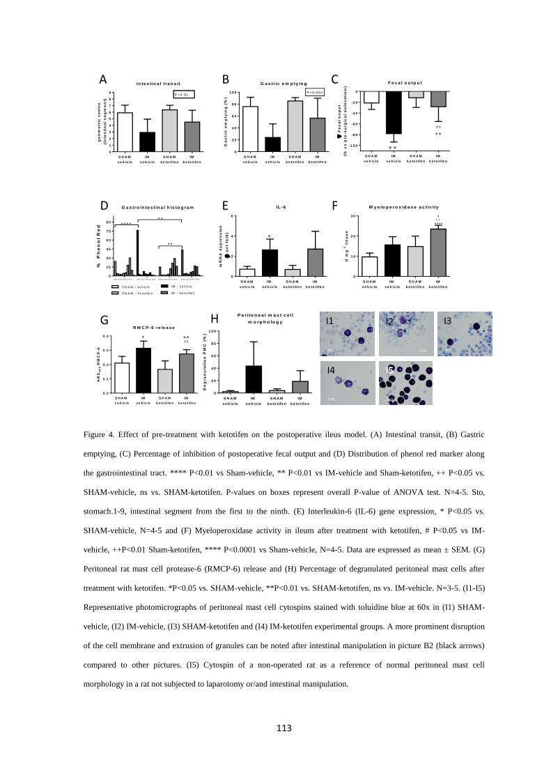
113
Figure 4. Effect of pre-treatment with ketotifen on the postoperative ileus model. (A) Intestinal transit, (B) Gastric
emptying, (C) Percentage of inhibition of postoperative fecal output and (D) Distribution of phenol red marker along
the gastrointestinal tract. **** P<0.01 vs Sham-vehicle, ** P<0.01 vs IM-vehicle and Sham-ketotifen, ++ P<0.05 vs.
SHAM-vehicle, ns vs. SHAM-ketotifen. P-values on boxes represent overall P-value of ANOVA test. N=4-5. Sto,
stomach.1-9, intestinal segment from the first to the ninth. (E) Interleukin-6 (IL-6) gene expression, * P<0.05 vs.
SHAM-vehicle, N=4-5 and (F) Myeloperoxidase activity in ileum after treatment with ketotifen, # P<0.05 vs IM-
vehicle, ++P<0.01 Sham-ketotifen, **** P<0.0001 vs Sham-vehicle, N=4-5. Data are expressed as mean ± SEM. (G)
Peritoneal rat mast cell protease-6 (RMCP-6) release and (H) Percentage of degranulated peritoneal mast cells after
treatment with ketotifen. *P<0.05 vs. SHAM-vehicle, **P<0.01 vs. SHAM-ketotifen, ns vs. IM-vehicle. N=3-5. (I1-I5)
Representative photomicrographs of peritoneal mast cell cytospins stained with toluidine blue at 60x in (I1) SHAM-
vehicle, (I2) IM-vehicle, (I3) SHAM-ketotifen and (I4) IM-ketotifen experimental groups. A more prominent disruption
of the cell membrane and extrusion of granules can be noted after intestinal manipulation in picture B2 (black arrows)
compared to other pictures. (I5) Cytospin of a non-operated rat as a reference of normal peritoneal mast cell
morphology in a rat not subjected to laparotomy or/and intestinal manipulation.
P e r ito n e a l m a s t c e l l
m o r p h o lo g y
De
gra
nu
lati
on
PM
C (
%)
S H A M
v e h ic le
IM
v e h ic le
S H A M
k e to t ife n
IM
k e to t ife n
0
2 0
4 0
6 0
8 0
1 0 0
In te s tin a l tra n s it
ge
om
etr
ic c
en
tre
(in
tes
tin
al
se
gm
en
t)
S H A M
v e h ic le
IM
v e h ic le
S H A M
k e to t ife n
IM
k e to t ife n
0
1
2
3
4
5
6
7
8
9P < 0 .0 1
G a s tr ic e m p ty in g
Ga
str
ic e
mp
tyin
g (
%)
S H A M
v e h ic le
IM
v e h ic le
S H A M
k e to t ife n
IM
k e to t ife n
0
2 0
4 0
6 0
8 0
1 0 0 P = 0 .0 0 4
G a s tro in te s tin a l h is to g r a m
%
Ph
en
ol
Re
d
0
1 5
3 0
4 5
6 0
7 5
9 0
S H A M - v e h ic le IM - v e h ic le
S H A M - k e to t ife n IM - k e to t ife n
sto1 2 3 4 5 6 7 8 9 sto1 2 3 4 5 6 7 8 9 sto1 2 3 4 5 6 7 8 9 sto1 2 3 4 5 6 7 8 9
* * * *
* *
* *
F e c a l o u tp u t
F
ec
al
ou
pu
t
(% v
s p
re
-su
rg
ica
l d
efe
ca
tio
n)
S H A M
v e h ic le
IM
v e h ic le
S H A M
k e to t ife n
IM
k e to t ife n
-1 0 0
-8 0
-6 0
-4 0
-2 0
0
* *
n s
+ +
M y e lo p e r o x id a s e a c tiv ity
U m
g-1
tis
su
e
S H A M
v e h ic le
IM
v e h ic le
S H A M
k e to t ife n
IM
k e to t ife n
0
1 0
2 0
3 0 #+ +
****
IL -6
mR
NA
ex
pre
ss
ion
(
-ac
t fo
ld)
S H A M
v e h ic le
IM
v e h ic le
S H A M
k e to t ife n
IM
k e to t ife n
0
2
4
6
*
M y e lo p e r o x id a s e a c tiv ity
U m
g-1
tis
su
e
S H A M
v e h ic le
IM
v e h ic le
S H A M
k e to t ife n
IM
k e to t ife n
0
1 0
2 0
3 0 #+ +
****
IL -6
mR
NA
ex
pre
ss
ion
(
-ac
t fo
ld)
S H A M
v e h ic le
IM
v e h ic le
S H A M
k e to t ife n
IM
k e to t ife n
0
2
4
6
*
In te s tin a l tra n s it
ge
om
etr
ic c
en
tre
(in
tes
tin
al
se
gm
en
t)
S H A M
v e h ic le
IM
v e h ic le
S H A M
k e to t ife n
IM
k e to t ife n
0
1
2
3
4
5
6
7
8
9P < 0 .0 1
G a s tr ic e m p ty in g
Ga
str
ic e
mp
tyin
g (
%)
S H A M
v e h ic le
IM
v e h ic le
S H A M
k e to t ife n
IM
k e to t ife n
0
2 0
4 0
6 0
8 0
1 0 0 P = 0 .0 0 4
G a s tro in te s tin a l h is to g r a m
%
Ph
en
ol
Re
d
0
1 5
3 0
4 5
6 0
7 5
9 0
S H A M - v e h ic le IM - v e h ic le
S H A M - k e to t ife n IM - k e to t ife n
sto1 2 3 4 5 6 7 8 9 sto1 2 3 4 5 6 7 8 9 sto1 2 3 4 5 6 7 8 9 sto1 2 3 4 5 6 7 8 9
* * * *
* *
* *
F e c a l o u tp u t
F
ec
al
ou
pu
t
(% v
s p
re
-su
rg
ica
l d
efe
ca
tio
n)
S H A M
v e h ic le
IM
v e h ic le
S H A M
k e to t ife n
IM
k e to t ife n
-1 0 0
-8 0
-6 0
-4 0
-2 0
0
* *
n s
+ +
A
D
G
B
E
H
C
F
I1 I2 I3
I4 I5
R M C P -6 re le a s e
AB
S4
50
RM
CP
-6
S H A M
v e h ic le
IM
v e h ic le
S H A M
k e to t ife n
IM
k e to t ife n
0 .0
0 .1
0 .2
0 .3
0 .4 * * *n s
114
Effects of ketotifen on mast cell activation after intestinal manipulation
Administration of ketotifen 60 min prior to surgery did not prevent the increase in peritoneal
RMCP-6 release induced by intestinal manipulation (P>0.05, Figure 4 G). Analysis of PMC
morphology by light microscopy revealed that intestinal manipulated-vehicle group presented
MCs with granules with lower content along with poor staining and extrusion of granules outside
of the cell outline, thus being compatible with features of MC degranulation (Figure 4 I2).
Intestinal manipulation induced degranulation of 43.40 ± 22.56% which was only partially
prevented by pre-treatment with ketotifen (18.60 ± 8.72%, Figure 4 H). On the other hand, MCs
from the SHAM groups were characterized by highly-defined intracytoplasmatic granules and
intact cell membrane (Figure 4 I1, I3). After treatment with ketotifen, PMCs in manipulated rats
showed a staining pattern more similar to that in SHAM-groups than that in the IM-vehicle group
(Figure 4 I4).
The number of MMCs in SHAM-vehicle (8.36 ± 0.45 MCs per high power field), IM-vehicle
(7.60 ± 0.30 MCs), SHAM-ketotifen (8.57 ± 0.50 MCs) and IM-ketotifen (8.50 ± 0.25 MCs) were
similar between the groups (P>0.05). Likewise, intestinal manipulation or ketotifen did not
change the number of CTMCs in the SHAM-vehicle (0.02 ± 0.01 MCs), IM-vehicle (0.0 ± 0.0
MCs), SHAM-ketotifen (0.0 ± 0.0 MCs) and IM-ketotifen group (0.10 ±0.06 MCs), P>0.05.
Expression of ileal RMCP-2 and -6 did not change by either intestinal manipulation or treatment
with ketotifen (P>0.05).
Effects of ketotifen on activation of dorsal root ganglia following intestinal manipulation.
Ketotifen treatment did not prevent the increased mRNA expression of CGRP and TrkA receptor
observed after intestinal manipulation (P>0.05, Figure 3 B).
115
DISCUSSION
In this study we report increased gene expression of specific sensory-related mediators in
DRGs during POI in an experimental model. Specific MC degranulation or stabilization proved
that dysmotility and inflammation observed in POI are mediated by degranulation of PMCs.
However, we were unable to prove an involvement of MC in the increase of gene expression of
sensory-related mediators induced by intestinal manipulation. We suggest that nociceptive
alteration in POI is independent of MC activation.
This is, to our knowledge, the first report demonstrating that intestinal manipulation
specifically evokes activation of gene expression of NGF, TrkA, CGRP and PAR-2 – all
mediators directed related to visceral hypersensitivity. NGF, acting through TrKA receptors
sensitises nerve terminals 22
, induces visceral hypersensitivity 23–25
and changes colon motility 26
.
Our study demonstrates that manipulation of the intestine induces an overexpression of NGF and
of its receptor which would increase sensitivity and contribute to symptoms of POI.
CGRP induces neurogenic inflammation, mediates abdominal pain 27
and can also inhibit
smooth muscle contractibility 28–30
. All of these actions could contribute to symptoms of POI.
Furthermore, CGRP up-regulation observed in the present study is in line with other studies in
which CGRP has been reported to be responsible for POI-like symptoms induced by activation of
splanchnic nerves16,18,31
.
In addition, both NGF and CGRP have been related to MC activation 32,33
and this,
together with the increase of their expression observed in our study, suggests a possible
interaction between MCs and sensory mediators expressed in DGRs. Therefore, our hypothesis
was that MC activation could contribute to the activation of DRGs by affecting the expression of
TrkA and PAR-2 on nerve terminals following release of tryptase (and other possible mediators)
from MCs.
Consequently we further aimed to investigate if MCs were involved in DRG activation.
Intestinal manipulation elicited release of RMCP-6 from PMCs. This is the first study reporting
such response in a rat model of POI and our results are in line with other studies performed on
116
mice and humans in which PMC degranulated upon intestinal manipulation 6–8,21,34
. However, in
our model, MC response seems to be limited to PMCs since we found no differences in MC
density or protease gene expression in the intestinal wall. This finding is in contrast to previous
studies reporting increased MC infiltration after intestinal manipulation 35,36
. While the divergence
of results may come from the methodology used for MC detection or from species differences, we
suggest that MC hyperplasia requires more than 24h to develop and thus to be detected by
changes in tissue MC density 4,37
.
Despite the attention to the role of MC in POI, there is still some controversy regarding
the involvement of MC in the pathophysiology of this disorder. While some studies point that
MCs play a clear role 6,21,38
others suggest that POI development is independent of MC activation
34. In our study, we addressed the involvement of MCs in symptoms of POI by a double
pharmacological way: 1) by inducing specific MC degranulation with compound 48/80, largely
proven as a specific mast cell degranulator, particularly of connective mast cells 39
, and 2) by
stabilization of MC with ketotifen, largely used in both experimental and clinical studies,
including treatments of POI 6,37,40
.
Compound 48/80 very efficiently reproduced all the motility changes induced by
intestinal manipulation as well as the increase in the inflammatory parameters. In parallel,
ketotifen in rats exposed to intestinal manipulation stabilised PMCs, which was verified by
reduced morphological traits of degranulation on peritoneal cytospins, and improved gastric
emptying and fecal output. Altogether, these results prove the role of PMCs in the induction of the
gastrointestinal motor alterations as well as the inflammation observed in POI.
In contrast, pharmacological modulation of MC activation by either C48/80 or ketotifen
did not alter expression of genes related to DRG activation. One could argue that intestinal
manipulation may be a more intense stimulus that C48/80 and that, therefore, a much large
activation of MCs could be induced by intestinal manipulation. However, the fact that C48/80 so
effectively reproduced both inflammatory and motor changes observed in POI, let us to conclude
that MC degranulation drives dysmotility and inflammation in POI but not DGRs stimulation.
Furthermore, the fact that C48/80 on its own resembled all parameters of motor alteration and
117
inflammation indicates that DGRs would be mainly responsible for other symptoms present in
POI, i.e. abdominal pain, but not motility disorders. In fact, a previous study demonstrated that
treatment with ketotifen prevented delayed gastric emptying but did not ameliorate pain score in
patients undergoing abdominal surgery 40
.
In summary, our results indicate that clinical handling of POI should contemplate a
double approach: 1) restoration of GI motor activity, where preventive treatments with ketotifen
or other mast stabilizers could be very effective; and 2) prevention of hypersensitivity and
hyperalgesia.
In summary, the present study describes the up-regulation in DRGs of NGF, TrkA, PAR-
2 and CGRP, mediators that are related to hypersensitivity and visceral function, suggesting a
potential role for POI pathogenesis. It also supports the role of MCs in POI and suggests these
cells as a target for the development of new strategies to prevent POI in patients undergoing
abdominal surgery. In addition, other strategies should also be provided to prevent neural
activation, altogether leading to a better approach to treat the multi-mechanistic pathogenesis of
POI.
118
ACKNOWLEDGMENTS, FUNDING AND DISCLOSURES
Acosta and E. Martínez are thanked for their assistance.
S.B and J.R designed and performed the study, analysed data and wrote the paper. P.V designed
the study and wrote the paper.
This study was supported by grants BFU2010-15401 from Ministerio de Ciencia e Innovación,
2009SGR708 and 2014SGR789 from Direcció General de Recerca de la Generalitat de
Catalunya. S.B. personal support: FPU program (AP2010-1692) from Ministerio de Educación,
Cultura y Deporte.
No competing interests declared.
119
REFERENCES
1. Doorly, M. G. & Senagore, A. J. Pathogenesis and Clinical and Economic Consequences
of Postoperative Ileus. Surg. Clin. North Am. 92, 259–272 (2012).
2. Lützelschwab, C., Pejler, G., Aveskogh, M. & Hellman, L. Secretory granule proteases in
rat mast cells. Cloning of 10 different serine proteases and a carboxypeptidase A from
various rat mast cell populations. J. Exp. Med. 185, 13–29 (1997).
3. Saavedra, Y. & Vergara, P. Hypersensitivity to ovalbumin induces chronic intestinal
dysmotility and increases the number of intestinal mast cells. Neurogastroenterol. Motil.
17, 112–22 (2005).
4. Fernández-Blanco, J. A., Hollenberg, M., Martínez, V. & Vergara, P. PAR-2-mediated
control of barrier function and motility differs between early and late phases of
postinfectious gut dysfunction in the rat. Am. J. Physiol. Gastrointest. Liver Physiol. 304,
G390–400 (2013).
5. Chen, Z. et al. Localization of rat tryptase to a subset of the connective tissue type of mast
cell. J. Histochem. Cytochem. 41, 961–9 (1993).
6. De Jonge, W. J. et al. Mast cell degranulation during abdominal surgery initiates
postoperative ileus in mice. Gastroenterology 127, 535–545 (2004).
7. Berdún, S. et al. Peritoneal mast cell degranulation and gastrointestinal recovery in
patients undergoing colorectal surgery. Neurogastroenterol. Motil. n/a–n/a (2015).
doi:10.1111/nmo.12525
8. The, F. O. et al. Intestinal handling-induced mast cell activation and inflammation in
human postoperative ileus. Gut 57, 33–40 (2008).
9. Stead, R. H. et al. Intestinal mucosal mast cells in normal and nematode-infected rat
intestines are in intimate contact with peptidergic nerves. Proc. Natl. Acad. Sci. U. S. A.
84, 2975–9 (1987).
10. Barbara, G. et al. Activated mast cells in proximity to colonic nerves correlate with
abdominal pain in irritable bowel syndrome. Gastroenterology 126, 693–702 (2004).
11. Suzuki, R. et al. Direct neurite-mast cell communication in vitro occurs via the
neuropeptide substance P. J. Immunol. 163, 2410–5 (1999).
12. De Jonge, F. et al. In vitro activation of murine DRG neurons by CGRP-mediated mucosal
mast cell degranulation. Am. J. Physiol. Gastrointest. Liver Physiol. 287, G178–91 (2004).
13. Mueller, M. H. et al. Intestinal afferent nerve sensitivity is increased during the initial
development of postoperative ileus in mice. J. Gastrointest. Surg. 13, 423–31 (2009).
14. Bonaz, B., Plourde, V. & Taché, Y. Abdominal surgery induces Fos immunoreactivity in
the rat brain. J. Comp. Neurol. 349, 212–22 (1994).
120
15. Barquist, E. et al. Neuronal pathways involved in abdominal surgery-induced gastric ileus
in rats. Am. J. Physiol. 270, R888–94 (1996).
16. Zittel, T. T., Reddy, S. N., Plourde, V. & Raybould, H. E. Role of spinal afferents and
calcitonin gene-related peptide in the postoperative gastric ileus in anesthetized rats. Ann.
Surg. 219, 79–87 (1994).
17. Zittel, T. T., Meile, T., Jehle, E. C. & Becker, H. D. Intraperitoneal capsaicin treatment
reduces postoperative gastric ileus in awake rats. Langenbecks. Arch. Surg. 386, 204–11
(2001).
18. Zittel, T. T. et al. Calcitonin gene-related peptide and spinal afferents partly mediate
postoperative colonic ileus in the rat. Surgery 123, 518–27 (1998).
19. Li, W.-W. et al. Substance P signaling controls mast cell activation, degranulation, and
nociceptive sensitization in a rat fracture model of complex regional pain syndrome.
Anesthesiology 116, 882–95 (2012).
20. Liu, S. & Chen, J. D. Z. Colonic electrical stimulation regulates colonic transit via the
nitrergic pathway in rats. Dig. Dis. Sci. 51, 502–5 (2006).
21. Snoek, S. A. et al. Mast cells trigger epithelial barrier dysfunction, bacterial translocation
and postoperative ileus in a mouse model. Neurogastroenterol. Motil. 24, 172–84, e91
(2012).
22. Zhang, X., Huang, J. & McNaughton, P. A. NGF rapidly increases membrane expression
of TRPV1 heat-gated ion channels. EMBO J. 24, 4211–23 (2005).
23. Barreau, F., Cartier, C., Ferrier, L., Fioramonti, J. & Bueno, L. Nerve growth factor
mediates alterations of colonic sensitivity and mucosal barrier induced by neonatal stress
in rats. Gastroenterology 127, 524–34 (2004).
24. Vasko, M. R. et al. Nerve growth factor mediates a switch in intracellular signaling for
PGE2-induced sensitization of sensory neurons from protein kinase A to Epac. PLoS One
9, e104529 (2014).
25. Delafoy, L., Raymond, F., Doherty, A. M., Eschalier, A. & Diop, L. Role of nerve growth
factor in the trinitrobenzene sulfonic acid-induced colonic hypersensitivity. Pain 105,
489–97 (2003).
26. Jardí, F., Martínez, V. & Vergara, P. NGF is involved in oral ovalbumin-induced altered
colonic contractility in rats: evidence from the blockade of TrkA receptors with K252a.
Neurogastroenterol. Motil. 24, e580–90 (2012).
27. Friese, N. et al. Involvement of prostaglandins and CGRP-dependent sensory afferents in
peritoneal irritation-induced visceral pain. Regul. Pept. 70, 1–7 (1997).
28. Plourde, V., St-Pierre, S., Fournier, A. & Taché, Y. CGRP 8-37[correction of 8-27] blocks
the inhibition of gastric emptying induced by intravenous injection of alpha-CGRP in rats.
Life Sci. 52, 857–62 (1993).
121
29. Maggi, C. A., Giuliani, S. & Zagorodnyuk, V. Calcitonin gene-related peptide (CGRP) in
the circular muscle of guinea-pig colon: role as inhibitory transmitter and mechanisms of
relaxation. Regul. Pept. 61, 27–36 (1996).
30. Holzer, P., Barthó, L., Matusák, O. & Bauer, V. Calcitonin gene-related peptide action on
intestinal circular muscle. Am. J. Physiol. 256, G546–52 (1989).
31. Plourde, V., Wong, H. C., Walsh, J. H., Raybould, H. E. & Taché, Y. CGRP antagonists
and capsaicin on celiac ganglia partly prevent postoperative gastric ileus. Peptides 14,
1225–9 (1993).
32. Stempelj, M. & Ferjan, I. Signaling pathway in nerve growth factor induced histamine
release from rat mast cells. Inflamm. Res. 54, 344–9 (2005).
33. Rychter, J. W. et al. CGRP1 receptor activation induces piecemeal release of protease-1
from mouse bone marrow-derived mucosal mast cells. Neurogastroenterol. Motil. 23,
e57–68 (2011).
34. Gomez-Pinilla, P. J. et al. Mast cells play no role in the pathogenesis of postoperative ileus
induced by intestinal manipulation. PLoS One 9, e85304 (2014).
35. Liu, X., Wu, T. & Chi, P. Inhibition of MK2 shows promise for preventing postoperative
ileus in mice. J. Surg. Res. 185, 102–12 (2013).
36. Kalff, J. C., Schraut, W. H., Simmons, R. L. & Bauer, A. J. Surgical manipulation of the
gut elicits an intestinal muscularis inflammatory response resulting in postsurgical ileus.
Ann. Surg. 228, 652–63 (1998).
37. Serna, H., Porras, M. & Vergara, P. Mast cell stabilizer ketotifen [4-(1-methyl-4-
piperidylidene)-4h-benzo[4,5]cyclohepta[1,2-b]thiophen-10(9H)-one fumarate] prevents
mucosal mast cell hyperplasia and intestinal dysmotility in experimental Trichinella
spiralis inflammation in the rat. J. Pharmacol. Exp. Ther. 319, 1104–11 (2006).
38. Van Bree, S. H. W. et al. Inhibition of spleen tyrosine kinase as treatment of postoperative
ileus. Gut 62, 1581–90 (2013).
39. Pearce, F. L., Befus, A. D., Gauldie, J. & Bienenstock, J. Mucosal mast cells. II. Effects of
anti-allergic compounds on histamine secretion by isolated intestinal mast cells. J.
Immunol. 128, 2481–6 (1982).
40. The, F. O. et al. The role of mast cell stabilization in treatment of postoperative ileus: a
pilot study. Am. J. Gastroenterol. 104, 2257–66 (2009).

122

123
DISCUSSION
124
Over the last decade, using different experimental approaches, researchers have
demonstrated that MCs contribute to the pathophysiology of POI 164,163,52,166,187
. As MCs
communicate with nerve endings within the GI tract131
, we hypothesized that MC-nerve
interactions are involved in the pathophysiology of POI. NGF is one of the mediators involved in
MC-nerve interactions189,190
and can induce MC degranulation144
. Therefore, we proposed a role
for NGF in mediating the activation of MCs during the development of POI. Thus, the main aim
of this work was to gain knowledge on the mechanisms involved in the degranulation of MC upon
intestinal manipulation, especially those involving nerve interactions and to further evaluate MCs
as a target to prevent the development of POI.
Intestinal manipulation induces degranulation of peritoneal mast cells during colorectal
surgery in human patients and in an experimental model of postoperative ileus in rats.
To evaluate if MCs degranulate during the development of POI we firstly focused on
patients undergoing colorectal surgery. We observed increased release of MC proteases into the
peritoneal cavity at the end of the surgical procedure. This finding suggests that resection of the
colon induces degranulation of PMCs. These results are in line with those observed previously in
abdominal surgeries that do not involve intestinal surgery166,187
. However, our study was the first
performed in patients undergoing intestinal handling, which is the main cause of POI. Therefore
our findings are of relevance to the development of this condition. Furthermore, our findings were
observed in patients developing POI. Thus, further supporting a potential role for MCs in POI as
suggested by previous reports166,187
.
Another observation we report is that degranulation of PMCs is not dependent on the
technique used for the resection of the colon. In fact, concentrations of released tryptase and
chymase were similar between open and laparoscopic colectomies. This is in contrast to
gynaecological surgery in which tryptase release was only detected following open hysterectomy
166.
Therefore, manipulation of the gut irrespective of the technique used appears to be
sufficient to evoke degranulation of MCs. Pharmacological strategies, i.e. MC blocking agents,
125
should therefore be taken into consideration to prevent release of MC mediators during surgery
and thereby prevent or diminish the severity of POI . Similar to the observations in patients
undergoing colectomy, we demonstrated an increased release of mast cell protease in peritoneal
lavage in our model of POI. This was confirmed in our two studies in the rat. Therefore, we
conclude that intestinal manipulation elicits a similar PMC response in both human and rat. Our
studies are the first reporting MC activation following intestinal manipulation in patients
undergoing colectomy and in rats, and therefore provide new data that complement previous
studies focusing on mice and on patients from non-gut surgeries 52,166,188,191
.
In the present study we focused on RMCP-6 which is a specific protease of the major MC
type residing in the peritoneal cavity. This allows us to conclude that CTMC activation constitutes
a major response during the development of POI and efforts should be made to specifically block
this type of MCs.
Summarised, these results indicate that PMCs are activated and release mediators that
potentially can alter gut homeostasis leading to intestinal disease such as POI as previously
reported84
. To further corroborate if MC degranulation causes POI, we investigated the effect of
drugs that modulate MC activity in the POI model.
Mast cells participate in experimental postoperative ileus in rat
The POI model we presented was characterized by delayed gastric emptying and
intestinal transit, up-regulation of IL-6 gene expression and increased MPO activity in the
intestinal wall. This is in line with previous animal studies reporting muscular layer inflammation
and hypomotility in rat after intestinal manipulation164,171
.
The use of MC degranulator C48/80 induced a pattern of GI hypomotility and
inflammation similar to that observed after intestinal manipulation. These findings suggest a
detrimental role of MCs in POI which is supported by a previous study in which C48/80 also
delayed intestinal motility and evoke an inflammatory response in the GI tract52
. We verified that
C48/80 induces β-hexosaminidase release from rat PMCs in vitro and in addition induces changes
in the release of RMCP-6 from PMCs in vivo. We subsequently tested the effects of the MC
126
stabilizer ketotifen in the rat POI model. In our study, ketotifen did prevent delayed gastric
emptying and reduced postoperative fecal output induced by intestinal manipulation. These results
complement other studies that reported POI amelioration in mice and humans52,187
.
In summary, these results suggest that MCs are implicated in the development of
inflammation and dysmotility occurring in the rat POI. This indicates MCs as a potential target for
the development of new strategies to shorten POI in the clinical setting.
However, it should be taken into consideration that the actions of ketotifen are not
specific to MCs, but can also act on other systems 192
. Complementary studies using MC deficient
animal models would be needed to confirm our results. A recent study using MC deficient mice
(Cpa3Cre/+
) indicated that absence of MCs does not result in the prevention of POI191
. As immune
responses may differ between mice and rat, a similar study using MC deficient rats would be
required to confirm our observations.
Intestinal manipulation induces mast cell-independent up-regulation of gene expression of
visceral sensory-related mediators in dorsal root ganglia.
In the next step of the present work, we investigated if MC-nerve interactions represent a
mechanism by which MCs are involved in the pathogenesis of POI. We observed an up-regulation
of CGRP, NGF, TrkA and PAR-2 gene expression in thoraco-lumbar dorsal root ganglia in the rat
model of POI 24h after intestinal manipulation. This indicates a role of these mediators in the
development of POI.
Since CGRP, NGF, TrkA and PAR-2 participate in MC-nerve communication193,194
, our
results suggest a bi-directional interaction between MCs and nerve endings during the initiation of
POI. The up-regulation of PAR-2 on nerve endings and RCMP-6 release after intestinal
manipulation observed in our study could indicate that MCs do communicate with nerve endings
during POI and therefore could result in alteration of motility and inflammation as reported in
other studies 195–197
. Stimulated MCs have been also shown to release NGF198
which can act on
up-regulated TrkA receptors expressed in nerve terminations after intestinal manipulation. CGRP
is a neuropeptide involved in activation of MCs 97
. Therefore, one can speculate that CGRP is
127
involved in the activation of MCs during POI, and in turn CGRP-induced MC degranulation can
activate PAR-2 and TrkA. Furthermore, our results indicate DRGs as the source of CGRP that has
been proved to lead to dysmotility in other POI models 199–202
.
Nonetheless, the use of the mast cell degranulator C48/80 did not increase gene
expression of the GCRP, NGF, TrkA or PAR-2 in the DRGs. Similarly, ketotifen did not prevent
the up-regulation of the investigated genes induced by intestinal manipulation. Based on this
result we conclude, that MCs are possibly responsible for motility disturbance and inflammation
during POI but do not drive activation of DRGs. It could be argued that C48/80 was a less intense
stimulus compared to the intestinal manipulation and therefore not sufficient to alter DRG gene
expression, but the present study suggests that modulation of motility and inflammation by
C48/80 and ketotifen was independent of DRG activation. This indicates that nerve endings do
not represent a target for MC activation in the context of POI. This observation is in line with the
suggestion that symptoms derived from activation of DRGs, such as abdominal pain or
hypomotility, in non-manipulated segments40
of GI tract should be targeted with strategies other
than MC blockers187
.
NGF-antagonist K252a stabilized mast cells and prevented IL-6 expression in postoperative
ileus rat model.
NGF is a well-known neurotrophin that induces degranulation of MCs203,204
and
participates in many functional motility disorders205,137
. On this basis, we evaluated if NGF could
serve as a pharmacological target in MC-mediated POI. Our study demonstrated that the alkaloid
K252a, an antagonist of the NGF receptor TrkA, inhibits degranulation of PMCs evoked by NGF
in vitro, which is in line with previous reports143,206
. In addition, we also report, for the first time,
inhibition of in vivo RMCP-6 release from PMCs and reduction of basal gene expression of
RMCP-2 in experimental POI. All these data suggest that K252a has a therapeutic potential as a
mast cell stabilizing agent. In addition we also described that K252a act on both MMCs and
CTMCs suggesting that K252a could be efficient in MC-mediated disorders irrespective of the
type of MC involved.
128
In the POI model, K252a also reduced IL-6 up-regulation induced by intestinal
manipulation which is in agreement with other studies reporting amelioration of inflammation in
experimental psoriasis, rhinitis and pulmonary infection following treatment with this agent207–209
.
Since MCs express TrkA receptors 137
and C48/80 induces over-expression of IL-6 as previously
discussed in this work, MCs can be the mechanism responsible for IL-6 down-regulation after
K252a treatment.
As an alternative, K252a could be acting on overexpressed TrkA on splanchnic nerve
endings as we previously described. In that case, non-mast cell derived NGF could sensitize nerve
endings and enhance CGRP release210–212
, or PAR-2 expression213
. Therefore, overexpression of
CGRP or PAR-2 induced by NGF/TrkA could be responsible for POI symptoms derived from
DRG activation. This would be in agreement with our previous results in which DRG activation
does not depend on MC degranulation.
In contrast to other parameters, K252a did not improve POI hypomotiliy. A previous
study described intestinal motility modulation by K252a 137
but dosage or route of administration
of K252a, used in this work, could underlie the lack of effect of this compound on motility in our
study, and explain the divergence of our results from that of others.
In summary, in our study we observed that intestinal manipulation induced degranulation of
PMCs in both human and rat. Manipulation of the intestine was also associated with an increase
of gene expression of CGRP, NGF-TrkA and PAR-2 in thoraco-lumbar DRG, thus indicating the
activation of splanchnic afferent fibers in the experimental model of POI. While MCs played a
role in the inflammation and POI related dysmotility, C48/80 and ketotifen did not demonstrate a
MC-dependent modulation of activation of DRGs. Therefore, under our experimental approach,
activation of DRG appears to be independent of MC activity. Interaction between NGF and its
receptor TrkA participate in degranulation of PMCs and inflammatory POI and therefore could be
one of the mechanisms mediating MC involvement in POI. Furthermore, K252a could be used as
MC blocking agent not only in POI but also in other disorders where MCs are involved.

129
CONCLUSIONS

130
131
1. In humans, colorectal surgery induced a local response of MCs characterized by release
of proteases into the peritoneal cavity. This release was irrespective of the technique
employed for resection of the colon, suggesting that the handling of the intestine to be
removed, rather than the surgical technique, determined the degranulation of PMCs. The
degranulation of PMCs following abdominal surgery was significantly correlated with the
development of POI in surgical patients, thus indicating an association between MCs and
POI.
2. In the rat model, intestinal manipulation lead to POI which was characterized by delayed
gastrointestinal motility and intestinal inflammation. Intestinal manipulation also induced
degranulation of MCs. This response was restricted to MCs residing in the peritoneal
cavity and therefore belonging to the connective tissue MC type.
3. The mast cell degranulator Compound 48/80 induced a delay of gastric emptying and
intestinal transit as well as an inflammatory response within the intestinal wall, thus,
reproducing the pattern of POI symptoms observed after intestinal manipulation.
Ketotifen reversed the delay of gastric emptying and restored the colonic propulsion
induced by intestinal manipulation. All together, these results indicate involvement of
MCs in the pathogenesis of POI.
4. Intestinal manipulation in the rat model increased the gene-expression of calcitonin gene-
related peptide (CGRP), nerve growth factor (NGF), TrkA and protease activated
receptor-2 (PAR-2) in thoraco-lumbar DRG. Therefore, intestinal manipulation seems to
be associated to plasticity of extrinsic primary sensory neurons during the development of
POI.
5. Exposure of the intestine to the mast cell degranulator compound 48/80 did not induced
up-regulation of CGRP, NGF, TrkA and PAR-2 in DRG. The mast cell stabilizer
ketotifen did not prevent up-regulation of these mediators following intestinal
manipulation. Therefore, under this pharmacological approach, activation of DRG is
independent of activation of MCs during the induction of POI. This suggests that
132
strategies other than MC stabilization should be employed in order to prevent DRG-
derived symptoms after intestinal manipulation.
6. The NGF receptor antagonist, K252a, inhibited the degranulation of rat PMCs in vitro
and in vivo in the experimental model of POI. In addition, K252a also decreased the basal
RMCP-2 expression in the intestinal wall. Thus, we confirm that K252a could be
employed as a MC stabilizing agent. In the POI model, K252a also diminished the up-
regulation of IL-6 gene expression. Therefore, K252a partially resolves inflammation and
modulates the immune response to intestinal manipulation in POI.
Intestinal manipulation evokes a response that involves hypomotility and
inflammation of the GI tract, degranulation of peritoneal mast cells and activation of
splanchnic afferent fibers leading to postoperative ileus. Of the components
mediating this response is the interaction between Nerve growth factor and TrkA.
Indeed, mast cell degranulation participates in the pathogenesis of postoperative
ileus.
133
REFERENCES

134
135
1. Furness, J. B., Alex, G., Clark, M. J. & Lal, V. V. Morphologies and projections of defined
classes of neurons in the submucosa of the guinea-pig small intestine. Anat. Rec. A.
Discov. Mol. Cell. Evol. Biol. 272, 475–83 (2003).
2. Hansen, M. B. The enteric nervous system II: gastrointestinal functions. Pharmacol.
Toxicol. 92, 249–57 (2003).
3. Bayliss, W. M. & Starling, E. H. The movements and innervation of the small intestine. J.
Physiol. 24, 99–143 (1899).
4. Raybould, H. E. & Taché, Y. Cholecystokinin inhibits gastric motility and emptying via a
capsaicin-sensitive vagal pathway in rats. Am. J. Physiol. 255, G242–6 (1988).
5. JE, K. in Comprehensive Human Physiology (eds. Greger, R. & Windhorst, U.) 1289–
1308 (Springer Berlin Heidelberg, 1996).
6. Furness, J. B. The enteric nervous system and neurogastroenterology. Nat. Rev.
Gastroenterol. Hepatol. 9, 286–94 (2012).
7. Costa, M. Anatomy and physiology of the enteric nervous system. Gut 47, 15iv–19 (2000).
8. Furness, J. . Types of neurons in the enteric nervous system. J. Auton. Nerv. Syst. 81, 87–
96 (2000).
9. Hansen, M. B. The enteric nervous system I: organisation and classification. Pharmacol.
Toxicol. 92, 105–13 (2003).
10. Furness, J. B., Jones, C., Nurgali, K. & Clerc, N. Intrinsic primary afferent neurons and
nerve circuits within the intestine. Prog. Neurobiol. 72, 143–64 (2004).
11. Grider, J. R. Neurotransmitters mediating the intestinal peristaltic reflex in the mouse. J.
Pharmacol. Exp. Ther. 307, 460–7 (2003).
12. Grider, J. R. & Piland, B. E. The peristaltic reflex induced by short-chain fatty acids is
mediated by sequential release of 5-HT and neuronal CGRP but not BDNF. Am. J.
Physiol. Gastrointest. Liver Physiol. 292, G429–37 (2007).
13. Chang, H. Y., Mashimo, H. & Goyal, R. K. Musings on the wanderer: what’s new in our
understanding of vago-vagal reflex? IV. Current concepts of vagal efferent projections to
the gut. Am. J. Physiol. Gastrointest. Liver Physiol. 284, G357–66 (2003).
14. Lomax, A. E., Sharkey, K. A. & Furness, J. B. The participation of the sympathetic
innervation of the gastrointestinal tract in disease states. Neurogastroenterol. Motil. 22, 7–
18 (2010).
15. Suda, K., Setoyama, H., Nanno, M., Matsumoto, S. & Kawai, M. Involvement of
parasympathetic pelvic efferent pathway in psychological stress-induced defecation. World
J. Gastroenterol. 19, 1200–9 (2013).
16. Tong, W. D., Ridolfi, T. J., Kosinski, L., Ludwig, K. & Takahashi, T. Effects of
autonomic nerve stimulation on colorectal motility in rats. Neurogastroenterol. Motil. 22,
688–93 (2010).
136
17. Michael J. Beyak, D. C. E. B. W. J. C. K. W. R. D. G. in Physiology of the gastrointestinal
tract (ed. Leonard R. Johnson, K. E. B. F. K. G. J. L. M. H. M. S. and J. D. W.) 685–725
(Academic press, 2006).
18. Blackshaw, L. A., Brookes, S. J. H., Grundy, D. & Schemann, M. Sensory transmission in
the gastrointestinal tract. Neurogastroenterol. Motil. 19, 1–19 (2007).
19. Holzer, P. Gastrointestinal afferents as targets of novel drugs for the treatment of
functional bowel disorders and visceral pain. Eur. J. Pharmacol. 429, 177–93 (2001).
20. Grundy, D. Neuroanatomy of visceral nociception: vagal and splanchnic afferent. Gut 51,
i2–i5 (2002).
21. Phillips, R. J. & Powley, T. L. Innervation of the gastrointestinal tract: patterns of aging.
Auton. Neurosci. 136, 1–19 (2007).
22. Berthoud, H. R., Blackshaw, L. A., Brookes, S. J. H. & Grundy, D. Neuroanatomy of
extrinsic afferents supplying the gastrointestinal tract. Neurogastroenterol. Motil. 16
Suppl 1, 28–33 (2004).
23. Knowles, C. H. & Aziz, Q. Basic and clinical aspects of gastrointestinal pain. Pain 141,
191–209 (2009).
24. Brookes, S. J. H., Spencer, N. J., Costa, M. & Zagorodnyuk, V. P. Extrinsic primary
afferent signalling in the gut. Nat. Rev. Gastroenterol. Hepatol. 10, 286–96 (2013).
25. Furness, J. B., Rivera, L. R., Cho, H.-J., Bravo, D. M. & Callaghan, B. The gut as a
sensory organ. Nat. Rev. Gastroenterol. Hepatol. 10, 729–40 (2013).
26. Moran, G. W., Leslie, F. C., Levison, S. E., Worthington, J. & McLaughlin, J. T.
Enteroendocrine cells: neglected players in gastrointestinal disorders? Therap. Adv.
Gastroenterol. 1, 51–60 (2008).
27. Kirchgessner, A. L., Tamir, H. & Gershon, M. D. Identification and stimulation by
serotonin of intrinsic sensory neurons of the submucosal plexus of the guinea pig gut:
activity-induced expression of Fos immunoreactivity. J. Neurosci. 12, 235–48 (1992).
28. Gunawardene, A. R., Corfe, B. M. & Staton, C. A. Classification and functions of
enteroendocrine cells of the lower gastrointestinal tract. Int. J. Exp. Pathol. 92, 219–31
(2011).
29. Hölzer, H. H., Turkelson, C. M., Solomon, T. E. & Raybould, H. E. Intestinal lipid inhibits
gastric emptying via CCK and a vagal capsaicin-sensitive afferent pathway in rats. Am. J.
Physiol. 267, G625–9 (1994).
30. Miyano, Y. et al. The role of the vagus nerve in the migrating motor complex and ghrelin-
and motilin-induced gastric contraction in suncus. PLoS One 8, e64777 (2013).
31. Mondal, A. et al. Coordination of motilin and ghrelin regulates the migrating motor
complex of gastrointestinal motility in Suncus murinus. Am. J. Physiol. Gastrointest. Liver
Physiol. 302, G1207–15 (2012).
137
32. Giralt, M. & Vergara, P. Glucagonlike Peptide-1 (GLP-1) Participation in Ileal Brake
Induced by Intraluminal Peptones in Rat. Dig. Dis. Sci. 44, 322–329 (1999).
33. Pironi, L. et al. Fat-induced ileal brake in humans: a dose-dependent phenomenon
correlated to the plasma levels of peptide YY. Gastroenterology 105, 733–9 (1993).
34. Mikkelsen, H. B. Interstitial cells of Cajal, macrophages and mast cells in the gut
musculature: morphology, distribution, spatial and possible functional interactions. J. Cell.
Mol. Med. 14, 818–32 (2010).
35. Harrison, O. J. & Maloy, K. J. Innate immune activation in intestinal homeostasis. J.
Innate Immun. 3, 585–93 (2011).
36. Doe, W. F. The intestinal immune system. Gut 30, 1679–85 (1989).
37. Khan, W. I. et al. Critical role for signal transducer and activator of transcription factor 6
in mediating intestinal muscle hypercontractility and worm expulsion in Trichinella
spiralis-infected mice. Infect. Immun. 69, 838–44 (2001).
38. Nalli, A. D. et al. Hypercontractility of intestinal longitudinal smooth muscle induced by
cytokines is mediated by the nuclear factor-κB/AMP-activated kinase/myosin light chain
kinase pathway. J. Pharmacol. Exp. Ther. 350, 89–98 (2014).
39. Kalff, J. C., Schraut, W. H., Billiar, T. R., Simmons, R. L. & Bauer, A. J. Role of
inducible nitric oxide synthase in postoperative intestinal smooth muscle dysfunction in
rodents. Gastroenterology 118, 316–27 (2000).
40. De Jonge, W. J. et al. Postoperative ileus is maintained by intestinal immune infiltrates
that activate inhibitory neural pathways in mice. Gastroenterology 125, 1137–47 (2003).
41. Sarker, S. A. & Gyr, K. Non-immunological defence mechanisms of the gut. Gut 33, 987–
93 (1992).
42. Hansen, M. B. Neurohumoral control of gastrointestinal motility. Physiol. Res. 52, 1–30
(2003).
43. Imeryüz, N. et al. Glucagon-like peptide-1 inhibits gastric emptying via vagal afferent-
mediated central mechanisms. Am. J. Physiol. 273, G920–7 (1997).
44. Cuche, G., Cuber, J. C. & Malbert, C. H. Ileal short-chain fatty acids inhibit gastric
motility by a humoral pathway. Am. J. Physiol. Gastrointest. Liver Physiol. 279, G925–30
(2000).
45. Raybould, H. E. Capsaicin-sensitive vagal afferents and CCK in inhibition of gastric motor
function induced by intestinal nutrients. Peptides 12, 1279–83
46. Gwynne, R. M. & Bornstein, J. C. Mechanisms underlying nutrient-induced segmentation
in isolated guinea pig small intestine. Am. J. Physiol. Gastrointest. Liver Physiol. 292,
G1162–72 (2007).
47. Gwynne, R. M., Thomas, E. A., Goh, S. M., Sjövall, H. & Bornstein, J. C. Segmentation
induced by intraluminal fatty acid in isolated guinea-pig duodenum and jejunum. J.
Physiol. 556, 557–69 (2004).
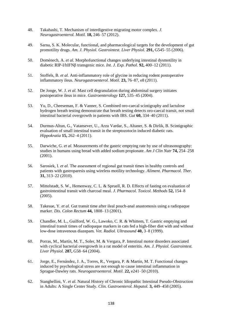
138
48. Takahashi, T. Mechanism of interdigestive migrating motor complex. J.
Neurogastroenterol. Motil. 18, 246–57 (2012).
49. Sarna, S. K. Molecular, functional, and pharmacological targets for the development of gut
promotility drugs. Am. J. Physiol. Gastrointest. Liver Physiol. 291, G545–55 (2006).
50. Domènech, A. et al. Morphofunctional changes underlying intestinal dysmotility in
diabetic RIP-I/hIFNβ transgenic mice. Int. J. Exp. Pathol. 92, 400–12 (2011).
51. Stoffels, B. et al. Anti-inflammatory role of glycine in reducing rodent postoperative
inflammatory ileus. Neurogastroenterol. Motil. 23, 76–87, e8 (2011).
52. De Jonge, W. J. et al. Mast cell degranulation during abdominal surgery initiates
postoperative ileus in mice. Gastroenterology 127, 535–45 (2004).
53. Yu, D., Cheeseman, F. & Vanner, S. Combined oro-caecal scintigraphy and lactulose
hydrogen breath testing demonstrate that breath testing detects oro-caecal transit, not small
intestinal bacterial overgrowth in patients with IBS. Gut 60, 334–40 (2011).
54. Durmus-Altun, G., Vatansever, U., Arzu Vardar, S., Altaner, S. & Dirlik, B. Scintigraphic
evaluation of small intestinal transit in the streptozotocin induced diabetic rats.
Hippokratia 15, 262–4 (2011).
55. Darwiche, G. et al. Measurements of the gastric emptying rate by use of ultrasonography:
studies in humans using bread with added sodium propionate. Am J Clin Nutr 74, 254–258
(2001).
56. Sarosiek, I. et al. The assessment of regional gut transit times in healthy controls and
patients with gastroparesis using wireless motility technology. Aliment. Pharmacol. Ther.
31, 313–22 (2010).
57. Mittelstadt, S. W., Hemenway, C. L. & Spruell, R. D. Effects of fasting on evaluation of
gastrointestinal transit with charcoal meal. J. Pharmacol. Toxicol. Methods 52, 154–8
(2005).
58. Takesue, Y. et al. Gut transit time after ileal pouch-anal anastomosis using a radiopaque
marker. Dis. Colon Rectum 44, 1808–13 (2001).
59. Chandler, M. L., Guilford, W. G., Lawoko, C. R. & Whittem, T. Gastric emptying and
intestinal transit times of radiopaque markers in cats fed a high-fiber diet with and without
low-dose intravenous diazepam. Vet. Radiol. Ultrasound 40, 3–8 (1999).
60. Porras, M., Martín, M. T., Soler, M. & Vergara, P. Intestinal motor disorders associated
with cyclical bacterial overgrowth in a rat model of enteritis. Am. J. Physiol. Gastrointest.
Liver Physiol. 287, G58–64 (2004).
61. Jorge, E., Fernández, J. A., Torres, R., Vergara, P. & Martin, M. T. Functional changes
induced by psychological stress are not enough to cause intestinal inflammation in
Sprague-Dawley rats. Neurogastroenterol. Motil. 22, e241–50 (2010).
62. Stanghellini, V. et al. Natural History of Chronic Idiopathic Intestinal Pseudo-Obstruction
in Adults: A Single Center Study. Clin. Gastroenterol. Hepatol. 3, 449–458 (2005).
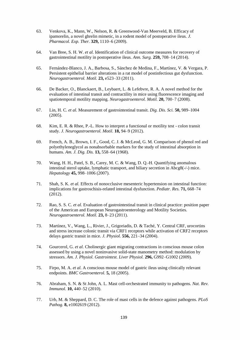
139
63. Venkova, K., Mann, W., Nelson, R. & Greenwood-Van Meerveld, B. Efficacy of
ipamorelin, a novel ghrelin mimetic, in a rodent model of postoperative ileus. J.
Pharmacol. Exp. Ther. 329, 1110–6 (2009).
64. Van Bree, S. H. W. et al. Identification of clinical outcome measures for recovery of
gastrointestinal motility in postoperative ileus. Ann. Surg. 259, 708–14 (2014).
65. Fernández-Blanco, J. A., Barbosa, S., Sánchez de Medina, F., Martínez, V. & Vergara, P.
Persistent epithelial barrier alterations in a rat model of postinfectious gut dysfunction.
Neurogastroenterol. Motil. 23, e523–33 (2011).
66. De Backer, O., Blanckaert, B., Leybaert, L. & Lefebvre, R. A. A novel method for the
evaluation of intestinal transit and contractility in mice using fluorescence imaging and
spatiotemporal motility mapping. Neurogastroenterol. Motil. 20, 700–7 (2008).
67. Lin, H. C. et al. Measurement of gastrointestinal transit. Dig. Dis. Sci. 50, 989–1004
(2005).
68. Kim, E. R. & Rhee, P.-L. How to interpret a functional or motility test - colon transit
study. J. Neurogastroenterol. Motil. 18, 94–9 (2012).
69. French, A. B., Brown, I. F., Good, C. J. & McLeod, G. M. Comparison of phenol red and
polyethyleneglycol as nonabsorbable markers for the study of intestinal absorption in
humans. Am. J. Dig. Dis. 13, 558–64 (1968).
70. Wang, H. H., Patel, S. B., Carey, M. C. & Wang, D. Q.-H. Quantifying anomalous
intestinal sterol uptake, lymphatic transport, and biliary secretion in Abcg8(-/-) mice.
Hepatology 45, 998–1006 (2007).
71. Shah, S. K. et al. Effects of nonocclusive mesenteric hypertension on intestinal function:
implications for gastroschisis-related intestinal dysfunction. Pediatr. Res. 71, 668–74
(2012).
72. Rao, S. S. C. et al. Evaluation of gastrointestinal transit in clinical practice: position paper
of the American and European Neurogastroenterology and Motility Societies.
Neurogastroenterol. Motil. 23, 8–23 (2011).
73. Martínez, V., Wang, L., Rivier, J., Grigoriadis, D. & Taché, Y. Central CRF, urocortins
and stress increase colonic transit via CRF1 receptors while activation of CRF2 receptors
delays gastric transit in mice. J. Physiol. 556, 221–34 (2004).
74. Gourcerol, G. et al. Cholinergic giant migrating contractions in conscious mouse colon
assessed by using a novel noninvasive solid-state manometry method: modulation by
stressors. Am. J. Physiol. Gastrointest. Liver Physiol. 296, G992–G1002 (2009).
75. Firpo, M. A. et al. A conscious mouse model of gastric ileus using clinically relevant
endpoints. BMC Gastroenterol. 5, 18 (2005).
76. Abraham, S. N. & St John, A. L. Mast cell-orchestrated immunity to pathogens. Nat. Rev.
Immunol. 10, 440–52 (2010).
77. Urb, M. & Sheppard, D. C. The role of mast cells in the defence against pathogens. PLoS
Pathog. 8, e1002619 (2012).
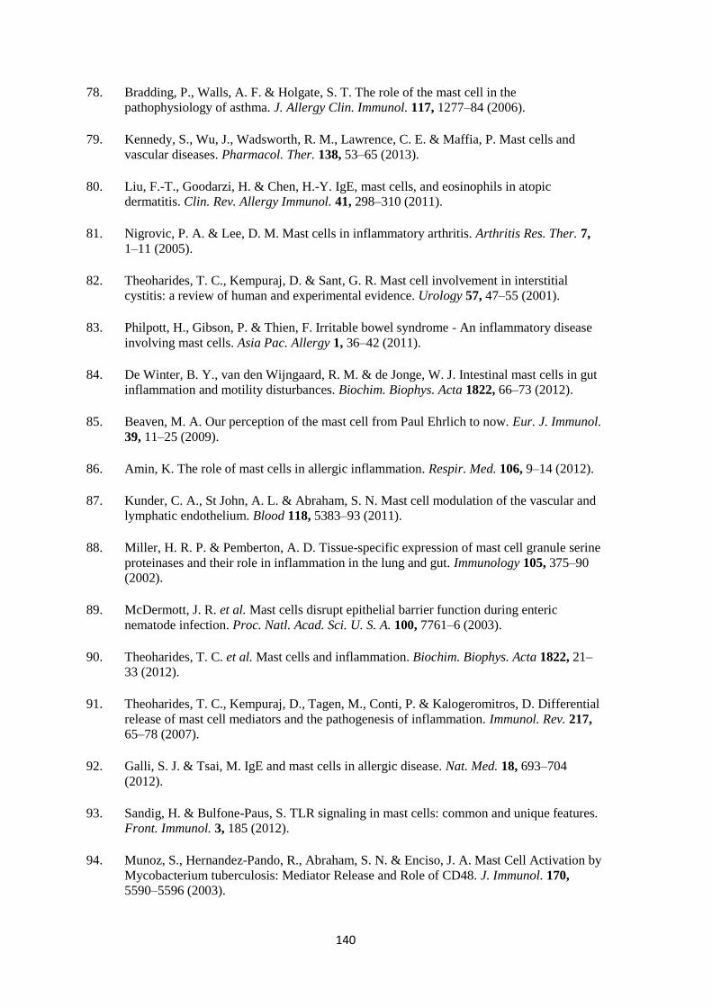
140
78. Bradding, P., Walls, A. F. & Holgate, S. T. The role of the mast cell in the
pathophysiology of asthma. J. Allergy Clin. Immunol. 117, 1277–84 (2006).
79. Kennedy, S., Wu, J., Wadsworth, R. M., Lawrence, C. E. & Maffia, P. Mast cells and
vascular diseases. Pharmacol. Ther. 138, 53–65 (2013).
80. Liu, F.-T., Goodarzi, H. & Chen, H.-Y. IgE, mast cells, and eosinophils in atopic
dermatitis. Clin. Rev. Allergy Immunol. 41, 298–310 (2011).
81. Nigrovic, P. A. & Lee, D. M. Mast cells in inflammatory arthritis. Arthritis Res. Ther. 7,
1–11 (2005).
82. Theoharides, T. C., Kempuraj, D. & Sant, G. R. Mast cell involvement in interstitial
cystitis: a review of human and experimental evidence. Urology 57, 47–55 (2001).
83. Philpott, H., Gibson, P. & Thien, F. Irritable bowel syndrome - An inflammatory disease
involving mast cells. Asia Pac. Allergy 1, 36–42 (2011).
84. De Winter, B. Y., van den Wijngaard, R. M. & de Jonge, W. J. Intestinal mast cells in gut
inflammation and motility disturbances. Biochim. Biophys. Acta 1822, 66–73 (2012).
85. Beaven, M. A. Our perception of the mast cell from Paul Ehrlich to now. Eur. J. Immunol.
39, 11–25 (2009).
86. Amin, K. The role of mast cells in allergic inflammation. Respir. Med. 106, 9–14 (2012).
87. Kunder, C. A., St John, A. L. & Abraham, S. N. Mast cell modulation of the vascular and
lymphatic endothelium. Blood 118, 5383–93 (2011).
88. Miller, H. R. P. & Pemberton, A. D. Tissue-specific expression of mast cell granule serine
proteinases and their role in inflammation in the lung and gut. Immunology 105, 375–90
(2002).
89. McDermott, J. R. et al. Mast cells disrupt epithelial barrier function during enteric
nematode infection. Proc. Natl. Acad. Sci. U. S. A. 100, 7761–6 (2003).
90. Theoharides, T. C. et al. Mast cells and inflammation. Biochim. Biophys. Acta 1822, 21–
33 (2012).
91. Theoharides, T. C., Kempuraj, D., Tagen, M., Conti, P. & Kalogeromitros, D. Differential
release of mast cell mediators and the pathogenesis of inflammation. Immunol. Rev. 217,
65–78 (2007).
92. Galli, S. J. & Tsai, M. IgE and mast cells in allergic disease. Nat. Med. 18, 693–704
(2012).
93. Sandig, H. & Bulfone-Paus, S. TLR signaling in mast cells: common and unique features.
Front. Immunol. 3, 185 (2012).
94. Munoz, S., Hernandez-Pando, R., Abraham, S. N. & Enciso, J. A. Mast Cell Activation by
Mycobacterium tuberculosis: Mediator Release and Role of CD48. J. Immunol. 170,
5590–5596 (2003).
141
95. Marianna Kulka, Cecilia H Sheen, Brian P Tancowny, Leslie C Grammer & Robert P
Schleimer. Neuropeptides activate human mast cell degranulation and chemokine
production . Immunology 123, 398–410 (2008).
96. De Jonge, F. et al. In vitro activation of murine DRG neurons by CGRP-mediated mucosal
mast cell degranulation. Am. J. Physiol. Gastrointest. Liver Physiol. 287, G178–91 (2004).
97. Rychter, J. W. et al. CGRP1 receptor activation induces piecemeal release of protease-1
from mouse bone marrow-derived mucosal mast cells. Neurogastroenterol. Motil. 23,
e57–68 (2011).
98. Thangam, E. B. et al. Airway smooth muscle cells enhance C3a-induced mast cell
degranulation following cell-cell contact. FASEB J. 19, 798–800 (2005).
99. Venkatesha, R. T., Berla Thangam, E., Zaidi, A. K. & Ali, H. Distinct regulation of C3a-
induced MCP-1/CCL2 and RANTES/CCL5 production in human mast cells by
extracellular signal regulated kinase and PI3 kinase. Mol. Immunol. 42, 581–7 (2005).
100. Kandere-Grzybowska, K. et al. IL-1 induces vesicular secretion of IL-6 without
degranulation from human mast cells. J. Immunol. 171, 4830–6 (2003).
101. Marshall, J. S., Stead, R. H., McSharry, C., Nielsen, L. & Bienenstock, J. The role of mast
cell degranulation products in mast cell hyperplasia. I. Mechanism of action of nerve
growth factor. J. Immunol. 144, 1886–92 (1990).
102. Bullock, E. D. & Johnson, E. M. Nerve growth factor induces the expression of certain
cytokine genes and bcl-2 in mast cells. Potential role in survival promotion. J. Biol. Chem.
271, 27500–8 (1996).
103. Chen, Z. et al. Localization of rat tryptase to a subset of the connective tissue type of mast
cell. J. Histochem. Cytochem. 41, 961–9 (1993).
104. Irani, A. M., Bradford, T. R., Kepley, C. L., Schechter, N. M. & Schwartz, L. B. Detection
of MCT and MCTC types of human mast cells by immunohistochemistry using new
monoclonal anti-tryptase and anti-chymase antibodies. J. Histochem. Cytochem. 37, 1509–
15 (1989).
105. Gurish, M. F. & Austen, K. F. Developmental origin and functional specialization of mast
cell subsets. Immunity 37, 25–33 (2012).
106. Gurish, M. F. & Boyce, J. A. Mast cell growth, differentiation, and death. Clin. Rev.
Allergy Immunol. 22, 107–18 (2002).
107. Gallwitz, M., Enoksson, M. & Hellman, L. Expression profile of novel members of the rat
mast cell protease (rMCP)-2 and (rMCP)-8 families, and functional analyses of mouse
mast cell protease (mMCP)-8. Immunogenetics 59, 391–405 (2007).
108. Lutzelschwab, C. Secretory Granule Proteases in Rat Mast Cells. Cloning of 10Different
Serine Proteases and a Carboxypeptidase A from Various Rat Mast Cell Populations. J.
Exp. Med. 185, 13–30 (1997).
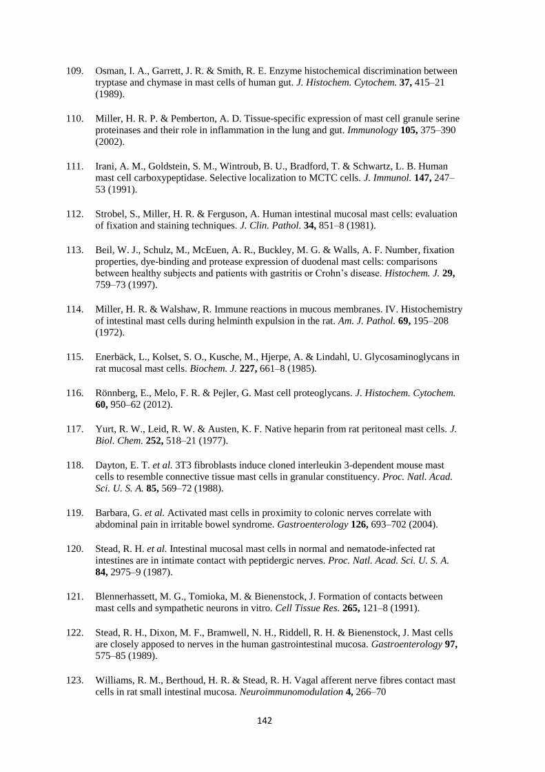
142
109. Osman, I. A., Garrett, J. R. & Smith, R. E. Enzyme histochemical discrimination between
tryptase and chymase in mast cells of human gut. J. Histochem. Cytochem. 37, 415–21
(1989).
110. Miller, H. R. P. & Pemberton, A. D. Tissue-specific expression of mast cell granule serine
proteinases and their role in inflammation in the lung and gut. Immunology 105, 375–390
(2002).
111. Irani, A. M., Goldstein, S. M., Wintroub, B. U., Bradford, T. & Schwartz, L. B. Human
mast cell carboxypeptidase. Selective localization to MCTC cells. J. Immunol. 147, 247–
53 (1991).
112. Strobel, S., Miller, H. R. & Ferguson, A. Human intestinal mucosal mast cells: evaluation
of fixation and staining techniques. J. Clin. Pathol. 34, 851–8 (1981).
113. Beil, W. J., Schulz, M., McEuen, A. R., Buckley, M. G. & Walls, A. F. Number, fixation
properties, dye-binding and protease expression of duodenal mast cells: comparisons
between healthy subjects and patients with gastritis or Crohn’s disease. Histochem. J. 29,
759–73 (1997).
114. Miller, H. R. & Walshaw, R. Immune reactions in mucous membranes. IV. Histochemistry
of intestinal mast cells during helminth expulsion in the rat. Am. J. Pathol. 69, 195–208
(1972).
115. Enerbäck, L., Kolset, S. O., Kusche, M., Hjerpe, A. & Lindahl, U. Glycosaminoglycans in
rat mucosal mast cells. Biochem. J. 227, 661–8 (1985).
116. Rönnberg, E., Melo, F. R. & Pejler, G. Mast cell proteoglycans. J. Histochem. Cytochem.
60, 950–62 (2012).
117. Yurt, R. W., Leid, R. W. & Austen, K. F. Native heparin from rat peritoneal mast cells. J.
Biol. Chem. 252, 518–21 (1977).
118. Dayton, E. T. et al. 3T3 fibroblasts induce cloned interleukin 3-dependent mouse mast
cells to resemble connective tissue mast cells in granular constituency. Proc. Natl. Acad.
Sci. U. S. A. 85, 569–72 (1988).
119. Barbara, G. et al. Activated mast cells in proximity to colonic nerves correlate with
abdominal pain in irritable bowel syndrome. Gastroenterology 126, 693–702 (2004).
120. Stead, R. H. et al. Intestinal mucosal mast cells in normal and nematode-infected rat
intestines are in intimate contact with peptidergic nerves. Proc. Natl. Acad. Sci. U. S. A.
84, 2975–9 (1987).
121. Blennerhassett, M. G., Tomioka, M. & Bienenstock, J. Formation of contacts between
mast cells and sympathetic neurons in vitro. Cell Tissue Res. 265, 121–8 (1991).
122. Stead, R. H., Dixon, M. F., Bramwell, N. H., Riddell, R. H. & Bienenstock, J. Mast cells
are closely apposed to nerves in the human gastrointestinal mucosa. Gastroenterology 97,
575–85 (1989).
123. Williams, R. M., Berthoud, H. R. & Stead, R. H. Vagal afferent nerve fibres contact mast
cells in rat small intestinal mucosa. Neuroimmunomodulation 4, 266–70
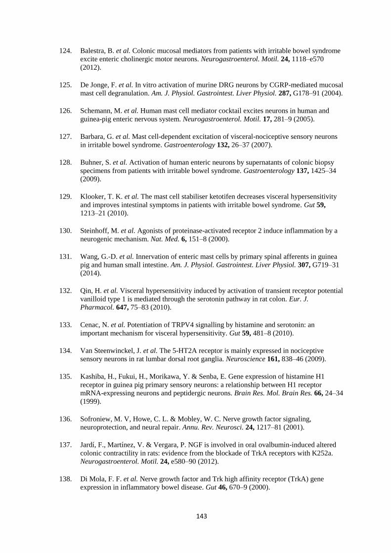
143
124. Balestra, B. et al. Colonic mucosal mediators from patients with irritable bowel syndrome
excite enteric cholinergic motor neurons. Neurogastroenterol. Motil. 24, 1118–e570
(2012).
125. De Jonge, F. et al. In vitro activation of murine DRG neurons by CGRP-mediated mucosal
mast cell degranulation. Am. J. Physiol. Gastrointest. Liver Physiol. 287, G178–91 (2004).
126. Schemann, M. et al. Human mast cell mediator cocktail excites neurons in human and
guinea-pig enteric nervous system. Neurogastroenterol. Motil. 17, 281–9 (2005).
127. Barbara, G. et al. Mast cell-dependent excitation of visceral-nociceptive sensory neurons
in irritable bowel syndrome. Gastroenterology 132, 26–37 (2007).
128. Buhner, S. et al. Activation of human enteric neurons by supernatants of colonic biopsy
specimens from patients with irritable bowel syndrome. Gastroenterology 137, 1425–34
(2009).
129. Klooker, T. K. et al. The mast cell stabiliser ketotifen decreases visceral hypersensitivity
and improves intestinal symptoms in patients with irritable bowel syndrome. Gut 59,
1213–21 (2010).
130. Steinhoff, M. et al. Agonists of proteinase-activated receptor 2 induce inflammation by a
neurogenic mechanism. Nat. Med. 6, 151–8 (2000).
131. Wang, G.-D. et al. Innervation of enteric mast cells by primary spinal afferents in guinea
pig and human small intestine. Am. J. Physiol. Gastrointest. Liver Physiol. 307, G719–31
(2014).
132. Qin, H. et al. Visceral hypersensitivity induced by activation of transient receptor potential
vanilloid type 1 is mediated through the serotonin pathway in rat colon. Eur. J.
Pharmacol. 647, 75–83 (2010).
133. Cenac, N. et al. Potentiation of TRPV4 signalling by histamine and serotonin: an
important mechanism for visceral hypersensitivity. Gut 59, 481–8 (2010).
134. Van Steenwinckel, J. et al. The 5-HT2A receptor is mainly expressed in nociceptive
sensory neurons in rat lumbar dorsal root ganglia. Neuroscience 161, 838–46 (2009).
135. Kashiba, H., Fukui, H., Morikawa, Y. & Senba, E. Gene expression of histamine H1
receptor in guinea pig primary sensory neurons: a relationship between H1 receptor
mRNA-expressing neurons and peptidergic neurons. Brain Res. Mol. Brain Res. 66, 24–34
(1999).
136. Sofroniew, M. V, Howe, C. L. & Mobley, W. C. Nerve growth factor signaling,
neuroprotection, and neural repair. Annu. Rev. Neurosci. 24, 1217–81 (2001).
137. Jardí, F., Martínez, V. & Vergara, P. NGF is involved in oral ovalbumin-induced altered
colonic contractility in rats: evidence from the blockade of TrkA receptors with K252a.
Neurogastroenterol. Motil. 24, e580–90 (2012).
138. Di Mola, F. F. et al. Nerve growth factor and Trk high affinity receptor (TrkA) gene
expression in inflammatory bowel disease. Gut 46, 670–9 (2000).
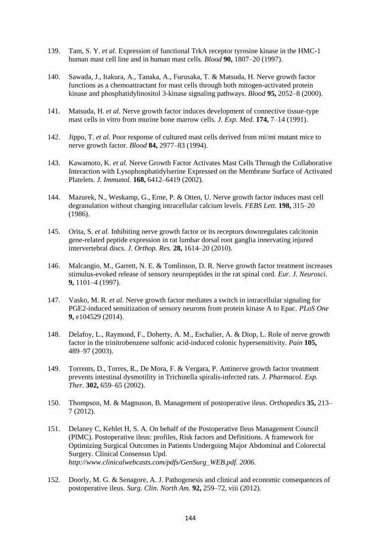
144
139. Tam, S. Y. et al. Expression of functional TrkA receptor tyrosine kinase in the HMC-1
human mast cell line and in human mast cells. Blood 90, 1807–20 (1997).
140. Sawada, J., Itakura, A., Tanaka, A., Furusaka, T. & Matsuda, H. Nerve growth factor
functions as a chemoattractant for mast cells through both mitogen-activated protein
kinase and phosphatidylinositol 3-kinase signaling pathways. Blood 95, 2052–8 (2000).
141. Matsuda, H. et al. Nerve growth factor induces development of connective tissue-type
mast cells in vitro from murine bone marrow cells. J. Exp. Med. 174, 7–14 (1991).
142. Jippo, T. et al. Poor response of cultured mast cells derived from mi/mi mutant mice to
nerve growth factor. Blood 84, 2977–83 (1994).
143. Kawamoto, K. et al. Nerve Growth Factor Activates Mast Cells Through the Collaborative
Interaction with Lysophosphatidylserine Expressed on the Membrane Surface of Activated
Platelets. J. Immunol. 168, 6412–6419 (2002).
144. Mazurek, N., Weskamp, G., Erne, P. & Otten, U. Nerve growth factor induces mast cell
degranulation without changing intracellular calcium levels. FEBS Lett. 198, 315–20
(1986).
145. Orita, S. et al. Inhibiting nerve growth factor or its receptors downregulates calcitonin
gene-related peptide expression in rat lumbar dorsal root ganglia innervating injured
intervertebral discs. J. Orthop. Res. 28, 1614–20 (2010).
146. Malcangio, M., Garrett, N. E. & Tomlinson, D. R. Nerve growth factor treatment increases
stimulus-evoked release of sensory neuropeptides in the rat spinal cord. Eur. J. Neurosci.
9, 1101–4 (1997).
147. Vasko, M. R. et al. Nerve growth factor mediates a switch in intracellular signaling for
PGE2-induced sensitization of sensory neurons from protein kinase A to Epac. PLoS One
9, e104529 (2014).
148. Delafoy, L., Raymond, F., Doherty, A. M., Eschalier, A. & Diop, L. Role of nerve growth
factor in the trinitrobenzene sulfonic acid-induced colonic hypersensitivity. Pain 105,
489–97 (2003).
149. Torrents, D., Torres, R., De Mora, F. & Vergara, P. Antinerve growth factor treatment
prevents intestinal dysmotility in Trichinella spiralis-infected rats. J. Pharmacol. Exp.
Ther. 302, 659–65 (2002).
150. Thompson, M. & Magnuson, B. Management of postoperative ileus. Orthopedics 35, 213–
7 (2012).
151. Delaney C, Kehlet H, S. A. On behalf of the Postoperative Ileus Management Council
(PIMC). Postoperative ileus: profiles, Risk factors and Definitions. A framework for
Optimizing Surgical Outcomes in Patients Undergoing Major Abdominal and Colorectal
Surgery. Clinical Consensus Upd.
http://www.clinicalwebcasts.com/pdfs/GenSurg_WEB.pdf. 2006.
152. Doorly, M. G. & Senagore, A. J. Pathogenesis and clinical and economic consequences of
postoperative ileus. Surg. Clin. North Am. 92, 259–72, viii (2012).
145
153. Boeckxstaens, G. E. & de Jonge, W. J. Neuroimmune mechanisms in postoperative ileus.
Gut 58, 1300–11 (2009).
154. Mueller, M. H. et al. Differential sensitization of afferent neuronal pathways during
postoperative ileus in the mouse jejunum. Ann. Surg. 247, 791–802 (2008).
155. Barquist, E. et al. Neuronal pathways involved in abdominal surgery-induced gastric ileus
in rats. Am. J. Physiol. 270, R888–94 (1996).
156. Fukuda, H. et al. Inhibition of sympathetic pathways restores postoperative ileus in the
upper and lower gastrointestinal tract. J. Gastroenterol. Hepatol. 22, 1293–9 (2007).
157. De Winter, B. Y. et al. Effect of adrenergic and nitrergic blockade on experimental ileus in
rats. Br. J. Pharmacol. 120, 464–8 (1997).
158. Meile, T., Glatzle, J., Habermann, F. M., Kreis, M. E. & Zittel, T. T. Nitric oxide synthase
inhibition results in immediate postoperative recovery of gastric, small intestinal and
colonic motility in awake rats. Int. J. Colorectal Dis. 21, 121–9 (2006).
159. Boeckxstaens, G. E. et al. Activation of an adrenergic and vagally-mediated NANC
pathway in surgery-induced fundic relaxation in the rat. Neurogastroenterol. Motil. 11,
467–74 (1999).
160. Fukuda, H. et al. Impaired gastric motor activity after abdominal surgery in rats.
Neurogastroenterol. Motil. 17, 245–50 (2005).
161. Kreiss, C., Birder, L. A., Kiss, S., VanBibber, M. M. & Bauer, A. J. COX-2 dependent
inflammation increases spinal Fos expression during rodent postoperative ileus. Gut 52,
527–34 (2003).
162. Cailotto, C. et al. Neuroanatomical evidence demonstrating the existence of the vagal anti-
inflammatory reflex in the intestine. Neurogastroenterol. Motil. 24, 191–200, e93 (2012).
163. Kalff, J. C., Schraut, W. H., Simmons, R. L. & Bauer, A. J. Surgical manipulation of the
gut elicits an intestinal muscularis inflammatory response resulting in postsurgical ileus.
Ann. Surg. 228, 652–63 (1998).
164. Kalff, J. C. et al. Biphasic response to gut manipulation and temporal correlation of
cellular infiltrates and muscle dysfunction in rat. Surgery 126, 498–509 (1999).
165. Kalff, J. C. et al. Intra-abdominal activation of a local inflammatory response within the
human muscularis externa during laparotomy. Ann. Surg. 237, 301–15 (2003).
166. The, F. O. et al. Intestinal handling-induced mast cell activation and inflammation in
human postoperative ileus. Gut 57, 33–40 (2008).
167. Stoffels, B. et al. Postoperative ileus involves interleukin-1 receptor signaling in enteric
glia. Gastroenterology 146, 176–87.e1 (2014).
168. Wehner, S. et al. Induction of IL-6 within the rodent intestinal muscularis after intestinal
surgical stress. Surgery 137, 436–46 (2005).
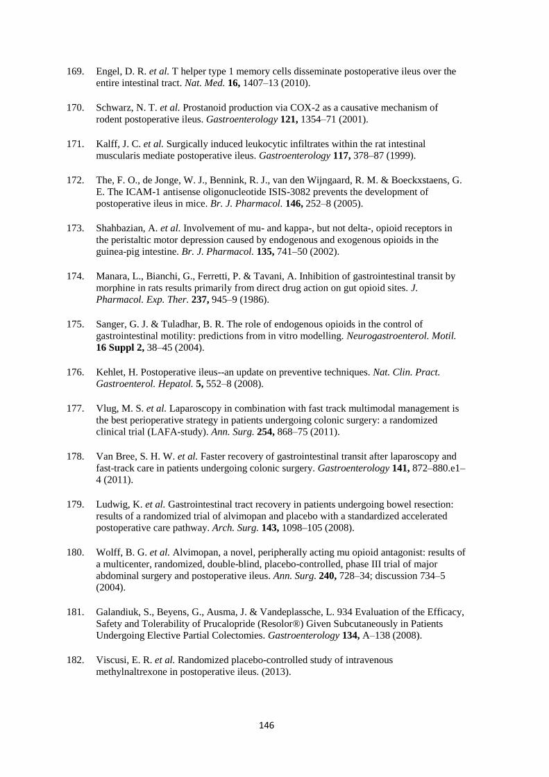
146
169. Engel, D. R. et al. T helper type 1 memory cells disseminate postoperative ileus over the
entire intestinal tract. Nat. Med. 16, 1407–13 (2010).
170. Schwarz, N. T. et al. Prostanoid production via COX-2 as a causative mechanism of
rodent postoperative ileus. Gastroenterology 121, 1354–71 (2001).
171. Kalff, J. C. et al. Surgically induced leukocytic infiltrates within the rat intestinal
muscularis mediate postoperative ileus. Gastroenterology 117, 378–87 (1999).
172. The, F. O., de Jonge, W. J., Bennink, R. J., van den Wijngaard, R. M. & Boeckxstaens, G.
E. The ICAM-1 antisense oligonucleotide ISIS-3082 prevents the development of
postoperative ileus in mice. Br. J. Pharmacol. 146, 252–8 (2005).
173. Shahbazian, A. et al. Involvement of mu- and kappa-, but not delta-, opioid receptors in
the peristaltic motor depression caused by endogenous and exogenous opioids in the
guinea-pig intestine. Br. J. Pharmacol. 135, 741–50 (2002).
174. Manara, L., Bianchi, G., Ferretti, P. & Tavani, A. Inhibition of gastrointestinal transit by
morphine in rats results primarily from direct drug action on gut opioid sites. J.
Pharmacol. Exp. Ther. 237, 945–9 (1986).
175. Sanger, G. J. & Tuladhar, B. R. The role of endogenous opioids in the control of
gastrointestinal motility: predictions from in vitro modelling. Neurogastroenterol. Motil.
16 Suppl 2, 38–45 (2004).
176. Kehlet, H. Postoperative ileus--an update on preventive techniques. Nat. Clin. Pract.
Gastroenterol. Hepatol. 5, 552–8 (2008).
177. Vlug, M. S. et al. Laparoscopy in combination with fast track multimodal management is
the best perioperative strategy in patients undergoing colonic surgery: a randomized
clinical trial (LAFA-study). Ann. Surg. 254, 868–75 (2011).
178. Van Bree, S. H. W. et al. Faster recovery of gastrointestinal transit after laparoscopy and
fast-track care in patients undergoing colonic surgery. Gastroenterology 141, 872–880.e1–
4 (2011).
179. Ludwig, K. et al. Gastrointestinal tract recovery in patients undergoing bowel resection:
results of a randomized trial of alvimopan and placebo with a standardized accelerated
postoperative care pathway. Arch. Surg. 143, 1098–105 (2008).
180. Wolff, B. G. et al. Alvimopan, a novel, peripherally acting mu opioid antagonist: results of
a multicenter, randomized, double-blind, placebo-controlled, phase III trial of major
abdominal surgery and postoperative ileus. Ann. Surg. 240, 728–34; discussion 734–5
(2004).
181. Galandiuk, S., Beyens, G., Ausma, J. & Vandeplassche, L. 934 Evaluation of the Efficacy,
Safety and Tolerability of Prucalopride (Resolor®) Given Subcutaneously in Patients
Undergoing Elective Partial Colectomies. Gastroenterology 134, A–138 (2008).
182. Viscusi, E. R. et al. Randomized placebo-controlled study of intravenous
methylnaltrexone in postoperative ileus. (2013).
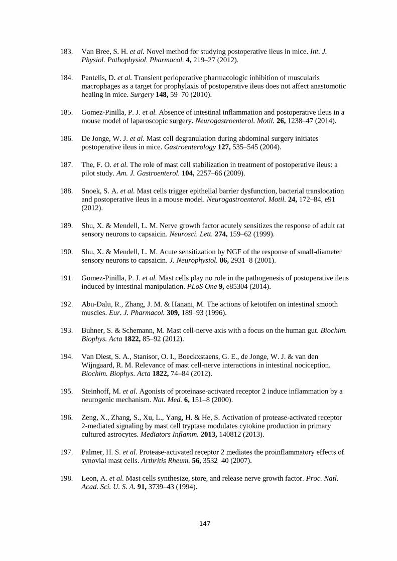
147
183. Van Bree, S. H. et al. Novel method for studying postoperative ileus in mice. Int. J.
Physiol. Pathophysiol. Pharmacol. 4, 219–27 (2012).
184. Pantelis, D. et al. Transient perioperative pharmacologic inhibition of muscularis
macrophages as a target for prophylaxis of postoperative ileus does not affect anastomotic
healing in mice. Surgery 148, 59–70 (2010).
185. Gomez-Pinilla, P. J. et al. Absence of intestinal inflammation and postoperative ileus in a
mouse model of laparoscopic surgery. Neurogastroenterol. Motil. 26, 1238–47 (2014).
186. De Jonge, W. J. et al. Mast cell degranulation during abdominal surgery initiates
postoperative ileus in mice. Gastroenterology 127, 535–545 (2004).
187. The, F. O. et al. The role of mast cell stabilization in treatment of postoperative ileus: a
pilot study. Am. J. Gastroenterol. 104, 2257–66 (2009).
188. Snoek, S. A. et al. Mast cells trigger epithelial barrier dysfunction, bacterial translocation
and postoperative ileus in a mouse model. Neurogastroenterol. Motil. 24, 172–84, e91
(2012).
189. Shu, X. & Mendell, L. M. Nerve growth factor acutely sensitizes the response of adult rat
sensory neurons to capsaicin. Neurosci. Lett. 274, 159–62 (1999).
190. Shu, X. & Mendell, L. M. Acute sensitization by NGF of the response of small-diameter
sensory neurons to capsaicin. J. Neurophysiol. 86, 2931–8 (2001).
191. Gomez-Pinilla, P. J. et al. Mast cells play no role in the pathogenesis of postoperative ileus
induced by intestinal manipulation. PLoS One 9, e85304 (2014).
192. Abu-Dalu, R., Zhang, J. M. & Hanani, M. The actions of ketotifen on intestinal smooth
muscles. Eur. J. Pharmacol. 309, 189–93 (1996).
193. Buhner, S. & Schemann, M. Mast cell-nerve axis with a focus on the human gut. Biochim.
Biophys. Acta 1822, 85–92 (2012).
194. Van Diest, S. A., Stanisor, O. I., Boeckxstaens, G. E., de Jonge, W. J. & van den
Wijngaard, R. M. Relevance of mast cell-nerve interactions in intestinal nociception.
Biochim. Biophys. Acta 1822, 74–84 (2012).
195. Steinhoff, M. et al. Agonists of proteinase-activated receptor 2 induce inflammation by a
neurogenic mechanism. Nat. Med. 6, 151–8 (2000).
196. Zeng, X., Zhang, S., Xu, L., Yang, H. & He, S. Activation of protease-activated receptor
2-mediated signaling by mast cell tryptase modulates cytokine production in primary
cultured astrocytes. Mediators Inflamm. 2013, 140812 (2013).
197. Palmer, H. S. et al. Protease-activated receptor 2 mediates the proinflammatory effects of
synovial mast cells. Arthritis Rheum. 56, 3532–40 (2007).
198. Leon, A. et al. Mast cells synthesize, store, and release nerve growth factor. Proc. Natl.
Acad. Sci. U. S. A. 91, 3739–43 (1994).
148
199. Plourde, V., St-Pierre, S., Fournier, A. & Taché, Y. CGRP 8-37[correction of 8-27] blocks
the inhibition of gastric emptying induced by intravenous injection of alpha-CGRP in rats.
Life Sci. 52, 857–62 (1993).
200. Plourde, V., Wong, H. C., Walsh, J. H., Raybould, H. E. & Taché, Y. CGRP antagonists
and capsaicin on celiac ganglia partly prevent postoperative gastric ileus. Peptides 14,
1225–9 (1993).
201. Zittel, T. T., Reddy, S. N., Plourde, V. & Raybould, H. E. Role of spinal afferents and
calcitonin gene-related peptide in the postoperative gastric ileus in anesthetized rats. Ann.
Surg. 219, 79–87 (1994).
202. Zittel, T. T. et al. Calcitonin gene-related peptide and spinal afferents partly mediate
postoperative colonic ileus in the rat. Surgery 123, 518–27 (1998).
203. Marshall, J. S., Stead, R. H., McSharry, C., Nielsen, L. & Bienenstock, J. The role of mast
cell degranulation products in mast cell hyperplasia. I. Mechanism of action of nerve
growth factor. J. Immunol. 144, 1886–92 (1990).
204. Stempelj, M. & Ferjan, I. Signaling pathway in nerve growth factor induced histamine
release from rat mast cells. Inflamm. Res. 54, 344–9 (2005).
205. Torrents, D., Torres, R., De Mora, F. & Vergara, P. Antinerve growth factor treatment
prevents intestinal dysmotility in Trichinella spiralis-infected rats. J. Pharmacol. Exp.
Ther. 302, 659–65 (2002).
206. Kim, J. Y., Kim, D. Y. & Ro, J. Y. Granule formation in NGF-cultured mast cells is
associated with expressions of pyruvate kinase type M2 and annexin I proteins. Int. Arch.
Allergy Immunol. 146, 287–97 (2008).
207. Mohtasham, L., Auais, A. & Piedimonte, G. Nerve growth factor mediates steroid-
resistant inflammation in respiratory syncytial virus infection. Pediatr. Pulmonol. 42, 496–
504 (2007).
208. Raychaudhuri, S. P., Sanyal, M., Weltman, H. & Kundu-Raychaudhuri, S. K252a, a high-
affinity nerve growth factor receptor blocker, improves psoriasis: an in vivo study using
the severe combined immunodeficient mouse-human skin model. J. Invest. Dermatol. 122,
812–9 (2004).
209. Wilfong, E. R. & Dey, R. D. Nerve growth factor and substance P regulation in nasal
sensory neurons after toluene diisocyanate exposure. Am. J. Respir. Cell Mol. Biol. 30,
793–800 (2004).
210. Gangula, P. R. et al. Regulation of calcitonin gene-related peptide expression in dorsal
root ganglia of rats by female sex steroid hormones. Biol. Reprod. 62, 1033–9 (2000).
211. Lindsay, R. M. & Harmar, A. J. Nerve growth factor regulates expression of neuropeptide
genes in adult sensory neurons. Nature 337, 362–4 (1989).
212. Supowit, S. C., Zhao, H. & DiPette, D. J. Nerve growth factor enhances calcitonin gene-
related peptide expression in the spontaneously hypertensive rat. Hypertension 37, 728–32
(2001).
149
213. Ye, Y. et al. Nerve growth factor links oral cancer progression, pain, and cachexia. Mol.
Cancer Ther. 10, 1667–76 (2011).

150
151
PUBLICATIONS AND
PARTICIPATIONS IN
SCIENTIFIC MEETINGS
DERIVED FROM THIS
THESIS

152
153
Publications
Berdún S, Bombuy E, Estrada O, Mans E, Rychter J, Clavé P, Vergara P.
Peritoneal mast cell degranulation and gastrointestinal recovery in patients undergoing colorectal
surgery. Neurogastroenterol Motil. 2015 Feb 9. doi: 10.1111/nmo.12525. [Epub ahead of print].
Submitted articles
Berdún S, Rychter J, Vergara P. Effects of nerve growth factor antagonist K252a on peritoneal
mast cell degranulation: implications for rat postoperative ileus. Submitted to Journal of
pharmacology and experimental therapeutics.
Berdún S, Rychter J, Vergara P. Intestinal manipulation increases gene expression of visceral-
afferent mediators in dorsal root ganglia: role for peritoneal mast cells. Submitted to
Neurogastroenterology and motility.
Scientific meetings
S Berdun, E Bombuy, O Estrada, E Mans, J Rychter, P Clavé, P Vergara. Peritoneal mast cell
degranulation correlates with gastrointestinal recovery in patients undergoing colorectal surgery.
Poster session. 20th United European Gastroenterology Week (UEGW). Amsterdam (The
Netherlands) 2012.
S Berdun, E Bombuy, O Estrada, E Mans, J Rychter, P Clavé, P Vergara. A pilot study: human
peritoneal supernatant presenting mast cell activation induced by intestinal surgery decreases
gastric emptying in rat. Poster session. International mast cell and basophil meeting 2012. Berlín
(Germany) 2012.
S Berdún, P Vergara. A refinement measure in a model of postoperative ileus in rat: comparative
effects of buprenorphine and meloxicam. Poster session. 12th FELASA and 12th SECAL
congress. Barcelona (Spain) 2013.
S Berdún, P Vergara. Intestinal manipulation activates peritoneal mast cells while it does not
affect expression of mast cell proteases within the ileal wall in the rat. Oral communication.
International mast cell and basophil meeting 2013. Udine (Italy) 2013.
154
S Berdun, E Bombuy, O Estrada, E Mans, J Rychter, P Clavé, P Vergara.
Correlació de la degranulació dels mastòcits peritoneals amb la recuperació gastrointestinal en
pacients sotmesos a cirurgia colorectal. Oral communication. Congreso de la sociedad catalana
de digestología. Lleida (Spain) 2014.
S Berdún, P Vergara. Intestinal manipulation induces degranulation of peritoneal mast cells and
activation of dorsal root ganglia in postoperative ileus in rat. Poster session. Digestive disease
week 2014. Chicago (US) 2014.

155

156


![líOs i]7TUT3 E,’• AMORhemeroteca-paginas.mundodeportivo.com/./EMD02/HEM/... · Creo que elhechode que ró cierta confianza en la humanidad. quepa un alfiler en los hoteles El](https://img.pdfslide.net/doc/110x75/60762613de486c452b74338c/los-i7tut3-eaa-amorhemeroteca-creo-que-elhechode-que-r-cierta-confianza.jpg)