Embed Size (px)
Citation preview
1
Improved Assessment and Treatment of
Abdominal Aortic Aneurysms: The Use of 3D
Reconstructions as a Surgical Guidance Tool
in Endovascular Repair
Barry J. Doyle,1§
Pierce A. Grace,1,2
Eamon G. Kavanagh,1,2
Paul E. Burke,1,2
Fintan Wallis,1,3
Michael T. Walsh1 and Timothy M. McGloughlin
1
1. Centre for Applied Biomedical Engineering Research (CABER),
Department of Mechanical and Aeronautical Engineering, and the
Materials and Surface Science Institute, University of Limerick, Ireland.
2. Department of Vascular Surgery, HSE Midwestern Regional Hospital,
Limerick, Ireland.
3. Department of Radiology, HSE Midwestern Regional Hospital, Limerick,
Ireland
§ Corresponding Author
Tel.: +35361202369
Fax: +35361202944
Email: [email protected]
2
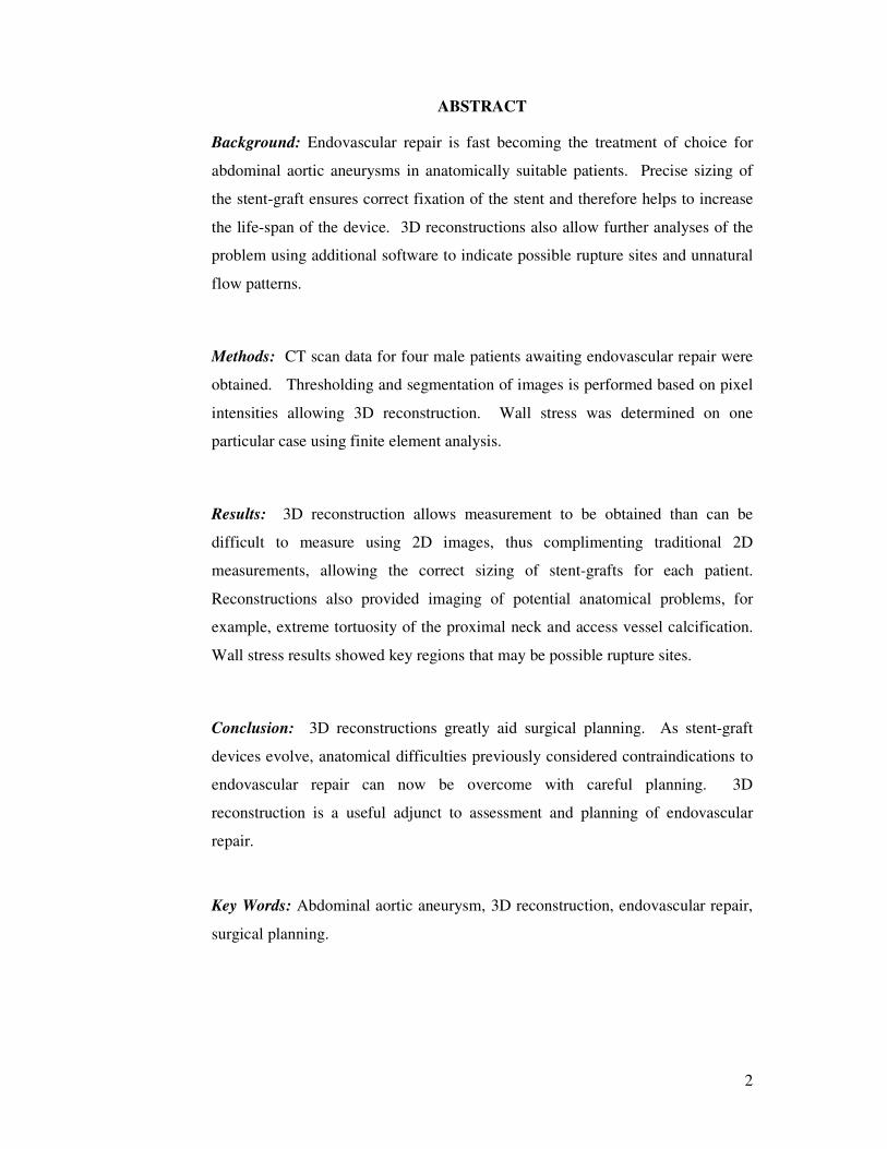
ABSTRACT
Background: Endovascular repair is fast becoming the treatment of choice for
abdominal aortic aneurysms in anatomically suitable patients. Precise sizing of
the stent-graft ensures correct fixation of the stent and therefore helps to increase
the life-span of the device. 3D reconstructions also allow further analyses of the
problem using additional software to indicate possible rupture sites and unnatural
flow patterns.
Methods: CT scan data for four male patients awaiting endovascular repair were
obtained. Thresholding and segmentation of images is performed based on pixel
intensities allowing 3D reconstruction. Wall stress was determined on one
particular case using finite element analysis.
Results: 3D reconstruction allows measurement to be obtained than can be
difficult to measure using 2D images, thus complimenting traditional 2D
measurements, allowing the correct sizing of stent-grafts for each patient.
Reconstructions also provided imaging of potential anatomical problems, for
example, extreme tortuosity of the proximal neck and access vessel calcification.
Wall stress results showed key regions that may be possible rupture sites.
Conclusion: 3D reconstructions greatly aid surgical planning. As stent-graft
devices evolve, anatomical difficulties previously considered contraindications to
endovascular repair can now be overcome with careful planning. 3D
reconstruction is a useful adjunct to assessment and planning of endovascular
repair.
Key Words: Abdominal aortic aneurysm, 3D reconstruction, endovascular repair,
surgical planning.
3
Introduction
An abdominal aortic aneurysm (AAA) can be defined as a permanent and
irreversible localised dilatation of the aorta [1]. Aneurysms form due to
alterations of the connective tissue in the aortic wall. This degradation of the
aortic wall is attributed to risk factors such as tobacco smoking, sex, age,
hypertension, chronic obstructive pulmonary disease, hyperlipidaemia, and family
history of the disorder [1]. With recent advancements in non-invasive diagnostic
imaging more AAAs are being detected. Approximately 150,000 new cases are
diagnosed each year in the US [2-4], and 15,000 deaths per year are attributed to
AAA rupture [5]. The normal diameter of the abdominal aorta can vary with
increasing age, male sex and bodyweight [1,2], and decreases progressively from
its entry into the abdominal cavity to the iliac bifurcation. The infrarenal
abdominal aortic diameter ranges from 15mm to 24mm for most elderly men [1]
and it was proposed [6] that an abdominal aortic aneurysm is an aorta with an
infrarenal diameter greater than 30mm. As the diameter of the infrarenal aorta is
dependent on age and sex [7], a size of at least 1.5 times greater than the expected
normal diameter of the infrarenal aorta [8], has been suggested as an appropriate
definition of an aneurysm.
Currently, the indication for surgical intervention is based on the maximum
diameter of the abdominal aortic aneurysm in the context of the patient’s general
health status, with most repairs performed when the aneurysm exceeds 50 to
60mm [9-12]. The aim of AAA treatment is to totally exclude the aneurysm,
isolating the aneurysm sac from systemic blood pressure and flow so as to
minimise the risk of rupture. As a result of AAA treatment, most aneurysms
should stabilise or shrink [13], although, it has been reported that aneurysms can
often enlarge after AAA treatment due to endotension [14]. Surgical repair can be
either traditional open repair or endovascular repair (EVAR). With traditional
surgical repair, the abdomen is entered via a long midline or a wide transverse
incision. Clamping of the infrarenal, or rarely the suprarenal, aorta is necessary
while suturing either a Dacron or expanded polytetrafluoroethylene (ePTFE) graft
to the neck of the aorta proximal to the aneurysm and either the distal aorta (tube
graft) or to both iliac arteries (bifurcated graft) distal to the aneurysm which is
effectively removed from the circulation. Endovascular repair consists of the
4
placement of a bifurcated graft via an intraluminal introducer across the aneurysm
and its fixation to the normal aortic and iliac walls with self-expanding stents at
both ends [1]. This treatment involves the surgical exposure of the common
femoral arteries where the endovascular graft can be inserted by an over-the-wire
technique. The early benefit of the minimally invasive EVAR approach has been
proven in several randomised trials in reducing the perioperative mortality
compared with open repair [14-17]. It is thought that this benefit is a result of the
avoidance of the large abdominal incision and consequent systemic inflammatory
response, respiratory and wound complications as well as the avoidance of aortic
clamping with resultant cardiovascular complications.
The conversion of computed tomography (CT) scans into 3D reconstructions of
patient-specific regions is of great use to surgeons and is becoming widely used
[18]. For many years regions of the human body have been reconstructed in order
to give clinicians a further insight into the particular problem region or condition.
The focus of this paper is on the use of 3D reconstruction of the human abdominal
aorta in patients suffering from aneurysmal disease. Also included in this study is
the methodology and results of a finite element analysis (FEA) of a particularly
extreme case of aneurysmal disease. FEA is a commonly used engineering
technique to determine stress distributions experienced in complex structures and
is used widely in biomedical engineering applications. The use of FEA in
assessing the propensity of an aneurysm to rupture is becoming more widespread
among researchers [11,12,19-22], and helps provide a useful insight into the
biomechanical behaviour of the aorta. Ultimately, these reconstructions provide
the clinician with valuable data regarding dimensions, geometry, and possible
problem areas that may arise during endovascular surgical repair.
Methods
3D Reconstructions
Computed tomography (CT) scan data was obtained from the Midwestern
Regional Hospital, Limerick, and St. James’s Hospital, Dublin, for 4 patients
awaiting surgical repair of an AAA. All patients were male with a mean age of
77.5yrs (range 59-87yrs). CT scans were obtained using a Somotom Plus 4
5
(Siemens AG, D-91052 Erlangen, Germany). The mean pixel size of the CT
scans was 0.675mm with all scans taken using a 3mm slice increment. The CT
scans were imported into the commercially available software Mimics v12
(Materialise, Belguim) for reconstruction. The full reconstruction technique has
been reported previously by our group [23]. Briefly, a thresholding technique is
applied to each scan in the series. This process assigns a pixel intensity value
measured in Hounsfield units (HU) to each pixel in the image. From this, the HU
value can be controlled so that only the regions of interest are thresholded.
Following this thresholding, segmentation of the image is possible. This assigns a
certain colour to a certain region of interest, in this case the diseased aorta. By
applying this algorithm to the series of CT scans, a complete 3D reconstruction
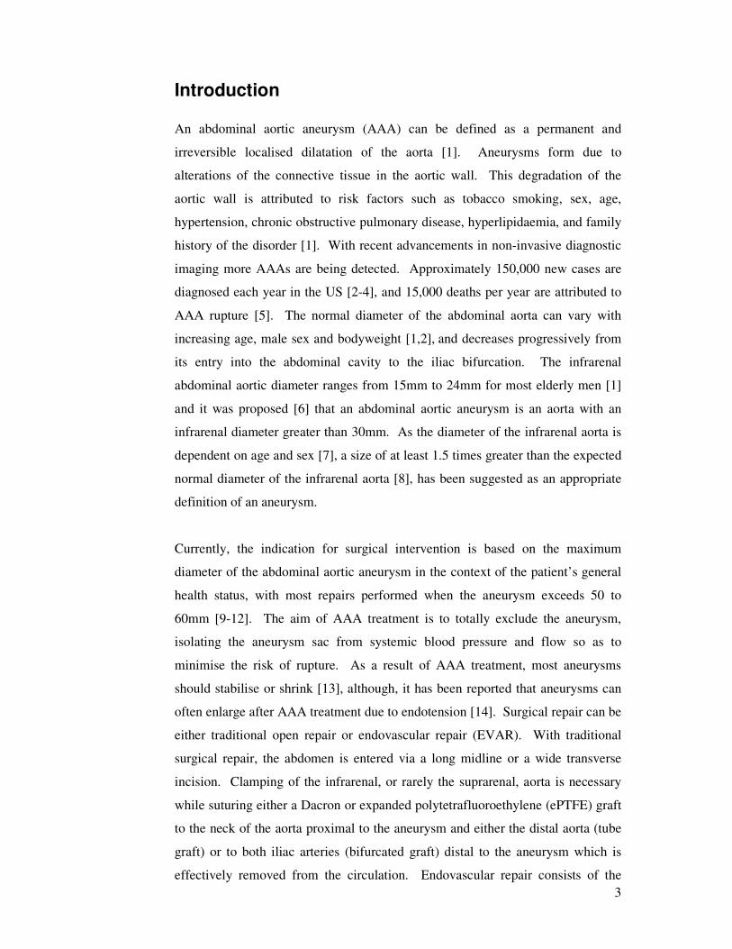
can be generated. This process can be seen in Figure 1.
Figure 1: (A) Typical CT scan showing aneurysmal aorta in centre of image, (B)
algorithm defines regions of interest and (C) software generates 3D reconstruction
of desired regions.
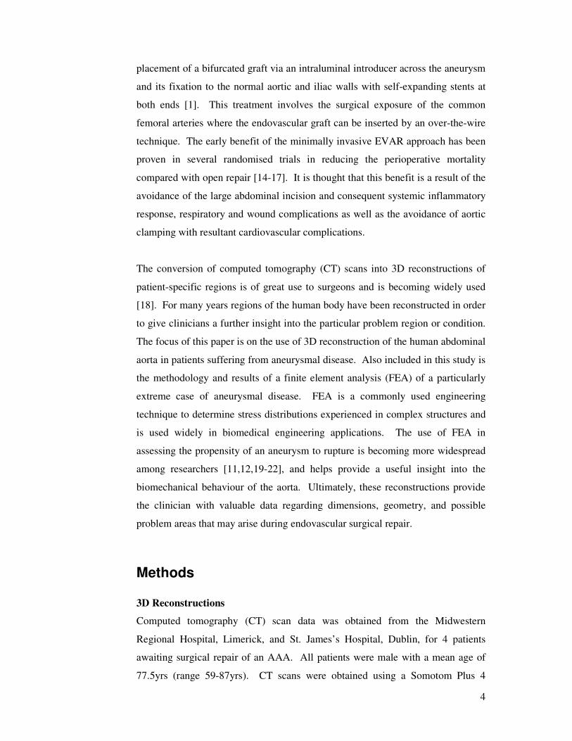
Assigning regions into different layers allows certain areas to be highlighted or
removed depending on the desired region of interest. An example of this layered
approach to reconstruction can be seen in Figure 2. In this particular
reconstruction, all major internal structures were generated for illustration
purposes.
6
Figure 2: Full 3D reconstruction of CT scan data set. Image shows the use of
layers to highlight/remove particular regions of interest depending on desired
areas.
For these particular reconstructions, only the lumen regions were of clinical
importance as this is the region to which the stent-graft is deployed and fixated.
For the purposes of FEA, the full aneurysmal sac of the extremely diseased aorta
was reconstructed.
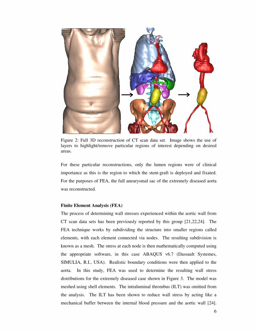
Finite Element Analysis (FEA)
The process of determining wall stresses experienced within the aortic wall from
CT scan data sets has been previously reported by this group [21,22,24]. The
FEA technique works by subdividing the structure into smaller regions called
elements, with each element connected via nodes. The resulting subdivision is
known as a mesh. The stress at each node is then mathematically computed using
the appropriate software, in this case ABAQUS v6.7 (Dassault Systemes,
SIMULIA, R.I., USA). Realistic boundary conditions were then applied to the
aorta. In this study, FEA was used to determine the resulting wall stress
distributions for the extremely diseased case shown in Figure 3. The model was
meshed using shell elements. The intraluminal thrombus (ILT) was omitted from
the analysis. The ILT has been shown to reduce wall stress by acting like a
mechanical buffer between the internal blood pressure and the aortic wall [24].
7
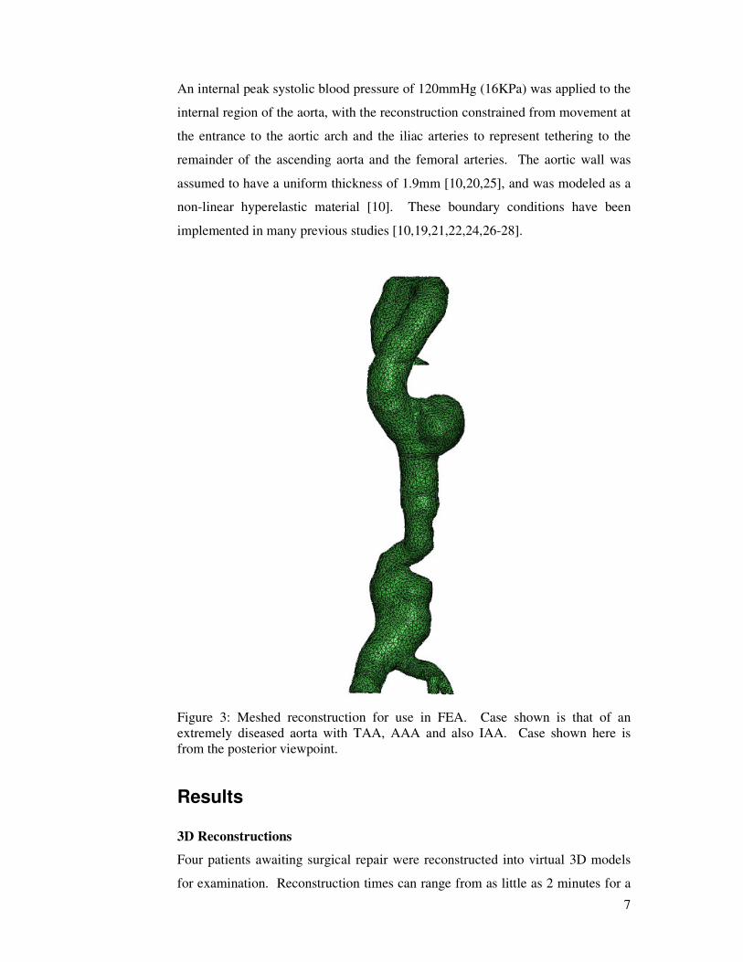
An internal peak systolic blood pressure of 120mmHg (16KPa) was applied to the
internal region of the aorta, with the reconstruction constrained from movement at
the entrance to the aortic arch and the iliac arteries to represent tethering to the
remainder of the ascending aorta and the femoral arteries. The aortic wall was
assumed to have a uniform thickness of 1.9mm [10,20,25], and was modeled as a
non-linear hyperelastic material [10]. These boundary conditions have been
implemented in many previous studies [10,19,21,22,24,26-28].
Figure 3: Meshed reconstruction for use in FEA. Case shown is that of an
extremely diseased aorta with TAA, AAA and also IAA. Case shown here is
from the posterior viewpoint.
Results
3D Reconstructions
Four patients awaiting surgical repair were reconstructed into virtual 3D models
for examination. Reconstruction times can range from as little as 2 minutes for a
8
basic model where minor details are ignored, to 1 hour where all details are
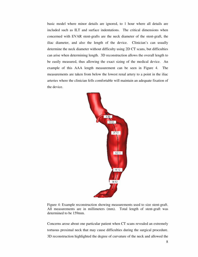
included such as ILT and surface indentations. The critical dimensions when
concerned with EVAR stent-grafts are the neck diameter of the stent-graft, the
iliac diameter, and also the length of the device. Clinician’s can usually
determine the neck diameter without difficulty using 2D CT scans, but difficulties
can arise when determining length. 3D reconstruction allows the overall length to
be easily measured, thus allowing the exact sizing of the medical device. An
example of this AAA length measurement can be seen in Figure 4. The
measurements are taken from below the lowest renal artery to a point in the iliac
arteries where the clinician fells comfortable will maintain an adequate fixation of
the device.
Figure 4: Example reconstruction showing measurements used to size stent-graft.
All measurements are in millimeters (mm). Total length of stent-graft was
determined to be 159mm.
Concerns arose about one particular patient when CT scans revealed an extremely
tortuous proximal neck that may cause difficulties during the surgical procedure.
3D reconstruction highlighted the degree of curvature of the neck and allowed the
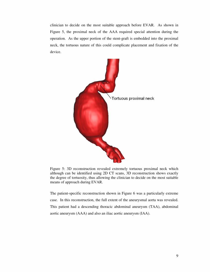
9
clinician to decide on the most suitable approach before EVAR. As shown in
Figure 5, the proximal neck of the AAA required special attention during the
operation. As the upper portion of the stent-graft is embedded into the proximal
neck, the tortuous nature of this could complicate placement and fixation of the
device.
Figure 5: 3D reconstruction revealed extremely tortuous proximal neck which
although can be identified using 2D CT scans, 3D reconstruction shows exactly
the degree of tortuosity, thus allowing the clinician to decide on the most suitable
means of approach during EVAR.
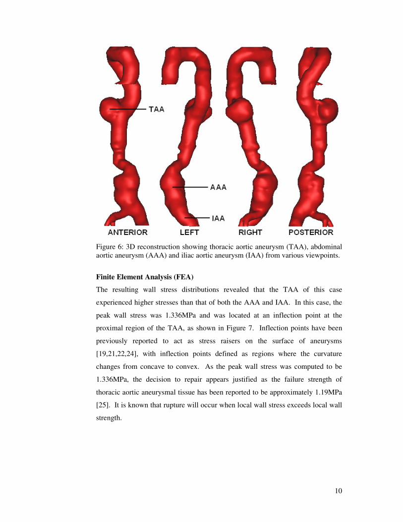
The patient-specific reconstruction shown in Figure 6 was a particularly extreme
case. In this reconstruction, the full extent of the aneurysmal aorta was revealed.
This patient had a descending thoracic abdominal aneurysm (TAA), abdominal
aortic aneurysm (AAA) and also an iliac aortic aneurysm (IAA).
10
Figure 6: 3D reconstruction showing thoracic aortic aneurysm (TAA), abdominal
aortic aneurysm (AAA) and iliac aortic aneurysm (IAA) from various viewpoints.
Finite Element Analysis (FEA)
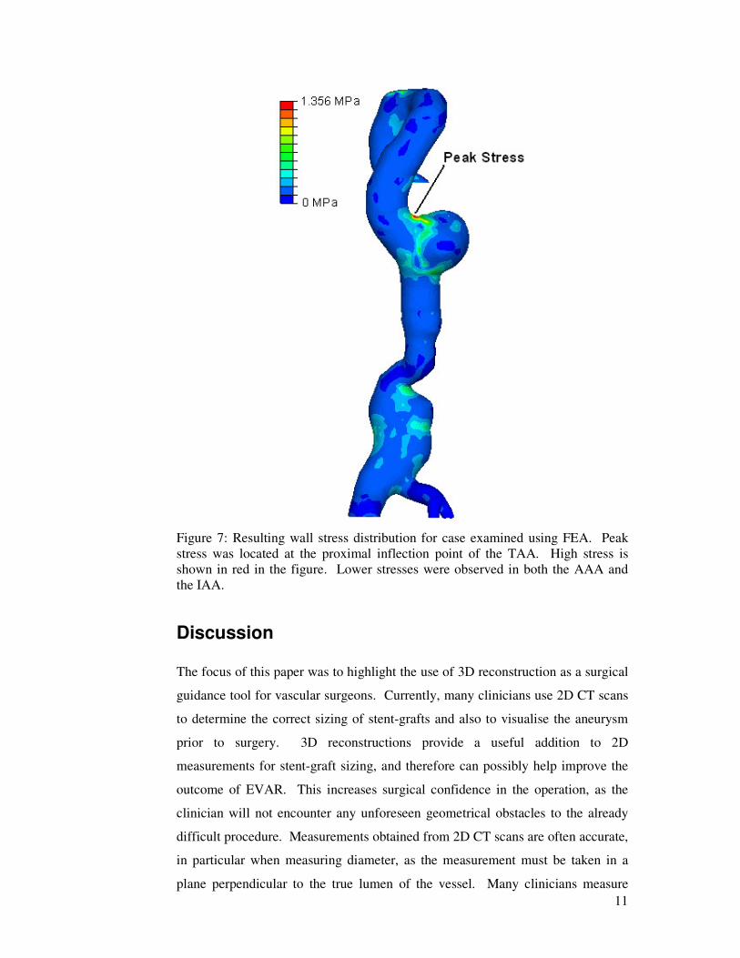
The resulting wall stress distributions revealed that the TAA of this case
experienced higher stresses than that of both the AAA and IAA. In this case, the
peak wall stress was 1.336MPa and was located at an inflection point at the
proximal region of the TAA, as shown in Figure 7. Inflection points have been
previously reported to act as stress raisers on the surface of aneurysms
[19,21,22,24], with inflection points defined as regions where the curvature
changes from concave to convex. As the peak wall stress was computed to be
1.336MPa, the decision to repair appears justified as the failure strength of
thoracic aortic aneurysmal tissue has been reported to be approximately 1.19MPa
[25]. It is known that rupture will occur when local wall stress exceeds local wall
strength.
11
Figure 7: Resulting wall stress distribution for case examined using FEA. Peak
stress was located at the proximal inflection point of the TAA. High stress is
shown in red in the figure. Lower stresses were observed in both the AAA and
the IAA.
Discussion
The focus of this paper was to highlight the use of 3D reconstruction as a surgical
guidance tool for vascular surgeons. Currently, many clinicians use 2D CT scans
to determine the correct sizing of stent-grafts and also to visualise the aneurysm
prior to surgery. 3D reconstructions provide a useful addition to 2D
measurements for stent-graft sizing, and therefore can possibly help improve the
outcome of EVAR. This increases surgical confidence in the operation, as the
clinician will not encounter any unforeseen geometrical obstacles to the already
difficult procedure. Measurements obtained from 2D CT scans are often accurate,
in particular when measuring diameter, as the measurement must be taken in a
plane perpendicular to the true lumen of the vessel. Many clinicians measure
12
stent-graft length from 2D images as the tortuous nature of the anerysmatic aorta
can often be reduced with the introduction of the stiff guide wire. A combination
of the methods may provide a better approximation of the exact length, thus
allowing more precise stent-graft lengths to be obtained. Measurements from the
virtual 3D model however, offer further insight into the morphology of the
diseased aorta. Iliac bifurcation angles can now be accurately measured in order
to determine the correct stent-graft for the particular application. Also, when
fenestrated stent-grafts are to be utilised, the distances between the mesenteric
arteries and the renal arteries can be accurately obtained. These exact
measurements can be difficult to determine from 2D images. It is also common
practice to use a graduated measuring catheter placed over the stiff guide wire
prior to deployment of the device, in order to ensure the correct length stent-graft
is to be introduced. Reconstruction times can vary depending on the complexity
of the case. Most reconstructions can be performed under 1 hour, which should
be adequate as the majority of CT scans are taken a considerable time prior to the
actual operation. Therefore, the clinician can often afford to examine a 3D
reconstruction without sacrificing the health status of the patient.
The reconstruction shown in Figure 6 revealed a tortuous proximal neck and iliac
aneurysm, both of which increase the difficulty of EVAR. Fixation of the distal
end of the device in the iliac arteries is hampered by the aneurysmal or tortuous
artery, and therefore, the device may dislodge and ultimately fail. Calcified or
tortuous iliac arteries may also rupture upon insertion or withdrawal of the
introducer. The problems associated with irregular iliac arteries were highlighted
in 2003 when Guidant were forced to withdraw the Ancure stent-graft from the
market [29]. The diameter of the iliac arteries is of obvious importance when
sizing stent-grafts, but so too is the tortuosity, as the introducer can place large
stresses on the artery wall whilst being navigated through the vessel. These
particular authors have experienced two intra-operative ruptures for this reason.
Can I say this??? Nowadays clinicians are tackling more challenging anatomy
with EVAR compared to recent years, and therefore, the ability to view the exact
morphology of the iliac arteries has clinical relevance, and may not always be
possible with 2D CT scans.
13
Reconstruction also gives the clinician valuable information about the aneurysm
before surgery. This ensures that the clinician does not encounter any unforeseen
problems upon surgical repair. This was particularly useful for cases where the
aneurysm displays extreme tortuosity and curvature, like the cases presented in
Figures 5 and 6. Enabling the clinician to visualise the AAA prior to surgery
increases confidence in the procedure as the degree of curvature of the neck or
iliac arteries can readily be determined. Extreme cases, such as that of Figure 6,
highlight the advantages of 3D reconstruction as a surgical tool. From the
reconstruction, precise measurements can be determined, such as length, degree of
curvature, angles, and exact distances over surfaces, therefore aiding in the choice
of a patient-specific stent-graft from the range currently available. Cases uschas
that presented in Figure 6 may often require tailor-made stent-grafts, and the use
of reconstructions aid this task. Tailor-made stent-grafts can be designed using
the centrelines generated from the reconstructions, and the resulting flow patterns
can be identified [30]. Clinicians will usually opt to have a range of device sizes
at hand during the operation so that the correct device can be obtained in the event
of unforeseen circumstances. Full examinations using 3D reconstructions can
help to alleviate this worry.
3D reconstructions also allow the clinical problem to be examined using various
software applications. Diseased aortas can now be modelled using complex
mathematical software to determine stresses and strains experienced in the wall,
and also the complex flow patterns of the blood through the diseased artery
allowing for stent-graft design [30]. The results of the FEA studied here show
that the aorta is experiencing a peak stress of 1.356MPa at the proximal inflection
point of the TAA, which exceeds the average failure strength of tissue in this area
as reported previously [25]. For this particular case, the stresses within the TAA
were greater than those of the AAA, which may have contributed to the sole
repair of the TAA and not the AAA by the clinician. It is also known that TAAs
can be more lethal than AAAs, which may have also influenced the decision of
the clinician. FEA also identifies hotspots on the diseased aorta that may indicate
possible rupture sites in vivo. The reconstruction of the stent-graft after placement
within the TAA can be seen in Figure 8. The device now excludes the region of
high stress and thus aids to return the aorta to a state that is relatively safe from
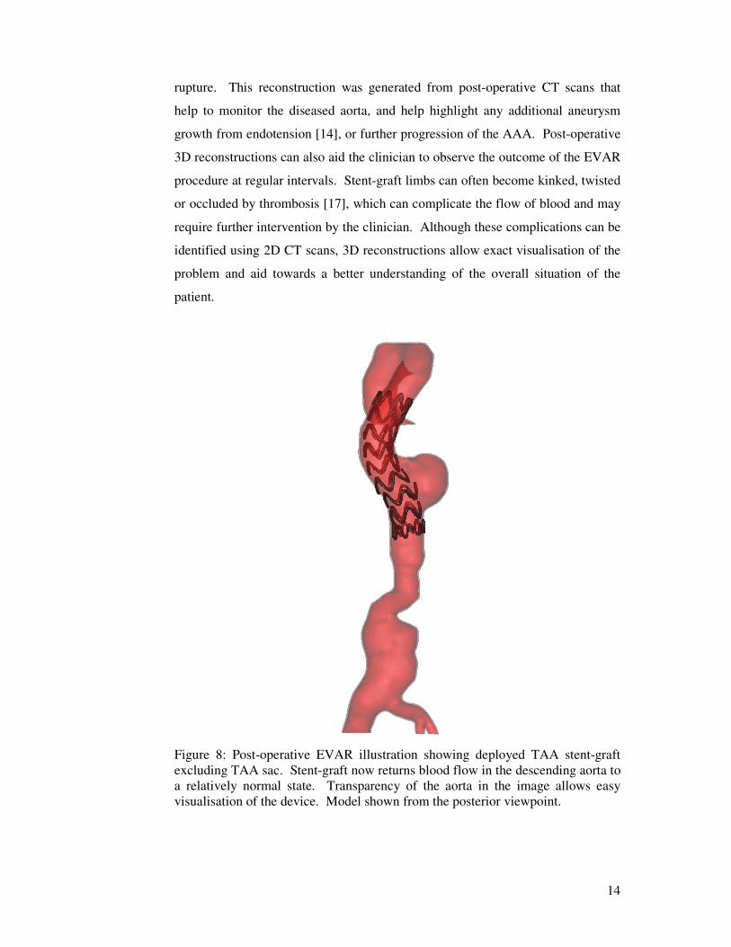
14
rupture. This reconstruction was generated from post-operative CT scans that
help to monitor the diseased aorta, and help highlight any additional aneurysm
growth from endotension [14], or further progression of the AAA. Post-operative
3D reconstructions can also aid the clinician to observe the outcome of the EVAR
procedure at regular intervals. Stent-graft limbs can often become kinked, twisted
or occluded by thrombosis [17], which can complicate the flow of blood and may
require further intervention by the clinician. Although these complications can be
identified using 2D CT scans, 3D reconstructions allow exact visualisation of the
problem and aid towards a better understanding of the overall situation of the
patient.
Figure 8: Post-operative EVAR illustration showing deployed TAA stent-graft
excluding TAA sac. Stent-graft now returns blood flow in the descending aorta to
a relatively normal state. Transparency of the aorta in the image allows easy
visualisation of the device. Model shown from the posterior viewpoint.
15
Conclusion
3D reconstruction of AAAs is a powerful and useful surgical guidance tool.
Reconstruction allows the clinician to obtain measurements useful for the sizing
of stent-grafts, and also allows the clinician to visualise the aneurysm prior to
surgery. Reconstructions are also necessary for further use with FEA which has
been shown to be a good method of determining wall stress in the diseased aorta.
FEA provides an additional source of information to the clinician and helps
towards a greater understanding of the biomechanical behaviour of the anatomy
prior to surgery. 3D reconstruction is quick to perform and could aid surgeons in
improving the treatment of AAAs.
ACKNOWLEDGMENTS
The authors would like to thank (i) the Irish Research Council for Science,
Engineering and Technology (IRCSET) Grant RS/2005/340 (ii) Grant #R01-HL-
060670 from the US National Heart Lung and Blood Institute (iii) the Department
of Vascular Surgery in the HSE Midwestern Regional Hospital, Ireland (iv) the
Department of Radiology in the HSE Midwestern Regional Hospital, Ireland (v)
Prof. David A. Vorp from the Centre for Vascular Remodeling and Regeneration,
McGowan Institute of Regenerative Medicine, University of Pittsburgh, USA and
(vi) Mr. Prakash Madhavan, Consultant Vascular Surgeon, St. James Hospital,
Dublin, Ireland.
REFERENCES
1. Sakalihasan N, Limet R, Defawe OD (2005) Abdominal aortic aneurysm.
Lancet 365:1577-1589.
2. Bengtsson H, Nilsson P, Bergqvist D (1998) Natural history of abdominal
aortic aneurysm detected by screening. Br J Surg 80:718-720.
3. Vorp DA (2007) Biomechanics of abdominal aortic aneurysm. J Biomech
40:1887-1902.
16
4. Ouriel K, Green RM, Donayre C, et al (1992) An evaluation of new
methods of expressing aortic aneurysm size: relationship to rupture. J Vasc
Surg 15:12-20.
5. Kleinstreuer C, Li Z (2006) Analysis and computer program for rupture-
risk prediction of abdominal aortic aneurysms. Biomed Eng Online 5:19.
6. McGregor JC, Pollock JG, Anton HC (1975) The value of ultrasonography
in the diagnosis of abdominal aortic aneurysm. Scott Med J 20:133-137.
7. Grimshaw GM, Thompson JM (1997) Changes in diameter of the
abdominal aorta with age: An epidemiological study. J Clin Ultrasound
25:7-13.
8. Johnston KW, Rutherford RB, Tilson MD, et al (1991) Suggested
standards for reporting on arterial aneurysms. Subcommittee on reporting
standards for arterial aneurysms, Ad hoc committee on reporting
standards, Society for Vascular Surgery and North American Chapter,
International Society for Cardiovascular Surgery. J Vasc Surg 13:452-458.
9. Sayers RD (2002) Aortic aneurysms, inflammatory pathways and nitric
oxide. Ann R Coll Surg Engl 84(4):239-246.
10. Raghavan ML, Vorp DA (2000) Toward a biomechanical tool to evaluate
rupture potential of abdominal aortic aneurysm: identification of a finite
strain constitutive model and evaluation of its applicability. J Vasc Surg
33:475-482.
11. Fillinger MF, Marra SP, Raghavan ML, et al (2003) Prediction of rupture
risk in abdominal aortic aneurysm during observation: wall stress versus
diameter. J Vasc Surg 37:724-732.
12. Fillinger MF, Raghavan ML, Marra SP, et al (2002) In vivo analysis of
mechanical wall stress and abdominal aortic aneurysm rupture risk. J Vasc
Surg 36:589-597.
13. Morris LG (2004) Numerical and experimental investigation of
mechanical factors in the treatment of abdominal aortic aneurysms. PhD
Thesis 2004, University of Limerick.
14. EVAR Trial Participants (2004) Comparison of endovascular aneurysm
repair with open repair in patients with abdominal aortic aneurysm (EVAR
trial 1), 30-day operative mortality results: randomised controlled trial.
Lancet 364:843-848.
17
15. EVAR trial participants (2005) Endovascular aneurysm repair versus open
repair in patients with abdominal aortic aneurysm (EVAR trial 1):
Randomised control trial. Lancet 365: 2179-2186.
16. Schermerhorn M, O’Malley J, Jhaveri A, et al (2008) Endovascular vs.
open repair of abdominal aortic aneurysms in the Medicare population. N
Engl J Med 358:464-474.
17. Corbet TJ, Callanan A, Morris LG, et al. (2008) In vivo and in vitro
biomechanical behaviour and performance of post-operative abdominal
aortic aneurysms and implanted stent-grafts: A review. J Endovasc Ther
15(4):468-484.
18. Resch TA. TeraRecon Clinical Case Studies: Endograft planning for
complex aortic aneurysms. Available via TeraRecon.
http://www.terarecon.com/downloads/news/clinical_case_studies_v2/cases
tudy_Resch-EndograftPlanning.pdf Accessed 10 March 2008.
19. Vorp DA, Raghavan ML and Webster MW (1998) Mechanical wall stress
in abdominal aortic aneurysm: influence of diameter and asymmetry. J
Vasc Surg 27(4):632-639.
20. Raghavan ML, Vorp DA, Federle MP, et al (2000) Wall stress distribution
on three-dimensionally reconstructed models of human abdominal aortic
aneurysm. J Vasc Surg 31:760-769.
21. Doyle BJ, Callanan A, Walsh MT, et al (2008) A finite element analysis
rupture index (FEARI) as an additional tool for abdominal aortic aneurysm
burst prediction. Vascular Disease Prevention, in press.
22. Doyle BJ, Callanan A, Burke PE, et al (2008) Vessel asymmetry as an
additional diagnostic tool for the assessment of abdominal aortic
aneurysms. J Vasc Surg, in press.
23. Doyle BJ, Morris LG, Callanan A, et al (2008) 3D reconstruction and
manufacture of real abdominal aortic aneurysms: From CT scan to silicone
model. J Biomech Eng 130:034501-5.
24. Doyle BJ, Callanan A and McGloughlin TM (2007) A comparison of
modelling techniques for computing wall stress in abdominal aortic
aneurysms. Biomed Eng Online 6:38.
18
25. Vorp DA, Shiro BJ, Ehrlich MP, et al (2003) Effect of aneurysm on the
tensile strength and biomechanical behaviour of the ascending thoracic
aorta. Ann Thorac Surg 75:1210-1214.
26. Thubrikar MJ, Al-Soudi J, Robicsek F (2001) Wall stress studies of
abdominal aortic aneurysm in a clinical model. Ann Vasc Surg 15:355-
366.
27. Truijers M, Pol JA, SchultzeKool LJ, et al (2007) Wall stress analysis in
small asymptomatic, symptomatic and ruptured abdominal aortic
aneurysms. Eur J Vasc Endovasc Surg 33:401-407.
28. Speelman L, Bohra A, Bosboom EMH, et al (2007) Effects of wall
calcifications in patient-specific wall stress analyses of abdominal aortic
aneurysms. J Biomech Eng 129:1-5.
29. Faries PL, Dayal R, Rhee J, et al (2004) Stent graft treatment for
abdominal aortic aneurysm repair: recent developments in therapy.
Diseases of the aorta, pulmonary, and peripheral vessels. Current Opinion
in Cardiology 19(6):551-557.
30. Molony DS, Callanan A, Morris LG, et al (2008) Geometrical
enhancements for abdominal aortic stent grafts. J Endovasc Ther, in press.