Embed Size (px)
Citation preview

Improved Efficacy and Reduced Toxicity of DoxorubicinEncapsulated in Sulfatide-Containing Nanoliposome in aGlioma Model
Author
Lin, Jia, Shigdar, Sarah, Fang, Ding Zhi, Xiang, Dognxi, Wei, Ming Q, Danks, Andrew, Kong,Lingxue, Li, Lianghong, Qiao, Liang, Duan, Wei
Published
2014
Journal Title
PloS One
DOI
https://doi.org/10.1371/journal.pone.0103736
Copyright Statement
© 2014 Lin et al. This is an Open Access article distributed under the terms of the CreativeCommons Attribution License CCAL. (http://www.plos.org/journals/license.html)
Downloaded from
http://hdl.handle.net/10072/64556
Griffith Research Online
https://research-repository.griffith.edu.au

Improved Efficacy and Reduced Toxicity of DoxorubicinEncapsulated in Sulfatide-Containing Nanoliposome in aGlioma ModelJia Lin1,2, Sarah Shigdar1, Ding Zhi Fang2, Dognxi Xiang1, Ming Q. Wei3, Andrew Danks4, Lingxue Kong5,
Lianghong Li6, Liang Qiao7, Wei Duan1*
1 School of Medicine, Faculty of Health, Deakin University, Waurn Ponds, Victoria, Australia, 2 Department of Biochemistry and Molecular Biology, West China School of
Preclinical and Forensic Medicine, Sichuan University, Chengdu, P. R. China, 3 School of Medical Science and Griffith Health Institute, Griffith University, Gold Coast
Campus, Southport, Australia, 4 Department of Surgery, Southern Clinical School, Monash University, Clayton, Victoria, Australia, 5 Institute for Frontier Materials, Deakin
University, Waurn Ponds, Victoria, Australia, 6 Liaoning Key Laboratory of Cancer Stem Cell Research, Dalian Medical University, Dalian, China, 7 Storr Liver Unit, Westmead
Millennium Institute, the University of Sydney at the Westmead Hospital, Westmead, NSW, Australia
Abstract
As a glycosphingolipid that can bind to several extracellular matrix proteins, sulfatide has the potential to become aneffective targeting agent for tumors overexpressing tenasin-C in their microenvironment. To overcome the dose-limitingtoxicity of doxorubicin (DOX), a sulfatide-containing nanoliposome (SCN) encapsulation approach was employed toimprove treatment efficacy and reduce side effects of free DOX. This study analysed in vitro characteristics of sulfatide-containing nanoliposomal DOX (SCN-DOX) and assessed its cytotoxicity in vitro, as well as biodistribution, therapeuticefficacy, and systemic toxicity in a human glioblastoma U-118MG xenograft model. SCN-DOX was shown to achieve highestdrug to lipid ratio (0.5:1) and a remarkable in vitro stability. Moreover, DOX encapsulated in SCN was shown to be deliveredinto the nuclei and displayed prolonged retention over free DOX in U-118MG cells. This simple two-lipid SCN-DOX nanodrughas favourable pharmacokinetic attributes in terms of prolonged circulation time, reduced volume of distribution andenhanced bioavailability in healthy rats. As a result of the improved biodistribution, an enhanced treatment efficacy of SCN-DOX was found in glioma-bearing mice compared to the free drug. Finally, a reduction in the accumulation of DOX in thedrug’s principal toxicity organs achieved by SCN-DOX led to the diminished systemic toxicity as evident from the plasmabiochemical analyses. Thus, SCN has the potential to be an effective and safer nano-carrier for targeted delivery oftherapeutic agents to tumors with elevated expression of tenascin-C in their microenvironment.
Citation: Lin J, Shigdar S, Fang DZ, Xiang D, Wei MQ, et al. (2014) Improved Efficacy and Reduced Toxicity of Doxorubicin Encapsulated in Sulfatide-ContainingNanoliposome in a Glioma Model. PLoS ONE 9(7): e103736. doi:10.1371/journal.pone.0103736
Editor: Maria G. Castro, University of Michigan School of Medicine, United States of America
Received April 12, 2014; Accepted June 24, 2014; Published July 29, 2014
Copyright: � 2014 Lin et al. This is an open-access article distributed under the terms of the Creative Commons Attribution License, which permits unrestricteduse, distribution, and reproduction in any medium, provided the original author and source are credited.
Data Availability: The authors confirm that all data underlying the findings are fully available without restriction. All relevant data are within the paper and itsSupporting Information files.
Funding: This work was supported by a grants from National Health and Medical Research Council, Australia (#479505), www.nhmrc.gov.au and Australia-IndiaStrategic Research Fund Programme–Indo-Australia Science and Technology Fund (#ST040007), http://www.innovation.gov.au/Science/InternationalCollaboration/aisrf/Pages/default.aspx. The funders had no role in study design, data collection and analysis, decision to publish, or preparationof the manuscript.
Competing Interests: The authors have declared that no competing interests exist.
* Email: [email protected]
Introduction
Indiscriminate exposure of all cells in the body to a systemically
administered chemotherapy agent kills healthy cells as well as the
tumor cells [1,2], causing severe toxicity to the patients and
leading to serious side effects, and poor quality of life [3,4]. This
non-specific biodistribution and the resulting side-effects limit the
clinical application of anticancer drugs [5]. Thus, there is an
urgent need to develop new chemotherapeutics that can target
tumor cells effectively.
Sulfatide, a lipid that is found in humans, is involved in a variety
of biological processes such as cell adhesion, platelet aggregation,
cell growth, protein trafficking, signal transduction, neuronal
plasticity and cell morphogenesis. Sulfatide is known to bind
several extracellular matrix glycoproteins including tenascin-C [6]
which is overexpressed in the microenvironment of most solid
cancers, including malignant brain tumors [7]. We have recently
shown that sulfatide was specifically required for robust uptake of
nanoliposomes by human glioblastoma U-87MG cells which
overexpress tenascin-C [8,9]. In addition, in vivo studies
demonstrated that the U-87MG tumor-bearing mice received
DOX encapsulated in nanoliposomes with sulfatide showed an
improvement in survival compared with those received DOX
encapsulated in nanoliposomes without sulfatide [8], suggesting
that sulfatide in the nanoliposome involves in the binding to
tenascin-C. The unique feature of this nanoliposome is that it is
comprised of two natural lipids found in human cells, namely
sulfatide and 1,2-dioleoyl-sn-glycero-3-phosphoethanolamine
(DOPE). Thus, this nanoliposome is totally human compatible
and degradable. The sulfatide-containing nanoliposomal DOX
(SCN-DOX) has been found to remain intact for hours after
uptake by the glioblastoma cells. Intracellular distribution study
PLOS ONE | www.plosone.org 1 July 2014 | Volume 9 | Issue 7 | e103736

has indicated a high accumulation of DOX in the nuclei where it
exerts its cytotoxic effect after 12 h incubation with SCN-DOX at
37uC [8,10].
Recognising the potential of the use of a tumor environment
targeting ligand as one of the main structural constituents of the
nanocarriers capable of both passive and active targeting, we
designed and carried out a series studies to investigate in vitro and
in vivo stability of the SCN-DOX, the pharmacokinetic behaviour,
the biodistribution pattern and tumor uptake of the SCN-DOX in
a gliomaxenograft model using human glioblastoma cells U-
118MG [11]. In the current study, we determined important
pharmacokinetic and pharmacodynamic properties of sulfatide-
containing nanoliposomal system. First, we studied the stability of
the formulation in the presence and absence of serum as well as its
in vivo blood clearance. Next, we examined cellular uptake and
retention of SCN-DOX in a monolayer culture. Finally, we
demonstrated the potential clinical utilities of this nanoliposome
via studies on its pharmacokinetics in healthy rats, biodistribution
in both healthy rats and tumor-bearing mice as well as antitumor
efficacy and toxicity profiles.
Methods and Materials
Cell cultureThe U-118MG (human glioblastoma) cell line was purchased
from American Type Culture Collection (ATCC, Manassas, VA).
Dulbecco’s modified eagle medium (DMEM, 4.5 g/L D-Glucose),
antibiotics and trypsin were from InvitrogenTM (Australia). Fetal
bovine serum (FBS)was from Hyclone (Canada). Tissue culture
flasks were purchased from BD FalconTM (Australia). Glass bottom
dishes were purchased from MatTek Corporation (Ashland,
MA).Cells were cultured in DMEM medium supplemented with
10% fetal bovine serum, penicillin (50 U/mL), and streptomycin
(50 mg/mL) in a humidified atmosphere containing 5% CO2 and
95% air at 37uC.
Chromatographic instrumentation and systemChromatographic instrumentation and system were used based
on a previously published method with some modifications [12].
Briefly, the high performance liquid chromatography (HPLC)
system used in this study consists of a Waters e2695 Separation
Module and a Waters 2475 Multi l Fluorescence Detector(-
Waters, USA). The excitation and emission wavelengths were set
at the 470 nm and 585 nm, respectively. Chromatographic
separation was performed on a Nova-PakH C18 column
(3.96150 mm i.d., 4 mm, Waters, USA) with a Nova-PakH C18
guard column (3.9620 mm i.d., 4 mm, Waters, USA). A mixture
of methanol and 0.01 M phosphate buffer (pH = 3.0) was used as
the mobile phase. The flow-rate used in the assay was 1 mL/min.
The column was maintained at 4065uC throughout the
chromatographic process. All solvents for HPLC procedures were
prepared freshly and filtered with 0.22 mm membrane before use.
Preparation of SCN-DOXLiposomes were prepared according to a previously published
method with modifications [8]. Briefly, as the uptake of SCN in
the glioma cells was optimal when the ratio between sulfatide and
DOPE was 3:7 [8], DOPE unilamellar vesicles containing 30%
(molar ratio) sulfatide were prepared by a hydration method
followed by polycarbonate membrane extrusion. DOPE
(13.35 mmol/mL) and sulfatide (6 mmol/mL, Avanti Polar Lipids,
Inc.)were dissolved in a mixture of chloroform and methanol (2:1,
v/v), and the lipid mixture, composed of DOPE/sulfatide (3:7,
mol/mol), was transferred to glass tubes. Samples were then
reduced to a minimum volume under a nitrogen stream, and
stored under vacuum for 24 h at 4uC to completely evaporate
the organic solvent. The thin lipid films were hydrated by 1 mL
of 250 mM ammonium sulfate (pH 8.5).The samples were
placed in an ice-water bath and sonicated under nitrogen for
2.5 min with 50% amplitude using a sonicator (Sonics &
Materials, Inc). Following sonication, the liposomes were
formed via extrusion through polycarbonate membranes (Avanti
Polar Lipids, Inc.) with consecutive pore sizes of 400 nm for 14
times, 200 nm for 14 times and 100 nm for 19 times at room
temperature. To establish a trans-bilayer ammonium sulfate
gradient, the extruded liposomes were dialyzed against a 250-
fold volume of 10% sucrose in 25 mM Trizma at pH 8.5 at 4uCfor 24 h. The external buffer was changed 3 times during
dialysis. After dialysis of the liposomes, DOX in 10% sucrose at
a final concentration of 5 mg/mL was added to the liposomes at
a drug-to-lipid ratio of 0.3:1 (w/w), followed by incubation in
the water bath at 60uC for 1 h. Non-encapsulated DOX was
removed by size exclusion chromatography using a Sephadex
G-50 column.
Determination of phospholipids in SCN-DOXThe concentration of phospholipids (DOPE) in liposomes was
determined as previously described [13]. Briefly, 1 mL chloroform
and 0.5 mL ferri-thiocyanate reagent were added in to a 100 mL
aliquot of SCN-DOX. The samples were then vortexed for 1 min,
and centrifuged at 12,0006g for 5 min. Following the removal of
supernatant,the absorbance of samples was measured at 488 nm
against the chloroform blank. The DOPE concentration in the
samples was calculated according to a standard curve of DOPE
concentration vs. its fluorescence intensity.
Determination of particle size and zeta potential of SCN-DOX
After the size exclusion chromatography, 10 mL aliquot of
liposome was diluted by 990 mL PBS and mixed gently. The
vesicle size and zeta potential of SCN were measured using
ZetasizerNano ZS Particle Characterization System from Malvern
Instruments (Malvern, UK).
Determination of drug loading efficiency of SCN-DOXFor determination of DOX loading efficiency, standard curves
of DOX (ranging from 50 to 10,000 ng/mL) were established via
using HPLC initially. Calibration curves were constructed by
plotting peak areas of fluorescence derived from DOX vs. DOX
concentrations. A linear regression was used for quantitation. The
standard formulas were determined by linear regression as
y = mx+b, where y is the peak area of DOX and x is the DOX
concentration. The DOX concentration in the samples was
calculated according to a standard curve of DOX concentration
vs. its fluorescence intensity.
The amount of DOX encapsulated in SCN was determined
by disrupting the liposomes with methanol, followed by
quantification of DOX using a fluorescence detector in HPLC.
Briefly, 10 mL aliquot of the liposomal drug eluted from a
Sephadex G-50 column was diluted in 100-fold phosphate
buffer/methanol(45:55,v/v), and the mixture was centrifuged at
20,0006 g for 5 min. Then, the supernatant was measured via
using HPLC. Encapsulation efficiency was calculated by the
following equation:
Pharmacokinetics and Efficacy of Sulfatide-Containing Nanoliposome
PLOS ONE | www.plosone.org 2 July 2014 | Volume 9 | Issue 7 | e103736

Encapsulation efficiency (%)~
DOX encapsulated in liposomes
DOX added to liposomes|100%
In vitro release kinetics of SCN-DOXThe in vitro leakage of DOX from SCN was measured by a
dialysis method [14,15]. Briefly, 2.5 mL SCN-DOX was added
into a Slide-A-Lyzer Dialysis Cassette (Pierce, molecular weight
cut-off of 2 kDa). The dialysis cassette was placed into a beaker
containing 250-fold excess of phosphate-buffer saline (PBS) or PBS
with 10% fetal bovine serum, penicillin (50 U/mL), and strepto-
mycin (50 mg/mL). The SCN-DOX was dialyzed with stirring for
72 hours at 37uC. At various time points (0 h, 0.5 h, 1 h, 4 h, 8 h,
24 h, 48 h and 72 h), 500 mL aliquots was withdrawn from the
external buffer for release kinetics analysis, and replaced with the
same volume of fresh external buffer. For HPLC measurement,
the aliquots were mixed with 1 mL methanol, followed by
centrifugation at 21,0006 g for 5 minutes. Supernatants were
collected for the measurement of HPLC. The drug concentration
in the external buffer was calculated according to a standard curve
of DOX concentration vs. its fluorescence intensity.
Analysis of cytotoxicityThe viabilities of treated and untreated cells were determined by
the MTT (3-(4,5-dimethylthiazol-2-yl)-2,5-diphenyl-2H-tetrazoli-
um bromide) assay which measures the mitochondrial conversion
of MTT to formazan as detected by the change of optical density
at 570 nm [16,17]. Briefly, MCF-7 and U-118MG cells were
plated at a density of 46103 and 36103 cells per well respectively
in 100 ml DMEM medium in 96-well plates and allowed to grow
for 24 hours. The cells were then exposed to a series of different
concentrations of free DOX, SCN-DOX or blank SCN for
48 hours at 37uC under 5% CO2. Then, 10 mL MTT solution
(5 mg/mL in PBS) was added into each well followed by
incubation for 4 h at 37uC. The reaction was terminated by
removing MTT before the addition of 150 mL/well solubilisation
reagent (dimethyl sulfoxide, DMSO). Absorbance was measured at
570 nm using a VICTOR TM X5 Multilabel HTS Reader
(PerkinElmer Life and Analytical Sciences). DOX concentration
leading to 50% cell-killing (IC50) was calculated using the statistical
software package SPSS 13.0.
Confocal microscopy analysis for cellular uptake andretention of SCN-DOX
U-118MG cells (1.56105 cells/well) were seeded in 35 mm glass
bottom dishes and incubated at 37uC in 5% CO2 for 24 h. The
medium was then replaced with full culture medium containing
2 mg/mL free DOX or SCN-DOX. After incubation for another
24 hours, cells were washed twice with PBS and incubated with
fresh culture medium. The cells were then imaged for cellular
uptake studies, followed by serial imaging at 1 h, 2 h, 4 h, and
24 h for retention study using a Fluoview FV10i fluorescence laser
scanning confocal microscopy (Olympus, Japan).
Study of pharmacokinetic properties and biodistributionin vivo
Male Sprague-Dawley (SD) rats (200 to 250 g) were housed in a
temperature controlled room (2561uC) with a 12-h light-dark
cycle. Rats were fed ad libitum with a standard diet but were fasted
overnight before free DOX or SCN-DOX administration. All
procedures involving animal experimentations were approved by
the Deakin University Animal Welfare Committee.
For determination of DOX concentration in the blood and
serum, standard curves of DOX (ranging from 2 to 10, 000 ng/
mL) were established via using HPLC initially. The construction of
calibration curves and the determination of standard formulas
were the same as described above. The DOX concentration in the
samples was calculated according to a standard curve of DOX
concentration vs. its fluorescence intensity.
To investigate the pharmacokinetics (PK) properties and
biodistribution (BD) of SCN-DOX in vivo, healthy SD rats were
injected i.v. with free DOX or SCN-DOX via the tail vein with a
single dose of 5 mg DOX/kg. For PK study, 250 mL blood was
serially collected from the same animal in heparinised tubes from
the tail at 2 min, 0.5 h, 2 h, 6 h, 24 h and 48 h. After collection,
samples were centrifuged at 3,0006 g at 4uC for 10 min to
separate the plasma. To determine DOX levels in plasma, 495 mL
Table 1. Physical properties of the liposomal formulations.
CompositionDOX loading efficiency(%) Particle size (nm)
DOX-to-DOPEratio (w/w) polydispersity index (PDI) Zeta potential (mV)
SCN 94.1162.27 92.3261.31 0.5:1 0.1560.01 226.3862.20
Data are shown as means 6 S.E. of at least three independent experiments.doi:10.1371/journal.pone.0103736.t001
Figure 1. In vitro stability of SCN-DOX. The stability of SCN-DOXwas studied by dialyzing the release of DOX from SCN-DOX into PBS orPBS with 10% FBS at 37uC. Aliquots of dialysis buffer were collected atdesigned time points (0, 0.5, 1, 4, 8, 24, 48 and 72 hour). DOX releasedinto the dialysis buffer was quantified. Data are shown as means 6 S.E.of at least three independent experiments.doi:10.1371/journal.pone.0103736.g001
Pharmacokinetics and Efficacy of Sulfatide-Containing Nanoliposome
PLOS ONE | www.plosone.org 3 July 2014 | Volume 9 | Issue 7 | e103736

of methanol and 405 mL of phosphate buffer were added into
100 mL plasma, vortexed for 1 min, and centrifuged at 21,0006gfor 10 min at 4uC. The supernatant was transferred to another
tube followed by the addition of 2 mL of perchloric acid (35%, v/
v). The samples were vortexed for 1 min, and centrifuged at
21,0006 g for 10 min at 4uC, followed by the measurement of
DOX concentration using HPLC [18]. For biodistribution study
in rats, animals were sacrificed by Lethabarb R (100 mg/kg) at
0.5 h, 2 h, 4 h and 24 h after a single dose of DOX or SCN-DOX
injection. The biodistribution study in tumor-bearing mice was
performed after the administration of 6 doses of DOX or SCN-
DOX. Tissues (heart, liver, spleen, lung and kidney) were
collected, followed by washing in cold physiological saline to
remove any excess blood, blot-dried using filter paper and
weighted. One hundred mg of tissue was added to 495 mL of
methanol and 405 mL of phosphate buffer in a tightly sealed 2-mL
tube followed by homogenization using the FastPrepH-24 tissue
and cell homogenizer (MP Biomedicals, US). The tissue homog-
enate was centrifuged at 21,0006 g for 10 min at 4uC and the
supernatant was transferred to a fresh tube and extracted with the
addition of 2 mL perchloric acid (35%, v/v) as described above.
The supernatant was analysed using HPLC [19].
Tumor implantation, treatment and evaluation in U-118MG tumor-bearing mice
Six-week-old female BALB/c-Foxn1nu mice were purchased
from The Animal Resources Centre (Perth, Australia) and used for
U-118MG xenograft models. The mice were housed in a
TECNIPLAST SealsafeTM Individually Ventilated Cages which
were placed in the temperature controlled room (2561uC) with a
12-h light-dark cycle. Mice were fed ad libitum with a standard
diet. All procedures, involving animal experimentations, were
approved by the Deakin University Animal Welfare Committee
(AEC Project Number: A61-2008 and A62-2008).Beddings, cages
and water were autoclaved at 121uC for 30 minutes while the
fodder was sterilized by ultraviolet irradiation before use. For
tumor implantation, a U-118MG single cell suspension was
prepared by trypsinization followed by washing and resuspension
in PBS at a concentration of 56107 cells/mL. Half million of cells
in 100 mL PBS were inoculated subcutaneously (s. c.) to the right
flank of the mice. Tumor diameters were measured using a digital
calliper every other day after implantation and approximate tumor
volume was calculated as length6width2/2 (V = lw2/2). The
tumor was allowed to grow until its volume reached approximately
150 mm3 [20].
Once the tumor reached a volume of 150 mm3, the nude mice
were randomly divided into 2 experimental groups (5 to 6 mice per
group). Formulations of either free or liposomal DOX at doses of
5 mg/kg DOX or equivalent were administered once a week via
the tail vein at rate 0.4 mL/min for 6 weeks. Twenty-four hours
after the last injection, mice were sacrificed by injection of
Lethabarb R (100 mg/kg). Tumors and tissues were collected and
processed as described in section 2.8 [20,21].
Therapeutic efficacy in tumor xenograft modelFor therapeutic studies, the nude mice were randomly divided
into 4 experimental groups (5 to 6 mice per group) when the
xenograft tumors reached 150 mm3 [22]. Treatment groups
consisted of (a) saline control group, (b) free DOX (5 mg/kg)
group and (c) SCN-DOX (5 mg/kg in DOX), (d) blank SCN
group. The treatment was administered by intravenous (i. v.)
injection via the tail vein once a week for six weeks [23]. Tumor
sizes, as well as animal weights, were measured every other day.
Determination of biodistribution and systemic toxicity inmice
For tissue distribution after repeated injections, mice were i.v.
injected with 5 mg/kg free DOX or SCN-DOX (5 mg/kg in
DOX) once a week for 6 weeks and were sacrificed 24 hours after
the last injection. Tumors and tissues were collected for HPLC
measurement of DOX. To evaluate the general toxicity of free
DOX and SCN-DOX, 20 female mice (6 weeks old) were
randomly divided into 4 groups, and treated as describe above.
The mice were sacrificed at 72 days after the 6th injection. Plasma
biochemical analyses were carried out by a veterinary pathology
laboratory (Gribbles Veterinary Pathology, Clayton, Vic). For the
measurement of the serum troponin, the method employed a cut-
off threshold of ,0.01 mg/L for normal subjects [24]. The
concentration of troponin for samples ,0.01 mg/L could not be
reliably determined nor reported by the veterinary pathology
laboratory.
Data analysisResults were reported as mean and standard error (mean 6
S.E.) unless otherwise stated. The pharmacokinetic parameters
were calculated from the average plasma concentrations using the
pharmacokinetic software DAS 2.0 software (Mathematical
Pharmacology Professional Committee of China, Shanghai,
China). The differences in the mean values among different
groups were analysed using a one-way analysis of variance
(ANOVA) using the statistical software package SPSS 13.0. Pvalues less than 0.05 were considered to be statistically significant.
Results
Characterization of liposomesThe physicochemical characteristics of SCN are presented in
Table 1. Mean vesicle size of SCN incorporating DOX was
92.3261.31 nm with polydispersity index (PDI) of 0.1560.01. At
an initial input weight ratio of DOX to DOPE of 0.3:1, the
efficiency of DOX loading to SCN using ammonium sulfate
gradient was 94.11%62.27%, in consistence with what is reported
in the literature [25]. Zeta potential value of SCN was 2
26.3862.20 mV. The DOX to DOPE weight ratio after DOX
encapsulation into SCN was determined to be 0.5:1.
Table 2. Mean IC50 values (mg/mL of doxorubicin) for treatment with free dox and sulfatide-containing nanoliposomal Dox.
Cell line Free DOX (mg/ml) SCN-DOX (mg/ml)
U-118MG 2.5160.33 19.5560.68
MCF-7 1.7660.41 29.5862.55
Data are shown as means 6 S.E. of three independent experiment performed in triplicate.doi:10.1371/journal.pone.0103736.t002
Pharmacokinetics and Efficacy of Sulfatide-Containing Nanoliposome
PLOS ONE | www.plosone.org 4 July 2014 | Volume 9 | Issue 7 | e103736

In vitro drug retention propertiesIn vitro DOX release from SCN was determined by dialysing
SCN-DOX against PBS or PBS with 10% FBS at 37uC and
measuring the DOX concentration over time from the fluid within
the dialysis container. As shown in Figure 1, there was minimal
DOX leakage from the SCN during the first 48 h dialysis period,
with more than 99% of DOX retained in the SCN after 48 h
under both PBS and PBS/serum dialysis conditions. The release of
DOX increased after 48 h incubation. The percentage of DOX
retained in the SCN after 72 h were 84.06%68.63% in PBS and
91.91%61.36% in PBS with 10% serum, respectively.
In vitro cytotoxicityThe cytotoxicity of free DOX and SCN-DOX to U-118MG
glioblastoma cells was compared. A breast cancer cell line, MCF-
7, that does not express detectable levels of tenascin-C [26] was
used as a negative control to verify the role of sulfatide in the
interaction between SCN and tenascin-expressing cancer cells.
The IC50 values for DOX and SCN-DOX are shown in Table 2.
The IC50 for free DOX (2.5160.33 mg/mL) in U-118MG was
lower than that observed in U-118MG treated with SCN-DOX
(19.5560.68 mg/mL). These results suggested that U-118MG cells
were relatively more sensitive to free DOX than to SCN-DOX
when exposed to a constant concentration of the agent. There was
no significant toxicity of blank SCN after 48 h incubation (data
not show). The lower toxicity of SCN-DOX to U-118MG in vitro
is in a good agreement with the literature for other liposomes
loaded with free DOX [27,28] and is consistent with the above
stability study as the cytotoxicity in vitro is related to the release
rate of DOX from nanocarriers [15]. The data suggest that under
in vitro conditions, the cellular uptake of free DOX via random
diffusion is more efficient than that of SCN-DOX via energy-
dependent endocytosis. Moreover, the IC50 value of SCN-DOX in
MCF-7 cells was significantly higher than that in U-118MG cells
(29.5862.55 mg/mL and 19.5560.68 mg/mL, respectively),
which might result from differential binding of SCN to cells with
or without tenascin-C and resultant difference in the subsequent
uptake of SCN-DOX.
Intracellular uptake and retention of SCN-DOX in U-118MG cells
The accumulation and retention of DOX and SCN-DOX in U-
118MG cells were studied using laser scanning confocal micros-
copy utilising the natural fluorescent property of DOX. We first
established that there was no apparent gross adverse effects in the
cells after 24 h incubation with 2 mg/ml DOX or equivalent
amounts of SCN-DOX followed by washing with PBS. Next,
cellular uptake of free DOX or SCN-DOX by U-118MG in
DOX- or liposome-free medium was examined. As shown in
Figure 2, both free DOX and SCN-DOX accumulated in the
cytoplasm of glioblastoma cells. However, there was slightly
stronger DOX fluorescence (red) in cells treated with free DOX
when compared to those treated with SCN-DOX after 24 h
incubation. The overlay of Hoechst staining (nucleus) and red
fluorescence (DOX) shown in Figure 3 indicated that SCN-DOX
were not adhered on the cell surface but actually penetrated into
the nucleus. Interestingly, SCN-DOX was better retained by the
glioma cells. As shown in Figure 3A, there was a significant
decrease of DOX fluorescence in cells treated with free DOX only
2 h after washing with PBS. In contrast, DOX fluorescence could
be found in glioma cells treated with SCN-DOX even 24 h after
washing (Figure 3B), suggesting more sustained retention of DOX
in U-118MG cells when delivered via SCN-DOX than that by free
DOX. The improved retention of DOX encapsulated with SCN
in vitro implies the potential of better treatment efficacy of SCN-
DOX in vivo.
Improved pharmacokinetic properties of SCN in healthyrats
The pharmacokinetic properties of both free DOX and SCN-
DOX were studied in healthy male SD rats. The main
pharmacokinetic parameters are shown in Table 3. In our study,
the terminal half-life with free DOX in plasma was 20.6561.34 h,
which is consistent with data from earlier studies by others [29,30].
Whereas it was 41.8963.58 h with SCN-DOX, showing a2.03-
fold increase in the terminal half-life. Indeed, the steady state
volume of distribution (Vd) of free DOX (18.36 L/kg) was 25-fold
higher than SCN-DOX (0.73 L/kg), suggesting that circulating
Figure 2. Intracellular uptake of SCN-DOX in U-118MG cells. U-118MG cells were incubated with 2 mg/mL free DOX or equivalent SCN-DOXfor 24 hours. Cells were washed twice with PBS and imaged with a confocal fluorescence microscope. (A) Cells treated with free DOX. (B) Cells treatedwith SLC-DOX. Red: fluorescence from DOX; blue: nuclei stained with Hoechst 33342. Data are representative of three independent experiments.Scale bars: 20 mm.doi:10.1371/journal.pone.0103736.g002
Pharmacokinetics and Efficacy of Sulfatide-Containing Nanoliposome
PLOS ONE | www.plosone.org 5 July 2014 | Volume 9 | Issue 7 | e103736

Pharmacokinetics and Efficacy of Sulfatide-Containing Nanoliposome
PLOS ONE | www.plosone.org 6 July 2014 | Volume 9 | Issue 7 | e103736

SCN-DOX sequester drug in the plasma compartment, contrib-
uting to the high Cmax observed for SCN-DOX. The area under
the plasma concentration-time curves (AUC0-‘) of DOX delivered
through SCN was 2.06-fold higher than free DOX
(3597.03699.36 mg/Lh and 1746.87669.94 mg/Lh, respectively),
suggesting a decreased in non-specific binding as well as a selective
sequestration of the drug to tissues when administered as
entrapped in SCN, leading to enhanced bioavailability.
Tissue distribution advantages of SCN-DOX in healthyrats
We next evaluated the biodistribution characteristics of SCN-
DOX after i. v. injection of a single dose of 5 mg/kg free DOX or
SCN-DOX in healthy SD rats. As shown in Figure 4, treating the
rats with SCN-DOX led to significantly decreased DOX
accumulation in the heart, the lung and the kidney. In the heart
and the lung, the DOX concentration was significantly lower in
the animals received SCN-DOX than those receiving free DOX at
all time points, typified by a 9-fold and 2-fold lower concentration
in the SCN-DOX group at 2-h for the heart and lung, respectively
(Figure 4A and 4D). Moreover, distribution of DOX at 0.5 h, 2 h
and 4 h time points in the kidney was significantly reduced by i. v.
administered SCN-DOX when compared to the free DOX
(Figure 4E). Consistent with the reported enhanced sequestration
of nanoparticles, including liposomes, by organs of the reticulo-
endothelial system [29,31], the DOX concentration was signifi-
cantly higher in the liver in animals received SCN-DOX than
those received free DOX (Figure 4B). In the spleen, the DOX
concentration in the SCN group was significantly higher than the
free DOX group only initially during the first 2 h after the
administration. There was no statistically significant difference of
the DOX concentration at 4 h and 24 h points in the spleen
between different treatment groups (Figure 4C).
Improved tumor uptake and biodistribution in U-118MGtumor xenograft model
Next, we investigated the ability of SCN-DOX to enhance the
delivery of therapeutic agents to tumor in vivo using a mouse
tumor xenograft model. Mice bearing U-118MG tumors were
administrated intravenously with5 mg/kg free DOX or SCN-
DOX once in a week for 6 weeks when subcutaneous implantation
tumors reached a volume of 150 mm3.The uptake of DOX in
various organs and tumor was determined 24 h after the last
injection. Consistent with the results from the study in healthy SD
rats in the preceding section, the DOX concentration in organ
responsible for dose-limiting toxicity in clinics, i.e. the heart, in the
tumor-bearing mice treated with SCN-DOX was significantly
lower than those treated with free DOX (0.8160.07 mg/g versus
1.4560.17 mg/g) (Figure 5A). The DOX concentration in the
other known major DOX toxicity organ, the skin, was also
statistically significantly lower in the SCN-DOX group compared
to the free DOX animals (,15 fold lower), so as in the kidneys
(28.0562.67 versus 20.9160.82 mg/g) (Figure 5C and 5E). On
the other hand, there were significantly higher levels of DOX in
the liver, spleen and lung in the groups treated with SCN-DOX
when compared to the free DOX group (6.10-fold, 3.16-fold and
1.22-fold, respectively) (Figure 5B and 5E).However, at least in the
case of the liver, the increased accumulation of DOX via SCN-
DOX delivery did not seem to translate into enhanced hepatic
toxicity (see below).As for the xenograft glioma (Figure 5D), there
was a statistically significant elevation of DOX level in tumor
tissue in the SCN-DOX group compared to that of free DOX
(1.30-fold), confirming the enhanced intratumoral DOX delivery
by SCN-DOX in vivo.
Enhanced therapeutic efficacy of SCN-DOX in U-118MGxenograft tumor model
Given our SCN-DOX is able to deliver more therapeutic agents
to the xenograft tumor (Figure 5), we proceeded to determine the
antitumor activity of SCN-DOX in vivo. Mice bearing U-118MG
tumors were injected with saline, blank SCN, DOX in solution or
encapsulated within SCN once in a week for 6 weeks when
subcutaneous implantation tumors reached a volume of 150 mm3.
As shown in Figure 6, there was no significant difference of tumor
sizes between mice treated with saline control and blank SCN
during the study period. At dose of 5 mg/kg, both DOX
formulations were effective in suppressing tumor growth compared
to saline and blank liposome control after the 2nd injection.
Importantly, tumors grew more rapidly in the mice receiving free
DOX when compared with those receiving SCN-DOX. The final
mean tumor load was 97.29610.71 mm3 in SCN-DOX treatment
group while in free DOX group was 154.76612.53 mm3. Thus,
SCN-DOX formulations displayed stronger tumor growth sup-
pression than free DOX.
To further confirm the antitumor efficacy, we compared the
survival rates of tumor-bearing mice after different treatment
regimen. As shown in Figure 7, the median survival days for
saline, free DOX and SCN-DOX group were 45, 61 and 93 days,
Figure 3. Intracellular retention of SCN-DOX in U-118MG cells. U-118MG cells were first incubated with 2 mg/mL free DOX or equivalent SCN-DOX for 24 hours. Cells were then washed with PBS twice and cultured in fresh full culture medium. The same wells of cells were imaged serially at1 h, 2 h, 4 h and 24 h after washing using fluorescence confocal microscopy. (A) Cells treated with free DOX. (B) Cells treated with SLC-DOX. Data aretypical of three independent experiments. Red: fluorescence from DOX; blue: nuclei stained with Hoechst 33342. Scale bars: 20 mm.doi:10.1371/journal.pone.0103736.g003
Table 3. Pharmacokinetic parameters for free DOX and SCN-DOX.
Formulations AUC0-‘(mg/Lh) t1/2(h) Vd(L/kg)
Free DOX 1746.87669.94 20.6561.34 18.3660.80
SCN-DOX 3597.03699.36** 41.8963.58** 0.7360.01**
Data are shown as means 6 S.E. of at least three independent experiments.AUC: Area under the plasma concentration-time curves.t1/2, Elimination half-life.Vd: Volume of distribution.**, P,0.01 compared to free DOX.doi:10.1371/journal.pone.0103736.t003
Pharmacokinetics and Efficacy of Sulfatide-Containing Nanoliposome
PLOS ONE | www.plosone.org 7 July 2014 | Volume 9 | Issue 7 | e103736

respectively. Thereby, the medium life-span was increased in mice
treated with SCN-DOX by 2.07-fold and 1.52-fold compared to
those treated with saline or free DOX, respectively. Taken
together, i. v. administration of SCN-DOX for 6 times over a 6-
week period displayed not only the stronger suppression on tumor
growth but also much improved survival of U-118MG xenograft-
bearing mice.
Reduced toxicity of SCN-DOX in vivoClinically, the efficacy of DOX is limited by dose-limiting
toxicities. One objective of delivery of chemotherapy agents in a
nano-formulaiton is to reduce systemic toxicities. For this aim, we
evaluated the toxicity of SCN-DOX and free DOX after repeated
injection in tumor-bearing nude mice. The tissue concentrations of
DOX were measured 24 h after the 6th administration of either
SCN-DOX or free DOX. As shown in Figure 5A and 5C,
consistent with our findings with healthy rats, the concentrations of
DOX in the heart and skin were significantly lower, ,1.8-fold and
,15-fold, respectively, in the tumor-bearing mice treated with
SCN-DOX than those treated with free DOX after repeated
administrations. As for plasma biochemistry analysis 72 days after
the last injection (Figure 8), treatment with free DOX induced a
significant increase in serum creatine kinase (CK) concentration
(1048.006100.95 U/L), indicative of heart damage, that exceeded
saline control (588.506167.37 U/L), blank SCN (543.40686.47)
and SCN-DOX groups (430.60682.94 U/L). Furthermore, com-
pared with the free DOX group, the plasma concentrations of
aspartate transaminase (AST), a sensitive indicator of liver
damage, in the SCN-DOX group (p,0.01) and blank SCN group
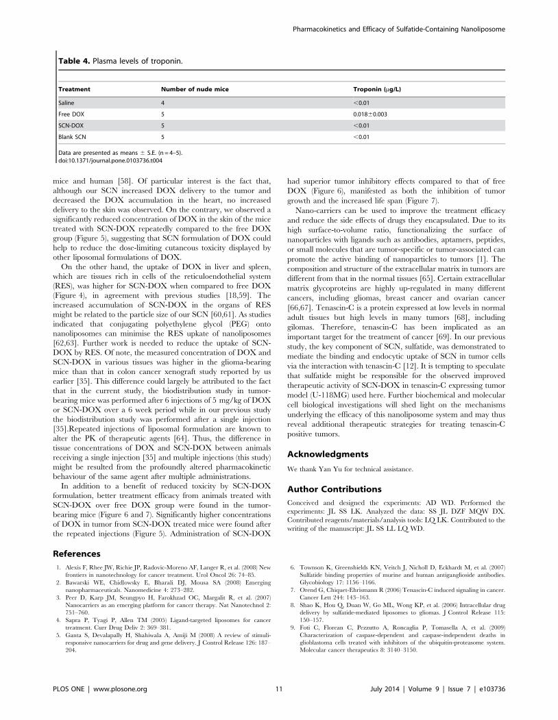
were significantly lower (p,0.05). Moreover, as a marker of DOX-
damaged myocytes, troponin was measured for the evaluation of
DOX-induced cardiomyopathy in vivo [32,33]. The method used
in the present study has a cut-off threshold of ,0.01 mg/L for
normal subjects [24]. As shown in Table 4, there was a 1.8-fold
higher level of serum troponin in the mice treated with free DOX
than those with saline control. In contrast, there is no elevation of
serum troponin level in the mice treated with SCN-DOX or blank
SCN compared to the controls. The results indicated that the
SCN-DOX has the potential to minimize the cardiotoxicity of free
DOX.
Discussion
As liposomes are composed of naturally biodegradable sub-
stances, they are metabolized and cleared while in circulation or
upon reaching the target sites, making them safe novel drug
delivery carriers [34]. Recently, we have shown that SCN-DOX
displays favourable pharmaceutical properties in a colorectal
cancer (HT-29) xenograft model, with an improved biodistribu-
tion, enhanced treatment efficacy and diminished toxicity com-
pared with the free DOX [35]. Here, we provided the first report
Figure 4. Biodistribution of DOX encapsulated in SCN in SD rats. Healthy rats were injected with a single dose of with 5 mg/kg free DOX orSCN-DOX i. v. Rats were euthanized at different time points. Organs were harvested, washed, weighed, and the DOX was extracted and quantified.Data are shown as means 6 S.E. for mg DOX per g of tissue (n = 5–6). ***P,0.001 compared to free DOX.doi:10.1371/journal.pone.0103736.g004
Pharmacokinetics and Efficacy of Sulfatide-Containing Nanoliposome
PLOS ONE | www.plosone.org 8 July 2014 | Volume 9 | Issue 7 | e103736

on detailed pharmacokinetic study, biodistribution in healthy
animals as well as in tumor-bearing mice and antitumor efficacy in
a glioma xenograft model [36] to demonstrate that the efficacy of
this novel nanoliposome is not restricted to gastrointestinal tumors.
In the literature, most of the reported DOX-to-lipid ratio after
loading was between 0.2:1 and 0.3:1 (w/w) [37,38,39]. In this
study, we have achieved a DOX-to-lipid ratio of 0.5:1 (w/w) for
SCN after encapsulation (Table 1). Therefore, our SCN may have
the potential to encapsulate more drugs than some of the other
types of nanoliposomes. Stable encapsulation is essential for
efficient drug delivery to the target site. Our in vitro stability study
suggest that SCN are very stable for 48 h 37uC and retain
substantial physical stability at 72 h in vitro (Figure 1). Interest-
ingly, the percentage of liposomal DOX leakage after 24 h
incubation in other studies is generally more than 5%
[15,40,41,42], in contrast to the minimal payload leakage of
SCN-DOX at 48 h reported here. Therefore, SCN display
superior DOX retention property in vitro under the experimental
conditions used. The prolonged half-life of our SCN-DOX in vivo
(Table 3) might have resulted from the slow release of DOX from
the nanoliposomal formulation [43].
As for in vitro cytotoxicity between different liposomes and
cancer cell lines, published studies reported contradictory results.
For example, in MLLB2 cells [44] and MCF-7/ADR cells [45],
the IC50 of the liposomal formulation was significantly lower than
free DOX, which indicated a higher cytotoxicity of liposomal
DOX in vitro. On the contrary, in HepG2 cells [20] and U-87
cells [8], free DOX seems to have higher intracellular uptake with
associated higher cytotoxicity than that of liposomal DOX.
Obviously, the kinetic properties are different between liposomal
DOX and free DOX in vivo. The half-life of liposomal DOX can
be up to several days while the free DOX can be eliminated in few
minutes [46,47,48]. Moreover, the MTT assay used to derive the
IC50 is carried with the monolayers in culture dishes, which are
very different when compared to the 3-dimentional tissue
architecture in vivo [49]. Thus, comparison of IC50 in vitro,
which is only relevant to the cytotoxicity under a consistent drug
concentration, is not a reliable predictor of the therapeutic efficacy
in vivo [50]. Furthermore, the SCN-DOX had been found to
remain in the nuclei for several hours even after washing under in
vitro condition (Figure 3). This is in agreement with other study
using the same DOX delivery system in a different cell line [8].
Notably, despite the higher IC50 value of SCN-DOX in vitro, it
had much better anti-tumor efficacy over the free DOX in U-
118MG tumor-bearing nude mice (Figure 6). Interestingly, IC50
for free DOX in MCF-7 cells was lower than that for U-118MG,
however, the IC50 for SCN-DOX was 1.5-fold higher in MCF-7
cells than that in U-118MG cells (Table 2). The higher toxicity of
SCN-DOX in U-118MG which overexpress tenascin-C might be,
at least in part, attributed to the interaction between sulfatide in
SCN and tenascin-C on the cells surface of U-118MG cells.
Our previous pharmacokinetic studies of the SLC-DOX in
healthy SD rats revealed that the clearance rate of free DOX is
1.93-fold higher than that of SCN-DOX [35]. The same
improvement was found in the area under the plasma concentra-
tion-time curve from time 0 to infinity and apparent volume of
distribution of free DOX in the current study (Table 3), indicating
prolonged circulation time, slow clearance rate and enhanced
bioavailability of SCN-DOX. In addition, there has been a
significant improvement of biodistribution profile. As a widely
used and efficient antitumor drug, however, DOX has a severe
cardiotoxicity that limits its clinic utility [51]. Encapsulation of
DOX into SCN resulted in a ,4-fold lower DOX concentration
in the heart of rats receiving SCN-DOX (Figure 4), which
represents a significant improvement over those reported by
others showing an approximately 1.5 times lower DOX accumu-
lation in the heart with other liposomal DOX formulation
compared with free DOX [18,19]. Therefore, the significant
reduction in accumulation of DOX in the heart indicates the
potential of SCN-DOX in reducing the cardiotoxicity of DOX.
This has been reinforced by our biochemical studies of the serum
creatine kinase activity, which is a toxicologic indicator of severe
cardiotoxicity [52] as well as cardiac troponin, another biomarker
used for the detection and prevention of cardiotoxicity at an earlier
phase [53]. Previous study by others revealed an increase of serum
troponin level from week 10 after the first administration of DOX
in Wistar rats [24]. Our present study revealed no discernable
increase in serum troponin level in mice treated with SCN-DOX
even 14 weeks after the onset of treatment, suggesting a
remarkable reduction in cardiotoxicity of DOX delivered via
SCN. Moreover, it is important to note that despite that SCN-
DOX resulted in a high accumulation of DOX in the liver, better
liver function than that in the animals received free DOX was
observed at the end of the study, as demonstrated by a significant
reduction of serum AST in mice treated with SCN-DOX
Figure 5. Biodistribution and tumor uptake of DOX encapsu-lated in SCN in glioma-bearing mice. Nude mice bearing humanglioblastomaU-118MGxenografts (s.c.) were treated with 5 mg/kg freeDOX or SCN-DOX i.v. once a week for 6 weeks. Mice were euthanized24 hours after the last treatment. Organs and tissues were harvested,washed, weighed, and the DOX content in tissues, expressed as mg DOXper g tissue, was determined. Data are shown as means 6 S.E. (n = 5–6).*, P,0.05 compared to free DOX; **, P,0.01 compared to free DOX;***, P,0.001 compared to free DOX.doi:10.1371/journal.pone.0103736.g005
Pharmacokinetics and Efficacy of Sulfatide-Containing Nanoliposome
PLOS ONE | www.plosone.org 9 July 2014 | Volume 9 | Issue 7 | e103736

(Figure 8).Of note, DOX is excreted predominantly through the
hepatobiliary route [54] and there is a good negative correlation
between serum AST activity and hepatic intrinsic clearance [55].
Therefore, it is tempting to speculate that SCN-DOX accumu-
lated in the liver was not significantly taken up the hepatocytes but
rather secreted via the bile duct. Further studies are under way to
explore the mechanism underlying the reduced hepatotoxicity
afforded by the SCN-DOX.
Although encapsulation with liposomes has been successful in
overcoming cardiotoxicity and myelosuppression (for free DOX),
the toxicity of liposomal DOX has shifted to the cutaneous toxicity
[56]. Palmar-plantar erythrodysesthesia (PPE), also called hand-
foot syndrome, is a toxic reaction associated with high accumu-
lation of cytotoxic chemotherapeutics, including pegylated liposo-
mal doxorubicin formulation in the skin [57]. High liposome
localization in the skin has been reported previously in both nude
Figure 6. Improved therapeutic activity of SCN-DOX against gliomaxenograft. Mice bearing U-118MG xenografts were injected i.v. withsaline, 5 mg/kg of free DOX, SCN-DOX or empty SCN once a week for 6 weeks when tumor volume reached approximately 150 mm3. Data shown aremeans 6 S.E. (n = 5–6). *, P,0.05 compared to saline; **, P,0.01 compared to saline; #, P,0.05 compared to free DOX; ***, P,0.001 compared tofree DOX; ##, P,0.05 compared to free DOX; &, P,0.01 compared to blank SCN; &&, P,0.01 compared to blank SCN; &&&, P,0.001 compared toblank SCN.doi:10.1371/journal.pone.0103736.g006
Figure 7. SCN-DOX enhanced survival of tumor-bearing mice.The Kaplan-Meier survival curve shows improvement of life span of U-118MG xenograft-bearing mice treated with SCN-DOX (n = 8–10 pergroup). Mice were treated as indicated in the legend for Fig. 6 and weresacrificed when the body weight loss was more than 15%.doi:10.1371/journal.pone.0103736.g007
Figure 8. SCN-DOX treatment had significantly reduced cardiacand hepatic toxicity. U-118MG xenograft-bearing mice were treatedas indicated in the legend for Fig. 6. Blood was collected immediatelyafter the mice were sacrificed upon reaching the end point. Serumenzymes indicative of cardiac and hepatic toxicity were analysed. Datashown are means 6 S.E. (n = 3–5). *, P,0.05 compared to saline; #, P,0.05 compared to free DOX; ##, P,0.01 compared to free DOX.doi:10.1371/journal.pone.0103736.g008
Pharmacokinetics and Efficacy of Sulfatide-Containing Nanoliposome
PLOS ONE | www.plosone.org 10 July 2014 | Volume 9 | Issue 7 | e103736
mice and human [58]. Of particular interest is the fact that,
although our SCN increased DOX delivery to the tumor and
decreased the DOX accumulation in the heart, no increased
delivery to the skin was observed. On the contrary, we observed a
significantly reduced concentration of DOX in the skin of the mice
treated with SCN-DOX repeatedly compared to the free DOX
group (Figure 5), suggesting that SCN formulation of DOX could
help to reduce the dose-limiting cutaneous toxicity displayed by
other liposomal formulations of DOX.
On the other hand, the uptake of DOX in liver and spleen,
which are tissues rich in cells of the reticuloendothelial system
(RES), was higher for SCN-DOX when compared to free DOX
(Figure 4), in agreement with previous studies [18,59]. The
increased accumulation of SCN-DOX in the organs of RES
might be related to the particle size of our SCN [60,61]. As studies
indicated that conjugating polyethylene glycol (PEG) onto
nanoliposomes can minimise the RES upkate of nanoliposomes
[62,63]. Further work is needed to reduce the uptake of SCN-
DOX by RES. Of note, the measured concentration of DOX and
SCN-DOX in various tissues was higher in the glioma-bearing
mice than that in colon cancer xenograft study reported by us
earlier [35]. This difference could largely be attributed to the fact
that in the current study, the biodistribution study in tumor-
bearing mice was performed after 6 injections of 5 mg/kg of DOX
or SCN-DOX over a 6 week period while in our previous study
the biodistribution study was performed after a single injection
[35].Repeated injections of liposomal formulation are known to
alter the PK of therapeutic agents [64]. Thus, the difference in
tissue concentrations of DOX and SCN-DOX between animals
receiving a single injection [35] and multiple injections (this study)
might be resulted from the profoundly altered pharmacokinetic
behaviour of the same agent after multiple administrations.
In addition to a benefit of reduced toxicity by SCN-DOX
formulation, better treatment efficacy from animals treated with
SCN-DOX over free DOX group were found in the tumor-
bearing mice (Figure 6 and 7). Significantly higher concentrations
of DOX in tumor from SCN-DOX treated mice were found after
the repeated injections (Figure 5). Administration of SCN-DOX
had superior tumor inhibitory effects compared to that of free
DOX (Figure 6), manifested as both the inhibition of tumor
growth and the increased life span (Figure 7).
Nano-carriers can be used to improve the treatment efficacy
and reduce the side effects of drugs they encapsulated. Due to its
high surface-to-volume ratio, functionalizing the surface of
nanoparticles with ligands such as antibodies, aptamers, peptides,
or small molecules that are tumor-specific or tumor-associated can
promote the active binding of nanoparticles to tumors [1]. The
composition and structure of the extracellular matrix in tumors are
different from that in the normal tissues [65]. Certain extracellular
matrix glycoproteins are highly up-regulated in many different
cancers, including gliomas, breast cancer and ovarian cancer
[66,67]. Tenascin-C is a protein expressed at low levels in normal
adult tissues but high levels in many tumors [68], including
gilomas. Therefore, tenascin-C has been implicated as an
important target for the treatment of cancer [69]. In our previous
study, the key component of SCN, sulfatide, was demonstrated to
mediate the binding and endocytic uptake of SCN in tumor cells
via the interaction with tenascin-C [12]. It is tempting to speculate
that sulfatide might be responsible for the observed improved
therapeutic activity of SCN-DOX in tenascin-C expressing tumor
model (U-118MG) used here. Further biochemical and molecular
cell biological investigations will shed light on the mechanisms
underlying the efficacy of this nanoliposome system and may thus
reveal additional therapeutic strategies for treating tenascin-C
positive tumors.
Acknowledgments
We thank Yan Yu for technical assistance.
Author Contributions
Conceived and designed the experiments: AD WD. Performed the
experiments: JL SS LK. Analyzed the data: SS JL DZF MQW DX.
Contributed reagents/materials/analysis tools: LQ LK. Contributed to the
writing of the manuscript: JL SS LL LQ WD.
References
1. Alexis F, Rhee JW, Richie JP, Radovic-Moreno AF, Langer R, et al. (2008) New
frontiers in nanotechnology for cancer treatment. Urol Oncol 26: 74–85.
2. Bawarski WE, Chidlowsky E, Bharali DJ, Mousa SA (2008) Emerging
nanopharmaceuticals. Nanomedicine 4: 273–282.
3. Peer D, Karp JM, Seungpyo H, Farokhzad OC, Margalit R, et al. (2007)
Nanocarriers as an emerging platform for cancer therapy. Nat Nanotechnol 2:
751–760.
4. Sapra P, Tyagi P, Allen TM (2005) Ligand-targeted liposomes for cancer
treatment. Curr Drug Deliv 2: 369–381.
5. Ganta S, Devalapally H, Shahiwala A, Amiji M (2008) A review of stimuli-
responsive nanocarriers for drug and gene delivery. J Control Release 126: 187–
204.
6. Townson K, Greenshields KN, Veitch J, Nicholl D, Eckhardt M, et al. (2007)
Sulfatide binding properties of murine and human antiganglioside antibodies.
Glycobiology 17: 1156–1166.
7. Orend G, Chiquet-Ehrismann R (2006) Tenascin-C induced signaling in cancer.
Cancer Lett 244: 143–163.
8. Shao K, Hou Q, Duan W, Go ML, Wong KP, et al. (2006) Intracellular drug
delivery by sulfatide-mediated liposomes to gliomas. J Control Release 115:
150–157.
9. Foti C, Florean C, Pezzutto A, Roncaglia P, Tomasella A, et al. (2009)
Characterization of caspase-dependent and caspase-independent deaths in
glioblastoma cells treated with inhibitors of the ubiquitin-proteasome system.
Molecular cancer therapeutics 8: 3140–3150.
Table 4. Plasma levels of troponin.
Treatment Number of nude mice Troponin (mg/L)
Saline 4 ,0.01
Free DOX 5 0.01860.003
SCN-DOX 5 ,0.01
Blank SCN 5 ,0.01
Data are presented as means 6 S.E. (n = 4–5).doi:10.1371/journal.pone.0103736.t004
Pharmacokinetics and Efficacy of Sulfatide-Containing Nanoliposome
PLOS ONE | www.plosone.org 11 July 2014 | Volume 9 | Issue 7 | e103736
10. Wu X, Li QT (1999) Hydration and stability of sulfatide-containing
phosphatidylethanolamine small unilamellar vesicles. Biochim Biophys Acta
1416: 285–294.
11. Adamsky K, Schilling J, Garwood J, Faissner A, Peles E (2001) Glial tumor cell
adhesion is mediated by binding of the FNIII domain of receptor protein
tyrosine phosphatase beta (RPTPbeta) to tenascin C. Oncogene 20: 609–618.
12. Alvarez-Cedron L, Sayalero ML, Lanao JM (1999) High-performance liquid
chromatographic validated assay of doxorubicin in rat plasma and tissues.
J Chromatogr B Biomed Sci Appl 721: 271–278.
13. Stewart JCM (1980) Colorimetric determination of phospholipids with
ammonium ferrothiocyanate. Anal Biochem 104: 10–14.
14. Rai S, Paliwal R, Vaidya B, Khatri K, Goyal AK, et al. (2008) Targeted delivery
of doxorubicin via estrone-appended liposomes. J Drug Target 16: 455–463.
15. Song H, Zhang J, Han Z, Zhang X, Li Z, et al. (2006) Pharmacokinetic and
cytotoxic studies of pegylated liposomal daunorubicin. Cancer Chemother
Pharmacol 57: 591–598.
16. Zhang FY, Du GJ, Zhang L, Zhang CL, Lu WL, et al. (2009) Naringenin
enhances the anti-tumor effect of doxorubicin through selectively inhibiting the
activity of multidrug resistance-associated proteins but not P-glycoprotein.
Pharm Res 26: 914–925.
17. Jung SH, Jung SH, Seong H, Cho SH, Jeong KS, et al. (2009) Polyethylene
glycol-complexed cationic liposome for enhanced cellular uptake and anticancer
activity. Int J Pharm 382: 254–261.
18. Xiong XB, Huang Y, Lu WL, Zhang H, Zhang X, et al. (2005) Enhanced
intracellular uptake of sterically stabilized liposomal Doxorubicin in vitro
resulting in improved antitumor activity in vivo. Pharm Res 22: 933–939.
19. Xiong XB, Huang Y, Lu WL, Zhang X, Zhang H, et al. (2005) Enhanced
intracellular delivery and improved antitumor efficacy of doxorubicin by
sterically stabilized liposomes modified with a synthetic RGD mimetic. J Control
Release 107: 262–275.
20. Li X, Ding L, Xu Y, Wang Y, Ping Q (2009) Targeted delivery of doxorubicin
using stealth liposomes modified with transferrin. Int J Pharm 373: 116–123.
21. Cheng C, Haouala A, Krueger T, Mithieux F, Perentes JY, et al. (2009) Drug
uptake in a rodent sarcoma model after intravenous injection or isolated lung
perfusion of free/liposomal doxorubicin. Interact Cardiovasc Thorac Surg 8:
635–638.
22. Lopes de Menezes DE, Hudon N, McIntosh N, Mayer LD (2000) Molecular and
pharmacokinetic properties associated with the therapeutics of bcl-2 antisense
oligonucleotide G3139 combined with free and liposomal doxorubicin. Clin
Cancer Res 6: 2891–2902.
23. Tang N, Du G, Wang N, Liu C, Hang H, et al. (2007) Improving penetration in
tumors with nanoassemblies of phospholipids and doxorubicin. J Natl Cancer
Inst 99: 1004–1015.
24. Koh E, Nakamura T, Takahashi H (2004) Troponin-T and brain natriuretic
peptide as predictors for adriamycin-induced cardiomyopathy in rats. Circ J 68:
163–167.
25. Fritze A, Hens F, Kimpfler A, Schubert R, Peschka-Suss R (2006) Remote
loading of doxorubicin into liposomes driven by a transmembrane phosphate
gradient. Biochim Biophys Acta 1758: 1633–1640.
26. Hancox RA, Allen MD, Holliday DL, Edwards DR, Pennington CJ, et al. (2009)
Tumour-associated tenascin-C isoforms promote breast cancer cell invasion and
growth by matrix metalloproteinase-dependent and independent mechanisms.
Breast cancer research: BCR 11: R24.
27. Chen Z, Deng J, Zhao Y, Tao T (2012) Cyclic RGD peptide-modified liposomal
drug delivery system: enhanced cellular uptake in vitro and improved
pharmacokinetics in rats. Int J Nanomedicine 7: 3803–3811.
28. Shmeeda H, Amitay Y, Gorin J, Tzemach D, Mak L, et al. (2010) Delivery of
zoledronic acid encapsulated in folate-targeted liposome results in potent in vitro
cytotoxic activity on tumor cells. J Control Release 146: 76–83.
29. Rahman A, Carmichael D, Harris M, Roh JK (1986) Comparative
pharmacokinetics of free doxorubicin and doxorubicin entrapped in cardiolipin
liposomes. Cancer Res 46: 2295–2299.
30. Wei G, Xiao S, Si D, Liu C (2008) Improved HPLC method for doxorubicin
quantification in rat plasma to study the pharmacokinetics of micelle-
encapsulated and liposome-encapsulated doxorubicin formulations. Biomed
Chromatogr 22: 1252–1258.
31. Sugiyama I, Sadzuka Y (2011) Correlation of fixed aqueous layer thickness
around PEG-modified liposomes with in vivo efficacy of antitumor agent-
containing liposomes. Curr Drug Discov Technol 8: 357–366.
32. Singal PK, Iliskovic N (1998) Doxorubicin-induced cardiomyopathy.
N Engl J Med 339: 900–905.
33. Herman EH, Zhang J, Lipshultz SE, Rifai N, Chadwick D, et al. (1999)
Correlation between serum levels of cardiac troponin-T and the severity of the
chronic cardiomyopathy induced by doxorubicin. J Clin Oncol 17: 2237–2243.
34. Zhu Q, Feng C, Liao W, Zhang Y, Tang S (2013) Target delivery of MYCN
siRNA by folate-nanoliposomes delivery system in a metastatic neuroblastoma
model. Cancer cell international 13: 65.
35. Lin J, Yu Y, Shigdar S, Fang DZ, Du JR, et al. (2012) Enhanced antitumor
efficacy and reduced systemic toxicity of sulfatide-containing nanoliposomal
doxorubicin in a xenograft model of colorectal cancer. PloS one 7: e49277.
36. Reardon DA, Zalutsky MR, Bigner DD (2007) Antitenascin-C monoclonal
antibody radioimmunotherapy for malignant glioma patients. Expert Rev
Anticancer Ther 7: 675–687.
37. Mayer LD, Tai LC, Ko DS, Masin D, Ginsberg RS, et al. (1989) Influence of
vesicle size, lipid composition, and drug-to-lipid ratio on the biological activity of
liposomal doxorubicin in mice. Cancer Res 49: 5922–5930.
38. Hussain S, Pluckthun A, Allen TM, Zangemeister-Wittke U (2007) Antitumor
activity of an epithelial cell adhesion molecule targeted nanovesicular drug
delivery system. Mol Cancer Ther 6: 3019–3027.
39. Hernandez J, Martı A, Estelrich J (1991) Interaction of doxorubicin with lipid
systems. Bioconjug Chem 2: 398–402.
40. Lim HJ, Masin D, Madden TD, Bally MB (1997) Influence of drug release
characteristics on the therapeutic activity of liposomal mitoxantrone. J Pharma-
col Exp Ther 281: 566–573.
41. Huang Y, Chen X-M, Zhao B-X, Ke X-Y, Zhao B-J, et al. (2010)
Antiangiogenic Activity of Sterically Stabilized Liposomes Containing Paclitaxel
(SSL-PTX): In Vitro and In Vivo. AAPS PharmSciTech 11: 752–759.
42. Sadzuka Y, Nakade A, Tsuruda T, Sonobe T (2003) Study on the
characterization of mixed polyethyleneglycol modified liposomes containing
doxorubicin. J Control Release 91: 271–280.
43. Ishida T, Takanashi Y, Doi H, Yamamoto I, Kiwada H (2002) Encapsulation of
an antivasospastic drug, fasudil, into liposomes, and in vitro stability of the
fasudil-loaded liposomes. International journal of pharmaceutics 232: 59–67.
44. Wang J, Goh B, Lu W, Zhang Q, Chang A, et al. (2005) In vitro cytotoxicity of
Stealth liposomes co-encapsulating doxorubicin and verapamil on doxorubicin-
resistant tumor cells. Biol Pharm Bull 28: 822–828.
45. Li B, Xu H, Li Z, Yao M, Xie M, et al. (2012) Bypassing multidrug resistance in
human breast cancer cells with lipid/polymer particle assemblies. Int J Nano-
medicine 7: 187–197.
46. Allen TM, Cheng WWK, Hare JI, Laginha KM (2006) Pharmacokinetics and
Pharmacodynamics of Lipidic Nano-Particles in Cancer. Anticancer Agents
Med Chem 6: 513–523.
47. Cabanes A, Even-Chen S, Zimberoff J, Barenholz Y, Kedar E, et al. (1999)
Enhancement of antitumor activity of polyethylene glycol-coated liposomal
doxorubicin with soluble and liposomal interleukin 2. Clin Cancer Res 5: 687–
693.
48. Unezaki S, Maruyama K, Takahashi N, Koyama M, Yuda T, et al. (1994)
Enhanced delivery and antitumor activity of doxorubicin using long-circulating
thermosensitive liposomes containing amphipathic polyethylene glycol in
combination with local hyperthermia. Pharm Res 11: 1180–1185.
49. Xu L, Anchordoquy T (2011) Drug delivery trends in clinical trials and
translational medicine: challenges and opportunities in the delivery of nucleic
acid-based therapeutics. J Pharm Sci 100: 38–52.
50. Wu J, Lu Y, Lee A, Pan X, Yang X, et al. (2007) Reversal of multidrug resistance
by transferrin-conjugated liposomes co-encapsulating doxorubicin and verapa-
mil. J Pharm Pharm Sci 10: 350–357.
51. Tokarska-Schlattner M, Zaugg M, da Silva R, Lucchinetti E, Schaub MC, et al.
(2005) Acute toxicity of doxorubicin on isolated perfused heart: response of
kinases regulating energy supply. Am J Physiol Heart Circ Physiol 289: H37–47.
52. Bagchi D, Sen CK, Ray SD, Das DK, Bagchi M, et al. (2003) Molecular
mechanisms of cardioprotection by a novel grape seed proanthocyanidin extract.
Mutat Res 523–524: 87–97.
53. Mercuro G, Cadeddu C, Piras A, Dessı M, Madeddu C, et al. (2007) Early
epirubicin-induced myocardial dysfunction revealed by serial tissue Doppler
echocardiography: correlation with inflammatory and oxidative stress markers.
Oncologist 12: 1124–1133.
54. Cosan D, Basaran A, Gunes HV, Degirmenci I, Aral E (2008) The effect of
doxorubicin on rats that received toxic and carcinogenic benzo(a)pyrene. Folia
Histochem Cytobiol 46: 367–372.
55. Yokogawa K, Ido A, Kurihara T, Mano Y, Nomura M, et al. (2006) Serum
aminotransferase activity as a predictor for estimation of total clearance of
hepatically metabolized drugs in rats with acute hepatic failure. Biol Pharm Bull
29: 141–145.
56. Charrois GJ, Allen TM (2003) Rate of biodistribution of STEALTH liposomes
to tumor and skin: influence of liposome diameter and implications for toxicity
and therapeutic activity. Biochim Biophys Acta 1609: 102–108.
57. Farr KP, Safwat A (2011) Palmar-plantar erythrodysesthesia associated with
chemotherapy and its treatment. Case Rep Oncol 4: 229–235.
58. Goren D, Horowitz AT, Zalipsky S, Woodle MC, Yarden Y, et al. (1996)
Targeting of stealth liposomes to erbB-2 (Her/2) receptor: in vitro and in vivo
studies. Br J Cancer 74: 1749–1756.
59. Elbayoumi TA, Torchilin VP (2009) Tumor-targeted nanomedicines: enhanced
antitumor efficacy in vivo of doxorubicin-loaded, long-circulating liposomes
modified with cancer-specific monoclonal antibody. Clin Cancer Res 15: 1973–
1980.
60. Campbell RB (2006) Tumor Physiology and Delivery of Nanopharmaceuticals.
Anticancer Agents Med Chem 6: 503–512.
61. Drummond DC, Meyer O, Hong K, Kirpotin DB, Papahadjopoulos D (1999)
Optimizing liposomes for delivery of chemotherapeutic agents to solid tumors.
Pharmacol Rev 51: 691–743.
62. Tanaka T, Mangala LS, Vivas-Mejia PE, Nieves-Alicea R, Mann AP, et al.
(2010) Sustained small interfering RNA delivery by mesoporous silicon particles.
Cancer research 70: 3687–3696.
63. Chen MH, Chang CH, Chang YJ, Chen LC, Yu CY, et al. (2010)
MicroSPECT/CT imaging and pharmacokinetics of 188Re-(DXR)-liposome
in human colorectal adenocarcinoma-bearing mice. Anticancer Res 30: 65–72.
Pharmacokinetics and Efficacy of Sulfatide-Containing Nanoliposome
PLOS ONE | www.plosone.org 12 July 2014 | Volume 9 | Issue 7 | e103736

64. Cui J, Li C, Wang C, Li Y, Zhang L, et al. (2008) Repeated injection of
pegylated liposomal antitumour drugs induces the disappearance of the rapiddistribution phase. The Journal of pharmacy and pharmacology 60: 1651–1657.
65. Liu F, Chen Z, Wang J, Shao X, Cui Z, et al. (2008) Overexpression of cell
surface cytokeratin 8 in multidrug-resistant MCF-7/MX cells enhances celladhesion to the extracellular matrix. Neoplasia 10: 1275–1284.
66. Fernando NT, Koch M, Rothrock C, Gollogly LK, D’Amore PA, et al. (2008)Tumor escape from endogenous, extracellular matrix-associated angiogenesis
inhibitors by up-regulation of multiple proangiogenic factors. Clin Cancer Res
14: 1529–1539.
67. Quemener C, Gabison EE, Naımi B, Lescaille G, Bougatef F, et al. (2007)
Extracellular matrix metalloproteinase inducer up-regulates the urokinase-type
plasminogen activator system promoting tumor cell invasion. Cancer Res 67: 9–
15.
68. Mackie EJ, Tucker RP (1999) The tenascin-C knockout revisited. J Cell Sci 112:
3847–3853.
69. Adams M, Jones JL, Walker RA, Pringle JH, Bell SC (2002) Changes in
tenascin-C isoform expression in invasive and preinvasive breast disease. Cancer
Res 62: 3289–3297.
Pharmacokinetics and Efficacy of Sulfatide-Containing Nanoliposome
PLOS ONE | www.plosone.org 13 July 2014 | Volume 9 | Issue 7 | e103736