Embed Size (px)
Citation preview
1323
Int. J. Morphol.,33(4):1323-1332, 2015.
Improved Sectioned Images and SurfaceModels of the Whole Female Body
Mejoramiento de Imágenes Seccionadas y Modelos de Superficie del Cuerpo Femenino Completo
Hyo Seok Park*; Dai Hai Choi** & Jin Seo Park*
PARK, H. S.; CHOI, D. H. & PARK, J. S. Improved sectioned images and surface models of the whole female body. Int. J. Morphol.,33(4):1323-1332, 2015.
SUMMARY: The objective of this research was to present high-quality sectioned images of a whole female body which wouldbe helpful in creating an atlas, virtual dissection, and various applications for medical education and clinical trial. In addition, the authorssought to demonstrate the applicabilities of sectioned images. A female cadaver was ground serially using the cryomacrotome andphotographed to make the sectioned images. Structures in the images were segmented to produce segmented images in Photoshop. In theself-developed browsing software, the sectioned and segmented images were stored. Based on the segmented images, surface modelswere built on commercial software and saved as PDF file. High-quality sectioned images of the female body were taken (intervals, 0.2mm or 1 mm; pixel size, 0.1 mm; color depth, 48 bit color). In the images obtained, very small and complicated structures could beidentified in color of living body. In order to ascertain the applicability of the images, the browsing software including sectioned andsegmented images and the PDF file including surface models were produced. The sectioned images and surface models produced duringthis research will prove to be a useful source for medical software. All data generated is available free of charge.
KEY WORDS: Whole body imaging; Cross sectional anatomy; Female; Three-dimensional imaging; Visible HumanProject.
INTRODUCTION
Sectioned images of the human body are of conside-rable use due to their high resolutions and natural colors ascompared with computer tomographs (CT) and magneticresonance images (MRIs) (Ackerman, 1999; Dai et al.,2012). Available images include those of the Visible HumanProject (VHP; male and female) conducted in the UnitedStates (Ackerman; Spitzer et al., 1996, 1998; Spitzer &Whitlock, 1998); the Chinese Visible Human (CVH; maleand female) (Zhang et al., 2006) and the Virtual ChineseHuman (VCH; male and female) (Tang et al., 2010; Yuan etal., 2008); and the Visible Korean (VK; male whole body,male head, and female pelvis) (Park et al., 2005a, 2009; Shinet al., 2013) (Table I). Male sectioned images of the VHP,CVH, and VK were used in many ways; for example, forcreating atlases (Cho, 2009; Spitzer et al., 1998), browsingsoftware (Shin et al.; Shin et al., 2011, 2012a), and virtualdissection software (Schiemann et al., 2000; Spitzer &Scherzinger, 2006), and provided the facility of free accessto three dimensional (3D) models in PDF files atlases (Shin
et al., 2012b; Park et al., 2013). Furthermore, the sectionedimages of VK were used that the dose conversion coefficientsof radiology were calculated virtually (Kim et al., 2008).
However, the usefulnesses of female-sectionedimages prepared were limited for the following reasons. InVHP images, degeneration was observed in the uterus andovaries because the subject was post-menopausal (59 yearsold), and the lateral sides of both arms were not shownbecause the subject was obese. In addition, image qualitywas not good because of the limited performances of thedigital camera and personal computer used (Spitzer et al.,1996, 1998). At some websites, digital atlases of the VHPfemale images could be shown (http://vhp.med.umich.edu/RegionalB.html). However, the shortcomings of the imagesappeared in the digital atlases. Furthermore, the digital atlasescould not downloaded, and therefore user could be accessedeasily at anytime and anywhere in case of off-line. In CVHand VCH images, images of small pixel size (>0.1 mm) and
* Department of Anatomy, School of Medicine, Dongguk University, Gyeongju, Republic of Korea.** Department of Emergency Medicine, School of Medicine, Dongguk University, Gyeongju, Republic of Korea. This work (2012R1A2A2A01012808) was supported by Mid-career Researcher Program through NRF grant funded by the MEST.
1324
24 bit color were made, but living body colors could not beshown because fixative was injected into the body and reddye perfused into arteries (Yuan et al.; Zhang et al.), and theVK provided no sectioned images of the whole female body(Table I). If there were high quality sectioned images ofwhole female body, the images would use very useful inmany ways like male images.
The aim of this research was to present improvedsectioned images of an adult female that show almost allbody structures in living body color. In addition, we soughtto demonstrate the usefulness of the sectioned imagesproduced for medical education and clinical trials. In orderto achieve these aims, serial sections of the whole body ofan adult female cadaver were prepared and photographed.For showing the high-qualities of the sectioned images toreaders, some images were inserted in this paper. In addition,sectioned images were segmented and reconstructed to pro-duce browsing software and a 3D PDF file.
MATERIAL AND METHOD
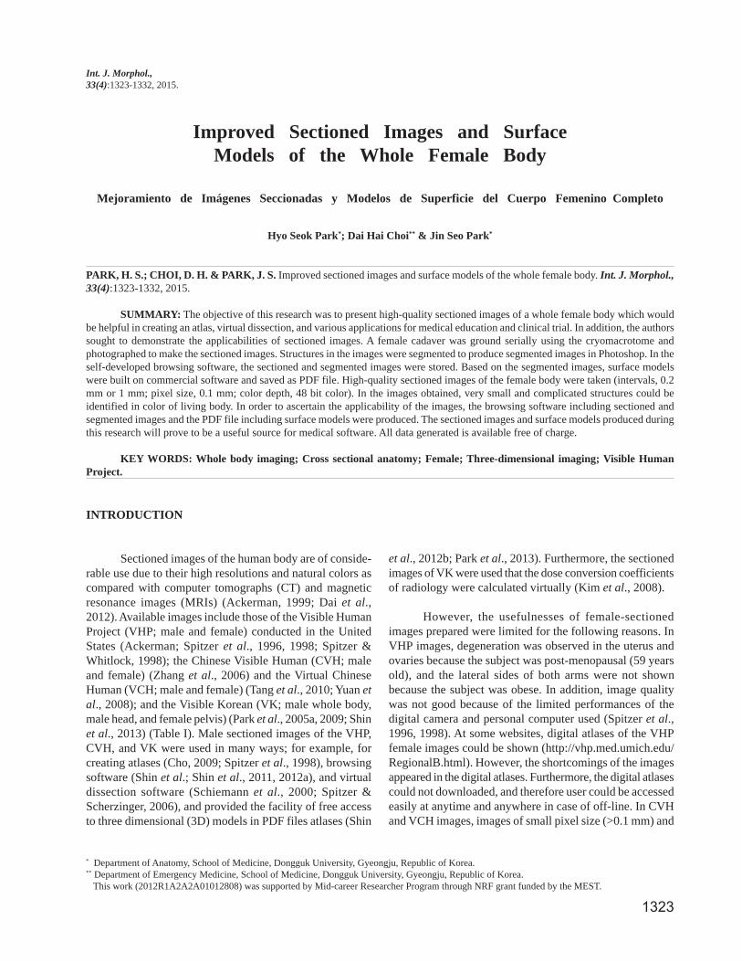
The subject was a Korean female cadaver aged 26years of standard body size (length, 1,690 mm; weight, 52kg) (Fig. 1a). After receiving consent from the family of thedeceased, the cadaver was scanned by computerizedtomography machine using a Philips Brilliance 64 channelCT scanner for pathological findings (Table II), and thenfrozen at -70 oC to prevent decay during a week. After thefrozen female cadaver with embedding agent (3% gelatinsolution containing 0.5% methylene blue) were put into anembedding box, the embedding box was refrozen at -70 oC.
For serial sectioning of the cadaver, a cryomacrotomewas used. The cryomacrotome used (width, 3 m x height, 4 mx length 5 m; 15 ton; HanwonTM of Republic of Korea) witha moving error of 0.001 mm enabled the whole body to beserially ground at a constant thickness, as previously described(Park et al., 2005b). Using the cryomacrotome, the frozenembedding box containing the cadaver was ground at 0.2 mm(from the vertex of the head and perineum; upper body) or at1.0 mm intervals (from under the perineum to the toes; lowerbody) to produce sectioned surfaces. After grinding, frost wasremoved using 10% ethyl alcohol and protruding denseconnective tissues were trimmed with a scalpel.
For photographing of the sectioned surfaces, a highperformance digital camera (CanonTM EOS-1Ds Mark IIITM
equipped with a CanonTM EF 50mm f/1.2L USM lens) wasused. This camera incorporated Canon's newest CMOS 21.1megapixel (effective) image sensor. The digital camera waslocated in front of the cryomacrotome, and two strobes(Digital S, ElinchromTM) connected to a power pack (Digital2, ElinchromTM), were positioned alongside the digitalcamera. The sectioned surfaces were photographed usingthe digital camera (ISO, 100; shutter speed, 1/200; apertureopening 9.0; manual focus). Images were checked and savedas in tagged image file format (TIFF) in Photoshop CS5version 12 (Adobe Systems, Inc., San Jose, CA, USA) at aresolution of 5,616 x 3,744. By repeating the proceduresectioned images of the whole body were obtained (Figs. 2and 3, Table II).
The next stage required post-processing of thesectioned images. Original images were re-sectioned on apersonal computer to produce coronal and sagittal imagesusing self developed software (Park et al., 2010). The coronal
Sectioned images Age Intervals (mm) Pixel size (mm) DrawbackVisible Human Project 59 0.33 0.33 Obese, menopausal
22 0.25–0.5 0.1–1 Embalmed, dyed21 0.1 --- Embalmed, dyed
Chinese Visible Human
25 0.2 0.2 Embalmed, dyedVirtual Chinese Human 19 0.2 0.2 Embalmed, dyedVisible Korean 43 0.1 0.1 Only pelvis
Table I. Comparison of female body sectioned images produced of each country.
Intervas Pixel size One TotalImages Region Resolution Number
(mm) (mm)Color depth
file size file size
CT Whole body 512_512 1,642 1.0 1.0 8 bit gray 257 KB 442 MBUpper body* 5,616 X 3,744 4,116 0.2 0.1 48 bit color 120 MB 484 GBSectioned imagesLower body§ 5,616 X 3,744 819 1.0 0.1 48 bit color 120 MB 96 GB
Segmented images Whole body 5,616 X 3,744 1,642 1.0 0.1 8 bit color 20 MB 32 GB
File format of all images was TIFF. *= Images of from the vertex of the head to perineum. §= Images from under the perineum to toe.
Table II. Features of CT, sectioned images, and segmented images.
PARK, H. S.; CHOI, D. H. & PARK, J. S. Improved sectioned images and surface models of the whole female body. Int. J. Morphol., 33(4):1323-1332, 2015.
1325
and sagittal images were then used to identify inconsistentalignments and brightness levels, and original images withthe problems were revised using the 'move' and 'Fouriertransform' commands in Photoshop.
Twenty-seven structures were chosen forsegmentation (Table III). After selecting sectioned imagesat 1 mm intervals, the boundaries of selected structures wereoutlined using the lasso or magnetic lasso tools in Photoshop,and the outlined structures were then filled with specificcolors (Park et al., 2005b; Park et al., 2010). Structure name,
Fig. 1. a) Koreanfemale cadaver andb) surface models.Real body shape isidentical with a skinsurface modelconstructed using thesectioned andsegmented images. c)Semitransparent skinwith inner organs andd) semitransparentbone with innerorgans are shownthrough surfacemodels in the PDFfile.
System StructuresSkeletal BonesAlimentary Pharynx, Esophagus, Stomach, Small intestine, Large intestine, Liver, GallbladderMuscular MusclesRespiratory Larynx, Trachea, LungsUrinary Kidneys, Urinary bladder, Female urethraGenital Ovaries, Uterine tubes, Uterus, VaginaCardiovascular HeartLymphoid SpleenNervous Spinal cord, Cerebrum, Cerebellum, BrainstemSensory EyeballsIntegumentary Skin
Table III. Twenty seven structures in the female body were segmented in sectioned images and reconstructedto build surface models.
PARK, H. S.; CHOI, D. H. & PARK, J. S. Improved sectioned images and surface models of the whole female body. Int. J. Morphol., 33(4):1323-1332, 2015.
and red, green, blue values constituting the color about everycolor-filled structure were saved as color.txt. Using thismethod, 1,642 segmented images of 27 structures wereobtained (resolution 5,616 x 3,744, color depth 8 bit; fileformat TIFF).
We used browsing software that was previouslydeveloped using the C# language of Microsoft VisualStudio.NET 2003 (Microsoft Corporation, Redmond, WA)composed of operating files with color.txt and image folders.The sectioned and segmented images were put into the soft-
1326
ware. If a user located the mouse pointer on a structure insectioned or segmented images, name of the structure wasseen as pop-up text (Park et al., 2013; Shin et al.; Shin etal., 2011, 2012b). In order to distribute the browsing soft-ware on-line, its file size was reduced by lowering theresolution of sectioned and segmented images from 5,616 x3,744 to 1,000 x 410. Furthermore, sectioned and segmentedimages representing 1.0 mm intervals were then stored inappropriate image folders. Operating files and new imagedata were then combined together using the NullsoftScriptable Install System of NSIS Media to produce aninstallation file (Fig. 4a).
Based on the segmented images, surface models ofthe segmented structures were built (Table III) on Mimicsversion 10.01 (Materialise, Leuven, Belgium). In the Mimics,a structure equivalent to a color in all segmented imageswas extracted, stacked, and reconstructed by surfacemodeling to create a surface model. In the same manner,other structures were reconstructed (Shin et al., 2009, 2013).After checking the shapes of the surface models, they weresaved as stereolithography (STL) files. Each structure in eachSTL file was then gathered and saved as a portable documentformat (PDF) file using 3D Reviewer accompanying soft-ware of Acrobat 9.0 Pro Extended. When the PDF file was
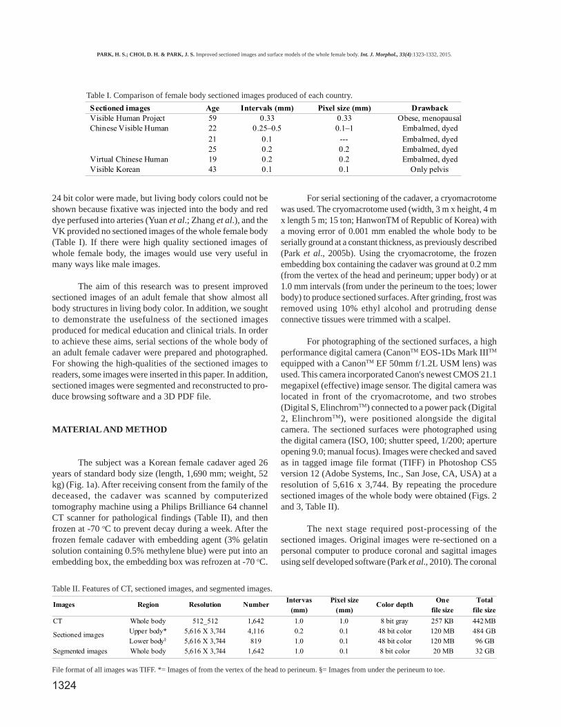
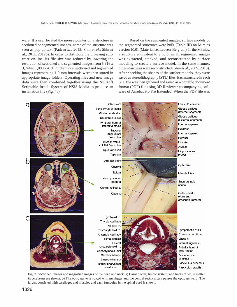
Fig. 2. Sectioned images and magnified images of the head and neck. a) Basal nuclei, limbic system, and tracts of white matterin cerebrum are shown. b) The optic nerve is coated with meninges and the central retina artery passes the optic nerve. c) Thelarynx consisted with cartilages and muscles and each funiculus in the spinal cord is shown.
PARK, H. S.; CHOI, D. H. & PARK, J. S. Improved sectioned images and surface models of the whole female body. Int. J. Morphol., 33(4):1323-1332, 2015.
1327
opened on Adobe Reader, the anatomical terms weredisplayed in the model tree window (Figs. 1 and 4b) (Parket al., 2013; Shin et al., 2012b).
RESULTS
A total of 1642 CT scans of the whole cadaver weretaken and carefully examined (Table II). Although the subjectdied of stomach cancer and pneumonia, stomach and lungshapes were relatively normal because she did not undergosurgery. Furthermore, her reproductive structures were nor-mal for a 26-year-old.
We prepared 4,116 sectioned images (pixel size 0.1mm) at 0.2 mm intervals of the upper body from the vertexof the head to the perineum; 819 images were prepared at1 mm intervals of the lower body from under the perineumto the toes. A total of 4,935 sectioned images of the wholebody were prepared (an image file size, 120 MB; total filesize, 580 GB; color depth, 48 bit color) (Figs. 2 and 3,Table II).
The sectioned images produced are of better imagequality than those produced during previous studies,including VHP, CVH, and VCH (Spitzer et al., 2008; Yuanet al.). The high quality of the sectioned images wasdescribed through the following examples.
In the cerebrum, the alveus and fimbria ofhippocampus are nerve fibers (white matter), and thus, appearlight. The hippocampus proper has many cell bodies (graymatter) and has a strong reddish color (Fig. 1a).
In the eyeball, the three layers, retina, choroid, andsclera, are observed. In the living body, a retina is naturallytransparent, and therefore, cannot be seen. However, in acadaver, the fibrous tissue of the retina is denatured, andthus, was semi-transparent. At the posterior chamber of theeyeball, the blind spot is observed as the optic disc createdby the absence of retina, choroid, and sclera. The maculalutea lies lateral to the optic disc (Fig. 2b).
In bilateral edges of the optic nerve, meninges (piamater) of dark color lines were apparent. In the junctionbetween the eyeball and optic nerve, the subarachnoid space
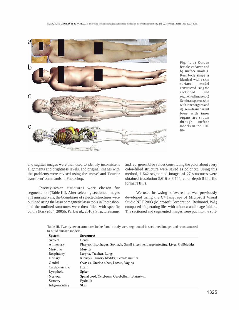
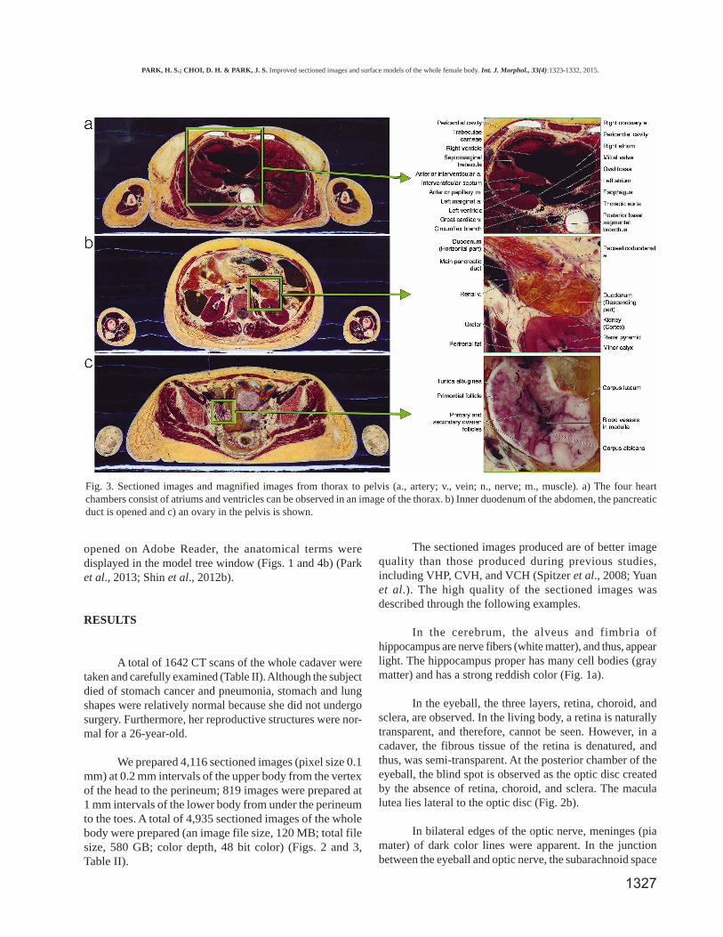
Fig. 3. Sectioned images and magnified images from thorax to pelvis (a., artery; v., vein; n., nerve; m., muscle). a) The four heartchambers consist of atriums and ventricles can be observed in an image of the thorax. b) Inner duodenum of the abdomen, the pancreaticduct is opened and c) an ovary in the pelvis is shown.
PARK, H. S.; CHOI, D. H. & PARK, J. S. Improved sectioned images and surface models of the whole female body. Int. J. Morphol., 33(4):1323-1332, 2015.
1328
is observed as an expanded space. In the inner optic nerves,the central retinal arteries supply blood to the eyeballs andare observed. In head images, the entire course of ophthalmicarteries was traced from the internal carotid artery to centralretinal artery in orbit (Fig. 2b).
In the larynx, cartilage and muscles are clearlyobserved due to their white and dark-red colors, respectively.Thyrohyoid muscle is attached in front of thyroid cartilage,and at its posterior, thyroarytenoid muscle pulls the arytenoidcartilage anteriorly. This cartilage is connected to cricoidcartilage by the lateral cricoarytenoid muscle. Furthermore,the space between the bilateral vocalis muscles is occupiedby the rima glottidis (Fig. 2c).
At the vertebral foramen of the cervical vertebra,gray and white matters of the spinal cord are clearlyidentified. In gray matter, the anterior horn is larger thanthe posterior horn because this region is at the sixth cervi-cal segment of the spinal cord, which includes many mo-tor neurons of the brachial plexus. In the white matter,
anterior, lateral, and posterior funiculi are distinctlydivided. Each funiculus is shown in detail; for example,the fasciculus gracilis and fasciculus cuneatus aredistinguishable in the posterior funiculus (Fig. 2c).
In the heart, atria and ventricles are evident. Theoval fossa appears as a thin wall between the atria. In theright ventricle, trabeculae carneae and a largerseptomarginal trabecula are observed at the inner wall ofthe interventricular septum. The mitral valve is locatedbetween the left atrium and left ventricle. The wall of theleft ventricle is thick due to a large amount of myocardium.Beyond the heart, coronary arteries are wrapped by avisceral layer of serous pericardium and frozen serous fluidof the pericardial cavity is observed in pericardium (Fig.3a).
The duodenum is divided into four parts in sectionedimages. The orifices of the main pancreatic duct and thebile duct of pancreas are present in its descending portion.The cortex and medulla of the kidney are distinct (Fig. 3b).
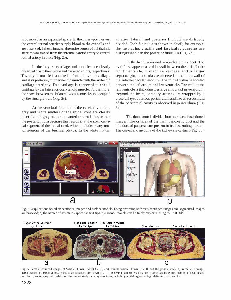
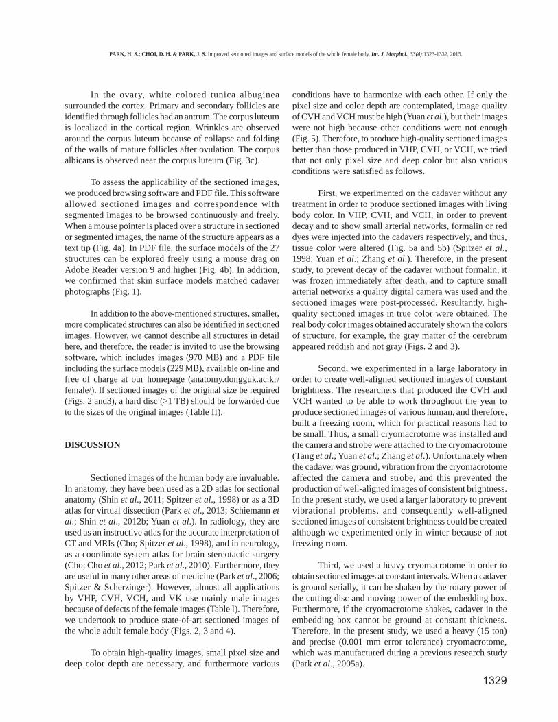
Fig. 4. Applications based on sectioned images and surface models. Using browsing software, sectioned images and segmented imagesare browsed; a) the names of structures appear as text tips. b) Surface models can be freely explored using the PDF file.
Fig. 5. Female sectioned images of Visible Human Project (VHP) and Chinese visible Human (CVH), and the present study. a) In the VHP image,degeneration of the genital organs due to an advanced age is evident. b) This CVH image shows a change in color caused by the injection of fixative andred dye. c) An image produced during the present study showing structures, including genital organs, at high definition in true color.
PARK, H. S.; CHOI, D. H. & PARK, J. S. Improved sectioned images and surface models of the whole female body. Int. J. Morphol., 33(4):1323-1332, 2015.
1329
In the ovary, white colored tunica albugineasurrounded the cortex. Primary and secondary follicles areidentified through follicles had an antrum. The corpus luteumis localized in the cortical region. Wrinkles are observedaround the corpus luteum because of collapse and foldingof the walls of mature follicles after ovulation. The corpusalbicans is observed near the corpus luteum (Fig. 3c).
To assess the applicability of the sectioned images,we produced browsing software and PDF file. This softwareallowed sectioned images and correspondence withsegmented images to be browsed continuously and freely.When a mouse pointer is placed over a structure in sectionedor segmented images, the name of the structure appears as atext tip (Fig. 4a). In PDF file, the surface models of the 27structures can be explored freely using a mouse drag onAdobe Reader version 9 and higher (Fig. 4b). In addition,we confirmed that skin surface models matched cadaverphotographs (Fig. 1).
In addition to the above-mentioned structures, smaller,more complicated structures can also be identified in sectionedimages. However, we cannot describe all structures in detailhere, and therefore, the reader is invited to use the browsingsoftware, which includes images (970 MB) and a PDF fileincluding the surface models (229 MB), available on-line andfree of charge at our homepage (anatomy.dongguk.ac.kr/female/). If sectioned images of the original size be required(Figs. 2 and3), a hard disc (>1 TB) should be forwarded dueto the sizes of the original images (Table II).
DISCUSSION
Sectioned images of the human body are invaluable.In anatomy, they have been used as a 2D atlas for sectionalanatomy (Shin et al., 2011; Spitzer et al., 1998) or as a 3Datlas for virtual dissection (Park et al., 2013; Schiemann etal.; Shin et al., 2012b; Yuan et al.). In radiology, they areused as an instructive atlas for the accurate interpretation ofCT and MRIs (Cho; Spitzer et al., 1998), and in neurology,as a coordinate system atlas for brain stereotactic surgery(Cho; Cho et al., 2012; Park et al., 2010). Furthermore, theyare useful in many other areas of medicine (Park et al., 2006;Spitzer & Scherzinger). However, almost all applicationsby VHP, CVH, VCH, and VK use mainly male imagesbecause of defects of the female images (Table I). Therefore,we undertook to produce state-of-art sectioned images ofthe whole adult female body (Figs. 2, 3 and 4).
To obtain high-quality images, small pixel size anddeep color depth are necessary, and furthermore various
conditions have to harmonize with each other. If only thepixel size and color depth are contemplated, image qualityof CVH and VCH must be high (Yuan et al.), but their imageswere not high because other conditions were not enough(Fig. 5). Therefore, to produce high-quality sectioned imagesbetter than those produced in VHP, CVH, or VCH, we triedthat not only pixel size and deep color but also variousconditions were satisfied as follows.
First, we experimented on the cadaver without anytreatment in order to produce sectioned images with livingbody color. In VHP, CVH, and VCH, in order to preventdecay and to show small arterial networks, formalin or reddyes were injected into the cadavers respectively, and thus,tissue color were altered (Fig. 5a and 5b) (Spitzer et al.,1998; Yuan et al.; Zhang et al.). Therefore, in the presentstudy, to prevent decay of the cadaver without formalin, itwas frozen immediately after death, and to capture smallarterial networks a quality digital camera was used and thesectioned images were post-processed. Resultantly, high-quality sectioned images in true color were obtained. Thereal body color images obtained accurately shown the colorsof structure, for example, the gray matter of the cerebrumappeared reddish and not gray (Figs. 2 and 3).
Second, we experimented in a large laboratory inorder to create well-aligned sectioned images of constantbrightness. The researchers that produced the CVH andVCH wanted to be able to work throughout the year toproduce sectioned images of various human, and therefore,built a freezing room, which for practical reasons had tobe small. Thus, a small cryomacrotome was installed andthe camera and strobe were attached to the cryomacrotome(Tang et al.; Yuan et al.; Zhang et al.). Unfortunately whenthe cadaver was ground, vibration from the cryomacrotomeaffected the camera and strobe, and this prevented theproduction of well-aligned images of consistent brightness.In the present study, we used a larger laboratory to preventvibrational problems, and consequently well-alignedsectioned images of consistent brightness could be createdalthough we experimented only in winter because of notfreezing room.
Third, we used a heavy cryomacrotome in order toobtain sectioned images at constant intervals. When a cadaveris ground serially, it can be shaken by the rotary power ofthe cutting disc and moving power of the embedding box.Furthermore, if the cryomacrotome shakes, cadaver in theembedding box cannot be ground at constant thickness.Therefore, in the present study, we used a heavy (15 ton)and precise (0.001 mm error tolerance) cryomacrotome,which was manufactured during a previous research study(Park et al., 2005a).
PARK, H. S.; CHOI, D. H. & PARK, J. S. Improved sectioned images and surface models of the whole female body. Int. J. Morphol., 33(4):1323-1332, 2015.
1330
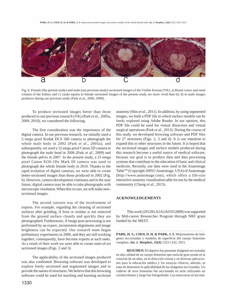
To produce sectioned images better than thoseproduced in our previous research (VK) (Park et al., 2005a,2009, 2010), we considered the following.
The first consideration was the importance of thedigital camera. In our previous research, we initially used a5 mega pixel Kodak DCS 560 camera to photograph thewhole male body in 2002 (Park et al., 2005a), andsubsequently, we used a 12 mega pixel Canon 5D camera tophotograph the male head in 2006 (Park et al., 2009) andthe female pelvis in 2007. In the present study, a 21-megapixel Canon EOS-1Ds Mark III camera was used tophotograph the whole female body in 2010. Thanks to therapid evolution of digital cameras, we were able to createbetter-sectioned images than those produced in 2002 (Fig.6). However, camera development continues and in the nearfuture, digital camera may be able to take photographs withmicroscopic resolution. When this occurs, we will make new-sectioned images.
The second concern was of the involvement ofexperts. For example, regarding the cleaning of sectionedsurfaces after grinding, if frost or residue is not removedfrom the ground surface cleanly and quickly they arephotographed. Furthermore, if image post-processing is notperformed by an expert, inconsistent alignments and imagebrightness can be expected. Our research team beganpreliminary experiments in 2000, and they are still workingtogether; consequently, have become experts at such tasks.As a result of their work we were able to create state-of-artsectioned images (Figs. 2 and 3).
The applicability of the sectioned images producedwas also confirmed. Browsing software was developed toexplore freely sectioned and segmented images and toprovide the names of structures. We believe that this browsingsoftware could be used for teaching and learning sectional
anatomy (Shin et al., 2011). In addition, by using segmentedimages, we built a PDF file in which surface models can befreely explored using Adobe Reader. In our opinion, thisPDF file could be used for virtual dissection and virtualsurgical operations (Park et al., 2013). During the course ofthis study, we developed browsing software and PDF filesfor 27 structures (Figs. 2, 3 and 4). It is our intention toexpand this to other structures in the future. It is hoped thatthe sectioned images and surface models produced duringthis research become a useful source of medical software,because our goal is to produce data and data processingsystems that contribute to the education of basic and clinicalmedicine. Recently, our data were used in the AnatomageTableTM (Copyright 2005© Anatomage, USA) of Anatomage(http://www.anatomage.com), which offers a life-sizeinteractive anatomy visualization table for use by the medicalcommunity (Chung et al., 2015).
ACKNOWLEDGEMENTS
This work (2012R1A2A2A01012808) was supportedby Mid-career Researcher Program through NRF grantfunded by the MEST.
PARK, H. S.; CHOI, D. H. & PARK, J. S. Mejoramiento de imá-genes seccionadas y modelos de superficie del cuerpo femeninocompleto. Int. J. Morphol., 33(4):1323-1332, 2015.
RESUMEN: El objetivo fue presentar imágenes seccionadasen alta calidad de un cuerpo femenino que sería de gran ayuda en lacreación de un atlas, en la disección virtual y en diversas aplicacio-nes para la educación médica y los ensayos clínicos; además, setrata de demostrar la aplicabilidad de las imágenes seccionadas. Uncadáver de sexo femenino fue seccionado en serie utilizando uncriomicrótomo y luego fue fotografiado. Las estructuras en las imá-
Fig. 6. Female (the present study) and male (our previous study) sectioned images of the Visible Korean (VK). a) Renal cortex and renalcolumn of the kidney and c) cauda equina in female sectioned images of the present study are more vivid than b); d) in male imagesproduces during our previous study (Park et al., 2006, 2009).
PARK, H. S.; CHOI, D. H. & PARK, J. S. Improved sectioned images and surface models of the whole female body. Int. J. Morphol., 33(4):1323-1332, 2015.

1331
genes fueron segmentadas para producir imágenes en Photoshop.En un programa de navegación de desarrollo propio se almacenaronlas imágenes seccionadas y segmentadas. Basado en las imágenessegmentadas, los modelos de superficie fueron construidas en elprograma y guardadas como archivo PDF. Las imágenes seccionadasde alta calidad del cuerpo femenino fueron tomadas con intervalosentre 0,2 o 1 mm; tamaño en píxeles de 0,1 mm y profundidad decolor de 48 bits). En las imágenes obtenidas, las estructuras muypequeñas y complicadas pudieron ser identificadas a color en el cuer-po. Con el fin de determinar la aplicabilidad de las imágenes, seprodujo un programa de navegación que incluye imágenesseccionadas y segmentadas y el archivo PDF que incluye modelosde superficie. Las imágenes seccionadas y los modelos de superfi-cie producidos durante esta investigación demostraron ser una fuen-te útil como programa médico. Todos los datos generados se en-cuentran disponibles gratuitamente.
KEY WORDS: Imagen de cuerpo completo; Anatomíaseccionada; Mujer; Imagen tridimensional; Proyecto humanovisible.
REFERENCES
Ackerman, M. J. The Visible Human Project: a resource foreducation. Acad. Med., 74(6):667-70, 1999.
Cho, Z. H. 7.0 Tesla MRI Brain Atlas. In Vivo Atlas withCryomacrotome Correlation. New York, Springer, 2009.
Cho, Z. H.; Calamante, F. & Chi, J. G. 7.0 Tesla MRI Brain WhiteMatter Atlas. Seoul, Panmun Book Company, 2012.
Chung, B. S.; Shin, D. S.; Brown, P.; Choi, J. & Chung, M. S.Virtual dissection table including the Visible Korean images,complemented by free software of the same data. Int. J.Morphol., 33(2):440-5, 2015.
Dai, J. X.; Chung, M. S.; Qu, R. M.; Yuan, L.; Liu, S. W. & Shin,D. S. The Visible Human Projects in Korea and China withimproved images and diverse applications. Surg. Radiol. Anat.,34(6):527-34, 2012.
Kim, C. H.; Choi, S. H.; Jeong, J. H.; Lee, C. & Chung, M. S.HDRK-Man: a whole-body voxel model based on high-resolution color slice images of a Korean adult male cadaver.Phys. Med. Biol., 53(15):4093-106, 2008.
Park, H. S.; Chung, M. S.; Shin, D. S.; Jung, Y. W. & Park, J. S.Accessible and informative sectioned images, color-codedimages, and surface models of the ear. Anat. Rec. (Hoboken),296(8):1180-6, 2013.
Park, J. S.; Chung, M. S.; Hwang, S. B.; Lee, Y. S.; Har, D. H. &Park, H. S. Visible Korean human: improved serially sectionedimages of the entire body. IEEE Trans. Med. Imaging,24(3):352-60, 2005a.
Park, J. S.; Chung, M. S.; Hwang, S. B.; Lee, Y. S. & Har, D. H.Technical report on semiautomatic segmentation using theAdobe Photoshop. J. Digit. Imaging, 18(4):333-43, 2005b.
Park, J. S.; Chung, M. S.; Hwang, S. B.; Shin, B. S. & Park, H. S.Visible Korean Human: its techniques and applications. Clin.Anat., 19(3):216-24, 2006.
Park, J. S.; Chung, M. S.; Shin, D. S.; Har, D. H.; Cho, Z. H.; Kim,Y. B.; Han, J. Y. & Chi, J. G. Sectioned Images of the CadaverHead Including the Brain and Correspondences With UltrahighField 7.0 T MRIs. Proc. IEEE, 97(12):1988-96, 2009.
Park, J. S.; Chung, M. S.; Chi, J. G.; Park, H. S. & Shin, D. S.Segmentation of cerebral gyri in the sectioned images byreferring to volume model. J. Korean Med. Sci., 25(12):1710-5, 2010.
Quackenbush, D.; Ratiu, P. & Kerr, J. Segmentation of the VisibleHuman datasets. The Visible Human Project ConferenceProceedings, October 7-8, 1996.
Schiemann, T.; Freudenberg, J.; Pflesser, B.; Pommert, A.;Priesmeyer, K.; Riemer, M.; Schubert, R.; Tiede, U. & Höhne,K. H. Exploring the Visible Human using the VOXEL-MANframework. Comput. Med. Imaging Graph., 24(3):127-32,2000.
Shin, D. S.; Chung, M. S.; Lee, J. W.; Park, J. S.; Chung, J.; Lee, S.B. & Lee, S. H. Advanced surface reconstruction technique tobuild detailed surface models of the liver and neighboringstructures from the Visible Korean Human. J. Korean Med.Sci., 24(3):375-83, 2009.
Shin, D. S.; Chung, M. S.; Park, H. S.; Park, J.S. & Hwang, S. B.Browsing software of the Visible Korean data used for teachingsectional anatomy. Anat. Sci. Educ., 4(6):327-32, 2011.
Shin, D. S.; Jang, H. G.; Hwang, S. B.; Har, D. H.; Moon, Y. L. &Chung, M. S. Two-dimensional sectioned images and three-dimensional surface models for learning the anatomy of thefemale pelvis. Anat. Sci. Educ., 6(5):316-23, 2013.
Shin, D. S.; Jang, H. G.; Park, J. S.; Park, H. S.; Lee, S. & Chung,M. S. Accessible and informative sectioned images and surfacemodels of a cadaver head. J. Craniofac. Surg.23(4):1176-80,2012a.
Shin, D. S.; Park, J. S.; Park, H. S.; Hwang, S. B. & Chung, M. S.Outlining of the detailed structures in sectioned images fromVisible Korean. Surg. Radiol. Anat., 34(3):235-47, 2012b.
Spitzer, V.; Ackerman, M. J.; Scherzinger, A. L. & Whitlock, D.The visible human male: a technical report. J. Am. Med. Inform.Assoc., 3(2):118-30, 1996.
Spitzer, V. M. & Scherzinger, A. L. Virtual anatomy: an anatomist'splayground. Clin. Anat., 19(3):192-203, 2006.
PARK, H. S.; CHOI, D. H. & PARK, J. S. Improved sectioned images and surface models of the whole female body. Int. J. Morphol., 33(4):1323-1332, 2015.
1332
Spitzer, V. M.; Whitlock, D. G. & National Library of Medicine(U. S.). Atlas of the Visible Human Male: Reverse Engineeringof the Human Body. Massachusetts, Jones & Bartlett Publishers,1998.
Spitzer, V. M. & Whitlock, D. G. The Visible Human Dataset: theanatomical platform for human simulation. Anat Rec.,253(2):49-57, 1998.
Tang, L.; Chung, M. S.; Liu, Q. & Shin, D. S. Advanced featuresof whole body sectioned images: Virtual Chinese Human. Clin.Anat., 23(5):523-9, 2010.
Yuan, Y.; Qi, L. & Luo, S. The reconstruction and application ofvirtual Chinese human female. Comput. Methods ProgramsBiomed., 92(3):249-56, 2008.
Zhang, S. X.; Heng, P. A. & Liu, Z. J. Chinese visible human project.Clin. Anat., 19(3):204-15, 2006.
Correspondence to:Jin Seo ParkDepartment of AnatomyDongguk University School of Medicine87 Dongdae-ro, Gyeongju, 780-350REPUBLIC OF KOREA
Tel: +82-54-770-2402Fax: +82-54-770-2402
Email: [email protected]
Received: 09-07-2015Accepted: 04-08-2015
PARK, H. S.; CHOI, D. H. & PARK, J. S. Improved sectioned images and surface models of the whole female body. Int. J. Morphol., 33(4):1323-1332, 2015.