Embed Size (px)
Citation preview

A peer-reviewed quarterly journal
October 2015Volume 13, Supplement 1
EL
SEV
IER
TH
E O
CU
LA
R SU
RFA
CE
V
olum
e 13 Sup
plem
ent 1 O
ctober 2015
www.theocularsurface.com
Indexed in MEDLINE/PubMed and EMBASE
Affiliated with the Tear Film & Ocular Surface Society
Special SupplementImproving Diagnosis and Outcomes of Sjögren’s
Disease Th rough Targeting Dry Eye PatientsA Continuing Medical Education Enduring Material
Contributing Authors Committee:Gary N. Foulks, MD (Chair)
Vatinee Y. Bunya, MDKatherine M. Hammitt, MA
Kelly K. Nichols, OD, MPH, PhDStephen C. Pfl ugfelder, MD
Frederick B. Vivino, MS, MD, FACR
Th is CME enduring material is available online at: http://www.theocularsurfacejournal.com/issue/S1542-0124(15)X0002-4.
A Continuing Medical Education Enduring Material
Target Audience Th is educational activity is intended for ophthalmologists, ophthalmologists in residency or fellowship training, and optometrists.
Learning ObjectivesUpon completion of this activity, participants will be able to:
(1) Recognize signs and symptoms suggestive of Sjögren’s disease among patients with dry eye disease.(2) Conduct a review of systems to ascertain risk for Sjögren’s disease among patients with dry eye. (3) Diagnose Sjögren’s disease on the basis of recognized consensus criteria developed by major
organizations and/or make appropriate referrals for multisystem evaluation and management.(4) Utilize antibody testing in the workup for Sjögren’s disease and potentially incorporate it into clinical prac-
tice for select dry eye patients.(5) Manage ocular manifestations of Sjögren’s disease.(6) Access practical resources for Sjögren’s disease diagnosis and management from the Sjögren’s Syndrome
Foundation and other sources.
JTOS_v13_i4_sS_COVER.indd 1JTOS_v13_i4_sS_COVER.indd 1 30-09-2015 16:49:0230-09-2015 16:49:02

Contributing Authors Committee
Gary N. Foulks, MD (Chair)Vatinee Y. Bunya, MDKathy Hammitt, MAKelly K. Nichols, OD, PhD, MPHStephen C. Pfl ugfelder, MDFrederick B. Vivino, MD
Gary N. Foulks, MD, is Professor Emeritus, Department of Ophthalmology & Visual Sciences, University of Louisville, Louisville, KY.Vatinee Y. Bunya, MD, is co-director of the Penn Dry Eye and Ocular Surface Center and an assistant professor of ophthalmology at the Hospital of the University of Pennsylvania, Philadelphia, PA.Kathy Hammitt MA, is Vice President of Research, Sjögren’s Syndrome Foundation, Bethesda, MD.Kelly K. Nichols, OD, PhD, MPH, is Dean of the University of Alabama at Birmingham School of Optometry, an executive board member for the Tear Film and Ocular Surface Society, and a founding member of Ocular Surface Society of Optometry.Stephen C. Pfl ugfelder, MD, is professor and holder of the James and Margaret Elkins Chair in ophthalmology at the Baylor College of Medicine, Houston, TX.Frederick B. Vivino, MD, is the chief of rheumatology and professor of clinical medicine at the Penn Presbyterian Medical Center and director of the Penn Sjögren’s Syndrome Center, Philadelphia, PA.
Improving Diagnosis and Outcomes of Sjögren’s Disease Th rough Targeting Dry Eye Patients is jointly sponsored by Candeo Clinical/Science Communications, LLC, and the University of Florida College of Medicine. Th is publication is administered by an independent editorial board and supported by an unrestricted educational grant from Bausch and Lomb/Nicox Inc.
Copyright 2015 Candeo Clinical/Science Communications, LLC. All rights reserved. Neither the University of Florida nor Candeo Clinical/Science Communications, LLC, assume any responsibility for injury or damage to persons or property arising from the use of information or ideas contained in this publication.
Course Director Anup Kubal, MDUniversity of FloridaGainesville, FL, USA
JTOS_v13_i4_sS_COVER.indd 3JTOS_v13_i4_sS_COVER.indd 3 30-09-2015 16:49:0430-09-2015 16:49:04

Special Supplement
Improving Diagnosis and Outcomes of Sjögren’s Disease Th rough Targeting Dry Eye Patients
A Continuing Medical Education Enduring Material
SECTION I. INTRODUCTION
SECTION II. A DRY EYE DISEASE TAXONOMY
SECTION III. SJÖGREN’S DISEASE: THE DISEASE STATE
SECTION IV. DIAGNOSIS OF DRY EYE DISEASE
SECTION V. AN ALGORITHM FOR SJÖGREN’S DISEASE DIAGNOSIS
SECTION VI. SJÖGREN’S DISEASE MANAGEMENT
SECTION VII. THE SJÖGREN’S SYNDROME FOUNDATION: A RESOURCE FOR DOCTORS AND PATIENTS
Contributing Authors Committee:
Gary N. Foulks, MD (Chair)Vatinee Y. Bunya, MD
Katherine M. Hammitt, MAKelly K. Nichols, OD, MPH, PhD
Stephen C. Pfl ugfelder, MDFrederick B. Vivino, MS, MD, FACR
Th is CME enduring material is available online at: http://www.theocularsurfacejournal.com/issue/S1542-0124(15)X0002-4.

Statement of NeedTh e last 5 years have seen a series of signifi cant advances in the diagnosis and treatment of ocular surface disease. However, most of the recent work on ocular surface disease has centered on locally caused conditions—that is, dry eye disease secondary to infectious or infl ammatory disease of the lids and/or lacrimal glands. While this accounts for most of the dry eye seen in the clinic, dry eye produced by systemic infl ammatory diseases such as Sjogren’s syndrome and rheumatoid arthritis still accounts for about 10% of all dry eye; and perhaps more im-portant, the lion’s share of the severe dry eye is caused by an underlying systemic infl ammatory disease.1
Sjogren’s syndrome accounts for roughly 90% of the systemically caused dry eye disease.2 In addition to of-ten severe dry eye, Sjogren’s syndrome cause xerostomia and dryness elsewhere in the body.3 Although a relative-ly common condition, the disease is underdiagnosed and oft en misdiagnosed. Typically, 5 years elapse between the time a patient notices symptoms and a diagnosis is made. Th is is a serious matter, because, when properly diagnosed, a multispecialty team can be assembled—consisting of an ophthalmologist, rheumatologist, and a dentist or oral surgeon. Care provided early can slow the disease process and make the patient more comfortable.
Although early diagnosis is clearly important, the detection of Sjogren’s syndrome has been hampered by, among other things, the lack of clear diagnostic test or a pathognomonic sign. Recently, however, serum markers have been isolated that promise to make diagnosis much more straightforward. Aimed at clinicians, this work presents an algorithm to guide diagnosis from dry eye presentation through to fi nal diagnosis of a systemic dry eye condition. Emphasis is placed on patient selection for Sjogren’s syndrome testing. In addition, the publication reviews recent work on Sjogren’s syndrome biomarkers and the treatment of ocular Sjogren’s syndrome.
References1. Akpek EK, Lindsley KB, Adyanthaya RS, et al. Treatment of Sjögren’s syndrome-associated dry eye an
evidence-based review. Ophthalmology. 2011 Jul;118(7):1242-52.2. Grayzel AI: What is Sjogren’s? In: Wallace, DJ, ed. Th e Sjogrens Book. 4th ed. New York, NY: Oxford Univer-
sity Press; 2012:7-10.3. Segal B, Bowman SJ, Fox PC, et al. Primary Sjögren’s Syndrome: health experiences and predictors of health
quality among patients in the United States. Health Qual Life Outcomes. 2009;7:46.doi:10.1186/1477-7525-7-46.
Off -label Use Statement Th is work discusses both on-label and off -label uses of drugs and devices.
General InformationTh is CME activity is sponsored by the University of Florida College of Medicine and is supported by an unre-stricted educational grant from Bausch + Lomb/Nicox Inc.
Directions: Select one answer to each question in the exam (questions 1–30) and in the evaluation (ques-tions 31–36). Th e University of Florida College of Medicine designates this enduring material for a maximum of 3.0 AMA PRA Category 1 Credits™. Th ere is no fee to participate in this activity. In order to receive CME credit, participants should read the report, and then take the post-test. A score of 80% is required to qualify for CME credit. Estimated time to complete the activity is 3 hours. On completion, tear out or photocopy the answer sheet and send it to:
University of Florida CME Offi cePO Box 100233, Gainesville, FL 32610-0233phone: 352-733-0064 fax: 352-733-0007Or, you can take the test online at http://cme.ufl .edu/ed/self-study/sjogrens-disease-dry-eye-supplement/.
System requirements for this activity are: For PC users: Windows® 2000, XP, 2003 Server, or Vista; Internet Ex-plorer® 6.0 or newer, or Mozilla® Firefox® 2.0 or newer (JavaScript™ and Java™ enabled). For Mac® users: Mac OS® X 10.4 (Tiger®) or newer; Safari™ 3.0 or newer, Mozilla® Firefox® 2.0 or newer; (JavaScript™ and Java™ enabled). Internet connection required: Cable modem, DSL, or better.

Date of Original Release October 2015. Approved for a period of 12 months.
Accreditation Statement Th is activity has been planned and implemented in accordance with the accreditation requirements and policies of the Accreditation Council for Continuing Medical Education (ACCME) through the joint providership of the University of Florida College of Medicine and Candeo Clinical/Science Communications, LLC. Th e University of Florida College of Medicine is accredited by the ACCME to provide continuing medical education for physicians.
Credit Designation Statement Th e University of Florida College of Medicine designates this enduring material for a maximum of 3.0 AMA PRA Category 1 Credits™. Physicians should claim only the credit commensurate with the extent of their participation in the activity.
Faculty Disclosure StatementsDr. Vatinee Bunya, MD is the co-director of the Penn Dry Eye & Ocular Surface Center and an assistant professor of ophthalmology at the hospital of the University of Pennsylvania. She states that in the past 12 months, she has not had a fi nancial relationship with any commercial organization that produces, markets, re-sells, or distributes healthcare goods or services consumed by or used on patients.
Gary Foulks, MD is an emeritus professor of ophthalmology at the University of Louisville School of Medi-cine. He is a consultant for Lexitas Pharmaceuticals, Kala Pharmaceuticals, Ocular Th erapeutix, Inc., and Parion Sciences. He is also a stock shareholder for TearLab Corporation.
Kathy Hammitt is the vice president of research at the Sjögren’s Syndrome Foundation. She states that in the past 12 months, she has not had a fi nancial relationship with any commercial organization that produces, markets, re-sells, or distributes healthcare goods or services consumed by or used on patients.
Kelly K. Nichols, OD, MPH, PhD, FAAO, is dean of the University of Alabama at Birmingham School of Optometry and a leading vision scientist in the area of dry eye disease. Nichols has received grant and research support from Allergan, Alcon, Johnson & Johnson (spouse), Kala Pharmaceuticals, and TearScience. Nichols is a consultant for Allergan, Alcon, Bausch + Lomb, Nicox, InSite Vision Incorporated, and Kala Pharmaceuticals. She is also a stock shareholder for TearLab (non-exercised options).
Stephen C. Pfl ugfelder, MD, is professor and holder of the James and Margaret Elkins Chair in ophthalmol-ogy at the Baylor College of Medicine. Pfl ugfelder has received grant and research support from Allergan. He is also a consultant for Allergan.
Frederick B. Vivino, MD, is the chief of rheumatology at the Penn Presbyterian Medical Center and director of the Sjögren’s Syndrome Center in Philadelphia. Vivino has received grant and research support from Novartis. He is also a consultant for Biogen Idec, Nicox Inc., and Takeda Pharmaceuticals.
Disclaimer Participants have an implied responsibility to use the newly acquired information to enhance pa-tient outcomes and professional development. Th e information presented in this activity is not meant to serve as a guideline for patient care. Procedures, medications, and other courses of diagnosis and treatment discussed or suggested in this activity should not be used by clinicians without evaluation of their patients’ conditions and possible contraindications or dangers in use, applicable manufacturer’s product information, and comparison with recommendations of other authorities.
Commercial SupportersTh is activity is supported by an unrestricted educational grant from Bausch + Lomb/Nicox Inc.
Th is CME enduring material is available online at: http://www.theocularsurfacejournal.com/issue/S1542-0124(15)X0002-4.

Special Supplement
Improving Diagnosis and Outcomes of Sjögren’sDisease through Targeting Dry Eye Patients: A
Continuing Medical Education Enduring Material
I. INTRODUCTIONREFERENCES1. Grayzel AI. What is Sjogren’s? In: Wallace DJ, ed. The Sjogrens Book. 4th
ed. New York, NY: Oxford University Press; 2012. p. 7-102. Segal B, Bowman SJ, Fox PC, et al. Primary Sjögren’s Syndrome: health
experiences and predictors of health quality among patients in theUnited States. Health Qual Life Outcomes 2009;7:46. http://dx.doi.org/10.1186/1477-7525-7-46
S jögren’s disease, or Sjögren’s syndrome as the con-dition has until recently been known, is a commonautoimmune disorder.1 An inflammatory condition
that affects many parts of the body but particularly theexocrine glands, Sjögren’s disease causes dryness of mucosalsurfaces, most frequently those of the eyes and mouth. Insurveys conducted by the Sjögren’s Syndrome Foundation(SSF), dry eye has consistently been named the feature ofSjögren’s disease that disturbs patients’ quality of life themost. Dry eye symptoms are typically what motivate Sjög-ren’s disease patients to seek medical attention, with theresult that eyecare practitioners are the ones who most ofteninitially encounter Sjögren’s disease.2
The dry eye of Sjögren’s disease can produce significantocular discomfort. Patients complain of sensations of gritti-ness, stinging, and burning, as well as visual disturbances,particularly when reading or performing intense near visiontasks. Historically, however, patients have faced long delaysin diagnosis of the disease, with some studies suggesting thatit can take as long as 7 years from onset of symptoms todiagnosis.2 Although this delay is due in part to the subtle,confusing signs and symptoms of early Sjögren’s disease,the lack of awareness in both the general and medicalcommunities regarding the frequency and morbidity of thedisease contributes to this delay. Early diagnosis of Sjögren’sdisease is critical so that patients can be effectively treatedand monitored for serious complications includinglymphoma.
Since eyecare providers are often the first medical profes-sionals to encounter patients with Sjögren’s disease, it isimperative that they be alert to the possibility of the diseasein any patient who presents with a dry eye. Because Sjögren’sdisease affects multiple body systems, eyecare professionalsshould have close working relationships with other healthcare providersdincluding oral health professionals, endocri-nologists, and especially rheumatologistsdto assurecomprehensive, collaborative care.
In the following pages, we review our current under-standing of dry eye disease (DED), including its relationshipto Sjögren’s disease. We discuss the methods available to di-agnose DED and some of the diagnostic challenges that
© 2015 Elsevier Inc. All rights reserved.
THE OCULAR SURFACE / OCTOBER 2015, VO
eyecare practitioners face. Because Sjögren’s disease repre-sents a significant but often overlooked subset of DED pa-tients, we offer a protocol for selecting those who shouldbe identified for additional testing as potential Sjögren’s dis-ease patients. Finally, we offer an approach to managementof Sjögren’s disease patients and describe resources availableto them.
Our goal throughout is to provide tools that will enableeyecare practitioners to more readily identify Sjögren’s andother autoimmune disease patients in the DED population.Earlier initiation of appropriate treatment can reducesuffering, improve quality of life, and lead to betteroutcomes.
II. A DED TAXONOMYThe past two decades have witnessed an explosion of
research in DED, with growing awareness of it as a signifi-cant cause of ocular morbidity. Numerous expert consortiahave attempted to gather and distill evidence-based dataabout DED into clinically useful classification schemes andrecommendations for care. As always, guidelines are merelyguidelines. However, due to the complex nature of DED eti-ology, the multiplicity of diagnostic tools and schema, andan ever-evolving knowledge base, research summaries andpractice recommendations continue to provide an importantbasis for communication within this young field. Highlightsfrom these and other sources are presented below.
A. Definition and Scope of DEDThe 2007 International Dry Eye Workshop (DEWS)
report defined DED as “a multifactorial disease of the tearsand ocular surface that results in symptoms of discomfort, vi-sual disturbance, and tear film instability with potential dam-age to the ocular surface. It is accompanied by increasedosmolarity of the tear film and inflammation of the ocularsurface.”1 Consistent with the DEWS report, the AmericanAcademy of Ophthalmology (AAO) Preferred PracticePattern Guidelines of 2013 separate DED into two main
L. 13 NO. 4S / www.theocularsurface.com S1

OUTLINE
I. Introduction
II. A Ded Taxonomy
A. Definition and Scope of DED
B. Prevalence of DED
C. DED Morbidity
D. Clinical Presentation of DED
E. Forms of DED
F. Aqueous Deficient DED
G. Evaporative DED
H. Primary DED Treatment Options
III. Sjögren’s Disease: The Disease State
A. Epidemiology
B. Presentations and Morbidity: Ocular Signs andSymptoms
C. Oral Manifestations
D. Other Xeroses
E. Systemic or Extraglandular Involvement
F. Lymphoproliferative/Myeloproliferative
G. Etiopathogenesis
IV. Diagnosis of Ded
A. Symptoms
B. DED Examination and Testing
C. External Examination
D. Tear Quantity and Quality
E. Tear Flow and Turnover
F. Tear Composition
G. Tear Proteins
H. Ocular Surface Dye Staining
I. Rose Bengal
J. Lissamine Green
K. Meibography and Proteomic Analysis
V. An Algorithm for Sjögren’s Disease Diagnosis
A. Current State of Antibody Testing for Sjögren’sDisease
B. Novel Autoantibiodies
C. Diagnostic Importance of Novel Antibodies
D. Novel Autoantibodies at a Glance
1. Specific Antibodies (Table V-1)
2. The Sjö�3. Sjö� Test Interpretation
4. Sjö� Test Evaluation
E. The Diagnostic Algorithm
VI. Sjögren’s Disease Management
A. Patient Management Team
B. Established Therapies
C. Artificial Tears and Lubricants
D. Ophthalmic Inserts
E. Autologous Serum Tears
F. Topical Ophthalmic Cyclosporine 0.05%
G. Topical Corticosteroids
H. Dietary Modalities
I. Antiinflammatory Diet
J. Punctal Occlusion
K. Systemic Agents
L. Systemic Immunomodulators
M. Other Methods
N. New Biologic Agents (In Trials)
1. Anti-CD20 (B-cell)
2. Anti-TNF
3. Anti-CD80/86
4. Anti-BLyS
VII. The Sjögren’s Syndrome Foundation: A Resource forDoctors and Patients
A. Major SSF Initiatives
1. Shorter Time to Diagnosis
2. Better Diagnostics
3. Improved Clinical Trials
4. Dry Eye Initiatives
5. Clinical Practice Guidelines
6. Access to Social Security Disability
B. SSF Resources for clinicians and patient
C. Looking Ahead, Together
CME PUBLICATION: SJÖGREN'S SYNDROME / SPECIAL SUPPLEMENT
S2 THE OCULAR SURFACE / OCTOBER 2015,
categories: DED due to “reduced tear production,” and DEDdue to “excessive tear evaporation.”2
Practice guidelines from the America Optometric Asso-ciation (AOA) refer to “dry eye” as any condition that resultsfrom “aqueous deficiency or disruption” and acknowledgethat different ocular surface disorders related to dry eyecan “overlap as well as coexist.”3 Dry eye is considered treat-able but generally not curable.2
Perhaps the best way to characterize DED is as a break-down or disruption of some aspect of the lacrimal functionalunit, which is comprised of the conjunctiva, cornea, lacrimaland meibomian glands, lacrimal drainage system, preoculartear film, and reflexive neural connections.4 An intact lacrimalfunctional unit maintains homeostasis of the ocular surfacevia the production, distribution, and drainage of a clear,balanced tear film. Dysfunction of the lacrimal functionalunit at any level can initiate a cascade of downstream eventsand provide an initial impetus for the development of DED.
B. Prevalence of DEDAs there is no single test (nor universally accepted
sequence of tests) for the diagnosis of DED, and aspatient-reported symptoms are often poorly concordantwith objective assessments, the exact prevalence of DED isdifficult to ascertain. However, using the most restrictivedefinition of DED, nearly 5 million individuals in the USsuffer from the condition, and many millions more presum-ably have subclinical or intermittent expressions of the dis-ease.5 Employing various diagnostic criteria andinvestigating diverse populations, different surveys haveshown between 5% and 33% DED prevalence, with thegreatest prevalence found among Asian, female, and olderpopulations.5 Among proposed risk factors for DED, those
VOL. 13 NO. 4S / www.theocularsurface.com
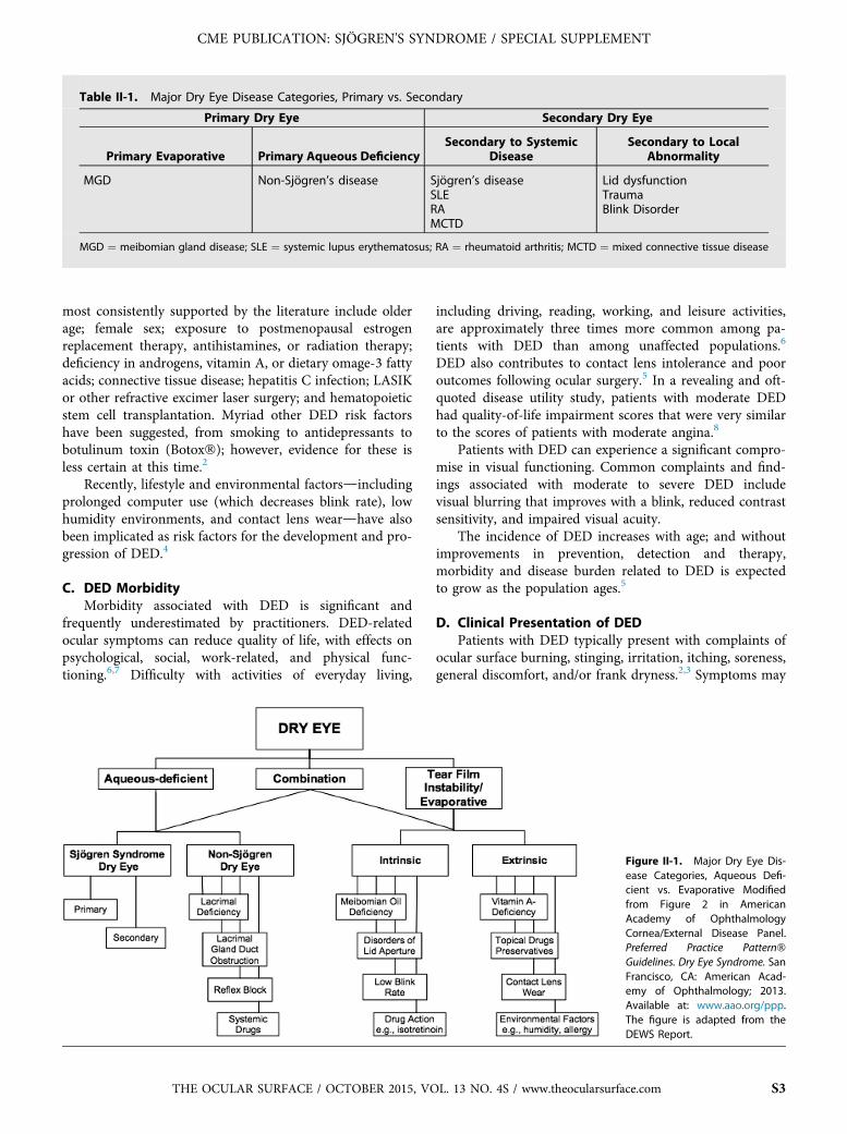
Table II-1. Major Dry Eye Disease Categories, Primary vs. Secondary
Primary Dry Eye Secondary Dry Eye
Primary Evaporative Primary Aqueous DeficiencySecondary to Systemic
DiseaseSecondary to Local
Abnormality
MGD Non-Sjögren’s disease Sjögren’s diseaseSLERAMCTD
Lid dysfunctionTraumaBlink Disorder
MGD ¼ meibomian gland disease; SLE ¼ systemic lupus erythematosus; RA ¼ rheumatoid arthritis; MCTD ¼ mixed connective tissue disease
CME PUBLICATION: SJÖGREN'S SYNDROME / SPECIAL SUPPLEMENT
most consistently supported by the literature include olderage; female sex; exposure to postmenopausal estrogenreplacement therapy, antihistamines, or radiation therapy;deficiency in androgens, vitamin A, or dietary omage-3 fattyacids; connective tissue disease; hepatitis C infection; LASIKor other refractive excimer laser surgery; and hematopoieticstem cell transplantation. Myriad other DED risk factorshave been suggested, from smoking to antidepressants tobotulinum toxin (Botox�); however, evidence for these isless certain at this time.2
Recently, lifestyle and environmental factorsdincludingprolonged computer use (which decreases blink rate), lowhumidity environments, and contact lens weardhave alsobeen implicated as risk factors for the development and pro-gression of DED.4
C. DED MorbidityMorbidity associated with DED is significant and
frequently underestimated by practitioners. DED-relatedocular symptoms can reduce quality of life, with effects onpsychological, social, work-related, and physical func-tioning.6,7 Difficulty with activities of everyday living,
THE OCULAR SURFACE / OCTOBER 2015, VO
including driving, reading, working, and leisure activities,are approximately three times more common among pa-tients with DED than among unaffected populations.6
DED also contributes to contact lens intolerance and pooroutcomes following ocular surgery.5 In a revealing and oft-quoted disease utility study, patients with moderate DEDhad quality-of-life impairment scores that were very similarto the scores of patients with moderate angina.8
Patients with DED can experience a significant compro-mise in visual functioning. Common complaints and find-ings associated with moderate to severe DED includevisual blurring that improves with a blink, reduced contrastsensitivity, and impaired visual acuity.
The incidence of DED increases with age; and withoutimprovements in prevention, detection and therapy,morbidity and disease burden related to DED is expectedto grow as the population ages.5
D. Clinical Presentation of DEDPatients with DED typically present with complaints of
ocular surface burning, stinging, irritation, itching, soreness,general discomfort, and/or frank dryness.2,3 Symptoms may
Figure II-1. Major Dry Eye Dis-ease Categories, Aqueous Defi-cient vs. Evaporative Modifiedfrom Figure 2 in AmericanAcademy of OphthalmologyCornea/External Disease Panel.Preferred Practice Pattern�Guidelines. Dry Eye Syndrome. SanFrancisco, CA: American Acad-emy of Ophthalmology; 2013.Available at: www.aao.org/ppp.The figure is adapted from theDEWS Report.
L. 13 NO. 4S / www.theocularsurface.com S3

Table II-2. Dry Eye Severity Grading Scheme
Dry Eye Severity Level 1 2 3 4*
Discomfort, severity &frequency
Mild and/or episodic;occurs under environ-mental stress
Moderate episodic orchronic, stress or nostress
Severe frequent orconstant without stress
Severe and/ordisabling and constant
Visual symptoms None or episodic mildfatigue
Annoying and/or activ-ity limiting, episodic
Annoying, chronic and/or constant, limitingactivity
Constant and/orpossibly disabling
Conjunctival injection None to mild None to mild þ/- þ/þþConjunctival staining None to mild Variable Moderate to marked Marked
Corneal staining(severity/location)
None to mild Variable Marked central Severe punctateerosions
Corneal/tear signs None to mild Mild debris, Ymeniscus
Filamentary keratitis,mucus clumping, [tear debris
Filamentary keratitis,mucus clumping, [tear debris, ulceration
Lid/meibomian glands MGD variably present MGD variably present Frequent Trichiasis, keratiniza-tion, symblepharon
TFBUT (sec) Variable �10 �5 Immediate
Schirmer score (mm/5min)
Variable �10 �5 �2
* Must have signs AND symptoms.
TFBUT ¼ fluorescein tear break-up time; MGD ¼ eibomian gland disease
Reproduced with permission from Lemp MA (Chair). Definition and Classification Subcommittee of the International Dry Eye Workshop. The
definition and classification of dry eye disease: report of the Definition and Classification Subcommittee of the International Dry Eye Workshop
(2007). Ocul Surf 2007;5:88.
Modified from Table A5-1 in American Academy of Ophthalmology Cornea/External Disease Panel. Preferred Practice Pattern� Guidelines. Dry Eye
Syndrome. San Francisco, CA: American Academy of Ophthalmology; 2013. Available at: www.aao.org/ppp. The table is adapted from the DEWS
Report.
CME PUBLICATION: SJÖGREN'S SYNDROME / SPECIAL SUPPLEMENT
be noticeable or worsened under circumstances of prolongedvisual focus (eg, computer work or reading), especially in thelater hours of the day. Symptoms may also worsen under theinfluence of certain medications (eg, antihistamine-containing cold or allergy remedies) or under adverseenvironmental conditions, such as air conditioning or aparticularly dry climate. In these circumstances, patientsmay experience an increasing need for topical ocular lubri-cating agents (eg, artificial tears) for symptom relief.2
Early in the course of disease, an insufficient and/or un-stable tear film may result in symptoms that are mild, inter-mittent, and/or brought about only by internal or externalstressors. As tear film hyperosmolarity and inflammation in-crease, symptoms may become more severe and persistentand may include visual complaints.3
An often-cited feature of DED is that patient-reportedsymptoms frequently do not correlate with objective signsof disease.9,10 In one study among patients with objectivesigns of DED, slightly more than half complained of symp-toms.10 While the basis for the disparity between subjectiveand objective findings in DED is not clear, corneal hypoes-thesia related to prolonged inflammation and superficialcorneal tissue disruption may be a contributing factor.11
S4 THE OCULAR SURFACE / OCTOBER 2015,
E. Forms of DEDDED is generally classified by etiology. Patients whose
DED does not stem from an identifiable extraocular source(such a systemic disorder or eyelid abnormality) are classi-fied as having “primary” DED. Primary DED is typicallydivided into evaporative dry eyedmost often caused by mei-bomian gland dysfunction (MGD)dor aqueous deficientdry eye. DED may be classified as “secondary” when an un-derlying etiologic mechanism can be identified; these mech-anisms include systemic diseases (eg, Sjögren’s disease,systemic lupus erythematosus [SLE], rheumatoid arthritis[RA], or mixed connective tissue disease [MCTD]) andanatomic or neurologic ocular conditions (eg, blink disorder,lid abnormality, or trauma). (Table II-1.)
A separate means for categorizing DED put forth by theDEWS committee starts with divisions between aqueousdeficient and evaporative mechanisms, then subdivides byprimary or secondary causation.1 (Figure II-1.)
F. Aqueous Deficient DEDAqueous deficient DED, which results from impaired
production of lacrimal fluid, is divided into Sjögren’s diseaseand non-Sjögren’s disease forms. Sjögren’s disease is
VOL. 13 NO. 4S / www.theocularsurface.com

Table II-3. Dry Eye Disease Treatments
Type of Therapy Treatment
Environmental/Exogenous d Education and environmental modifications* (e.g., humidifier)d Elimination of offending topical or systemic medications
Medication
Topical medication d Artificial tear substitutes, gels/ointments*d Anti-inflammatory agents (topical cylosporine and corticosteroids)d Mucolytic agentsd Autologous serum tears
Systemic medication d Omega-3 fatty acids (may increase prostate cancer risk in males)d Tetracyclines* (for meibomian gland dysfunction, rosacea)d Systemic anti-inflammatory agentsd Secretagogues
Surgical d Punctal plugsd Permanent punctal occlusiond Tarsorrhaphy*d Repair of eyelid malpositions or exposure*d Mucous membrane, salivary gland, amniotic membrane transplantation
Other d Eyelid therapy (warm compresses and eyelid hygiene)*d Contact lensesd Moisture chamber spectacles*
* Particularly helpful for increased evaporative loss.
Data from Pflugfelder SC (Chair). Management and Therapy Subcommittee of the International Dry Eye Workshop. Management and therapy of
dry eye disease: report of the Management and Therapy Subcommittee of the International Dry Eye Workshop (2007). Ocul Surf 2007;5:163-78.
Modified from Table 4 in American Academy of Ophthalmology Cornea/External Disease Panel. Preferred Practice Pattern� Guidelines. Dry Eye
Syndrome. San Francisco, CA: American Academy of Ophthalmology; 2013. Available at: www.aao.org/ppp. The table is adapted from the DEWS
Report.
CME PUBLICATION: SJÖGREN'S SYNDROME / SPECIAL SUPPLEMENT
sometimes further divided into primary and secondaryforms, ie, secondary to other autoimmune disease such asSLE, RA, MCTD, or other.
Some experts have questioned the use of a category forSjögren’s disease secondary to another autoimmune disease,proposing instead that patients with two autoimmune dis-eases be identified without primary and secondary designa-tions.12 They argue that until the etiology is understood, wecannot know which process came first and therefore shouldnot make presumptions.
However, among Sjögren’s disease patients, there is valuein attempting to determine which autoimmune process isprimary, as internal manifestations and prognosis may differbetween the two. For example, unlike patients with primarySjögren’s disease, those with secondary Sjögren’s diseasehave not been shown to be at increased risk of lymphoma.Thus, primary and secondary Sjögren’s disease may wellrepresent slightly different disease processes.13 On occasion,two rheumatologic conditions occur simultaneously and/orare diagnosed at the same time. In such instances, a clearprimary and secondary designation cannot be made andthe designation of “overlap syndrome” is used. In instanceswhere individuals have signs and symptoms of a connectivedisorder, including DED, but fail to meet any criteria for aspecific autoimmune disease, the term “undifferentiatedconnective tissue disease” is used.
THE OCULAR SURFACE / OCTOBER 2015, VO
G. Evaporative DEDIn contrast to aqueous deficient forms of DED, evapora-
tive DED is the result of impaired production or balance oflipid and/or mucin components of the tear film or impairedeyelid/blink function. The most common cause of evapora-tive DED (and the most common form) is MGD.14
MGD results from hyperkeratinization of ductal epithe-lial cells and subsequent blockage of terminal meibomiangland ducts, or by accumulation of abnormally thick meibo-mian secretions. Gland obstruction leads to cystic dilation,meibocyte death, gland dropout, and reduced meibum secre-tion. Reduced meibum in the tear film and insufficient mei-bum quality causes evaporative dry eye, hyperosmolarity,tear film instability, and local bacterial overgrowth. Age,gender, endogenous and exogenous hormones, and topicalmedications may all play a role in MGD development.14
A prolonged interval between blinks (as may occur withblink abnormalities or computer use), as well as drying med-ications and environments, can also cause or exacerbateevaporative DED. In terms of prevalence, evaporativeDED > mixed evaporative/aqueous deficient DED >aqueous deficient DED.2,5
H. Primary DED Treatment OptionsDED can also be categorized according to severity
without respect to etiopathogenesis, as many etiologic
L. 13 NO. 4S / www.theocularsurface.com S5
Table II-4. Dry Eye Disease Treatment by Severity
Mild d Education and environmental modificationsd Elimination of offending topical or systemic medicationsd Aqueous enhancement using artificial tear substitutes, gels/ointmentsd Eyelid therapy (warm compresses and eyelid scrubs)d Treatment of contributing ocular factors such as blepharitis or meibomianitis (see Blepharitis PPP87)d Correction of eyelid abnormalities
Moderate In addition to above treatments:d Anti-inflammatory agents (topical cydosporine88,89 and corticosteroids90-93), systemic omega-3 fattyacids supplements94,96
d Punctal plugsd Spectade side shields and moisture chambers
Severe In addition to above treatments:d Systemic cholinergic agonists96-98
d Systemic anti-inflammatory agentsd Mucolytic agentsd Autologous serum tears99,100
d Contact lensesd Permanent punctal ocdusiond Tarsorrhaphy
Adapted with permission from Pflugfelder SC (Chair). Management and Therapy Subcommittee of the International Dry Eye Workshop.
Management and therapy of dry eye disease: report of the Management and Therapy Subcommittee of the International Dry Eye Workshop
(2007). Ocul Surf 2007:5:174.
Modified from Table 5 in American Academy of Ophthalmology Cornea/External Disease Panel. Preferred Practice Pattern� Guidelines. Dry Eye
Syndrome. San Francisco, CA: American Academy of Ophthalmology; 2013. Available at: www.aao.org/ppp. The table is adapted from the DEWS
Report.
REFERENCES1. The definition and classification of dry eye disease: Report of the defi-
nition and classification subcommittee of the international Dry EyeWorkShop (2007). Ocul Surf 2007;5:75-92
2. American Academy of Ophthalmology Cornea/External Disease Panel.Preferred Practice Pattern� Guidelines. Dry Eye Syndrome. San Fran-cisco, CA: American Academy of Ophthalmology. Available at: www.aao.org/ppp; 2013. Accessed on Aug 10, 2014
3. American Optometric Association. Optometric Clinical Practice Guide-line: Care of the Patient with Ocular Surface Disorders. Available at:www.aoa.org/documents/optometrists/CPG-10.pdf. Accessed on Aug10, 2014.
4. Bron AJ, Tomlinson A, Foulks GN, et al. Rethinking Dry Eye Disease: APerspective on Clinical Implications. Ocul Surf 2014;12(2S):S1-31
5. The epidemiology of dry eye disease: report of the Epidemiology Sub-committee of the International Dry Eye WorkShop. Ocul Surf 2007;5:93-107
6. Miljanovic B, Dana R, Sullivan DA, et al. Impact of dry eye syndromeon vision-related quality of life. Am J Ophthalmol 2007;143:409-15
7. Uchino M, Schaumberg DA. Dry eye disease: impact on quality of lifeand vision. Curr Ophthalmol Rep 2013;1:51-7
8. Schiffman RM, Walt JG, Jacobsen G, et al. Utility assessment among pa-tients with dry eye disease. Ophthalmology 2003;110:1412-9
9. Nichols KK, Nichols JJ, Mitchell GL. The lack of association betweensigns and symptoms in patients with dry eye disease. Cornea 2004;23:762-70
10. Sullivan BD, Crews LA, Messmer EM, et al. Correlations betweencommonly used objective signs and symptoms for the diagnosisof dry eye disease: clinical implications. Acta Ophthalmol 2014;92:161-6
CME PUBLICATION: SJÖGREN'S SYNDROME / SPECIAL SUPPLEMENT
mechanisms and combinations of mechanisms result insimilar downstream signs and symptoms. (Table II-2) Stagedtreatment algorithms for DED are often based on severityrather than etiology.
Our evolving understanding of DED pathophysiologyhas led to increasingly sophisticated management strategiesin which the entire lacrimal functional unit is considered.The current management paradigm goes beyond ocular sur-face rehydration and lubrication to include improving glan-dular health and function, combating inflammation, andmaintaining ocular surface epithelial barrier integrity. Opti-mally, treatment will address underlying and contributingfactors as well as disease severity. Aggressive treatment earlyin the course of disease can improve quality of life andprotect patients from serious, even sight-threatening,complications.15
DED treatment options may be categorized by typeinto: (1) environmental/exogenous; (2) topical medication;(3) systemic medication; (4) surgical; and (5) other.(Table II-3) Disease severity and patient response to priortreatments are major considerations when crafting a DEDtherapeutic regimen. (Table II-4) DED treatment is thor-oughly covered in other reviews.2,3,15 Clinical guidelinesfor management of dry eye disease developed by the Sjog-ren’s Syndrome Foundation have been recently been pub-lished.16 Details related to DED treatment amongSjögren’s disease patients is presented in section VI: Sjög-ren’s Disease Management.
S6 THE OCULAR SURFACE / OCTOBER 2015, VOL. 13 NO. 4S / www.theocularsurface.com

11. Bourcier T, Acosta MC, Borderie V, et al. Decreased corneal sensitivityin patients with dry eye. Invest Ophthalmol Vis Sci 2005;46:2341-5
12. Shiboski S, Shiboski C, Criswell L, , et alfor the Sjogren’s InternationalCollaborative Clinical Alliance (SICCA) Research Groups. AmericanCollege of Rheumatology classification criteria for Sjogren’s syndrome:a data driven, expert consensus approach in the Sjogren’s InternationalCollaborative Clinical Alliance Cohort. Arthritis Care Res 2012;64:475-87
13. Whitcher JP, Shiboski CH, Shiboski SC, , et alfor the Sjogren’s Interna-tional Collaborative Clinical Alliance (SICCA) Research Groups.A simplified quantitative method for assessing keratoconjunctivitis siccafrom the Sjogren’s Syndrome International Registry. Am J Ophthalmol2010;149:405-15
14. Nichols KK, Foulks GN, Bron AJ, et al. The international workshop onmeibomian gland dysfunction: executive summary. Invest OphthalmolVis Sci 2011;52:1922-9
15. Management and therapy of dry eye disease: report of the managementand therapy subcommittee of the international Dry Eye WorkShop.Ocul Surf 2007;5:163-78
16. Foulks GN, Forstot SL, Donshik PC, Forstot JZ, et al. Clinical guidelinesfor the management of dry eye associated with Sjogren’s disease. OculSurf 2015;13:118-32
Table III-1. Unusual Presentations of Sjögren’s disease
Seropositive polyarthritis Renal tubular acidosis
Polymyalgia rheumatica Fever of unknown origin
Leukocytoclastic vasculitis Chronic fatigue syndrome
Peripheral neuropathy Elevated ESR
Demyelinating disease Positive ANA or RF in anasymptomatic patient
Inflammatory myositis Corneal melt or perforation
Salivary gland swelling
Accelerated caries
ANA ¼ antinuclear antibody; RF ¼ rheumatoid factor
Source: Vivino FB. Sjogren’s syndrome: a disease in evolution.
Medscape Rheumatology. 2004;5:1-8.
CME PUBLICATION: SJÖGREN'S SYNDROME / SPECIAL SUPPLEMENT
III. SJÖGREN’S DISEASE: THE DISEASE STATESjögren’s disease is a chronic autoimmune disease of the
exocrine glands characterized principally by lymphocyticinfiltration and damage to lacrimal and salivary glands,commonly resulting in aqueous deficient DED.1 Its uniquepathogenesis, array of clinical findings, and prognosis distin-guish it from other forms of aqueous deficient DED.Furthermore, it is not unusual for Sjögren’s disease patientsto have evidence of MGD in addition to aqueous deficiency.2
The vast majority of patients with Sjögren’s disease presentwith insidious onset of sicca symptoms that evolve over aperiod of months to years. A minority of patients, perhaps20%, present in an atypical fashion, with minimal or nil siccasymptoms. (Table III-1)
Studies suggest that quality of life for Sjögren’s diseasepatients is poor and generally comparable to that of patientswith other rheumatic diseases.3,4 Fatigue and musculoskel-etal pain are two of the most important contributors topoor quality of life among Sjögren’s disease patients.5 In En-gland, annual healthcare costs for Sjögren’s disease werefound to be more than twice those of community controls(£949) and comparable (£2,188 vs. £2,693) to those of pa-tients with RA.6 In another study, dental care costs aresignificantly increased in Sjögren’s disease patientscompared to normal controls.7
Since 1965, 12 classification schemes for the diagnosis ofSjögren’s disease have been proposed, the two most recentbeing the revised American-European Consensus Group(AECG) and the American College of Rheumatology(ACR) classification schemes.8,9 The revised AECG classifi-cation criteria defines six components of the evaluationdoc-ular symptoms, oral symptoms, ocular signs, oral signs,labial minor salivary gland biopsy, and presence of markerautoantibodies. (Table III-2) A diagnosis of primary Sjög-ren’s disease requires that four of six categories be positive,
THE OCULAR SURFACE / OCTOBER 2015, VO
including either lip biopsy or the marker autoantibodies;diagnosis of secondary Sjögren’s disease requires the pres-ence of at least one ocular or oral complaint in a patientwith established connective tissue (eg, RA) plus at leasttwo of the following: positive ocular sign, oral sign or lip bi-opsy.9 The AECG criteria can facilitate the diagnosis of Sjög-ren’s disease even in patients who don’t have a lip biopsy orin patients who do not have sicca symptoms. Nevertheless,sicca symptoms comprise an important part of this classifi-cation scheme.
Although the AECG criteria for Sjögren’s disease havebeen validated in prospective studies, this classificationsscheme is frequently criticized for the inclusion of symptomsas two major categories in the diagnostic algorithm. Thiscriticism stems from the observation that the correlation be-tween severity of ocular and oral symptoms and that ofocular and oral signs is often poor.8 Additionally, the alter-native tests proposed to document objective evidence of dryeyes (e.g. Schirmer’s vs. vital dye staining) and dry mouthhave different sensitivities and specificities and are, there-fore, not diagnostically equivalent. Finally, some of the pro-posed studies (e.g. salivary scintigraphy) are not yetstandardized and results may vary from center to center.
In an effort to create more robust diagnostic guidelineswith higher specificity for Sjögren’s disease (thereby mini-mizing the chance of exposing non-Sjögren’s disease patientsto the risks of biologics in clinical trials for the treatment ofSjögren’s disease), the NIH-funded Sjögren’s InternationalCollaborative Clinical Alliance (SICCA) recruited patientswith Sjögren’s disease-like complaints or findings(N¼1618) and published their own guidelines in 2012 basedsolely on objective criteria. The system defined Sjögren’s dis-ease as the presence of at least two of the following criteria:(1) positive serum autoantibodies (anti-Sjögren’s syndrome-A [anti-SSA] and/or anti-Sjögren’s syndrome-B [anti-SSB],or rheumatoid factor [RF] and antinuclear antibodies[ANA]); (2) positive minor labial salivary biopsy; and (3)keratoconjunctivitis sicca (KCS) as defined by fluorescein
L. 13 NO. 4S / www.theocularsurface.com S7

Table III-2. American European Consensus Group Criteria for Diagnosis of Sjögren’s Disease
1. Ocular symptoms (any 1 of 3)Dry eyes >3 monthsTear use > tidForeign body sensation in eyes
2. Oral symptoms (1 of 3)Dry mouth > 3 monthsSwollen salivary glandsNeed liquids to swallow/chew food
3. Ocular signs (1 of 2)Unanesthetized Schirmer’s < 5mm/5min (either eye)Positive vital dye staining (rose bengal, fluorescein, lissamine green)van Bijsterveld score > 4*
4. Oral signs (1of 3)Abnormal salivary scintigraphyAbnormal parotid sialographyAbnormal sialometry (unstimulated salivary flow < 0.1ml /min)
5. Positive lip biopsyFocal lymphocytic sialadenitis (focus score > 1 / 4mm2)
6. Positive Anti-SSA and /or SSB antibodiesd Exclusions: hepatitis C, graft vs. host disease, anticholinergic medications, HIV, head/neck irradiation, pre-existing lym-phoma, sarcoidosis.
d Diagnosis of 1� Sjögren’s disease requires 4 of 6 criteria including # 5 or 6.d Diagnosis of 2� Sjögren’s disease requires established connective tissue disease, plus one sicca symptom (#1 or 2) plus 2of 3 objective tests for dry eyes and mouth (#3-5).
d Diagnosis of Sjögren’s disease can be made in patients who have no sicca symptoms if 3 of 4 objective criteria are ful-filled (#3-6).
* Van Bijsterveld OP. Diagnostic tests in the sicca syndrome. Arch Ophthalmol. 1969;82:10-4.
Source: Vitali C, Bombardieri S, Jonsson R, et al. Classification criteria for Sjogren’s syndrome: a revised version of the European criteria proposed
by the American-European consensus group. Ann Rheum Dis.2002;61:554-8.
CME PUBLICATION: SJÖGREN'S SYNDROME / SPECIAL SUPPLEMENT
and lissamine green ocular surface staining score (OSS).8
(Table III-3)The SICCA registry included adults with at least one of
the following: dry eye symptoms, dry mouth symptoms,bilateral parotid enlargement, increasing dental caries, aSjögren’s disease diagnosis, or elevated titers of ANA, RF,anti-SSA, or anti-SSB antibodies. Analyses of this cohort us-ing ACR criteria confirmed the variability of patient presen-tations consistent with Sjögren’s disease. Among patientswho met the criteria for Sjögren’s disease (N¼515), 60%(n¼309) had all three findings: evidence of serum
Table III-3. American College of Rheumatology Preliminary Cri
1. Serologic: þSSA or SSB or þRF (any titer)/ANA >1:3202. Histologic: þBiopsy ¼ Focal lymphocytic sialadenitis (foc3. Keratoconjuctivitis sicca: þOcular Surface Staining score*d Exclusions: IgG-4 syndrome; otherwise same as AECGd Diagnosis of Sjögren’s disease requires fulfillment of 2 of
* Whitcher JP, Shiboski CH, Shiboski SC, et al. for the Sjogren’s Internationquantitative method for assessing keratoconjunctivitis sicca from2010;149:405-15.
y No distinction made between primary and secondary Sjogren’s.
Source: Shiboski S, Shiboski C, Criswell L, et al. for the Sjogren’s Internatio
College of Rheumatology classification criteria for Sjogren’s syndrome: a d
Collaborative Clinical Alliance Cohort. Arthritis Care Res. 2012; 64:475-87.
S8 THE OCULAR SURFACE / OCTOBER 2015,
autoantibodies, ocular findings, and oral findings. However,40% only had two: antibodies plus ocular findings butwithout oral signs (15% or n¼76); antibodies plus oral find-ings but without ocular signs (8% or n¼40); or oral andocular signs but without antibodies (17% or n¼90). Interest-ingly, a great number of patients within the SICCA registrymet only one Sjögren’s disease criterion (n¼405), including260 individuals with severe ocular staining (OSS � 4). Thesepatients represent a subset of non-Sjögren’s disease patientswith severe DED.1 The underlying pathogenesis of this sub-set remains unknown.
teria for the Classification of Sjögren’s Disease
us score >1/ 4mm2) (same as AECG)> 3 either eye using fluorescein and lissamine green
3 objective tests for classification as SSy
al Collaborative Clinical Alliance (SICCA) Research Groups. A simplifiedthe Sjogren’s Syndrome International Registry. Am J Ophthalmol.
nal Collaborative Clinical Alliance (SICCA) Research Groups. American
ata driven, expert consensus approach in the Sjogren’s International
VOL. 13 NO. 4S / www.theocularsurface.com

CME PUBLICATION: SJÖGREN'S SYNDROME / SPECIAL SUPPLEMENT
Both sets of criteriadthe AECG and ACR/SICCAdarewidely used with an approximate sensitivity and specificity of90-95%, and both offer reasonable albeit imperfect reli-ability.1,8,10 Interestingly, when both criteria were applied tothe same patient group, the level of agreement was approxi-mately 80%.10 Since considerable overlap in testing require-ments exists, clinicians may choose to apply both sets ofcriteria in order to extend their diagnostic capability. Recently,an international consensus committee was formed to develop anew hybrid model utilizing both the AECG and ACR-SICCAcriteria. The diagnostic dilemma should ease as understandingof the pathologic basis of Sjögren’s disease evolves and diag-nostic tools based on pathogenesis become available.10 Onesuch test, the new multi-antibody blood test called Sjö�, wasapproved by the US FDA in 2013.11 The Sjö� test detects threenovel serum autoantibodies that may be early biomarkers forSjögren’s disease. Clinical trials evaluating the place of Sjö�in Sjögren’s disease diagnosis are ongoing.
A. EpidemiologyEstimates of Sjögren’s disease prevalence are compli-
cated by lack of consensus around diagnostic criteriaand lack of a single, well-accepted test for the disease.Estimated prevalence within the general population hasbeen reported as 0.1-3% in the US and, similarly, 0.5-1% in Europe.12,13 Thus, approximately 2 to 4 million in-dividuals in the US have Sjögren’s disease, many of whomremain undiagnosed.14 Studies suggest that Sjögren’s dis-ease may represent at least 10% of clinically significantaqueous deficient DED cases.12 A prospective study byLiew and coworkers demonstrated that a diagnosis ofSjögren’s disease could be made in 11.6% of patientswith at least 6 months of symptomatic DED with thefollowing ocular findings: unanesthetized Schimer test�7 mm, corneal fluorescein staining score �4 (NationalEye Institute Scale), and moderate-to-severe symptomscore on the modified Ocular Comfort Index question-naire.12 The authors concluded that this figure is likelyan underestimate of prevalence within the general popula-tion, as no salivary gland assessments were included intheir protocol.
Sjögren’s disease predominantly affects women 40 to 55years of age. The female-to-male ratio is approximately 9:1;other estimates suggest 20:1.14,15 However, as disease aware-ness and knowledge of diagnostic tests and criteria improvesamong providers, cases are identified among youngerwomen, more and more men and, pediatric patients aswell. Among primary autoimmune conditions, Sjögren’s dis-ease is considered to be second in prevalence only to RA.16,17
Approximately 60% of Sjögren’s disease cases are secondaryto other autoimmune disorders.14
B. Presentations and Morbidity: Ocular Signs andSymptomsAmong patients with dry eye who present to an eyecare
provider, suspicion of Sjögren’s disease generally arises
THE OCULAR SURFACE / OCTOBER 2015, VO
when the patient’s dry eye signs or symptoms are severe,the patient is a middle-aged female, and/or the character-istic extraocular findingsddry mouth, fatigue, joint pain,and suspected or confirmed autoimmunitydare present.As a systemic disease, Sjögren’s has the potential to affectnearly every organ system in the body.14 Here we reviewcommon and less common manifestations of Sjögren’sdisease.
Dry eye is the most common ocular presentation ofSjögren’s disease.14 Dry eye associated with Sjögren’s dis-ease tends to be relatively severe; otherwise ocular mani-festations associated with Sjögren’s disease may bedifficult to distinguish from other forms of aqueous defi-cient DED. Patients may complain of ocular itching,stinging, grittiness, soreness, photosensitivity, glare, ery-thema, discharge, decreased visual acuity, or visual blur-ring. Symptoms may be worse in low humidityenvironments, such as air conditioned rooms or dry cli-mates, with smoke exposure, or when taking anticholin-ergic medications.14
Sjögren’s disease-related DED is a form of aqueous defi-cient DED, although MGD may also be present.18 Ocularsurface inflammation is commonly seen in Sjögren’s diseasepatients and may be severe. However, clinical inflammationis minimal or absent in some Sjögren’s disease patients.(Inflammation, therefore, is an unreliable marker for Sjög-ren’s disease, as many patients with DED unrelated to Sjög-ren’s disease have significant ocular surface inflammation.)Due to the overall severity of ocular involvement, Sjögren’sdisease patients are at a higher risk for sight-threateningcorneal involvement compared with non-Sjögren’s diseasepatients, although comparative studies demonstrating thisare lacking.
Due to a deficiency of aqueous tear fluid andinflammation-induced alterations in mucin production,thick rope-like discharge may be observed in the innercanthus of some eyes with Sjögren’s disease.14,18 Cornealepithelial erosions, corneal ulceration, and, in severe cases,corneal perforation requiring corneal transplant mayoccur.14 Infectious complications such as bacterial conjunc-tivitis, keratitis, and blepharitis may also be present. In aretrospective study of patients with primary Sjögren’sdisease treated between 1999 and 2013 at Wilmer Eye Insti-tute (N¼183), 13% (n¼23) had sight-threatening ocularinvolvement including corneal ulceration, corneal perfora-tion, cicatrizing conjunctivitis, uveitis, optic neuritis, scleri-tis, or retinal vasculitis.19 Serious ocular involvement wasassociated with an increased risk for systemic manifestationsof Sjögren’s disease.
Among Sjögren’s disease patients, there is considerablevariation in the degree of ocular involvement andresponse to treatment. Although severity may fluctuate,a majority of Sjögren’s disease patients have some levelof chronic DED throughout life. More rarely, patientsmay have periods of severe dry eye interspersed with pe-riods of milder or even seemingly remittent signs and
L. 13 NO. 4S / www.theocularsurface.com S9

CME PUBLICATION: SJÖGREN'S SYNDROME / SPECIAL SUPPLEMENT
symptoms, depending upon their general health orambient environmental challenges.
C. Oral ManifestationsPatients with oral manifestations of Sjögren’s disease
may present with xerostomia (dry mouth) or less obviouscomplaints such as difficulty eating dry food, unusual taste,or difficulty speaking and swallowing or swelling of the sali-vary glands. With advanced oral dryness, patients may havediminished levels of pooling saliva, drying and wrinkling ofthe oral mucosa, and sticking of the tongue to the roof of themouth producing a clicking sound during speech. Tonguemanifestations secondary to Sjögren’s disease may includeredness, lobulation, depapillation, and, in advanced cases,fissuring.14
Complications of untreated xerostomia may includeaccelerated caries, loss of dentition, poorly fitting dentures,recurrent oral candidiasis, sialolithiasis (salivary stones),acute bacterial sialadenitis (usually due to Staphylococcus,Streptococcus or gram negative bacteria), dysphagia leadingto weight loss and malnutrition, and chronic sleepdisturbance.14
Sjögren’s disease patients are also at risk for the develop-ment of chronic erythematous candidiasis, which can bepainful and difficult to treat. Symptoms of chronic erythem-atous candidiasis include stomatopyrosis (burning of themouth and tongue) and intolerance to spicy foods. Physicalfindings include atrophy of filiform papillae, mucosal ery-thema, and angular cheilitis.20
D. Other XerosesXeroses beyond the eyes and mouth can significantly
compromise quality of life in patients with Sjögren’s disease;however, these features of the disease tend to receive lessclinical attention. Dry skin (xeroderma) due to impairedsweating is common among Sjögren’s patients. Patientsmay complain of dryness, pruritis, or prickly sensations,and they may observe rough and scaling skin.21 In addition,deficient glandular secretions of the upper respiratory trackcan cause dryness of the nose, throat, and trachea (xerotra-chea), leading to hoarseness and a chronic, nonproductivecough.14 Vaginal dryness (vaginitis sicca) may affect femaleSjögren’s disease patients and may cause dyspareunia anditching.14 Since most Sjögren’s disease patients are peri- orpostmenopausal females, symptoms of vaginal and vulvardryness may relate to hormonal changes, autoimmunedysfunction, or both and can be overlooked as a symptomof Sjögren’s disease.
E. Systemic or Extraglandular InvolvementSystemic and/or extraglandular involvement is
frequently present among patients with Sjögren’s disease.(Table III-4) Some studies suggest that upwards of 50% ofSjögren’s disease patients develop extraglandular manifesta-tions of the disease, most commonly articular, pulmonary,and neurologic.22,23 Others show that when hematologic
S10 THE OCULAR SURFACE / OCTOBER 2015,
and dermatologic findings are excluded, extraglandularinvolvement is fairly low, particularly as it pertains to thy-roid, kidney, liver, and perhaps neurologic complications.15
Sjögren’s can also be associated with a variety of other auto-immune diseases, including autoimmune thyroiditis, celiacsprue, chronic active autoimmune hepatitis, interstitialcystitis, and primary biliary cirrhosis. Clinicians should bevigilant for systemic involvement in all Sjögren’s disease pa-tients and should keep a low threshold for patient referralwhen a new or recurrent systemic manifestation issuspected.
Systemic complications may occur for a variety of rea-sons, including exocrine dysfunction (eg, recurrent bron-chitis or sinusitis), extraglandular spread of lymphocyticinfiltration (eg, interstitial lung disease), or nervous systemdysfunction (eg, peripheral neuropathy). Other manifesta-tions may be a consequence of hematologic or lymphoproli-ferative complications (eg, lymphomas). Someextraglandular manifestations of Sjögren’s disease resemblethose seen in related autoimmune conditions such as SLE;these include arthralgia and myalgia with or without objec-tive signs of arthritis, myositis, and Raynaud’s syndrome.
Hematologic manifestations include anemia, thrombocy-topenia, and leukopenia, the last of which raises concernabout vulnerability to infection.18 Gastrointestinal complica-tions include difficulty swallowing, heartburn, autoimmunepancreatitis, autoimmune liver disease, chronic bloatingwith diarrhea due to small bowel bacterial overgrowth syn-drome (similar to that seen in SLE), abdominal angina(due to medium vessel vasculitis), and constipation. Themost common Sjögren’s disease-related renal disorder isinterstitial nephritis due to lymphocytic infiltration; glomer-ular nephritis and renal tubular acidosis types I and II canalso result.
Sjögren’s disease patients are at risk for a range of pul-monary complications. Impaired moisture-production inthe airways (xerotrachea) typically manifests as a chronic,dry cough. The combination of dryness and impaired ciliaryfunction places patients at risk for recurrent bronchitis andpneumonia. Lymphocytic infiltration of the pulmonaryinterstitium can lead to interstitial pneumonitis, which, inearly stages, can mimic pneumonia. As it advances, patientsdevelop chronic coughing and shortness of breath on exer-tion. If undiagnosed and untreated, permanent scarring ofthe lungs (pulmonary fibrosis)da very serious complica-tiondmay develop. Bronchiolitis obliterans, a lymphocyticinfiltration of the small airway walls, typically presents aswheezing and shortness of breath. Coughing and sputumproduction may signal the development of bronchiectasis,a condition characterized by airway dilatation and bacterialcolonization.
In addition to xeroderma, dermatologic manifestationsof Sjögren’s disease include hives, sun-induced rashes, andvarious types of maculopapular rashes. The most severeform of dermatologic involvement is leukocytoclastic vascu-litis. This typically presents as petechiae or palpable or
VOL. 13 NO. 4S / www.theocularsurface.com

Table III-4. Common Extraocular and Extraoral Mani-festations of Sjögren’s Disease
ConstitutionalFatigueMalaiseFevers
RheumatologicArthralgiasPolyarthritisMyalgiasRaynaud’s phenomena
Skin/ Mucous MembranesXeroderma (dry skin)Purpura (leukocytoclastic vasculitis orhypergammaglobulinemic purpura)
UrticariaMouth soresCandidadoral and vaginalVaginitis sicca (vaginal dryness)
Hematologic/ NeoplasticLeukopeniaLymphopeniaNeutropeniaAnemiaThrombocytopeniaCryoglobulinemiaLymphadenopathyLymphoma, most commonly non-Hodgkin’s B-cell
GastrointestinalDysphagia associated with xerostomiaEsophageal dysmotilityEsophageal websGastroesophageal refluxAtrophic gastritisAutoimmune pancreatitisHepatitisConstipation
OtolaryngologyEpistaxisOtitis mediaHearing LossRecurrent sinusitisLaryngeal tracheal reflux
Renal/ UrinaryInterstitial nephritisRenal tubular acidosis, types I and IIGlomerulonephritis
PulmonaryXerotracheaInterstitial pneumonitisPulmonary fibrosisRecurrent bronchitis and pneumoniaBronchiectasisBronchiolitis obliteransLung nodules
NeuromuscularPeripheral neuropathyMononeuritis multiplexDemyelinating diseaseCranial neuropathyAutonomic neuropathyCognitive dysfunctionMyositisPregnancy-relatedFetal complications, eg, congenital heart block
Sources: Fox RI, Saito I. Sjogren’s syndrome. In: Hertl M, ed.
Autoimmune Diseases of the Skin. Second Revison. New York, NY:
SpringerWein; 2005; Derk CT, Vivino FB. A primary care approach
to Sjogren’s syndrome. Postgraduate Medicine. 2004 ;116:49-65.
CME PUBLICATION: SJÖGREN'S SYNDROME / SPECIAL SUPPLEMENT
THE OCULAR SURFACE / OCTOBER 2015, VO
nonpalpable purpura on the distal lower extremities and isthe result of neutrophilic infiltration of small blood vesselsof the skin.18,21
Common Sjögren’s disease-related neurologic disordersinclude headache, cognitive dysfunction involving memoryand concentration (sometimes referred to as brain fog),and mood disorders.24 Demyelinating disease similar tomultiple sclerosis has also been reported in associationwith Sjögren’s disease. Patients may also develop a widevariety of peripheral neuropathic conditions, which maymanifest as pain, sensory or motor impairment, ataxia,or autonomic dysfunction.25 One of the most commonextraglandular manifestations of Sjögren’s disease is fa-tigue, ranging in severity from mild to debilitating. Fatiguemay be the result of an inflammatory process, sleep distur-bance, hormonal imbalance (eg, thyroid disorder), vitamindeficiency or other cause.18,26 Fatigue may manifest as ageneral tiredness throughout the day or waning energyby afternoon or evening compromising the ability to com-plete a full workday. Midday fatigue may relate in partincreasing dry eye symptomsdeg, ocular surface irritabil-ity, light sensitivity, more blinkingdover the course ofthe day, which can impede work efficiency and leave pa-tients feeling tired. Fatigue may also contribute todepressed mood, a common finding among patients withSjögren’s disease.
Pregnancy among Sjögren’s disease patients may occa-sionally be complicated by miscarriage or thrombosis; fetalrisk for congenital heart block is increased.18
F. Lymphoproliferative/MyeloproliferativeAmong autoimmune diseases, Sjögren’s disease has the
highest incidence of associated benign and malignantlymphoproliferative disorders.27 Lymphoproliferative com-plications of Sjögren’s disease include lymphadenopathy,
L. 13 NO. 4S / www.theocularsurface.com S11

REFERENCES1. Whitcher JP, Shiboski CH, Shiboski SC, , et alfor the Sjögren’s Interna-
tional Collaborative Clinical Alliance (SICCA) Research Groups.A simplified quantitative method for assessing keratoconjunctivitis siccafrom the Sjogren’s Syndrome International Registry. Am J Ophthalmol2010;149:405-15
2. Shimazaki J, Goto E, Ono M, et al. Meibomian gland dysfunction in pa-tients with Sjögren syndrome. Ophthalmology 1998;105:1485-8
3. Strombeck B, Ekdahl C, Manthorp R, et al. Health related quality of lifein primary Sjogren’s syndrome, rheumatoid arthritis and fibromyalgiacompared to normal population data. Scand J Rheumatol 2000;29:20-8
4. Valtýsdóttir ST, Gudbjörnsson B, Hällgren R, et al. Psychological well-being in patients with primary Sjogren’s syndrome. Clinical and Exper-imental Rheumatology 2000;18:597-600
5. Segal B, Bowman S, Fox P, Vivino F, et al. Primary Sogren’s syndrome;health experiences and predictors of health quality among patients inthe United States. Health and Quality of Life Outcomes 2009;7:46:1-9
6. Callaghan R, Prabu A, Allan RB, et al. Direct healthcare costs and pre-dictors of costs in patients with primary Sjogren’s syndrome. Rheuma-tology 2007;46(1):105-11
7. Fox P, Bowman S, Segal B, Vivino F, et al. Oral involvement in primarySjogren syndrome. J Am Dent Assoc 2008;139:1592-601
8. Shiboski S, Shiboski C, Criswell L, , et alfor the Sjogren’s InternationalCollaborative Clinical Alliance (SICCA) Research Groups. AmericanCollege of Rheumatology classification criteria for Sjogren’s syndrome:a data driven, expert consensus approach in the Sjogren’s InternationalCollaborative Clinical Alliance Cohort. Arthritis Care Res 2012;64:475-87
9. Vitali C, Bombardieri S, Jonsson R, et al. Classification criteria for Sjog-ren’s syndrome: a revised version of the European criteria proposed bythe American-European consensus group. Ann Rheum Dis 2002;61:554-8
10. Rasmussen A, Ice JA, Li H, et al. Comparison of the American-European Consensus Group Sjogren’s syndrome classification criteriato newly proposed American College of Rheumatology criteria in alarge, carefully characterized sicca cohort. Ann Rheum Dis 2014;73:1-17
CME PUBLICATION: SJÖGREN'S SYNDROME / SPECIAL SUPPLEMENT
enlarged salivary glands, pulmonary infiltrates, and spleno-megaly.14 Additionally, between 1% and 10% of patientswith primary Sjögren’s disease develop non-Hodgkin’sB-cell lymphomas, a rate up to 40 times that of unaffectedpeers.18,22,27 The most common cell types are mucosa asso-ciated lymphoid tissue (aka marginal zone B cell) lym-phomas. Lymphoma originating in parotid andsubmandibular glands is especially common.18 Althoughthe etiopathogenesis of Sjögren’s disease-related lymphomais not known, several risk factors have been identified,including severe exocrine gland involvement, parotidswelling, splenomegaly, leukocytoclastic vasculitis, hypo-complementemia, and cryoglobulinemia.22,27 A recentreport documented a strong association between a mutationin the A20 (TNFAIP3) tumor suppressor gene and mucosaassociated lymphoid tissue lymphomas in primary Sjögren’spatients.28
Rare reports of other hematologic malignancies amongSjögren’s disease patients include multiple myeloma, pri-mary nodal plasmacytoma, lymphocytic leukemia, and mul-ticentric Castelman’s disease.27 Mortality among patientswith primary Sjögren’s disease is slightly higher than thatof the general population due to Sjögren’s disease patients’increased risk for hematologic malignancy.22
G. EtiopathogenesisThe etiopathogenesis of Sjögren’s disease has been
recently reviewed.29 Genes, hormones, and environmentalfactors (eg, infection with a sialotropic virus) may all playa role. Animal models of Sjögren’s disease suggest thatdisease susceptibility may be multigenic, meaning that het-erogeneous combinations of genetic polymorphisms maycontribute to the development or predisposition for devel-opment of Sjögren’s disease. This is evidenced by the arrayof presentations and findings among Sjögren’s disease pa-tients as well as the variable presence of autoantibodies,which serve not only as markers but also likely play a rolein pathogenesis.30
A leading pathogenic theory related to Sjögren’s disease(as well as other autoimmune diseases) involves a combi-nation of genetic and infectious factors. Simply stated,the idea is that an immunogenetically susceptible individ-ual who encounters a certain virus or bacterium mountsan immune attack against the pathogen; however, becausethe antigen toward which the immune attack is directedbears a strong resemblance to a human cellular epito-pedsuch as a bit of ribonucleic acid (RNA) or a cellularheat shock proteindthe immune system misdirects its ef-forts and harms human tissue rather than the invader. As-sociations of Sjögren’s disease with Epstein-Barr virus(EBV), hepatitis C virus, human T-cell leukemia virustype 1 (HTLV-1), and Helicobacter pylori have been sug-gested, but proof is lacking.31,32
Hormonal changes during menopause may also play arole in the development of Sjögren’s disease. Estrogen is
S12 THE OCULAR SURFACE / OCTOBER 2015,
protective of exocrine cells and indirectly of secretory acinarcells; therefore, declining levels leave exocrine cells vulner-able to dysfunction and apoptotic death. Sjögren’s diseasedevelopment may relate to a breakdown in autotolerancethat occurs when cell turnover is high and debris-clearingmechanisms overwhelmed.33 Baseline DED-associatedocular surface inflammation (related to computer overuse,medications or other non-Sjögren’s disease factors) mayraise the cell death toll, causing fragment release and thuscontributing to an overall state of autoimmune susceptibil-ity. Indeed, studies have demonstrated that exocrine glandsin Sjögren’s disease not only suffer immune insult but alsoperpetuate the cycle.31
One of the fastest moving fields in medical research sur-rounds the role of the normal human flora in maintaininghealth and preventing disease. Commensal bacteria in thegut may play a role in immune tolerance, while alterationsin gut flora may contribute to pathogenesis in SLE, Sjögren’sdisease, and other common systemic autoimmunedisorders.34
VOL. 13 NO. 4S / www.theocularsurface.com

11. SJO Information. Available at: http://www.mynicox.com/products/sjo-sAccessed August 15, 2014.
12. Liew M, Zhang M, Kim E, et al. Prevalence and predictors of Sjogren’ssyndrome in a prospective cohort of patients with aqueous-deficient dryeye. Br J Ophthalmol 2012;96:1498-503
13. Binard A, Devauchelle-Pensec V, Fautrel B, et al. Epidemiology of Sjög-ren’s syndrome: where are we now? Clin Exp Rheumatol 2007;25:1-4
14. Kassan S, Moutsopoulos HM. Clinical manifestations and early diag-nosis of Sjogren syndrome. Arch Intern Med 2004;164:1275-84
15. Malladi AS, Sack KE, Shiboski S, et al. Primary Sjogren’s syndrome as asystemic disease: a study of participants enrolled in an internationalSjogren’s Syndrome registry. Arthritis Care Res 2012;64:911-8
16. Thomas E, Hay EM, Hajeer A, et al. Sjogren’s syndrome: a community-based study of prevalence and impact. Br J Rheumatol 1998;37:1069-76
17. Lawrence RC, Felson DT, Helmick CG, et al. Estimates of the preva-lence of arthritis and other rheumatic conditions in the United States.Arthritis Rheum 2008;58:15-25
18. Fox RI, Saito I. Sjogren’s syndrome. In: Hertl M, ed. Autoimmune Dis-eases of the Skin. Second Revision. New York, NY: SpringerWein, 2005
19. Mathews PM, Hahn S, Baer AN, et al. 2013. Analysis of ocular and sys-temic morbidities in patients with primary Sjogren’s Syndrome. Posterpresented at: Tear Film and Ocular Surface Society (TFOS) meeting;September 18-21, 2013; Taormina, Sicily.
20. Daniels T, Newbrun E. Oral treatment and prevention of tooth decay.In: Carsons S, Harris E, eds. The New Sjogren’s Syndrome Handbook.New York: Oxford University Press, 1998. p. 156-62
21. Roguedas AM, Misery L, Sassolas B, et al. Cutaneous manifestations ofprimary Sjögren’s syndrome are underestimated. Clin Exp Rheumatol2004;22:632-6
22. Voulgarelis M, Tzioufas AG, Moutsopoulos HM. Mortality in Sjögren’ssyndrome. Clin Exp Rheumatol 2008;26(5 Suppl 51):S66-71
23. Ramos-Casals M, Brito-Zeron P, Solans R, et al. Systemic involvementin primary Sjogren’s syndrome evaluated by the EULAR-SS disease ac-tivity index: analysis of 921 Spanish patients (GEAS-SS Registry). Rheu-matology 2014;53:321-31
24. Morreale M, Marchione P, Giacomini P, et al. Neurological involvementin primary Sjogren Syndrome: a focus on central nervous system. PLoSONE 2014;9:e84605
25. Mori K, Iijima M, Koike H, et al. The wide spectrum of clinical mani-festations in Sjogren’s syndrome-associated neuropathy. Brain
2005;128:2518-3426. Mishra R, Vivino FB. Diagnosis and Management of Fatigue in Sjog-
ren’s Syndrome. In: Wallace D, Alexander E, Carson S, Hammitt K,Vivino FB, eds. The Sjögren’s Book. 4th ed. New York: Oxford Univer-sity Press, 2012
27. Manganelli P, Fietta P, Quaini F. Hematologic manifestations of pri-mary Sjogren’s syndrome. Clin Exp Rheumatol 2006;24:438-48
28. Nocturne G, Boudaoud S, Miceli-Richard C, et al. Germline and so-matic genetic variations of TNFAIP3 in lymphoma complicating pri-mary Sjogren’s syndrome. Blood 2013;122:4068-76
29. Gottenberg J, Mariette X. Looking into the future-Emerging therapiesbased on pathogenesis. In: Fox RI, Fox CM, eds. Sjogren’s Syndrome:Practical Guidelines to Diagnosis and Therapy. New York: Springer,2011
30. Huang YF, Cheng Q, Jiang CM, et al. The immune factors involved inthe pathogenesis, diagnosis, and treatment of Sjogren’s syndrome. ClinDev Immunol 2013:160491
31. Baudouin C. The pathology of dry eye. Surv Ophthalmol 2001;45:S211-20
32. Amital H, Govoni M, Maya R, et al. Role of infectious agents in systemicrheumatic disease. Clin Exp Rheumatol 2008;26:S27-32
33. Tincani A, Andreoli L, Cavazzana I, et al. Novel aspects of Sjogren’ssyndrome in 2012. BMC Medicine 2013;11:93
34. Szymula A, Rosenthal J, Szczerba BM, et al. T cell epitope mimicry be-tween Sjögren’s syndrome Antigen A(SSA)/Ro60 and oral, gut, skin andvaginal bacteria. Clin Immunol 2014;152:1-9
CME PUBLICATION: SJÖGREN'S SYNDROME / SPECIAL SUPPLEMENT
THE OCULAR SURFACE / OCTOBER 2015, VO
IV. DIAGNOSIS OF DEDA. Symptoms
DED is usually accompanied by characteristic symptomsof discomfort and/or visual disturbance. (Table IV-1) Theocular discomfort may be described as irritation, burning,grittiness, foreign body sensation, itching, or ocular fatigue.1
A number of questionnaires have been developed to cata-logue and quantify the degree of discomfort; evidence showsthat some questionnaires are better for detecting evaporativeversus aqueous deficient DED.2,3
Disturbance of visual function is most often described asblurred vision, typically experienced when the patient isreading or concentrating on computer work.4 Symptomsare typically aggravated by prolonged near work or environ-mental conditions such as a dry or drafty milieu and may betransiently improved with topical lubricants.5 Paradoxically,patients with early DED may retain good ocular sensitivityand experience greater symptoms than those with more se-vere DED, in which corneal sensation is decreased. As aresult, the patient may experience less severe symptomsdespite greater signs of DED.6-8
Table IV-1. Dry Eye Disease (Sjögren’s Disease and non-Sjögren’s Disease) Symptoms *y
Irritation
Burning
Grittiness
Foreign body sensation
Itchiness
Ocular fatigue
Blurred vision (particularly with reading or concentratedwork at display terminal)
Temporary relief with topical lubricants
Aggravating factors
Prolonged near work
Low humidity
Drafty air currents
* Symptoms do not necessarily correlate with severity.
y Sjögren’s disease patients may have more severe or treatment-recalcitrant symptoms compared with non-Sjögren’s diseasedry eye patients.
L. 13 NO. 4S / www.theocularsurface.com S13

CME PUBLICATION: SJÖGREN'S SYNDROME / SPECIAL SUPPLEMENT
Recent studies have identified patients with confirmedDEDwho lack symptoms; such patients present a unique chal-lenge to the clinician.9 Sjögren’s disease-associatedDEDman-ifests itself in the same way as non-Sjögren’s disease DED, butthe severity of symptoms and signs is often greater, and thedisease is often more difficult to treat effectively.
B. DED Examination and TestingClinical evaluation of the patient with DED symptoms
must include examination of the eyelids, tear film, tearfilm composition, and integrity of the ocular surface. Testingfor the presence of DED has traditionally involved deter-mining tear film stability and tear volume, as well as evalu-ating the health of the ocular surface by use of topicallyapplied vital stains.1 Recent advances in technology haveenabled more precise testing of tear film stability, volume,and composition.4,5
C. External ExaminationThe eyelid margin is a particularly important area to
examine with the slit lamp, as MGD is the most commoncause of evaporative DED. It is important to determinewhether MGD is presentdeither alone or in combinationwith aqueous deficient DEDdin any given patient.10,11
This is especially true in patients with Sjögren’s disease, asrecent studies have confirmed an association between Sjög-ren’s disease and MGD as well as aqueous deficient DED.12
Erythema of the posterior lid margin with dilation of thevessels occurs in acute inflammatory MGD and may beaccompanied by telangiectasia of the lid margin in chronicMGD. Meibomian gland orifices should be assessed forplugging and inspissation of secretions, and the expressibil-ity of the glands should be evaluated.13,14
D. Tear Quantity and QualityDetermination of tear film stability is an important early
step in evaluating any form of DED. Tear film instability is ahallmark of DED and is reflected in both tear film breakuptime (TFBUT) and in composition of the tear fluid.4
Measured in seconds, TFBUT denotes how long the tearfilm remains intact after blink and is the most widely usedmeasure of tear film stability. The test is performed by add-ing dilute fluorescein dye to the tear film and observing withthe slit lamp under cobalt blue filtered light. The traditionalthreshold value for diagnosis of DED has been TFBUT lessthan 10 seconds, but recent studies suggest a shorter TFBUT(< 5 seconds) when instilling small volumes of fluoresceinsolution has more specificity but less sensitivity.1,15,16 Recentadvances in technology allow non-invasive measurement oftear film stability with rapid and reproducible opticalmethods.17,18
Measurement of tear volume and change in tear volumehave traditionally been performed using the Schirmer test, inwhich paper strips are applied over the lower eyelid marginfor 5 minutes in eyes that either have or have not been
S14 THE OCULAR SURFACE / OCTOBER 2015,
anesthetized.1 Normal wetting of the paper strip is 10 mmor more in 5 minutes. Diagnostic accuracy increases as thethreshold is reduced to 5 mm or even 3 mm, but this risksmissing patients with early or mild DED. The AECG con-siders � 5 mm of wetting in 5 minutes in a Schirmer testwithout anesthetic as a diagnostic feature of Sjögren’s dis-ease.19 Documentation of the above findings is only requiredin one eye for a positive test.
Use of the Schirmer strip following instillation of topicalanesthesia usually reduces the risk of measuring reflextearing, but liquid from the topical anesthetic can alter thetest result, and many clinicians remove the added volumewith the corner of a tissue prior to Schirmer strip place-ment.1,10 Recent studies have shown that, particularly withmild DED, the accuracy of the Schirmer test is equivocaland not diagnostic of evaporative DED.20-22 A recent studyalso indicated that Schirmer testing was not very efficient indiagnosis of Sjögren’s disease-associated DED, although asecond study indicated that use of a combination ofSchirmer test with phenol red test increased accuracy.10,23
The phenol red thread test also measures tear volume; itrequires less time and is associated with less reflex secretionthan Schirmer testing. The normal value is 10 mm or greaterper 15 seconds.1,23
Measurement of tear volume can now be done withadvanced technology that measures the curvature of theinferior tear meniscus by reflectometry or by optical coher-ence tomography (OCT).24-27 Schirmer testing is inexpen-sive and readily available, but newer methods that aremore accurate and more comfortable to the patient mayeventually replace the Schirmer test.28
E. Tear Flow and TurnoverThe rate of tear flow can be determined by dilution
methods after instillation of an appropriate marker dye. Inresearch settings, fluorophotometric measurement of therate of decay in fluorescence of instilled fluorescein hasbeen used; however, the necessary instrumentation is notgenerally available in primary care settings.1,29,30 An alterna-tive approach to determining tear turnover is to use sequen-tial sampling of the fluorescein stained tear film byapplication of Schirmer strips over a 20-minute time periodwith assessment of dilution of the dye in the strip.31
F. Tear CompositionThe determination of tear osmolarity as a characteristic
feature of DED was extensively studied by Farris and co-workers in the 1970s.32 Tomlinson and coworkers per-formed an extensive meta-analysis of published reports ofosmolarity testing and determined that 316 mOsm/L wasan effective diagnostic cutoff value, but the population stud-ied probably reflected more severely diseased patients.33 Amore recent study of a larger population of patients withmild DED suggested that 305 mOsm/L was a more accuratethreshold value.34,35 A study more representative of the
VOL. 13 NO. 4S / www.theocularsurface.com

print&web4C=FPO
print&web4C=FPO
print&web4C=FPO
Figure IV-1. TearLab Osmometer for point of care testing. Point-of-Care testing of osmolarity provides early diagnostic and staginginformation. Courtesy of TearLab, Inc.
CME PUBLICATION: SJÖGREN'S SYNDROME / SPECIAL SUPPLEMENT
entire spectrum of DED recommended >308 mOsm/L asthe most sensitive diagnostic referent value, especiallywhen there is variability between eyes or variability overtime.36,37 Normal eyes without DED demonstrate a very sta-ble osmolarity, both between eyes and in the same eye overtime. In contrast, patients with DED have considerable vari-ability in osmolarity, both between eyes and over time in thesame eye.28,37
“Lab-on-a-chip” technology has allowed rapid point-of-care determination of osmolarity in the clinical setting withan FDA approved device.4,21,36 Several studies have indi-cated that tear osmolarity is a valuable method of diag-nosing DED across the severity spectrum of the disease,while others have raised questions about its clinicalutility.21,34,36-39 The present recommendation with use ofthe TearLab Osmometer is to test both eyes. If the osmolar-ity in either eye is greater than 308 mOsm/L a diagnosis ofDED can be made. If the measurement is below 308mOsm/L but the suspicion for DED is high, repeated mea-surements can be taken at different clinic visits. It is impor-tant to test for osmolarity before drops of any sort areinstilled. Osmolarity testing does not distinguish betweenevaporative and aqueous deficient DED. The lab-on-a-chip osmometer should be housed at room temperaturein a temperature-controlled room away from radiators orair conditioning vents and periodic calibration with theprovided standard solutions should be done. (Figure IV-1)
THE OCULAR SURFACE / OCTOBER 2015, VO
G. Tear ProteinsChanges occur in many protein constituents of the tear
fluid in DED. Some components are decreased (eg, lyso-zyme, lactoferrin, Mucin 5AC [MUC5AC]) and others areincreased (eg, matrix metalloproteinase, some cytokines).1,40
Measuring tear protein concentrations is difficult due tosmall test volumes and complicated methods for measuringthe proteins. Nonetheless, in-office testing of selected pro-teins with relevance to DED is possible. Matrixmetalloproteinase-9 (MMP9) is one such protein that isassociated with ocular surface inflammation.41,42 A semi-quantitative commercial immunoassay to measure tearMMP9 has been approved in Europe, Canada, and theUnited States. The RPS InflammaDry� test is a semiquanti-tative method that identifies MMP9 levels greater than40 ng/mL.43 (Figure IV-2)
Published studies have found good sensitivity and spec-ificity detecting DED-associated inflammation, but the testcan be positive in any condition associated with ocular sur-face inflammation, including allergy. Its most appropriateuse may be in detecting those DED-related conditions asso-ciated with inflammation, including Sjögren’s disease.43,44
H. Ocular Surface Dye StainingOcular surface staining is a hallmark of DED and is usu-
ally associated with moderate to severe disease that hasresulted in damage to the ocular surface.1 Ocular surfacestaining with any vital dye can be utilized to document theocular component of the modified AECG criteria for thepresence of Sjögren’s disease.19
The dye most frequently used to detect staining of theocular surface is fluorescein, which stains both cornea andconjunctiva. Instilled as a drop of 0.5% to 1% solution orby wetting a fluorescein impregnated strip, the dye stainsdamaged surface cells and adherent mucus. Fluoresceinstaining is most easily seen on the cornea at the slitlamp biomicroscope using cobalt blue light and is optimalat 2 to 2.5 minutes following instillation. The intensity ofcorneal staining can be enhanced by using a yellow bar-rier filter (Wratten #11) before the ocular examination.The yellow filter also helps in detecting conjunctivalstaining.45
The classical staining pattern is an interpalpebral bandacross the center of the cornea and nasal and temporalconjunctival surface, but analysis of recent clinical trialshas shown that corneal staining intensity typically occursas follows: inferior cornea > nasal cornea > central ¼ tem-poral cornea > superior cornea.46
Figure IV-2. RPS InflammaDryMMP9 test strip for point of caretesting. Point-of-Care testing forpresence of MMP-9 allowsdetermination of presence ofinflammation. Courtesy of RPS,Inc.
L. 13 NO. 4S / www.theocularsurface.com S15

Table IV-2. Dry Eye Disease (Sjögren’s Disease and non-Sjögren’s Disease) Tests
Test Measure of Units measured Normal Abnormal Notes
Fluorescein Tear BreakUp Time (FTBUT)
Tear film stability Seconds to break up af-ter blink
10-20 <10 possibly abnormal;<5 abnormal
Tear film stability analysissystem (TSAS)
Tear film stability Time from completeblink to distortion ofimage
Tear thinning time byKeratography
Tear film stability
Schirmer basic secretiontest, anesthetized
Tear volume mm over 5 minutes >5 <5 Assesses lacrimal gland function
Schirmer test, unanes-thetized (Schirmer I)
Tear volume mm over 5 minutes >5 < or ¼ 5 Not diagnostic of evaporative
Reflectometry/ Tearmeniscus height
Tear volume mm curvature of inferiortear meniscus
>0.2 mm (range 0.1 e0.6 mm)
Scanty or absenttear meniscusindicative of ADDE
OCT Tear volume
Phenol red thread test Tear volume mm/ 15 seconds >10 <10 Less reflex tearing vs. Schirmers
Fluorescein fluorometryassessment
Tear flow Limited availability in clinical setting
Osmometry Tear osmolarity/ lacrimalgland function
mOsm/L <305 mOsm/L >308 mOsm/L Does not distinguish betweenevaporative and ADDE
MMP-9 test Ocular surface inflamma-tion (tear MMP-9 level)
þ or e (positive ¼ >or ¼ 40 ng/mL)
- (negative) þ (positive) Nonspecific for surfaceinflammation
Fluorescein staining- vonBjisterveld (AECS)
Ocular surface staining;microepithelial defects;mucus deficiency
Score of 0 e 9 No staining or < or ¼ 3 >4 Stains cornea and conjunctiva; Mayreveal micropunctate dots, macro-punctate spots, coalescent staining,frank epithelial defects
Rose Bengal staining Staining of non mucin-coated epithelium
Score 0 - 9 No staining visible Micropunctate staining Increased temporal conjunctivastaining may indicate Sjogren’s; maysting on installation; interferes withherpes culture
Lissamine green staining Staining of non mucin-coated epithelium
Score 0 - 9 No staining visible Micropunctate staining Use adequate volume; better toler-ated than rose bengal
Meibography Meibomian gland struc-ture and number
Table IV-2. continues on the following page
CMEPUBLIC
ATIO
N:SJÖ
GREN'SSY
NDROME/SP
ECIA
LSU
PPLE
MENT
S16THEOCULA
RSU
RFA
CE/OCTOBER2015,V
OL.
13NO.4S
/www.theocularsurface.com

Table
IV-2.
Dry
EyeDisease
(Sjögren
’sDisease
andno
n-Sjög
ren’sDisease)Tests(con
tinuedfrom
previous
page
)
Test
Mea
sure
ofUnitsmea
sured
Normal
Abnormal
Notes
Invivo
confocal
microscop
yMeibo
mianglan
dstruc-
ture
andinflam
matory
infiltrate
En-facespectral
domain
ocular
CT
Meibo
mianglan
dstruc-
ture
andinflam
matory
infiltrate
SSAor
anti-Rh
o;SSBor
anti-La;R
F;ANA
Serum
autoan
tibod
ies
Neg
ative
Positiv
eVa
ryingratesof
sensitivity
and
specificity
forSS
CME PUBLICATION: SJÖGREN'S SYNDROME / SPECIAL SUPPLEMENT
THE OCULAR SURFACE / OCTOBER 2015, VOL
Several grading systems have been developed to quan-tify fluorescein stain. In the earliest system (described byvon Bjisterveld) staining is graded from 0 to 3 in thecornea, nasal bulbar conjunctiva, and temporal bulbar con-junctiva, with a possible maximum score of 9 for eacheye.47 This is the scoring system recommended by themodified AECG, which requires a minimum score � 4to diagnose Sjögren’s disease.19 Early-stage staining pat-terns include micropunctate dots; progressive severitycan lead to macropunctate spots, coalescent staining, orfrank epithelial defects. Adherent mucus and filamentson the cornea also stain with fluorescein. When frankepithelial defects are present, fluorescein can penetrateinto the stroma as a diffuse haze, obscuring detail of theother surface staining.
Other grading systems include the National Eye Insti-tute/Industry Workshop scale, which evaluates the corneain five sectors, with a scale of 0 to 3 in each sector for acombined maximum score of 15;48 the Oxford System,which utilizes 5 grades including cornea and conjunctiva;45
the Japanese Grading System, which evaluates each of thesuperior, central, and inferior regions of the cornea on a 0-3 scale or by area and density of staining;49 and the SICCAOSS scale, which utilizes vital dyes and scores the areas ofnasal bulbar conjunctiva, temporal bulbar conjunctiva, andcornea by counting dots of staining in each area on a 0-3scale and adding them together. Additional points areadded for areas of confluent staining, involvement of thepupils and ocular surface filaments for a maximum scoreof 12 in each eye. 50
The SICCA OSS was recently developed by an interna-tional team of ophthalmologists in order to simplify andstandardize ocular surface assessment and grading amongpatients with DED and suspected Sjögren’s disease.50 Ituses lissamine green to stain the conjunctiva and fluores-cein to stain the cornea, arriving at a grade between 0 to12 for each eye depending on severity and extent ofinvolvement. A score �3 in either eye represents a clini-cally significant abnormal OSS.
I. Rose BengalRose bengal is a dye related to fluorescein but with a
red color. Applied as a 0.5% or 1% solution to the conjunc-tival cul-de-sac, it stains both cornea and conjunctiva whenthere is altered mucin expression of the ocular surfaceepithelium or if adherent mucus is present (including fila-ments). The staining of the cornea is occasionally difficultto see, but the conjunctival staining is prominent, evenwith white light.
Some data suggest that increased staining of the tem-poral conjunctiva is a reliable indicator of Sjögren’sdisease-related DED, but the association may also beexplained by increased severity of staining of the temporalconjunctiva due to the greater severity of Sjögren’sdisease-related DED.51 Rose bengal stings upon instilla-tion, and patients often do not tolerate the sensation.This stain can cause cells to be sensitive to infrared light,
. 13 NO. 4S / www.theocularsurface.com S17

6. Xu KP, Yagi Y, Tsubota K. Decrease in corneal sensitivity and change intear function in dry eye. Cornea 1996;15:235-9
7. Bourcier T, Acosta MC, Borderie V, et al. Decreased corneal sensitivityin patients with dry eye. Invest Ophthalmol Vis Sci 2005;46:2341-5
8. Adatia FA, Michaeli-Cohen A, Naor J, et al. Correlation betweencorneal sensitivity, subjective dry eye symptoms and corneal stainingin Sjogren’s syndrome. Can J Ophthalmol 2004;39:767-71
9. Sullivan BD, Crews LA, Messmer EM, et al. Correlations betweencommonly used objective signs and symptoms for the diagnosis ofdry eye disease: clinical implications. Acta Ophthalmol 2014;92(2):161-6
10. The definition and classification of dry eye disease: report of the Defi-nition and Classification Subcommittee of the International Dry EyeWorkShop (2007). Ocul Surf 2007;5(2):75-92
11. Lemp MA, Crews LA, Bron AJ, et al. Distribution of aqueous-deficientand evaporative dry eye in a clinic-based patient cohort: A retrospectivestudy. Cornea 2012;31(5):472-8
12. Shimazaki J, Goto E, Ono M, et al. Meibomian gland dysfunction in pa-tients with sjogren syndrome. Ophthalmology 1998;105(8):1485-8
13. The International Workshop on Meibomian Gland Dysfunction: Reportof the Diagnosis Subcommittee. Invest Ophthal Vis Sci 2011;52(4):2006-50
14. Foulks GN, Bron AJ. Meibomian gland dysfunction: a clinical schemefor description, diagnosis, classification, and grading. Ocul Surf2003;1:107-26
15. Lemp MA, Hamill JR. Factors affecting tear film breakup in normaleyes. Arch Ophthalmol 1973;89:103-5
16. AbelsonM,Ousler 3rdG,Nally LA, et al. Alternate reference values for tearfilm break-up time in normal and dry eye populations. Adv Exp Med Biol2002;506(Part B):1121-5
17. Gumus K, Crockett CH, Rao K, et al. Noninvasive assessment of thetear film stability in patients with tear dysfunction using the TearFilm Stability Analysis System (TSAS). Invest Ophthalmol Vis Sci2011;52(1):456-61
18. Hong J, SunX,WeiA, et al.Assessment of tearfilmstability indry eyewithanewly developed keratograph. Cornea 2013;32(5):716-21
19. Vitali C, Bombardieri S, Jonsson R, et al. Classification criteria for Sjog-ren’s syndrome: a revised version of the European criteria proposed bythe American-European Consensus Group. Ann Rheum Dis 2002;61:554-8
20. Wang J, Palakuru JR, Aquavella JV. Correlations among upper andlower tear menisci, noninvasive tear break-up time, and the Schirmertest. Am J Ophthalmol 2008;145(5):795-800
21. Sullivan BD, Whitmer D, Nichols KK, et al. An objective approach todry eye disease severity. Invest Ophthalmol Vis Sci 2010;51(12):6125-30
22. Arita R, Itoh K, Maeda S, et al. Efficacy of diagnostic criteria for the dif-ferential diagnosis between obstructive meibomian gland dysfunction
CME PUBLICATION: SJÖGREN'S SYNDROME / SPECIAL SUPPLEMENT
and it is known to inhibit culture recovery of herpes sim-plex virus.52
J. Lissamine GreenLissamine green is a food dye that stains the ocular sur-
face in the same pattern as rose bengal but without the sting-ing sensation or possible damage to epithelial cells.53
Instilled as a 1% solution, the optimal drop size is 10 micro-liters, and it is important to use adequate volume to elicitproper staining.54 Staining is best viewed at the slit lamp un-der low illumination between 1 and 4 minutes followinginstillation and after several complete blinks. Interpretationand grading of the staining is the same as with rose bengal.Patient tolerance of application is much better than rosebengal.
K. Meibography and Proteomic AnalysisMeibography is now possible with new instrumentation
that images the glandular structure and allows quantificationof gland dropout.55 In vivo confocal microscopy and en-facespectral domain ocular computed tomography have shownpromising accuracy in identifying meibomian gland struc-ture and adjacent inflammatory cell infiltration in MGD.56,57
Proteomic analysis has been applied to evaluation of tearproteins and may hold promise in providing diagnosis ofDED.58-60 To date, 1543 different proteins have been foundin normal tears, and patterns of altered protein compositionin DED have been identified.60,61 Although the sophisticatedequipment needed to perform proteomic testing is stilllimited to academic centers, there are reports of techniquesapplicable to the clinical setting.61,62
All of the traditional clinical tests for DED diagnosis arewell described in the DEWS Report, along with analysis ofthe specificity, sensitivity, and predictive value of eachtest.1 DED is a multifactorial disorder that requires evalua-tion of both symptoms and signs for diagnosis but withrecognition that there is often only a weak correlation be-tween symptoms and signs, particularly in patients withvery mild or very severe disease. Multiple clinical testsmay be needed to confirm the diagnosis, and variability be-tween eyes and over time is to be expected, especially in pa-tients with early or mild disease.9,21 (Table IV-2)
REFERENCES1. Methodologies to diagnose and monitor dry eye disease: report of the
Diagnostic Methodology Subcommittee of the International Dry EyeWorkShop (2007). Ocul Surf 2007;5(2):108-52
2. The epidemiology of dry eye disease: report of the Epidemiology Sub-committee of the International Dry Eye WorkShop (2007). Ocul Surf2007;5(2):93-107
3. Finis D, Pischel N, König C, et al. Comparison of the OSDI and SPEEDquestionnaires for the evaluation of dry eye disease in clinical routine(German). Ophthalmologe 2014 Jul 18. [Epub ahead of print]
4. Bron AJ, Tomlinson A, Foulks GN, et al. Rethinking dry eye disease: aperspective on clinical implications. Ocul Surf 2014;12(suppl):S1-31
5. Foulks GN, Pflugfelder SC. New testing options for diagnosing andgrading dry eye disease. Am J Ophthalmol 2014;157(6):1122-9
and aqueous deficiency dry eye. Jpn J Ophthalmol 2010;54(5):387-9123. de Monchy I, Gendron G, Miceli C, et al. Combination of the Schirmer
I and phenol red thread tests as a rescue strategy for diagnosis of oculardryness associated with Sjögren’s syndrome. Invest Ophthalmol Vis Sci2011;52(8):5167-73
24. Yokoi N, Bron A, Tiffany J, et al. Reflective meniscometry: a non-invasive method to measure tear meniscus curvature. Br J Ophthalmol1999;83(1):92-7
25. Mengher LS, Pandher KS, Bron AJ. Non-invasive tear film break-uptime: Sensitivity and specificity. Acta Ophthalmol (Copenh)1986;64(4):441-4
26. Ibrahim OM, Dogru M, Takano Y, et al. Application of visante opticalcoherence tomography tear meniscus height measurement in the diag-nosis of dry eye disease. Ophthalmology 2010;117(10):1923-9
27. Qiu X, Gong L, Lu Y, et al. The diagnostic significance of Fourier-domain optical coherence tomography in Sjögren syndrome, aqueous
S18 THE OCULAR SURFACE / OCTOBER 2015, VOL. 13 NO. 4S / www.theocularsurface.com

tear deficiency and lipid tear deficiency patients. Acta Ophthalmol2012;90(5):e359-66
28. Bunya VY, Langelier N, Chen S, et al. Tear osmolarity in sjögren’s syn-drome. Cornea 2013;32(7):922-7
29. Van Best JA, Benitez del Castillo JM, Coulangeon LM. Measurement ofbasal tear turnover using a standardised protocol. Graefe’s Arch ClinExp Ophthalmol 1995;233:1-7
30. Afonso AA, Monroy D, Stern ME, et al. Correlation of tear fluoresceinclearance and Schirmer test scores with ocular irritation symptoms.Ophthalmology 1999;106(4):803-10
31. Prabhasawat P, Tseng SC. Frequent association of delayed tear clear-ance in ocular irritation. Br J Ophthalmol 1998;82(6):666-75
32. Farris RL. Tear osmolarityda new gold standard? Adv Exp Med Biol1994;350:495-503
33. Tomlinson A, Khanal S, Ramaesh K, et al. Tear film osmolarity: Deter-mination of a referent for dry eye diagnosis. Invest Ophthalmol Vis Sci2006;47(10):4309-15
34. Suzuki M, Massingale ML, Ye F, et al. Tear osmolarity as a biomarkerfor dry eye disease severity. Invest Ophthalmol Vis Sci 2010;51(9):4557-61
35. Versura P, Profazio V, Campos EC. Performance of tear osmolaritycompared to previous diagnostic tests for dry eye diseases. Curr EyeRes 2010;35(7):553-64
36. Lemp MA, Bron AJ, Baudouin C, et al. Tear osmolarity in the diagnosisand management of dry eye disease. Am J Ophthalmol 2011;151(5):792-8
37. Sullivan BD, Crews LA, Sonmez B, et al. Clinical utility of objective testsfor dry eye disease: Variability over time and implications for clinicaltrials and disease management. Cornea 2012;31(9):1000-8
38. Amparo F, Jin Y, Hamrah P, et al. What is the value of incorporatingtear osmolarity measurement in assessing patient response to therapyin dry eye disease? Am J Ophthalmol 2014;157:69-77
39. Khanal S, Millar TJ. Barriers to clinical uptake of tear osmolarity mea-surements. Br J Ophthalmol 2012;96:341-4
40. Zhou L, Beuerman RW, Chan CM, et al. Identification of tear fluid bio-markers in dry eye syndrome using iTRAQ quantitative proteomics.J Proteome Res 2009;8(11):4889-905
41. Chotikavanich S, de Paiva CS, Li de Q, et al. Production and activity ofmatrix metalloproteinase-9 on the ocular surface increase in dysfunc-tional tear syndrome. Invest Ophthalmol Vis Sci 2009;50(7):3203-9
42. Acera A, Rocha G, Vecino E, et al. Inflammatory markers in the tears ofpatients with ocular surface disease. Ophthalmic Res 2008;40(6):315-21
43. Sambursky R, Davitt 3rd WF, Latkany R, et al. Sensitivity and specificityof a point-of-care matrix metalloproteinase 9 immunoassay for diag-nosing inflammation related to dry eye. JAMA Ophthalmol2013;131(1):24-8
44. Sambursky R, Davitt 3rd WF, Friedberg M, et al. Prospective, multi-center, clinical evaluation of point-of-care Matrix Metalloproteinase-9Test for confirming dry eye disease. Cornea 2014;33(8):812-8
45. Bron AJ, Evans VE, Smith JA. Grading of corneal and conjunctivalstaining in the context of other dry eye tests. Cornea 2003;22(7):640-50
46. Foulks GN, Gardler RG, eds. Ophthalmology Clinics of North America,Cornea and External Disease. Philadelphia: W. B. Saunders Company,2003
47. VanBijsterveldOP.Diagnostic tests in the sicca syndrome.ArchOphthalmol1969;82:10-4
48. Lemp MA. Report of National Eye Institute/Industry Worksh op onclinical trials in dry eyes. CLAO J 1995;21:221-32
49. Takamura E, Tsubota K, Watanabe H, , et alfor the Diquafoso1Ophthalmic Solution Phase 3 Study Group. A randomized double-masked comparison study of diquafosol versus sodium hyaluronate
ophthalmic solutions in dry eye patients. Br Jour Ophthalmol 2012;96:1310-5
50. Whitcher JP, Shiboski CH, Shiboski SC, , et alfor the Sjögren’s Interna-tional Collaborative Clinical Alliance Research Groups. A simplifiedquantitative method for assessing keratoconjunctivitis sicca from theSjögren’s Syndrome International Registry. Am J Ophthalmol2010;149:405-15
51. Caffery B, Simpson T, Wang S, et al. Rose bengal staining of the tem-poral conjunctiva differentiates Sjögren’s syndrome from keratocon-junctivitis sicca. Invest Ophthalmol Vis Sci 2010;51(5):2381-7
52. Kim J, Foulks GN. Evaluation of lissamine green and rose bengal on hu-man corneal epithelial cells. Cornea 1999;18(3):328-32
53. Manning F, Wehrly S, Foulks GN. Patient tolerance and ocular surfacestaining characteristics of lissamine green and rose bengal. Ophthal-mology 1995;102:1953-7
54. Hamrah P, Alipour F, Jiang S, et al. Optimizing evaluation of lissaminegreen parameters for ocular surface staining. Eye 2011;25(11):1429-34
55. Arita R. Validity of noninvasive meibography systems: noncontact mei-bography equipped with a slit-lamp and a mobile pen-shaped meibo-graph. Cornea 2013;32(Suppl 1):S65-70
56. Alhatem A, Cavalcanti B, Hamrah P. In vivo confocal microscopy indry eye disease and related conditions. Semin Ophthalmol 2012;27:138-48
57. Hassani RTJ, Liang H, EL Sanharawi M, et al. En-face Optical CoherenceTomography as a Novel Tool for Exploring the Ocular Surface: A PilotComparative Study to Conventional B-Scans and in Vivo Confocal Mi-croscopy. Ocul Surf 2015. In press
58. Grus FH, Joachim SC, Pfeiffer N. Proteomics in ocular fluids. Prote-omics Clin Appl 2007;1(8):876-88
59. Jacob JT, Ham B. Compositional profiling and biomarker identificationof the tear film. Ocul Surf 2008;6(4):175-85
60. Zhou L, Zhao SZ, Koh SK, et al. In depth analysis of the human tearproteome. J Proteomics 2012;75(13):3877-85
61. Zhou L, Beuerman RW, Chan CM, et al. Identification of tear fluid bio-markers in dry eye syndrome using iTRAQ quantitative proteomics.J Proteome Res 2009;8(11):4889-905
62. Versura P, Bavelloni A, Blalock W, et al. A rapid standardized quanti-tative microfluidic system approach for evaluating human tear proteins.Mol Vis 2012;18:2526-37
CME PUBLICATION: SJÖGREN'S SYNDROME / SPECIAL SUPPLEMENT
THE OCULAR SURFACE / OCTOBER 2015, VO
V. AN ALGORITHM FOR SJÖGREN’S DISEASEDIAGNOSIS
It is important for eyecare providers to maintain a highdegree of suspicion of Sjögren’s disease when evaluatingDED patients so that referrals for Sjögren’s disease evalua-tions can be made in a timely manner. Determining whichDED patients should be referred for a Sjögren’s diseasework-up has historically been challenging. Because DED ishighly prevalent, one can’t simply refer all DED patientsfor a work-up. Referral for a rheumatology work-up shouldbe considered in DED patients with any of the following: (1)a positive review of systems for extraocular involvementconsistent with Sjögren’s disease; (2) moderate-to-severeocular surface findings; or (3) symptoms or signs refractoryto standard treatment.
While the classic manifestations of Sjögren’s disease aresicca symptoms (primarily dry eye and dry mouth), it isimportant to perform a thorough review of systems, as
L. 13 NO. 4S / www.theocularsurface.com S19

CME PUBLICATION: SJÖGREN'S SYNDROME / SPECIAL SUPPLEMENT
this can help identify cases in which the probability offinding Sjögren’s disease is high.
As discussed in Section III (Sjögren’s Disease: The Dis-ease State), patients with Sjögren’s disease can have a widevariety of extraglandular symptomsdin addition to drymouth, these may include dry nose, dry throat, chroniccough, dry skin, vaginal dryness, arthralgias, and fatigue.1
A recent study found that the most common systemic symp-toms in a cohort of Sjögren’s disease patients involved thejoints, lungs, skin, and peripheral nerves.2 Another study re-ported that the presence of three symptoms (dry mouth,sore mouth, and dry eye) correctly classified 93% of casesand 97.7% of controls.3 As a result, any DED patient withany systemic symptoms suggestive of Sjögren’s disease, espe-cially dry mouth or sore mouth, should be considered for arheumatology work-up.
All patients with moderate-to-severe ocular surface dam-age due to DED should be considered for a Sjögren’s diseasework-up. One approach would be to refer all patients whofall into a DEWS dry eye severity level 3 or 4.4 For example,those patients with Schirmer-without-anesthetic scores lessthan or equal to 5 mm/5 minutes should be referred for awork-up. In addition, patients with a moderate-severe de-gree of ocular surface staining (for example 2þ to 3þ stain-ing of conjunctiva with lissamine green or rose bengal; or 2þto 3þ fluorescein staining of the cornea according to theDEWS grading system), should also be worked up for Sjög-ren’s disease.
Previous studies in the literature provide some guidanceas to which DED patients would benefit from a rheuma-tology work-up based on the ocular surface exam. Cafferyand colleagues found that the presence of temporal stainingof the conjunctiva with rose bengal correlated with a diag-nosis of Sjögren’s disease.5 The authors also found that tem-poral conjunctival staining with rose bengal and severity ofdry mouth symptoms were the major factors in distinguish-ing primary Sjögren’s disease patients from non-Sjögren’sDED patients. In addition, in a prospective study, Liewand colleagues found that patients with Sjögren’s diseasehad worse conjunctival lissamine green staining, cornealfluorescein staining, Schirmer test scores (with and withoutanesthetic), and total Ocular Surface Disease Index (OSDI)scores than DED patients without Sjögren’s disease.1 How-ever, the authors did not find a correlation between temporalconjunctival lissamine green staining and a diagnosis ofSjögren’s disease. Other studies have not found ocular sur-face tests useful in distinguishing Sjögren’s disease-relatedfrom non-Sjögren’s disease related DED. A small retrospec-tive study found that TFBUT, Schirmer test, and cornealfluorescein staining did not correlate with a diagnosis ofSjögren’s disease in a cohort of DED patients.6
New DED diagnostic tests, such as tear film OCT andtear osmolarity, have been evaluated for their ability todistinguish Sjögren’s disease from non-Sjögren’s DED. In astudy by Qui and coworkers, which included 53 patientswith Sjögren’s disease, tear meniscus parameters measuredby OCT were significantly lower in Sjögren’s disease
S20 THE OCULAR SURFACE / OCTOBER 2015,
compared to non-Sjögren’s disease aqueous deficient DEDpatients.7 However, further larger studies are needed to eval-uate the potential utility of OCT-tear film analysis in the se-lection of DED patients who have a high likelihood ofhaving Sjögren’s disease.
There have been conflicting results regarding the utilityof tear osmolarity in diagnosing Sjögren’s disease. Bunyaand coworkers found an elevated mean tear osmolarity in49 subjects with Sjögren’s disease.8 However, Szalai and co-workers did not find any significant difference in tear osmo-larity between DED patients (which included Sjögren’sdisease patients) and controls, although this study’s method-ology has been criticized.9,10
Finally, a diagnosis of Sjögren’s disease should be consid-ered in any DED patient whose ocular surface disease is re-fractory to the full array of standard therapies, includingphysical measures, dietary modification, tear substitutes,topical corticosteroids, and topical cyclosporine. The timeneeded to ascertain treatment response can be expected tovary from patient to patient, as it depends upon diseaseseverity at presentation, types and sequence of therapies pre-scribed, frequency and methods of monitoring used, and in-dividual patient course. For some DED patients, “treatmentrefractory”may be an appropriate designation after only 6 to8 months of unsuccessful therapy. For other patients, alonger time period may be warranted, especially consideringthat improvements related to topical cyclosporine can takeseveral months to achieve. Once standard treatment optionshave been exhausted, patients experiencing continued ocularsurface disease should be referred for a rheumatologywork-up.
A. Current State of Antibody Testing for Sjögren’sDiseaseThe presence of conventional autoantibodiesdtypically
anti-SSA (also called anti-Ro), anti-SSB (also called anti-La), rheumatoid factor (RF), and anti-nuclear antibodies(ANA)dcontributes to establishing the diagnosis. However,their usefulness is limited by low specificity and sensitivity,particularly low sensitivity at early stages of disease.
RF and ANA autoantibodies are associated with a rangeof autoimmune disorders; neither is highly specific nor sen-sitive for Sjögren’s disease. Studies suggest that 36% to 74%of patients with Sjögren’s disease have a positive test for RF;59% to 85% are ANA-positive.11
Anti-Ro and anti-La are human autoantibodies that atta-ch to distinct epitopes on nuclear or cytoplasmic RNA,creating Ro/La-RNA complexes. They may be detected inthe serum, salivary secretions, and/or tear film of patientswith Sjögren’s disease and are thought to participate in theantigen-driven immune response that underlies Sjögren’sdisease pathogenesis.12,13 Anti-Ro and anti-La are the mostprevalent and the most disease specific among conventionalserum biomarkers.12 However, depending on the methodemployed for testing, serum anti-Ro and anti-La is detect-able among only 33% to 74% and 23% to 52% of primarySjögren’s disease patients, respectively. Thus, sensitivity is
VOL. 13 NO. 4S / www.theocularsurface.com

print&web4C=FPO
print&web4C=FPO
Figure V-1. Proportion of IL-14fTG Mice with Autoantibodies at 6 and14 Months. Source: Shen L, Suresh L, Lindemann M, et al. Novel auto-antibodies in Sjogren’s Syndrome. Clinical Immunology; 2012: 145; 251-5.
CME PUBLICATION: SJÖGREN'S SYNDROME / SPECIAL SUPPLEMENT
low; between 26% and 67% of Sjögren’s disease patients donot have detectable levels of these biomarkers and wouldgo undiagnosed without additional assessments.11 Amongpatients with secondary Sjögren’s disease, sensitivity iseven lower: only 5% to 15% of patients with Sjögren’s diseasesecondary to RA and approximately 40% of patients withSjögren’s disease secondary to SLE have detectable anti-Laand/or anti-Ro autoantibodies.12
Current ACR and AECG diagnostic criteria for Sjögren’sdisease utilize anti-Ro and anti-La antibodies.14 Detection ofanti-Ro and/or anti-La antibodies is correlated with longerdisease duration, higher lymphocytic infiltration of the sali-vary glands, and a higher rate of extraglandular disease.11,12
Some studies have also shown that anti-Ro and anti-La pos-itivity correlate with greater subjective and objective ocularsurface involvement.11
Although conventional serologic markers for Sjögren’sdiseasedanti-Ro, anti-La, RF, and ANAdare frequentlyrelied upon for diagnosis, the diagnostic process would begreatly helped by the discovery of serological markers thatappear earlier in the course of disease. Recent identificationof target-organ specific serological markers may expedite thediagnosis of Sjögren’s disease in the future. These markershave the potential to improve the sensitivity and specificityof detection of Sjögren’s disease, particularly in early-stagepatients.
B. Novel AutoantibiodiesThe initial discovery of novel antibodies occurred in a
mouse model for Sjögren’s disease.15 Shen and colleaguesstudied autoantibody patterns in interleukin-14f transgenicmice (IL-14fTG), an animal model of Sjögren’s disease. IL-14fTG mice exhibit features of Sjögren’s disease, includingthe development of lacrimal and salivary lymphocytic infil-trates, spontaneous production of anti-Ro/La, lymphomas,loss of salivary function, and dry eye symptoms.15 Theyalso studied autoantibody emergence in non-obese diabetic(NOD) mice, a separate Sjögren’s disease animal modelthat experiences lacrimal and salivary injuries similar toIL-14fTG.16 They made several important observations.First, IL-14fTG and NOD mice (but not control mice) pro-duced autoantibodies against several novel proteins: salivaryprotein-1 (SP-1), parotid secretory protein (PSP), and car-bonic anyhydrase 6 (CA-6).16 Second, on average, antibodiesdirected against SP-1, PSP, and CA-6 developed earlier inthe course of illness in these animals than antibodies againstRo and La. (Figure V-1)
Prior work by the same group of researchers character-ized histopathophysiologic stages of Sjögren’s disease inthe same mouse model.15 In those studies, autoantibodydeposition in salivary glands and decreased salivary flowwere present at 6 months; however, lymphocytic infiltrationof the salivary and lacrimal glands had not yet begun. Lym-phocytic infiltration could be demonstrated at 9 months inthe submandibular glands, at 12 months in the lacrimalglands, and at 15 months in the parotid glands. By 18months, malignant transformations were noted in
THE OCULAR SURFACE / OCTOBER 2015, VO
submandibular and parotid glands, and salivary functionhad steadily declined.
These findings suggest that, in mouse models, a humoralstage of Sjögren’s disease, characterized by autoantibodyproduction and deposition, precedes a cellular stage, charac-terized by progressive glandular inflammation, destruction,and malignant transformation.15 Taken together, these find-ings indicate that, in mice, and potentially in humans, theappearance of novel autoantibodies coincides with earlierstages of diseasedie, prior to lymphocytic infiltration ofglands.15,16
C. Diagnostic Importance of Novel AntibodiesPreliminary evidence suggests that these antibodies may
be important in the human pathogenesis of Sjögren’s diseaseas well. For example, these novel antibodies have been foundin patients with Sjögren’s disease both together and withoutanti-Ro and anti-La, as well as in patients with idiopathicdry mouth and DED.16-19 As a diagnostic tool, antibodiesto SP-1, PSP, and CA-6 may be useful for identifying earlySjögren’s disease, particularly among patients who areanti-Ro/La negative.
In a cohort analysis of 20 patients who met the full diag-nostic criteria for Sjögren’s disease (including positive sali-vary biopsy) but who were anti-Ro and anti-La negative,45% were positive for anti-SP-1 and 5% were positive forCA-6.16,19 In a separate cohort of 29 patients consideredto have early idiopathic xerostomia and xerophthalmia(symptoms for less than 2 years) who met at least threecriteria for Sjögren’s disease, 76% had anti-SP-1/ CA-6 anti-bodies present compared with only 31% with anti-Ro/La.16
While promising, additional larger studies are needed tounderstand the role of these autoantibodies in humans andtheir possible role in the pathophysiology of Sjögren’s dis-ease. For example, there is currently no known human ho-molog of SP-1, and the function of SP-1 is unknown.16
D. Novel Autoantibodies at a Glance1. Specific Antibodies (Table V-1)
SP-1 is a salivary protein highly expressed in murinelacrimal and salivary glands. Its exact function is not known.
L. 13 NO. 4S / www.theocularsurface.com S21

Table V-1. Novel autoantibodies and proteins they target
Protein Site of Production Proposed FunctionNovel AutoAb
associated with SSInciting event forAutoAb production
SP-1 (mouse; humanequivalent has notbeen identified)1
Submandibular andlacrimal glands(murine)1
Unknown; May modu-late T lymphocytereactivity to salivarygland tissue1
Anti-SP1 Unknown; Possiblecross-reaction tonormal or abnormalflora in gut or lung1
PSP Parotid and subman-dibular glands, gingivalepithelial cells(human)2
Host defense1,2,3 Anti-PSP Unknown; Possiblecross-reaction tomicroorganism boundto cleaved PSP1
CA-6 4 Parotid and subman-dibular glands(human)1,4
Maintain saliva pH4 Anti-CA6 Unknown
1. Shen L, Suresh L, Lindemann M, et al. Novel autoantibodies in Sjogren’s Syndrome. Clinical Immunology; 2012: 145; 251-5.2. Albdolhosseini M, Sotsky JB, Shelar AP, et al. Human parotid secretory protein is a lipopolysaccharide-binding protei: identification of an anti-
inflammatory peptide domain. Mol Cell Biochem. 2012; 359:1-8.3. Gorr S-U, Venkatesh SG, Darling DE. Parotid secretory granules: crossroads of secretory pathways and protein storage. J Dent Res.
2005;84:500-9.4. Pertovaara M, Bootorabi F, Kuuslahti M, et al. Novel CA autoantibodies and renal manifestations in patients with pSS. Rheumatology.
2011;1453-7.
SP-1 ¼ salivary protein 1; PSP ¼ parotid secretory protein; CA-6 ¼ carbonic anhydrase 6
CME PUBLICATION: SJÖGREN'S SYNDROME / SPECIAL SUPPLEMENT
Detection of antibodies against SP-1 is the most sensitiveand specific indicator of early Sjögren’s disease in the novelantibody group.16
PSP is produced and secreted by parotid and subman-dibular glands and is involved in binding and clearing infec-tious agents. Antibodies against PSP tend to occurconcomitantly with antibodies against CA-6.16
Carbonic anhydrases are enzymes that regulate acid-basebalance at various sites in the body including the kidneys.CA-6 is found in serous acinar cells of the salivary glandsand is involved in the buffering capacity of saliva. Likeanti-SP1, anti-CA-6 antibodies are hypothesized to appearearly in the course of Sjögren’s disease.16 Pertovaara and co-workers found significantly higher levels of anti-CA-6 (andanti-CA I, II, and VII) in patients with primary Sjögren’sdisease compared to subjects with sicca symptoms but noSjögren’s disease.20 The authors postulated that these anti-bodies may be involved in acid-base homeostasis in Sjög-ren’s disease patients.
2. The Sjö�The Sjö� test is a panel of novel and classic biomarkers
designed to help eyecare providers and/or rheumatologistsassess Sjögren’s disease autoantibody status among patientswith sicca symptoms including dry eye. In addition to con-ventional biomarkers SSA, SSB, RF (IgM, IgG, and IgA), andANA (titer and pattern), the Sjö� test includes novel bio-markers anti-SP-1 (IgM, IgG, and IgA), anti-CA-6 (IgM,IgG, and IgA), and anti-PSP (IgM, IgG, and IgA). Foreach patient tested, resulting titers may be entered into anonline interpretation app provided by the manufacturer,which suggests whether Sjögren’s disease is likely or unlikely
S22 THE OCULAR SURFACE / OCTOBER 2015,
based on their complete autoantibody pattern as detected bythe Sjö� test.21 Results may be printed but are not storednor transmitted from the site.
3. Sjö� Test InterpretationAs a new tool, best practices around Sjö� testing have
not been firmly established. Although preliminary evidencein mouse models and humans suggests that novel Sjögren’sdisease autoantibodies may be markers of early Sjögren’sdisease, these antibodies are not included in the currentdiagnostic criteria for Sjögren’s disease. Additionally, theprevalence of these novel autoantibodies and closely relatedautoimmune disorders such as SLE and RA has not beenstudied in large groups of patients. In some instances, titersof novel antibodies may be consistent with those of conven-tional antibodies, lending additional support to either a pos-itive or negative diagnosis. For example, a fully negativeSjö� test panel (ie, undetectable titers for novel and conven-tional antibodies) supports a non-Sjögren’s disease diagnosisand continued DED treatment with close follow-up. Simi-larly, a fully positive Sjö� test (ie, elevated titers of noveland conventional antibodies) supports the diagnosis of Sjög-ren’s disease and should prompt a referral to a rheumatolo-gist and a dentist.
For patients with mixed results on Sjö� testingdeg,positive novel antibodies but negative or borderline conven-tional antibodiesdthe meaning and implications are lessclear. Take, for example, a patient who has a clinical pheno-type consistent with Sjögren’s disease but who does not fullymeet the diagnostic criteria. If this patient tested positive fornovel antibodies, some rheumatologists would diagnose thepatient with Sjögren’s disease and treat accordingly. Others
VOL. 13 NO. 4S / www.theocularsurface.com

print&web4C=FPO
print&web4C/FPO
CME PUBLICATION: SJÖGREN'S SYNDROME / SPECIAL SUPPLEMENT
might choose to follow patients who are positive for novelantibodies more closely, suspecting that they are risk ofdeveloping Sjögren’s disease over time. Still others mightrequire continued workup, likely a lip biopsy, to establisha diagnosis and determine treatment. Further studies areneeded to determine the optimal use of the Sjo� Test inclinical practice.
4. Sjö� Test EvaluationThe chief advantage of detecting novel serum anti-
bodies for Sjögren’s disease is that it may allow earlierdiagnosis of disease and thus enable earlier collaborationwith members of the care team, including a dentist andrheumatologist. Earlier intervention could conceivably pre-vent the permanent loss of gland function and systemiccomplications.
THE OCULAR SURFACE / OCTOBER 2015, VO
Blood samples for Sjö� testing are relatively easily ob-tained by simple finger prick using the lancet contained inthe kit.22 Most insurance covers the testing.
However, there are currently some limitations to use ofthe Sjö� test. One difficulty with the Sjö� test is that welack evidence-based guidelines regarding which DED pa-tients to test. Studies aimed at identifying DED patientswho have the highest likelihood of having Sjögren’s diseaseare needed. An algorithm for positioning the Sjö� testwithin the diagnostic process is presented. (Figure V-2)
Data is also lacking on the meaning of a positive test fornovel autoantibodies in a patient who does not meet othercriteria for Sjögren’s disease. Whether such patients will goon to develop the disease is currently unknown.
Finally, in some ophthalmic clinical settings, collectingand sending out blood specimens may present logistical
Figure V-2. Algorithm for early diagnosisof Sjogren’s disease.
L. 13 NO. 4S / www.theocularsurface.com S23

11. Bournia VK, Vlachoyiannopoulos PG. Subgroups of Sjogren’s syn-drome patients according to serological profiles. J Autoimmunity2012;39:15-26
12. Routsias JG, Tzioufas AG. Sjogren’s syndromedstudy of autoantigensand autoantibodies. Clinical Reviews in Allergy and Immunology2007;32(3):238-51
13. Toker E, Yavuz S, Direskeneli H. Anti-Ro/SSA and anti-La/SSB autoan-tibodies in the tear fluid of patients with SS. Br J Ophthalmol 2004;88:384-7
14. Rasmussen A, Ice JA, Li H, et al. Comparison of the American-European Consensus Group Sjogren’s syndrome classification criteriato newly proposed American College of Rheumatology criteria in alarge, carefully characterised sicca cohort. Annals of the Rheumatic Dis-eases 2014;73(1):31-8
15. Shen L, Suresh L, Li H, et al. The nexus for primary Sjogren’s disease inmice and umans. Clinical Immunology 2009;130:304-12
16. Shen L, Suresh L, Lindemann M, et al. Novel autoantibodies in Sjogren’sSyndrome. Clinical Immunology 2012;145:251-5
17. Vishwanath S, Everett S, Shen L, et al. Xerophthalmia of SS diagnosedwith anti-salivary gland protein1 antibodies. Case Rep Ophthalmol2014;5:186-9
18. Shen L, Kapsogeorgou EK, Yu M, et al. Evaluation of salivary protein 1antibodies in patients with primary and secondary Sjogren’s syndrome.Clinical Immunology 2014;155(1):42-6
19. Ambrus JL, Shen L, Kapsogeorgou EK, et al. Novel autoantibody, anti-salivary protein 1 (SP-1) in patients with Sjogren’s syndrome and otherautoimmune diseases. Poster no. P3e09 presented at: International So-ciety for the Systems Sciences (ISSS); July 14-19, 2013; Hai Phong City,Vietnam.
CME PUBLICATION: SJÖGREN'S SYNDROME / SPECIAL SUPPLEMENT
challenges, including personnel, storage, patient flow, andreimbursement issues.
E. The Diagnostic AlgorithmEyecare providers should maintain a high index of suspi-
cion for Sjögren’s disease among their patients with DED.The algorithm we present offers a rational roadmap for eval-uating patients with significant DED. All patients with sig-nificant DED should undergo a detailed review of systemsto uncover extraocular symptoms suggestive of Sjögren’s dis-ease. Patients with a positive review of systems for extraoc-ular involvement consistent with Sjögren’s disease orsignificant ocular surface staining and/or signs or symptomsrefractory to standard DED treatment should be consideredfor referral to a rheumatologist for further workup. When indoubt, a rheumatology referral should be made soonerrather than later.
The Sjö� test can provide eyecare practitioners a morerobust profile of both novel and conventional autoantibodiesassociated with Sjögren’s disease and ancillary support for(or against) a diagnosis of Sjögren’s disease. As a comple-ment to more preliminary assessments, the Sjö� test serol-ogies can assist eyecare providers with decision makingaround referral and management.23 When used by arheumatologist, the Sjö� test may facilitate the diagnosisin patients with mixed or borderline findings suggestive ofearly Sjögren’s disease.
REFERENCES1. Kassan SS, Moutsopoulos HM. Clinical manifestations and early diag-
nosis of Sjögren syndrome. Arch Intern Med 2004;164:1275-842. Ramos-Casals M, Brito-Zeron P, Solans R, et al. Systemic involvement
in primary Sjogren’s syndrome evaluated by the EULAR-SS disease ac-tivity index: analysis of 921 Spanish patients (GEAS-SS Registry). Rheu-matology 2014;53:321-31
3. Al-Hashimi I, Khuder S, Haghighat N, et al. Frequency and predictivevalue of the clinical manifestations in Sjögren’s syndrome. J Oral PatholMed 2001;30:1-6
4. Methodologies to diagnose and monitor dry eye disease: report of theDiagnostic Methodology Subcommittee of the International Dry EyeWorkShop (2007). Ocul Surf 2007;5:108-52
5. Caffery B, Simpson T, Wang S, et al. Factor analysis of the clinical char-acteristics of primary Sjogren syndrome. Optom Vis Sci 2010;87:742-50
6. Akpek EK, Klimava A, Thorne JE, et al. Evaluation of patients with dryeye for presence of underlying Sjogren’s Syndrome. Cornea 2009;28:493-7
7. Qiu X, Gong L, Lu Y, et al. The diagnostic significance of Fourier-domain optical coherence tomography in Sjögren syndrome, aqueoustear deficiency and lipid tear deficiency patients. Acta Ophthalmol2012;90:e359-66
8. Bunya VY, Langelier N, Chen S, et al. Tear osmolarity in Sjögren’s syn-drome. Cornea 2013;32(7):922-7
9. Szalai E, Berta A, Szekanecz Z, et al. Evaluation of tear osmolarity innon-Sjögren and Sjögren syndrome dry eye patients with the TearLabsystem. Cornea 2012;31:867-71
10. Lemp MA, Foulks GN, Pepose JS. Evaluation of tear osmolarity in non-Sjögren and Sjögren syndrome dry eye patients with the TearLab sys-tem. Cornea 2013;32:379-81
20. Pertovaara M, Bootorabi F, Kuuslahti M, et al. Novel CA autoantibodiesand renal manifestations in patients with pSS. Rheumatology 2011:1453-7
21. Nicox Sjo Interpretation. Available at: www.mynicox.com/apps/sjo.Accessed on October 1, 2014.
22. Sjo [package insert]. Buffalo, NY: Immco Diagnostics, Inc.; 2014.23. Beckman KA. Detection of early markers for Sjogren Syndrome in dry
eye patients. Cornea 2014;33:1262-4
S24 THE OCULAR SURFACE / OCTOBER 2015,
VI. SJÖGREN’S DISEASE MANAGEMENTThere is as yet no cure for Sjögren’s disease, nor is there
a remittive agentdie, one that stops disease progressiondasthere are for other rheumatic conditions, such as RA. Untilsuch agents are developed, the universal goals of treatmentare to palliate symptoms, improve quality of life, and preventcomplications. Rheumatologists have the additional goal ofidentifying patients who might benefit from immunosup-pressive agents.
A. Patient Management TeamAs a complex, chronic multisystem disorder, Sjögren’s
disease requires coordinated, cross-specialty care deliveredby a team of medical experts. Ideally, the team will includea corneal specialist (or an ophthalmologist or optometristwith a special interest in DED), a dentist or oral medicinespecialist, a rheumatologist, a primary care provider (eg,family practitioner or internist), and, depending on organinvolvement, appropriate medical subspecialists (eg, pulmo-nologist for patients with lung involvement and a
VOL. 13 NO. 4S / www.theocularsurface.com
CME PUBLICATION: SJÖGREN'S SYNDROME / SPECIAL SUPPLEMENT
gastroenterologist for gastrointestinal complications). At abare minimum, the Sjögren’s disease medical team shouldconsist of an optometrist or ophthalmologist with significanttraining and experience in DED, a dentist, and arheumatologist.
Patients should be offered educational and psychosocialsupport to cope with the significant physical and emotionaltoll of the disease. The Sjögren’s Syndrome Foundation, Inc.,Bethesda, M.D. (SSF) offers a range of invaluable resourcesfor patients.1 Additionally, professional counseling maybenefit patients in order to detect, treat, and prevent depres-sion and to optimize overall functioning and well-being.2
Generally, a rheumatologist (or sometimes a primarycare physician) serves as the coordinator of care. Alterna-tively, whendas is often the casedthe bulk of the symp-toms are ocular, it may be desirable to have anophthalmologist lead the care team. No matter who leadsit, each member of the team should be well versed in andalert to the gamut of Sjögren’s disease-related complicationsso that new complications can be identified as quickly aspossible and prompt referrals made as needed.
B. Established TherapiesEstablished therapies used in the treatment of Sjögren’s
disease-related DED are essentially the same as those forthe treatment of severe DED due to other causes. However,patients with Sjögren’s disease are often more challenging totreat and are more likely to progress along the treatment al-gorithm due to lack of response. A paucity of clinical evi-dence exists to support the use of various modalities inSjögren’s disease treatment specifically, with no DED ther-apy achieving level I strength of evidence on recent review.3
However, several small studies have been performed in Sjög-ren’s disease patients (reviewed below) and provide somemeasure of evidence to the therapeutic knowledge base.
Consistent with the treatment of DED in general, agents/interventions used to treat Sjögren’s disease are aimed atlubricating the ocular surface, reducing local inflammation,and/or increasing secretion or conservation of tears.3
C. Artificial Tears and LubricantsOcular lubricant dropsdwhich contain a range of active
and vehicular ingredientsdare cornerstones of mild-moderate DED therapy. In clinical trials, improved signsand symptoms of DED have been associated with use of awide variety of ocular tear preparations.4 Active lubricatingingredients, inactive ingredients, and formulation can all in-fluence the efficacy, the safety, and the patient experiencewith artificial tears. Solutions and suspensions drain quicklyfrom the ocular surface and so require more frequent dosingthan thicker formulations.
Topical ocular lubricants that come in a reusable bottletypically contain a preservative. Since preservatives can irri-tate the eye with frequent use, application more than fourtimes per day should prompt the clinician to switch a patientto preservative-free unit-dose formulations, which can beapplied more liberally. “Liquid gel” and “plus” formulations
THE OCULAR SURFACE / OCTOBER 2015, VO
are more viscous and may provide longer lasting reliefthrough prolonged contact of the lubricating ingredientagainst the ocular surface. Ointments containing petrolatum,mineral oil, or lanolin help retain moisture against theocular surface and are best used at night due to theirvision-blurring effect. Patients with allergy to wool maynot tolerate lanolin-containing products.5
Patients who do not experience relief from a singletopical lubricant may benefit from combining different arti-ficial tears products for concomitant use.
Of the numerous topical ocular lubricants available overthe counter, only a few active ingredients have been tested inclinical trials with Sjögren’s disease patients. Hypotonic(150 mOsm/L) 0.4% sodium hyaluronate eye drops, 0.5%hydroxypropyl methylcellulose inserts, and sodium sucrosesulfate have been associated with improved symptoms andsigns among Sjögren’s disease patients.3
D. Ophthalmic InsertsSoluble polymeric inserts (eg, hydroxypropyl cellulose
ophthalmic inserts) are small rod-shaped pellets designedfor placement in the inferior cul-de-sac of the eye to provideslow release of a lubricating agent throughout the day inplace of repeated drops.6 They are indicated for moderateto severe DED among patients who fail on artificial tear so-lutions or those with exposure keratitis, decreased cornealsensitivity, and recurrent corneal erosions. Care must betaken so as to not abrade the cornea on insertion. As withall artificial tear formulations, contraindications include hy-persensitivity to any ingredient.
E. Autologous Serum TearsAutologous serum tears are derived from patients’ own
blood, thus providing an unpreserved, non-antigenic, pro-tein-rich tear substitute.5 Because they contain growth fac-tors, immunoglobulins, enzymes, vitamins, and othernutrients that support a healthy epithelial surface, theymay offer greater benefit than over-the-counter artificialtears.3 The process of making autologous serum tears re-quires testing and processing multiple tubes of blood by acompounding pharmacy; a 3- to 6-month supply of frozenserum aliquots is then returned to the patient. A single, mul-tidose dropper is thawed and used daily.7 Patients may findautologous serum tears soothing and longer lasting thanartificial tears, possibly due to their biochemical similarityto natural tears.
Autologous serum tears have been shown to improvesigns and symptoms of severe DED; they are also used totreat persistent epithelial defects, superior limbal keratocon-junctivitis, and other conditions.4,7 They are generally safeand well tolerated; however, complications, including scleralvasculitis and immune complex deposition, have beenreported. Problems with stability, standardization, and infec-tion risk have not been entirely resolved. Contraindicationsmay include infection with blood borne pathogens such ashepatitis C or HIV.7 Autologous serum tears have notbeen well studied in Sjögren’s disease; a few studies have
L. 13 NO. 4S / www.theocularsurface.com S25

CME PUBLICATION: SJÖGREN'S SYNDROME / SPECIAL SUPPLEMENT
shown symptomatic improvement and reduced cornealstaining associated with their use.3
F. Topical Ophthalmic Cyclosporine 0.05%Studies suggest that treatment of ocular surface inflam-
mation improves both symptoms and signs of ocular surfacedisease.8
Cyclosporine is an immunomodulatory agent formu-lated for topical ophthalmic use that is indicated for thetreatment of keratoconjunctivitis sicca with an inflammatorycomponent.9 Treatment with topical cyclosporine hasimproved signs and symptoms of moderate-to-severe DEDin clinical trials.4,10 Adding topical cyclosporine to a pa-tient’s DED regimen can often help reduce the frequencyof artificial tear dosing.
Topical ophthalmic cyclosporine 0.05% is contraindi-cated in patients with hypersensitivity to any of its ingredi-ents. It is generally well tolerated; however, 15% to 20% ofpatients report stinging on application.9 Stinging can bereduced by chilling drops in the refrigerator before applyingthem to the eye. Alternatively, patients may use a drop ofartificial tear first as a barrier, wait for five minutes, theninstill a drop of cyclosporine. Some clinicians prescribe atopical corticosteroid such as lotoprednol etabonate twicedaily for the first two weeks of topical cyclosporine therapyto relieve the stinging and speed antiinflammatory effect.11
A 3-year study of topical cyclosporine used twice dailydemonstrated good tolerance and effect, but longer termtopical cyclosporine use for the treatment of chronic DEDhas not been well studied.2,12
At least four studies have tested the effect of varying con-centrations of topical cyclosporine among Sjögren’s diseasepatients.13-16 Objective improvements in TFBUT (onestudy), conjunctival staining scores (two studies), Schirmertest (one study), and conjunctival biopsy (reduced numberof activated lymphocytes; one study) have been reported.One crossover study found no objective or subjective benefitof cyclosporine 2% over placebo. As a drug with a goodsafety record in DED and clinical and histopathologic evi-dence of efficacy, topical ophthalmic cyclosporine 0.05% isrecommended for treatment of Sjögren’s disease-relatedocular surface inflammation.3
G. Topical CorticosteroidsTopical corticosteroids are potent inhibitors of ocular
surface inflammation. In the treatment of DED, a numberof ophthalmic corticosteroid preparationsdloteprednol eta-bonate 0.5% ophthalmic suspension, fluorometholone, andmethylprednisolonedhave been associated with improve-ments in signs and symptoms, providing level 1 evidencefor their short term use in steroid-responsive ocularconditions.4,17
The potential benefits of corticosteroids must always bebalanced against the risk for ocular side effects, includingraised intraocular pressure/glaucoma, cataract formation,and infection. Careful patient monitoring is critical to usingthem successfully in any condition. Due to the chronicity of
S26 THE OCULAR SURFACE / OCTOBER 2015,
Sjögren’s disease, corticosteroids are somewhat less favoredfor topical antiinflammatory treatment in Sjögren’s diseasecompared with cyclosporine; corticosteroids should bereserved for occasions when a pulse of fast, potent antiin-flammatory action is required or for adjunctive use to relievethe initial stinging sensation of cyclosporine therapy.11 Care-ful patient monitoring for adverse effects should accompanytheir use. A single study of short term (1 month) topicalmethylprednisolone therapy in patients with Sjögren’s dis-ease revealed early reduction in symptoms and corneal stain-ing and improved Schirmer test, TFBUT, and goblet cellnumber by end of therapy. Long-term drug-free remissiontimes were 56 weeks after one pulse therapy and 72 weeksafter a second pulse.3
H. Dietary ModalitiesIn the past 100 years, industrialization of food produc-
tion has resulted in major shifts in the typical Westerndiet and, consequently, physical health. Reduced availabilityof vitamins and other nutrients in soil and the foods it sup-ports, increased consumption of saturated fats and processeditems, and an inversion of the ratio of omega-6 and omega-3fatty acid consumption have contributed to widespreadobesity and undernourishment and contributed to variousailments that develop slowly over time.18 Dietary modifica-tion and/or supplementation for the support of ocular healthand the treatment of disease, principally the macula but alsothe tear film, are popular subjects of research.18,19
Long chain omega polyunsaturated fatty acids are vitalto cellular health and are available only by dietary means.Omega-3 fatty acids, abundant in fish, flax seeds, and othersources, comprise a group of nutrients with an antiinflam-matory role when consumed in adequate amounts. Dietschronically low in omega-3 fatty acids are thought to beproinflammatory and damaging to cells over time.20 Highdietary intake and/or supplementation with omega-3-fattyacids have been shown to curb inflammation in chronicautoimmune disease, including RA.21
There is evidence to suggest that oral fatty acid supple-mentation reduces symptoms and inflammation among pa-tients with DED.2,4,18 A prospective, masked trial comparingoral omega-3 supplementation (daily flax seed oil) with pla-cebo (olive oil) revealed improvements in the systemicomega-6: omega-3-fatty acid ratio as well as improvedTFBUT, OSDI score, and meibum quality.22 While generallyconsidered safe, potential benefit must be balanced with apossible increased risk for prostate cancer among male pa-tients and bleeding problems in patients taking coumadin.2
Among Sjögren’s disease patients specifically, high qualitytrials have been few and results have been mixed.3 Furtherresearch on the subject is needed.
I. Antiinflammatory DietPatients with an interest in dietary management should
be counseled to follow an antiinflammatory diet, such asthe diet recommended by the SSF.23 Recommendationsinclude increasing intake of colorful fruits and vegetables,
VOL. 13 NO. 4S / www.theocularsurface.com
CME PUBLICATION: SJÖGREN'S SYNDROME / SPECIAL SUPPLEMENT
fiber, healthy fats, and spices with antiinflammatory proper-ties such as tumeric, ginger, and garlic. Consumption ofvegetable- or grass-fed, organic, free-range (rather thanfactory-farmed) meat is recommended. Avoidance of proin-flammatory foodsdincluding trans- or hydrogenated fats,refined oils, processed foods, and artificial sweetenersdisalso recommended.
Adequate whole body hydration is also thought to sup-port a healthy tear film.18 Screening for and correction ofvitamin deficiencies, particularly of vitamins A, B12, andD, may benefit Sjögren’s disease patients. Vitamin D is apro-hormone with important immune regulatory capacity.Deficiency of vitamin D is common among patients withautoimmune diseases; levels have been shown to correlatewith disease severity. Low vitamin D among Sjögren’s dis-ease patients may contribute to musculoskeletal pain, weak-ness, fatigue, and neuropathy and may increase risk forlymphoma.24,25
J. Punctal OcclusionArtificial tears approximate but cannot replicate the
biochemical complexity of the natural tear film, which isrich in biologically active molecules interacting with theocular surface in ways that are only beginning to be eluci-dated. Blocking tear drainage via punctal occlusion retainsthe tears and prolongs the contact time between the tearfilm and the ocular surface and mitigates lacrimal gland un-derproduction aqueous deficient DED.
When preservative-free tear replacement and antiinflam-matory therapies prove insufficient, punctal occlusion maybe useful. Occlusion of the lacrimal canaliculi by plug inser-tion is the most common temporary method used in thetreatment of moderate to severe aqueous deficient DED.3
In clinical trials, punctal plugs have been shown to signifi-cantly reduce symptoms and signs of severe DED within 4to 8 weeks of placement. Tear film quantity and qualityare improved. Plugs are generally well tolerated; adverseevents include epiphora, foreign body sensation, ocular irri-tation, and spontaneous plug ejection.26 Some patients withinflammatory DED experience increased symptoms uponplug placement.27
Collagen plugs, which last four to seven days on average,may be tried on a temporary basis to test efficacy and toler-ability prior to the placement of semi-permanent siliconeplugs. Silicone plugs may be removed by an eyecare provideror may dislodge spontaneously.26 Approximately 70% of tearvolume drains through the inferior lacrimal canaliculus and30% through the superior lacrimal canaliculus; placingpunctal plugs in the inferior canaliculi alone may be suffi-cient to control symptoms.4 After an adequate trial, plugplacement in the superior canaliculus may be performedfor added efficacy.
Patients with punctal plugs should be monitored regu-larly for their presence and position; they should also becautioned against rubbing their eyes to minimize the riskof losing the plug. With repeated plug loss, permanent punc-tal occlusion by cautery or argon laser may be undertaken.
THE OCULAR SURFACE / OCTOBER 2015, VO
Thermal cautery may be preferred as it is more effectiveand less expensive than laser cautery. In the most severeand recalcitrant cases, limited tarsorrhaphy (in which theeyelids are partially sewn together to narrow the palpebralfissure) may be considered.2
Several prospective studies have evaluated the use ofpunctal plugs in patients with Sjögren’s disease. In onestudy, Sjögren’s disease patients who underwent inferiorpunctum plug placement experienced significant improve-ment in Schirmer score and TFBUT at 1 year follow-up.27
In a separate study, symptom and ocular surface stainingimprovement was associated with upper and lower punctaplacement.28
K. Systemic AgentsSecretagogues are oral muscarinic receptor (cholinergic)
agents that stimulate glandular flow of secretions. (In thesame way that anticholinergics decrease moisture, secreta-gogues [cholinergics] increase moisture.) Two secretagogues,pilocarpine and cevimeline, have been shown in clinical trialsto be effective in the treatment of oral and ocular sicca signsand symptoms, particularly at higher doses tested.29-32
Pilocarpine is a naturally occurring plant alkaloid derivedfrom the South American shrub Pilocarpus jaborandi.29 Cevi-meline is a quinuclidine derivative of acetylcholine.32
While approved in the US only for the treatment ofxerostomia (related to radiation therapy or Sjögren’s dis-ease), secretagogues are sometimes employed in the treat-ment of extraoral symptoms, including DED that isrecalcitrant to other treatments.33,34 Pilocarpine and cevime-line are contraindicated in patients with uncontrolledasthma, hypersensitivity to ingredients, and/or intoleranceof miosis (eg, acute iritis or narrow-angle glaucoma). Theseagents also carry cardiovascular, pulmonary, ocular, renal,and gastrointestinal warnings. Drug interactions includebeta-adrenergic antagonists and drugs with parasympatho-mimetic and/or antimuscarinic effects.2,34
Common adverse effects include sweating, increased uri-nary frequency, and flushing.24 It is important to note thatthe efficacy and side effect profile of the two agents varywithin the class. Patients who fail one secretagogue mayfind success when switched to the other.35 Excessive sweat-ing and flushing are generally more common among pa-tients taking pilocarpine than cevimeline. These effectsmay reduce tolerability, although increased sweating maybe of benefit for patients with dry skin. Cevimeline is gener-ally better tolerated; however, it is more likely to be associ-ated with gastrointestinal side effects.3,35 Cevimelineshould generally be avoided in patients with Sjögren’sdisease-related intestinal involvement. Patients with consti-pation, on the other hand, may benefit from increasedgastrointestinal secretions associated with cevimeline.
In prospective randomized, controlled studies, Sjögren’sdisease patients treated with oral pilocarpine reportedreduced symptoms, including visual blurring and interfer-ence with focus and reduced reliance on artificial tears.Objective assessments revealed improved conjunctival
L. 13 NO. 4S / www.theocularsurface.com S27
CME PUBLICATION: SJÖGREN'S SYNDROME / SPECIAL SUPPLEMENT
staining.3 In one trial, sweating was reported as an adverseevent among 40% of subjects.29 In a separate series of ran-domized controlled trials evaluating cevimeline among Sjög-ren’s disease patients, subjective and objective improvementswere also noted.3 Side effects, including abdominal pain,nausea, headache and sweating, were reported among 82%of subjects in one trial.32
L. Systemic ImmunomodulatorsImmunosuppressive agents are commonly employed in
the treatment of systemic manifestations associated withSjögren’s disease. For example, hydroxychloroquine andmethotrexate are used as first-line and second-line agentsin the treatment of Sjögren’s disease-related adenopathyand arthralgia; corticosteroids, azathioprine, or cyclophos-phamide are standard treatments for interstitial pneumo-nitis; and rituximab has been shown to reduce Sjögren’sdisease-related fatigue. However, treatment of ocular mani-festations of Sjögren’s disease using systemic immunosup-pression has proven largely ineffective. For example, trialsof oral cyclosporine and systemic corticosteroid have shownno effect on Schirmer scores or Schirmer score and conjunc-tival staining.3
Hydroxychloroquine is a widely used immunomodula-tory treatment for a range of autoimmune disorders; it isone of several disease modifying antirheumatic drugs(DMARDs) used in the treatment of RA.36 In Sjögren’s dis-ease, hydroxychloroquine is the treatment of choice for fa-tigue, adenopathy, parotid swelling, arthralgias, myalgias,and hypergammaglobulinemic purpura.37 Due to its regula-tory effect on B-cells, hydroxychloroquine may play a role inlymphoma prevention, although this has not beensubstantiated.3
Studies evaluating the role of hydroxychloroquine onocular manifestations of Sjögren’s disease have been few.One showed a rebound increase in ocular signs and symp-toms on withdrawal of hydroxychloroquine, indicating anocular benefit for its use. A separate prospective trial showedno ocular benefit. Without better evidence supporting itsuse, hydroxychloroquine is not recommended for Sjögren’sdisease-associated sicca symptoms at this time.3
M. Other MethodsMoisture chamber goggles, hydrophilic bandage lenses,
collagen corneal shields, and as a last resort, tarsorrhaphymay be considered in individual cases.5
Progression of ocular treatment for Sjögren’s disease-related DED (a guideline based upon review of availableresearch and collective clinical experience of this committee)For mild Sjögren’s disease-related DED:
d Discontinue medications with drying properties, eg,anticholinergic agents, if possible.38
d Start preserved artificial tears as needed up to fourtimes daily.
d Address other sources of ocular irritation (eg, smokeexposure, prolonged computer use) or environmentaldehydration (eg, air with low humidity).
S28 THE OCULAR SURFACE / OCTOBER 2015,
For mild-moderate Sjögren’s disease-related DED:d Switch to preservative-free tears, for use up to everyhalf hour as needed.
d Add ocular ointment or gel before bed for better night-time relief. Ocular gels and ointments are available infour formulation types: preservative free; preserved;lanolin-free; and lanolin-containing. Lanolin is soothingto some patients and poorly tolerated by others.
d Consider long-acting hydroxypropyl cellulose pellets orhigher viscosity artificial tear formulations (generallylabeled “liquid gel” or “plus”) for patients intolerantof or not responding to above.
d Add oral omega-3 fatty acid supplementation.For moderate-severe Sjögren’s disease-related DED with in-flammatory component:
d Add topical antiinflammatory agent for persistentsymptoms due to ocular surface inflammation.
B Topical corticosteroid (eg, fluorometholone, lotepred-nol or prednisolone acetate, one drop each eye oncedaily for 1 month, then stop.)
B Or, topical 0.05% cyclosporine drops (one drop ineach eye twice daily). Studies suggest treatment ofocular surface inflammation may not only improvesymptoms but ocular surface damage as well.8
d Consider autologous serum tears for patients intolerantof or not responding to above.
For Sjögren’s disease-related DED with evaporative compo-nent:
d Treat for MGD/blepharitis as indicated:B Lid hygiene, scrubs, and warm compresses.B Topical cyclosporine 0.05% and oral omega-3-fatty acid supplementation for antiinflammatoryaction.
B Topical antibiotic and lubricant, eg, erythromycin orazithromycin ophthalmic or oral tetracycline or doxy-cycline for antimicrobial/antiinflammatory actions.
B Thermal pulsation technology may be used.39
For patients with severe Sjögren’s disease-related DED whoare using topical antiiflammatory agents:
d Punctal occlusion via plugs. Take a step-wise approach,testing effectiveness with temporary collagen plugthen progressing to permanent silicone plug; startwith lower canaliculi, then plug upper canaliculi asneeded.
d Punctal occlusion via cautery or laser.d Trial of oral secretagogues for patients in whom othertherapies have failed.29-32
d Moisture chamber glasses or glasses with windshields.d Therapeutic contact lens, such the PROSE device.40,41
N. New Biologic Agents (In Trials)Biologic agents comprise a new, rapidly evolving cate-
gory of advanced therapeutic molecules that target specificfacets of immune dysregulationdsuch as cytokines, B-cellreceptors, and co-stimulatory moleculesdthat contributeto oncologic and autoimmune disease. Their effectivenessstems from a genetically engineered design that typically
VOL. 13 NO. 4S / www.theocularsurface.com

CME PUBLICATION: SJÖGREN'S SYNDROME / SPECIAL SUPPLEMENT
combines a highly specific antibody-binding domain to afunctional or therapeutic domain that inhibits immune pro-liferation.42 Due to good efficacy and overall safety, biologicsare becoming increasingly important as adjuncts andsecond-line therapies in the management of RA, SLE, andother autoimmune diseases. However, cost (often exceeding$12,000 per year), risk for serious adverse events (includingcytopenias, infections, and infusion reactions), and the needfor intravenous administration limit their use.42,43 Patientsmust be screened for occult infection, including tuberculosis,and brought up to date on vaccinations prior to initiation ofbiologic therapy; during and after treatment, patients mustbe carefully monitored.
While several biologic agents have been evaluated for thetreatment of Sjögren’s disease and show promise, none iscurrently recommended. Further research is needed to iden-tify the best target molecule in Sjögren’s disease and themost effective inhibitor of that target.
1. Anti-CD20 (B-cell)Rituximab, the most thoroughly studied biologic in the
treatment of Sjögren’s disease to date, is a chimeric mono-clonal antibody directed against the CD20 molecule on thesurface of B-cells.3 CD20 is expressed on normal and malig-nant B-cells and is essential to the regulation of B-cell pro-liferation. Rituximab is thought to work by depleting Bcells through one or more of the following mechanisms:halting B-cell proliferation and/or inducing direct and indi-rect (complement-mediated and antibody-mediated) B-celldeath.44 Early depletion of the autoreactive B-cell populationmay allow for a resetting of immune tolerance.45
Rituximab is currently indicated in the treatment of RA,ANCA-associated vasculitis, and certain types of lymphomaand leukemia.46 It is contraindicated during pregnancy andbreastfeeding, active infection, live vaccination, and amongpatients with past medical history of hepatitis B, certain can-cers, cardiovascular, or demyelinating diseases.43 As a B-celldepleting therapy, rituximab is a logical choice for evalua-tion in Sjögren’s disease patients, because B-cell dysregula-tion and proliferation are fundamental pathophysiologicfeatures of the disease.
Several studies have demonstrated clinical improvementamong primary Sjögren’s disease patients with rituximab. Ina randomized, placebo-controlled pilot study of rituximab inprimary Sjögren’s disease patients (N¼17), Dass and co-workers demonstrated statistically significant fatigue reduc-tion by visual analog scale (VAS) from baseline andimproved social functioning score by Short Form 36 HealthSurvey (SF-36).47 Other endpoints, including Schirmer test,salivary flow, and the primary endpoint of fatigue reductionvs. placebo, did not reach statistical significance. Five seriousadverse events, including heart palpitations (1) and infusionreactions (2), were reported.10
In a separate randomized, placebo-controlled, double-blind trial (N¼32), treatment of primary Sjögren’s diseasepatients with rituximab resulted in significant improvementin stimulated and unstimulated salivary flow rates, lacrimal
THE OCULAR SURFACE / OCTOBER 2015, VO
function as demonstrated by lissamine green staining, andVAS of oral and ocular dryness when compared to baseline.Schirmer test and TFBUT were not significantly affected byrituximab treatment. The side effects reported included mildserum sickness (1) and infection (12).48
In a recent, prospective, nonrandomized trial amongearly, active Sjögren’s disease patients (N¼41), Carubbiand coworkers demonstrated significantly reduced overalldisease activity using the European League Against Rheuma-tism Sjögren’s syndrome disease activity index (ESSDAI)with rituximab compared to DMARDs (hydroxychloro-quine, methotrexate, and cyclosporine).45 Salivary flow rateand Schirmer test improved significantly amongrituximab-treated but not DMARD-treated patients. Therewere no significant adverse events and no study discontinu-ations. The authors suggested that rituximab efficacy may beincreased when therapy is initiated within two years of Sjög-ren’s disease symptom onset.
The largest (N¼120) doubleeblind, placebo controlledtrial to date for rituximab in Sjögren’s failed to show any sta-tistically significant benefit for primary endpoints: > 30 mmimprovement of at least two of four visual analogue scalesfor global disease activity, fatigue, pain and dryness at sixmonths. However, modest benefit was observed for globaldisease activity, fatigue and dryness at some earlier timepoints.49
2. Anti-TNFOverexpression of the proinflammatory cytokine tumor
necrosis factor (TNF) by immune cells and target tissues isthought to factor significantly in the pathophysiology ofautoimmune diseases, including Sjögren’s disease. Threeanti-TNF biologic agentsdetanercept, adalimumab, andinfliximabdare currently used in the management of auto-immune diseases and at least two of these have been evalu-ated in patients with Sjögren’s disease.44
Infliximab showed promise in early, open-label pilotstudies, even inducing remission for up to 1 year following3 months of therapy in a minority of patients.44 Subsequentstudies, however, including a randomized, placebo-controlled, trial (N¼103), failed to show improvement inSjögren’s disease patients.44,50 Similarly, etanercept has notproven effective in Sjögren’s disease patients.44,51
Adalimumab is indicated for treatment of RA, anky-losing spondylitis, psoriasis, Crohn’s disease, and ulcerativecolitis.52 It has not been evaluated in clinical trials in Sjög-ren’s disease patients. In a case of primary Sjögren’s diseasecomplicated by Crohn’s disease, adalimumab therapyinduced remission of both diseases (diarrhea and arthralgiaby 2 months and complete symptomatic remission by 4months), which was sustained for the duration of follow-up (> 1.5 years).53
TNF-a inhibitors are sometime used off label to treat in-flammatory joint and muscle pain in Sjögren’s. However,further study is required to define the role of anti-TNF ther-apies in Sjögren’s disease, and at present none arerecommended.
L. 13 NO. 4S / www.theocularsurface.com S29
10. Ramos-Casals M, Tzioufas AG, Stone JH, et al. Treatment of primarySjogren’s syndrome: a systematic review. JAMA 2010;304:452-60
11. Sheppard JD, Donnenfeld ED, Holland EJ, et al. Effect of loteprednoletabonate 0.5% on initiation of dry eye treatment with topical cyclo-sporine 0.05%. Eye Contact Lens 2014;40:289-96
12. Barber LD, Pflugfelder SC, Tauber J, et al. Phase III safety evaluation ofcyclosporine 0.1% ophthalmic emulsion administered twice daily to dryeye disease patients for up to 3 years. Ophthalmology 2005;112:1790-4
13. Gunduz K, Ozdemir O. Topical cyclosporin treatment of keratocon-junctivitis sicca in secondary Sjogren’s syndrome. Acta Ophthalmol(Copenh) 1994;72:438-42
14. Fan WS, Hung HL, Liao HP, et al. Topical cyclosporine therapy forkeratoconjunctivitis sicca in Sjögren’s syndrome. Tzu Chi Med J2003;15:85-9
15. Jain AK, Sukhija J, Dwedi S, et al. Effect of topical cyclosporine on tearfunctions in tear-deficient dry eyes. Ann Ophthalmol (Skokie) 2007;39:19-25
16. Kunert KS, Tisdale AS, Stern ME, et al. Analysis of topical cyclosporinetreatment of patients with dry eye syndrome: effect on conjunctivallymphocytes. Arch Ophthalmol 2000;118:1489-96
17. Management and therapy of dry eye disease: report of the Managementand Therapy Subcommittee of the International Dry Eye WorkShop(2007). Ocul Surf 2007;5:163-78
18. Jalbert I. Diet, nutraceuticals, and the tear film. Experimental Eye Res2013;117:138-46
19. Chiu CJ, Chang ML, Zhang FF, et al. The relationship of major Amer-ican dietary patterns to age-related macular degeneration. Am J Oph-thalmol 2014;158:118-27
20. Keicolt-Glaser JK. Stress, food and inflammation: psychoneuroimmu-nology and nutrition at the cutting edge. Psychosom Med 2010;72:365-9
21. Calder PC, Zurier RB. Polyunsaturated fatty acids and rheumatoidarthritis. Curr Opin Clin Nutr Metab Care 2001;4:115-21
22. Macsai MS. The role of omega-3 dietary supplementation in blepharitisand meibomian gland dysfunction (an AOS thesis). Trans Am Ophthal-mol Soc 2008;106:336-56
23. Wilkinson K. Anti-inflammatory Diet: SSF Patient Education Sheet.
CME PUBLICATION: SJÖGREN'S SYNDROME / SPECIAL SUPPLEMENT
3. Anti-CD80/86T-cell activation, which is thought to play an important
role in autoimmune inflammation, including Sjögren’s dis-ease, requires co-stimulation of CD80/CD86 and CD28molecules.
Abatacept is a cytotoxic T-lymphocyte-associated-4 pro-tein that blocks CD80/86, thus interrupting CD28-inducedT-cell activation.43,54 It is indicated for the treatment ofadult and juvenile RA.54 Abatacept has also been used off-label in the treatment of polyarthritis associated with SLE.43
A recent, open-label pilot study (N¼15) demonstratedsafety and some efficacy in the treatment of primary Sjög-ren’s disease. Treatment was associated with improvementsin the numbers of lymphocytic foci, disease activity, fatigue,and other patient-reported symptoms, laboratory parame-ters, and quality of life compared to baseline; however, sali-vary and lacrimal function were not improved.55
4. Anti-BLySB-cell activating factor or B-lymphocyte stimulator
(BLyS) is thought to stimulate the differentiation of B-cellsand protect B-cells from apoptosis. An anti-BLyS biologic,belimumab is a human monoclonal antibody designed tobind and inhibit the soluble form of BLyS, thus disruptingthe survival of B-cells including autoreactive B-cells.43,56
Belimumab is currently indicated for the treatment of mildto moderate SLE.43
In a recent, open-label phase II trial (N¼30), patientswith primary Sjögren’s disease treated with belimumabexperienced statistically significant improvements frombaseline in disease activity, patient-reported symptoms,and dryness (by VAS); salivary flow and Schirmer testwere unaffected.57
REFERENCES1. Sjogren’s Syndrome Foundation. Available at: www.sjogrens.org.
Accessed on August 10, 2014.2. American Academy of Ophthalmology Cornea/External Disease Panel.
Preferred Practice Pattern� Guidelines: Dry Eye Syndrome. San Fran-cisco, CA: American Academy of Ophthalmology. Available at: www.aao.org/ppp; 2013. Accessed November 7, 2014
3. Akpek EK, Lindsley KB, Adyanthaya RS, et al. Treatment of Sjogren’ssyndrome-associated dry eye. Ophthalmol 2011;118:1242-52
4. Alves M, Fonseca EC, Alves MF, et al. Dry eye disease treatment: a sys-tematic review of published trials and a critical appraisal of therapeuticstrategies. Ocul Surf 2013;11:181-92
5. American Optometric Association. Care of the Patients with OcularSurface Disorders: American Optometric Association Optometric Clin-ical Practice Guideline. Available at: www.aoa.org/documents/optometrists/CPG-10.pdf. Accessed on August 10, 2014.
6. Lacrisert [prescribing information]. Lawrenceville, NJ: Aton Pharma,Inc; 2007
7. Geerling G, MacLennan S, Hartwig D. Autologous serum eye drops forocular surface disorders. Br J Ophthalmol 2004;88:1467-74
8. Sall K, Stevenson OD, Mundord TK, et al. Two multicenter, randomizedstudies of the efficacy and safety of cyclosporine ophthalmic emulsion inmoderate to severe dry eye disease. Ophthalmology 2000;107:631-9
9. Restasis [prescribing information]. Irvine, CA: Allergan, 2013
Available at: www.sjogrens.org/files/brochures/anti-inflammatory_diet.pdf. Accessed on August 20, 2014.
24. Tincani A, Andreoli L, Cavazzana I, et al. Novel aspects of Sjogren’ssyndrome in 2012. BMC Medicine 2013;11:93
25. Agmon-Levin N, Kivity S, Tzioufas A, et al. Low levels of vitamin D areassociated with neuropathy and lymphoma among patients with Sjog-ren’s syndrome. J Autoimmunity 2012;39:234-9
26. Ervin AM, Wojciechowski R, Schein O. Punctal occlusion for dry eyesyndrome. Cochrane Database Syst Rev 2013;9:CD006775
27. Egrilmez S, Aslan F, Karabulut G, et al. Clinical efficacy of theSmartPlug� in the treatment of primary Sjogren’s syndrome with kera-toconjunctivitis sicca: one-year follow-up study. Rheumatol Int 2011;31:1567-70
28. Mansour K, Leonhardt CJ, Kalk WW, , et alfor the Sjögren Workgroup.Lacrimal punctum occlusion in the treatment of severe keratoconjunc-tivitis Sicca caused by Sjögren syndrome: a uniocular evaluation. Cornea2007;26:147-50
29. Vivino FB, Al-Hashimi I, Khan Z, , et alfor the P92-01 Study Group.Pilocarpine tablets for the treatment of dry mouth and dry eye symp-toms in patients with Sjogren syndrome: a randomized, placebo-controlled, fixed-dose, multicenter trial. Arch Intern Med 1999;159:174-81
30. Papas A, Sherrer YS, Charney M, et al. Successful treatment of drymouth and dry eye symptoms in Sjogren’s syndrome patients withoral pilocarpine: A randomized, placebo-controlled, dose-adjustmentstudy. J Clin Rheumatol 2004;10:169-77
S30 THE OCULAR SURFACE / OCTOBER 2015, VOL. 13 NO. 4S / www.theocularsurface.com

31. Fife RS, Chase WF, Dore RK, et al. Cevimeline for the treatment ofxerostomia in patients with Sjogren’s syndrome. Arch Intern Med2002;162:1293-300
32. Petrone D, Condemi JJ, Fife R, et al. A double-blind, randomized,placebo-controlled study of cevimeline in Sjogren’s syndrome patientswith xerostomia and keratoconjunctivitis sicca. Arthritis Rheum2002;46:748-54
33. FDA letter to Daiichi Sankyo regarding Cevimeline. Available at: www.fda.gov/downloads/Drugs/GuidanceComplianceRegulatoryInformation/EnforcementActivitiesbyFDA/WarningLettersandNoticeofViolationLetterstoPharmaceuticalCompanies/ucm054351.pdf. Accessed on August 20,2014.
34. Salagen (pilocarpine hydrochloride) PI. Available at: www.mims.com/Singapore/drug/info/Salagen/?type¼full#Indications. Accessed onAugust 20, 2014.
35. Noaiseh G, Baker JF, Vivino FB. Comparison of the discontinuationrates and side-effect profiles of pilocarpine and cevimeline forxerostomia in primary Sjögren’s syndrome. Clin Exp Rheumatol2014;32:575-7
36. Singh JA, Furst DE, Bharat A, et al. 2012 update of the 2008 ACR rec-ommendations for the use of disease-modifying antirheumatic drugsand biologic agents in the treatment of rheumatoid arthritis. ArthritisCare Res 2012;64:625-39
37. Vivino FB. Sjogren’s syndrome. In: Brennan M, Fox P, eds. Clinician’sGuide: Salivary Gland and Chemosensory Disorders. Edmonds, WA:The American Academy of Oral Medicine, 2009
38. Fraunfelder FT, Sciubba JJ, Mather WD. The role of medications incausing dry eye. J Ophthalmol 2012;2012:285851
39. Finis D, Hayajneh J, König C, et al. Evaluation of an automated thermo-dynamic treatment (LipiFlow�) system for meibomian gland dysfunc-tion: a prospective, randomized, observer-masked trial. Ocul Surf2014;12:146-54
40. Romero-Rangel T, Stavrou P, Cotter J, et al. Gas-permeable scleral con-tact lens therapy in ocular surface disease. Am J Ophthalmol 2000;130:25-32
41. Rosenthal P, Croteau A. Fluid-ventilated, gas-permeable scleral contactlens is an effective option for managing severe ocular surface diseaseand many corneal disorders that would otherwise require penetratingkeratoplasty. Eye Contact Lens 2005;31:130-4
42. Biologic Treatments for Rheumatoid Arthritis. American College ofRheumatology. Available at: www.rheumatology.org/Practice/Clinical/Patients/Medications/Biologic_Treatments_for_Rheumatoid_Arthritis/.Accessed on August 20, 2014.
43. Rosman Z, Shoenfeld Y, Zandman-Goddard G. Biologic therapy forautoimmune diseases: an update. BMC Medicine 2013;11:88
44. Meijer JM, Pijpe J, Bootsma H, et al. The future of biologic agents in thetreatment of Sjögren’s syndrome. Clin Rev Allergy Immunol 2007;32:292-7
45. Carubbi F, Cipriani P, Marrelli A, et al. Efficacy and safety of rituximabtreatment in early primary Sjogren’s syndrome: a prospective, multi-center, follow-up study. Arthritis Res Ther 2013;15:R172
46. Rituxan [prescribing information]. South San Francisco, CA: Genen-tech, Inc; 2014
47. Dass S, Bowman SJ, Vital EM, et al. Reduction of fatigue in Sjögren syn-drome with rituximab: results of a randomized, double-blind, placebo-controlled pilot study. Ann Rheum Dis 2008;67:1541-4
48. Meijer JM, Meiners PM, Vissink A, et al. Effectiveness of rituximabtreatment in primary Sjögren’s syndrome: a randomized, double blind,placebo-controlled trial. Arthritis Rheum 2010;62:960-8
49. Devauchelle-Pensec V, Mariette X, Jousse-Joulin S, et al. Treatment ofprimary Sjögren syndrome with rituximab: a randomized trial. AnnIntern Med 2014;160:233-42
50. Mariette X, Ravaud P, Steinfeld S, et al. Inefficacy of infliximab in pri-mary Sjögren’s syndrome: results of the randomized, controlled Trial ofRemicade in Primary Sjögren’s Syndrome (TRIPSS). Arthritis Rheum2004;50:1270-6
51. Sankar V, Brennan MT, Kok MR, et al. Etanercept in Sjögren’s syn-drome: a twelve week randomized, double-blind, placebo-controlled pi-lot clinical trial. Arthritis Rheum 2004;50:2240-5
52. Humira [prescribing information]. North Chicago, IL: Abbvie, Inc; 201353. Tursi A. Sjögren’s syndrome associated with Crohn’s disease success-
fully treated with adalimumab. J Crohns Colitis 2012;6:26354. Orencia [prescribing information]. Princeton, NJ: Bristol-Myers
Squibb, 201355. Meiners PM, Vissink A, Kroese FG, et al. Abatacept treatment reduces
disease activity in early primary Sjögren’s syndrome (open-label proofof concept ASAP study). Ann Rheum Dis 2014;73:1393-6
56. Benlysta [prescribing information]. GlaxoSmithKline, NC: AtonPharma, Inc; 2014
57. Mariette X, Seror R, Quartuccio L, et al. Efficacy and safety of belimu-mab in primary Sjogren’s syndrome: results of the BELISS open-labelphase II study. Ann Rheum Dis 2013. [Epub ahead of print]
CME PUBLICATION: SJÖGREN'S SYNDROME / SPECIAL SUPPLEMENT
THE OCULAR SURFACE / OCTOBER 2015, VO
VII. THE SJÖGREN’S SYNDROME FOUNDATION: ARESOURCE FOR DOCTORS AND PATIENTS
The SSF is the only national nonprofit organization thatspecifically serves Sjögren’s disease patients and the healthcare professionals who diagnose and treat them. The SSFprovides patients with practical information and copingstrategies, advocates for Sjögren’s disease patients in theUS and abroad, and operates as the leading clearinghousefor medical information on Sjögren’s disease. Founded in1983, the mission of the SSF is to:
d Educate patients and their families about Sjögren’sdisease.
d Increase public and professional awareness of Sjögren’sdisease.
d Encourage research into new treatments and a cure.
A. Major SSF InitiativesThe SSF is currently engaged in numerous national and
international initiatives that will help clinicians diagnose,manage and treat Sjögren’s disease patients. Here are someinitiatives that eyecare professionals should know about:
1. Shorter Time to DiagnosisThe SSF is committed to shortening the lag time between
patient suffering and disease recognition. Ophthalmologistsand optometrists are critical to this pursuit, as dry eye symp-toms are the most frequent complaint of Sjögren’s diseasepatients.
A major 2007 SSF study executed by Harris Interactivefound that 92% to 94% of Sjögren’s disease patients considerdry eye symptoms to be their most significant complaint. Inaddition, 97% to 98% of Sjögren’s disease patients had usedeye drops and/or ointments at one time, and 92% to 93% ofthem currently use those medications. For Sjögren’s diseasepatients, eye drops and ointments were the most commonlyused of all medications. Diagnosis took nearly 7 years fromonset of symptoms.1
L. 13 NO. 4S / www.theocularsurface.com S31
CME PUBLICATION: SJÖGREN'S SYNDROME / SPECIAL SUPPLEMENT
A smaller SSF study carried out by Polaris MarketingResearch in 2012 looking at recently diagnosed Sjögren’s dis-ease patients again found dry eye symptoms to be their chiefcomplaint. In this study, 69.9% of Sjögren’s disease patientsrated dry eye as their most difficult symptom. When askedwhich health care provider diagnosed them, the majoritysaid a rheumatologist (50%), the second largest group citedtheir primary care physician (18%), and the next largestgroup said they were diagnosed by an ocular specialist (8%).2
Since 2007, the delay in diagnosis has improved slightlyto an average of 4.7 years, a duration still considered muchtoo long by the SSF.2 Thus, the SSF set a “5-Year Break-through Goal” in 2012 “to shorten the time to diagnose Sjög-ren’s by 50% in 5 years” and launched major initiatives tocombat the delay. Interim polls are demonstrating thattime-to-diagnosis is improving significantly, and the SSFhopes to reach its goal of an average of 2.5 years from onsetof symptoms to diagnosis by 2017.
2. Better DiagnosticsEncouraging the development of novel diagnostics
became a natural priority for the SSF to help reach its 5-Year Breakthrough Goal. New and better diagnostics shouldalso encourage clinical trials in Sjögren’s disease and helpraise professional and public awareness of the condition.The SSF Research Grants Program has most recentlyemphasized research projects that could lead to better diag-nostics, and the SSF is partnering with companies producingor investigating new diagnostics in Sjögren’s disease. Exam-ples of the latter include the new Sjö� serum test, whichidentifies proteins that may appear earlier in the diseasecourse than traditional markers. The SSF is also supportingwork on salivary biomarkers that is currently undergoingvalidation.
3. Improved Clinical TrialsThe SSF has launched an international clinical trials con-
sortium to increase interest in developing new therapeuticsfor Sjögren’s disease. As part of the consortium’s efforts,the SSF has hosted ongoing discussions to ensure that thelatest classification criteria and outcome measures are inter-nationally validated and accepted. The SSF is also workingwith government agencies to establish guidance documentsfor clinical trials and identifying companies that focus onnew therapeutics or have therapies already in existence forclosely related diseases and symptoms. The consortiumthen links those companies to medical centers that haveSjögren’s disease populations that meet their trial needsand experienced investigators to oversee those trials.
4. Dry Eye InitiativesThe SSF hopes to participate in the next International
DEWS, sponsored by the Tear Film & Ocular Surface Soci-ety (TFOS), which is planning to update earlier findings. The2007 DEWS led to groundbreaking reports on all aspects ofDED, including classification and epidemiology; diagnosis,management and treatment; and research and clinical trial
S32 THE OCULAR SURFACE / OCTOBER 2015,
design. Made publicly available by TFOS and published byThe Ocular Surface, the DEWS Report dramatically changedthe clinical and research landscape for dry eye.3,4
Another TFOS special meeting held in 2010 on “GlobalTreatments for Dry Eye Syndrome: An Unmet Need”included an SSF patient speaker to provide “The Voice ofa Patient” and ensure that attendees understood the humanaspects of dealing with DED and Sjögren’s disease, makingtheir mission all that more crucial. This meeting, attendedby SSF staff and medical members, addressed the challengesof getting new treatments to market.
5. Clinical Practice GuidelinesThe SSF Clinical Practice Guidelines initiative is creating
the first-ever guidelines for managing and treating all aspectsof Sjögren’s disease. The largest medical professional initia-tive undertaken by the SSF to date, the Guidelines processinvolves more than 100 hundred specialists who are engagedin a highly rigorous process to develop recommendations onevery aspect of Sjögren’s disease. Goals are to provide consis-tent, high quality care, guide insurance reimbursement, andhighlight areas where research is needed. Final recommen-dations will be publicized in professional journals, at profes-sional meetings, through SSF publications, and online.
6. Access to Social Security DisabilityFinally, the SSF obtained a specific listing for Sjögren’s dis-
ease under Social Security Disability in 2008. Previously, Sjög-ren’s disease patients had to meet criteria for related diseasesthat often did not fit their condition and were spreadthroughout the disability guidelines. The new criteria have alldisabling aspects of Sjögren’s disease in one place, includingthe disabling aspects of DED, making the application andapproval process much easier for patients and their physicians.
B. SSF Resources for Clinicians and PatientsResources available from the SSF help clinicians and pa-
tients stay up-to-date with the latest information about Sjög-ren’s disease diagnosis and treatment, making the jobs ofthose who care for patients with Sjögren’s disease easier.Forthe clinician, the SSF offers:
d Complimentary brochures for clinician’s offices.These include “What is Sjögren’s Syndrome?” “DryEye: A Hallmark Symptom of Sjögren’s Syndrome,”and more subjects related to Sjögren’s disease. Hardcopy brochures can be mailed to a doctor’s office andalso may be downloaded from the SSF website atwww.sjogrens.org.5
d Complimentary resource sheets. Available online atwww.sjogrens.org and through the SSF professionalnewsletter, these include “Questions to Ask Your Doc-tor About Sjögren’s Syndrome and Dry Eye” and one-page tip sheets on managing DED.
d Complimentary subscription to the Sjögren’s Quar-terly. This SSF publication for health care professionalsincludes “Clinician’s Corner,” a feature for clinicianson specific aspects of Sjögren’s disease, clinical studies,
VOL. 13 NO. 4S / www.theocularsurface.com

CME PUBLICATION: SJÖGREN'S SYNDROME / SPECIAL SUPPLEMENT
clinical and research news and abstracts of interest, re-ports on Sjögren’s disease research, and informationon SSF initiatives of interest to professionals.
d Annual national patient conferences. While theseconferences are geared toward a patient audience,health care professionals also frequently find the talksby clinicians and researchers useful. Complimentaryregistration is provided for health care professionals,and audiotapes on topics of interest can be purchasedfrom the SSF.
d The Sjögren’s Book. Published by the SSF and OxfordUniversity Press, this comprehensive and authoritativebook offers chapters on every topic associated withSjögren’s disease. It’s a must-have for patients and cli-nicians alike and can be ordered from the SSF website.
d SSF Website (www.sjogrens.org). A special section forhealth care professionals and researchers provides in-formation on SSF resources and programs.6
For patients, the SSF offers:d Support groups. The SSF has over 65 support groupsin cities across the US and Canada as well as an addi-tional 200 telephone contacts. The SSF also coordinatesa website page for the International Sjögren’s Network,which includes groups from around the worldproviding assistance and support to Sjögren’s diseasepatients.
d The Moisture Seekers newsletter. Published 10 times ayear, this newsletter contains practical tips and the lat-est information on Sjögren’s disease for patients and ismailed to those who join the Foundation.
d Annual national patient conferences. Held in adifferent US city each year, this two-day conferencedraws more than 400 patients and family membersto hear educational talks on different aspects of Sjög-ren’s disease and provides a unique opportunity tointeract with other patients and meet health care pro-fessional speakers. SSF members receive a discountedregistration.
d Patient education sheets. As noted above, these can beobtained from health care providers who receive theSjögren’s Quarterly or downloaded from the SSFwebsite.
d Books and other resources. These are available fromthe SSF on topics ranging from coping with the diseaseto understanding and managing symptoms and com-plications. Discounts are provided for SSF members.
d Media sources for information and support. The SSFoffers education and involvement via its website, Face-book, and Twitter.
d Advocacy, awareness and fundraising initiatives. TheSSF advocates for all stakeholders and offers opportu-nities to join in activities to increase awareness andraise funds for the SSF. For advocacy, for example,
THE OCULAR SURFACE / OCTOBER 2015, VO
the SSF campaigns for increased research fundingand serves on coalitions working to include medicallynecessary over-the-counter products and drugs suchas moisturizing eye drops and ointments covered byflexible health spending accounts.
C. Looking Ahead, TogetherTreating Sjögren’s disease patients means treating a sys-
temic disease with manifestations that must be addressed bydifferent specialists. Forming partnershipsdbetween doc-tors, between doctor and patient, and with the SSFdisessential. For ophthalmologists and optometrists, this meansworking closely with the patient’s rheumatologist or pri-mary care provider and maintaining awareness that ocularsymptoms may reflect systemic disease. Referrals to otherkey specialists and regular communication with those spe-cialists are important to ongoing treatment of Sjögren’s dis-ease patients.
A partnership between clinician and patient is also vitalfor good care. Asking patients the right questions initiallycan lead to a more rapid diagnosis; and regular communica-tion throughout management is crucial to a patient’s overallcare. Patients should be empowered to play active roles intheir own management, communicating openly about symp-toms and taking charge of basic tasks such as humidifyingthe environment; using warm, wet compresses; and applyingmoisturizing drops before feeling dry.
The SSF is an essential partner for clinicians, patients,patients’ families, companies and other stakeholders in Sjög-ren’s disease. The Foundation works to catalyze new ideas,support research, and increase awareness and education inSjögren’s disease. Fortunately, the tools we have availablefor diagnosing, managing and treating Sjögren’s disease, aswell as the expanding knowledge base, are changing andimproving. Joining the SSF and taking advantage of themany resources it has to offer will keep health care providerson the cutting edge of their profession. To learn more aboutthe Foundation and sign up for brochures and the Sjögren’sQuarterly, visit www.sjogrens.org.
REFERENCES1. Segal B, Bowman SJ, Fox PC, et al. Primary Sjögren’s Syndrome: health
experiences and predictors of health quality among patients in theUnited States. Health Qual Life Outcomes 2009;7:46. http://dx.doi.org/10.1186/1477-7525-7-46
2. Sjogren’s Syndrome Foundation. Polaris Marketing Research study, 2012.3. Available at: www.tearfilm.org/dewsreport. Accessed on November 7,
2014.4. Report of the International Dry Eye WorkShop (DEWS). Ocul Surf
2007;5(2):65-2045. Available at: www.sjogrens.org/home/about-sjogrens-syndrome/
brochures-and-fact-sheets. Accessed on November 7, 2014.6. Available at: www.sjogrens.org/home/research-programs/healthcare-
providers. Accessed on November 7, 2014.
L. 13 NO. 4S / www.theocularsurface.com S33

This CME enduring material is available online at: http://www.theocularsurfacejournal.com/issue/S1542-0124(15)X0002-4.
Examination Questions
This CME program is sponsored by the University of Florida College of Medicine and supported by an unrestricted educational grant fromBausch þ Lomb/Nicox Inc. Directions: Select the one best answer to each question in the exam (Questions 1e30) and in the evaluation(Questions 31e36) below by circling one letter for each answer. Participants must score at least 80% on the questions and complete the entireEvaluation section on the form below. The University of Florida College of Medicine designates this enduring material for a maximum of 3.0AMA PRA Category 1 Credits™. There is no fee to participate in this activity. You can take the test online at http://cme.ufl.edu/ed/self-study/sjogrens-disease-dry-eye-supplement/.
CME QUESTIONS
1. According to surveys conducted by the Sjogren’s Syndrome Foundation, patients with Sjogren’s disease state that the symptom thatmost diminishes quality of life is
A. Dry eyeB. Dry mouthC. Vaginal drynessD. Fatigue
2. A patient with Sjogren’s disease associated dry eye may present with which of the following complaints?A. Ocular stingingB. Visual disturbances while readingC. Visual disturbance while working at a computerD. Any of the above
3. According to the 2007 DEWS report, which of the following is characteristic of dry eye disease?A. Symptoms of discomfortB. Reduced tear film osmolarityC. Both A and BD. Neither A nor B
4. Which of the following is NOT a component of the lacrimal function unit?A. ConjunctivaB. PlasmaC. Lacrimal glandsD. Reflexive neural connections
5. Which of the following are considered risk factors for the development of dry eye disease?A. Male sexB. Excessive androgen productionC. Older ageD. None of the above is a risk factor for dry eye disease
6. In a study of disease utility conducted by Schiffman and colleagues, patients with dry eye disease reported quality of life impairmentsimilar to that associated with
A. OsteoarthritisB. AsthmaC. AnginaD. Tinnitus
7. Which of the following complaints is/are common among patients with dry eye disease?A. Ocular irritation that worsens as the day progressesB. Ocular stinging or irritation that worsens with prolonged computer useC. Symptomatic relief after artificial tear instillationD. All of these may be associated with dry eye disease
8. Which of the following patients would be considered to have “overlap syndrome”?A. A patient with Sjogren’s disease with both ocular and oral manifestationsB. A patient with rheumatoid arthritis and Sjogren’s disease diagnosed at the same timeC. A patient with Sjogren’s disease diagnosed 1 year following diagnosis of systemic lupus erythematosusD. A patient with both aqueous deficient and evaporative dry eye
THE OCULAR SURFACE / OCTOBER 2015, VOL. 13 NO. 4S / www.theocularsurface.com v

VI
9. Sicca symptoms associated with Sjogren’s disease typicallyA. Manifest acutelyB. Evolve over days to weeksC. Evolve over months to yearsD. Evolve over decades
10. Which of the following categories does NOT contribute to the diagnosis of Sjogren’s disease according to the American College ofRheumatology (ACR)/Sjogren’s International Collaborative Clinical Alliance (SICCA) diagnostic classification?
A. Ocular symptomsB. Serum autoantibodiesC. Labial salivary biopsyD. Presence of keratoconjunctivitis sicca as defined by ocular surface staining score
11. In the US, Sjogren’s disease is estimated to affect approximatelyA. 200,000 individualsB. 0.5 to 1 million individualsC. 2 to 5 million individualsD. 10% of the population
12. Which of the following represents a classic presentation of Sjogren’s disease?A. Female patient in her 40s to 50s; severe dry eye; extraocular findings including oral sorenessB. Female patient in her 60s to 70s; mild dry eye; visual impairment; dry skinC. Male patient in his 20s to 40s; moderate dry eye/grittiness following LASIK; photophobiaD. None of the above presentations is suspicious for Sjogren’s disease
13. Which of the following ocular conditions may be present among patients with Sjogren’s disease?A. Ocular surface inflammationB. Meibomian gland diseaseC. Altered mucin productionD. All of the above
14. Which of the following is NOT seen in patients with Sjogren’s associated xerostomia?A. Wrinkling of oral mucosaB. Expressive aphasiaC. Clicking sound during speechD. Tongue changes
15. Which of the following pathogenic mechanisms may contribute to systemic complications of Sjogren’s disease?A. Exocrine dysfunctionB. Lymphocytic infiltration/ proliferationC. Nervous system dysfunctionD. All of the above
16. Which rheumatologic disease is associated with the highest prevalence of lymphoproliferative disorders?A. Sjogren’s diseaseB. Ankylosing spondylitisC. Rheumatoid arthritisD. Systemic lupus erythematosus
17. Compared with the general public, Sjo
gren’s disease patients’ risk of developing non-Hodgkin’s B-cell lymphoma isA. Increased two-foldB. Increased 10-foldC. Increased 40-foldD. Slightly reduced18. Dry eye symptoms associated with Sjo
gren’s disease are often similar to those associated with non-Sjogren’s related dry eye, exceptthat in Sjogren’s disease they are typicallyA. More severeB. More amenable to treatmentC. More acuteD. All of the above
19. Telangiectasia of the lid margin is visible in patients withA. Acute inflammatory meibomian gland diseaseB. Chronic inflammatory meibomian gland disease
C. Non-Sjogren’s related dry eye due to aqueous deficiencyD. Sjogren’s related dry eye due to aqueous deficiency
THE OCULAR SURFACE / OCTOBER 2015, VOL. 13 NO. 4S / www.theocularsurface.com

20. Which of the following tests is NOT used to measure tear volume?A. Schirmer testB. Phenol red thread testC. Tear film breakup timeD. Optical coherence tomography
21. The von Bjisterveld, Oxford system, Japanese system, and SICCA OSS are examples ofA. Grading systems for ocular stainingB. Meibography technology systemsC. Dry eye symptom surveysD. Tear composition analysis technology
22. In a study by Al-Hashimi and colleagues, the presence of this triad of findings was highly predictive of Sjogren’s disease:A. Dry eye, blurry vision, and weight lossB. Dry eye, fatigue, and hematuriaC. Dry eye, dry mouth, and sore mouthD. Ocular burning, dyspareunia, arthralgia
23. Which of the following combinations of serum antibodies is currently used to establish the diagnosis of Sjogren’s disease in thecurrent ACR and AECG guidelines?
A. Anti-La and anti-RoB. Anti-CA6, anti-SP1, and anti-PSPC. RF and ANAD. Both A and C
24. According to the algorithm presented in this supplement, which findings should prompt eye care providers to conduct a review ofsystems/inquire about sicca symptoms in their patients with dry eye?
A. Unanesthetized Schirmer test <5 mm/5 minB. Conjunctival staining 2-3+C. Corneal staining 2-3+D. Any of the above
25. Referral to a rheumatologist for workup for Sjogren’s disease would be appropriate for all of the following dry eye patients EXCEPT:A. A 40-year-old female with 3+ corneal staining, chronic dry cough, and fatigueB. A. 42-year-old female with unanesthetized Schirmer test of 12mm/5 min and negative review of symptomsC. A 51-year-old male with severe dry eye unresponsive to maximal treatmentD. A 47-year-old female with positive Sjogren’s antibody test and dry mouth
26. Which of the following groups of providers represents the baseline requirement for the Sjogren’s disease patient management team?A. Eye care provider, general practitioner, and ENTB. Dentist, gastroenterologist, and rheumatologistC. Eye care provider, dentist, and rheumatologistD. Comprehensive eye care provider, corneal specialist, and social worker
27. Psychosocial support may help patients with Sjogren’s disease toA. Prevent, detect, and manage depressionB. Optimize overall functioning and well-beingC. Cope with the physical distress and limitations of the conditionD. All of the above
28. An antiinflammatory diet recommended by the Sjogren’s Syndrome Foundation (SSF) includes which of the following?A. Turmeric, ginger, and garlicB. Artificial sweetenersC. Colorful fruits and vegetablesD. Both A and C
29. Which of the following is NOT a common side effect of oral secretagogues?A. SweatingB. ConstipationC. Urinary frequencyD. Flushing
30. A major SSF initiative is aimed at reducing the time between symptom onset and Sjogren’s diagnosis to an average of _____ by 2017.A. 2.5 yearsB. 5 yearsC. 6 yearsD. 2 months
THE OCULAR SURFACE / OCTOBER 2015, VOL. 13 NO. 4S / www.theocularsurface.com vii

VIII
Examination Answer Sheet
This CME activity is jointly sponsored by the University of Florida and Candeo Clinical/Science Communications, LLC, and supported by anunrestricted educational grant from Bausch þ Lomb/Nicox Inc. Mail to: University of Florida CME Office, PO Box 100233, Gainesville, FL32610-0233. Directions: Select the one best answer for each question in the exam (Questions 1e30). Participants must score at least 80% onthe questions and complete the entire Evaluation [Questions 30-36] to receive CME credit. CME exam expires October 31, 2016.
ANSWERS:1. A B C D2. A B C D3. A B C D4. A B C D5. A B C D6. A B C D7. A B C D8. A B C D9. A B C D10. A B C D11. A B C D12. A B C D13. A B C D14. A B C D15. A B C D16. A B C D17. A B C D18. A B C D19. A B C D20. A B C D21. A B C D22. A B C D23. A B C D24. A B C D25. A B C D26. A B C D27. A B C D28. A B C D29. A B C D30. A B C D
Evaluation Questions:
1¼Poor 2¼Fair 3¼Satisfactory 4¼Good 5¼Outstanding
31. Extent to which the activity met the identified:
SEE FRONT COVER FOR OBJECTIVES:Objective 1: 1 2 3 4 5Objective 2: 1 2 3 4 5Objective 3: 1 2 3 4 5Objective 4: 1 2 3 4 5Objective 5: 1 2 3 4 5Objective 6: 1 2 3 4 5
32. Rate the overall effectiveness of how the activity:Related to my practice: 1 2 3 4 5Will influence how I practice: 1 2 3 4 5Will help me improve patient care: 1 2 3 4 5Stimulated my intellectual curiosity: 1 2 3 4 5Overall quality of material: 1 2 3 4 5Overall met my expectations: 1 2 3 4 5Avoided commercial bias/influence: 1 2 3 4 5
33. Will the information presented cause you to make any changes in your practice? Yes No
34. If yes, please describe: ____________________________________________________________________
THE OCULAR SURFACE / OCTOBER 2015, VOL. 13 NO. 4S / www.theocularsurface.com

35. How committed are you to making these changes? 1 2 3 4 5
36. Are future activities on this topic important to you? _______Yes _______No
If you wish to receive credit for this activity, please fill in the following information. Retain a copy for your records.
Please print clearly
____________________________________________________________________________
First Name Last name Degree
____________________________________________________________________________
Organization/Institute
____________________________________________________________________________
Address line 1
____________________________________________________________________________
Address line 2
____________________________________________________________________________
City State Zip
____________________________________________________________________________
Phone Fax
____________________________________________________________________________
E-mail address
THE OCULAR SURFACE / OCTOBER 2015, VOL. 13 NO. 4S / www.theocularsurface.com ix