Embed Size (px)
Citation preview
3/7/2015
1
BIOL 241, Winter 2015
Handout for March 7, 2015
Today’s agenda:• In-class activity on Chapter 11• Quiz on the “top 24 muscles” from Chapter 11• Laboratory Exercise 19 (The Spinal Cord and Spinal Nerves)
• see introductory slides & instructions in last week’s handout• Laboratory Exercise 17 (Gross Anatomy of the Brain and Cranial
Nerves)
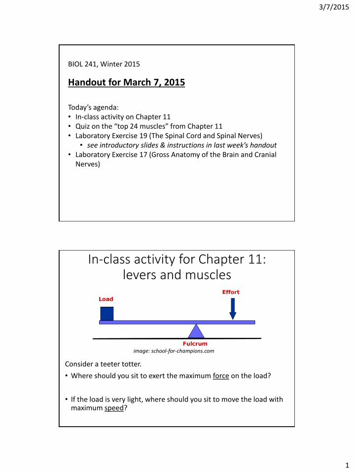
In-class activity for Chapter 11: levers and muscles
Consider a teeter totter.
• Where should you sit to exert the maximum force on the load?
• If the load is very light, where should you sit to move the load with maximum speed?
image: school-for-champions.com

3/7/2015
2
Label in-lever (Li) and out-lever (Lo).
Maximize the ratio Li/Lo to maximize [ force speed ] (circle one).
Maximize the ratio Lo/Li to maximize [ force speed ] (circle one).
image: school-for-champions.com
Now consider the joints below. (Images: mentone-educational.com.au; Patrick J. Lynch via Wikimedia Commons.)
• Where is each fulcrum? (label images with “F”)
• Where is each muscle’s insertion? (label images with “I”)
• Label each Li. Is it longer or shorter than Lo?
• Based on these examples, do human muscles seem more optimized for high force or high speed?
Biceps brachii Rectus femoris Masseter

3/7/2015
3
Introductory slides on the brain(10th Martini Chapter 14)
• How have brain structures’ functions been discovered?
• Brief summary of anatomy and functions• Brain structures
• Cranial nerves
Understanding a region of the brain: the example of Phineas Gage
H.J. Bigelow, via Wikipedia

3/7/2015
4
Corpus callosum (clinical note, p. 488)• The corpus callosum was sometimes cut as a treatment for
severe epilepsy. Consequences?
Image: D. Wolman, “The split brain: a tale of two halves,” Nature 483: 260-263, 2012
Cortical maps of body (motor & sensory)
Image: merckmanuals.com

3/7/2015
5
BRIEF summary of brain structures/functionsStructure Major functions (partial list)
Frontal lobe Includes motor cortex (control of voluntary muscles) and prefrontal cortex (decision-making, problem-solving)
Parietal lobe Perceives touch/pressure, taste, pain, and temperature
Temporal lobe Perceives smells and sounds
Occipital lobe Perceives visual info
Cerebellum Subconsciously adjusts posture and movement
Pons Relays sensory and motor info
Medulla Controls basic functions like heart rate, blood pressure, rate of breathing
Superior
colliculus
Relays/processes visual info
Inferior colliculus Relays/processes auditory info
Corpus callosum Connects the 2 cerebral hemispheres
Thalamus Relays/processes auditory and visual info, etc.
Hypothalamus Regulates body temperature, heart rate, blood pressure, fluid loss, etc. (negative feedback central!)
Cranial nerves
Why are they special?
Image: Marieb et al., Exercise 17

3/7/2015
6
Cranial nerve mnemonic #1:nerve names
Number Saying Nerve Name
I Oh Olfactory
II Oh Optic
III Oh -- Oculomotor
IV To Trochlear
V Touch Trigeminal
VI And Abducens
VII Feel Facial
VIII Very Vestibulocochlear
IX Good Glossopharyngeal
X Velvet -- Vagus
XI Ah, Accessory
XII Heaven! Hypoglossal
Cranial nerve mnemonic #2:
What kind of info is carried by each nerve?• Sensory (S)? • Motor (M)? • Both (B)?
Number Saying
I Some
II Say
III “Money
IV Matters”
V But
VI My
VII Brother
VIII Says
IX “Big
X Brains
XI Matter
XII More!”

3/7/2015
7
Cranial nerve mnemonic #3: functionsNumber Function
12 Moving your tongue
11 Your back and your neck
10 Parasympathetic stuff
9 & 7 Saliva and taste
8 Balance and hearing
5 Facial senses, chewing
4, 6-4-3 Controlling all the muscles that adjust your gaze
Links to demo MP3 and sheet music: http://faculty.washington.edu/crowther/Misc/Songs/cranial.shtml
Guidelines for Laboratory Exercise 17• Main objective: get introduced to the anatomy and
functions of the brain and cranial nerves.
• Complete Activity 1.• In your lab notebook, sketch a lateral view of a brain in which
you annotate the following features and information:• Central sulcus – what lobes are divided by this?
• Lateral sulcus – what lobes are divided by this?
• Frontal lobe – what are its major functions?
• Parietal lobe – what are its major functions?
• Temporal lobe – what are its major functions?
• Occipital lobe – what is its major function?
• Cerebellum – what are its major functions?
• Pons – what are its major functions?
• Medulla – what are its major functions?
• Spinal cord – what is its major function?

3/7/2015
8
Guidelines for Laboratory Exercise 17• Complete Activity 2.
• In your lab notebook, sketch a midsagittal section of a brain in which you annotate the following features and information:• Cerebellum – what are its major functions?
• Pons – what are its major functions?
• Medulla – what are its major functions?
• Spinal cord – what is its major function?
• Corpus callosum – what is its major function?
• Thalamus – what are its major functions?
• Hypothalamus – what are its major functions?
• Lateral ventricle and fourth ventricle – what do they contain?
• Inferior colliculus – what is its major function?
• Superior colliculus – what is its major function?
Guidelines for Laboratory Exercise 17• Complete Activity 3 (sheep brain dissection; you don’t
need to fill in the table of cranial nerve ganglia).• In your lab notebook, answer the questions embedded in the
dissection instructions. Also report which of the cranial nerves you are able to see in your sheep brain.
• Complete the Group Challenge and write the answers in your lab notebook.
• Answer the following Review Sheet questions in your lab notebook: #1, #2, #11, #12, #15, #16, #17.

BIOL 241 Winter 2015
9
Study Guide for March 14 Lab Test (Exercises 10, 11, 12, 13, 17, and 19)
Below is the material that could be covered on the upcoming lab test. As with the previous lab test, questions may be posed in a variety of formats, using 3D models, drawings, etc. Please note that I have decided NOT to make this lab test cumulative; that is, it will not include material from Exercises 1, 3,4, 5, 6, 7, 8, or 9 – except that you WILL need to know the bones to which the muscles in Exercise 13 connect. Lab Manual, Exercise 10: The Appendicular Skeleton Pectoral girdle: clavicle, scapula. Upper limb: humerus, radius, ulna, carpals (8), metacarpals (5), phalanges (3 per digit except thumb). Pelvic girdle: pubis, ilium, ischium. Lower limb: femur, patella, tibia, fibula, tarsals (7: talus, calcaneus – know these specifically!), metatarsals (5), phalanges (3 per digit except big toe). Other appendicular features: Acromion, Anterior border of tibia, Iliac crest, Lateral malleolus, Medial malleolus, Olecranon, Pubic arch, Pubic symphysis. Lab Manual, Exercise 11: Articulations and Body Movements Movements: flexion/extension, abduction/adduction, rotation vs. circumduction, pronation/supination, dorsiflexion/plantar flexion, inversion/eversion. Joint names to recognize (which bones form them?): know the joints listed in section 9.3 of Crowther’s Tenth Martini (“Names of individual joints”).
atlantoaxial joint: between the atlas (C1 vertebra) and the axis (C2 vertebra)
atlantooccipital joint: between the atlas (C1 vertebra) and the occipital bone
carpometacarpal joint: between carpals and metacarpals
claviculosternal joint: between the clavicle and the sternum
femoropatellar joint: between the femur and the patella
intercarpal joint: between carpals
interphalangeal joint: between phalanges
intertarsal joint: between tarsals
intervertebral joint: between vertebrae
metacarpophalangeal joint: between metacarpals and phalanges
metatarsophalangeal joint: between metatarsals and phalanges
radiocarpal joint: between the radius and carpals

BIOL 241 Winter 2015
10
radioulnar joint: between the radius and the ulna
sacroiliac: between the sacrum and the ilium
tarsometarsal joint: between tarsals and metatarsals
temporomandibular joint: between the temporal bone and the mandible
tibiofemoral joint: between the tibia and the femur
tibiofibular: between the tibia and the fibula
acromioclavicular joint: between the scapula and the clavicle
costovertebral: between ribs and vertebrae
glenohumeral: between the scapula and the humerus
sternocostal: between the sternum and ribs Synovial joint anatomy: in a diagram like Figure 11.2, be able to identify articular (hyaline) cartilage, joint cavity, synovial fluid, and ligament. Lab Manual, Exercise 12: Microscopic Anatomy and Organization of Skeletal Muscle Know and be able to identify the following in pictures: actin, fascicle, mitochondria, myosin, sarcolemma, sarcomere, sarcoplasmic reticulum (SR), synaptic cleft, tendon. Lab Manual, Exercise 13: Gross Anatomy of the Muscular System You are responsible for the muscles covered in section 11.6 of Crowther’s Tenth Martini (“The top 24 muscles”). Be able to identify these muscles in models or drawings and know their origins and insertions (to the level of the bone) and their functions. Lab Manual, Exercise 19: The Spinal Cord and Spinal Nerves Know and be able to identify the following in pictures: 3 layers of spinal meninges (arachnoid mater, dura mater, pia mater), dorsal and ventral horns, dorsal and ventral roots, dorsal root ganglion, gray and white matter. Know the information in the table below.
Plexus Ventral rami
Body areas covered
Cervical C1 to C5 Neck, shoulders, diaphragm
Brachial C5 to T1 Shoulders, arm, forearm, hand
Lumbar L1 to L4 Lower abdominopelvic region, anterior thigh
Sacral L4 to S4 Butt, posterior thigh, leg, foot
Lab Manual, Exercise 17: Gross Anatomy of the Brain and Cranial Nerves Know the brain and cranial nerve structures and functions covered in the introductory slides for today.