Embed Size (px)
Citation preview
Research Collection
Doctoral Thesis
Aerobic microbial degradation of chloromethane
Author(s): Studer, Alexander
Publication Date: 2001
Permanent Link: https://doi.org/10.3929/ethz-a-004227599
Rights / License: In Copyright - Non-Commercial Use Permitted
This page was generated automatically upon download from the ETH Zurich Research Collection. For moreinformation please consult the Terms of use.
ETH Library

Diss. ETH No. 14294
AEROBIC MICROBIAL DEGRADATION OF
CHLOROMETHANE
A dissertation submitted to the
SWISS FEDERAL INSTITUTE OF TECHNOLOGY ZÜRICH
for the degree of
DOCTOR OF NATURAL SCIENCE
presented by
ALEXANDER STUDER
Dipl. phil. II University of Basel
born on October 17th, 1971
from Grafenried (BE)
accepted on the recommendation of
Prof. Dr. T. Leisinger, examiner
Prof. Dr. L. Thöny-Meier, co-examiner
PD Dr. Stéphane Vuiiieumier, co-examiner
Zürich 2001


meinen Eltern


DANK
Meinem Doktorvater Thomas Leisinger mochte ich fur die Möglichkeit danken,
dass ich meine Dissertation in seinem Labor durchfuhren konnte Seine offene
Tur lud stets zum Gesprach, wobei er es mit seiner Ruhe und Erfahrung immer
geschafft hat, dass ich sein Büro mit neuer Zuversicht wieder verlassen habe
Ein herzlicher Dank geht an meinen Mitdenker Stéphane Vuiiieumier, welcher
mit seinem Enthusiasmus fur die Forschung diese Arbeit erheblich mitgepragt
hat Sein ewig kritischer Geist schärfte den Blick fur neue Losungen und dies
nicht nur in wissenschaftlichen Belangen
Linda Thony-Meyer danke ich fur die Übernahme des Korreferats
Ein besonderer Dank richtet sich an meine Eltern, die durch ihre langjährige
Unterstützung diese Ausbildung ermöglicht haben, sowie an meine Freundin
Annina, welche mich stets auf meinem Weg bestärkt hat
Ich danke allen meinen jetzigen und ehemaligen Mitstreitern aus der Leisi-
Gruppe Antje, Claudia, Craig, Dani, Gareth, Gunter, Jan, Jörg, Julia, Julien,
Kann, Martin, Manangela, Mario, Michael, Paul, Peter, Rainer, Sandra, Sue,
Todd, Yongneng und Zohre fur ihre wissenschaftliche Unterstützung und so
manch gemütliche Apéro-Stunde
Antje, Palmira, Paul, Alain und Jacques danke ich fur die Aufrechterhaltung der
Infrastruktur am Institut, ohne welche ein sinnvolles Arbeiten nicht möglich
gewesen ware


Table of contents
Abbreviations 13
Enzymes 13
Summary 15
Zusammenfassung 17
1 General Introduction 19
1.1 The global chloromethane budget 21
1.2 Microbial degradation of chloromethane 26
1.3 Biochemistry of methylotrophic metabolism in Methylobacterium 27
1.4 Chloromethane degradation in M. chloromethanicum CM4 31
1.5 Outline of the Ph.D. Thesis 37
2 A corrinoid-dependent catabolic pathway for growth of a
Methylobacterium strain with chloromethane 39
2.1 Abstract 40
2.2 Introduction 41
2.3 Experimental procedures 43
2.3.1 Materials 43
2.3.2 Bacterial Strains 43
2.3.3 DNA manipulations 43
2.3.4 Sequence analysis 43
2.3.5 N-terminal sequencing 44
2.3.6 Preparation of cell-free extract 44
2.3.7 Activity measurements with crude extracts 44
2.3.8 Determination of protein concentration 45
2.4 Results 46
2.4.1 Identification of genes involved in chloromethane utilization 46
2.4.2 In vitro dehalogenation of chloromethane 50
2.4.3 Enzyme activities in cell-free extracts of emu negative mutants 52
2.5 Discussion 55
Table of contents
3 Properties of the methylcobalamin:H4folate methyltransferase
involved in chloromethane utilizationby Methylobacterium sp.
strain CM4 59
3.1 Abstract 60
3.2 Introduction 61
3.3 Experimental Procedures 63
3.3.1 Materials 63
3.3.2 Bacterial strains and growth conditions 63
3.3.3 DNA manipulations 63
3.3.4 Construction of the cmuB expression plasmid 63
3.3.5 Preparation of cell-free extracts 64
3.3.6 Determination of enzyme activity 64
3.3.7 Purification of methylcobalamin:H4folate methyltransferase
overexpressed in E. coll 65
3.3.8 Protein determination. 66
3.4 Results 67
3.4.1 Enzyme purification and molecular properties 67
3.4.2 Catalytic properties 70
3.4.3 Restoration of dehalogenation activity in cell extract
of a cmuB mutant 73
3.5 Discussion 74
4 Chloromethane:tetrahydrofolate methyl transfer by two proteins
from Methylobacterium chloromethanicum strain CM4 79
4.1 Abstract 80
4.2 Introduction 81
4.3 Experimental procedures 83
4.3.1 Materials 83
4.3.2 Growth conditions and preparation of cell-free extract 83
4.3.3 Isolation of corrinoids from M. chloromethanlcum strain CM4 83
4.3.4 CmuA purification 84
4.3.5 Molecular mass determination 84
4.3.6 Other protein chemical methods 85
Table of contents
4.3.7 Enzyme assays 85
4.4 Results 87
4.4.1 Cobalt requirement of M. chloromethanlcum strain CM4 87
4.4.2 Purification of the CmuA protein 88
4.4.3 Molecular properties of the CmuA protein 90
4.4.4 Chloromethane:H4folate transferase activity 91
4.4.5 Dependence of the chloromethane: H4folate methyltransferase
activity on the ratio of CmuA to CmuB 92
4.4.6 Chloromethane:halide methyltransferase activity 94
4.5 Discussion 95
Chloromethane induced genes that encode a third Ci oxidation
pathway in Methylobacterium chloromethanlcum CM4 99
5.1 Abstract 100
5.2 Introduction 101
5.3 Experimental procedures 104
5.3.1 Materials 104
5.3.2 Bacterial strains and plasmids 104
5.3.3 Media and growth conditions 104
5.3.4 DNA manipulations 104
5.3.5 Construction of M. chloromethanlcum CM4 metF
mutant strain 106
5.3.6 Construction of plasmids containing transcriptional
xylE fusions 106
5.3.7 Catechol-2,3-dioxygenase activity 107
5.3.8 RNA isolation 108
5.3.9 Mapping of transcriptional start sites 108
5.4 Results 109
5.4.1 Identification of genes encoding pterin-dependent Ci
oxidation enzymes in Methylobacterium 109
5.4.2 Expression analysis of plasmid-borne transcriptional
xylE fusions 109
5.4.3 Determination of chloromethane induced transcription

Table of contents
initiation sites in M. chloromethanlcum CM4 112
5.4.4 Growth characteristics of metF and purLI mutants 114
5.5 Discussion 116
6 General Discussion 121
6.1 Genes involved in the metabolism of chloromethane 122
6.1.1 Comparative sequence analysis of the emu genes
in chloromethane utilizing methylotrophic bacteria 123
6.1.2 Genes encoding possible chloromethane responsive
regulatory proteins 126
6.1.3 Clusters III and IV contain genes whose functions in
chloromethane metabolism are not known 129
6.2 The cobalam in-dependent chloromethane dehalogenation
reaction 132
6.2.1 The corrinoid binding domain of CmuA 135
6.2.2 The zinc-binding methyltransferase domain of CmuA 138
6.2.3 How many methyltransferases are needed for
growth with chloromethane ? 143
6.2.4 A reactivation mechanism for the CmuA corrinoid protein 144
References 145
Appendix 158
Bacterial strains 158
Plasmids 159
Oligonucleotides 164
Curriculum Vitae 167
Publications 169

Abbreviations 13
ABBREVIATIONS
CoM
CH3-H4folate
H4folate
H4MPT
ICP-MS
IPTG
MALDI-TOF
coenzyme M
A/5-methyltetrahydrofolate
tetrahydrofolate
tetrahydromethanoptenn
inductively coupled plasma-atomic emission
mass spectrometry
isopropyl thio-ß-D-galactoside
matrix assisted laser desorption / ionization
time-of-fhght mass spectrometry
ENZYMES
Chloromethane corrinoid methyltransferase (EC 2 1 1 -)
Methylcobalamin tetrahydrofolate methyltransferase (EC 2 1 1 -)
NADH-dependent methylene tetrahydrofolate reductase (EC 1 5 1 -)


Summary 15
SUMMARY
The monohalomethanes CH3CI and CH3Br originate from both natural and
anthropogenic sources They are abundantly present in the atmosphere and
significantly contribute to the depletion of the ozone layer To allow an accurate
budgeting of monohalomethanes in the atmosphere the magnitude of the
source and sinks has to be determined However, only little information is
available on the biological sinks for monohalomethanes Some aerobic and
anaerobic bacteria are known to degrade monohalomethanes, but detailed
biochemical or genetic studies had not been carried out when this work was
initiated
Methylobacterium chloromethanlcum CM4, an aerobic methylotrophic a-
proteobactenum, is able to grow with chloromethane as the sole carbon and
energy source Mutants of this strain, previously obtained by miniTn5
mutagenesis, were unable to grow with chloromethane but could still grow with
methanol, methylamine or formate The transposon insertion sites in six of
these mutants were mapped to two distinct DNA fragments Sequence analysis
suggested the presence of a set of genes encoding a multistep pathway for the
conversion of chloromethane to formate
Based on enzyme activity measurements in cell-free extracts of CM4 mutant
strains, the cmuA and cmuB genes were found to be essential for
dehalogenation of chloromethane The CmuA protein represents a so far unique
two-domain protein, combining a methyltransferase and a corrinoid binding
protein It contains a noncovalently bound corrinoid identified as vitamin B12 and
one mol zinc per mol protein CmuB is a homodimenc protein and was shown to
catalyze methyl group transfer from free methylcobalamin to tetrahydrofolate
Together, purified CmuA and CmuB proteins were sufficient to catalyze
chloromethane dehalogenation The dehalogenation mechanism proceeds via
two subsequent methyl transfer reactions, which involve binding of a methyl
group to the vitamin B12 cofactor on CmuA Thus, current data suggest that
CmuA catalyzes the first methyl transfer from chloromethane onto its prosthetic
vitamin B12 group, and that the second methyl transfer reaction, from the
methylated CmuA protein to tetrahydrofolate, is catalyzed by the CmuB protein
16 Summary
The Ci unit of methyl-tetrahydrofolate formed upon dehalogenation of
chloromethane is thought to be oxidized by a set of tetrahydrofolate-dependent
enzymes to formate Sequence analysis suggested that three genes, metF, folD
and purLI located near cmuA and cmuB in the CM4 genome, encode enzymes
involved in such a Ci oxidation pathway
Studies with transcriptional reporter gene fusions demonstrated
chloromethane-dependent expression of these genes Transcriptional start sites
were mapped by primer extension analysis, and three promoter regions were
identified that are active during growth on chloromethane The corresponding
sequences were well conserved, but differed from the Methylobacterium
promoters described so far This suggests that at least three coordinately
regulated transcriptional units are expressed during growth of M
chloromethanlcum CM4 with chloromethane These units comprise cmuA,
cmuB as well as the metF, folD and purLI genes Mutational inactivation of the
metF and purLI genes resulted in CM4 strains deficient in growth with
chloromethane and thereby provided additional evidence for the involvement of
the corresponding gene products in a specific catabolic pathway for
chloromethane
Summary 17
ZUSAMMENFASSUNG
Emissionen der Monohalomethane CH3CI and CH3Br stammen sowohl aus
natürlichen wie auch aus anthropogenen Quellen In der Atmosphäre liefern
diese Gase einen signifikanten Beitrag zum Abbau der Ozonschicht Fur das
Erstellen einer genauen Bilanz der atmosphärischen Monohalomethane ist es
unerlasshch, den Umfang der Quellen und Senken dieser Verbindungen zu
bestimmen Bis heute ist aber erst wenig über den biologischen Abbau von
Monohalomethanen bekannt Der Abbau von Monohalomethanen wurde zwar
fur einige aerobe und anaerobe Bakterien gezeigt, bis zu Beginn dieser Arbeit
fehlten aber detaillierte genetische und biochemische Untersuchungen
Methylobacterium chloromethanlcum CM4 ist ein aerobes, methylotrophes a-
Proteobaktenum, welches auf Chlormethan als einziger Kohlenstoff- und
Energiequelle wachsen kann Mittels miniTn5 Mutagenese wurden Mutanten
von Stamm CM4 erhalten, welche immer noch auf Methanol, Methylamin oder
Formiat, aber nicht mehr mit Chlormethan wachsen Dies war auf Transposon-
Insertionen in zwei anscheinend ungekoppelte DNA Regionen des CM4
Genoms zurückzuführen Eine Sequenzanalyse dieser zwei Gencluster führte
zur Identifizierung mehrerer Gene, welche fur einen mehrstufigen Abbauweg
von Chlormethan zu Formiat kodieren
Mittels Bestimmung von Enzymaktivitaten in zellfreiem Rohextrakt der
entsprechenden Mutanten des Stammes CM4 wurden die Gene cmuA and
cmuB als essentiell fur die Chlormethan-Dehalogenierung identifiziert CmuA ist
ein bisher einzigartiges Zwei-Domanen-Protein, welches aus einer
Methyltransferase und einem Cornnoid-bindenden Teil besteht Es zeigte sich,
dass im Enzym ein nicht kovalent gebundener Vitamin B12 Cofaktor sowie ein
Zink Molekül enthalten sind CmuB ist ein homodimeres Protein, welches den
Methylgruppentransfer von freiem Methylcobalamin auf Tetrahydrofolat
katalysiert Zusammen sind die gereinigten Proteine CmuA und CmuB
ausreichend fur die Dehalogenierung von Chlormethan Der
Dehalogenierungmechanismus umfasst zwei aufeinanderfolgende
Methyltransferreaktionen und eine intermediäre Bindung der Methylgruppe an
18 Summary
den Vitamin B12 Cofaktor. Im aktuellen Modell katalysiert CmuA den ersten
Methyltransfer von Chlormethan auf die prosthetische Vitamin B12 Gruppe.
Danach katalysiert CmuB den zweiten Methylgruppentransfer vom methylierten
CmuA Protein auf Tetrahydrofolat.
Die an Tetrahydrofolat gebundene Methylgruppe scheint dann durch eine
Reihe von Tetrahydrofolat-abhängigen Enzymen zu Formiat oxidiert zu werden.
Basierend auf einer Sequenzanalyse scheinen die drei Gene metF, folD und
purLI die Enzyme eines solchen Ci Abbauweges zu kodieren.
Experimente mit transkriptionellen Reportergen-Fusionen zeigten eine
Chlormethan-abhängige Expression dieser Gene. Mit Hilfe von „Primer
Extension"-Analyse wurden drei Promotoren identifiziert, welche während dem
Wachstum auf Chlormethan spezifisch aktiviert sind. Die entsprechenden DNA-
Sequenzen sind stark konserviert, haben aber keine Ähnlichkeit zu bereits
bekannten Promotoren von Methylobacterium. Während des Wachstums von
M. chloromethanlcum CM4 auf Chlormethan ist somit die Expression von
mindestens drei transkriptionellen Einheiten induziert. Diese Einheiten
schliessen die Gene cmuA, cmuB, sowie metF, folD und purLI ein. Die
Inaktivierung von metF und purLI durch Mutagenese führte erwartungsgemäss
zu CM4 Stämmen mit defizientem Wachstum auf Chlormethan. Dies wurde als
weiterer Beweis für die Funktion dieser Genprodukte in einem spezifischen
Abbauweg für Chlormethan gewertet.

Chapter 1
General Introduction

20 Chapter 1
Scientific interest in the degradation of halogenated methanes dates back to
the seventies It was initiated by concerns that emissions of chlorofluorocarbons
(CFCs) to the atmosphere might pose environmental problems It was
anticipated that CFCs might make their way up to the stratosphere where
halogen atoms released by ultraviolet radiation would then react with the ozone
layer [1] Along with the chloromethanes, the CFCs were the commercially most
important classes of halogenated methanes (for an overview see [2]) Due to
their low toxicity and inflammability, these inexpensive and chemically inert
compounds were ideal for industrial processes This included usage as liquid
phase for large-scale biochemical reactions and as degreasing agents for
cleaning equipment In particular the CFCs have found widespread application,
as coolants for refrigerators, later also as propellants for aerosols and in air-
conditioning systems The utilization of CFCs started in the thirties and their
production increased massively during the following fifty years The extensive
use and the high volatility of these compounds caused their immoderate entry
into the atmosphere In 1985 Farman and coworkers discovered a significant
depletion of the stratospheric ozone over Antarctica and provided the first clear
evidence for the previously suspected negative influence of man-made CFCs
on the atmosphere [3] Since then enormous research efforts were undertaken
to identify and quantify the sources and sinks of halogenated methanes and to
determine their concentration in the atmosphere Accurate budgeting of these
compounds is necessary to understand their impact on the atmosphere, and to
monitor a possible recovery of the stratospheric ozone concentration after
constraints in their use In fact, since 1987 the use of CFCs was gradually
phased out, but due to their longevity in the atmosphere an improvement of the
stratospheric ozone concentration is not expected before 2050 (European
Ozone Research Coordination Unit, www ozone-sec ch cam ac uk)
Within the scope of finding sinks for CFCs, bacteria were isolated which are
able to metabolize halogenated methanes, and numerous studies addressing
the enzymatic mechanisms responsible for dehalogenation were conducted
(reviewed in [2,4,5]) Nevertheless, until about 5 years ago practically nothing
was known about the biochemistry of microbial degradation of the structurally
General Introduction 21
simplest halogenated methanes, the monohalomethanes. The reason might be
that, in contrast to man-made CFCs, emissions of both chloromethane and
bromomethane to the atmosphere are for the most part of natural origin (see
below). Today it is known that monohalomethanes significantly contribute to the
ongoing ozone layer depletion. This implies that halogen based ozone depletion
is partly a natural process, which has been brought out of balance by the
extensive emissions of anthropogenic halomethanes. The natural origin of
monohalomethanes further suggests that bacteria able to degrade theses
compounds may be widespread in the environment (see below). These
organisms are likely to represent a significant sink for monohalomethanes and
might therefore play an important role in preserving the natural atmospheric
equilibrium of these compounds. From a molecular point of view, it is also
interesting whether enzymatic mechanisms for the degradation of naturally
occurring monohalomethanes are evolutionary related to mechanisms involved
in degradation of xenobiotic halomethanes. A biochemical characterization of
enzymatic dehalogenation reactions in such organisms is thus both of
environmental relevance and of evolutionary interest. In addition, the isolation of
the genes involved in chloromethane-utilization would provide genetic markers
for evaluating the environmental distribution of monohalomethane-utilizing
organisms.
1.1 THE GLOBAL CHLOROMETHANE BUDGET
Chloromethane is a gas at ambient temperature (bp - 24°C) and it represents
the most abundant source of chlorine in the atmosphere [6]. Over the past 16
years measurements of atmospheric chloromethane concentrations have been
performed at many locations. The data suggest an average tropospheric
concentration of 606 pptv (parts per 1012/vol). Therefore the total atmospheric
burden of chloromethane is estimated to be around 5.3 million tons (Fig. 1.1,
[6]). The dominant process for removal of chloromethane from the atmosphere
is the reaction with tropospheric hydroxyl radicals. From the known rate of this
reaction, an average global lifetime for chloromethane of about 1.3 years was
estimated [7]. By dividing the total amount of chloromethane by the mean of its
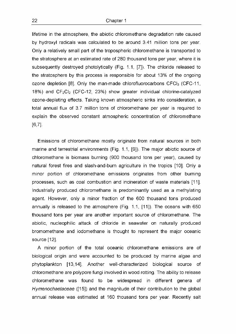
22 Chapter 1
lifetime in the atmosphere, the abiotic chloromethane degradation rate caused
by hydroxyl radicals was calculated to be around 3 41 million tons per year
Only a relatively small part of the tropospheric chloromethane is transported to
the stratosphere at an estimated rate of 280 thousand tons per year, where it is
subsequently destroyed photolytically (Fig 1 1, [7]) The chloride released to
the stratosphere by this process is responsible for about 13% of the ongoing
ozone depletion [8] Only the man-made chlorofluorocarbons CFCb (CFC-11,
18%) and CF2CI2 (CFC-12, 23%) show greater individual chlorine-catalyzed
ozone-depleting effects Taking known atmospheric sinks into consideration, a
total annual flux of 3 7 million tons of chloromethane per year is required to
explain the observed constant atmospheric concentration of chloromethane
[6,7]
Emissions of chloromethane mostly originate from natural sources in both
marine and terrestrial environments (Fig 1 1, [9]) The major abiotic source of
chloromethane is biomass burning (900 thousand tons per year), caused by
natural forest fires and slash-and-burn agriculture in the tropics [10] Only a
minor portion of chloromethane emissions originates from other burning
processes, such as coal combustion and incineration of waste materials [11]
Industrially produced chloromethane is predominantly used as a methylating
agent However, only a minor fraction of the 600 thousand tons produced
annually is released to the atmosphere (Fig 1 1, [11]) The oceans with 650
thousand tons per year are another important source of chloromethane The
abiotic, nucleophihc attack of chloride in seawater on naturally produced
bromomethane and lodomethane is thought to represent the major oceanic
source [12]
A minor portion of the total oceanic chloromethane emissions are of
biological origin and were accounted to be produced by marine algae and
phytoplankton [13,14] Another well-characterized biological source of
chloromethane are polypore fungi involved in wood rotting The ability to release
chloromethane was found to be widespread in different genera of
Hymenochaetaceae ([15]) and the magnitude of their contribution to the global
annual release was estimated at 160 thousand tons per year Recently salt

General Introduction 23
marshes were identified as an important, previously overlooked terrestrial
source of both chloromethane and bromomethane [16]. These emissions are
thought to originate predominantly from salt tolerant (halophytic) plants. On the
basis of In situ measurements, a minimum worldwide production of 170
thousand tons of chloromethane per year was estimated to result from this
source.
uv
CH3CI -* CH, + CI
SINK 280
Photodissociation
CIO
STRATOSPHERE
CIONO
TOTAL ATMOSPHERIC BURDEN
OFCHLOROMETHANE
5300 103 tons
TROPOSPHERE
SINK 3410
Reaction with
OH radicals
ANNUAL FLUX TO THE ATMOSPHERE 3700
SOURCES
Identified
Unidentified
4000
2100
1900
OCEANS
ABIOTIC
transhalogenation
BIOLOGICAL
phytoplankton
macroalgae
650
LAND 1450
ABIOTIC
Biomass burning 900
Coal combustion 107
Incineration 45
Industrial 10
BIOLOGICAL
Salt marshes 170
halophytes
Woodrotting fungi 160
Wetlands 48
SINK
BIOLOGICAL
Soil microbes
260
CH3CIC02 + H+C|-
biomass
Fig. 1.1. Schematic overview of global chloromethane fluxes. Data were taken from
[6,9,12]. With the exception of the total atmospheric burden of chloromethane, units are
thousand tons per year.

24 Chapter 1
Enzymatic mechanisms of halomethane formation seem to be conserved
among chloromethane-emitting organisms An enzyme able of halomethane
production was first isolated from the red alga Endocladia muncata [13] and
characterized as a S-adenosylmethionine/hahde ion methyltransferase This
activity was also found in the wood-rotting fungus Phellmus pomaceous [17]
and in several halophytic plants [13,18,19], The main function of such enzymes
is thought to be the biosynthesis of secondary metabolites in fungi or in the
regulation of intracellular chloride concentration in halophytes [9,18]
In addition, bromomethane is the single largest carrier of bromide to the
atmosphere, where it is degraded by the same mechanisms as chloromethane
The total annual flux to the atmosphere is estimated to be around 185 thousand
tons, where it accounts for about 15% of the stratospheric ozone depletion [8]
Due to its high reactivity bromine is about 50-60 fold more effective in ozone
depletion than chlorine Thus, the ozone depletion caused by bromomethane is
in the same order of magnitude as that caused by chloromethane The identified
sources are essentially the same as for chloromethane Natural sources, such
as the oceans (60 thousand tons per year), biomass burning (20 thousand tons
per year) and salt marshes (14 thousand tons per year, [9]) appear to dominate
However, there is also a sizeable anthropogenic flux of circa 47 thousand tons
per year to the atmosphere through its use as a fumigant [20]
In contrast to the various sources presently known, sinks for halomethanes
remain poorly characterized Soils are the only significant terrestrial sink for
chloromethane and bromomethane identified so far [6,21] On the basis of soil-
atmosphere exchange measurements, a total global uptake of chloromethane in
the order of 260 to 500 thousand tons per year was estimated [6,7,9] These
numbers must be regarded as highly speculative until measurements from a
broader selection of soil, from a larger variety of climatic zones are available
In general, the currently identified sources of chloromethane account for only
about half of the estimated annual flux to the atmosphere (Fig 1 1) This
indicates a deficiency in our current understanding of the global chloromethane
cycle There are three possible explanations that may cause this deficiency, (i)
General Introduction 25
the production from the known sources is underestimated, (ii) some major
sources have not yet been identified or (iii) the atmospheric sinks for
chloromethane have been greatly overestimated.
Hyphomicrobium vulgare
Hyphomicrobium aestuam
| Hyphomicrobium demtnficans
H j Hyphomicrobium chloromethanlcum CM2 (AF198623)
T" Hyphomicrobium facilis
*—Hyphomicrobium methylovorum
^^^—^^^— Methylosinus trichosponum
Methylobacterium organophilum— Methylobacterium rhodinum
|— Methylobacterium zatmanu
L Methylobacterium rhodesiamum
\ Methylobacterium chloromethanlcum CM4 (AF198624)'Methylobacterium extorquens
Bradyrhizobiumjapomcum— Rhodopseudomonas palustris^—^— Sphingomonas paucimobilis•^^^^^^^^— Zymomonas mobilis
-L
Rhizobium leguminosarum— Agrobactenum tumefaciens
HT,
K
Sinorhizobium meliloti
Mesorhizobium tianshanese
Mesorhizobium mediterraneum
Mesorhizobium plunfariumMesorhizobium huakuu
Mesorhizobium loti
Mesorhizobium cecen
— Strain ER2
Aminobacter aganoensis
Aminobacter nugataensis
i- Aminobacter sp. strain IMB-1 (AF034798)'Chelatobacter heintzu
i- Strain CC495 (AF107722)' Aminobacter aminovorans
Pseudaminobacter defluvu
Pseudaminobactersalicylatoxidans
Ruegena atlantica
Ruegena gelatinovorans
Ruegena algicolaStrain MB2
Roseobacter gallaeciensis
Roseobacter litoralis
Roseobacter demtrificans
Rhodobacter capsulatus
Paracoccus demtrificans
Paracoccus versutus
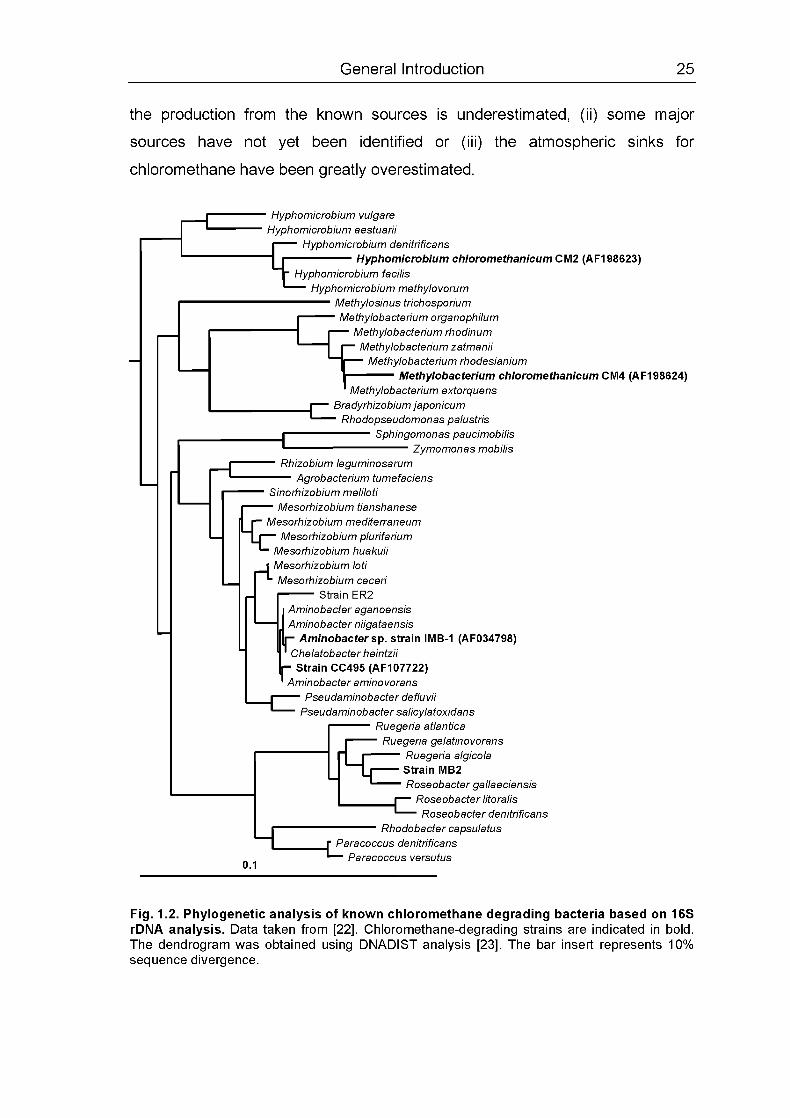
Fig. 1.2. Phylogenetic analysis of known chloromethane degrading bacteria based on 16S
rDNA analysis. Data taken from [22]. Chloromethane-degrading strains are indicated in bold.
The dendrogram was obtained using DNADIST analysis [23]. The bar insert represents 10%
sequence divergence.

26 Chapter 1
1.2 MICROBIAL DEGRADATION OF CHLOROMETHANE
In the last 5 years it became evident that bacteria are the agents responsible
for the substantial degradation of monohalomethanes in soils [24,25]. In fact it
has already been known for some years that a variety of microorganisms can
oxidize chloromethane to formaldehyde and inorganic chloride. Until recently,
however, all strains with this property were unable to use the compound as a
growth substrate. This cometabolic degradation of chloromethane was
demonstrated for nitrifying bacteria [26] and methanotrophs [27], and was
attributed to the action of ammonium- or methane monooxygenase,
respectively. It is questionable that these bacteria represent a substantial sink
for chloromethane in the environment since the enzymatic activities obtained
under laboratory conditions are likely to be insignificant under natural
conditions. The subsequent isolation of bacteria capable of growth with
chloromethane as the sole energy and carbon source thus represented a major
breakthrough. Several isolates have now been characterized. They comprise a
strictly anaerobic homoacetogen, Acetobacterlum dehalogenans (formerly
named strain MC, [28]), and several isolates of strictly aerobic, methylotrophic
a-proteobacteria (Fig. 1.2, [29-32]). Hyphomicrobium sp. strain MC1, isolated
from a sewage plant in Switzerland in 1986, was the first bacterium, for which
growth with chloromethane was described [32]. Unfortunately this strain is no
longer available. Five other Hyphomicrobium and three Methylobacterium
strains were isolated from soil samples taken from a petrochemical factory in
Russia [30]. Analysis of these strains by 16S rDNA sequencing showed that
these represented only two distinct species, subsequently named
Hyphomicrobium chloromethanlcum CM2 and Methylobacterium
chloromethanlcum CM4 [33]. Recently, Aminobacter sp. strain IMB-1, isolated
from soil fumigated with bromomethane [31], was shown to grow either with
chloromethane or bromomethane. In contrast to these strains, which were all
isolated from polluted sites or from sewage effluent, two strains were recently
isolated from a pristine environment. Strain CC495 was obtained from the litter
layer of woodland soil [29] and strain MB2, which does not stem from soil but
from seawater [34]. The latter strain grows with bromomethane and has so far
General Introduction 27
not been tested for growth with other halomethanes, but it seems reasonable to
assume that it is also capable of growth with chloromethane. The diverse origin
of chloromethane-degrading bacteria provides a first indication that such
organisms may be widespread in the environment and thus represent a
significant sink for monohalomethanes.
1.3 BIOCHEMISTRY OF METHYLOTROPHIC METABOLISM IN
METHYLOBACTERIUM
Microorganisms capable of growth with chloromethane are per definition
methylotrophic. Methylotrophy is defined as the ability to grow with compounds
more reduced than CO2 that lack any carbon-carbon bonds. Methylotrophic
microorganisms do not represent a distinct taxonomic group but include both
aerobic and anaerobic bacteria, archaea and yeasts.
Among methylotrophic bacteria, Methylobacterium represents a discreet
genus of pink-pigmented, strictly aerobic facultative methylotrophs. All strains
characterized so far grow with a limited range of substrates with carbon-carbon
bonds and with several C1 compounds, but not with methane (Fig. 1.3, [35]).
Degradation of methanol has been investigated most extensively. Its oxidation
is catalyzed by the periplasmatic pyrrolo-quinoline quinone (PQQ)-linked
enzyme methanol dehydrogenase, whose genes have been cloned from M.
extorquens AM1 and from a variety of other methylotrophs. Genetic analysis
suggested that at least 24 genes were involved in the oxidation of methanol to
formaldehyde in M. extorquens AM1. These include structural genes, regulatory
genes and genes for the synthesis of PQQ [36]. Methylamine is also
metabolized by a dehydrogenase, that is functionally analogous to methanol
dehydrogenase, but involves a covalently bound tryptophan tryptophylquinone
prosthetic group instead of PQQ. Genes encoding the enzyme and proteins
involved in cofactor biosynthesis have also been described in M. extorquens
AM1 [36].
In addition to chloromethane, dichloromethane is the only halogenated C1
compound used as energy and carbon source by aerobic methylotrophs. The
dehalogenase enzyme from Methylobacterium dlchloromethanlcum DM4 was

28 Chapter 1
demonstrated to transform dichloromethane in a glutathione-dependent reaction
into formaldehyde and inorganic chloride [37].
CH3NH3+ CH2 CI2
2-phospho-
glycerate
\
CH3OH
serine
Serine
cycle
H,0
2[H] + NH3 2 HCl
4 V ^Y 5
HCOOH
2[H]
CO,
H4folate 6
CH2=H4folate CH2=H4MPTA
15 ^10 V2[H] 2[H]
glycine CH = H4f0|ate CH = H4MPT
11 16
malyl-CoA 8 glyoxylate adp-^A L- h2o
CHO-H4folate CHO-H4MPT
MFU
ATP +
13
^ H4folate
17
!^H, MPT
HCOOH
acetyl-CoA
3-phospho-
glycerate2[H]
CO,
CHO-MFU
18,
2[H]
Cell material
Fig. 1.3. Metabolism of C1 compounds in Methylobacterium. 1, methanol dehydrogenase; 2,
formaldehyde dehydrogenase; 3 formate dehydrogenase; 4, methylated amine dehydrogenase;5, dichloromethane dehalogenase; 6, spontaneous methylene-H4folate formation; 7, serine
hydroxymethyltransferase; 8, malyl-CoA lyase; 9, phosphoglycerate mutase; 10, NADP-
dependent methylene-H4folate dehydrogenase MtdA; 11, methenyl-H4folate cyclohydrolaseFchA; 12, formyl-H4folate synthase; 13, 10-formyl-H4folate synthase; 14 formaldehyde activating
enzyme Fae; 15, NAD(P)-dependent methylene-H4MPT dehydrogenases MtdA and MtdB; 16,
methenyl-H4MPT cyclohydrolase Mch; 17 formyl-methanofuran: H4MPT-formyltransferase; 18,
formyl-methanofuran dehydrogenase. Dashed arrows indicate conversions involving several
enzymatic steps.
General Introduction 29
In summary, it is important to stress that all Ci compounds mentioned above
are metabolized via formaldehyde, which is situated at a metabolic crossroad in
Methylobacterium (Fig 1 3) Part of the formaldehyde is assimilated into
biomass and the remainder is oxidized to carbon dioxide for the gain of energy
for growth This implies that the relative flux of formaldehyde between the
assimilatory and dissimilatory pathways has to be tightly regulated and depends
on the growth conditions In this context it is also important to mention that
formaldehyde is toxic for bacteria and growth of Methylobacterium on
formaldehyde is only possible at concentrations below 1 mM ([38], Vuiiieumier
and Kayser unpublished) Therefore, during growth with Ci compounds, cells
must have mechanisms to cope with the increased formation of formaldehyde
The accumulation of formaldehyde is avoided by its efficient enzymatic
oxidation to carbon dioxide So far four possible enzymatic pathways for
formaldehyde degradation have been described in methylotrophic bacteria
(i) A linear pathway involves the sequential action of formaldehyde
dehydrogenase and formate dehydrogenase Two different types of
formaldehyde dehydrogenases have been described, a glutathione-dependent
enzyme [39,40] and a mycothiol-dependent (previously called NAD-factor
linked) enzyme [41] These enzymes have not been reported for
Methylobacterium Both methanol and aldehyde dehydrogenases, are known to
react non-specifically with formaldehyde and the corresponding activities were
detected in cell-free extracts of Methylobacterium However, the physiological
relevance of these reactions m vivo is uncertain [42]
(ii) The oxidation and assimilation of formaldehyde via the nbulose
monophosphate cycle has been observed in a variety of methylotrophs [42]
This pathway is not present in Methylobacterium, which uses the serine cycle
for Ci assimilation The serine cycle starts with the formation of serine from
glycine, a reaction catalyzed by serine hydroxymethyltransferase (Fig 1 3, [42])
The methylene-H4folate required for this reaction is formed from formaldehyde
and H4folate, in a reaction which is thought to occur abiotically (see [38])
30 Chapter 1
(mi) Methylene-H4folate is also the starting point of a H4folate-dependent
formaldehyde oxidation pathway in M extorquens AM1 [43] The pathway
involves two enzymes, a methylene-H4folate dehydrogenase MtdA [44] and a
methenyl-H4folate cyclohydrolase FchA [45], both so far only described in M
extorquens AM1 The next step in this pathway is presumably catalyzed by a
formyl H4folate hydrolase, but such an enzyme has not yet been characterized
in Methylobacterium The formate formed by such an enzyme is then oxidized
to carbon dioxide by formate dehydrogenase A search of the M extorquens
AM1 genome database (Chistoserdova, pers comm ) revealed the presence of
genes potentially encoding a 10-formyl-H4folate synthase and at least three
putative formate dehydrogenases
(iv) A tetrahydromethanoptenn (H4MPT)-dependent oxidation pathway for
formaldehyde and the presence of the cofactor dephospho-H4MPT were
recently demonstrated in M extorquens AM1 [43] The occurrence of H4MPT
was previously thought to be restricted to methanogenic and sulfate-reducing
archaea A survey among 13 genera of methylotrophic proteobactena
suggested that this pathway is indeed present in many methylotrophic bacteria
[46] Based on enzyme studies and on thermodynamic considerations, the
H4MPT-dependent oxidation pathway is proposed to be the main pathway for
energy production in M extorquens AM1, whereas the H4folate-dependent
pathway is thought to provide one-carbon moieties for biosynthesis [43,47]
Three enzymes participating in the H4MPT Ci oxidation pathway in M
extorquens AM1 have already been biochemically characterized In contrast to
methylene-H4folate, methylene-H4MPT is enzymatically formed This reaction is
catalyzed by the formaldehyde-activating enzyme Fae [47] The next step in this
pathway is the transformation of methylene-H4MPT to methenyl-H4MPT, which
is catalyzed by the MtdA paralog MtdB MtdB does not react with methylene-
H4folate, whereas MtdA can use both methylene-pterins as substrates [46] The
third step in the pathway is catalyzed by methenyl-H4MPT cyclohydrolase Mch,
which also specifically uses H4MPT as a cofactor [45] The following steps in
the pathway are presumably catalyzed by formyl methanofuran H4MPT
formyltransferase and by formyl methanofuran dehydrogenase The genes

General Introduction 31
putatively encoding these enzymes have been identified in regions of the M.
extorquens AM1 genome closely associated with other genes of Ci metabolism.
However, neither of the enzymes has yet been characterized, nor has the
presence of the cofactor methanofuran in M. extorquens AM1 been confirmed.
1.4 CHLOROMETHANE DEGRADATION IN M. CHLOROMETHANICUM
CM4
M. chloromethanlcum CM4 is an ideal candidate for a biochemical and
genetic analysis of chloromethane metabolism. Its 16S rDNA sequence is 98%
identical to that of M. extorquens AM1 [33] and the central Ci metabolism is
therefore expected to be similar in the two organisms (see 1.3). The results of
an initial physiological characterization of M. chloromethanlcum CM4 and other
chloromethane-degraders are shown in Table 1.1. The presence of serine
hydroxymethyltransferase, hydroxypyruvate reductase and malyl-CoA lyase
activities in cell-free extracts suggested that M. chloromethanlcum CM4
assimilates Ci compounds via the serine cycle [30]. This is the typical
assimilation pathway found in all Methylobacterium strains described up to now
[42].
First insights into the chloromethane metabolism in strain CM4 were obtained
from studies with whole cells and from the analysis of chloromethane non-
utilizing mutants of the organisms, summarized in Vannelli et al. 1998 [48]. This
work served as a starting point for the studies in the present thesis and will
therefore be described in some detail in the following section.
Measurements of oxygen uptake and chloride release by resting cells
suggested that 1 mol chloromethane is oxidized to 1 mol carbon dioxide,
producing 1 mol of hydrochloric acid and consuming 1.5 mol of oxygen. The
chloromethane degrading activity appeared inducible, since it was not detected
in methanol-grown cells. Chloromethane-grown cells were also capable of
dehalogenating bromomethane and iodomethane, but not dichloromethane or
32 Chapter 1
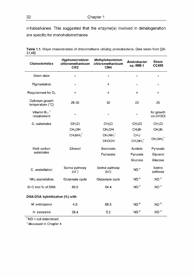
n-haloalkanes. This suggested that the enzyme(s) involved in dehalogenation
are specific for monohalomethanes.
Table 1.1. Major characteristics of chloromethane utilizing proteobacteria. Data taken from [29-
31,48]
Hyphomicrobium Methylobacterium . . . _. .
~u *-
*- J, *•. l.1 *l.Aminobacter Strain
Characteristics chloromethanlcum chloromethanlcum
CM2 CM4sp. IMB-1 CC495
Gram stain - - - -
Pigmentation - + - -
Requirement for 02 + + + +
Optimum growth
temperature (°C)28-30 30 22 25
Vitamin B-|2brequirement
- - -
for growthon CH3CI
C1 substrates CH3CI CH3CI CH3CI CH3CI
CH3OH CH3OH CH3Br CH3Br
CH3NH3+ CH3NH3+
CHOOH
CH3I
CH3NH3+CH3NH3+
Multi carbon Ethanol Succinate Acetate Pyruvatesubstrates
Fumarate Pyruvate
Glucose
Glycerol
Glucose
C1 assimilationSerine pathway
(id+)Serine pathway
(id)NDa
Serine
pathway
NH4 assimilation Glutamate cycle Glutamate cycle NDa NDa
G+C mol % of DNA 60.0 64.4 NDa NDa
DNA-DNA hybridization (%) with
M. extorquens 4.0
H. zavarzinii 29.4
68.5
5.2
ND'
ND:
ND
ND
ND = not determined
'discussed in Chapter4

General Introduction 33
Three possible mechanisms for the dehalogenation of chloromethane in
strain CM4 were considered, which would all lead to the formation of
formaldehyde, the central intermediate in Ci metabolism of Methylobacterium
(Fig. 1.4). (i) Monooxygenation of chloromethane would result in the formation
of an unstable chlorohydrin compound, from which formaldehyde would then be
spontaneously formed by abiotic elimination of chloride. This mechanism was
previously proposed for chloromethane utilization in Hyphomicrobium strain
MC1 [32]. (ii) Substitutive, hydrolytic dehalogenation would yield methanol and
hydrochloric acid without a requirement of molecular oxygen. Such a
mechanism was demonstrated for the haloalkane dehalogenase DhIA of
Xanthobacter autotrophicus which grows on 1,2-dichloroethane as the sole
carbon source (reviewed by [49]).
hydrolase CH3CI
H20^^ H+ Cl-
CH3OH
methyl-transferase/
monooxygenase^2[H] dehydrogenase
2[H] + 02 ' ' H20 HX
f*i i f*i ^k CH2CIOH
abiotich. Ulf*l lf^ ^
Jr*\ 1 v ^ ^ r*\ 1 r*\
t»H3U x *"
>^ nunu ^ /• 0H3A
" s on3oi
H20 H+Ci-
H20^2[H] + HX H+Ci-
' ^2[H]
HCC)OH
V
CO,
2[H]
Fig. 1.4. Possible mechanisms for the metabolism of chloromethane by methylotrophs.Each of the pathways follows the stoichiometry of CH3CI + 3/2 02 - C02 + H20 + HCl (adaptedfrom [48]).
(iii) In a putative methyltransferase/dehydrogenase mechanism,
formaldehyde would be produced without the formation of methanol as an

34 Chapter 1
intermediate. The nature of the methyl group acceptor involved in the first step
of this mechanism could be either a free cofactor, such as glutathione, found as
a cofactor in dichloromethane dehalogenation of Methylobacterium
dlchloromethanlcum DM4 [37], or a catalytic thiol group of the protein itself.
The methyltransferase/dehydrogenase mechanism was considered to be
most likely based on the following experimental evidence, (i) Growth yields
strongly argued against a monooxygenase driven reaction. The growth yield of
M. chloromethanlcum CM4 with chloromethane (2.8 ±0.1 g of protein per mol of
C) was within the same range as with methanol (2.9 ±0.1 of protein per mol of
C). In contrast, with formate (0.94 ± 0.2 g of protein per mol of C) the growth
yield amounted to only a third of that obtained for methanol. This is in
agreement with the two electron equivalents produced from the oxidation of
formate compared to the six electron equivalents gained from the oxidation of
methanol (Fig. 1.4). A monooxygenase driven dehalogenation reaction would
consume two reducing equivalents for the activation of chloromethane, a net
yield of only two electron equivalents would be expected. In this case the
growth yield with chloromethane would be expected to be of the same order of
magnitude as with formate, (ii) In a hydrolytic pathway chloromethane is
transformed to methanol in an oxygen-independent manner. However, resting
cell assays demonstrated that in absence of oxygen no chloride is released
from chloromethane, which was a first indication that a hydrolytic mechanism
appeared rather unlikely, (iii) Convincing evidence against a hydrolytic
mechanism came from transposon mutagenesis of M. chloromethanlcum CM4.
4032 exconjugants were isolated and subsequently tested for growth on various
Ci compounds. Among these, 53 did not grow on methanol, 7 did not grow on
methylamine and 2 were unable to grow on formate. 11 exconjugants could not
grow on either methanol or methylamine, which probably accounts for mutations
in genes encoding enzymes essential for growth on both substrates. However,
most interesting in course of this study were 9 mutants which were unable to
grow with chloromethane, but grew normally on all the other Ci substrates
tested. The fact that no mutants were isolated which could not grow on
methanol and chloromethane, provided further evidence against a hydrolytic
General Introduction 35
pathway, since it indicated that methanol is not an intermediate in the
metabolism of chloromethane by M. chloromethanlcum CM4.
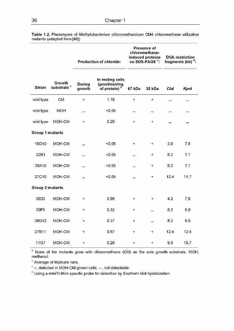
The nine çhloromethane-utilization (emu) mutants were subsequently divided
into two phenotypically distinct groups. All mutants were unable to use
chloromethane as a growth substrate. However, whereas five mutants released
chloride in the presence of chloromethane, the other four mutants had lost this
ability (Table 1.2). This observation suggested that the latter mutants are
deficient in the dehalogenation step, whereas the other five mutants were
deficient in unknown reactions required for growth with chloromethane. Analysis
of chloromethane-induced proteins in mutant and wild type CM4 strains led to
the identification of two proteins with estimated sizes of 35 kDa and 67 kDa.
Both proteins were absent in methanol-grown cells and were also missing in
some of the mutants grown with a methanol/chloromethane mixture. Southern
blot analysis with probes against the transposon sequence further suggested
that several different loci in the chloromethane-utilizing mutants were affected
by transposon insertion (Table 1.2).
In summary, studies with cell-suspension of CM4 and transposon
mutagenesis provided first evidence for a specific multistep pathway for the
degradation of chloromethane in M. chloromethanlcum CM4, which apparently
involves a dehalogenation mechanism so far not described for aerobic
methylotrophic bacteria.
36 Chapter 1
Table 1.2. Phenotypes of Methylobacterium chloromethanlcum CM4 chloromethane utilization
mutants (adapted from [48])
Presence of
chloromethane-
induced proteins DNA restriction
Production of chloride: on SDS-PAGE c: fragments (kb) d:
In resting cellsGrowth
^During (umol/min/mg
Strain substrate growth of protein)b 67 kDa 35 kDa C/al Kpn\
wild type CM 1.16
wild type MOH <0.05
wild type MOH-CM 0.29
Group 1 mutants
19D10 MOH-CM <0.05 3.9 7.8
22B3 MOH-CM
38A10 MOH-CM
27C10 MOH-CM
<0.05
<0.05
<0.05
8.2
8.2
7.1
7.1
12.4 11.7
Group 2 mutants
36D3 MOH-CM
30F5 MOH-CM
0.86
0.32
4.2
8.2
7.8
6.9
38G12 MOH-CM 0.37 8.2 6.9
27B11 MOH-CM 0.67 12.4 12.4
11G7 MOH-CM 0.28 9.9 15.7
None of the mutants grew with chloromethane (CM) as the sole growth substrate. MOH,methanol.
b
Average of triplicate runs.
c
+, detected in MOH-CM-grown cells; —, not detectable.
d
Using a miniTn5Km specific probe for detection by Southern blot hybridization.

General Introduction 37
1.5 OUTLINE OF THE PH.D. THESIS
In the present thesis the aerobic degradation of chloromethane was
addressed at the molecular level. The previously isolated mutants of M.
chloromethanlcum CM4 unable to grow with chloromethane provided a suitable
basis to investigate the biochemical mechanisms which allow this strain to grow
on that compound. The main focus of the present study was to unravel the
enzymatic mechanisms and to identify the essential cofactors of chloromethane
dehalogenation. Chapter 2 describes the isolation of two genetic loci in M.
chloromethanlcum CM4 involved in chloromethane utilization. Sequence
analysis and enzymatic measurements in cell-free extracts suggested a
multistep pathway for the oxidation of chloromethane to formate. Chapters 3
and 4 are dedicated to the first step in this pathway and describe the purification
and characterization of the two proteins essential for the dehalogenation
reaction. The reaction involves a vitamin B12-mediated methyl transfer from
chloromethane to H4folate with concomitant release of chloride. Chapter 5
summarizes expression studies and transcriptional analyses performed in M.
chloromethanlcum CM4. The data demonstrate the presence of further
chloromethane-induced genes besides the genes required for dehalogenation.
These experiments, along with mutational inactivation of some of the identified
genes, point to a specific tetrahydrofolate-dependent Ci oxidation pathway in M.
chloromethanlcum CM4.

Chapter 2
A corrinoid-dependent catabolic pathway for growth of a
Methylobacterium strain with chloromethane
Todd Vannelli, Michael Messmer, Alex Studer,
Stéphane Vuiiieumier, and Thomas Leisinger
Proceedings of National Academy of Science USA 96, 4615-4620 (1999)
40 Chapter 2
2.1 ABSTRACT
Methylobacterium sp. strain CM4, an aerobic methylotrophic a-
proteobacterium, is able to grow with chloromethane as carbon and energy
source. Mutants of this strain that still grew with methanol, methylamine or
formate but were unable to grow with chloromethane were previously obtained
by miniTn5 mutagenesis. The transposon insertion sites in six of these mutants
mapped to two distinct DNA fragments. The sequences of these fragments,
which extended over more than 17 kb, were determined. Sequence analysis,
mutant properties, and measurements of enzyme activity in cell-free extracts
allowed to define a multistep pathway for the conversion of chloromethane to
formate. The methyl group of chloromethane is first transferred by the protein
CmuA (emu: chloromethane utilization) to a corrinoid protein, from where it is
transferred to H4folate by CmuB. Both CmuA and CmuB display sequence
similarity to methytransferases of methanogenic archaea. In its C-terminal part,
CmuA is also very similar to corrinoid binding proteins, indicating that it is a
bifunctional protein consisting of two domains that are expressed as separate
polypeptides in methyl transfer systems of methanogens. The methyl group
derived from chloromethane is then processed via pterine-linked intermediates
to formate by a pathway which appears to be distinct from those already
described in Methylobacterium. Remarkable features of this novel pathway for
the catabolism of chloromethane thus include the involvement of a corrinoid-
dependent methyltransferase system for dehalogenation in an aerobe, and a
set of enzymes specifically involved in funnelling the Ci moiety derived from
chloromethane into central metabolism.
A catabolic pathway for chloromethane 41
2.2 INTRODUCTION
Attention has been focused on chloromethane and bromomethane because
of their role as sources of stratospheric chlorine and bromine, the primary
agents of ozone destruction. Chloromethane (CH3CI) is the most abundant
halocarbon in the atmosphere and is responsible for 15-20% of chlorine-
catalyzed ozone destruction in the stratosphere [50]. It is released at an
estimated global rate of 3.5-5 x 10⁶ tons per year, primarily from natural
sources, and less than 1% of the global chloromethane flux is due to industrial
production of the compound (reviewed in [50]). For bromomethane, current
estimates of ocean emission and natural formation during combustion of
vegetation fall in the range of 8 x 10⁴ tons per year, slightly more than the
amount annually emitted by the use of this compound in soil fumigation [51]. On
a molar basis, bromine is 40-100 times more effective than chlorine in depleting
ozone [52]. Thus, chloromethane and bromomethane contribute about equally
to an estimated 40% of the total global loss of stratospheric ozone.
Green plants [53] and soil bacteria [21,24,25,30,54] represent terrestrial sinks
for chloromethane and bromomethane. Evidence for bacterial degradation of
halogenated methanes in seawater was also reported [55]. Microbial
metabolism of monohalomethanes includes oxidative [56] and hydrolytic [57]
cometabolic processes as well as mineralization by methylotrophic bacteria that
utilize chloromethane as a growth substrate. The homoacetogenic bacterium
Acetobacterium dehalogenans [58] is the only known strictly anaerobic
representative of the latter group [59]. Anoxic dehalogenation of chloromethane
by this organism was shown to be catalyzed by enzymes that transfer the
methyl group of chloromethane via a corrinoid protein to H4folate to yield
chloride and CH3-H4folate, an intermediate of the acetyl-CoA pathway [60]. In
contrast, the reactions by which some recently isolated strictly aerobic
methylotrophic bacteria [30] utilize chloromethane as a growth substrate have
yet to be elucidated. The physiological properties of the wild type and of
chloromethane utilization-negative mutants of a representative strain,
Methylobacterium sp. CM4, led us to propose that this organism metabolizes
chloromethane by initial dehalogenation via a methyl transfer reaction [48].

42 Chapter 2
Here we report on the sequence of two large DNA fragments containing at
least four genes essential for chloromethane metabolism in strain CM4 We
present experimental evidence that this aerobic bacterium is able to catalyze
transfer of the methyl moiety of chloromethane to H4folate via a corrinoid
intermediate to yield CH3-H4folate, a key intermediate of methylotrophic
metabolism

A catabolic pathway for chloromethane 43
2.3 EXPERIMENTAL PROCEDURES
2.3.1 Materials
Reagents for molecular biology were obtained from Fermentas and
Boehringer Mannheim. (6S)-5,6,7,8-tetrahydrofolic acid trihydrochloride
(H4folate) and (6S)-5-methyl tetrahydrofolate (CH3-H4folate) were purchased
from Dr. Schircks Laboratorium (Jona, Switzerland). ATP, S-adenosyl
methionine and methylcobalamin were from Sigma, and NADPH:FMN
oxidoreductase was from Boehringer. All other chemicals were reagent grade or
better and were purchased from Fluka.
2.3.2 Bacterial Strains
Bacterial strains in this study included Methylobacterium sp. strain CM4 that
grows with chloromethane [30], and miniTn5 [61] insertion mutants whose
phenotypes have been described previously [48]. E. coli K12 strain DH5a?
(GIBCO/BRL-Life Technologies) was used as a host in DNA work.
2.3.3 DNA manipulations
Preparation of genomic DNA, restriction enzyme digestions, ligations and
transformations were performed using standard procedures [62]. Plasmid
pBluescript-KSII(+) (Stratagene) was used for cloning. DNA fragments from
mutants of strain CM4 containing a miniTn5 insertion were cloned by selection
of transformants for kanamycin resistance (25 ng/ml).
2.3.4 Sequence analysis
The cloned genomic DNA was sequenced on both strands using PCR
methods with fluorescent dideoxynucleotide terminators and an ABI-Prism
automatic sequencer (Perkin Elmer). The precise site of insertion of the
minitransposon was determined for all mutants. The sequences of cluster I
(8863 nt, Ace. No. AJ011316) and cluster II (8457 nt, Ace. No. AJ011317) were
assembled from sequence fragments obtained from DNA cloned from the
different mutants with the GCG sequence analysis package (Version 8.1,
Genetics Computer Group, Madison Wl). Similarity searches were performed

44 Chapter 2
using gapped BLAST and PSI-BLAST programs [63] against public protein and
gene databases.
2.3.5 N-terminal sequencing
The 67 kDa protein induced during growth with chloromethane [48] was
partially purified, and the N-terminal sequence of the corresponding protein
band on SDS-PAGE was determined by Edman degradation using an Applied
Biosystems 476A automatic sequencer.
2.3.6 Preparation of cell-free extract
Methylobacterium sp. strain CM4 and mutants were grown as described
previously [48]. Bacteria were harvested at an OD6oo of 0.5 to 0.7 (10'000 x g,
15 min) and resuspended (1 g wet cells per ml) in 50 mM Tris-S04 buffer (pH
7.2) containing 5 mM DTT. Cells were disrupted by two passages through a
French pressure cell (120 MPa, 4°C), and DNasel (50 ng/ml final) was added to
the suspension, which was centrifuged (17000 rpm, 45 min) to remove cell
debris. The resulting supernatant was cleared from membrane components by
ultracentrifugation (100'000 x g, 45 min), and the cell-free extract obtained
(approx. 15 mg protein/ml) was flash-frozen in liquid nitrogen and stored at -
20°C.
2.3.7 Activity measurements with crude extracts
Solutions were made anoxic by degassing with N2 plus H2 (95:5 [vol/vol]).
Enzyme reactions, manipulations and measurements were performed under the
same atmosphere. Assays of enzymatic activity were done at 30°C in a 3 ml
volume in 12.4 ml serum flasks with gas-tight rubber stoppers. The H4folate-
dependent dehalogenation of chloromethane was measured by following the
consumption of chloromethane by gas chromatography [64] using a Henry
constant of 0.43 calculated by interpolation to 30°C of the published value for
chloromethane [65]. Dehalogenation was also determined by monitoring the
chloromethane-dependent formation of CH3-H4folate from H4folate. CH3-
H4folate was separated from the incubation mixture by HPLC [59] and detected
spectrophotometrically at 320 nm. The incubation mixture contained cell-free

A catabolic pathway for chloromethane 45
extract (0.8-4 mg/ml protein) in 100 mM Tris-S04 (pH 7.8) buffer, 5 mM DTT,
2.4 mM H4folate (of which 1 mM was biologically available [66]), and 1 mM
titanium(lll)citrate. Cell-free extracts dialysed against 100 mM Tris-S04 (pH 7.8)
were also used in order to check whether any endogenous cofactors in the
extracts were involved in dehalogenase activity. The dehalogenation reaction
was initiated by the addition of 0.5 mM chloromethane gas (based on the liquid
phase volume) through the rubber stopper with a gas-tight syringe. For
determination of methylcobalamin:H4folate methyltransferase activity, the assay
mixture contained 0.5 mM methylcobalamin instead of chloromethane. The
numbers reported are from representative individual experiments which were
performed at least twice and all yielded very similar results.
2.3.8 Determination of protein concentration
Protein was determined by the method of Bradford [67], using a commercial
dye reagent (Bio-Rad) with bovine serum albumin as a standard.

46 Chapter 2
2.4 RESULTS
2.4.1 Identification of genes involved in chloromethane utilization
We previously isolated miniTn5 transposon insertion mutants of
Methylobacterium sp. strain CM4 that were unable to grow with chloromethane
[48]. Thus, nine emu negative mutants were obtained that were still able to grow
with methanol, methylamine, or formate. Conversely, 73 transposon mutants
defective in the utilization of methanol, methylamine, methanol plus
methylamine, or formate could still grow with chloromethane [48]. This
suggested that chloromethane was metabolized in Methylobacterium sp. CM4
by reactions different from those involved in the metabolism of methanol and
methylamine. The genes whose insertional inactivation caused loss of the ability
to grow with chloromethane were isolated by selection of the kanamycin
resistance gene present on the minitransposon [61]. The DNA fragments
carrying a transposon insertion were sequenced from all emu negative mutants.
Transposon insertion mutants, arbitrarily labeled in order of their detection [48],
were found in four apparently unlinked DNA regions that were termed cluster I
(mutants 30F5, 38G12, 22B3, 38A10), cluster II (mutants 19D10, 36D3), cluster
III (mutant 11G7), and cluster IV (mutant 27B11). The emu negative mutant
27C10 was not analyzed in detail since it appeared to carry a partial duplication
of the transposon. A schematic representation of the 14 open reading frames
identified in the DNA sequences of clusters I and II is shown in Fig. 2.1.
Most of the encoded polypeptides displayed significant sequence identity to
proteins of known functions (Table 2.1). With respect to their possible role in
metabolism, the proteins encoded in DNA clusters I and II fell into four groups:
methyltransferases, pterine-dependent enzymes, proteins associated with
cobalam in biosynthesis, and proteins of unknown function. Similarity searches
of the protein sequences encoded in clusters III and IV (data not shown) did not
provide insights as to their association with chloromethane transformation and
are not discussed further here.

TABLE
1.Genesandopen
readingframes
inDNA
regionsassociatedwithchloromethane
utilization
Gene
(orf)
Length
calcMr
Gene
(aa)
(kDa)
Begin
Gene
Inferredfunction
End
Sequencecomparison
ofrepresentative
hit
%pr
otei
nsequence
identity
(AceNo)
a
Identity
(%)
Cluster
I
cobU
342
349
1502
474
cobalamin
biosynthesis
folC
467
498
2942
1539
folylglutamate
synthetase
folD
306
324
3865
2945
5,10-methylene-H4folate
dehydrogenase/5,10-methenyl
H4folate
cyclohydrolase
purl)
287
327
4849
3986
10-formyl-H4folate
hydrolase
orf414
414
437
5628
6872
unknown
emuA
617
67
06897
8750
methyltransferase/
corrinoid
prot
ein
Clustern
cobQ
>424
ND
<1
1275
cobalamin
biosynthesis
cobD
330
342
2275
1283
cobalamin
biosynthesis
orf219
219
249
3345
2686
unknown
metF
320
343
3507
4469
5,10-methylene-H4folatereductase
cmuB
311
333
4703
5638
methyltransfer
cmuC
378
412
5635
6771
methyltransfer
orf361
361
375
7971
6886
cobalamin
biosynthesis
cobC
>162
ND
>8456
7968
cobalamin
biosynthesis
CobU(P
demt
rifi
cans
,P29935)
57
FolC(£
coli,P08192)
32
FolD(£
coli,P24186)
49
PurU(Corynebactenum
sp
,Q46339)
47
Orf{Mycobacterium
tuberculosis
,P72042)
31(143aa
)
MtbA,{Methanosarcinabarken,O30640)/
24/32
c
MtmC(Methanosarcinabarken,030641)
CobQ(P
demt
rifi
cans
,P29932)
59
CobD(P
demt
rifi
cans
,P21634)
55
{Synechocystis
sp
,Q55963)
32(117aa
)
Orf(Saccharomycescerevisiae,P53128)
24(156aa
)
MtrH(M
thermoautotrophicum
,P80187)
30
MtaA
(Methanosarcinabarken,Q48949)
28
(104aa
)
MTH808(M
thermoautotrophicum
,026899)
35
CobC(P
demt
rifi
cans
,P21633)
38
ND,
notdetermined
aAccessionnumbersfromSwissprot
orTrembldatabases
b
Sequence
identity
isoverthe
entirelength
oftheshorterofthetwocomparedsequences,exceptwherenoted
cTheMtbAsequence(339aa)canbeal
ignedtoresidues7-353
ofCmuA,andtheMtmCsequence(217aa)canbeal
igne
dtoresiduess401-607
ofCmuA,
resp
ecti
vely

48 Chapter 2
38G12 22B3 38A10
cobU folC folD purU orf414 emuA
cobQ cobD orf219 metF cmuB cmuC orf361 cobC
Fig. 2.1. Schematic view of gene clusters I (A) and II (B) of Methylobacterium sp. CM4.
Genes encoding methyltransferases are marked in black, genes encoding putative pterine-
dependent enzymes of C1 metabolism are marked in grey, and genes encoding enzymes of
cobalamin biosynthesis or proteins with unknown function are marked in white. The position and
orientation of the transposon insertions in the genome of emu negative mutants is also shown.
The products of emuA, cmuB and cmuC in clusters I and II showed sequence
similarity to methyltransferases or corrinoid binding proteins from archaea
(Table 2.1). The C-terminal part of CmuA was found to be most similar to
MtmC, the 29 kDa corrinoid protein which, when methylated by methylamine,
acts as the substrate of the methyltransferase catalyzing the methylation of
coenzyme M in Methanosarcina barker! [68]. The C-terminal part of CmuA also
showed similarity to many other corrinoid-binding proteins, including methionine
synthases [69]. The N-terminal part of CmuA showed considerable sequence
identity to MtbA, the 36 kDa methyltransferase that transfers the methyl group
from MtmC to coenzyme M [68,70]. The similarity in sequence between the two
proteins extended over the entire length of MtbA (Table 2.1). It thus appears
that CmuA, whose calculated molecular weight is 67 kDa, represents an
unprecedented fusion of two proteins that are expressed as separate but
closely associated polypeptides in methyl transfer systems of methanogenic
archaea. CmuB, the second methyltransferase-like protein suggested from
sequence analysis, showed most sequence identity (30%, Table 2.1) with
subunit MtrH of the membrane associated N5-methyl-tetrahydromethano-

A catabolic pathway for chloromethane 49
pterin co-enzyme M methyltransferase complex from Methanobactenum
thermoautotrophicum that catalyzes transfer of the methyl group of N5-
methyltetrahydromethanoptenn to the corrinoid protein MtrA [71] CmuB, unlike
CmuA, also showed low but significant pairwise identity (23%) to the CH3-
H4folate binding domain of MetH from E coli (residues 337-648 in the protein
sequence)
CmuC, the third methyltransferase-hke putative protein, was most similar to
MtaA, another corrinoid coenzyme M methyltransferase characterized in M
barken [70] (Table 2 1) CmuC was 19% identical to the N-terminal domain of
CmuA, and displayed significant but low identity (14%) over its entire length to
MtbA and MtaA from M barken and to DcuP from E coli in multiple alignments
The second group of proteins detected in clusters I and II were similar to
enzymes involved in interconversion pathways of one-carbon compounds A
emu phenotype was observed in mutant 30F5, in which the open reading frame
encoding a protein with strong similarity to bacterial 10-formyl-H4folate
hydrolases (Table 2 1) was disrupted Accordingly, the gene was named purU
Proteins similar to bacterial FolD and FolC (Table 2 1), enzymes involved in the
metabolism of one-carbon compounds, are encoded by genes downstream of
purU It is noteworthy that cell-free extracts of mutant 38G12, in which the
transposon insertion is located upstream of the purU gene (Fig 2 1), lack a
protein of about 35 kDa that is induced by chloromethane [48] This protein
could therefore be PurU (32 7 kDa calculated molecular mass) and perhaps
also FolD (32 4 kDa, see Table 2 1) Finally, the gene tentatively named metF in
cluster II codes for a protein similar to enzymes of the 5,10-methylene-H4folate
reductase family in part of its sequence The role in chloromethane degradation
of this and other open reading frames detected in the DNA sequence of clusters
I and II, however, remains uncertain since no mutants are yet available in which
these genes have been knocked out
50 Chapter 2
2.4.2 In vitro dehalogenation of chloromethane
Transposon insertions into genes cmuA and cmuB (Fig. 2.1) led to a
dechlorination negative phenotype in the corresponding mutants, that could
neither dehalogenate chloromethane nor grow with this compound. The other
emu negative mutants released chloride from chloromethane in a resting cell
assay [48], but were unable to grow with chloromethane. The dechlorination
negative phenotype of cmuA and cmuB mutants strongly indicated that the
proteins encoded by these genes were directly involved in chloromethane
dehalogenation.
Table 2.2. Components required for dehalogenation of chloromethane by cell-free
extracts of Methylobacterium sp. CM4
Maximum rate (nmol/min mg protein)CH3-H4folate formation
Growth Assay mixturea CH3CI consumption CH3-H4folate formation
substrate
MeOH complete <0.1 <0.1
CH3CI complete 3.9 3.9
CH3CI without CH3CI - <0.1
CH3CI without H4folate 0.3 <0.1
CH3CI without Ti(lll) citrate 2.6 1.7
aSee Materials and Methods
In previous work with strain CM4, chloromethane dehalogenation activity
could only be detected in cell suspensions [48]. The inferred function of several
open reading frames (Table 2.1) suggested that assay mixtures containing
H4folate and chloromethane (Table 2.2) might allow activity measurements in
cell-free extracts of Methylobacterium sp. CM4, as previously observed in
chloromethane dehalogenation in A. dehalogenans [59]. Indeed, chloromethane
was consumed with the concomitant formation of CH3-H4folate from H4folate by
cell-free extracts of the chloromethane-grown wild type strain CM4 (Fig. 2.2) at
0.5% of the in vivo chloromethane degradation rate. The data presented in

A catabolic pathway for chloromethane 51
Table 2.2 demonstrated that the dehalogenation activity was not present in
extracts of cells grown with methanol, confirming the previously observed
inducibility of chloromethane utilization in strain CM4 [48]. CH3-H4folate-
formation was strictly dependent on chloromethane and H4folate. The
chloromethane dehalogenase activity converting chloromethane and H4folate to
CH3-H4folate was stimulated by the non-physiological reductant
titanium(lll)citrate (Table 2.2). Most notably, low molecular weight components
of known corrinoid protein reactivation systems, such as ATP, as well as GTP,
S-adenosyl-methionine, FMNH2 and FADH2 were without effect on the
dehalogenase activity of Methylobacterium sp. CM4 (data not shown). In
contrast, chloromethane dehalogenase activity in cell-free extracts of the strict
anaerobe A. dehalogenans requires the addition of ATP, presumably in order to
maintain the cobalt ion of the corrinoid cofactor in the reduced Co(l) state
[59,60,72].
0.5
^
0.4
s"
c 0.3o
«a
0.1
0.0
0 50 100 150 200
Time (min)
Fig. 2.2. Disappearance of chloromethane (circles), and formation of CH3-H4folate from
chloromethane and H4folate (squares), by cell-free extracts of chloromethane-grown
Methylobacterium sp. CM4. Chloromethane was determined by gas chromatography and CH3-
H4folate by HPLC (see Materials and Methods). The assay mixture contained 2.4 mg protein,0.5 mM chloromethane, 1 mM H4folate and 1 mM titanium(lll) citrate.
52 Chapter 2
2.4.3 Enzyme activities in cell-free extracts of emu negative mutants
Methylcobalamin could replace chloromethane as a methyl donor in the
formation of CH3-H4folate from H4folate catalyzed by cell-free extracts of strain
CM4 grown with chloromethane (Table 2.3). This suggested that the
transformation of chloromethane and H4folate to CH3-H4folate and chloride in
strain CM4 resulted from two sequential methyl transfer reactions involving a
methylated corrinoid intermediate (Fig. 2.3). Such sequential methyl transfer
reactions were previously documented in enzyme systems of methanogens
catalyzing the formation of methyl-CoM from coenzyme M and methanol or
methylamine [73], and most likely also operate in the chloromethane
dehalogenase of A. dehalogenans [60]. In these systems, methylcobalamin
presumably acts as a surrogate for the physiological, protein-bound methyl-
corrinoid. This may explain the about three-fold lower specific activity of the
methylcobalamin:H4folate methyltransferase (methyltransferase II) activity, as
compared to the chloromethane dehalogenase activity representing the overall
rate of the transformation of chloromethane to CH3-H4folate by
methyltransferase I and methyltransferase II reactions (Fig. 2.3).
Table 2.3. Methyltransferase activities in cell-free extracts of Methylobacterium
sp. CM4 wild-type and emu negative mutantsa
Initial rate of CH3-H4folate formation
_ „ . . .
(nmol/min mgprotein)
Gene affected by* a v '
Strainmini Tno insertion
from CH3CI from CH3B
wild type - 2.6b
0.8b
30F5 purU 1.7 0.5
38G12 purU (upstream) 2.1 0.8
22B3 emuA <0.1 1.0
38A10 emuA <0.1 0.7
19D10 cmuB <0.1 <0.1
36D3 cmuC 2.2 0.7
a
grown with 20 mM methanol and 2% vol/vol CH3CIbThe initial rate of CH3-H4folate formation in extracts from wild type bacteria
grown with methanol was < 0.1 nmol/min mg
A catabolic pathway for chloromethane 53
CmuA
CmuB
CH
CmuB
CH
MetF
CH3CI
^-/col/^
i^^ HCl
3- /Colli/
1
«S"
1
^ FH4 /
- FH4 orH4MPT
N^[2H]
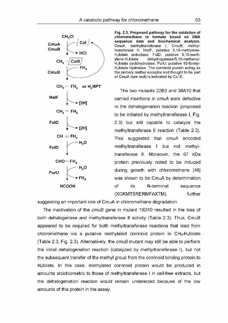
Fig. 2.3. Proposed pathway for the oxidation of
chloromethane to formate based on DNA
sequence data and biochemical analysis.
CmuA, methyltransferase I; CmuB, methyl¬transferase II; MetF, putative 5,10-methylene-H4folate reductase; FolD, putative 5,10-meth-
ylene-H4folate dehydrogenase/5,10-methenyl-H4folate cyclohydrolase; PurU; putative 10-formyl-
H4folate hydrolase. The corrinoid protein acting as
the primary methyl acceptor and thought to be partof CmuA (see text) is indicated by Co-E.
CH, FH.
FolD
[2H]
CH == FH4
FolD/- H2°
' '
CHO-FH4
Purl 1
/- H2°
1 N^FH4HCOOH
The two mutants 22B3 and 38A10 that
carried insertions in cmuA were defective
in the dehalogenation reaction (proposed
to be initiated by methyltransferase I, Fig.
2.3) but still capable to catalyze the
methyltransferase II reaction (Table 2.3).
This suggested that cmuA encoded
methyltransferase I but not methyl¬
transferase II. Moreover, the 67 kDa
protein previously noted to be induced
during growth with chloromethane [48]
was shown to be CmuA by determination
of its N-terminal sequence
(XGKMTSRERMFAXTM), further
suggesting an important role of CmuA in chloromethane degradation.
The inactivation of the cmuB gene in mutant 19D10 resulted in the loss of
both dehalogenase and methyltransferase II activity (Table 2.3). Thus, CmuB
appeared to be required for both methyltransferase reactions that lead from
chloromethane via a putative methylated corrinoid protein to CH3-H4folate
(Table 2.3, Fig. 2.3). Alternatively, the cmuB mutant may still be able to perform
the initial dehalogenation reaction (catalyzed by methyltransferase I), but not
the subsequent transfer of the methyl group from the corrinoid binding protein to
H4folate. In this case, methylated corrinoid protein would be produced in
amounts stoichiometric to those of methyltransferase I in cell-free extracts, but
the dehalogenation reaction would remain undetected because of the low
amounts of this protein in the assay.

54 Chapter 2
Mutants of Methylobacterium sp CM4 disrupted in purU and in cmuC were
unable to grow with chloromethane but exhibited wild type levels of both
dehalogenase and methyltransferase II activity (Table 2 3) These mutants thus
are unaffected in the dehalogenation reaction but are deficient in some later
step of chloromethane metabolism

A catabolic pathway for chloromethane 55
2.5 DISCUSSION
The results presented here lead us to propose the corrinoid-dependent
pathway for chloromethane catabolism shown in Fig. 2.3. This pathway implies
that the dehalogenation reaction proceeds with the Co(l) of a corrinoid protein
acting as primary acceptor for the methyl group of chloromethane. It requires
methyltransferase I to form methylated corrinoid protein from chloromethane,
and methyltransferase II for the transfer of the methyl group to the pterine
cofactor.
The amino acid sequence of CmuA (Table 2.1) supports the view that this
protein not only encodes methyltransferase I activity detected in cell-free
extracts, but also acts as the corrinoid binding protein indicated in the model
(compare Table 2.3). We thus hypothesize that the CmuA protein carrying a
methylated corrinoid serves as the methyl-donating substrate for CH3-H4folate
formation from H4folate by the methyltransferase II encoded by cmuB.
Gift«*»
KtatC
•fell
HutA
CnuA
HtniC
H«tB
But*.
«a i,lk|Bfo||100 V&lHOlHl T&fl. OaI
KfIMeFAB£TlHe
Ha. EPIplïlHMBm . . , BO 5*2
«S- FJUlKlliRTgl« . ..VU W
5*3 *¥*!Iii viiL
79S jfcJJKi!
TNeBTVvfiEKBAEIfMfcsRBeAOKVLi
ÄtSw ma J» m m |
m*sosbmbmewsju|&m d svk.
«.Ol»LBPimiKIÜHItH. RPDXL
W » . » . VSDKW 1Ï2
Fig. 2.4. Alignment of the sequences from CmuA, MtmC of M. barker! (Swissprot Ace. No.
030641), MetH of E. coli (P13009) and MutA, the human methylmalonyl-CoA mutase (P22033),in the conserved C-terminal sequence region of CmuA around the corrinoid binding site. This
site was defined from a structure-based sequence fingerprint for cobalamin dependentmethionine synthases and mutases [69]. The most conserved residues in these motifs
(indicated by grey boxes) are labelled by dots below the alignment. Positions in which amino
acids were identical or similar to the CmuA sequence in at least two other sequences were
boxed in black or grey, respectively, using the program BOXSHADE.
Sequence alignments of the C-terminal domain of the CmuA protein with that
of the corrinoid binding domain of methionine synthase of E. coli, whose
structure has been solved [74], allows one to further speculate on the properties
of the CmuA protein (Fig. 2.4). The most striking feature of the CmuA
sequence, when compared to the motifs which were defined for cobalamin
dependent methionine synthases [69,74], is residue Gln504, equivalent to
56 Chapter 2
His759 in E. coll (Fig. 2.4). The three residues His759, Asp757 and Ser810 in E.
coli methionine synthase form a ligand catalytic triad with the histidine residue
as the lower axial ligand of the "base-off/His-on" corrinoid [69]. His759 in E. coli
was shown to be essential for enzyme turnover [75]. It is unclear in what way
the corresponding glutamine in CmuA can be isofunctional to this residue. No
other sequence in the database so far matches the corrinoid-binding motif so
closely as CmuA but lacks the histidine residue. A glutamine residue, however,
was recently described to be the axial ligand of the nickel porphinoid F430 of
methyl-coenzyme M reductase of M. thermoautotrophicum [76], and the
manually aligned sequence of the AcsD corrinoid iron-sulfur protein of
Clostridium thermoaceticum also features a Gin residue in register with His759
of methionine synthases (S. Ragsdale, personal communication). A glutamine
at position 504 in CmuA is expected to contribute much weaker ligation to a
corrinoid-bound cobalt than a histidine residue. As a consequence, it is also
expected to render the reduction potential of the Co(ll)/Co(l) couple less
negative and thus to stabilize the corrinoid bound by CmuA in its Co(l) state. A
reactive Co(l) species in CmuA would readily react with chloromethane, which
is known to be a good corrinoid alkylating agent [77]. This could contribute to
maintain the methyltransferase I in an active form by preventing oxidation of the
cobalt to Co(ll). In support of this idea, the ATP- and/or reductant-dependent
reactivation system essential for activity of methyltransferases from anaerobes
[60,78,79] and of methionine synthase [80] was not required for chloromethane
dehalogenase activity in cell-free extracts of strain CM4 (Table 2.3). The
sequence similarity of CmuA to E. coli methionine synthase does not include
the C-terminal "AdoMet" domain of methionine synthase involved in reactivation
of the cobalt center [80]. In addition, none of the other protein sequences
deduced from the genes of cluster I or II in strain CM4 (Table 2.1) showed any
detectable similarity to involved in the reductive activation of
methyltransferases.
A catabolic pathway for chloromethane 57
The reactions in the second part of the proposed chloromethane utilization
pathway (Fig 2 3) lead from CH3-H4folate to formate Indication for a H4folate-
dependent pathway specific for the conversion of CH3-H4folate derived from
chloromethane to formate is of interest in the light of recent findings on Ci
metabolism of Methylobacterium extorquens AM1 [43] This organism
possesses a dephospho-tetrahydromethanoptenn-mediated Ci transfer
pathway that is essential for growth with Ci compounds and brings about the
conversion of formaldehyde to carbon dioxide The sequences of many proteins
involved in this pathway most closely resemble those of enzymes that
participate in reduction of carbon dioxide to methane in methanogenic archaea
Dephospho-tetrahydromethanoptenn was previously thought to be unique to
methanogenic and sulfate-reducing archaea [81] In parallel to the
tetrahydromethanoptenn-mediated pathway, M extorquens AM1 appears to
operate a H4folate dependent pathway for the oxidation of formaldehyde to
carbon dioxide With the exception of the gene for NADP-dependent methylene-
H4folate dehydrogenase (mtdA), however, the genes encoding the enzymes of
this pathway are still unknown [43]
The proposed ptenne-dependent pathway for the conversion of
chloromethane to formate in Methylobacterium sp CM4 (Fig 2 3) represents
yet a third variant of the reactions for interconverting Ci compounds in
Methylobacterium Since the emu negative mutant with a disrupted copy of the
purU gene still grew with methanol or methylamine, this pathway would be
specific for processing a methylated ptenne-based cofactor derived from
chloromethane While the nature of the pterin cofactor in this pathway needs to
be determined, we have found that purified CmuB protein catalyses methyl
transfer from methylcobalamin using H4folate but not tetrahydromethanoptenn
as the methyl group acceptor (unpublished data)
58 Chapter 2
In conclusion, our biochemical and genetic data suggest that growth of the
strict aerobe Methylobacterium sp CM4 with chloromethane is based on a
specific catabolic pathway involving corrinoid-dependent enzymes that was
hitherto unknown in organisms with an aerobic lifestyle The similarity in
sequence of CmuA and CmuB to other proteins of related function in
methanogenic archaea (Table 2 1) is interesting from an evolutionary
standpoint, since it extends the emerging notion that genes involved in
methylotrophy and methanogenesis share a common origin [43], and that
strictly anaerobic archaea and aerobic bacteria may use similar reactions to
exploit Ci substrates for metabolism
Chapter 3
Properties of the methylcobalamin:H4folate
methyltransferase involved in chloromethane utilization by
Methylobacterium sp. strain CM4
Alex STUDER, Stéphane VUILLEUMIER and Thomas LEISINGER
European Journal of Biochemistry 264, 242-249 (1999)
60 Chapter 3
3.1 ABSTRACT
Methylobacterium sp. strain CM4 is a strictly aerobic methylotrophic
proteobacterium growing with chloromethane as the sole carbon and energy
source. Genetic evidence and measurements of enzyme activity in cell-free
extracts have suggested a multistep pathway for the conversion of
chloromethane to formate. The postulated pathway is initiated by a corrinoid-
dependent methyltransferase system involving methyltransferase I (CmuA) and
methyltransferase II (CmuB) which transfer the methyl group of chloromethane
onto tetrahydrofolate (H4folate). We report the overexpression in Escherichia
coli and the purification to apparent homogeneity of methyltransferase II. This
homodimeric enzyme with a subunit molecular mass of 33 kDa catalyzed the
conversion of methylcobalamin and H4folate to cob(l)alamin and methyl-
H4folate with a specific activity of 22 nmol min"1 (mg protein) ~1. The apparent
kinetic constants for H4folate were: Km = 240 |j,M, Vmax = 28.5 nmol min_1
(mg
protein) ~1. The reaction appeared first order with respect to methylcobalamin at
concentrations up to 2 mM, presumably reflecting the fact that methylcobalamin
is an artificial substitute for the methylated methyltransferase I, the natural
substrate of the enzyme. Tetrahydromethanopterin, a coenzyme also present in
Methylobacterium, did not serve as a methyl group acceptor for
methyltransferase II. Purified methyltransferase II restored chloromethane
dehalogenation by a cell free extract of a strain CM4 mutant defective in
methyltransferase II.
A methyltransferase for chloromethane degradation 61
3.2 INTRODUCTION
Methylobacterium sp. strain CM4 is a strictly aerobic methylotrophic a-
proteobacterium capable of growth with chloromethane as the sole carbon and
energy source [30]. To elucidate the pathway of chloromethane utilization,
miniTn5 transposon insertion mutants unable to grow with chloromethane were
isolated and characterized. All emu (chloromethane utilization) negative mutants
obtained were still able to grow with methanol, methylamine, or formate, an
observation suggesting that chloromethane was metabolized by reactions
different from those involved in the metabolism of methanol and methylamine
[48]. Sequence analysis of the DNA fragments affected by transposon insertion,
mutant properties, and measurements of enzyme activity in cell-free extracts led
to the proposal of a multistep pathway for the conversion of chloromethane to
formate (see Chapter 2).
This pathway is thought to be initiated by a dehalogenation reaction in which
the Co(l) center of a corrinoid protein acts as primary acceptor for the methyl
group of chloromethane. CmuA, the protein suggested to catalyze this reaction,
has a calculated molecular mass of 67 kDa and appears to represent a fusion of
two proteins that are expressed as separate polypeptides in methyl transfer
systems of methanogenic archaea (see Chapter 2). In its N-terminal part CmuA
shows considerable sequence identity to the 36 kDa methyltransferase MtbA of
Methanosarcina barken that transfers the methyl group from the 29 kDa
corrinoid protein MtmC to coenzyme M [68]. The C-terminal part of CmuA was
found to be similar to MtmC [68,70] as well as to many other corrinoid-binding
proteins. By analogy to similar methyltransferase systems [58,82,83], this
suggested that CmuA acts as both the methyltransferase I and the corrinoid
binding protein in the dehalogenation of chloromethane (Fig. 3.1). Sequence
analysis of cmuB and enzyme activity measurements in crude extracts suggest
that protein CmuB is a methyltransferase II that transfers the methyl group from
Co(lll)-methylated CmuA onto H4folate thereby yielding methyl-H4folate (Fig.
3.1, see Chapter 2). The methyl-H4folate derived from chloromethane is then
processed to formate via pterin-linked intermediates by a pathway which is
62 Chapter 3
proposed to differ from the Ci transfer pathways recently described in
Methylobacterium [44].
A dehalogenation reaction based on a corrinoid-dependent methyltransferase
system is one of the striking features of the proposed pathway for
chloromethane degradation in strain CM4, a strictly aerobic bacterium. A similar
system for chloromethane dehalogenation has been described in the strict
anaerobe Acetobacterium dehalogenans [60]. In contrast to chloromethane
dehalogenation by Methylobacterium sp. CM4, it requires an ATP- and
reductant-dependent reactivation system to maintain the corrinoid cobalt in the
Co(l) state. To reach an understanding of Co(l)-mediated, reactivation-
independent chloromethane dehalogenation in strain CM4 and to determine the
nature of the cofactors involved in this process, we have set out to purify the
proteins involved in the dehalogenation reaction. Here we report on the
purification of CmuB and demonstrate its activity in vitro as a
methylcobalamin:H4folate methyltransferase.
/Co'/
CH3CI^ fMTI >^ -rCH3-H4folate
).
CH3I CmuB (MTII)
HCl V \ / Co"' /.' V H4folatev
CmuA y
(MTI)
Fig. 3.1. Postulated scheme of chloromethane dehalogenation. The reactions catalyzed by
methyltransferase I (MTI, CmuA) and methyltransferase II (MTII, CmuB) are shown.

A methyltransferase for chloromethane degradation 63
3.3 EXPERIMENTAL PROCEDURES
3.3.1 Materials
Restriction endonucleases and T4 ligase were obtained from MBI Fermentas,
Pfu DNA polymerase from Stratagene, methylcobalamin from Sigma, H4folate
and methyl-H4folate from Dr. Schircks Laboratory (Jona, Switzerland). All other
chemicals were of reagent grade from Fluka, Merck or Sigma.
3.3.2 Bacterial strains and growth conditions
E. coli strains XL1-Blue (Stratagene) and BL21(DE3) [84] were grown at 37°
or 30°C in Luria-Bertani medium with constant shaking at 180 rpm. When
required, kanamycin was added at 25 ng/ml and ampicillin at 100 ng/ml. The
media and growth conditions for Methylobacterium sp. strain CM4 [30] and its
miniTn5 insertion mutants have been described previously [48].
3.3.3 DNA manipulations
Preparation of genomic DNA, restriction digests, ligations and
transformations were performed using standard protocols [62]. The plasmid
pBluescript-KSII(+) (Stratagene) was used for cloning.
3.3.4 Construction of the cmuB expression plasmid
A 7.5 kb Kpn\ fragment of genomic DNA was cloned from the
Methylobacterium sp. CM4 mutant 36D3 by selection for kanamycin resistance,
yielding plasmid pME1742. The cloned fragment carried the miniTn5
transposon plus bp 3281 to 8456 of cluster II (accession number AJ011317)
which included an intact copy of the cmuB gene. Sequence data and mutant
properties have been described previously (see Chapter 2).
The cmuB gene was placed under the control of the T7 RNA polymerase
promoter of vector pET24a(+) (Novagene) in a two-step cloning procedure.
First, the cmuB gene was amplified by polymerase chain reaction from
pME1742. The oligonucleotide primers used were 5'-
GGGAGGTTAAATÇATATGAATAAG-3', with a change of two bases
(underlined) to introduce an N-terminal Nde\ site (bold) and 5'-
64 Chapter 3
CGATCAGCAGTCGACCGGTCGGTTGTCCC-3'. The 1012-base pair
polymerase chain reaction product was cloned into the EcoRV site of
pBluescript KSII(+) by blunt-end ligation, generating plasmid pME1747. In a
second step, a 1009-base pair Nde\-Hind\\\ fragment from pME1747 was cloned
into pET24a(+), resulting in the expression plasmid pME1748.
3.3.5 Preparation of cell-free extracts
Cell extracts from Methylobacterium sp. strain CM4 were prepared as
described previously (see Chapter 2). For the production of CmuB protein in E.
coll, strain BL21(DE3) harboring the expression plasmid pME1748 was grown
at 30°C in 5-liter Erlenmeyer flasks containing 1000 ml of LB. When the culture
had reached an A6oo of 0.6, CmuB expression was induced by addition of IPTG
to a final concentration of 500 |j,M and the culture was incubated for another 3 h
to a final A6oo of about 2.0 (3.2 g wet weight). Cells were collected by
centrifugation at 5000 x g for 15 min at 4°C and resuspended in 50 mM Na-
phosphate buffer (pH 8.0; 0.5 g wet cells per ml). The cells were disrupted by
three passages through a French pressure cell (120 mPa, 4°C) and cell debris
was removed by centrifugation (14'000 x g, 30 min, 4°C). Crude cell-free extract
was obtained after ultracentrifugation of the resulting supernatant (100'000 x g,
30 min, 4°C).
3.3.6 Determination of enzyme activity
All solutions used were made anoxic by degassing with 95% N2/5% H2 (v/v).
Subsequent manipulations, enzymatic reactions and measurements were
carried out in an anaerobic chamber under the same conditions. All reactions
were performed in glass cuvettes or reaction vials with gas-tight rubber stoppers
at 30°C for 40 min after addition of the enzyme. Methyl transfer from
methylcobalamin to methyl-H4folate was performed at 30°C with a mixture that
contained 100 mM Tris/HCI (pH 8.5), 2.4 mM H4folate, 0.2 mM methylcobalamin
and between 20 and 40 \ig CmuB protein per ml. Commercially available
H4folate was abiotically converted to methylene-H4folate by the addition of
formaldehyde, and biologically active H4folate was quantified by its NADH-
A methyltransferase for chloromethane degradation 65
dependent conversion to methyl-H4folate using purified methylene-H4folate
reductase from Peptostreptococcus productus [66].
Methylcobalamin:H4folate methyltransferase activity of CmuB was
determined using the photometric assay described by Kreft and Schink [85], by
measuring the rate of cob(l)alamin formation from methylcob(lll)alamin from the
absorbance decrease at 528 nm (A s = 6.2 mM"1 cm"1). Enzyme activity of
CmuB was also determined by measuring the formation of methyl-H4folate from
H4folate and methylcobalamin. Methyl-H4folate was separated from the other
components of the incubation mixture by high-performance liquid
chromatography (HPLC) on a C8-reversed phase column using 0.175% H3P04
in H20 containing 20% methanol as eluent [59]. This method was also used to
monitor chloromethane dehalogenation activity in crude extracts of
Methylobacterium sp. CM4. One unit is defined as the amount of enzyme that
catalyzes the conversion of 1 nmol of methylcobalamin to cob(l)alamin or the
formation of 1 nmol of methyl-H4folate per minute.
3.3.7 Purification of methylcobalamin :H4folate methyltransferase
overexpressed in E. coli
Ammonium sulfate was added to crude cell-free extract from E coli
BL21(DE3)(pME1748) up to a concentration of 1.72 M and the resulting
suspension was centrifuged at 45'000 x g for 30 min. The precipitate was
discarded and the filtered supernatant was fractionated on a Porös PE 4.6/10
hydrophobic-interaction column with a BioCAD SPRINT apparatus (PerSeptive
Biosystems Inc.) at a flow rate of 3 ml/min, using a stepwise gradient from 1.72
to 0 M in 50 mM Na-phosphate buffer pH 8.0. Active CmuB eluted at an
ammonium sulfate concentration of about 0.4 M. Active fractions were pooled
and subsequently desalted by repeated concentration and dilution in 50 mM Na-
phosphate buffer (pH 8.0) using a Biomax 10K centrifugal filter (Millipore). The
pooled fractions containing CmuB were then applied on a 1 ml ResourceQ
anion-exchange column (Pharmacia) at a flow rate of 3 ml/min and eluted by a
linear gradient of increasing NaCI in 50 mM Na-phosphate buffer (pH 8.0). The
CmuB protein eluted at a concentration of about 270 mM NaCI.

66 Chapter 3
3.3.8 Protein determination.
Protein was determined by the method of Bradford [67] using the BioRad
reagent. Bovine serum albumin (Sigma) was used as the standard.
A methyltransferase for chloromethane degradation 67
3.4 RESULTS
It was previously found that cell-free extract of Methylobacterium sp. CM4
grown with chloromethane catalyzed methyl transfer from chloromethane (3
mU/mg protein) onto H4folate. This extract, but not extract from methanol-grown
cells, also catalyzed methyl transfer from methylcobalamin to H4folate at a rate
of 0.8 mU/mg protein. Methyltransferase II of the chloromethane
dehalogenation system thus appeared to accept methylcobalamin as an
artificial methyl donor in place of methylated corrinoid protein. Cell-free extract
from strain 19D10, a transposon insertion mutant that did not grow with
chloromethane, lacked activity with chloromethane or methylcobalamin as a
methyl donor (see Chapter 2). Since the transposon had inserted into cmuB,
this gene was proposed to encode methyltransferase II (Fig. 3.1). The use of
methylcobalamin as an artificial substrate for the CmuB protein enabled to
monitor its activity. We thus have purified CmuB and characterized it as a
methylcobalamin:H4folate methyltransferase whose function is in accordance
with the scheme proposed in Fig. 3.1.
Examination of the protein pattern of crude extracts of Methylobacterium sp.
CM4 and of the cmuB mutant 19D10 by SDS/PAGE showed that the CmuB
protein accounted for only about 2% of the total soluble protein. This estimation
was based on the staining intensity of a 33 kDa chloromethane-induced protein
which was absent from extracts of mutant 19D10 (data not shown). To obtain
extract with a high concentration of methylcobalamin:H4folate methyltransferase
as starting material for enzyme purification, the cmuB gene from
Methylobacterium sp. CM4 was expressed in E. coli.
3.4.1 Enzyme purification and molecular properties
The cmuB gene was amplified by the polymerase chain reaction and ligated
into a pET-type expression vector [84] to yield plasmid pME1748. E. coli
transformants carrying pME1748 produced a soluble protein with an apparent
molecular mass of 33 kDa. This protein was not present in extract from
untransformed recipient cells and its formation was dependent on the addition
of IPTG to the growing culture of the recombinant E. coll strain (Fig. 3.2, lanes 1

68 Chapter 3
and 2). The apparent molecular mass of the CmuB protein produced in the
heterologous host was in accordance with the subunit molecular mass of 33.3
kDa calculated from the translated cmuB gene sequence. Crude cell-free
extract of E. coli BL21(DE3)(pME1748) contained about 20% CmuB protein
which accounted for 5.2 mU/mg protein of methylcobalamin:H4folate
methyltransferase. The increase in enzyme specific activity and in the
concentration of CmuB thus was seven- and tenfold as compared to the values
measured in crude extract of chloromethane-induced Methylobacterium sp.
CM4. From this it appears that the specific activity of the recombinant CmuB
protein from E coli was similar to that of the enzyme in cell-free extracts of
Methylobacterium.
(kDa) M 1 2 3 4 M
200
21.5
Fig. 3.2. Purification of the Methylobacterium sp. CM4 methylcobalamin:tetrahydrofolate
methyltransferase from cell-free extract of E. coli BL21(DE3) (pME1748). Protein samples
(10 |ig) were analyzed at different stages of purification on a denaturing 12% SDS-
polyacrylamide gel and stained by Coomassie brilliant blue. Lane M, molecular weight markers
in kDa; lane 1, cell-free extract of non-induced cells; lane 2, cell-free extract of IPTG-induced
cells; lane 3, fraction after PE hydrophobic interaction chromatography; lane 4, purified CmuB
obtained after ResourceQ ion exchange chromatography.
116.2
97.4
66.2
45.0

A methyltransferase for chloromethane degradation 69
Recombinant CmuB protein was purified under aerobic conditions by
hydrophobic interaction and by anion-exchange chromatography. Copurification
of the activity catalyzing methylcobalamin-dependent methyl-H4folate formation
and of the activity promoting H4folate-dependent methylcobalamin consumption
was observed, thus demonstrating that both of these activities are characteristic
of the methylcobalamin:H4folate methyltransferase activity of CmuB. Purification
was 4.3-fold with 39% yield (Table 3.1). The purified enzyme retained about
80% of its activity after 5 months of storage at -20°C in buffer containing 20%
(v/v) of glycerol.
Table 3.1. Purification of the overexpressed CmuB protein from E. coli
Specific activity
Purification Total Total methyl-H4folate methyl-cobalamin Purification Yield
step protein activity formation formation (-fold) (%)
(mg) (mU) (mU/mg) (mU/mg)
Cell-free 17.7 101 5.2 5.7 1.0 100
extract
(NH4)2S04 16.5 106 6.4 6.8 1.2 105
HICPEPoros 3.2 41 11.4 12.8 2.2 41
ResourceQ 1.6 39 22.3 24.3 4.3 39
As shown by SDS/PAGE, the preparation obtained after two steps of column
chromatography contained a single polypeptide (Fig. 3.2, lane 4). Its N-terminal
amino acid sequence (15 amino acids) determined by Edman degradation
included the N-terminal methionine and corresponded to the sequence
predicted from the cmuB gene. The purified enzyme eluted from a Superdex 75
HR column with an apparent molecular mass of 66 kDa (data not shown),
suggesting that the enzyme has a homodimeric structure. The UV-visible
spectrum of the enzyme gave no indication for the presence of a chromophoric
prosthetic group. Addition of EDTA (10 mM final) had no effect on the activity of
the pure enzyme.
70 Chapter 3
3.4.2 Catalytic properties
The effect of pH and temperature on enzyme activity and the substrate
affinity of the purified enzyme were determined and this led to the standard
assay conditions described in Materials and Methods. The enzyme activity was
only weakly affected by the pH, and a broad activity optimum was observed
around pH 9.2. The temperature optimum for the reaction was 48°C. When the
dependence of the rate of methyl transfer on the concentrations of the
substrates was examined, Michaelis-Menten kinetics were observed with
respect to H4folate (Fig. 3.3A). At a methylcobalamin concentration of 0.2 mM,
the apparent Km for H4folate was 240 ± 10 |j,M and the apparent Vmax was 28.5
± 1.0 mU/mg protein.The reverse reaction, methyl transfer from H4folate to
cob(l)alamin also appeared to follow Michaelis-Menten kinetics Fig. 3.3B). At a
cob(l)alamin concentration of 0.2 mM, the apparent Km for methyl-H4folate was
12.5 + 1 mM and the apparent Vmax was about 1200 ± 100 mU/mg protein. It
was not possible to obtain an apparent Km for methylcobalamin since the rate of
methyl-H4folate formation was found to be first order with respect to
methylcobalamin up to 2 mM (Fig. 3.3C). However, the methyl transfer reaction
to H4folate presumably occurs under saturation conditions in vivo, since the
physiological cobamide mimicked by methylcobalamin in vitro is thought to be
protein-associated.
In view of the recent discovery that Methylobacterium, in addition to H4folate,
possesses H4MPT as a carrier of one-carbon units [43], it was tested whether
H4MPT served as a methyl group acceptor in the reaction catalyzed by CmuB.
No reaction occurred when purified enzyme was provided with 1 mM H4MPT in
the place of H4folate under standard assay conditions.
A methyltransferase for chloromethane degradation 71
^ 30ö)
E
3E
9- 20 -
10 -
1 2
s (H4folate [mM])
600 -
B•
500 -
//*
400 -
/**
300 -
• /
200 -
•A
100 -
0 -
2 4 6 8 10
s (CH3H4folate [mM])
12
180 -
160
140
120
100
80
60
40
20
0
0 1
s (methylcobalamin [mlV
Fig. 3.3. Dependence of methylcobalamin:tetrahydrofolate methyltransferase activity on
the substrate concentration. Assays were conducted with 5 |ig of pure CmuB protein at 30°C
in 100 mM Tris/HCI buffer pH 8.5. (A) Dependence of the rate of methylcobalamin consumptionon the concentration of H4folate. The inset shows the same data plotted according to Hanes
[86]. The assay mixtures contained 0.2 mM methylcobalamin and H4folate at the concentrations
indicated. The rates were determined by following the absorbance change at 528 nm. (B)
Dependence of the rate of conversion of cob(l)alamin to methylcob(lll)alamin on the
concentration of methyl-H4folate. The assay mixture contained 0.2 mM cobalamin, 2.5 mM
titanium(lll)citrate, and methyl-H4folate at the concentrations indicated. Rates were determined
by following the change in absorbance at 528 nm. (C) Dependence of the rate of methyl-H4folate production on the concentration of methylcobalamin. The assay mixtures contained 4
mM H4folate and methylcobalamin at the concentrations indicated. The reaction rates were
determined by HPLC analysis of methyl-H4folate formation.
72 Chapter 3
The time-dependence of the visible spectrum of the incubation mixture after
addition of CmuB is shown in Fig. 3.4. A time-dependent decrease of the
methylcobalamin absorbance peak at 528 nm was observed, which could be
used to determine the initial rate of the reaction catalyzed by CmuB.
Concomitantly to the decrease in absorbance at this wavelength, there was an
increase at 388 nm where cob(l)alamin exhibits a typical absorbance peak [85].
This is strong evidence that, in accordance to the scheme proposed in Fig. 3.1,
cob(l)alamin is indeed the product of the methyltransferase II reaction catalyzed
by CmuB. In mixtures that were exposed to air or contained dithiothreitol, the
spectrum showed absorbance characteristics typical for cob(ll)alamin (not
shown), indicating oxidation of Co(l) to Co(ll) by oxygen or by traces of disulfide
present in the DTT preparation [87].
350 400 450 500 550 600 650
Wavelength (nm)
Fig. 3.4. Formation of cob(l)alamin by purified methylcobalamin:tetrahydrofolate
methyltransferase. The reaction was carried out under anaerobic conditions at 40°C in 100
mM Tris/HCI buffer pH 8.5 containing 0.2 mM methylcobalamin and 4 mM H4folate. The reaction
was started by the addition of 50|ig of purified CmuB protein. The spectra plotted were taken at
different times after addition of the enzyme. The increase of absorbance at 388 nm is
characteristic for the formation of the cob(l)alamin species [85].
A methyltransferase for chloromethane degradation 73
3.4.3 Restoration of dehalogenation activity in cell extract of a cmuB
mutant
Cell-free extract from Methylobacterium wild type cells was able to
dehalogenate chloromethane, i.e. to transfer the methyl group onto H4folate by
the concerted action of methyltransferase I and methyltransferase II. Because
of its deficiency in methyltransferase II, extract from the cmuB mutant 19D10
was unable to carry out this reaction. As shown in Fig. 3.5, the ability to convert
chloromethane to methyl-H4folate was restored to wild type level by the
addition to the mutant extract of purified CmuB protein in threefold molar excess
over CmuA. This suggests that the heterologously expressed and purified
CmuB protein catalyzed not only the methylcobalamin:H4folate
methyltransferase reaction but also the physiological methyl transfer from
methylated CmuA protein onto H4folate. It also indicates that the proteins
possessing methyltransferase I and methyltransferase II activities need not be
coexpressed for chloromethane dehalogenation activity to be obtained.
08
Ö 06E
E,
1 04
X
f 02
00
0 100 200 300
Time (mm)
Fig. 3.5. In vitro complementation of chloromethane dehalogenation by purified
methylcobalamin:tetrahydrofolate methyltransferase. Formation of methyl-H4folate from
chloromethane was measured in a 2 ml assay mixture containing 50 mM Tris/HCI pH 8.5, 4 mM
H4folate and 2 mM chloromethane in the gas-phase, and either (•) 400|ig cell-free extract from
Methylobacterium sp. CM4 wild type, or (O) 400|ig cell-free extract from the cmuB mutant
19D10, or (t) a mixture of 400|ig cell-free extract from the cmuB mutant 19D10 and 50|ig
purified CmuB protein.

74 Chapter 3
3.5 DISCUSSION
The results reported here support the concept that chloromethane
dehalogenation by Methylobacterium sp. CM4 proceeds by two sequential
methyltransferase reactions. In the first reaction, the bifunctional protein CmuA
acts as a methyltransferase I and at the same time provides a cobalamin
prosthetic group which serves as an intermediate methyl carrier. In the second
reaction, protein CmuB acts as a methyltransferase II and transfers the methyl
group from CmuA onto H4folate (Fig. 3.1). Mutational loss of either CmuA or
CmuB activity has previously been shown to abolish the liberation of chloride
from chloromethane as well as the conversion of chloromethane to methyl-
H4folate (see Chapter 2, [48]). Addition of purified methyltransferase II
reconstituted the in vitro transformation of chloromethane to methyl-H4folate by
a cell-free extract of a mutant devoid of CmuB activity, thus demonstrating the
expected interaction of methyltransferase II with methylated methyltransferase I
(Fig. 3.5).
Based on its reaction with methylcobalamin, methyltransferase II of
Methylobacterium sp. CM4 was purified and characterized as
methylcobalamin:H4folate methyltransferase. Non-physiological methyl transfer
from free methylcobalamin has previously been reported for corrinoid-
dependent methyltransferases involved in the metabolism of one-carbon
compounds by anaerobic Archaea and Bacteria. This applies for example to the
methyltransferases involved in the formation of methyl-coenzyme M from
methanol and coenzyme M by Methanosarcina barker!. Protein MtaB, the
methyltransferase I of this system, catalyzes the hydrolysis of methylcobalamin
to cob(l)alamin plus methanol [83] and protein MtaA, the methyltransferase II,
can use free methylcobalamin as the methyl donor for the formation of methyl-
coenzyme M [88]. Similarly, the methyltransferase II of the Halophaga foetida
enzyme system for O-demethylation of methoxylated aromatic compounds
catalyzed the methylation of H4folate with methylcobalamin as an artificial
substrate [85]. These enzymes exhibited either a very high apparent Km value
for methylcobalamin [88] or, similar to methyltransferase II of Methylobacterium

A methyltransferase for chloromethane degradation 75
sp CM4, displayed first order kinetics with respect to the concentration of
methylcobalamin [83,85]
The CmuB sequence is most similar to the sequence of subunit H of the
methyl-H4MPT coenzyme M methyltransferase from methanogens (see
Chapter 2) It can best be aligned to domain 9948 of the ProDom database [89]
that was defined on the basis of the sequences of the MtrH protein from
Methanobactenum thermoautotrophicum [90] and related proteins This is
somewhat intriguing given that in CmuB, methyltransferase activity was only
detected with H4folate as the methyl acceptor Modest similarity of MtrH to the
putative H4folate binding domain of cobalamin-dependent methionine synthase
from E coli (MetH), however, was noted previously [91] A more detailed
analysis of the CmuB sequence using pattern-initiated BLAST (PHI-BLAST,
[92]) with the short conserved motif D-[FY]-[IVL]-X-[FY]-G-[PL]-[IV]-[ED]
common to CmuB and sequences in the MtrH-based Prodom domain yields that
only MtrH- and MetH-hke sequences as significant hits The ProDom domain
9873 corresponding to the putative H4folate binding domain in cobalamin-
dependent methionine synthases [93] also includes protein AcsE of Clostridium
thermoaceticum, which is involved in methyl transfer from methyl-H4folate to an
iron/sulfur corrinoid protein in acetate synthesis from C02 [94] Overall, the level
of sequence similarity between MtrH, MetH, and CmuB is quite low (Fig 3 6) A
sequence motif can be defined that is both common and unique to all these
proteins (see Fig 3 6, legend) However, in the absence of knowledge on the
residues involved in catalysis or cofactor binding in any of these proteins, the
significance of this motif remains unclear

76 Chapter 3
CmuB
MtrH
MetH
CmuB
MtrH
MetH
CmuB
MtrH
MetH
CmuB
MtrH
MetH
CmuB
MtrH
MetH
CmuB
MtrH
MetH
1 lFRFDKEgl\^DJAET^BQp|341 r. .lsgl.epBIWedIlfv:
5 7 vndth^kyEmqmafdviaas61 BF,j3MSnVmBNl3HTVOTFGOTPl
390 ffvgNffi^QIigiNMDEGMLDAE.
114 gflLBC118 EvgLEjl448 ..JQGK
|L^DTEI1
169. . .FRPJLDAlgvEAP. .McG§MD . PPSlglTNlAIKL JRETY. .[
17 7 DKGLLEiAAlc&äDKY. . LmHva|tPLGQMvA\^TSFA ftSKW- -BP"!!^3502 rraykilteBvBfppediHfwpnifavatBHeehnnyaodfigacediBr.elBhalisH
* * *
214 a|ajScfpq|tBIkdlgB1 fvnlslgaJl|sirf HlMlL
226 BH£SASD^REYKHHKEA WPVCP|GJNfllQQM ,
561 MSiMFSFRËNnPvBFATHAvBlYYAIRNSMDMGBlNlGOLAIYDDLPAELRDaVEBVI
* **
253 HBLJRTVPAAHMsGE^LGFBQELDGHTLPRPaVLSMFKLAlPATGPAAVAAE*268 pÉBNAKMAFiciMADlllSEiKDIGTEPV.EDÏFFK|L*62l |nrrddgterllelaekyrgtktddtanaqqaewrswevnkr|eysljkgitefieqdte
Fig. 3.6. Sequence analysis of the CmuB protein. The alignment shows sequence similarityof CmuB to the H subunit of the A/5-methyltetrahydromethanopterin:coenzyme M methyl¬transferase complex of Methanobacterium thermoautotrophicum (MtrH [90], 30% identity) and
to the putative tetrahydrofolate binding domain in E. coli cobalamin-dependent methionine
synthase (MetH [93], 13% identity). The short ad hoc sequence pattern (PROSITE format [95])
D-[FY]-[IVL]-X-[FY]-G-[PL]-[IV]-[ED] (dots) unique to CmuB and MtrH-like sequences was used
in the pattern-hit initiated sequence analysis [92] of CmuB (see text). Absolutely conserved
residues in MtrH-like sequences (ProDom domain 9948; release 99.1, [89]), or in MetH-like
sequences (domain 9873) including the AcsE protein from Clostridum thermoaceticum [94], are
boxed in black. Other residues identical in two or more sequences in the alignment are boxed in
grey. The asterisks indicate a sequence pattern, G-[EA]-X(2)-[TNG]-X(47,56)-[LIM]-X(16,20)-
[DN]- [RSA]- X(8) - [GA] - X (10,15) - [NA] - S-X(19,24) -[AL]- X(10,18)-[GA]- X(9,23) -G- X(6,8)-
D-X(19,27)-[KRA]-X(8,10)-[GA]-X-[HAS]-N, that is present in all 8 MtrH- and in 9 MetH-related
sequences only (Swissprot release 38; Embl release 55).
The physiological reaction catalyzed by CmuB is suggested to be the transfer
of a methyl group from a corrinoid protein to H4folate. The corresponding methyl
transfer reactions in the methyl-H4folate: corrinoid/iron-sulfur protein
methyltransferase from Clostridium thermoaceticum [96], in methyl-H4MPT:
coenzyme M methyltransferases from methanogens [97], or in methionine
synthases [93], proceed in the reverse direction in vivo. In these cases, methyl-
H4folate or methyl-H4MPT serves as the alkylating agent for a corrinoid protein
with cobalt in the Co(l) state. The absorption maxima and isobestic points that
arise in the CmuB-catalyzed reaction of methylcobalamin in the presence of
H4folate are characteristic of Co(l) (Fig. 3.4) and suggest a Sn2 type reaction
A methyltransferase for chloromethane degradation 77
involving formal transfer of 2 electrons rather than a radical reaction [96] Most
notably, cob(l)alamin is produced by CmuB under conditions (95% N2/5% H2)
that cannot elicit the regeneration of cob(l)alamin from cob(ll)alamin CmuB was
found to have a much lower apparent Km for H4folate (250 |j,M, Fig 3 3A) than
for methyl-H4folate (12 5 mM, Fig 3 4B), in contrast to what was observed
previously for the AcsE methyltransferase from Clostridium thermoaceticum
[96]
Active CmuB protein purified from E coli is now helping to isolate and
characterize the other protein(s) comprising the chloromethane dehalogenase
of Methylobacterium sp CM4 Coupling of methyltransferase I to purified
methyltransferase II provides an activity-based assay for methyltransferase I
that is now being exploited for the purification and characterization of the CmuA
protein By affording the physiological, protein-associated methylcobamide
methyl donor, purified CmuA protein should allow investigation of the kinetic
properties of the CmuB protein in catalyzing the transfer of a cobalt-bound
methyl group to H4folate

Chapter 4
Chloromethane:tetrahydrofolate methyl transfer by two
proteins from Methylobacterium chloromethanlcum strain
CM4
Alex STUDER, Erhard STUPPERICH1, Stéphane VUILLEUMIER
and Thomas LEISINGER
1Mikrobiologie und Biotechnologie, Universität Ulm,
D-89069 Ulm, Germany
European Journal of Biochemistry 268, 2931 -2938 (2001 )

80 Chapter 4
4.1 ABSTRACT
The cmuA and cmuB genes are required for growth of Methylobacterium
chloromethanlcum strain CM4 with chloromethane as the sole carbon source.
While CmuB was previously shown to possess methylcobalamin:tetra-
hydrofolate methyltransferase activity, sequence analysis indicated that CmuA
represented a novel and so far unique two-domain methyltransferase/corrinoid-
binding protein involved in methyl transfer from chloromethane to a corrin
moiety. CmuA was purified from wild-type M. chloromethanlcum strain CM4 and
characterized as a monomeric, cobalt and zinc containing enzyme of a
molecular mass of 67 kDa with a bound vitamin B12 cofactor. In combination,
CmuA and CmuB proteins catalyze the in vitro transfer of the methyl group of
chloromethane to tetrahydrofolate, thus affording a direct link between
chloromethane dehalogenation and core Ci metabolism of Methylobacterium.
Chloromethane dehalogenase activity in vitro is limited by CmuB, as formation
of methyltetrahydrofolate from chloromethane displays apparent Michaelis-
Menten kinetics with respect to methylated CmuA, with an apparent Km of 0.27
|j,M and a Vmax of 0.45 U mg"1. This contrasts with sequence-related systems for
methyl transfer from methanogens, which involve methyltransferase and
corrinoid protein components in well-defined stoichiometric ratios.

Chloromethane dehalogenase 81
4.2 INTRODUCTION
Chloromethane, a compound which to a large extent is naturally produced, is
the most abundant halogenated compound in the atmosphere and accounts for
about 15% of the destruction of the ozone layer by halogenated chemicals [8,9].
Characterization of the sources and sinks of chloromethane in the environment
is thus of topical interest. While the global budget for chloromethane presents a
significant deficit in known sources of the compound [8], additional sinks of
bacterial origin have recently emerged [29,30,59,98]. The strictly aerobic,
facultatively methylotrophic a-proteobacterium Methylobacterium chloro¬
methanlcum strain CM4 [30,33,48] is capable of growth with chloromethane as
the sole source of carbon and energy. Physiological and genetic studies with
this organism have shown that the products of the cmuA and cmuB genes are
essential for the dehalogenation of chloromethane and growth with this
compound, and have further suggested that chloromethane is degraded to
formate by a multistep pathway (Fig. 4.1; see Chapter 2, [48]).
The cmuA gene encodes a 67 kDa protein with unique features. CmuA
shows similarity to known methyltransferases as well as to their cognate
corrinoid protein binding proteins in the N-terminal and C-terminal part of its
sequence, respectively (see Chapter 2). In analogy to the scheme proposed for
the chloromethane dehalogenase of anaerobic bacteria [60], the CmuA protein
was suggested to catalyze the first step of the chloromethane degradation
pathway, comprising the dehalogenation of chloromethane and the methylation
of an associated cobalt corrin moiety. The second step of the pathway likely
involves CmuB, which was shown to be a homodimeric enzyme of 33 kDa
subunits capable of using methylcobalamin as a substitute for methylated
CmuA protein as a methyl donor to generate methyltetrahydrofolate (CH3-
H4folate) from tetrahydrofolate (H4folate, see Chapter 3). Later steps in the
chloromethane degradation pathway (Fig. 4.1) remain to be investigated in
detail. However, genes metF, folD and purU encode proteins similar to enzymes
involved in the oxidation of pterin-linked one-carbon units to formate. These
genes were found nearby the cmuA and cmuB genes in M. chloromethanlcum
82 Chapter 4
strain CM4, and mutational inactivation of the operon containing purU and folD
genes prevented growth of this strain with chloromethane (see Chapter 2). The
chloromethane degradation pathway postulated for M. chloromethanlcum strain
CM4 may be widespread among methylotrophic bacteria that grow with this
compound. Indeed, cmuA and cmuB genes were also identified in
Hyphomicrobium chloromethanlcum strain CM2 [99], and a protein of similar
size and 81% identical N-terminal sequence to CmuA was described from the
chloromethane degrading strain CC495 [29].
CH»CI
f ï
/ Co" /
CmuA
/ Co1"/CmuA
(MTII) H4to1ati
CH3-H4folati
CH2-H4folate
i FolDi
i PurUf
formate
' Serine I
k eye I» jj
^
Fig. 4.1. Postulated scheme for chloromethane
utilization in M. chloromethanlcum strain CM4.
Transformations involving single enzymes are
indicated by continuous arrows and multistep
enzymatic pathways by broken arrows. MetF (5,10-
methylene-H4folate reductase), FolD (5,10-methy-lene-H4folate dehydrogenase / 5,10-methenyl-
H4folate-cyclohydrolase), PurU (10-formyl-H4folate
hydrolase), proteins involved in the catabolic
pathway basing on previous sequence analysis
(see Chapter 2).
In the present study, we report on the reconstitution in vitro of the novel
dehalogenation and methyl transfer reaction catalyzed in the two initial steps of
the pathway (Fig. 4.1). We present the purification of the CmuA protein from M.
chloromethanlcum strain CM4 and identify vitamin B12 as a prosthetic group of
the enzyme. It is shown that purified CmuA displays the properties inferred from
sequence analysis in that it functions both as a chloromethane
methyltransferase and as the substrate of the CmuB protein, which transfers the
corrinoid-bound methyl group derived from chloromethane to H4folate.

Chloromethane dehalogenase 83
4.3 EXPERIMENTAL PROCEDURES
4.3.1 Materials
H4folate and CH3-H4folate were purchased from Dr. Schircks Laboratory
(Jona, Switzerland). Protein markers for SDS-PAGE came from New England
Biolabs, and reference proteins for molecular mass determination from
Pharmacia. N-ethylmaleimide, o-phenanthroline and EDTA were from Fluka. All
other chemicals were reagent grade and obtained from Fluka, Merck or Sigma.
4.3.2 Growth conditions and preparation of cell-free extract
Methylobacterium chloromethanlcum strain CM4 is deposited in the All-
Russian Collection of Microorganisms as VKM B-2223. Media and the growth
conditions for the organism have been described previously [48]. Bacteria were
harvested from a chloromethane-grown culture (12 I) at late exponential phase
(OD6oo 0.8 - 0.9) by centrifugation for 30 min at 10,000 g. The cell pellet (14.5 g
wet weight) was resuspended in 40 ml of 50 mM sodium phosphate buffer (pH
8.0), disrupted by four passages through a French pressure cell (120 MPa, 4°C)
and cell debris removed by centrifugation (14,000 g, 30 min, 4°C). Crude cell-
free extract was obtained after ultracentrifugation of the resulting supernatant
(100,000 g, 1 h, 4°C).
4.3.3 Isolation of corrinoids from M. chloromethanlcum strain CM4
Bacteria were grown on chloromethane in the presence of 0,1 |j,M [57Co]
(Amersham Buchler) cobalt chloride (275 Bqnmol"1), harvested during
exponential phase (8.3 g wet weight) and resuspended in 50 ml 100 mM
sodium acetate buffer pH 5.0. Corrinoids were isolated using a method adapted
from [100]. After addition of potassium cyanide (final concentration 0.1 mM) and
boiling for 20 min, the resulting suspension was centrifuged (10,000 g, 10 min)
after cooling and the pellet reextracted with 100 mM sodium acetate buffer pH
5.0 containing 0.1 mM KCN (26 ml). Supernatants were pooled and loaded onto
a XAD-4 column 10/3 (Serva). The column was washed with 60 ml H20 and the
corrinoids subsequently eluted with 10 ml methanol. The methanol fraction was
flash-evaporated at 30°C, the residue redissolved in 1 ml H20 and analyzed by
84 Chapter 4
reversed phase HPLC (C18 Nucleosil 120-10, 3.9 mm x 21 cm column)
equilibrated in a mixture of 17 mM aqueous acetic acid and methanol in a 23/77
(vol/vol) ratio. Corrinoids were eluted (12 ml/min) with a linear gradient of the
same solvent components from 23/77 to 40/60 in 30 minutes. The elution was
monitored spectrophotometrically at 254 and 546 nm and 0.5 ml fractions were
collected, which were subsequently analyzed in a Packard Minaxi auto-gamma
5000 gamma counter.
4.3.4 CmuA purification
Ammonium sulfate (final concentration, 1.64 M) was added to crude cell-free
extract of M. chloromethanlcum strain CM4 and the resulting suspension
centrifuged at 45,000 g for 60 min. After further ammonium sulfate addition to
the resulting supernatant (final concentration 2.46 M), the precipitate was
harvested by centrifugation (45,000 g, 60 min) and treated with 20 ml 1.64 M
ammonium sulfate in 50 mM sodium phosphate buffer (pH 8.0). After clearing
by centrifugation (45,000 g, 60 min), the supernatant was filtered through a 0.45
nm filter disk (Millipore) and loaded on a Source 15ISO hydrophobic interaction
column 10/10 (Pharmacia) connected to a BioCAD SPRINT apparatus
(PerSeptive Biosystems). Protein was fractionated at a flow rate of 1.5 ml min"1
with a stepwise gradient from 1.64 M to 0.82 M ammonium sulfate in 50 mM
sodium phosphate buffer, pH 8.0. Fractions eluting between 1.25 and 1.0 M
ammonium sulfate were pooled, concentrated using a Biomax 50 K centrifugal
filter (Millipore), and eluted with 50 mM Tris chloride pH 8.0 through a PD10
size exclusion cartridge (Pharmacia) equilibrated in the same buffer. The
protein fraction was then applied to a MonoQ anion exchange column 5/5
(Pharmacia) and eluted at a flow rate of 1.5 ml min"1 in a gradient of 0-200 mM
NaCI in 50 mM Tris chloride, pH 8.0. Purified CmuA protein eluted at a
concentration of approximately 125 mM NaCI.
4.3.5 Molecular mass determination
The apparent molecular mass of the CmuA protein was determined by
analytical gel filtration on a Superdex 200 column (Pharmacia) equilibrated and
eluted in 50 mM Tris chloride (pH 8.0) containing 200 mM NaCI. The column

Chloromethane dehalogenase 85
was calibrated using ferritin (440 kDa), catalase (232 kDa), aldolase (158 kDa),
bovine serum albumin (67 kDa), ovalbumin (43 kDa) and RNAse (13.7 kDa) as
reference proteins.
4.3.6 Other protein chemical methods
Protein was determined by the method of Bradford [67] using a commercial
reagent (BioRad) and bovine serum albumin (Sigma) as the standard. Protein
purification was followed by SDS-PAGE (12% acrylamide) and Coomassie blue
staining. UV-visible absorbance spectra were recorded on a Hitachi U3300
spectrophotometer (1 nm interval, 300 nm min"1), at room temperature in quartz
cuvettes of 1 cm pathlength. MALDI-TOF analysis was performed on a
Voyager-DE Elite (Perseptive Biosystems) mass spectrometer operated in the
reflection mode, using 2,5-dihydroxybenzoic acid as the matrix. Two-
dimensional gel electrophoresis was carried out as previously described [101]
using the broad pi calibration kit (Pharmacia) for pi determination. ICP analysis
was performed in metal-free 5 mM Tris chloride buffer pH 8.0 on a SCIEX ELAN
6100 DRC ICP-MS (Perkin Elmer) in the Laboratory of Inorganic Chemistry
ETH Zürich (Prof. D. Günther). The N-terminal sequence of the CmuA protein
was determined by Edman degradation (Applied Biosystems 476A) after
electrophoresis of the purified protein by 8% SDS-PAGE and electroblotting of
the protein band onto a PVDF membrane (Immobilon-P, Millipore) as described
by the manufacturer.
4.3.7 Enzyme assays
Methyl transfer from chloromethane catalyzed by the CmuA protein was
determined by following CH3-H4folate formation in a coupled assay with CmuB
protein purified from E. coli (see Chapter 3). Methyl-H4folate formation was
quantified by HPLC with spectrophotometric detection at 320 nm as described
previously (see Chapter 2). Solutions were made anoxic by degassing with
N2/H2 (95/5 [vol/vol]) and enzyme assays were performed under the same
atmosphere. Reactions (500 jLtl liquid volume) were performed at 30°C in 9 ml
crimp vials sealed with gas-tight butyl rubber stoppers in 100 mM Tris sulfate
buffer (pH 8.7) containing 2.4 mM H4folate (of which 50% was biologically

86 Chapter 4
available, [66]), 2 mM titanium(lll)citrate, 1.82 \M purified CmuB protein and
CmuA containing protein fraction (4-12 ngml"1). The enzymatic reaction was
started by addition, through the rubber stopper with a gas-tight syringe, of 200
jLtl (8.9 nmol) chloromethane corresponding to an initial concentration of 2.1 mM
in the liquid phase assuming a Henry constant of 0.43 [65]. One unit is defined
as the amount of enzyme that catalyzes the formation of 1 nmol CH3-H4folate
per minute.
The halomethane:halide exchange activity of the CmuA protein was
measured by a previously described method [29] in a total reaction volume of 1
ml comprising 50 mM sodium phosphate buffer (pH 7.0), 5 mM DTT and 2 mg
cell-free extract or 50 \ig (0.75 |j,M) purified CmuA protein using the same vials
as above. Chloromethane (0.5 mM based on the liquid phase volume) was
added through a gas-tight syringe and the sealed vial incubated at 30°C for 1 h,
after which it was opened and allowed to stand in air for 1 h. Chloromethane
was again added, followed by potassium iodide (3 mM) to initiate the halide
exchange reaction. The vial was sealed again, incubated at 30°C, and
chloromethane consumption and iodomethane production were quantified by
gas chromatography using flame ionization detection (Hewlett Packard 8700
gas Chromatograph). Headspace samples (200 nl) were injected on a 180 x 0.2
cm Poropack P (80/100 mesh) column (SupeIco) at an oven temperature of
150°C, with N2 (30 ml min"1) as the carrier gas. Under these conditions,
chloromethane and iodomethane eluted from the column at retention times of
58 s and 91 s, respectively.
Chloromethane dehalogenase 87
4.4 RESULTS
4.4.1 Cobalt requirement of M. chloromethanlcum strain CM4
Sequence analysis of the DNA comprising the cmuA and cmuB genes
essential for chloromethane dehalogenation and growth with chloromethane by
M. chloromethanlcum strain CM4 revealed the presence of several open
reading frames encoding homologs of enzymes involved in cobalamin
biosynthesis (see Chapter 2). The cmuA gene appeared to encode a corrinoid
binding domain in its C-terminal half (see Chapter 2). We thus investigated the
cobalt requirement and the corrinoid content of M. chloromethanlcum strain
CM4 growing with chloromethane. Growth of this bacterium on a mineral salts
medium with chloromethane as the only carbon and energy source showed a
strict dependence on the presence of cobalt in the cultivation medium (Fig. 4.2),
whereas no such requirement was observed with methylamine (Fig. 4.2) or
methanol (not shown).
1.2
1.0
0.8
i
J 0.0
o
0.4
§J
0.0
§ 30 Ü SO 120 150
Tim« {h}
Fig. 4.2. Cobalt dependence of growth of M. chloromethanlcum strain CM4 with
chloromethane. Minimal medium (100 ml) devoid of Co(N03)26H20 was inoculated to a final
OD600 of 0.01 with a preculture grown with 50 mM methylamine and supplemented with 5 %
(v/v) chloromethane together with either 86 nM (), 8.6 nM (n) or no Co(N03)2 6H20 (? ), or with
50 mM methylamine without Co(N03)26H20 (î) as a control.

88 Chapter 4
HPLC analysis of the cyano-corrinoids extracted from M. chloromethanlcum
strain CM4 grown with chloromethane in the presence of radiolabeled [57Co]-
cobalt chloride then showed that the major radioactive fraction (53% of the total
radioactivity) co-chromatographed with authentic vitamin Bi2, suggesting that M.
chloromethanlcum strain CM4 requires vitamin B12 for growth with
chloromethane, and that it can synthesize it de novo. In contrast, the
chloromethane degrading strain CC495 depended on supplementation with
cyanocobalamin for growth with chloromethane [29]. The estimated vitamin B12
content of M. chloromethanlcum strain CM4 was about 4 nmol/g dry weight, at
the low end of the range reported for both aerobic and anaerobic prokaryotes
[102,103].
4.4.2 Purification of the CmuA protein
Initial fractionation of cell-free extract by anion exchange chromatography led
to complete loss of chloromethane:H4folate methyltransferase activity,
suggesting that proteins involved in this reaction were being separated from
each other in this process. Addition of purified CmuB protein with
methylcobalamin:H4folate methyltransferase activity (see Chapter 3) to the
collected fractions restored CH3-H4folate formation from chloromethane in some
fractions. This observation was exploited for the purification to apparent
homogeneity of a 67 kDa protein required for chloromethane dehalogenase
activity in addition to CmuB, by a combination of ammonium sulfate
fractionation, hydrophobic-interaction chromatography and anion-exchange
chromatography under aerobic conditions (Fig. 4.3, Table 4.1). The purified
protein was of the size expected for CmuA, and Edman degradation yielded a
10-residue N-terminal sequence corresponding to that predicted for CmuA from
the sequence of the corresponding gene.

Chloromethane dehalogenase 89
(kDa) M 1 2 3 4 M
175
83
Fig. 4.3. CmuA purification. Protein samples (15 |ig) were analyzed at different stages of
purification on a 8 % SDS-PAGE gel stained with Coomassie Brilliant Blue. Lane M, molecular
mass markers in kDa; lane 1, cell-free extract of chloromethane grown cells; lane 2, ammonium
sulfate fraction (40-60% saturation); lane 3, Source 15ISO hydrophobic interaction
chromatography fraction; lane 4, purified CmuA protein obtained after MonoQ anion exchange
chromatography.
Table 4.1. Purification of the CmuA protein from M. chloromethanlcum strain CM4
Purification step Total protein Total activity Specific activity Purification
(mg) (U) (%) (ml) mg-1) (-fold)
Cell-free extract 1449.0 33.6 100.0 23.2 1.0
(NH4)2S04 468.9 34.5 102.6 73.6 3.2
Source 15ISO 39.0 9.5 28.3 248.9 10.7
MonoQ 1.9 1.1 3.7 577.3 28.7

90 Chapter 4
4.4.3 Molecular properties of the CmuA protein
Purified CmuA eluted as a symmetrical peak with a molecular mass of 67
kDa upon gel filtration on Superdex 200 (data not shown), in good agreement
with the apparent molecular mass of 66 kDa estimated by SDS-PAGE (Fig. 4.3)
and the relative molecular mass of 66988 Da calculated from the gene
sequence, suggesting that the CmuA protein is monomeric. In two-dimensional
SDS-PAGE, the CmuA protein migrated to an isolectric point of 4.8 ±0.1,
compared to a value of 5.3 calculated from the CmuA amino acid sequence. A
solution of purified CmuA protein was of light orange color. As isolated, the
CmuA protein displayed an UV-Vis spectrum similar to that of the vitamin B12-
containing methionine synthase [104], with an absorption peak at 470 nm (Fig.
4.4) typical of the spectrum of free cobalamin in the Co(ll) form [105]. Upon
reduction of the protein with titanium(lll)citrate, the apparition of a sharp
absorption peak at 390 nm signalled a change to the Co(l) state (Fig. 4.4,
[105]).
0,3 -
Üc
aJO
oM
JS
<
0.1 -
0.0
500 4« 5W «00 7011
Wavelength (nm}
Fig. 4.4. UV-visible spectra of purified CmuA protein in the oxidized and reduced states.
Purified CmuA protein (0.24 mg in 100 \i\ 50 mM Tris chloride (pH 8.0) containing 10 % glycerol)was analyzed as isolated in air after MonoQ ion exchange chromatography (continuous line),and after reduction with 0.5 mM titanium(lll)citrate (dotted line).

Chloromethane dehalogenase 91
The purified CmuA protein was analyzed by MALDI-TOF to determine the
size of its corrinoid cofactor. An intense signal was obtained at a molecular
mass of 1329.3 ± 0.5, corresponding to the mass of vitamin Bi2 (1329.4).
Further, ICP spectrometry demonstrated the presence of 0.68 mol Co as well as
0.89 mol Zn per mol purified CmuA protein, suggesting partial loss of CmuA-
bound vitamin B12 cofactor during purification.
4.4.4 Chloromethane:H4folate transferase activity
Purified CmuA and CmuB (see Chapter 3) proteins in combination were
sufficient to catalyze methyl-group transfer from chloromethane to H4folate (Fig.
4.5), but neither CmuA nor CmuB protein was able to do so by itself (Fig. 4.5).
^400
o
c
E
Q
t§ö
5 ioo
So
0 20 * 80 80
Time (min)
Fig. 4.5. In vitro reconstitution of chloromethane:H4folate methyltransferase activity. Time
course of CH3-H4folate formation in 500 |il 100 mM Tris sulfate (pH 8.7) containing 30.2 |ig
(0.91 nmol) purified CmuB methylcobalamin:H4folate methyltransferase (see Chapter 3), 2.4
mM H4folate, 2 mM titanium(lll)citrate, 2.1 mM chloromethane (in the liquid phase) and different
amounts of purified CmuA protein: no CmuA added (o), 1.65 |ig (25 pmol) CmuA (), 3.3 |ig (50
pmol) CmuA (? ), and 6.6 |ig (100 pmol) CmuA (n); (p ), 5 |ig (75 pmol) CmuA in the absence of
CmuB protein.
Notably, however, the rate of CH3-H4folate formation measured under
standard assay conditions (Fig. 4.5) was not proportional to the amount of
CmuA protein in the reaction mixture (see below and Fig. 4.6B). The effect of
pH on enzyme activity was examined over a range from pH 5.5 to pH 9.5, and

92 Chapter 4
maximal chloromethane:H4folate methyltransferase activity was found at pH 8.7
in Tris sulfate buffer. Exposure of CmuA to oxygen led to complete inactivation.
Activity was restored under anoxic conditions by addition of the low-potential
electron donor titanium(lll)citrate. There was no indication for the requirement of
an ATP- and reductant-dependent reactivation system [60] to maintain the
corrinoid bound by CmuA in its active Co(l) state. The purified CmuA enzyme
retained full activity after two months of storage at -80°C in 50 mM Tris sulfate
buffer (pH 8.0) containing 10 % glycerol.
In view of the sequence similarity of CmuA to methylcobamide:CoM
methyltransferases from Archaea in its N-terminal half (see Chapter 2), the
possibility of coenzyme M (CoM) being an alternative methyl acceptor in
chloromethane dehalogenation was investigated. However, inclusion of
coenzyme M up to 14 mM showed no effect on chloromethane:H4folate
methyltransferase activity under standard assay conditions.
Chloromethane:H4folate methyltransferase activity was also determined in
the presence of various potential inhibitors. Metal chelating agents appeared to
show little effect, as EDTA had no effect on enzyme activity up to a
concentration of 20 mM, and 50 % inhibition by o-phenanthroline required a 2
mM concentration of this compound. Similar findings were reported for the zinc-
dependent methionine synthase MetE of E. coli with EDTA [106]. In contrast,
methylcobamide:CoM methyltransferase activity of M.barkeri was shown to be
reduced by half upon addition of 5 |j,M EDTA only [88]. The thiol active agent N-
ethylmaleimide (NEM), in contrast, caused 50 % inhibition at 0.6 mM and
complete inhibition at 1 mM. Inhibition by NEM was not reversible by dilution
and independent of the presence of titanium(lll)citrate during preincubation.
4.4.5 Dependence of the chloromethane:H4folate methyltransferase
activity on the ratio of CmuA to CmuB
The overall rate of the reaction catalyzed by CmuA and CmuB, as measured
by CH3-H4folate production, increased linearly with the amount of CmuB protein
up to at least a 35-fold molar excess of CmuB over CmuA (Fig. 4.6A). In the
presence of saturating chloromethane (2.1 mM in liquid phase), it reached 1.6
nmol min"1 mg"1 CmuA protein. In contrast, the reaction followed apparent

Chloromethane dehalogenase 93
Michaelis-Menten kinetics when CmuB was kept constant and CmuA was
varied (Fig. 4.6B), allowing an apparent Km of 0.27 nM with respect to CmuA
and a Vmax of 0.45 U mg"1 to be determined.
0.00 0.05 0.10 0.18 0.20 0.25 0.31
CmuB (mmol) CmuA [nmol)
4.3*2 4.1 t.B fl.1 0-2 0.3 6.4 0.6 0.6
s (CmuA [pM]I
Fig. 4.6. Dependence of chloromethane:H4folate methyltransferase activity on the ratio of
CmuA to CmuB in the reaction mixture. Reactions were performed as described (Fig. 4.5,
legend) with variable amounts of CmuA and CmuB proteins. (A) Rate of CH3-H4folate formation
from chloromethane in the presence of purified CmuA (5 |ig, 75 pmol) and variable amounts of
purified CmuB protein. (B) Rate of CH3-H4folate formation from chloromethane in the presence
of purified CmuB (30.2 |ig, 0.91 nmol) and variable amounts of purified CmuA protein. (C)Hanes plot of the data from Fig. 4.6B.
94 Chapter 4
The specificity of the interaction between CmuA and CmuB proteins in this
reaction is emphasized when these parameters are compared with the lack of
saturation observed previously for the CmuB protein with methylcobalamin as
the methyl donor, where the observed rate of CH3-H4folate formation was
proportional to the concentration of the methylcobalamin substrate up to 2 mM
(see Chapter 3). In other words, it appears that methylated CmuA is the specific
physiological substrate for CmuB which, under the chosen assay conditions, is
rate-limiting for the chloromethane:H4folate methyltransferase reaction.
4.4.6 Chloromethane:halide methyltransferase activity
The molecular mass and the N-terminal sequence that were reported for the
cobalt-dependent methyltransferase protein purified from the chloromethane-
degrading strain CC495 [29] are similar to those of CmuA. The protein from
strain CC495, however, was characterized as a halomethane:bisulphide/halide
ion methyltransferase catalyzing the interconversion of methyl halides, with a
specific activity of 58.9 nmol min"1 mg"1 for the conversion of chloromethane to
iodomethane in the presence of iodide. This activity was also measured for
CmuA from M. chloromethanlcum strain CM4, albeit at a lower rate (16.3 ± 0.5
nmol min"1 mg"1). This rate corresponded to an approximately 20-fold increase
in specific activity from cell-free extract (0.8 ± 0.1 nmol min"1 mg"1), in
reasonable agreement with the increase in chloromethane: H4folate
methyltransferase activity observed for the same purified preparation of CmuA
(Table 4.1). Notably, however, the chloromethane:halide methyltransferase
activity of CmuA amounted to at most 5% of the measured
chloromethane:H4folate methyltransferase activity (Fig. 4.6A, lowest point).
Chloromethane dehalogenase 95
4.5 DISCUSSION
The work described here confirms that the initial dehalogenation step in the
M. chloromethanlcum strain CM4 pathway for chloromethane utilization requires
two proteins, CmuA and CmuB, which are functionally similar to components of
the Methanosarcina barker! systems for the utilization of methylamines and
methanol as growth substrates [107,108]. The latter archaeal pathways for
methanogenesis from monomethylamine [109], dimethylamine [108],
trimethylamine [110] or methanol [83] all involve three proteins. These are a
pathway-specific methyltransferase I, a cognate corrinoid protein, and a
methyltransferase II that transfers the methyl group from the corrinoid protein to
CoM. The interaction of the methyltransferases I with the corresponding
corrinoid proteins is specific, whereas a single methyltransferase II (MtbA)
demethylates the three corrinoid proteins carrying methyl groups originating
from mono,- di- or trimethylamine, and another methyltransferase II (MtaA)
preferentially demethylates the corrinoid protein involved in the methanol
utilization pathway [82]. For methanogenesis from dimethylsulfide in M. barker's
only two proteins are required [111]. The same protein (MtsA) methylates and
demethylates its cognate corrinoid protein (MtsB) and thus exhibits both
methyltransferase I and II activity [112]. In some cases, the interaction of
methyltransferase I with its matching corrinoid protein involves the formation of
a stable complex between the two components [108,112]. In the chloromethane
utilization system of M. chloromethanlcum strain CM4, the association between
a methyltransferase I and its cognate corrinoid protein appears to have reached
an extreme in that the two corresponding homologs are linked in the single
polypeptide CmuA, resulting in a methyl transfer pathway that involves the
fusion protein CmuA and another methyltransferase, protein CmuB.
Whereas the analogy between the chloromethane methyl transfer system of
M. chloromethanlcum and the M. barker's systems holds at the level of overall
organization of these proteins, it does not when the cosubstrates used by the
different systems are compared. In the chloromethane system, the metabolic
methyl group acceptor is H4folate, in the archaeal systems it is CoM. The
96 Chapter 4
archaeal methyltransferases II thus catalyze methyl transfer onto the thiol group
of CoM whereas CmuB methylates H4folate This difference is reflected by
sequence comparisons The archaeal representatives MtaA, MtbA, and MtsA
are about 50% identical [70,88,113], but unrelated to CmuB, whose sequence is
more similar to that of subunit H of the methyl-H4MPT CoM methyltransferase
from methanogens (see Chapter 3) MtrH is thought to catalyze the transfer of a
methyl group from H4MPT to a cognate corrinoid protein [91], which represents
a reversal of the physiological reaction proposed for CmuB in the aerobic
chloromethane degradation pathway
In contrast to CmuB, the archaeal methyltransferases II also contain a
conserved zinc binding motif (H-X-C Xn-C) typical of enzymes that catalyze the
alkylation of a thiol group, such as the cobalamin-independent methionine
synthases of various organisms [114] and the epoxyalkane CoM
methyltransferase of Xanthobacter strain Py2 [115] Indeed, MtaA, the
methyltransferases II involved in methanol utilization, and MtsA, the afunctional
methyltransferase involved in dimethylsulfide utilization by M barken, have
been shown to contain 1 mol of zinc per mol of enzyme [88,116] Zinc was
found to be essential for catalysis, presumably acting in the activation of the
thiol group of CoM for nucleophihc attack of the comnoid-bound methyl group
[107] The N-terminal part of CmuA displays evident similarity to both MtaA and
MtsA of M barken (see Chapter 2), and also features the corresponding zinc-
binding motif [114] In the present work, we have determined that purified CmuA
contains 0 9 mol of zinc per mol of protein Thus, by analogy to the reaction
catalyzed by its methanoarchaeal counterpart, the N-terminal domain of CmuA
may catalyze methyl transfer by means of a zinc-activated protein thiol The
inhibition of chloromethane H4folate methyltransferase activity by NEM
observed here, while rather weak, constitutes preliminary experimental
evidence in favor of such a hypothesis Another possibility is that zinc is capable
of activating chloromethane to facilitate nucleophihc attack by the corrinoid as
previously demonstrated for MtaB in methanol utilization [83] However such a
role of zinc seems questionable since vitamin Bi2 in the Co(l) state is a powerful
nucleophile and readily reacts with chloromethane in aqueous solution with a

Chloromethane dehalogenase 97
pseudo-first-order rate constant in the order of 30 M"1 s"1 [77]. It thus remains to
be investigated whether a CmuA-encoded methyltransferase activity is actually
required for methyl transfer to CmuA-bound vitamin Bi2.
The specific activity of chloromethane:H4folate methyltransferase in crude
extracts of M. chloromethanlcum was 23 nmol min"1mg"1 protein under standard
assay conditions (Table 4.1), a value amounting to only 3% of the specific
activity observed in bacteria growing with chloromethane at a rate of 0.12 h"1
[48]. With purified CmuA and CmuB high specific chloromethane:H4folate
methyltransferase activities are only achieved when CmuB is present in large
excess to CmuA (Fig. 4.6A). This is surprising given that an approximately
equimolar ratio of protein components yielded maximal activity in other
methyltransferase systems [108,117]. The difference in the concentrations of
CmuA and CmuB needed for maximum activity of chloromethane H4folate
methyltransferase in vitro cannot be explained by the relative content of these
proteins in the cell, estimated at a few percent of the total soluble protein in both
cases (see Chapter 2 and Table 4.1). It is tempting to speculate that CmuA, in
addition to serving as a substrate for the H4folate-specific CmuB
methyltransferase, also is a substrate for other yet uncharacterized
methyltransferases. Methylobacterium extorquens AM1 was shown to possess
a tetrahydromethanopterin-dependent pathway, which is likely to be essential
for the oxidation of Ci units to C02, in addition to the usual H4folate-dependent
pathway of Ci-metabolism [44]. Hence, the presence in strain CM4 of another
methyltransferase able to catalyze the transfer of a methyl group from CmuA to
tetrahydromethanopterin is an attractive hypothesis. The CmuC protein
encoded directly downstream of the cmuB gene in M. chloromethanlcum strain
CM4 is a possible candidate for such a methyltransferase. Its sequence shows
similarity to some methyltransferases, and its mutational inactivation leads to
lack of growth with chloromethane (see Chapter 2).

Chapter 5
Chloromethane induced genes that encode a third Ci
oxidation pathway in Methylobacterium chloromethanlcum
CM4
Alex STUDER, Rainer BÜCHELE,
Thomas LEISINGER and Stéphane VUILLEUMIER

100 Chapter 5
5.1 ABSTRACT
Methylobacterium chloromethanlcum CM4 is an aerobic a-proteobacterium
which is capable of growth on chloromethane as sole energy and carbon
source. The proteins CmuA and CmuB were previously shown to catalyze the
dehalogenation of chloromethane by a vitamin Bi2 mediated methyl group
transfer to tetrahydrofolate. A set of genes, designated metF, folD and purU,
located nearby the cmuA and cmuB genes suggested the presence of an
oxidation pathway from methyl-tetrahydrofolate to formate. Southern blot
analysis indicates that these genes are distinct for CM4 and are not present in
other Methylobacterium strains. Studies with transcriptional xylE fusions
demonstrated chloromethane-dependent expression of these genes.
Transcriptional start sites mapped by primer extension led to the identification of
three promoter regions specifically active during growth with chloromethane.
The promoters display a high degree of conservation and are structurally
different from the Methylobacterium promoters described so far. Mutational
inactivation of the metF and purU genes resulted in strains deficient in growth
with chloromethane. Complementation of these mutants and the expression
patterns observed with transcriptional xylE fusions suggest the presence of at
least three transcriptional units, each of them comprising several genes. Taken
together this is evidence that M. chloromethanlcum CM4 requires a set of
tetrahydrofolate-dependent enzymes for growth with chloromethane.
A Ci oxidation pathway specific for chloromethane 101
5.2 INTRODUCTION
Aerobic methylotrophic a-proteobacteria of the genus Methylobacterium are
capable of growth with methanol and methylamine as sole carbon and energy
source [42]. These substrates are oxidized via formaldehyde, a central
intermediate in methylotrophic metabolism [118]. Formaldehyde is then either
assimilated into cell material by means of the serine cycle, or completely
oxidized to carbon dioxide [42]. Historically, the oxidation of formaldehyde was
thought to proceed via a linear pathway, involving the sequential action of a
formaldehyde dehydrogenase and formate dehydrogenase. However, the
formaldehyde dehydrogenases described for Methylobacterium are non-specific
aldehyde dehydrogenases with often low specific activities with formaldehyde
[119]. It was therefore considered questionable that this pathway is responsible
for growth of Methylobacterium with Ci compounds [42,119]. An alternative
hypothesis indicated that formaldehyde oxidation proceeds via pterin-dependent
intermediates [120]. Indeed, two pterin-dependent pathways have recently been
shown to be essential for growth with methanol in Methylobacterium extorquens
AM1 (Fig. 5.1, [43,121]). One of these pathways is tetrahydrofolate (H4folate)-
dependent and it has so far been described only in M. extorquens AMI The
other pathway is tetrahydromethanopterin (H4MPT)-dependent and appears to
be present in most methylotrophic bacteria [46]. It was proposed that the
H4MPT-dependent pathway predominantly operates in the oxidative direction,
whereas the H4folate-dependent pathway works in either direction, depending
upon the cellular pools of Ci intermediates available for biosynthesis or energy
generation [43-45].
With 16S rDNA sequence identity of 98%, Methylobacterium
chloromethanlcum CM4 is closely related to M. extorquens AM1 [33]. Central Ci
metabolism is thus expected to be similar in these bacteria. M.
chloromethanlcum CM4, however, is distinct from all other Methylobacterium
species described so far in its ability to grow with chloromethane as sole carbon
and energy source [30]. Physiological and genetic studies demonstrated that
the cmuA and cmuB genes are essential for growth on chloromethane and that

102 Chapter 5
they encode the proteins responsible for the dehalogenation of the compound
(see Chapter 2, [48]). These two proteins were recently purified and were
demonstrated to catalyze the dehalogenation of chloromethane in vitro (see
Chapter 3 and 4).
CH3CI CH3OH
CmuA
CmuB'
MxaF
CH3-H4folate . CH20
metF Fae
serine
cycle
1
S
0 CH2=H4folate CH2=H4MPT
folD MtdA
'
MtdA
MtdB
CH=H4folate CH=H4MPT
folD
,
FchA MchA
' '< ' ' •
CHO-H4folate CHO-H4MPT
purU
1 1
'
ffsA
F
HCC)()H CHO--MFU
CO,
Fig. 5.1. Pterin-dependent C1 metabolism in M. chloromethanlcum CM4. Transformations
shown by light arrows indicate reactions common to methylotrophs and previouslydemonstrated to be essential for growth with methanol of M. extorquens AM1. Transformations
shown by fat arrows are specific for M. chloromethanlcum CM4. CmuA, chloromethanexorrinoid
methyltransferase; CmuB, methylcobalamin:H4folate methyltransferase; metF, methyl-H4folatereductase; folD, bifunctional methylene-H4folate dehydrogenase/cyclohydrolase; purU, 10-
formyl-H4folate hydrolase; MtdA, NADP-dependent methylene-H4folate dehydrogenase; FchA,
methenyl-H4folate cyclohydrolase; Fae, formaldehyde activating enzyme; MtdB, NAD(P)-
dependent methylene-H4MPT dehydrogenase; MchA, methenyl-H4MPT cyclohydrolase; ffsA,
formylmethanofuran:H4MPTformyltransferase.

A Ci oxidation pathway specific for chloromethane 103
Dehalogenation occurs by a methyl group transfer from chloromethane onto
a vitamin B12 cofactor bound to the protein CmuA The methyl group is then
transferred from CmuA to H4folate by CmuB, an enzyme specific for H4folate
and unable to catalyze methyl-transfer to H4MPT (see Chapter 3) The
formation of methyl-H4folate suggests that the degradation of chloromethane
does not involve formaldehyde as an intermediate, as it is the case for other Ci
substrates in Methylobacterium [118] In fact, it appears more likely that the
methyl-H4folate derived from chloromethane is oxidized to formate via H4folate-
hnked intermediates in M chloromethanlcum CM4 (Fig 5 1) The presence of
genes metF, folD and purU encoding proteins similar to H4folate-dependent
enzymes nearby cmuA and cmuB in the CM4 genome is supportive of a
H4folate-dependent oxidation pathway (see Chapter 2) This pathway, however,
would be distinct from the H4folate-dependent oxidation pathway already known
in M extorquens AM1 [43,121] Indeed, the latter involves two proteins, the
methylene-H4folate dehydrogenase MtdA [44] and methenyl-H4folate
cyclohydrolase FchA [45], which are different from the afunctional methylene-
H4folate dehydrogenase/cyclohydrolase potentially encoded by folD in strain
CM4
It is not obvious why M chloromethanlcum CM4 would need yet a third
formaldehyde oxidation pathway in addition to the two already described In this
contribution we apply transcriptional reporter gene fusions, analysis of
transcription start sites and mutational inactivation to explore whether the genes
encoding this pathway are specific for growth with chloromethane

104 Chapter 5
5.3 EXPERIMENTAL PROCEDURES
5.3.1 Materials
Restriction endonucleases, T4 ligase and T4 polymerase were obtained from
MBI Fermentas. Pfu DNA polymerase was from Stratagene. All other chemicals
were from Fluka, Sigma or Merck.
5.3.2 Bacterial strains and plasmids
The bacterial strains and plasmids used in this work are listed in Table 4.1.
Conjugation of plasmids into M. chloromethanlcum CM4 from Escherichia coli
donor strains S17-1 or S17-1(Apir) and selection of transconjugants were
performed as described previously [122].
5.3.3 Media and growth conditions
Luria-Bertani medium [123] was used for growth of E. coli. M.
chloromethanlcum was grown in 500 ml rubber-stoppered serum bottles filled
with 100 ml minimal medium [48]. Chloromethane gas was added with a syringe
through the rubber stopper to a final concentration of 4.5 mM (2% [vol/vol]),
corresponding to an initial concentration of 1.4 mM in the liquid phase assuming
a Henry constant of 0.43 [65]. Methanol, methylamine and formate were added
to a concentration of 20-40 mM from sterile filtered solutions. Where required,
the following concentrations (\ig per ml) of antibiotics were used: ampicillin, 100;
kanamycin, 25; and tetracycline 25.
5.3.4 DNA manipulations
Preparation of total and plasmid DNA, recombinant DNA work and Southern
analysis were performed according to standard protocols [62].

A Ci oxidation pathway specific for chloromethane 105
Table 4.1. Bacterial strains and plasmids used in this study.
Strains
plasmids
orRelevant phenotype or genotype
Source or
reference
E. coli
DH5a
CC118(/lp;r)
S17-1
S17-1 {Xpir)
M. chloromethanlcum
sup E44 A/acU169 (O80/acZAM15) hsdRM recA1 endM gyrA96thiA re/A1
A(ara-teu), araD, A/acX74, galE, galK, phoA20, thiA, rpsE, rpoB,
argE, recA 1, X pir lysogen
hsdR (RP4-2 Kmr::Tn5Tc::MU chromosomally integrated)
>.pir lysogen of E. coli SMA
CM4 wild type
30F5 purl/::miniTn5-Km
19D10 cmuß::miniTn5-Km
22B3 cmuA:miniTn5-Km
PK1 mefF::pME1781
Plasmids
pBLS KS If Apr, cloning vector
pKNOCK-Km Kanr, broad-host-range suicide vector
pCM62 Tetr, broad-host range vector
pCM130 Tetr, xylE promoter-probe vector
pCM131 Tetr, mxaF::xylE fusion in pCM130
pME1703 M. chloromethanlcum 1.9 kb Hind\\\ fragment from cmuB mutant
19D10inpBLSKSlf
pME1742 M. chloromethanlcum 7.5 kb Kpnl fragment from cmuC mutant
36D3 in pBLS KS if
pME1712 8.0 kb C/al chromosomal fragment in pBLS KS if
pME1754 7.0 kb Sad chromosomal fragment in pBLS KS if; purU folD
folC cobU genes
pME1776 2.8 kb ßamHI/Sad fragment from pME1754 in pCM62; contains
purU
pME1781 0.6 kb Smal/C/al fragment in pKNOCK-Km
pME1789 2.8 kb H/ndlll/Fspl pME1742 fragment in pCM62; contains metF
pME1790 779 bp PCR fragment in pCM130; purUr.xylE fusion
pME1791 779 bp PCR fragment in pCM130; orf414::xylE fusion
pME1796 159 bp PCR fragment in pCM130; orf219::xylE fusion
pME1797 159 bp PCR fragment in pCM130; metFr.xylE fusion
pME1798 1.4 kb EcoRI chromosomal fragment in pBLS KS if; contains
purU
pME1799 1.7 kb H/'ndlll/Xhol chromosomal fragment in pCM130; metF
cmuB::xylE fusion
pME8250 0.6 kb Cla\/Xho\ chromosomal fragment in pCM130; cmuB/.xylEfusion
pME8251 1.4 kb EcoRI chromosomal fragment cloned as /-//ndlll/ßamHI
fragment from pME1798 into pCM130; purU folD/.xylE fusion
pME8252 0.7 kb Xho\/EcoR\ chromosomal fragment cloned as Xho\/BamH\
fragment from pME1798 into pCM130; purUr.xylE fusion
pME8253 0.7 kb EcoRI/Xhol chromosomal fragment cloned as Hind\\\/Xho\
fragment from pME1798 fragment in pCM130; folD::xylE fusion
BRL
[124]
[125]
[30]
[48]
[48]
[48]
This work
Stratagene
[126]
[127]
[127]
[127]
this work
this work
this work
this work
this work
this work
this work
this work
this work
this work
this work
this work
this work
this work
this work
this work

106 Chapter 5
5.3.5 Construction of M. chloromethanlcum CM4 metF mutant strain
The metF gene was mutagenized by integration of the 2666 bp plasmid
pME1781 in a single crossover event (Fig. 5.2). Plasmid pME1781 is a
pKNOCK-Km [126] derivative containing the 597 bp Sma\/Cla\ internal fragment
of the M. chloromethanlcum CM4 metF gene cloned from plasmid pME1703
(Table 5.1).
CM4 wt —I
pME1781
metF
metF cmuB
fT r z>\ 'metF' Kmri
1kb
metF' Kmr
/fzi£^
'metF cmuB
mM ¥
Fig. 5.2. Construction of metF mutant
RB1. Abbreviations: T, RP4 plasmid oriT
region; R, R6K y-origin of replication; Kmr,Kanamycin resistance gene [126].
5.3.6 Construction of plasmids containing transcriptional xylE fusions
Transcriptional xylE fusions of the folD and purU genes were obtained by the
insertion of different fragments from plasmid pME1742 into the xylE reporter
plasmid pCM130 (Fig. 5.3, [127]). Plasmid pME1742 harbors a 1.4 kb genomic
EcoRI fragment, (i) A 1.4kb /-//ndlll/ßamHI fragment of plasmid pME1742 was
cloned into pCM130 digested with the same restriction enzymes, yielding
plasmid pME8251. (ii) A 0.7 kb H/ndlll/Xnol fragment of pME1742 with the Xho\
site filled in was cloned into pCM130 sequentially digested with Pst\ blunted and
digested with Hind\\\ to yield plasmid pME8252. (iii) Plasmid pME8253 harbors
a 0.7 kb Xho\IBamH\ fragment and was obtained by digestion of plasmid
pME8251 with Hind\\\ andXnol, filling in and religation.
The purU-orf414 promoter region was inserted into pCM130 as a PCR
fragment from pME1712 generated with primers ast26 (5'-
TTCCGCCATCTAGAGATTCC-3'; nucleotide position 4841-4860 in AJ011316)
with a change of two bases (underlined) containing a Xba\ site (bold) and ast25
A Ci oxidation pathway specific for chloromethane 107
(5'-GGCGACATATGACGGCACC-3'; nucleotide position 5635-5617 in
AJ011316) with a change of two bases from the wild type CM4 sequence
(underlined) to introduce a A/del site. This PCR fragment was digested with
Xba\ and A/del, its ends were filled in, and then cloned into the unique Hind\\\
site of pCM130 by blunt-end ligation, yielding plasmids pME1790 (purUr.xylE
fusion) and pME1791 (orf414::xylE fusion).
The transcriptional cmuB/.xylE fusions of plasmids pME1799 and pME8250
were constructed by cloning a 1.7 kb Hin6\\\IXho\ genomic fragment and a 0.65
kb Cla\IXho\ genomic fragment into vector pCM130, respectively. Cohesive
ends were filled in and the fragments were cloned into the unique Hind\\\ site on
pCM130 by blunt-end ligation.
The orf219-metF promoter region was PCR-amplified from plasmid pME1742
using the primers ast30 (5'-GCTTTCGGATCCATCAGACG-3'; nucleotide
position 3332-3351 in AJ011317) and ast31 (5'-
CCACATATGCGGATCCGTC3494-3'; nucleotide position 3512-3494 in
AJ011317). A BamHI site (bold) was introduced in primer ast30 and changes to
the wild type CM4 sequence are underlined. The PCR fragment was then
cloned as a BamHI fragment into pCM130 digested with the same enzyme,
resulting in plasmid pME1796 (orf219::xylE fusion) and pME1797 {metFr.xylE
fusion, opposite orientation of insert).
5.3.7 Catechol-2,3-dioxygenase activity
M. chloromethanlcum CM4 cultures were grown at 30°C and harvested in the
exponential phase (A6oo 0.6-0.7). Samples (1.5 ml) were centrifuged and
resuspended in 0.2 ml cold catechol-2,3-dioxygenase (C230) assay buffer (50
mM potassium phosphate pH 7.5, 10% acetone). A 50 jLtl aliquot was added to 1
ml of C230 buffer, the reaction was initiated by the addition of 10 |jJ of 0.1 M
catechol-1,2-dihydroxybenzene and the change in absorbance at 375 nm was
recorded at 25°C. A molar extinction coefficient (s375) of 4.4 x 10⁴ was used for
calculating the specific activity of C230 [128], which was expressed as nmol
product liberated per min per 108 cells assayed. An A6oo of 1 was shown to
correspond 4 x 10⁸ cells per ml (M. Kayser, unpublished data).
108 Chapter 5
5.3.8 RNA isolation
Total RNA was isolated from M. chloromethanlcum CM4 cells grown to A6oo
of 0.6-0.7 according to Völker et al. [129]. A culture aliquot (25 ml) was added to
20 ml of frozen and crushed 20 mM Tris chloride buffer pH 7.5 containing 5 mM
MgCI2 and 20 mM NaN3. Cells were harvested by centrifugation (20000 g, 15
min), resuspended in 2.5 ml ice cold 1 mM EDTA in 20 mM sodium acetate pH
5.5, and the aqueous phase extracted with 2.5 ml of prewarmed (65°C) acidic
phenol (pH 5.5) containing 0.5% sodium dodecyl sulfate (wt/vol). The aqueous
phase was further extracted with 2.5 ml of phenol:chloroform:isoamylalcohol
(49.5:49.5:1) and then with 2.5 ml of dichloromethane. The RNA was
precipitated with 2.5 volumes of ethanol and dissolved in diethyl pyrocarbonate-
treatedH2O(0.1%).
5.3.9 Mapping of transcriptional start sites
M. chloromethanlcum CM4 was grown on either 40 mM methanol or 5%
chloromethane, and RNA was isolated as described above. Approximately 15-
20 \ig of RNA and 2 - 3 x 10⁵ cpm of radiolabeled primer were used for primer
extension experiments, which were performed as previously described [130].
Extension products were purified by phenol extraction followed by ethanol
precipitation, before separation on 6% denaturing Polyacrylamide gels. The
primers used were ast27 (5'-CGCACCTGAAACGGCAGCGACGATGC-3';
nucleotide position 4775-4800 in AJ011316) for purU, ast32 (5'-
CGACAGACCCGAACCTCGCCATTGG-3'; nucleotide position 5509-5533 in
AJ011316) for orf414 and ast33 (5'- GGGAGACCTCCAATGACAGATCGCG-
3'; nucleotide position 3627-3603 in AJ011317) for metF. Sequencing reactions
were carried out with the same primers and a suitable plasmid as template,
using the fmol cycle sequencing kit (Promega).
A Ci oxidation pathway specific for chloromethane 109
5.4 RESULTS
5.4.1 Identification of genes encoding pterin-dependent C1 oxidation
enzymes in Methylobacterium
Measurements of enzymes involved in the interconversion of H4folate
derivatives in cell-free extracts from wild type M. chloromethanlcum CM4 grown
on chloromethane or methanol did not lead to conclusive results (data not
shown). This might reflect the fact that different pterin-dependent enzymes are
active during growth of M. chloromethanlcum CM4 on different Ci sources (Fig.
5.1). Southern blot analyses with genomic DNA from M. extorquens AM1 and
from the dichloromethane-degrading strain M. dichloromethanicum DM4 using
specific probes against the purU, folD and metF genes of M. chloromethanlcum
CM4 were performed. No signals were detected, suggesting that these genes
are present in strain CM4 but not in the other two strains (data not shown). This
finding is supported by an analysis of the database of the M. extorquens AM1
sequencing project (Chistoserdova, pers. comm.), which showed that neither
purU nor folD is present in strain AM1. A putative metF homologue was found in
the genome sequence of strain AM1, but the gene had only low similarity to
metF from strain CM4, which might be the reason why it was not detected by
Southern hybridization.
Hybridization analysis with probes against the genes mtdA, fae and mchA of
M. extorquens AM1 (Fig. 5.1) indicated that these genes are present in CM4
(Kayser, Ucurum and Vuiiieumier unpublished).
5.4.2 Expression analysis of plasmid-borne transcriptional xylE fusions
In order to gain insight into the chloromethane-dependent regulation in M.
chloromethanlcum CM4, plasmids harboring transcriptional xylE fusions with
metF, folD, purU, cmuB and orf414 were introduced into the wild type strain
(Fig. 5.3).

110 Chapter 5
BamHI EcoRI Xho\ EcoRI
LJ I L_
Sacl
folD purU orf414 cmuA
pME8251
pME8252
pME8253
•4 pME1790
1.50 n
"to"1.25-
>-°
CO
o.EE Eo »<D —
Q. O« E
1.00-
0.75
0.50
n~lr
rh
0.25
o -
r-idM MC C M MC C M MC C M MC C M MC C
folL
(pM
)::x
E82
VIE
51)
purUr.x
(pME82
yIE
52)
folD::xylE
(pME8253)
purU::xylE
(pME1790)
orf414::xylE
(pME1791)
BHind\\\ C/al Xftol
>-°
CÖ
o.EE Eo -~
<D —
Q. O</> E
orf219 metF cmuB cmuC
1.50
1.25
1.00
0.75
0.50
0.25
0
- pME1799
- pME8250
1r— pME1797
<- pME1796
1kb
tme """ï"""" ^-
t~*^ 1 PMjfeM^1
^.
M MC C M MC C M MC C
cmuBr.xylE cmuBr.xylE metFr.xylE
(pME1799) (pME8250) (pME1797)
M MC C
orf219::xylE
(pME1796)
M MC C M MC C
Control mxaFr.xylE
(pCM130) (pCM131)
Fig. 5.3. Expression of plasmid-borne xylE fusions in Methylobacteriumchloromethanlcum CM4. Genetic organization of the gene clusters I (A) and II (B) involved in
chloromethane utilization in M. chloromethanlcum CM4. Genes encoding methyltransferases
are shown in black, genes encoding putative H4folate-dependent enzymes in Ci metabolism are
in dark gray, vitamin B12 biosynthesis genes in light gray and genes of unknown functions in
white. Bar diagrams show catechol dioxygenase activity in transconjugants of wild type M.
chloromethanlcum CM4 with different putative intragenic regions fused to the the promoterless
xylE gene in vector pCM130 [127]. Plasmid constructs are discussed in the text and
schematically reproduced below the sequence, The orientation of the xylE gene is indicated bya black arrowhead. M, methanol; MC, methanol and chloromethane; C; chloromethane.

A Ci oxidation pathway specific for chloromethane 111
Transconjugant strains were grown with methanol, with chloromethane or
with a mixture of both Ci sources Catechol oxygenase activity was measured
in exponentially growing bacteria Both the folD and purU gene from cluster I
were induced in the presence of chloromethane (Fig 5 3A) This suggests that
the two genes are co-expressed, and that the promoter is located upstream of
the purU gene (Fig 5 3A, plasmids pME8251 and 8252) Construct pME8253
misses this region and showed no chloromethane-induced catechol
dioxygenase activity Further, a DNA fragment PCR-amplified between the purU
and orf414 genes showed high chloromethane-induced promoter activities in
the direction of purU (Fig 5 3A, pME1790)
Construct pME1791 contains the same insert as pME1790 in the inverse
orientation and thus represents a promoter probe vector for orf414 and cmuA
Catechol dioxygenase activities of the same magnitude were measured as folD
and purU This suggests the presence of two divergent chloromethane-
dependent promoters in cluster I
As for the genes located in cluster II, cmuB seems to be expressed from a
promoter upstream of the metF gene and not from a promoter in the non-coding
region between the two genes (Fig 5 3B, plasmids pME1799 and 8250)
However, the short fragment between the start sites of the metF gene and
orf219 is not itself sufficient to promote chloromethane-induced xylE reporter
activity in either direction (Fig 5 3B, plasmids pME1796 and 1797) The
controls used in these experiments were M chloromethanlcum CM4 cells
harboring the xylE-expression vector pCM130 without insert and pCM131 with
the xylE gene under the control of part of the mxaF promotor The mxaF gene
encodes the large subunit of methanol dehydrogenase of M extorquens AM1
and was shown to be highly expressed during growth on Ci compounds (Fig
5 3B, [127]) In summary, expression studies suggest the presence of at least
three chloromethane-inducible promoters, which are located upstream of purU,
metF and orf414, respectively

112 Chapter 5
5.4.3 Determination of chloromethane induced transcription initiation
sites in M. chloromethanlcum CM4
The transcriptional start sites were mapped by primer extension using RNA
isolated from wild type M. chloromethanlcum CM4 grown on either
chloromethane or methanol. Specific elongation products were observed in
reactions performed with RNA from chloromethane-grown cells. Their 3'-ends
were located upstream of purU, metF and orf414 (Fig. 5.4). In contrast, no
extension products were obtained using primers designed to detect
transcriptional start sites upstream of cmuA, folD and orf219 (data not shown).
The promoters upstream of the chloromethane-induced transcription start sites
in strain CM4 exhibited significant sequence conservation (Fig. 5.5). Their -35
region is identical to the minus -35 region of the dichloromethane
dehalogenase promoter of Methylobacterium dichloromethanicum DM4 [131].
Conservation is less extensive in the -10 region, and the deduced consensus
has no similarity to the -10 promoter regions identified in Methylobacterium
(Fig. 5.5). A transcriptional start site was observed 340 nucleotides upstream of
the translational start of orf414. Detailed analysis of the sequence upstream of
orf414 indicated the presence of a previously unidentified open reading frame
encompassing 168 nucleotides, termed orf55, which appears to be
translationally coupled to orf414 (Fig. 5.4).

A Ci oxidation pathway specific for chloromethane 113
3
C
A
A
T
C
G
C
A
G
C
C
T
A
T
T
A
A
G A T C CH3CI CH3OH G A T C CH3CI CH3OH
3'
G
C
A
A
T
T
G
C
C
C
T
A
T
A
G
C
T
folD purU orf414
orf55
cmuA
orf219 metF cmuB cmuC
Fig. 5.4. Transcriptional start sites of chloromethane induced genes in Methylobacteriumchloromethanlcum CM4. RNA was isolated from M. chloromethanlcum CM4 grown on
chloromethane or methanol and reverse transcribed using primers indicated by black arrows on
the schematic view of the two gene clusters (see legend Fig. 5.3). Lanes A,T,C and G show
sequencing ladders obtained using the same primers. The transcriptional start sites (T1-T3,hooked arrows) identified are marked in bold in the sequence.
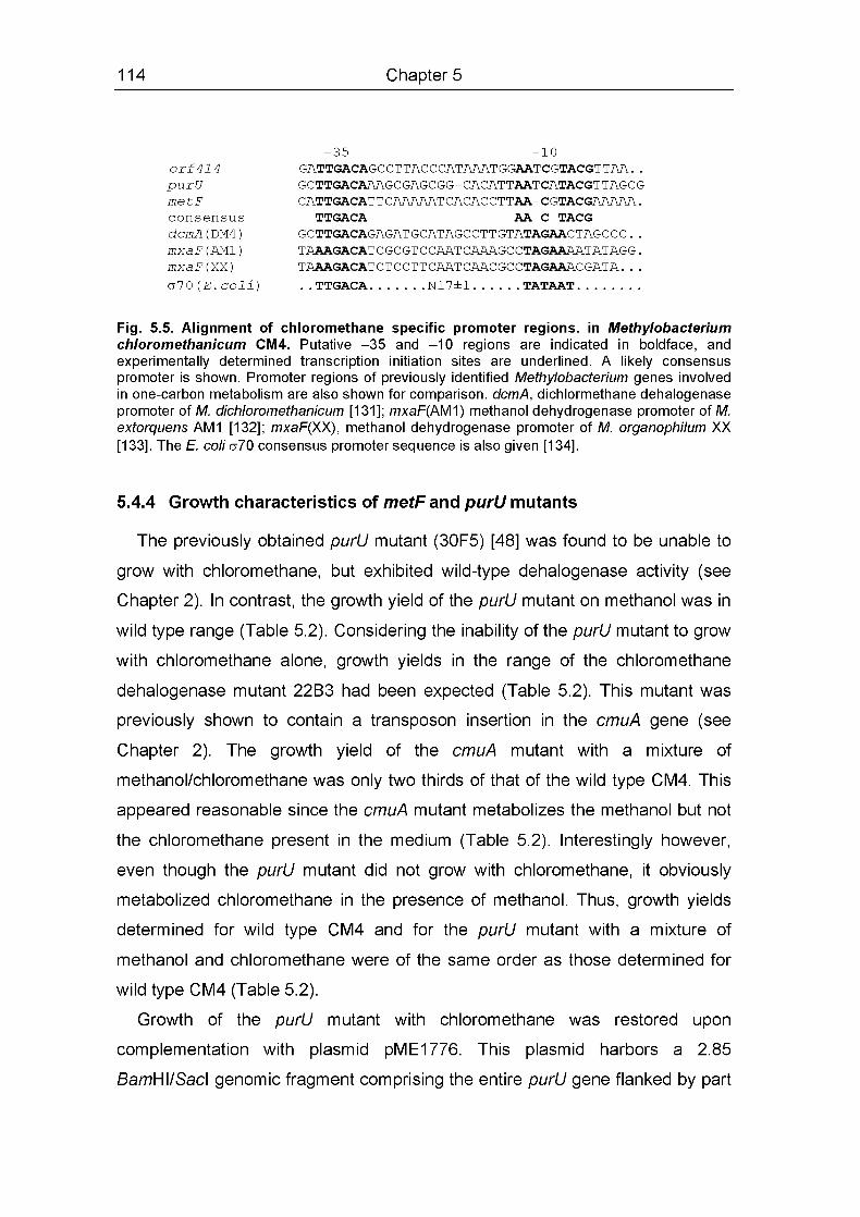
114 Chapter 5
-35 -10
orf414 GATTGACAGCCTTACCCATAAATGGAATCGTACGTTAA..
purU GCTTGACAAAGCGAGCGG-CACATTAATCATACGTTAGCG
metF CATTGACATTCAAAAATCACACCTTAA-CGTACGAAAAA.
consensus TTGACA AA C TACG
dcmA(DM4) GCTTGACAGAGATGCATAGCCTTGTATAGAACTAGCCC..
mxaF(AMI) TAAAGACATCGCGTCCAATCAAAGCCTAGAAAATATAGG.
mxaF(XX) TAAAGACATCTCCTTCAATCAACGCCTAGAAACGATA...
GlO (E. coli) ..TTGACA N17±l TATAAT
. . .
.^.. .
Fig. 5.5. Alignment of chloromethane specific promoter regions, in Methylobacteriumchloromethanlcum CM4. Putative -35 and -10 regions are indicated in boldface, and
experimentally determined transcription initiation sites are underlined. A likely consensus
promoter is shown. Promoter regions of previously identified Methylobacterium genes involved
in one-carbon metabolism are also shown for comparison. dcmA, dichlormethane dehalogenase
promoter of M. dichloromethanicum [131]; mxaF(AM1) methanol dehydrogenase promoter of M.
extorquens AM1 [132]; mxaF(XX), methanol dehydrogenase promoter of M. organophilum XX
[133]. The E. coli a70 consensus promoter sequence is also given [134].
5.4.4 Growth characteristics of metF and purU mutants
The previously obtained purU mutant (30F5) [48] was found to be unable to
grow with chloromethane, but exhibited wild-type dehalogenase activity (see
Chapter 2). In contrast, the growth yield of the purU mutant on methanol was in
wild type range (Table 5.2). Considering the inability of the purU mutant to grow
with chloromethane alone, growth yields in the range of the chloromethane
dehalogenase mutant 22B3 had been expected (Table 5.2). This mutant was
previously shown to contain a transposon insertion in the cmuA gene (see
Chapter 2). The growth yield of the cmuA mutant with a mixture of
methanol/chloromethane was only two thirds of that of the wild type CM4. This
appeared reasonable since the cmuA mutant metabolizes the methanol but not
the chloromethane present in the medium (Table 5.2). Interestingly however,
even though the purU mutant did not grow with chloromethane, it obviously
metabolized chloromethane in the presence of methanol. Thus, growth yields
determined for wild type CM4 and for the purU mutant with a mixture of
methanol and chloromethane were of the same order as those determined for
wild type CM4 (Table 5.2).
Growth of the purU mutant with chloromethane was restored upon
complementation with plasmid pME1776. This plasmid harbors a 2.85
BamH\/Sac\ genomic fragment comprising the entire purU gene flanked by part
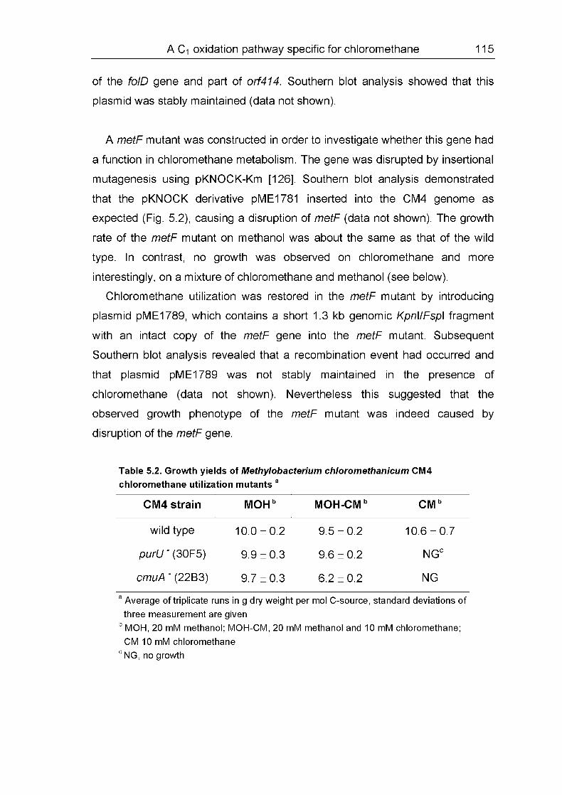
A Ci oxidation pathway specific for chloromethane 115
of the folD gene and part of orf414. Southern blot analysis showed that this
plasmid was stably maintained (data not shown).
A metF mutant was constructed in order to investigate whether this gene had
a function in chloromethane metabolism. The gene was disrupted by insertional
mutagenesis using pKNOCK-Km [126]. Southern blot analysis demonstrated
that the pKNOCK derivative pME1781 inserted into the CM4 genome as
expected (Fig. 5.2), causing a disruption of metF (data not shown). The growth
rate of the metF mutant on methanol was about the same as that of the wild
type. In contrast, no growth was observed on chloromethane and more
interestingly, on a mixture of chloromethane and methanol (see below).
Chloromethane utilization was restored in the metF mutant by introducing
plasmid pME1789, which contains a short 1.3 kb genomic Kpn\IFsp\ fragment
with an intact copy of the metF gene into the metF mutant. Subsequent
Southern blot analysis revealed that a recombination event had occurred and
that plasmid pME1789 was not stably maintained in the presence of
chloromethane (data not shown). Nevertheless this suggested that the
observed growth phenotype of the metF mutant was indeed caused by
disruption of the metF gene.
Table 5.2. Growth yields of Methylobacterium chloromethanlcum CM4
chloromethane utilization mutantsa
CM4 strain MOHb MOH-CMb CM"
wild type 10.0 + 0.2 9.5 + 0.2 10.6 + 0.7
purU' (30F5) 9.9 + 0.3 9.6 + 0.2 NGC
cmuA'(22B3) 9.7 + 0.3 6.2 + 0.2 NG
a
Average of triplicate runs in g dry weight per mol C-source, standard deviations of
three measurement are givenbMOH, 20 mM methanol; MOH-CM, 20 mM methanol and 10 mM chloromethane;
CM 10 mM chloromethane
CNG, no growth

116 Chapter 5
5.5 DISCUSSION
Expression analysis of transcriptional reporter gene fusions suggests the
presence of at least three transcriptional units for chloromethane utilization in
two gene clusters identified in M. chloromethanlcum CM4 (Fig. 5.5). Mapping of
the respective transcriptional start sites upstream of purU, metF and orf414
supports this hypothesis. The three promoter regions identified show a
significant degree of conservation and are different from any other
Methylobacterium promoter described until now. This suggests that genes
cmuA and cmuB as well as metF, purU and folD are regulated by similar
mechanisms at the level of transcription initiation. A database search using the
promoter consensus proposed in Fig. 5.6 as a pattern did not reveal the
presence of further similar promoters in both gene clusters of M.
chloromethanlcum CM4 (AJ011316 and AJ011317). Moreover, the same
analysis performed with the sequences identified in the chloromethane
degraders Aminobacter sp. strain IMB-1 (AF281260, [135]) and
Hyphomicrobium chloromethanlcum CM2 (AF281259, [99]) did not reveal any
similar promoter regions. This indicates that the identified promoter consensus
in strain CM4 does not represent an ubiquitous motif for the regulation of
chloromethane genes in methylotrophic bacteria and that regulation of the emu
genes is probably genus-specific.
The analysis of gene induction and the phenotypic properties of mutants
strongly support the operation of Ci oxidation pathway from methyl-H4folate to
formate that is specific for growth of strain CM4 with chloromethane. Expression
studies suggest that purU and folD are co-expressed from a promoter identified
upstream of purU (Fig. 5.5). Since it is very likely that the transposon insertion
in the purU mutant 30F5 has a polar effect on the folD gene, the question arose
as to whether both genes or just purU or folD are involved in chloromethane
degradation. Complementation of the chloromethane minus phenotype of the
purU mutant 30F5 by providing an intact copy of the purU gene in trans using
plasmid pME1776 (Table 5.1) indicated that folD is not essential for growth on
chloromethane. It is conceivable that the isofunctional enzyme pair of
methylene-H4folate dehydrogenase MtdA [44] and methenyl-H4folate

A Ci oxidation pathway specific for chloromethane 117
cyclohydrolase FchA [45] are able to compensate for the lack of FolD. Indeed a
recently obtained folD mutant does not have a chloromethane minus growth
phenotype (McAnulla unpublished). FolD thus appears to be non-essential for
growth with chloromethane, but might allow efficient oxidation of methylene-
H4folate during growth with chloromethane. The fact that a folD homologue is
present near the chloromethane-dehalogenase gene cmuA in Aminobacter
strain sp. IMB-1 [135] supports such a hypothesis.
Methylobacterium chloromethanlcum CM4 (CAB40737)
Aminobacter sp. IMB-1 (AAK38769)
-
Streptomyces nogalater (AAG42847)
Bacillus subtilis (C69840)
Thermotoga maritima (D72397)
Synechococcus sp (BAB18721 )
,02,
Rhodothermus mannus (CAC08536)
— Leptospira interrogans (AAF64321 )
— Methylobacterium extorquens AM1
Caulobacter crescentus (AAK24111 )
r~ Erwinia carotovora (P71319 )
*— Eschenchia coli (P00394)
- Vibrio cholerae (C820045)
Haemophilus influenzae (P45208)
Neissena meningitidis (F81880)
Buchnera aphidicola (P57154)
^
Pseudomonas aeruginosa (F83591 )
Xylella fastidiosa (F82720)
Streptomyces lividans (054235)
- Aquifex aeolicus (067422)
Campylobacterjejuni (D81326)
Lactococcus lactis (AE006357)
- Arabidopsis thaliana (T47821 )
Saccharomyces cerevisiae (P53128)
- Homo sapiens (P42898)
Fig. 5.6. Phylogenetic analysis of MetF proteins from bacteria. A multiple alignment of MetF
sequences (see text) obtained with T_Coffee [136] was used to generate a phylogenetic tree
with FITCH from PHYLIP [23]. The tree was drawn with NJPIot [137]. Accession numbers are
given in brackets.
118 Chapter 5
Proteins related to PurU, the putative 10-formyl-H4folate hydrolase, are
present in a variety of bacteria. The protein from Escherichia coli is 37%
identical in its amino acid sequence with PurU from M. chloromethanlcum CM4.
The E. coli PurU catalyzes the hydrolysis of 10-formyl-H4folate to H4folate and
formate, and was shown to be important for setting the ratio of alkylated to free
H4folate in the cell [138]. The observed phenotype of the M. chloromethanlcum
CM4 purU mutant is interesting in this context. The higher growth yield
observed with a mixture of methanol and chloromethane than with methanol
alone suggested that chloromethane is utilized for biomass formation in the
presence of methanol as a co-substrate. It is possible that a yet unknown
enzyme(s) that is only induced in the presence of methanol can complement for
the lack of 10-formyl-H4folate hydrolase in the purU mutant. Whatever the case
may be, it is likely that a lack of PurU negatively affects H4folate metabolism in
strain CM4.
In contrast to FolD and PurU, MetF of strain CM4 displays only low sequence
similarity to its mammalian and bacterial counterparts (see Chapter 2). In E
coli, MetF catalyzes the reduction of methylene-H4folate to methyl-H4folate. This
commits the one-carbon precursor for use in the synthesis of methionine from
homocysteine [139]. The low sequence similarity of the CM4 MetF protein might
be due to the fact that in M. chloromethanlcum CM4 MetF is supposed to
catalyze the reverse reaction to perform its proposed physiological role (Fig.
5.1). Indeed, the halomethane-utilizer Aminobacter sp. IMB-1 contains a metF
gene with the so far highest sequence similarity to MetF from CM4 (Fig. 5.6).
These two proteins, together with an unknown gene product from Streptomyces
nogalater form a subgroup of MetF homologues whose members are different
from the MetF proteins usually found in bacteria (Fig. 5.6). In contrast, a gene
encoding a putative MetF protein identified in the M. extorquens AM1 genome
was more related to the E. coli protein. This implies that strain CM4 might have
a second, so far not identified metF gene.
Interestingly, the metF mutant of strain CM4 was not only unable to grow with
chloromethane, but was also sensitive to the presence of chloromethane during
A Ci oxidation pathway specific for chloromethane 119
growth with methanol. A physiological explanation for this phenomenon might
be that the cofactor is trapped by the methylation catalyzed by chloromethane
dehalogenase, at the expense of all H4folate derivatives in the cell. Other
biosynthetic pathways relying on Ci moieties provided by H4folate might thereby
be abolished. This is reminiscent of the situation prevailing in human patients
with pernicious anemia. According to the currently favored "methyl trap" model
developed to account for the observed clinical picture, a disfunctional
methionine synthase leads to the accumulation of methyl-H4folate [140]. The
consequence is poor availability of the H4folate cofactor for other important
physiological processes.


Chapter 6
General Discussion

122 Chapter 6
This study addressed chloromethane degradation in aerobic bacteria at both
genetic and biochemical levels. Genes involved in chloromethane utilization by
Methylobacterium chloromethanlcum CM4 were isolated and their expression
was demonstrated to be regulated at the level of transcription. The mechanism
of chloromethane dehalogenation was investigated in more detail, which
required the purification and characterization of the responsible proteins. This
Chapter discusses results and current ideas concerning chloromethane
utilization in strain CM4 to provide a stimulus for future work on this topic.
6.1 GENES INVOLVED IN THE METABOLISM OF CHLOROMETHANE
As described in Chapter 2, a genetic approach was instrumental in the
discovery of the chloromethane utilization pathway of M. chloromethanlcum
CM4. In the following, I discuss the genes that have been proven or are
suspected to be involved in the metabolism of chloromethane by this organism.
Such genes fall into 3 classes (Fig. 5.1). The first class comprises genes
whose roles in chloromethane metabolism have been elucidated by biochemical
and/or genetic experiments as described in the previous Chapters. The second
class is formed by genes which encode proteins with a possible function in
chloromethane metabolism, some of which were previously analyzed in Chapter
2. Others will be presented in some detail below. Finally, a third class comprises
genes which are not, or only indirectly, involved in chloromethane utilization.
These include genes of clusters I and II encoding proteins involved in cobalamin
biosynthesis in strain CM4 (see Chapter 2), and genes located downstream of
transposon insertions in clusters III and IV which were isolated from so far
uncharacterized emu minus mutants (Fig. 5.1).

General Discussion 123
I (10'658bp) |1kb 38G12
30F5 nt 3903
nt3167\ y
22B3 38A10
nt8136 nt9460
V V
<z^ccobU folC folD purU orf55 orf414 cmuA orf98 hutl
(8'457 bp)19D10 36D3
nt5032 nt5919
cobQ cobD orf219 metF cmuB cmuC orf361 cobC
(5'280 bp) 11G7
nt 2863
^7
IV (1757 kb) 27B11
nt1757
IC <^3CdeoA deoC xapA IpxK waaA orf107 radC map
^| chloromethane dehalogenase 1dass ^ genes inv0|vec| in
^J H4folate dependent C, oxidation J chloromethane metabolism
<( | class 2, genes with a suspected function in chloromethane metabolism
<( | class 3, genes probably not involved in chloromethane metabolism
Fig. 6.1. Schematic view of gene clusters I - IV of Methylobacterium chloromethanlcum
CM4. The position and orientation of the transposon insertions in the genome of the emu minus
mutants are shown. Genes in cluster III and IV were named according to Table 5.1, genes in
clusters I (AJ011316) and II (AJ011317) are discussed in Chapter 2 and in the present Chapter.
6.1.1 Comparative sequence analysis of the emu genes in chloromethane
utilizing methylotrophic bacteria
Gene clusters I and II of M. chloromethanlcum CM4 were previously
described in detail (Fig. 6.1, see Chapter 2). In accordance with the properties
of the corresponding mutants (see Chapter 1, [48]), the gene products of cmuA
and cmuB were demonstrated to catalyze the formation of methyl-H4folate from
chloromethane (see Chapters 3 and 4). Further, the metF, folD and purU genes
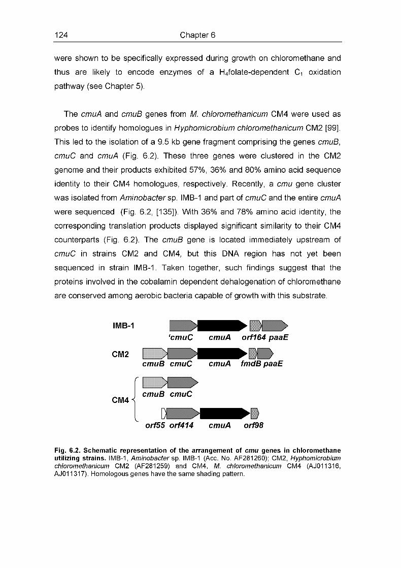
124 Chapter 6
were shown to be specifically expressed during growth on chloromethane and
thus are likely to encode enzymes of a H4folate-dependent Ci oxidation
pathway (see Chapter 5).
The cmuA and cmuB genes from M. chloromethanlcum CM4 were used as
probes to identify homologues in Hyphomicrobium chloromethanlcum CM2 [99].
This led to the isolation of a 9.5 kb gene fragment comprising the genes cmuB,
cmuC and cmuA (Fig. 6.2). These three genes were clustered in the CM2
genome and their products exhibited 57%, 36% and 80% amino acid sequence
identity to their CM4 homologues, respectively. Recently, a emu gene cluster
was isolated from Aminobacter sp. IMB-1 and part of cmuC and the entire cmuA
were sequenced (Fig. 6.2, [135]). With 36% and 78% amino acid identity, the
corresponding translation products displayed significant similarity to their CM4
counterparts (Fig. 6.2). The cmuB gene is located immediately upstream of
cmuC in strains CM2 and CM4, but this DNA region has not yet been
sequenced in strain IMB-1. Taken together, such findings suggest that the
proteins involved in the cobalamin dependent dehalogenation of chloromethane
are conserved among aerobic bacteria capable of growth with this substrate.
IMB-1
CM2
CM4S
'cmuC cmuA orf164 paaE
cmuB cmuC emuA fmdB paaE
cmuB cmuC
orf55 orf414 emuA orf98
Fig. 6.2. Schematic representation of the arrangement of emu genes in chloromethane
utilizing strains. IMB-1, Aminobacter sp. IMB-1 (Ace. No. AF281260); CM2, Hyphomicrobiumchloromethanicum CM2 (AF281259) and CM4, M. chloromethanicum CM4 (AJ011316,
AJ011317). Homologous genes have the same shading pattern.
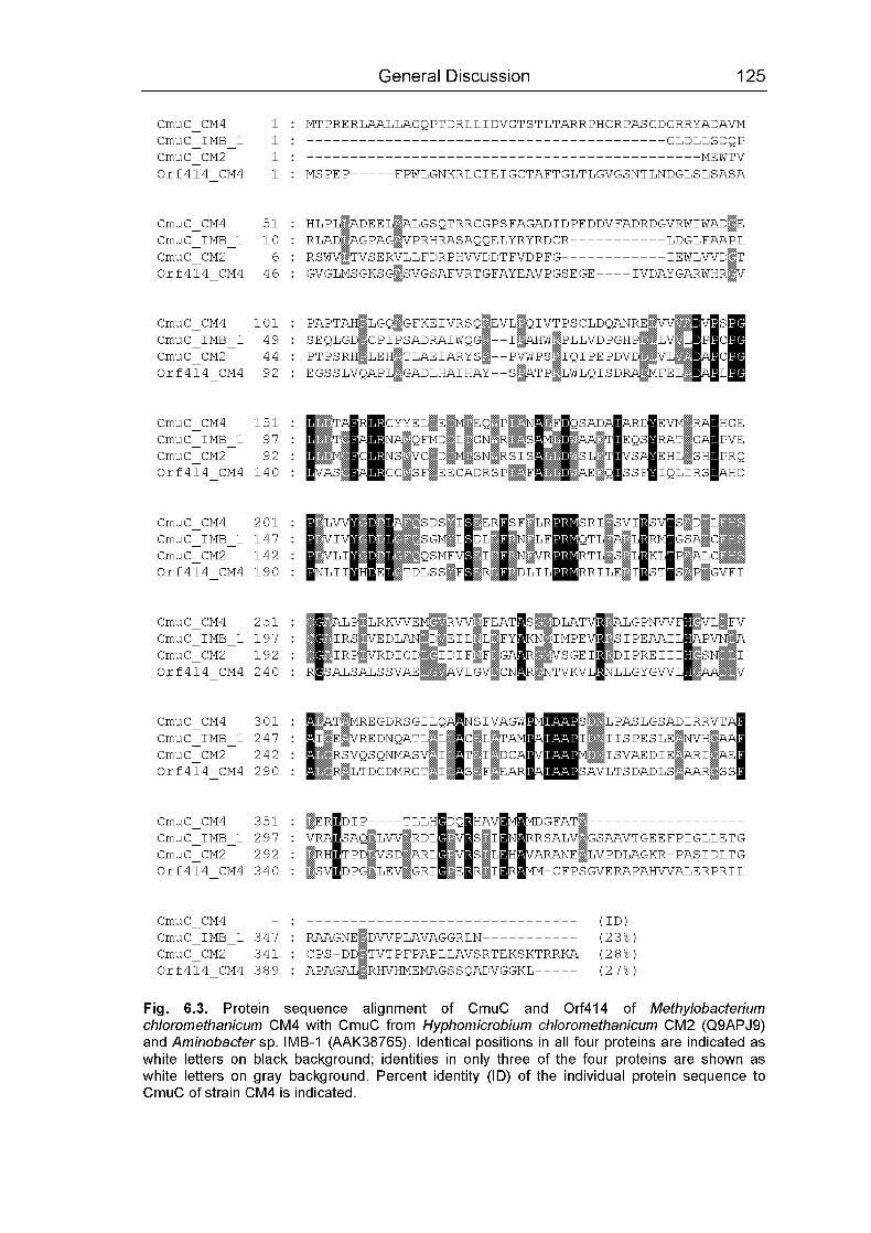
General Discussion 125
CmuC CM4 1
CmuC 1MB 1 1
CmuC CM2 1
Orf414 CM4 1
CmuC CM4 51
CmuC 1MB 1 10
CmuC CM2 6
Orf414 CM4 46
MTPRERLAALLAGQPTDRLLIDVGTSTLTARRPHCRPASGDGRRYADAVM
GLDLLSDQP
MEWTV
MSPEP FPWLGNKRLCIEIGCTAFTGLTLGVGSNTLNDGLSLSASA
hlpljjadeelIalgsqtrrcgpsfagadidpeddvfadrdgvrwiwadEerladjlagpaggvprhrasaqqelyryrdcr ldglfaapl
rswv[Jtvservllfdrphvvddtfvdpfg iewlvvdEt
gvglmsgksgHsvgsafvrtgfayeavpgsege ivdaygarwhrIv
CmuC_CM4 101
CmuC_IMB_l 4 9
CmuC_CM2 44
Orf414 CM4 92
paptahhlgqggfkeivrsqhevlhqivtpscldqanregvvmlseqlgdhcpipsadraiwqgI—i|ahwHpllvdpghpMlv1lBp|ptpsrh|§lehBtlaeiarys|§—pvwpsBioipepdvdBvli-EGSSLVQAPLgGADLHAIHAY—s[§ATp|jLWLQISDRa|§MFEl|
CmuC_CM4 151
CmuC_IMB_l 97
CmuC_CM2 92
Orf414 CM4 140
lÉgnBrHs!Il
gyyelBe
iNAiQFMDRD»EVM|gRAfflHGE
tHeqsHratIgaHpve
thvsaHehlJshHprqqhssfHiqlirsHahd
CmuC_CM4 2 01
CmuC_IMB_l 147
CmuC_CM2 142
Orf414 CM4 190
III[QSMFvBl[WtdlssIfHrI
cdTBQVT
ûqtl||a{|lIPTT BqIItr\ 1 J-ig^Hii
I'D "D T T pHtKK1 J_iI!jHJ.
SVHSBDËL|
CmuC_CM4 251
CmuC_IMB_l 197
CmuC_CM2 192
Orf414 CM4 240
\LP|jLRKVVEJIRslvEDLANiJlRPgfvRDICD
RffiSALSALSSVAE
eiljlBfySknIimpev !
idifmfHgaSrWvsgei~
vlgvicnIrIntvkvl!
.LGPNVVF
IsipeaailI(dipreiii
inllgygvvli
CmuC_CM4 301
CmuC_IMB_l 2 47
CmuC_CM2 2 42
Orf414 CM4 290
Ëmregdrs gi lqaSns ivagw|IvrednoatlPIlIPcBlPIt
RSVQSQNMASvBigTHlHDC.rBltdgdmrgtIiIIsBfIearI
^saslilLPAS LGSADI rrvt;
^saillli i s pe s leHnvh!
ISVAEDIEl
^saSAVLTSDADLsl
CmuC_CM4 351
CmuC_IMB_l 2 97
CmuC_CM2 2 92
Orf414 CM4 340
BeRJJdipivraHsaqIrhHtpdisvHdp
JmdgfatH
InIrrsalvIgsaavtgeefpiglletgIvARANEgLVPDLAGKR-PASIDLTGgMM-CFPSGVERAPAHVVALERPRIL
CmuC_CM4
CmuC_IMB_l 347
CmuC_CM2 341
Orf414 CM4 389
(ID)
raagneHdvvplavaggrln (23°
CPS-DDHTVTPFPAPLLAVSRTEKSKTRRKA (28t
APAGALHRHVHMEMAGS SQADVGGKL (2 71
Fig. 6.3. Protein sequence alignment of CmuC and Orf414 of Methylobacteriumchloromethanicum CM4 with CmuC from Hyphomicrobium chloromethanicum CM2 (Q9APJ9)and Aminobacter sp. IMB-1 (AAK38765). Identical positions in all four proteins are indicated as
white letters on black background; identities in only three of the four proteins are shown as
white letters on gray background. Percent identity (ID) of the individual protein sequence to
CmuC of strain CM4 is indicated.

126 Chapter 6
In contrast to strains IMB-1 and CM2, the emu genes of M.
chloromethanicum CM4 are found at two separate loci (Fig. 6.2). Since the emu
genes of both clusters are surrounded by genes putatively involved in
cobalamin biosynthesis, it is possible that the emuA and cmuBC genes are
arranged in close proximity to each other on the CM4 genome.
Of interest with respect to future studies is the striking sequence similarity of
cmuC and orf414 (Fig. 6.3). The arrangement of cmuC immediately upstream of
cmuA in H. chloromethanicum CM2 and in Aminobacter sp. IMB-1 suggests that
a gene duplication event had occured which, combined with a translocation, has
led to the separation of the emu genes in M. chloromethanicum CM4 (Fig. 6.2).
Mutant studies demonstrated that in strain CM4 the cmuC gene product
performs an essential but so far unknown function in chloromethane metabolism
(see Chapter 1). In contrast, it is not known whether orf414 is essential for
growth on chloromethane, since an orf414 mutant is not yet available. However,
the intact orf414 gene is not sufficient to allow growth on chloromethane in the
cmuC mutant, so that the two genes are clearly not isofunctional.
6.1.2 Genes encoding possible chloromethane responsive regulatory
proteins
Three promoter regions with significant similarity to each other were identified
in clusters I and II and expression of the genes under their control was
specifically induced during growth on chloromethane (see Chapter 5).
Regulatory protein(s) or a conserved operator region potentially involved in
chloromethane dependent transcriptional regulation have yet to be identified in
strain CM4. A possible candidate for a regulatory protein is encoded by orf219
in cluster II (Fig. 6.1). Orf219 is predicted to encode a 219 amino acid protein
similar to proteins annotated as members of the family of MerR response
regulators (Fig. 6.4). These regulatory proteins occur in a range of bacteria and
respond to a wide variety of external stimuli. The prototype of this family is the
mercury resistance regulator MerR, present in both Gram negative and Gram
positive bacteria [141]. The MerR family includes mainly metal-responsive
regulators of gene expression which, for example, respond to cobalt in

General Discussion 127
Synechocystis PCC 6803 (CoaR; [142]), cadmium in Pseudomonas putida
(CadR; [143]), copper (CueR, [144]) and zinc in E. coli (ZntR; [145]).
merr_ECOLI
merR_BACIL
MERR_PSEAE
CadR_PSEPU
CUER_ECOLI
ZNTR_ECOLIOrf219
<
mennlenlt8BjvfB1aaBvnve
mkfrHelsdkcIvnkeMENNLENLTjWFBjAAgVNVE
mkBelBatdcaveMNiDw*Ts
myriIIelBIImaevtpdi
MDVKAARKS GLKKVgEVgDIFg^TT PRj
rlBIipeiorkIBlleiREMLPE
ekBBVtpikqqmmehevrteggf
eqEhs-Irktargt
merr_ECOLI
merR_BACIL
MERR_PSEAE
CadR_PSEPU
CUER_ECOLI
ZNTR_ECOLIOrf219
47
42
47
40
40
41
50
GEADVVBVKBVKSAQRjMBlSsqqyvd1lh|ikrmqe|B|tGEADVTHVr||vKSAQR|B|stqahve(3lt||irncrt[§dmttqqhlneltllrqarqvBïntesdlqBlk(§irharqjm|sSDADIRHFEVAYKLScRfP
RLDDG THjEEASSLAEHGVVDRDE AKjRDMYDFTILRLEDG THBEEASSLAEH
RLRDSPD DSgGSVNALIDENLFNDPQ RHSADVKRRTLE
SIRIDPEH—HTgûESKGIVQE.LTRHNALTGDESGRKLSNIIDG
merr_ECOLI
merR_BACIL
MERR_PSEAE
CadR_PSEPU
CUER_ECOLI
ZNTR_ECOLIOrf219
92
89
92
87
87
89
100
klkdvrekmadHar|1etv s
kiediqrk||ed krierm m-
KLKDVREKMAD Ar||EAV S-
hiehvqarBdg VALQEQ V E
KVAEIERHgEERLQEVEARjIAElvedlqroJIrh
QSÏRDQ
QsIqrs
eHvc.
D KERgPENKDVCAHHARRG
ERGRLMIR
ÏHARKG—N-VSH
I-YEB
N-VSH
—AEH
—ADS
NDAfflCGTAH—SSVYHSILE7
cwdhknkpsrltcpnBIceihm
RRRHNAQG-
Al ANAfflPGDDS
-R
merr_ECOLI 133
merR_BACIL 13 0
MERR_PSEAE 133
CadR_PSEPU 12 6
CUER ECOLI 127
ZNTR ECOLI 131
Orf219 150
merr ECOLI _
merR BACIL -
MERR PSEAE -
CadR PSEPU -
CUER ECOLI -
ZNTR ECOLI -
Orf219 200
QGEAGLARSAMP
MKK
QGGASLAGSAMP
ETNGAVSVPETEHSHVGRSHGH
SGCCHHRAG
EQGASGVKSGC
EQSQLLWLTWDADREVSAQGPLNPPRTKCMEASLPNVPTEAVADASTSRV
(ID)
(16^
(24^
(16^
(17^
(16^
(18^
DPARRRRRGRQPLERTRNSK
Fig. 6.4. Amino acid sequence alignment of Orf219 of Methylobacterium chloromethanicum
CM4 with members of the MerR-type family of transcriptional activators, including MerR of E.
coli (AAC33922), Pseudomonas aeruginosa (P06688) and Bacillus subtilis (P22853), as well as
ZntA of £. coli (P36676), CadR of Pseudomonas putida (AAK48830) and CueR of £. coli
(P77565). Amino acids are indicated as white letters on black background when conserved in all
seven proteins and as white letters on gray background when conserved in five of the seven
proteins. The identities (ID) of the individual amino acid sequences to Orf219 of strain CM4 are
indicated. *, conserved cysteine residues; <—>, helix-turn-helix motif.
128 Chapter 6
In addition, MerR-type regulatory proteins are involved in the response to
superoxide stress in E coli, in thiostrepton resistance in Streptomyces, and in
regulation of nodulation in Bradyrhizobium and of multidrug efflux protein
synthesis in Bacillus [141] MerR type regulatory proteins display stretches of
conserved sequence in their ammo-terminal domain which includes a hehx-turn-
hehx DNA-binding motif (Fig 6 4, [141]) The C-terminal domain is less
conserved and thought be involved in sensing of specific effectors Specifically,
two cysteines (Cys131 and Cys143), of the three cysteine residues required for
mercury-binding and promoter activation in MerR, are conserved in Orf219 (Fig
6 4, [146])
Currently, the role of Orf219 as a regulator in chloromethane-utihzation is
purely hypothetical It is based on the location of orf219 adjacent to metF and
their divergent direction of transcription, which are features typical of MerR-type
regulators and their target genes [141] However, the conserved inverted repeat
and the extended spacer region between the -35 and -10 regions typical of
known MerR-type regulators [146,147] were not found in chloromethane-
induced promoters of strain CM4 (see Chapter 5)
Another candidate for a regulatory protein in chloromethane degradation is
Orf98 in cluster I Originally, the orf98 gene was overlooked until now due to the
lack of significant similarity of Orf98 to any gene product in the database With
the availability of the emu gene clusters of Aminobacter sp IMB-1 and H
chloromethanicum CM2 [99,135], however, it has become evident that all three
organisms have an orf98-\\We open reading frame located immediately
downstream of cmuA (Fig 6 2) Further, all three orf98-\\We proteins have a
conserved CX2CX19CX2C motif at their ammo-terminal end (Fig 6 5), which is
typical of metal-binding sites in zinc-finger domains [148] Interestingly, all three
gene products show some low level of similarity (<19% identity) to the protein
FmdB, a likely positive transcriptional regulator for the expression of
formamidase in Methylophilus methylotrophus [149]

General Discussion 129
FmdB {M.m)
Orfl46
FmdB (H. c)
Orf98
* *
ü|lHeyeHdsHg-vft;üSsBt^MgdHrHdBqI
üSlhqpmdkBdBcBdiiSI3frE9tiliiPlIBI j\ î\TIB"R1 B(-!?!PH
* *
rkmses-sHpaeHdcB
tdlatadgBIIvHtsb/NYELAKkBIJiHTSFERSRS^HvHTG
WSPRILSVPKLAIM
s ïtraIIlvvIs"|t-
-ît
1-
FmdB (M.m)
Orfl46
FmdB (H. c)
Orf98
FmdB (M.m)
Orfl46
FmdB (H. c)
Orf98
49
47
47
47
97
89
94
90
dklqrsSherneksane—prtHrrssc|vigp-Itearraan—-dakIeeifkpi
iLRSRSsTANRSGSSRDVAGEgQRQGK[JjLSSFSgDAVTSDRP TSSVTASD)
|ktkpp|nqdtgk@g_G"RBSK1
-AI HBT Kl
GfRlSKlaih!
|n-*1srI
lqmqtmtarpwmvgh
BkqeiBahgfvded-BreeirIangiadqd-BsvasHtns
(ID)
(19^
(42^
(39^
Fig. 6.5. Sequence alignment of Orf98 of Methylobacterium chloromethanicum CM4 with FmdB
from Hyphomicrobium chloromethanicum CM2 (AAK01348), the C-terminal part of Orf146 from
Aminobacter sp. IMB-1 (AAK38766) and FmdB from Methylophilus methylotrophus S74214.
Amino acids are indicated as white letters on black background when conserved in all four
proteins and as white letters on gray background when conserved in three of the four protein
sequences. Cysteine residues in the "CXXC" motifs are marked by asterisks. Identity (ID) of the
individual sequences to Orf98 of strain CM4 are indicated.
Clearly, in the case of expanded regulation studies in strain CM4, both orf219
and orf98 would be interesting targets for mutational inactivation.
6.1.3 Clusters III and IV contain genes whose functions in chloromethane
metabolism are not known
Only six transposon mutants displaying a chloromethane utilization negative
phenotype have been studied in some detail. In these cases, the transposon
insertion was found in clusters I or II (Fig. 6.1). However, three other mutants
with a emu minus phenotype, mutants 11G7, 27B11 and 27C10 were isolated
(Fig. 6.1, see Chapter 1, [48]). In the latter mutant, orf414 was affected by
insertion of the transposon, but some uncharacterized additional genetic
rearrangement had occurred in this mutant, which was therefore not
characterized further. The DNA regions affected by transposon insertions in
strains 11G7 and 27B11, termed clusters III and IV in the following, appeared
unrelated to each other or to clusters I and II (Fig. 6.1).

130 Chapter 6
A 5.3 kb region covering both sides of the transposon insertion site in mutant
11G7 was sequenced. The insertion mapped to a DNA sequence of M.
chloromethanicum CM4 that either is non-coding or encodes a protein with no
similarity to any of the proteins stored in the database (Fig. 6.1). The reason for
the emu minus phenotype of mutant 11G7 is thus not obvious. Moreover, the
putative genes identified near by the transposon insertion site have no
immediate relation to chloromethane metabolism and showed similarity to
proteins involved in nucleotide degradation or lipid biosynthesis (Table 6.1).
A 1.75 kb region on one side of the miniTn5 insertion of a 17 kb Cla\
fragment cloned from mutant 27B11 (in plasmid pME1733) was also
sequenced. The insertion mapped to an open reading frame encoding a
putative gene product with similarity to methionine aminopeptidase (Map) from
a variety of bacteria [150]. This protein is responsible for the removal of the N-
terminal methionine after translation. In this case as well, a link between the
putative function of the disrupted gene in mutant 27B11 and chloromethane
metabolism is difficult to envisage. Immediately downstream of map, however,
lies radC, which presumably encodes a protein involved in the repair of UV-
induced DNA damage [151]. It can be envisaged that the transposon insertion
in mutant 27B11 has a polar effect on the expression of radC which leads to an
impairment of the DNA repair machinery. That such a transposon insertion
would correlate with a emu minus phenotype is of interest, considering the
known systemic methylating potency of monohalomethanes, which can lead to
DNA damage [152]. Indeed, related observations were recently made with
Methylobacterium dichloromethanicum DM4. In this organism the polA gene,
which encodes DNA polymerase I, an enzyme with a well-known role in DNA
repair [151], was found to be essential for growth with dichloromethane [122].
Growth with dichloromethane was also impaired in an uvrA mutant of M.
dichloromethanicum DM4 (Kayser and Vuiiieumier unpublished), a gene likely
to encode a protein involved in nucleotide excision repair [151]. These
observations suggest that due to the reactivity of halogenated methanes,
Methylobacterium needs an efficient DNA repair machinery for growth with such
compounds.
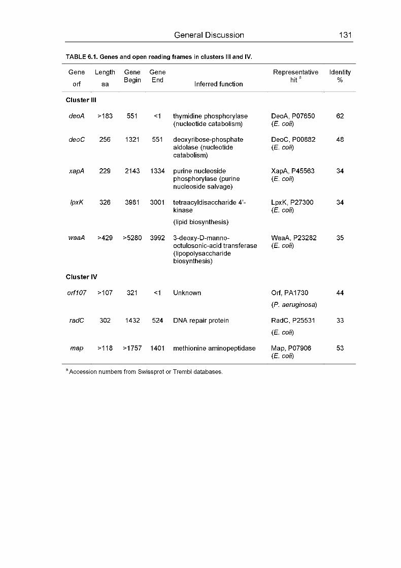
General Discussion 131
TABLE 6.1. Genes and open readingI frames in clusters III and IV.
Gene
orf
Length
aa
Gene
Begin
Gene
EndInferred function
Representativehita
Identity%
Cluster III
deoA >183 551 <1 thymidine Phosphorylase
(nucleotide catabolism)
DeoA, P07650
(E. coli)
62
deoC 256 1321 551 deoxyribose-phosphatealdolase (nucleotide
catabolism)
DeoC, P00882
(E. coli)
48
xapA 229 2143 1334 purine nucleoside
Phosphorylase (purinenucleoside salvage)
XapA, P45563
(E. coli)
34
IpxK 326 3981 3001 tetraacyldisaccharide 4'-
kinase
(lipid biosynthesis)
LpxK, P27300
(E. coli)
34
waaA >429 >5280 3992 3-deoxy-D-manno-octulosonic-acid transferase
WaaA, P23282
(E. coli)
35
(lipopolysaccharide
biosynthesis)
Cluster
orf107
IV
>107 321 <1 Unknown
radC 302 1432 524 DNA repair protein
map >118 >1757 1401 methionine aminopeptidase
Orf, PA1730 44
(P. aeruginosa)
RadC, P25531 33
(E. coli)
Map, P07906 53
(E. coli)
a
Accession numbers from Swissprot or TrembI databases

132 Chapter 6
6.2 THE COBALAMIN-DEPENDENT CHLOROMETHANE
DEHALOGENATION REACTION
The main features and components of chloromethane dehalogenation were
characterized in the course of this study (see Chapter 3 and 4). The reaction
involves a cobalamin-dependent methyl transfer from chloromethane to
H4folate. This finding came as a surprise, since catabolic reactions based on a
cobalamin-dependent methyl transfer had so far been observed only in strictly
anaerobic bacteria and archaea [60,153].
Table 6.2. Classes of cobalamin-dependent methyltransferases
proteinsa
reaction
Methyltransferase donor (CH3-X)d acceptor (Y)e
methinone synthase MetH CH3-H4folate homocysteine
CH3X:CoM
methyltransferase
MtaA,B,C
MtbA, MtmB,C
methanol
monomethylamine
HSCoM
HSCoM
MtbA,B,C dimethylamine HSCoM
MtbA, MttB.C trimethylamine HSCoM
MtsA,B methanethiol HSCoM
methyl-H4MPT:CoM
methyltransferase
MtrA-E CH3-H4MPT HSCoM
acetyl coenzyme A synthase AcsD, E CH3-H4folate carbon monoxide
dehydrogenase
aromatic O-demethylase
chloromethane
dehalogenase
OmdA' phenylmethylethers
MtvA,B,CD
CmuA, CmuB chloromethane
H4folate
H4folate
a
Biochemically characterized proteins for which the corresponding genes were sequencedbThe aromatic O-demethylase of Acetobacterium dehalogenans [58] consists of three com¬
ponents of which only the gene encoding the corrinoid binding protein was sequenced so far.
cAromatic O-demethylase of Moorella thermoacetica [154] of unknown sequence
Abbreviations according to Fig. 6.6.

General Discussion 133
Several classes of cobalamin-dependent methyl transfer reactions have been
described (Table 6.2), which all feature a common reaction mechanism (Fig.
6.6). The crucial step in the reaction takes place at the corrinoid-binding protein
(E, Fig. 6.6), which is methylated by a methyltransferase I (MTI) exhibiting
binding affinity for the methyl group donor (CH3-X). The methylated corrinoid
protein is demethylated by another methyltransferase (MTII) with affinity to the
methyl group acceptor (Fig. 6.6). In cobalamin dependent methyl transfer
reactions, the methyl-cobalt bond is cleaved heterolytically. Both bonding
electrons stay on the cobalt and the reaction thus formally corresponds to a
transfer of a methyl carbocation. The cob(l)alamin form of the protein formed in
such a reaction is a powerful nucleophile able to demethylate various substrates
(Table 6.2). It is therefore essential for catalytic activity. However, during
turnover the cob(l)alamin can occasionally be oxidized into its inactive
cob(ll)alamin form (Fig. 6.6). The reductive reactivation of the protein to the
active cob(l)alamin form requires a fourth enzymatic reaction (AE). Reactivation
mechanisms differ in various corrinoid dependent methyltransferases and are
discussed in some detail below.
S-Ado + 1e-
N-His-E
CH3-Y
Fig. 6.6. General scheme of corrinoid-dependent methyl transfer reactions. MT,
methyltransferase; AE, possible reactivation enzyme; N-His-E, protein histidine residue (see
text). CH3X represents the methyl group donor and Y the methyl group acceptor (see Table
6.2).
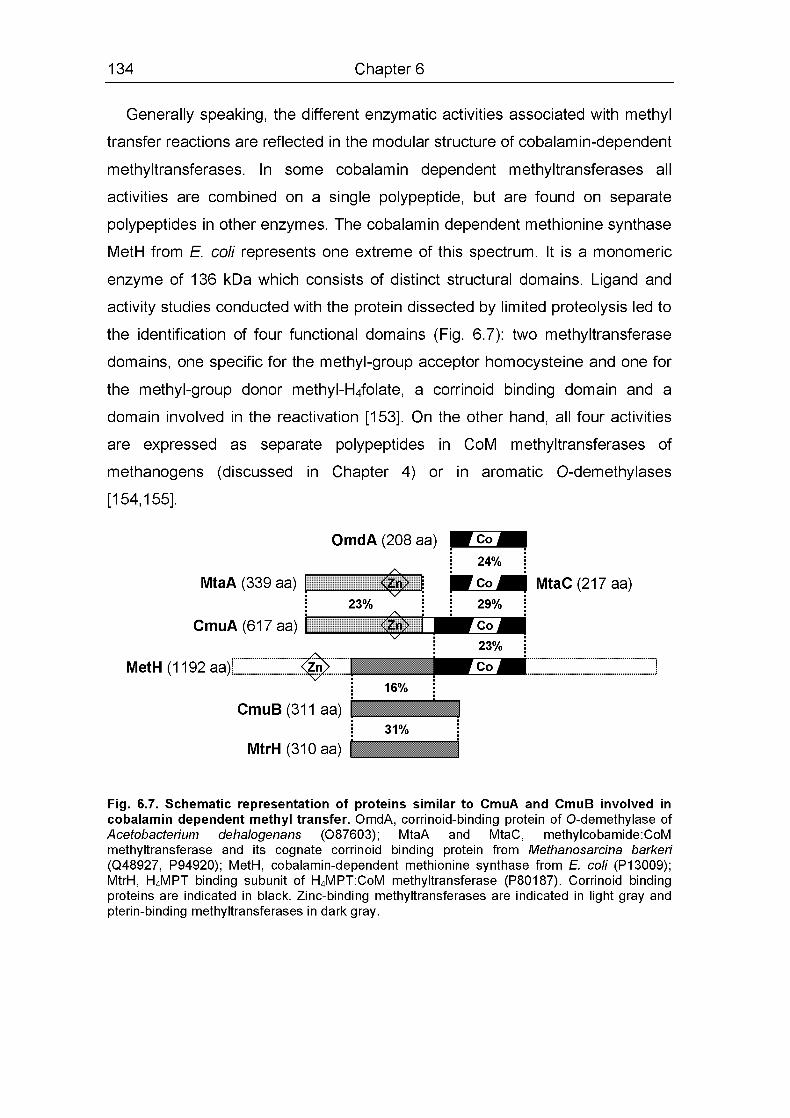
134 Chapter 6
Generally speaking, the different enzymatic activities associated with methyl
transfer reactions are reflected in the modular structure of cobalamin-dependent
methyltransferases. In some cobalamin dependent methyltransferases all
activities are combined on a single polypeptide, but are found on separate
polypeptides in other enzymes. The cobalamin dependent methionine synthase
MetH from E. coli represents one extreme of this spectrum. It is a monomeric
enzyme of 136 kDa which consists of distinct structural domains. Ligand and
activity studies conducted with the protein dissected by limited proteolysis led to
the identification of four functional domains (Fig. 6.7): two methyltransferase
domains, one specific for the methyl-group acceptor homocysteine and one for
the methyl-group donor methyl-H4folate, a corrinoid binding domain and a
domain involved in the reactivation [153]. On the other hand, all four activities
are expressed as separate polypeptides in CoM methyltransferases of
methanogens (discussed in Chapter 4) or in aromatic O-demethylases
[154,155].
MtaA (339 aa)
CmuA (617 aa) [
MetH (1192 aa)
OmdA (208 aa)
CmuB (311 aa)
MtrH (310 aa)
MtaC(217aa)
Fig. 6.7. Schematic representation of proteins similar to CmuA and CmuB involved in
cobalamin dependent methyl transfer. OmdA, corrinoid-binding protein of O-demethylase of
Acetobacterium dehalogenans (087603); MtaA and MtaC, methylcobamide:CoM
methyltransferase and its cognate corrinoid binding protein from Methanosarcina barker!
(Q48927, P94920); MetH, cobalamin-dependent methionine synthase from E. coli (P13009);MtrH, H4MPT binding subunit of H4MPT:CoM methyltransferase (P80187). Corrinoid binding
proteins are indicated in black. Zinc-binding methyltransferases are indicated in light gray and
pterin-binding methyltransferases in dark gray.
General Discussion 135
The chloromethane dehalogenase of M. chloromethanicum CM4 represents
an intermediate case since the methyltransferase specific for chloromethane
and its cognate corrinoid binding protein are fused to a single polypeptide
CmuA. The methyltransferase II CmuB represents a separate protein which
uses methylated CmuA as a substrate for methyl transfer onto H4folate.
Nevertheless, domains with similar function often exhibit a significant degree of
conservation between different classes of cobalamin-dependent
methyltransferases. Sequence similarities of CmuA and CmuB to other
cobalamin-dependent methyltransferases discussed in Chapter 2 are shown
schematically in Fig. 6.7.
The properties of the different domains in the Cmu proteins are discussed in
relation to similar proteins of other cobalamin-dependent methyltransferases in
the following. The main emphasis is put on the CmuA protein, which catalyzes
the dehalogenation of chloromethane and represents the only known enzyme to
combine the methyltransferase I and the corrinoid binding domain. Finally, the
last part of the Chapter will describe the reactivation mechanisms in corrinoid
dependent methyltransferases, since the reactivation system in strain CM4 is
not yet known.
6.2.1 The corrinoid binding domain of CmuA
As discussed in detail in Chapter 4, CmuA is a bifunctional protein containing
a vitamin B12 cofactor bound in a non covalent manner. In free
methylcobalamin, the cobalt is in the +3 oxidation state and coordinated to six
ligands [153]. These are the four nitrogens of the corrin ring, termed the
equatorial ligands, a methyl group as upper ligand and the
dimethylbenzimidazole moiety of the cofactor as lower ligand. In contrast, the
unmethylated cob(l)alamin form of the cofactor (oxidation state +1) is only
tetrahedrally coordinated by the equatorial nitrogen ligands of the corrin ring
with the dimethylbenzimidazole residue dissociated. The two free electrons in
the orbital perpendicular to the plane of the corrin ring impart cob(l)alamin its
strong nucleophihc character [156].

136 Chapter 6
In corrinoid proteins, the axial ligand is replaced by a histidine residue of the
protein, which modulates the reactivity of the corrinoid A conserved sequence
motif D-X-H-X(2)-G-X(41 )-S-X-L-X(26)-G-G was proposed to be involved in the
binding of the cobalamin cofactor [157] This motif is evident in sequence
alignments (Fig 6 8) and present in most corrinoid binding proteins, but only
poorly conserved in CmuA
Determination of the three-dimensional structure of the corrinoid binding
domain of methionine synthase MetH from E coli confirmed the involvement of
residues in this sequence motif in cofactor binding [74] Residues D757, H759
and S810 were identified as members of a ligand triad important for catalytic
activity, in which the histidine residue replaces the dimethylbenzimidazole tail as
lower axial ligand to the cobalt of the corrinoid cofactor Further, the three
conserved glycine residues at amino acid positions 762, 833 and 834 were
shown to provide the necessary space for the dissociated
dimethylbenzimidazole tail of the prosthetic cobalamin cofactor [74,153] H759
is thought to be instrumental in stabilizing the cob(l)alamin and
methylcobalamin states of the cofactor by dissociation and association, and
thereby to facilitate catalysis of methyl group transfer [153,156] Similar results
were obtained for the MtaC protein from Methanosarcina barken, where, using
site directed mutagenesis, a histidine was also identified as the essential axial
ligand of the corrinoid [117]
Although the C-terminal region of the CmuA protein from M
chloromethanicum CM4 showed considerable sequence similarity with other
corrinoid-binding proteins involved in methyl transfer, the cobalamin-binding
motif is only partially conserved (Fig 6 8, see Chapter 2) The sequence
alignment suggests that in CmuA the histidine is replaced by a glutamine
residue as lower axial ligand (Fig 6 8) This change in the corrinoid binding
motif is conserved among all CmuA sequences obtained so far [99,135] It is not
clear whether a glutamine residue is able to act as the lower axial ligand of
cobalamin and if so, whether it can exert the same function as a histidine
residue The potential impact of such an amino acid change was briefly
discussed in Chapter 2

General Discussion 137
CmuA 4 01
MetH 651
MtaC 1
MtmC_ 1
OmdA 1
EGHVYEKLVEAIMDY
QAEWRSwIvNKRLEYSLVKGMLDFTEASLKKVLTRYNVALEKALTPEEAAEELYPKDHLIYPIAKAIFEG
MANQgiFDKLRDAIVNQMKMEEVKAKVEAG
CmuA 417
MetH 671
MtaC 51
MtmC 18
OmdA 14
CmuA 4 67
MetH 721
MtaC 101
MtmC_ 68
OmdA 63
dadkakqwvqvgldrEisaqkivfdgBslBkiitefieqdteeBrqqatrpievHegppmdBnv|
eeddvveglqaIie.nvagtpelckeIl.ksklvpglvqeIlde
KDPIDLMDD.
PALDl|jTKG|§SV|sapgeJjlqamvdsjJjgvj
DMYERNERHVTDMLK
|DL FGE GKmHlHqVVKSGVraiRLYDEGViHlHNVMM
»KlfcDKFEAAElHLHQIMMEKFSSGElBvllEMLI
AcsD 106 : DPEGANHSVDQCV ATVKEVLOAvIBvPLVVVGcHllvEKDHEVLEAVAE
aHktIdaamplI llhssgsdggptgtvivglvrfiIaskeqgktn1kmviatvk|
SLEGIEYCK HNSGATPKTkItVVCHVAE
E LHKNKKE GEEaIlAIT FVAE|da§
Af,
AgKAÎsKGVEvl
SJRVIKQAVAY
SGKAffiiSNAMEV|- LMAGDGSAS LffiT CVIGTV.
AcsD 153 : aaaBf.nll-BBnaf.of.nyksltaacmvhkhnttarBpBdtntckqlntlt
CmuA 517 KAiÏÏFKVI
MetH 771
148
QCl
RAj
INYFT
MtaC YNViMtmC 118 GA| Bfqi IOmdA 111 es;«FDM1I
IknbkpHqfietaereg-paIkilrtakevn-
IRDhpaIevlaavqkek-DfflLNgNVVEEAAKHKGEKVLLVDfflPADT fvqavkdntn-vklvaci
-avaigmhvmtns stvyvekva
-adliglJgIIijjpsldemvnva-pimltgtaImIIttmyafkevn
TSMLGQKDLM
LlTTMPALKEAV
AcsD 202 NEMNLPLDHIVIDPSIgELGYGIEYSFSIMERIRLGALQGDK
CmuA
MetH
MtaC
MtmC
565
819
196
168
OmdA 160
EMLKSQGKADKYLLMM^gAANRGVAEKFGVRYGLDANAAVSLVRDHVESKEMERQ—GFTIPLLI^HtTSKAHTAVKIEQNYSGPTVYVQNASRTVGVDMLLEN—GIKIPFAcfflGAVNQDFVSQFALGVYGEEAADAPKIADAIIA
DRLNEEKLRDSVKCMF^gPVSDKWIEEIGADATAENAAEAAKVALEVMKQTIKAA—YPDMKVTvHBpVTPFYAAFVGADGYAPDAGSAAVKARFTAT
CmuA 614
MetH 867
MtaC 244
MtmC_OmdA 2 0 8
RAAA
VAALLSDTQRDDFVARTRKEYETVRIQHGR
GTTDVTELREKFHKH
A
Fig. 6.8. Amino acid sequence alignment of the corrinoid-binding domain of CmuA with
other corrinoid proteins. MetH from £. coli (P13009), MtaC (P94920) and MtmC (030641)from Methanosarcina barker! and OmdA from Acetobacterium dehalogenans (087603). Amino-
acids are indicated as white letters on black background when conserved in all five proteins and
as white letters on gray background when conserved in four of the five protein sequences. The
corrinoid binding motif [157] is marked by asterisks. Part of the corrinoid binding protein AcsD
from Clostridium thermoaceticum (Q07341) was aligned separately and shaded according to
the alignment.

138 Chapter 6
Even though histidine was found to be essential as a lower axial ligand in
both MetH and MtaC, such a structural arrangement does not seem to be
universal for catalysis of cobalamin-dependent methyl transfer. For example,
the corrinoid/iron-sulfur protein AcsD involved in acetyl coenzyme A synthesis in
Clostridium thermoaceticum has neither the dimethylbenzimidazole nucleotide
nor a nitrogenous amino acid ligand from the protein coordinated to the cobalt
[158]. Part of the AcsD protein sequence aligned with other corrinoid-binding
proteins suggests little relatedness to the corrinoid binding motif (Fig. 6.8).
6.2.2 The zinc-binding methyltransferase domain of CmuA
Purified CmuA protein was found to contain 1 mol of zinc per mol protein
(see Chapter 4). The transition metal zinc has been identified as a ligand in
various types of proteins and enzymes, where it serves both structural and
catalytic roles [159]. Zinc is usually 4-coordinated, the ligands being nitrogen or
sulfur atoms from His, Cys, Asp and Glu residues. In zinc binding sites involved
in catalysis, two of the amino acids participating are often separated by only 1 to
3 residues in the protein sequence, while a third residue is found at a larger
distance [159]. Interestingly, such a H-X-C-Xn-C zinc-binding motif is also found
in the CmuA protein (Fig. 6.9, see Chapter 4). Two other methyltransferases are
known which contain one equivalent of zinc per mol protein and display the
same zinc-binding motif. These are the cobalamin-independent methionine
synthase MetE from E coll [114,160] and the methylcobamide:CoM
methyltransferase MtaA from Methanosarcina barkerii [88,116].

General Discussion 139
CmuA 241
MetE 634
MtaA 230
MtbA 2 32
MtsA 229
CmuA 289
MetE 682
MtaA 27 9
MtbA 27 9
MtsA 27 6
iggigftgtg-tftqpiwrdiaengclnfngdmypg-mehakraiggqisddtqihtfflmhycefndimdsiaaldadvitietsrsdmellesfe—efdvnsvtvlHih-gkvnailsdmadcEfeglsveekigdaaegkkvigdrarldvatvlHih-gnttnglgimdktIvngisvdqkvdiktatgsvk—NAIISCPLIlHiB-GDTSKLLPYIKQsIiDCFSFDAVP—VWYCRQVMGNEMS
lmEtlsp-fstlmhEst-tdvanevBklIaevEynggficmpc
-PFTLLPgPI--vavlwnbtp-
-idlmpnItp-
-DKlKAEAa-V|LEC »ID VLAPG
-EEIAEASj-KgLDAgVG LLTVG
-eqvynrtr-ecilqIad—ivgt;
*
BDID-
IGLK1
IGIA-
IGTV-
DVS-
288
681
278
278
275
333
730
319
319
316
Fig. 6.9. Amino acid sequence alignment of the zinc-binding motif of CmuA with similar
methyltransferases. MetE from E. coli (P25665), MtaA (Q48927), MtbA (030640) and MtsA
(Q48924) from Methanosarcina barker! Amino acids are indicated as white letters on black
background when conserved in all four proteins and as white letters on gray background when
conserved in three of the four protein sequences. Zinc-binding residues discussed in text are
marked by asterisks.
MtaA and related methylcobamide:CoM methyltransferases from
methanogenic archaea show highest similarity to the N-terminal domain of
CmuA (Fig. 6.7, see Chapter 2). Zinc was recently shown to be involved in CoM
activation in MtaA, and to be essential for methyl group transfer from
methylcobalamin to CoM [107]. The zinc atom was therefore postulated to
activate the thiol group of CoM for nucleophihc attack on the corrinoid-bound
methyl group (Fig. 6.10B, [161]). This hypothesis is based on observations
formulated previously for the activation of homocysteine by the cobalamin
independent methionine synthase MetE from E. coli [160,162]. In the latter
case, homocysteine was identified as the fourth ligand of zinc, in addition to
three amino acid ligands of the protein. Zinc was proposed to stabilize the
thiolate of the homocysteine substrate at neutral pH and thereby to activate the
thiol for nucleophihc attack on methyl-H4folate (Fig. 6.10A).
Such a thiol activation mechanism was first described for the Ada protein of
E. coli, which is the transcriptional regulator in the adaptive response to DNA
alkylation [163]. The Ada protein is automethylated by DNA
methylphosphotriesters at a specific cysteine residue which is also one of four
conserved cysteines involved in zinc-binding (Fig. 6.10C). Upon methylation,
Ada undergoes a conformational change and acquires the ability to bind to
140 Chapter 6
operator sites on the DNA This leads to the transcription of genes that confer
resistance to methylating agents [163] It is thus tempting to speculate that
chloromethane-responsive gene regulation in M chloromethanicum CM4 could
be based on a mechanism similar to that of the Ada protein from E coli (Fig
6 10C) Indeed, alignments of Orf219 and Orf98 suggest the presence of
possible zinc-binding sites in both cases As pointed out above, both proteins
are candidate regulatory proteins in chloromethane metabolism in strain CM4
In such a scenario, methylation of one or both of these proteins by
chloromethane might enable them to act as transcriptional activators
Given the presence of zinc and a zinc binding motif in the sequence of CmuA
(Fig 6 9), it is conceivable that methyl transfer in chloromethane
dehalogenation is based on a mechanism similar to that of Mta, MetE or Ada
(Fig 6 10A-C) Such a putative reaction mechanism would theoretically involve
four zinc ligands, instead of three zinc ligands as for MetE or MtaA Cys326 in
CmuA could possibly play this role (Fig 6 9) In this case, the zinc binding site
in CmuA would be reminiscent of that of Ada Alternatively, a low molecular
weight thiol compound, such as CoM or methanethiol, might be involved in zinc
binding of CmuA However, no evidence has been obtained so far for the
requirement of such a cofactor in chloromethane degradation
Should a catalytic thiol group in CmuA indeed be involved in cornnoid
dependent chloromethane dehalogenation, two possible mechanisms are
conceivable a priori (Fig 6 1 OD) Either chloromethane is dehalogenated by
thiol conjugation prior to methyl transfer onto the cornnoid cofactor (Fig
6 10D1), or the methyl group of chloromethane is transferred from the cornnoid
to the thiol to facilitate nucleophihc attack by H4folate (Fig 6 10D2)
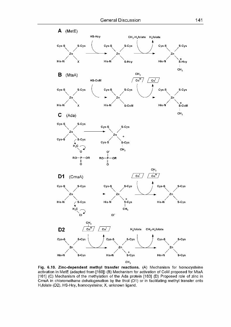
General Discussion 141
A (MetE)
HS-Hcy
Cys-S S-Cys
Zn
/ \His-N x
Cys-S S-Cys
- Zn
/ \His-N s-Hcy
Cys-S S-Cys
Zn
/ \ +
His-N s-Hcy
I
CH,
B (MtaA) CH,
HS-CoM ëu bu
Cys-S S-Cys
Zn
/ \His-N x
C (Ada)
Cys-S S-Cys
\ /Zn —
Cys-S S-Cys
- Zn
/ \His-N S-CoM
Cys-S S-Cys
—*~ \ /Zn
Cys-S S-Cys
Zn
/ \ +
His-N S-CoM
I
CH,
Cys-S s S-CysC Cys-S S-Cys
O
IO"
I
I
CH,
RO—P—OR RO—P—OR
Il IIO O
CH,
D1 (CmuA)
Cys-S S-Cys
\ /Zn —
BU BU
Cys-S S-Cys
\ /* Zn
His-N y- S-CysC
H,C
His-N
^
\ +
S-CysI
CH,
Cys-S S-Cys
\ /Zn
/ \His-N S-Cys
CI CI"
CH,
D2 / Co'"/ / Co' / H4folate CH3-H4folate
Cys-S S-Cys
Zn
/ \His-N S-Cys
Cys-S S-Cys
». Zn
His-N S-CysI
Cys-S S-Cys
Zn
/ \His-N S-Cys
CH,
Fig. 6.10. Zinc-dependent methyl transfer reactions. (A) Mechanism for homocysteineactivation in MetE (adapted from [160]) (B) Mechanism for activation of CoM proposed for MtaA
[161] (C) Mechanism of the methylation of the Ada protein [163] (D) Proposed role of zinc in
CmuA in chloromethane dehalogenation by the thiol (D1) or in facilitating methyl transfer onto
H4folate (D2). HS-Hcy, homocysteine; X, unknown ligand.
142 Chapter 6
Concerning the first mechanism, it should be remembered that there is in
principle no need for enzymatic dehalogenation since chloromethane readily
reacts abiotically with cob(l)alamin [77] On the other hand it appears likely that
the CmuA protein should offer a certain protection of the cornnoid cofactor
against oxidative inactivation so that dehalogenation of chloromethane by
CmuA-bound cobalamin may not proceed as readily as the chemical reaction in
solution The second mechanism (Fig 6 10D2), in contrast, may provide a
means to facilitate nucleophihc attack of H4folate onto the methyl group derived
from chloromethane Future biochemical studies, perhaps involving selected
site-directed mutants of CmuA and CmuB proteins, will no doubt allow to
distinguish between these different possibilities and to elucidate mechanisms of
chloromethane dehalogenation in more detail
General Discussion 143
6.2.3 How many methyltransferases are needed for growth with
chloromethane ?
CmuA and CmuB were sufficient to catalyze chloromethane dehalogenation
in vitro (see Chapter 4). CmuA exhibits methyltransferase I activity and CmuB
catalyzes the methyltransferase II reaction. Based on sequence, the conserved
cmuC gene (Fig. 6.2) seems to encode yet another methyltransferase essential
for growth on chloromethane besides CmuA and CmuB (see Chapter 2). One
may thus speculate that CmuC exhibits a function similar to CmuB and may
also use CmuA as a substrate for methyl group transfer onto an alternative, so
far unidentified cofactor. The existence of H4MPT-dependent Ci oxidation in M.
extorquens AM1 (see Chapter 1, [43]) and the identification of the cofactor
H4MPT in strain CM4 (Studer and Keltjens unpublished) in particular are
indications for a possible methyl group transfer from CmuA to H4MPT catalyzed
by CmuC (discussed in Chapter 4).
This hypothesis is in accordance with the phenotypes of cmuB and cmuC
mutants [48]. Both mutants were unable to grow with chloromethane, but in
contrast to the cmuB mutant, the cmuC mutant was still able to release chloride
in the presence of chloromethane. This observation could be explained by the
polar effect of the miniTn5 insertion in cmuB on the expression of the cmuC
gene. Such an effect would inactivate both methyltransferases in the cmuB
mutant, whereas in the cmuC mutant one methyltransferase (CmuB) would still
be active. However, the recent complementation of the two mutants with the
individual genes did not support the hypothesis of CmuC acting in
chloromethane dehalogenation (McAnulla unpublished). The cmuC mutant was
complemented in presence of the cmuC gene, whereas the cmuB mutant was
not complemented with only cmuB present. This is consistent with the polar
effect in the cmuB mutant on cmuC. However, the cmuB mutant complemented
with the cmuC gene alone did not show any chloride release in the presence of
chloromethane (McAnulla unpublished), which demonstrates that CmuC is not
directly involved in the dehalogenation of chloromethane. Therefore the
essential function of CmuC in chloromethane metabolism of strain CM4 remains
to be elucidated.
144 Chapter 6
6.2.4 A reactivation mechanism for the CmuA corrinoid protein
One problem associated with corrinoid-dependent dehalogenation is the low
midpoint potential of the cob(l)alamin/cob(ll)alamin couple, which is - 606 mV
for the free cofactor [164]. Bound to an enzyme, the potential is usually 100 to
150 mV higher, still below that of many natural electron donors, especially in
aerobic organisms [165]. Corrinoid-dependent methyltransferase are therefore
prone to inactivation by oxidation and the cell requires an efficient mechanism
to maintain these proteins in an active form. Such reactivation systems are
functionally diverse among different organisms. However, they share the
requirement for a low potential electron donor and for a high energy compound
(Fig. 6.6). For example, methanoLCoM methyltransferase activation requires an
ATP-dependent methyltransferase-activating protein and involves a ferredoxin,
a hydrogenase and molecular hydrogen as electron donor[165]. In contrast,
MetH, the inactive cob(ll)alamine enzyme is reactivated by a reductive
methylation that involves a reduced flavodoxin and adenosylmethionine
[166,167].
The physiological reactivation system is not known and the midpoint potential
of the cob(l)alamin/cob(ll)alamin couple has not been determined for
chloromethane dehalogenase. Thus, activity of the enzyme could so far only be
maintained under anoxic conditions using the artificial low-potential electron
donor titanium(lll)citrate. This suggests that the midpoint potential is in the
range of other corrinoid dependent methyltransferases. Recently, a gene
encoding a putative oxidoreductase was identified immediately downstream of
CmuA in H. chloromethanicum CM2 and Aminobacter sp. IMB-1 (Fig. 6.2),
which might be part of a reductive activation system for chloromethane
dehalogenase in these organisms [135]. However, a similar gene is not present
in strain CM4 judging Southern analysis (McAnulla unpublished). Therefore, the
nature of the corrinoid reactivation system remains elusive in strain CM4.

References 145
REFERENCES
[I] Molina, M J, & Rowland, F S (1974) Stratospheric sink for
chlorofluoromethanes chlorine atomic-atalysed destruction of ozone Nature
249, 810-812
[2] Wackett, L P, Logan, MSP, Blocki, F A, & Bao-h, C (1992) A
mechanistic perspective on bacterial metabolism of chlorinated methanes
Biodegradation 3, 19-36
[3] Farman, J C,Gardiner, B G
,& Shankhn, J D (1985) Large losses of
total ozone in Antarctica reveal seasonal CIOx/NOx interaction Nature 315, 207-
210
[4] Leisinger, T,Gisi, D
, Magh, A ,Sornbas, H
,Vannelh, T
,& Vuiiieumier,
S (1997) Bacterial dehalogenation of chlorinated methanes In Mechanisms of
biohalogenation and dehalogenation (Janssen, D B,Soda, K, & Wever, R
,
eds), Vol 1 pp 185-194 North-Holland, Amsterdam
[5] Fetzner, S (1998) Bacterial dehalogenation Appl Microbiol Biotechnol
50, 633-657
[6] Khahl, M A K,& Rasmussen, R A (1999) Atmospheric methyl chloride
Atmos Environ 33, 1305-1321
[7] Keene, W C,Khahl, M A K
,Enckson, D J
,McCulloch, A
,Graedel, T
E,Lobert, J M
,Aucott, M L
, Gong, S L, Harper, D B
,Kleiman, G
, Midgley,P
,Moore, R M
,Seuzaret, C
, Sturges, W T, Benkovitz, C M, Koropalov, V,
Barrie, L A,& Li, Y F (1999) Composite global emissions of reactive chlorine
from anthropogenic and natural sources reactive chlorine emissions inventoryJ Geophys Res 104, 8429-8440
[8] Butler, J H (2000) Better budgets for methyl hahdes? Nature 403, 260-
261
[9] Harper, D B (2000) The global chloromethane cycle biosynthesis,biodégradation and metabolic role Nat Prod Rep 17,337-348
[10] Andreae, M O, Atlas, E, Harris, G W, Helas, G, deKock, A,
Koppmann, R,Maenhaut, W, Mano, S
,Pollock, W H
, Rudolph, J,Scharffe,
D,Schebeske, G
,& Welling, M (1996) Methyl halide emissions from savanna
fires in southern Africa J Geophys Res 101,23603-23613
[II] McCulloch, A, Aucott, M L,Benkovitz, C M
,Graedel, T E
,Kleiman,
G, Midgley, P M
,& Li, Y F (1999) Global emissions of hydrogen chloride and
chloromethane from coal combustion, incineration and industrial activities
reactive chlorine emissions inventory J Geophys Res 104,8391-8403
[12] Khahl, M A K, Moore, R M, Harper, D B
,Lobert, J M
,Enckson, D
J, Koropalov, V , Sturges, W T
,& Keene, W C (1999) Natural emissions of
chlorine-containing gases reactive chlorine emissions inventory J GeophysRes 104, 8333-8346
[13] Wuosmaa, A M,& Hager, L P (1990) Methyl chloride transferase a
carbocation route for biosynthesis of halometabohtes Science 249, 160-162

146 References
[14] Scarratt, M. G., & Moore, R. M. (1998) Production of methyl bromide and
methyl chloride in laboratory cultures of marine phytoplankton II. Marine
Chemistry 59, 311-320.
[15] Waitling, R., & Harper, D. B. (1998) Chloromethane production by wood-
rotting fungi and an estimate of global flux to the atmosphere. Mycol. Res. 107,769-787.
[16] Rhew, R. C, Miller, B. R., & Weiss, R. F. (2000) Natural methyl bromide
and methyl chloride emissions from costal salt marshes. Nature 403, 292-295.
[17] Saxena, D., Aouad, S., Attieh, J., & Saini, H. S. (1998) Biochemical
characterization of chloromethane emission from the wood-rotting fungusPhellinus pomaceus. Appl. Environ. Microbiol. 64, 2831-2835.
[18] Ni, X. H., & Hager, L. P. (1999) Expression of Bâtis maritima methylchloride transferase in Escherichia coli. Proc. Natl. Acad. Sei. U. S. A. 96, 3611-
3615.
[19] Saini, H. S., Attieh, J. M., & Hanson, A. D. (1995) Biosynthesis of
halomethanes and methanethiol by higher plants via a novel methyltransferasereaction. Plant Cell Environ. 18, 1027-1033.
[20] Miller, M. (1996) Methyl bromide and the environment. In The methylbromide issue (Bell, C. H., Price, N., & Chakrabarti, B., eds), Vol. 1 pp. 91-148.
John Wiley & Sons, Chichester.
[21] Shorter, J. H., Kolb, C. E., Grill, P. M., Kerwin, R. A., Talbot, R. W.,
Hines, M. E., & Harriss, R. C. (1995) Rapid degradation of atmospheric methylbromide in soils. Nature 377, 717-719.
[22] McAnulla, C. (2000) Chloromethane metabolism by Gram-negativemethylotrophs. Department of Biological Sciences, University of Warwick,Dissertation.
[23] Felsenstein, J. (1993) PHYLIP (Phylogeny inference package) version
3.5c. Distributed by the author. Department of Genetics, University of
Washington, Seattle.
[24] Miller, L. G., Connell, T. L, Guidetti, J. R., & Oremland, R. S. (1997)Bacterial oxidation of methyl bromide in fumigated agricultural soils. Appl.Environ. Microbiol. 63, 4346-4354.
[25] Hines, M. E., Grill, P. M., Varner, R. K., Talbot, R. W., Shorter, J. H.,
Kolb, C. E., & Harriss, R. C. (1998) Rapid consumption of low concentrations of
methyl bromide by soil bacteria. Appl. Environ. Microbiol. 64, 1864-1870.
[26] Rasche, M. E., Hicks, R. E., Hyman, M. R., & Arp, D. J. (1990) Oxidation
of monohalogenated ethanes and n-chlorinated alkanes by whole cells of
Nitrosomonas europaea. J. Bacteriol. 172, 5368-5373.
[27] Stirling, D. I., & Dalton, H. (1979) The fortuitous oxidation and
cometabohsm of various carbon compounds by whole-cell suspensions of
Methylococcus capsulatus (Bath). FEMS Microbiol. Lett. 5, 315-318.
References 147
[28] Traunecker, J., Preuss, A., & Diekert, G. (1991) Isolation and
characterization of methyl chloride utilizing, strictly anaerobic bacteria. Arch.
Microbiol. 156,416-421.
[29] Coulter, C, Hamilton, T. G., McRoberts, W. C, Kulakov, L, Larkin, M. L,& Harper, D. B. (1999) Halomethane:bisulfide/halide ion methyltransferase, an
unusal corrinoid enzyme of environmental significance isolated from an aerobic
methylotroph using chloromethane as the sole carbon source. Appl. Environ.
Microbiol. 65, 4301-4312.
[30] Doronina, N. V., Sokolov, A. P., & Trotsenko, Y. A. (1996) Isolation and
initial characterization of aerobic chloromethane-utilizing bacteria. FEMS
Microbiol. Lett. 142, 179-183.
[31] Hancock, T. L. C, Costello, A. M., Lidstrom, M. E., & Oremland, R. S.
(1998) Strain IMB-1, a novel bacterium for the removal of methyl bromide in
fumigated agricultural soils. Appl. Environ. Microbiol. 64, 2899-2905.
[32] Hartmans, S., Schmuckle, A., Cook, A. M., & Leisinger, T. (1986) Methylchloride: naturally occurring toxicant and C-1 growth substrate. J. Gen.
Microbiol. 132, 1139-1142.
[33] McDonald, I. R., Doronina, N. V., Trotsenko, Y. A., McAnulla, C, &
Murrell, J. C. (2001) Hyphomicrobium chloromethanicum sp. nov.,
Methylobacterium chloromethanicum sp. nov., chloromethane utilising bacteria
isolated from a polluted environment. Int. J. Syst. Evol. Microbiol., 119-122.
[34] Goodwin, K. D., Schaefer, J. K., & Oremland, R. S. (1998) Bacterial
oxidation of dibromomethane and methyl bromide in natural waters and
enrichment cultures. Appl. Environ. Microbiol. 64, 4629-4636.
[35] Green, P. N. (1992) The genus Methylobacterium. In The prokaryotes
(Balows, A., Trüper, H. G., Dworkin, M., Harder, W., & Schleifer, K.-H., eds),Vol. 3 pp. 2342-2349. Springer-Verlag, New York.
[36] Goodwin, K. D., & Anthony, C. (1995) The biosynthesis of periplasmicelectron-transport proteins in methylotrophic bacteria. Microbiology 141, 1051-
1064.
[37] Vuiiieumier, S. (2001) Coping with a halogenated one-carbon diet:
aerobic dichloromethane-minerahsing bacteria. In Focus on Biotechnologyseries (Agathos, S., & Reineke, W., eds), Vol. 3 pp. in press. Kluwer Academic
Publishers.
[38] Attwood, M. M., & Quayle, J. R. (1984) Formaldehyde as a central
intermediary metabolite of methylotrophic metabolism. In Microbial growth on Ci
compounds (Crawford, R. L, & Hanson, R. S., eds), Vol. 1 pp. 315-323.
American Society of Microbiology, Washington, D.C.
[39] Barber, R. D., & Donohue, T. J. (1998) Function of a glutathione-dependent formaldehyde dehydrogenase in Rhodobacter sphaeroides
formaldehyde oxidation and assimilation. Biochemistry 37, 530-537.

148 References
[40] Harms, N,Ras, J
, Reijnders, W N M, vanSpanning, R J M
,&
Stouthamer, A H (1996) S-formylglutathione hydrolase of Paracoccus
demtrificans is homologous to human esterase D A universal pathway for
formaldehyde detoxification'? J Bactenol 178,6296-6299
[41] Norm, A, VanOphem, P W, Piersma, S R
,Persson, B
,Duine, J A
,&
Jornvall, H (1997) Mycothiol-dependent formaldehyde dehydrogenase, a
prokaryotic medium-chain dehydrogenase/reductase, phylogenetically links
different eukaryotic alcohol dehydrogenases - Primary structure, conformational
modelling and functional correlations Eur J Biochem 248, 282-289
[42] Lidstrom, M E (1992) The aerobic methylotrophic bacteria In The
prokaryotes (Balows, A , Truper, H G,Dworkin, M
,harder, W, & Schleifer, K -
H, eds), Vol 3 pp 431-445 Springer-Verlag, New York
[43] Chistoserdova, L,Vorholt, J A
,Thauer, R K
,& Lidstrom, M E (1998)
C-1 transfer enzymes and coenzymes linking methylotrophic bacteria and
methanogenic Archaea Science 281, 99-102
[44] Vorholt, J A,Chistoserdova, L
,Lidstrom, M E
,& Thauer, R K (1998)
The NADP-dependent methylene tetrahydromethanoptenn dehydrogenase in
Methylobacterium extorquens AM1 J Bactenol 180,5351-5356
[45] Pomper, B K, Vorholt, J A, Chistoserdova, L, Lidstrom, M E, &
Thauer, R K (1999) A methenyl tetrahydromethanoptenn cyclohydrolase and a
methenyl tetrahydrofolate cyclohydrolase in Methylobacterium extorquens AM1
Eur J Biochem 261, 475-80
[46] Vorholt, J A, Chistoserdova, L, Stolyar, S M,Thauer, R K, &
Lidstrom, M E (1999) Distribution of tetrahydromethanoptenn-dependent
enzymes in methylotrophic bacteria and phylogeny of methenyltetrahydromethanoptenn cyclohydrolases J Bactenol 181,5750-5757
[47] Vorholt, J A,Marx, C J
,Lidstrom, M E
,& Thauer, R K (2000) Novel
formaldehyde-activating enzyme in Methylobacterium extorquens AM1 requiredfor growth on methanol J Bactenol 182,6645-6650
[48] Vannelh, T, Studer, A, Kertesz, M, & Leisinger, T (1998)Chloromethane metabolism by Methylobacterium sp strain CM4 Appl Environ
Microbiol 64, 1933-1936
[49] Janssen, D B,Pries, F
,& van der Ploeg, J R (1994) Genetics and
biochemistry of dehalogenating enzymes Annu Rev Microbiol 48, 163-191
[50] Harper, D B (1998) Chloromethane production by wood-rotting fungiand an estimate of the global flux to the atmosphere Mycol Res 102, 769-787
[51] Yvon-Lewis, S A, & Butler, J H (1997) The potential of oceanic
biological degradation on the lifetime of atmospheric CH3Br Geophys Res
Lett 24, 1227-1230
[52] Montzka, S A,Butler, J H
, Myers, R C,Thomson, T M
,Swanson, T
H,Clarke, A D
,Lock, L T
,& Elkins, J W (1996) Decline in the tropospheric
abundance of halogen from halocarbons implications for stratospheric ozone
depletion Science 272, 1318-1322
References 149
[53] Jeffers, P. M., Wolfe, N. L., & Nzengung, V. (1998) Green plants: a
terrestrial sink for atmospheric CH3Br. Geophys. Res. Lett. 25, 43-46.
[54] Connell Hancock, T. L, Costello, A. M., Lidstrom, M. E., & Oremland, R.
S. (1998) Strain IMB-1, a novel bacterium for the removal of methyl bromide in
fumigated agricultural soils. Appl. Environ. Microbiol. 64, 2899-2905.
[55] Goodwin, K. D., Lidstrom, M. E., & Oremland, R. S. (1997) Marine
bacterial degradation of brominated methanes. Environ. Sei. Technol. 31, 3188-
3192.
[56] Oremland, R. S., Miller, L. G., Culbertson, C. W., Connell, T. L, &
Jahnke, L. (1994) Degradation of methyl bromide by methanotrophic bacteria in
cell suspensions and soils. Appl. Environ. Microbiol. 60, 3640-3646.
[57] Keuning, S., Janssen, D. B., & Witholt, B. (1985) Purification and
characterization of hydrolytic haloalkane dehalogenase from Xanthobacter
autotrophicus GJ10. J. Bacteriol. 163, 635-639.
[58] Kaufmann, F., Wohlfarth, G., & Diekert, G. (1998) O-demethylase from
Acetobacterium dehalogenans substrate specificity and function of the
participating proteins. Eur. J. Biochem. 253, 706-711.
[59] Messmer, M., Wohlfarth, G., & Diekert, G. (1993) Methyl chloride
metabolism of the strictly anaerobic, methyl chloride-utilizing homoacetogenstrain MC. Arch. Microbiol. 160, 383-387.
[60] Wohlfarth, G., & Diekert, G. (1997) Anaerobic dehalogenases. Curr Op.Biotechnol. 8, 290-295.
[61] De Lorenzo, V., & Timmis, K. N. (1994) Analysis and construction of
stable phenotypes in Gram negative bacteria with Tn5- and Tn^O-derived
minitransposons. Methods Enzymol. 235, 386-405.
[62] Ausubel, F. M., Brent, R., Kingston, R. E., Moore, D. D., Seidman, J. G.,
Smith, J. A., & Struhl, K. (2001) Current Protocols in Molecular Biology. Greene
Publishing Associates & John Wiley and Sons, New York.
[63] Altschul, S. F., Madden, T. L., Schaeffer, A. A., Zhang, J., Zhang, Z.,
Miller, W., & Lipman, D. J. (1997) Gapped BLAST and PSI-BLAST: A new
generation of protein database search programs. Nucleic Acids Res. 25, 3389-
3402.
[64] Mägli, A., Messmer, M., & Leisinger, T. (1998) Metabolism of
dichloromethane by the strict anaerobe Dehalobacterium formicoaceticum.
Appl. Environ. Microbiol. 64, 646-650.
[65] Gossett, J. M. (1987) Measurement of Henry's law constants for C1 and
C2 chlorinated hydrocarbons. Environ. Sei. Technol. 21, 202-208.
[66] Wohlfarth, G., Geerligs, G., & Diekert, G. (1990) Purification and
properties of a NADH-dependent 5,10-methylenetetrahydrofolate reductase
from Peptostreptococcus productus. Eur. J. Biochem. 192, 411-417.
[67] Bradford, M. M. (1976) A rapid and sensitive method for the quantitationof microgram quantities of protein utilizing the principle of protein-dye binding.Anal. Biochem. 72, 248-254.

150 References
[68] Burke, S. A., Lo, S. L, & Krzycki, J. A. (1998) Clustered genes encodingthe methyltransferases of methanogenesis from monomethylamine. J. Bacteriol.
180, 3432-3440.
[69] Drennan, C. L, Dixon, M. M., Hoover, D. M., Jarrett, J. T., Goulding, C.
W., Matthews, R. G., & Ludwig, M. L. (1998) Cobalamin-dependent methionine
synthase from Escherichia coli: structure and reactivity. In Vitamin Bn and En-
proteins (Kräutler, B., Arigoni, D., & Golding, B. T., eds), Vol. 1 pp. 133-155.
Wiley-VCH, Weinheim.
[70] Harms, U., & Thauer, R. K. (1996) Methylcobalamin:coenzyme M
methyltransferase isoenzymes MtaA and MtbA from Methanosarcina barker!.
Cloning, sequencing and differential transcription of the encoding genes, and
functional overexpression of the mtaA gene in Escherichia coll. Eur. J. Biochem.
235, 653-639.
[71] Harms, U., & Thauer, R. K. (1997) Identification of the active site histidine
in the corrinoid protein MtrA of the energy-conserving methyltransferasecomplex from Methanobacterium thermoautotrophicum. Eur. J. Biochem. 250,783-788.
[72] Messmer, M., Reinhardt, S., Wohlfarth, G., & Diekert, G. (1996) Studies
on methyl chloride dehalogenase and O-demethylase in cell extracts of the
homoacetogen strain MC based on a newly developed coupled enzyme assay.
Arch. Microbiol. 165, 18-25.
[73] Thauer, R. K. (1998) Biochemistry of methanogenesis: a tribute to
Marjory Stephenson. Microbiology 144, 2377-2406.
[74] Drennan, L. C, Huang, S., Drummond, J. T., Matthews, R. G., & Ludwig,M. L. (1994) How a protein binds B12: a 3.0 A X-ray structure of B12-bindingdomains of methionine synthase. Science 266, 1669-1674.
[75] Jarrett, J. T., Amaratunga, M., Drennan, C. L., Schölten, J. D., Sands, R.
H., Ludwig, M. L., & Matthews, R. G. (1996) Mutations in the Bi2-binding regionof methionine synthase: How the protein controls methylcobalamin reactivity.Biochemistry 35, 2464-2475.
[76] Ermler, U., Grabarse, W., Shima, S., Goubeaud, M., & Thauer, R. K.
(1997) Crystal structure of methyl-coenzyme M reductase: the key enzyme of
biological methane formation. Science 278, 1457-1462.
[77] Schrauzer, G. N., & Deutsch, E. (1969) Reactions of cobalt(l)supernucleophiles. The alkylation of vitamin B12s, cobaloximes (I), and related
compounds. J. Amer. Chem. Soc. 91, 3341-3350.
[78] Menon, S., & Ragsdale, S. W. (1998) Role of the [4Fe-4S] cluster in
reductive activation of the cobalt center of the corrinoid center of the corrinoid
iron-sulfur protein from Clostridium thermoautotrophicum during acetate
biosynthesis. Biochemistry 37, 5689-5698.

References 151
[79] Daas, P. J. H., Wassenaar, R. W., Willemsen, P., Theunissen, R. J.,
Keltjens, J. T., van der Drift, C, & Vogels, G. D. (1996) Purification and
properties of an enzyme involved in the ATP-dependent activation of the
methanol:2-mercaptoethanesulfonic acid methyltransferase reaction in
Methanosarcina barken. J. Biol. Chem. 271, 22339-22345.
[80] Jarrett, J. T., Hoover, D. M., Ludwig, M. L., & Matthews, R. G. (1998) The
mechanism of adenosylmethionine-dependent activation of methionine
synthase: a rapid kinetic analysis of intermediates in reductive methylation of
cob(ll)alamin enzyme. Biochemistry 37, 12649-12658.
[81] White, R. H. (1998) Methanopterin biosynthesis: methylation of the
biosynthetic intermediates. Biochim. Biophys. Acta 1380, 257-267.
[82] Ferguson, D. J., Jr., Krzycki, J. A., & Grahame, D. A. (1996) Specificroles of methylcobamide:coenzyme M methyltransferase isozymes in
metabolism of methanol and methylamines in Methanosarcina barker!. J. Biol.
Chem.21\, 5189-5194.
[83] Sauer, K., Harms, U., & Thauer, R. K. (1997) Methanohcoenzyme M
methyltransferase from Methanosarcina barker!. Purification, properties and
encoding genes of the corrinoid protein MT1. Eur. J. Biochem. 243, 670-677.
[84] Studier, F. W., Rosenberg, A. H., Dunn, J. J., & Dubendorff, J. W. (1990)Use of T7 RNA polymerase to direct expression of cloned genes. Methods
Enzymol. 185, 60-89.
[85] Kreft, J. U., & Schink, B. (1994) O-demethylation by the homoacetogenicanaerobe Holophaga foetida studied by a new photometric methylation assay
using electrochemically produced cob(l)alamin. Eur. J. Biochem. 226, 945-951.
[86] Cornish-Bowden, A. (1995) Fundamentals of enzyme kinetics. Portland
Press.
[87] Hogenkamp, H. P. C, Bratt, G. T., & Sun, S. (1985) Methyl transfer from
methylcobalamin to thiols. A reinvestigation. Biochemistry 24, 6428-6432.
[88] LeClerc, G. M., & Grahame, D. A. (1996) Methylcobamide:coenzyme M
methyltransferase isoenzymes from Methanosarcina barker!. J. Biol. Chem.
271, 18725-18731.
[89] Corpet, F., Gouzy, J., & Kahn, D. (1999) Recent improvements of the
ProDom database of protein domain families. Nucleic Acids Res. 27, 263-267.
[90] Harms, U., Weiss, D. S., Gärtner, P., Linder, D., & Thauer, R. K. (1995)The energy conserving N5-methyltetrahydromethanopterin:coenzyme M
methyltransferase complex from Methanobacterium thermoautotrophicum is
composed of eight different subunits. Eur. J. Biochem. 228, 640-648.
[91] Harms, U., & Thauer, R. K. (1998) EPR spectroscopic evidence that in
the energy conserving methyltransferase complex from methanogenic archaea
a histidine residue is ligated to the cobamide-cobalt. In Vitamin B12 and B12-
proteins (Kräutler, B., Arigoni, D., & Golding, B. T., eds), Vol. 1 pp. 157-165.
Wiley-VCH, Weinheim.
152 References
[92] Zhang, Z,Schaffer, A A, Miller, W, Madden, T L
, Lipman, D J,
Koonin, E V, & Altschul, S F (1999) Protein sequence similarity searches
using patterns as seeds Nucleic Acids Res 26, 3986-3990
[93] Goulding, C W, Postigo, D, & Matthews, R G (1997) Cobalamin-
dependent methionine synthase is a modular protein with distinct regions for
binding homocysteine, methyltetrahydrofolate, cobalamin and
adenosylmethionine Biochemistry 36, 8082-8091
[94] Roberts, D L, Zhao, S, Doukov, T, & Ragsdale, S W (1994) The
reductive acetyl coenzyme A pathway sequence and heterologous expression
of active methyltetrahydrofolate cornnoid/iron-sulfur protein methyltransferasefrom Clostridium thermoaceticum J Bactenol 176,6127-6130
[95] Hofmann, K,Bûcher, P
, Falquet, L,& Bairoch, A (1999) The PROSITE
database, its status in 1999 Nucleic Acids Res 27, 215-219
[96] Zhao, S,Roberts, D L
,& Ragsdale, S W (1995) Mechanistic studies
of the methyltransferase from Clostridium thermoaceticum origin of the pH
dependence of the methyl group transfer from methyltetrahydrofolate to the
comnoid/iron-sulfur protein Biochemistry 34, 15075-15083
[97] Weiss, D S,Gartner, P
,& Thauer, R K (1994) The energetics and
sodium-ion dependence of N5-methyltetrahydromethanoptenn coenzyme M
methyltransferase studied with cob(l)alamin as methyl acceptor and
methylcob(lll)alamin as methyl donor Eur J Biochem 226, 799-809
[98] Schaefer, J K,& Oremland, R S (1999) Oxidation of methyl hahdes by
the facultative methylotroph strain IMB-1 Appl Environ Microbiol 65, 5035-
5041
[99] McAnulla, C,Woodall, C A
,McDonald, I R
,Studer, A
,Vuiiieumier, S
,
Leisinger, T, & Murrell, J M (2001) Chloromethane utilization gene cluster
from Hyphomicrobium chloromethanicum strain CM2T and development of
functional gene probes to detect halomethane-degrading bacteria ApplEnviron Microbiol 67, 307-316
[100] Stuppench, E,Steiner, I
,& Ruhlemann, M (1986) Isolation and analysis
of bacterial cobamides by high-performance liquid chromatography Anal
Biochem 155, 365-370
[101] Hummerjohann, J,Kuttel, E
,Quadroni, M
, Ragaller, J, Leisinger, T, &
Kertesz, M A (1998) Regulation of the sulfate starvation response in
Pseudomonas aeruginosa role of cysteine biosynthetic intermediates
Microbiology 144, 1375-1386
[102] Florent, J (1986) Vitamins In Biotechnology {Rehm, H J,& Redd, G
,
eds), Vol 4 pp 115-158 VCH Verlagsgesellschaft mbH, Weinheim, Federal
Republic of Germany
[103] Stuppench, E, Eisinger, H J, & Schurr, S (1990) Corrinoids in
anaerobic bacteria FEMS Microbiol Rev 87, 355-360
[104] Fujii, K ,& Huennekens, F M (1974) Activation of methionine synthetase
by a reduced tnphosphopyndine nucleotide-dependent flavoprotein system J
Biol Chem 249, 6745-6753
References 153
[105] Giannotti, C. (1982) Electronic spectra of B12and related systems. In B12
(Dolphin, D., ed), Vol. 1 pp. 393-430. John Wiley & Sons, New York.
[106] Gonzalez, J. C, Peariso, K., Penner-Hahn, J. E., & Matthews, R. G.
(1996) Cobalamin-independent methionine synthase from Escherichia coli: a
zinc metalloenzyme. Biochemistry 35, 12228-12234.
[107] Sauer, K., & Thauer, R. K. (2000) Methyl-coenzyme M formation in
methanogenic archaea. Involvement of zinc in coenzyme M activation. Eur. J.
Biochem. 267, 2498-2504.
[108] Ferguson, D. J., Gorlatova, N., Grahame, D. A., & Krzycki, J. A. (2000)Reconstitution of dimethyhcoenzyme M methyl transfer with a discrete corrinoid
protein and two methyltransferases purified from Methanosarcina barker!. J.
Biol. Chem. 275, 29053-29060.
[109] Burke, S. A., & Krzycki, J. A. (1997) Reconstitution of monomethylamine
coenzyme M methyl transfer with a corrinoid protein and two methyltransferasespurified from Methanosarcina Barker!. J. Biol. Chem. 272, 16570-16577.
[110] Ferguson, D. J., & Krzycki, J. A. (1997) Reconstitution of trimethylamine-
dependent coenzyme M methylation with the trimethylamine corrinoid proteinand the isozymes of methyltransferase II from Methanosarcina barker!. J.
Bacteriol. 179, 846-852.
[111] Tallant, T. C, & Krzycki, J. A. (1997) Methylthiohcoenzyme M
methyltransferase from Methanosarcina barker's, an enzyme of methanogenesisfrom dimethylsulfide and methylmercaptopropionate. J. Bacteriol. 179, 6902-
6911.
[112] Tallant, T. C, Paul, L, & Krzycki, J. A. (2001) The MtsA subunit of the
methylthiohcoenzyme M methyltransferase of Methanosarcina barker! catalysesboth half-reactions of corrinoid dependent dimethylsulfide:coenzyme M methyltransfer. J. Biol. Chem. 276, 4485-4493.
[113] Paul, L., & Krzycki, J. A. (1996) Sequence and transcript analysis of a
novel Methanosarcina barker! methyltransferase II homolog and its associated
corrinoid protein homologous to methionine synthase. J. Bacteriol. 178, 6599-
6607.
[114] Zhou, Z. H. S., Peariso, K., Penner-Hahn, J. E., & Matthews, R. G.
(1999) Identification of the zinc ligands in cobalamin-independent methionine
synthase (MetE) from Escherichia coli. Biochemistry 38, 15915-15926.
[115] Krum, J. G., & Ensign, S. A. (2000) Heterologous expression of bacterial
epoxyalkane:coenzyme M transferase and inducible coenzyme M biosynthesisin Xanthobacter strain Py2 and Rhodococcus rhodochrous B276. J. Bacteriol.
182, 2629-2634.
[116] Sauer, K., & Thauer, R. K. (1997) Methanohcoenzyme M
methyltransferase from Methanosarcina barker!. Zinc dependence and
thermodynamics of the methanol:cob(l)alamin methyltransferase reaction. Eur.
J. Biochem. 249, 280-285.

154 References
[117] Sauer, K., & Thauer, R. K. (1998) Methanohcoenzyme M
methyltransferase from Methanosarcina barker!. Identification of the active-site
histidine in the corrinoid-harboring subunit MtaC by site-directed mutagenesis.Eur. J. Biochem. 253, 698-705.
[118] Murrell, J. C, & McDonald, I. R. (2000) Methylotrophy. In Encyclopediaof microbiology (Lederberg, J., ed), Vol. 3 pp. 245-255. Academic Press, New
York.
[119] Anthony, C. (1982) The biochemistry of methylotrophs. Academic Press,London.
[120] Marison, I. W., & Attwood, M. M. (1982) A possible alternative
mechanism for the oxidation of formaldehyde to formate. J. Gen. Microbiol. 128,1441-1446.
[121] Chistoserdova, L. V., & Lidstrom, M. E. (1994) Genetics of the serine
cycle in Methylobacterium extorquens AM1: identification of sgaA and mtdA and
sequences of sgaA, hprA, and mtdA. J. Bacteriol. 176, 1957-68.
[122] Kayser, M. F., Stumpp, M. T., & Vuiiieumier, S. (2000) DNA polymerase I
is essential for growth of Methylobacterium dichloromethanicum DM4 with
dichloromethane. J. Bacteriol. 182, 5433-5439.
[123] Miller, J. H. (1972) Experiments in molecular genetics. Cold SpringHarbor Laboratory Press, Cold Spring Harbor, N. Y.
[124] Herrero, M., De Lorenzo, V., & Timmis, K. N. (1990) Transposon vectors
containing non-antibiotic resistance selection markers for cloning and stable
chromosomal insertion of foreign genes in Gram-negative bacteria. J. Bacteriol.
172, 6557-6567.
[125] Simon, R., Priefer, U., & Pühler, A. (1983) A broad host range
mobilization system for in vivo genetic engineering: transposon mutagenesis in
gram negative bacteria. BioTechniques 1, 784-790.
[126] Alexeyev, M. F. (1999) The pKNOCK series of broad-host-rangemobilizable suicide vectors for gene knockout and targeted DNA insertion into
the chromosome of gram-negative bacteria. BioTechniques 26, 824-828.
[127] Marx, C. J., & Chistoserdova, L. (2001) Development of improvedversatile broad-host-range vectors for use in methylotrophs and other Gram-
negative bacteria. Microbiology 147, 2065-2075.
[128] Rothmel, R. K., Chakrabarty, A. M., Berry, A., & Darzins, A. (1991)Genetic systems in Pseudomonas. Methods. Enzymol. 204, 485-514.
[129] Völker, U., Engelmann, S., Maul, B., Riethdorf, S., Völker, A., Schmid, R.,
Mach, H., & Hecker, M. (1994) Analysis of the induction of general stress
proteins of Bacillus subtilis. Microbiology 140, 741-752.
[130] Babst, M., Hennecke, H., & Fischer, H. M. (1996) Two different
mechanisms are involved in the heat-shock regulation of chaperonin gene
expression in Bradyrhizobium japonicum. Mol. Microbiol. 19, 827-839.
References 155
[131] La Roche, S D, & Leisinger, T (1990) Sequence-analysis and
expression of the bacterial dichloromethane dehalogenase structural gene, a
member of the glutathione S-transferase supergene family J Bacteriol 172,164-171
[132] Anderson, D J,Morris, C J
,Nunn, D N
, Anthony, C,& Lidstrom, M
E (1990) Nucleotide sequence of the Methylobacterium extorquens AM1 moxF
and moxJ genes involved in methanol oxidation Gene 90, 173-176
[133] Machhn, S M, & Hanson, R S (1988) Nucleotide sequence and
transcriptional start site of the Methylobacterium organophilum XX methanol
dehydrogenase structural gene J Bacteriol 170,4739-4747
[134] Wagner, R (2000) Transcription in prokaryotes Oxford University Press,New York
[135] Woodall, C A, Warner, K L, Oremland, R S, Murrell, J C, &
McDonald, I R (2001) Identification of methyl hahde-utihzing genes in the
methyl bromide-utilizing bacterial strain IMB-1 suggests a high degree of
conservation of methyl hahde-specific genes in gram negative bacteria ApplEnviron Microbiol 67, 1959-1963
[136] Notredame, C, Higgins, D G, & Hennga, J (2000) T-Coffee a novel
method for fast and accurate multiple sequence alignment J Mol Biol 305,202-217
[137] Perrière, G,& Gouy, M (1996) WWW-Query An on-line retrieval system
for biological sequence banks Biochimie 78, 364-369
[138] Nagy, P L, Marolewski, A, Benkovic, S J, & Zalkin, H (1995)
Formyltetrahydrofolate hydrolase, a regulatory enzyme that functions to balance
pools of tetrahydrofolate and one-carbon tetrahydrofolate adducts in
Escherichia coli J Bacteriol 177, 1292-1298
[139] Matthews, R G (1987) One-carbon metabolism In Escherichia coli and
Salmonella (Neidhardt, F C, Ingraham, J L
, Magasanki, B,Low, B K,
Schaechter, M,& Umbarger, E H
, eds), Vol 1 pp 600-611 American Societyfor Microbiology, Washington, D C
[140] Matthews, R G (1984) Methionine biosynthesis In Folates and pterinschemistry and biochemistry of folates (Blakely, R L
,& Benkovic, S J
, eds),Vol 1 pp 497-553 Wiley, New York
[141] Hobman, J L, & Brown, N L (1997) Bacterial mercury-resistance
genes In Metal ions in biological systems (Sigel, A ,& Sigel, H
, eds), Vol 34
pp 527-568 Marcel Dekker, Inc,New York
[142] Rutherford, J C, Cavet, J S, & Robinson, N J (1999) Cobalt-
dependent transcriptional switching by a dual-effector MerR-hke protein
regulates a cobalt-exporting variant CPx-type ATPase J Biol Chem 274,25827-25832
[143] Lee, S W, Ghckmann, E,& Cooksey, D A (2001) Chromosomal locus
for cadmium resistance in Pseudomonas putida consisting of a cadmium-
transporting ATPase and a MerR family response regulator Appl Environ
Microbiol 67, 1437-1444

156 References
[144] Stoyanov, J. V., Hobman, J. L, & Brown, N. L. (2001) CueR (Ybbl) of
Escherichia coli is a MerR family regulator controlling expression of the copper
exporter CopA. Mol. Microbiol. 39, 502-511.
[145] Brocklehurst, K. R., Hobman, J. L, Lawley, B., Blank, L, Marshall, S. J.,
Brown, N. L, & Morby, A. P. (1999) ZntR is a Zn(ll)-responsive MerR-like
transcriptional regulator of zntA in Escherichia coli. Mol. Microbiol. 31, 893-902.
[146] Summers, A. 0. (1992) Untwist and shout: a heavy metal-responsivetranscriptional regulator. J. Bacteriol. 174, 3097-3101.
[147] Ansah, A. Z., Bradner, J. E., & Ohalloran, T. V. (1995) DNA-bend
modulation in a repressor-to-activator switching mechanism. Nature 374, 371-
375.
[148] Berg, J. M. (1990) Zinc fingers and other metal-binding domains -
elements for interactions between macromolecules. J. Biol. Chem. 265, 6513-
6516.
[149] Wyborn, N. R., Mills, J., Williams, S. G., & Jones, C. W. (1996) Molecular
characterisation of formamidase from Methylophilus methylotrophus. Eur. J.
Biochem. 240, 314-322.
[150] Lowther, W. T., & Matthews, B. W. (2000) Structure and function of the
methionine aminopeptidases. Biochim. Biophys. Acta 1477, 157-167.
[151] Friedberg, E. C, Walker, G. C, & Siede, W. (1995) DNA repair and
mutagenesis. ASM Press, Washington, D.C.
[152] Bolt, H. M., & Gansewendt, B. (1993) Mechanisms of carcinogenicity of
methyl halides. Cht. Rev. Toxicol. 23, 237-253.
[153] Ludwig, M. L., & Matthews, R. G. (1997) Structure-based perspectives on
Bi2-dependent enzymes. Annu. Rev. Biochem. 66, 269-313.
[154] Naidu, D., & Ragsdale, S. W. (2001) Characterization of a three-
component vanillate O-demethylase from Moorella thermoacetica. J. Bacteriol.
183, 3276-3281.
[155] Kaufmann, F., Wohlfarth, G., & Diekert, G. (1998) O-demethylase from
Acetobacterium dehalogenans: cloning, sequencing, and active expression of
the gene encoding the corrinoid protein. Eur. J. Biochem. 257, 515-521.
[156] Kräutler, B. (1998) B12 coenzymes, the central theme. In Vitamin B12 and
Bn-proteins (Kräutler, B., Arigoni, D., & Golding, B. T., eds), Vol. 1 pp. 3-44.
Wiley-VCH, Weinheim.
[157] Marsh, E. N. G., & Holloway, D. E. (1992) Cloning and sequencing of
glutamate mutase component S from Clostridium tetanomorphum homologieswith other cobalamin- dependent enzymes. FEBS Lett. 310, 167-170.
[158] Ragsdale, S. W., Lindahl, P. A., & Munck, E. (1987) Mössbauer, EPR,and optical studies of the corrinoid iron-sulfur protein involved in the synthesisof acetyl coenzyme A by Clostridium thermoaceticum. J. Biol. Chem. 262,14289-14297.
[159] Vallée, B. L, & Auld, D. S. (1990) Active-site zinc ligands and activated
H20 of zinc enzymes. Proc. Natl. Acad. Sei. U. S. A. 87, 220-224.

References 157
[160] Peariso, K, Goulding, C W, Huang, S, Matthews, R G, & Penner-
Hahn, J E (1998) Characterization of the zinc binding site in methionine
synthase enzymes of Escherichia coli the role of zinc in the methylation of
homocysteine J Amer Chem Soc 120, 8410-8416
[161] Sauer, K (1999) Methanol Coenzym M Methyltransferase aus
Methanosarcina barken Fachbereich Biologie, Phihpps-Universitat Marburg,Dissertation
[162] Matthews, R G, & Goulding, C W (1997) Enzyme-catalyzed methyltransfers to thiols the role of zinc Curr Op Chem Biol 1,332-339
[163] Landini, P,& Volkert, M R (2000) Regulatory responses of the adaptive
response to alkylation damage a simple regulon with complex regulatoryfeatures J Bacteriol 182, 6543-6549
[164] Pratt, J M (1993) Making and breaking the Co-alkyI bond in B12
derviatives In Metal ions in biological systems (Sigel, A ,& Sigel, H
, eds), Vol
29 pp 229-286 Marcel Dekker, New York
[165] Daas, P J H, Hagen, W R
, Keltjens, J T,van der Drift, C
,& Vogels,
G D (1996) Activation mechanism of methanol 5-
hydroxybenzimidazolylcobamide methyltransferase from Methanosarcina
barken J Biol Chem 271,22346-22351
[166] Banerjee, R V, Harder, S R, Ragsdale, S W, & Matthews, R G
(1990) Mechanism of reductive activation of cobalamin-dependent methionine
synthase an electron paramagnetic resonance spectroelectrochemical studyBiochemistry 29, 1129-1135
[167] Jarrett, J T, Huang, S
,& Matthews, R G (1998) Methionine synthase
exists in two distinct conformations that differ in reactivity toward
methyltetrahydrofolate, adenosylmethionine, and flavodoxin Biochemistry 37,5372-5382
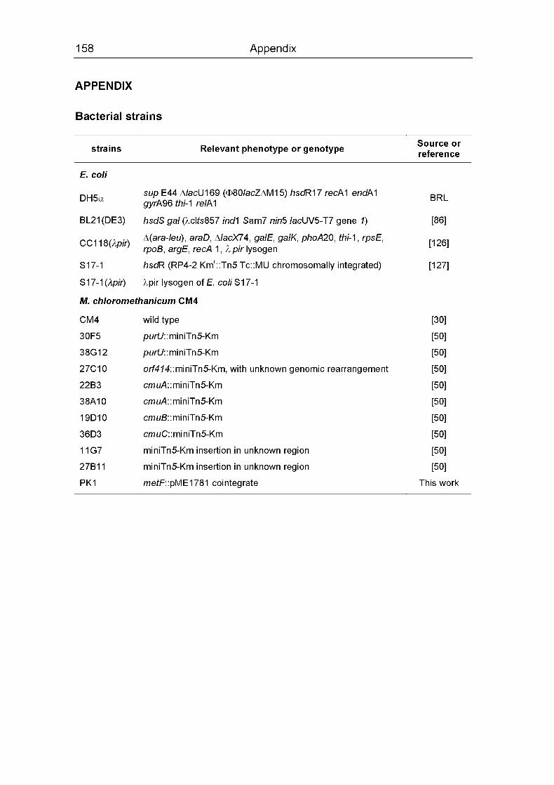
158 Appendix
APPENDIX
Bacterial strains
strains Relevant phenotype or genotypeSofurce or
r IV a IV reference
E. coli
_.,,,. sup E44 A/acU169 (O80/acZAM15) /?sdR17 recA1 endMDDI
DH5a
gyrA96 thiA relMBRL
BL21 (DE3) fisc/S ga/ (^clfs857 ind-\ Sam7 n/n5 /acUV5-T7 gene 1) [86]
CC118ttp/r)A(3ra-leu), araD, A/acX74, ga/E, ga/K, phoA20, thiA, rpsE,
„2„rpoß, argE, rec/A 1, A p/'r lysogen
S17-1 /?sdR (RP4-2 Kmr::Tn5Tc::MU chromosomally integrated) [127]
S17-1 {Xpir) >.pir lysogen of E. co//' S17-1
M. chloromethanicum CM4
CM4 wild type [30]
30F5 purl/::miniTn5-Km [50]
38G12 purl/::miniTn5-Km [50]
27C10 o/f474::miniTn5-Km, with unknown genomic rearrangement [50]
22B3 cmuA:miniTn5-Km [50]
38A10 cmuA:miniTn5-Km [50]
19D10 cmuß::miniTn5-Km [50]
36D3 cmuC::miniTn5-Km [50]
11G7 miniTn5-Km insertion in unknown region [50]
27B11 miniTn5-Km insertion in unknown region [50]
PK1 mefF::pME1781 cointegrate This work
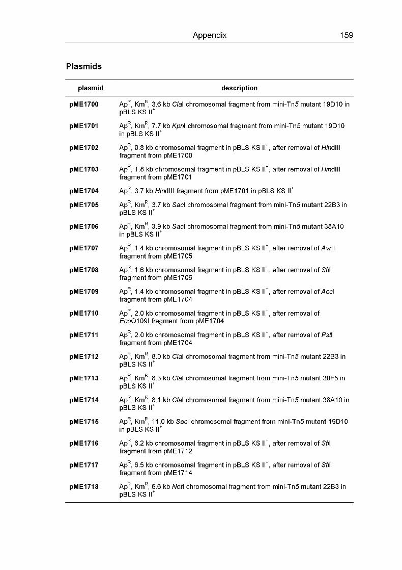
Appendix 159
Plasmids
plasmid description
pME1700
pME1701
pME1702
pME1703
pME1704
pME1705
pME1706
pME1707
pME1708
pME1709
pME1710
pME1711
pME1712
pME1713
pME1714
pME1715
pME1716
pME1717
pME1718
ApR, KmR, 3.6 kb C/al chromosomal fragment from mini-Tn5 mutant 19D10 in
pBLS KS If
ApR, KmR, 7.7 kb Kpnl chromosomal fragment from mini-Tn5 mutant 19D10
in pBLS KS if
ApR, 0.8 kb chromosomal fragment in pBLS KS if, after removal of H/ndlll
fragment from pME1700
ApR, 1.8 kb chromosomal fragment in pBLS KS If, after removal of Hind\\\
fragment from pME1701
ApR, 3.7 kb H/ndlll fragment from pME1701 in pBLS KS if
ApR, KmR, 3.7 kb Sac\ chromosomal fragment from mini-Tn5 mutant 22B3 in
pBLS KS If
ApR, KmR, 3.9 kb Sad chromosomal fragment from mini-Tn5 mutant 38A10
in pBLS KS if
ApR, 1.4 kb chromosomal fragment in pBLS KS I
fragment from pME1705
ApR, 1.6 kb chromosomal fragment in pBLS KS I
fragment from pME1706
ApR, 1.4 kb chromosomal fragment in pBLS KS I
fragment from pME1704
ApR, 2.0 kb chromosomal fragment in pBLS KS I
EcoO109l fragment from pME1704
ApR, 2.0 kb chromosomal fragment in pBLS KS I
fragment from pME1704
after removal of/Avril
after removal of Sfi\
after removal of Acc\
after removal of
after removal of Psti
ApR, KmR, 8.0 kb C/al chromosomal fragment from mini-Tn5 mutant 22B3 in
pBLS KS If
ApR, KmR, 8.3 kb C/al chromosomal fragment from mini-Tn5 mutant 30F5 in
pBLS KS If
ApR, KmR, 8.1 kb C/al chromosomal fragment from mini-Tn5 mutant 38A10 in
pBLS KS If
ApR, KmR, 11.0 kb Sad chromosomal fragment from mini-Tn5 mutant 19D10
in pBLS KS if
ApR, 6.2 kb chromosomal fragment in pBLS KS if, after removal of Sfi\
fragment from pME1712
ApR, 6.5 kb chromosomal fragment in pBLS KS if, after removal of Sfi\
fragment from pME1714
ApR, KmR, 6.6 kb Not\ chromosomal fragment from mini-Tn5 mutant 22B3 in
pBLS KS If

160 Appendix
plasmid description
pME1719 Ap ,Km
,7.7 kb Not\ chromosomal fragment from mini-Tn5 mutant 38A10 in
pBLS KS If
pME1720 ApR, 1.5 kb chromosomal fragment in pBLS KS if, after Kpnl digest of
pME1716
pME1721 4.7 kb chromosomal fragment in pBLS KS if, after A/ofl digest of pME1716
pME1722 ApR, KmR, 7.6 kb A/ofl chromosomal fragment from mini-Tn5 mutant 19D10
pME1723 ApR, 2.3 kb chromosomal fragment in pBLS KS if, after Pst\ digest of
pME1713
pME1724 ApR, 1.2 kb H/ndlll fragment from pME1719 in pBLS KS if
pME1725 ApR, 9.0 kb EcoRI fragment from pME1715 in pBLS KS if
pME1726 ApR, 5.5 kb chromosomal fragment in pBLS KS if, after Psti digest of
pME1725
pME1727 ApR, KmR, 8.7 kbXJbal chromosomal fragment from mini-Tn5 mutant 27C10
in pBLS KS if
pME1728 ApR, KmR, 9.0 kb Sacl chromosomal fragment from mini-Tn5 mutant 30F5 in
pBLS KS If
pME1729 ApR, 4.0 kb chromosomal fragment in pBLS KS if, after H/ndlll digest of
pME1728
pME1730 ApR, 3.5 kb chromosomal fragment in pBLS KS if, after Hind/// digest of
pME1727
pME1731 ApR, 3.0 kb H/ndlll fragment from pME1727 in pBLS KS if
pME1732 ApR, KmR, 10.8 kb C/al chromosomal fragment from mini-Tn5 mutant 11G7
in pBLS KS if
pME1733 ApR, KmR, 17.0 kb C/al chromosomal fragment from mini-Tn5 mutant 27B11
in pBLS KS if
pME1734 ApR, KmR, 9.0 kb A/ofl chromosomal fragment from mini-Tn5 mutant 27C10
in pBLS KS if
pME1735 ApR, KmR, 3.6 kb A/ofl/EcoRV chromosomal fragment from mini-Tn5 mutant
27C10inpBLSKSlf
pME1736 ApR, 3.2 kb chromosomal fragment in pBLS KS if, after H/ndlll cut digest of
pME1732
pME1737 ApR, 2.6 H/ndlll fragment from pME1732 in pBLS KS if
pME1738 ApR, KmR, 4.6 kb C/al chromosomal fragment from mini-Tn5 mutant 36D3 in
pBLS KS If
pME1739 ApR, KmR, 4.5 kb chromosomal fragment in pBLS KS if, after EcoRV digest
ofpME1733
pME1740 ApR, KmR, 10.0 kb C/al chromosomal fragment from mini-Tn5 mutant 38G12
in pBLS KS if
pME1741 ApR, 1.4 kb H/ndlll fragment of pME1735
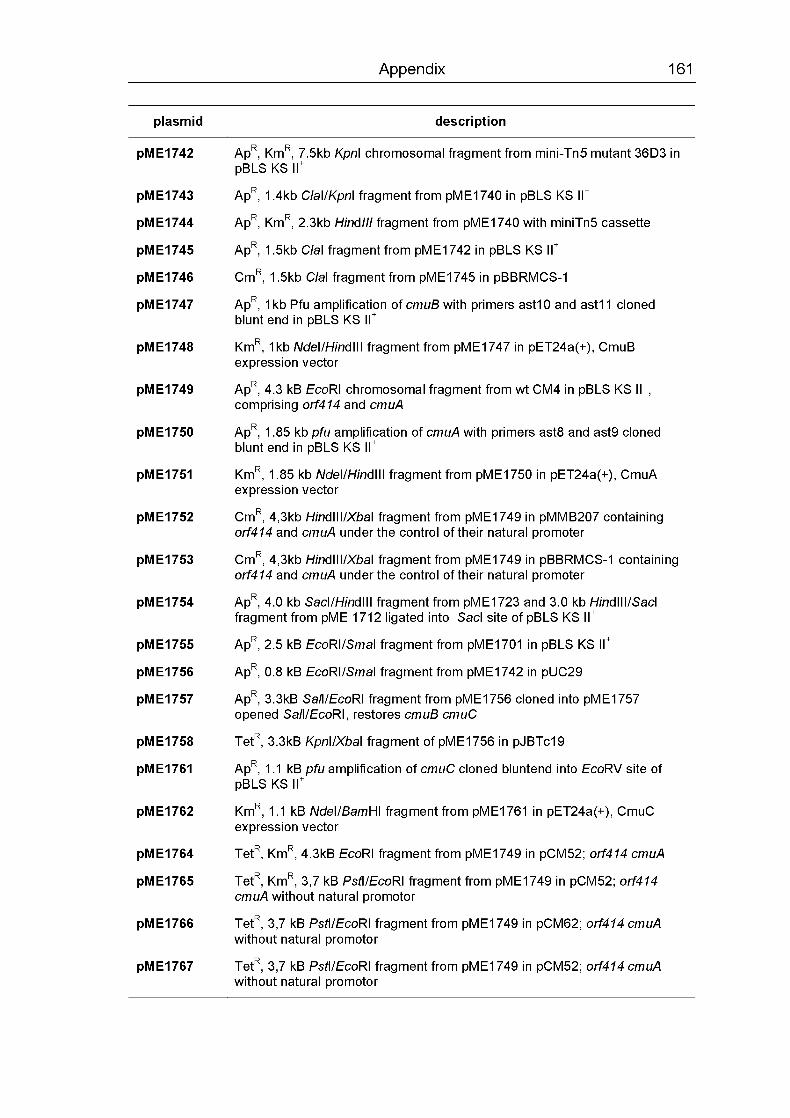
Appendix 161
plasmid description
pME1742 Ap ,Km
,7.5kb Kpnl chromosomal fragment from mini-Tn5 mutant 36D3 in
pBLS KS If
pME1743 ApR, 1.4kb C/al/Kpnl fragment from pME1740 in pBLS KS if
pME1744 ApR, KmR, 2.3kb Hindill fragment from pME1740 with miniTn5 cassette
pME1745 ApR, 1.5kb C/al fragment from pME1742 in pBLS KS if
pME1746 CmR, 1.5kb C/al fragment from pME1745 in pBBRMCS-1
pME1747 ApR, 1kb Pfu amplification of cmuB with primers ast10 and ast11 cloned
blunt end in pBLS KS if
pME1748 KmR, 1kb A/c/el/H/ndlll fragment from pME1747 in pET24a(+), CmuB
expression vector
pME1749 ApR, 4.3 kB EcoRI chromosomal fragment from wt CM4 in pBLS KS if,comprising orf414 and cmuA
pME1750 ApR, 1.85 kbpfu amplification of cmuA with primers ast8 and ast9 cloned
blunt end in pBLS KS if
pME1751 KmR, 1.85 kb A/c/el/H/ndlll fragment from pME1750 in pET24a(+), CmuA
expression vector
pME1752 CmR, 4,3kb H/ndlll/Xbal fragment from pME1749 in pMMB207 containingorf414 and cmuA under the control of their natural promoter
pME1753 CmR, 4,3kb H/ndlll/Xbal fragment from pME1749 in pBBRMCS-1 containingorf414 and cmuA under the control of their natural promoter
pME1754 ApR, 4.0 kb Sacl/H/ndlll fragment from pME1723 and 3.0 kb H/ndlll/Sacl
fragment from pME 1712 ligated into Sad site of pBLS KS if
pME1755 ApR, 2.5 kB EcoRI/Smal fragment from pME1701 in pBLS KS if
pME1756 ApR, 0.8 kB EcoRI/Smal fragment from pME1742 in pUC29
pME1757 ApR, 3.3kB Sa/l/EcoRI fragment from pME1756 cloned into pME1757
opened Sa/l/EcoRI, restores cmuB cmuC
pME1758 TetR, 3.3kB Kpnl/XJbal fragment of pME1756 in pJBTc19
pME1761 ApR, 1.1 kB pfu amplification of cmuC cloned bluntend into EcoRV site of
pBLS KS If
pME1762 KmR, 1.1 kB A/ctel/ßamHI fragment from pME1761 in pET24a(+), CmuC
expression vector
pME1764 TetR, KmR, 4.3kB EcoRI fragment from pME1749 in pCM52; orf414 cmuA
pME1765 TetR, KmR, 3,7 kB Psfi/EcoRI fragment from pME1749 in pCM52; orf414
cmuA without natural promotor
pME1766 TetR, 3,7 kB Psfl/EcoRI fragment from pME1749 in pCM62; orf414 cmuA
without natural promotor
pME1767 TetR, 3,7 kB Psfl/EcoRI fragment from pME1749 in pCM52; orf414 cmuA
without natural promotor

162 Appendix
plasmid description
pME1768 Tef, 3,7 kB Psfl/EcoRI fragment from pME1749 in pCM52; orf414 cmuA
without natural promotor
pME1769 TetR, 4.3kB EcoRI fragment from pME1749 in pCM52; orf414 cmuA
pME1770 ApR, 0.7 kb Pfu amplification of purU orf414 intragenic region with primerast25 and ast26 Xbal/A/c/el digested and cloned into pME1750; fusion of
natural promoter region directly to cmuA
pME1771 TetR, 2,6 kbXbal/H/ndlll fragment from pME1770 in pCM62
pME1772 TetR, 1.9 kb Psfl/ßamHI fragment from pME1750 in pCM62; cmuA under
lacP promoter
pME1773 ApR, 0.5 kb EcoRI/ßamHI fragment from pME1716 in pBLS KS if
pME1774 KmR, 0.5 kb A/ofi/Xnol fragment from pME1773 in pKNOCK-Km; internal folD
fragment
pME1775 TetR, 5.8 bpXJbal/Sad fragment from pME1754 in CM62; purU folD folC
cobU
pME1776 TetR, 2.8 kb ßamHI/Sacl fragment from pME1754 in pCM62; purU
pME1777 TetR, 3.1 kb Kpnl(blunted)/Sacl fragment from pME1754 in CM62 opened
H/ndlll(blunted)/Sacl
pME1778 TetR, 4.8 kb A/col(blunted)/Sacl fragment from pME1754 in CM62
H/ndlll(blunted)/Sacl
pME1780 KmR, 592bp ßamHI/H/ncll fragment of pME1749 containing orf414 part
pME1781 KmR, 0.6 kb Smal/C/al internal metF fragment in pKNOCK-Km
pME1782 ApR, KmR, 1.2 kb Mlu\ fragment of pKNOCK-Km (KmR gene) in pME1749Mlu\ opened
pME1784 TetR, KmR, 3.4kbXJbal fragment from pME1757 in pCM52; cmuB cmuC
pME1785 TetR, KmR, 3.4kbXbal fragment from pME1757 in pCM62; cmuB cmuC
pME1786 TetR, KmR, 3.4kbXbal fragment from pME1757 in pCM80; cmuB cmuC
pME1787 TetR, KmR, 3.2 kb Kpnl fragment of pME1782 (orf414 with KmR gene) in
pWM41
pME1788 TetR, KmR, 3.2 kb Kpnl fragment of pME1782 (orf414 with KmR gene) in
pWM41
pME1789 TetR, KmR, 1.2 kb H/ndlll/Fspl fragment from pME1742 in pWM41 ; metF
pME1790 779 bp PCR fragment in pCM130; purU::xylE fusion
pME1791 779 bp PCR fragment in pCM130; orf414::xylE fusion
pME1792 ApR, 0.3 kb blunted Nde\/Xho\ internal orf219 fragment from pME1726 in
pBLS KS If EcoRV opened
pME1793 TetR, 1.7 kb H/ndlll/Xnol fragment from pME1726 in pME 1785 H/ndlll/Xnol
opened; metF cmuB cmuC under control of natural promoter region

Appendix 163
plasmid description
pME 1794 Ap ,0.2 kB Pfu amplification of o/f279/mefF intragenic region with primer
ast30 and ast31 ligated blunted into EcoRV opened pBLS KS if
pME 1795 ApR, 0.2 kB Pfu amplification of orf219/metF intragenic region with primerast30 and ast31 ligated blunted into EcoRV opened pBLS KS if
pME 1796 TetR, 159 bp BamHI fragment from pME1795 in pCM130; orf219::xylE fusion
(checked by sequencing)
pME 1797 TetR, 159 bp BamHI fragment from pME1795 in pCM130; metFrxylE fusion
(checked by sequencing)
pME1798 ApR, 1.4 kb EcoRI fragment from pME1716 in pBLS KS if; purUfolD'
pME1799 TetR, 1.7 kb H/ndlll/Ynol fragment from pME1793 in pCM130; metF
cmuBr.xylE fusion
pME8250 TetR, 0.6 kb Cla\/Xho\ fragment from pME1793 in pCM130; cmuBr.xylEfusion
pME8251 TetR, 1.4 kb H/ndlll/ßamHI from pME1798 fragment in pCM130; EcoRI
chromosomal fragment with purU folDrxylE fusion
pME8252 TetR, 0.7 kbXnol/ßamHI pME1798 fragment in pCM130; Xnol/EcoRI
chromosomal fragment with purUrxylE fusion
pME8253 TetR, 0.7 kb H/ndlll/Xnol fragment in pCM130 with folDrxylE fusion, obtained
after removal of aXno//ßamHI fragment from pME8251 and religation of the
blunted plasmid
pBLS II KS+ Apr, cloning vector, Stratagene
pKNOCK-Km Kmr, broad-host-range suicide vector, oriR6K, Stratagene
pET24a(+) Kmr,expression vector
pCM52 Tetr, Kmr, broad-host range vector
pCM62 Tetr, broad-host range vector
pCM80 Tetr, broad-host range vector
pCM130 Tetr, xylE promoter-probe vector
pCM131 Tetr, mxaFrxylE fusion in pCM130
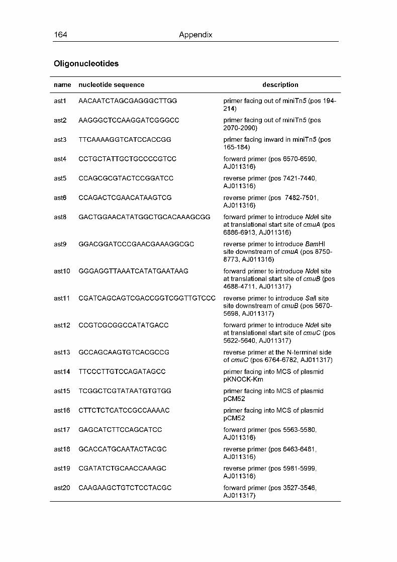
164 Appendix
Oligonucleotides
name nucleotide sequence description
asti AACAATCTAGCGAGGGCTTGG
ast2 AAGGGCTCCAAGGATCGGGCC
ast3 TTCAAAAGGTCATCCACCGG
ast4 CCTGCTATTGCTGCCCCGTCC
ast5 CCAGCGCGTACTCCGGATCC
ast6 CCAGACTCGAACATAAGTCG
ast8 GACTGGAACATATGGCTGCACAAAGCGG
ast9 GGACGGATCCCGAACGAAAGGCGC
ast10 GGGAGGTTAAATCATATGAATAAG
astl 1 CGATCAGCAGTCGACCGGTCGGTTGTCCC
ast12 CCGTCGCGGCCATATGACC
ast13 GCCAGCAAGTGTCACGCCG
ast14 TTCCCTTGTCCAGATAGCC
ast15 TCGGCTCGTATAATGTGTGG
ast16 CTTCTCTCATCCGCCAAAAC
ast17 GAGCATCTTCCAGCATCC
ast18 GCACCATGCAATACTACGC
ast19 CGATATCTGCAACCAAAGC
ast20 CAAGAAGCTGTCTCCTACGC
primer facing out of miniTn5 (pos 194-
214)
primer facing out of miniTn5 (pos
2070-2090)
primer facing inward in miniTn5 (pos
165-184)
forward primer (pos 6570-6590,
AJ011316)
reverse primer (pos 7421-7440,
AJ011316)
reverse primer (pos 7482-7501,
AJ011316)
forward primer to introduce Nde\ site
at translational start site oîcmuA (pos
6886-6913, AJ011316)
reverse primer to introduce ßamHI
site downstream of cmuA (pos 8750-
8773, AJ011316)
forward primer to introduce Nde\ site
at translational start site of cmuB (pos4688-4711, AJ011317)
reverse primer to introduce Sa/I site
site downstream of cmuB (pos 5670-
5698, AJ011317)
forward primer to introduce Nde\ site
at translational start site of cmuC (pos
5622-5640, AJ011317)
reverse primer at the N-terminal side
of cmuC (pos 6764-6782, AJ011317)
primer facing into MCS of plasmid
pKNOCK-Km
primer facing into MCS of plasmid
pCM52
primer facing into MCS of plasmid
pCM52
forward primer (pos 5563-5580,
AJ011316)
reverse primer (pos 6463-6481,
AJ011316)
reverse primer (pos 5981-5999,
AJ011316)
forward primer (pos 3527-3546,
AJ011317)

Appendix 165
name nucleotide sequence description
ast21 GCTCGCGAAACTATATCTGC
ast22 GCAACTAGGAGGATCTCACG
ast25 GGCGACATATGACGGCACC
ast26 TTCCGCCATCTAGAGATTCC
ast27 CGCACCTGAAACGGCAGCGACGATGC
ast28 CCGAGCCACGGAAACGGTTCAGGCG
ast29 CGAACATCCGCTCACGGCTCGTCATC
ast30 GCTTTCGGATCCATCAGACG
ast31 CCACATATGCGGATCCGTC
ast32 CGACAGACCCGAACCTCGCCATTGG
ast33 GGGAGACCTCCAATGACAGATCGCG
ast34 CCCATCTATCACTTGTGCAATCATG
ast35 CGGAGAAATGATGCCTTGCTCCTCA
ast36 CCTCGATCTCTCAATCCCTGG
ast37 GAATGTCAATGGGATCGTGCG
ast38 CAGATACGGTAAACTAGCCTCG
reverse primer (pos 4417-4436,
AJ011316)
reverse primer (pos 3852-3871,
AJ011316)
reverse primer to introduce Nde\ at
translational start site of o/f474 (pos
5617-5635, AJ011316)
forward primer to introduce XJbal site
at translational start site of purU (pos4841-4860, AJ011316)
forward primer for primer extension
analysis of purU (pos 4775-4800,
AJ011316)
reverse primer for primer extension
analysis of o/f474 (pos 5632-5656,
AJ011316)
reverse primer for primer extension
analysis of cmuA (pos 6917-6942,
AJ011316)
forward primer to introduce ßamHI
site at translational start site of orf219
(pos 3332-3351, AJ011317)
reverse primer to introduce Nde\ at
translational start site of metF (pos3494-3512, AJ011317)
reverse primer for primer extension
analysis of o/f474 (pos 5509-5533,
AJ011316)
reverse primer for primer extension
analysis o\metF (pos 3603-3627,
AJ011317)
forward primer for primer extension
analysis of folD (pos 3842-3866,
AJ011316)
forward primer for primer extension
analysis oforf219 (pos 3229-3253
AJ011317)
forward primer (pos 3360-3380,
AJ011317)
reverse primer (pos 3440-3460,
AJ011317)
primer facing into MCS of plasmid
pCM130


CURRICULUM VITAE
Alexander Studer
citizen of Grafenried (BE), Switzerland
born October 17th, 1971 in Liestal (BL), Switzerland
1978 - 1982 Primary education in Pratteln (BL), Switzerland
1982 - 1986 Secondary education (Progymnasium) in Pratteln (BL),
Switzerland
1986 - 1990 Secondary education (Gymnasium) in Muttenz (BL),
Switzerland
Graduation: Matura C
1992 - 1995 Studies of Molecular Biology and Biochemistry at the
University of Basel, Biozentrum, Switzerland
1995 - 1996 Masters degree (Diploma) in Microbiology: "Type IC
restriction-modification system EcoRI 24" at the University
of Basel, Biozentrum, Switzerland
1997 - 2001 Research assistant at the Swiss Federal Institute of
Technology, Institute of Microbiology, Zürich, Switzerland
2001 Ph.D. thesis at the Swiss Federal Institute of Technology,
Institute of Microbiology, Zürich, Switzerland


PUBLICATIONS
Vannelli, T., Studer, A., Kertesz, M., & Leisinger, T. (1998) Chloromethane
metabolism by Methylobacterium sp. strain CM4. Appl. Environ. Microbiol. 64,
1933-1936.
Vannelli, T., Messmer, M., Studer, A., Vuiiieumier, S., & Leisinger, T. (1999)
A corrinoid-dependent catabolic pathway for growth of a Methylobacterium
strain with chloromethane. Proc. Natl. Acad. Sei. U. S. A. 96, 4615-4620.
Studer, A., Vuiiieumier, S., & Leisinger, T. (1999) Properties of the
methylcobalamin:H4folate methyltransferase involved in chloromethane
utilization by Methylobacterium sp. strain CM4. Eur. J. Biochem. 264, 242-249.
McAnulla, C, Woodall, C. A., McDonald, I. R., Studer, A., Vuiiieumier, S.,
Leisinger, T., & Murrell, J. M. (2001) Chloromethane utilization gene cluster
from Hyphomicrobium chloromethanicum strain CM2T and development of
functional gene probes to detect halomethane-degrading bacteria. Appl.
Environ. Microbiol. 67, 307-316.
Studer, A., Stupperich, E., Vuiiieumier, S., & Leisinger, T. (2001)
Chloromethane:tetrahydrofolate methyl transfer by two proteins from
Methylobacterium chloromethanicum strain CM4. Eur. J. Biochem. 268, 2931-
2938.