Embed Size (px)
Citation preview

In silico dynamics of COVID-19 phenotypes foroptimizing clinical managementChrysovalantis Voutouria,1, Mohammad Reza Nikmaneshib,c,1, C. Corey Hardind, Ankit B. Patele, Ashish Vermae,Melin J. Khandekarf, Sayon Duttag, Triantafyllos Stylianopoulosa,2, Lance L. Munnc,2, and Rakesh K. Jainc,2
aCancer Biophysics Laboratory, Department of Mechanical and Manufacturing Engineering, University of Cyprus, 1678 Nicosia, Cyprus; bDepartment ofMechanical Engineering, Sharif University of Technology, Tehran, Iran, 11155; cEdwin L. Steele Laboratories, Department of Radiation Oncology,Massachusetts General Hospital and Harvard Medical School, Boston, MA 02114; dDepartment of Pulmonary and Critical Care Medicine, MassachusettsGeneral Hospital and Harvard Medical School, Boston, MA 02114; eDepartment of Medicine/Renal Division, Brigham and Women’s Hospital and HarvardMedical School, Boston, MA 02115; fDepartment of Radiation Oncology, Massachusetts General Hospital and Harvard Medical School, Boston, MA 02114;and gDepartment of Emergency Medicine, Massachusetts General Hospital and Harvard Medical School, Boston, MA 02114
Contributed by Rakesh K. Jain, December 2, 2020 (sent for review October 20, 2020; reviewed by Narendra M. Dixit and Libin Rong)
Understanding the underlying mechanisms of COVID-19 progressionand the impact of various pharmaceutical interventions is crucial forthe clinical management of the disease. We developed a comprehen-sive mathematical framework based on the knownmechanisms of thesevere acute respiratory syndrome coronavirus 2 (SARS-CoV-2) infec-tion, incorporating the renin−angiotensin system and ACE2, which thevirus exploits for cellular entry, key elements of the innate and adap-tive immune responses, the role of inflammatory cytokines, and thecoagulation cascade for thrombus formation. The model predicts theevolution of viral load, immune cells, cytokines, thrombosis, and oxy-gen saturation based on patient baseline condition and the presenceof comorbidities. Model predictions were validated with clinical datafrom healthy people and COVID-19 patients, and the results wereused to gain insight into identified risk factors of disease progres-sion including older age; comorbidities such as obesity, diabetes,and hypertension; and dysregulated immune response. We thensimulated treatment with various drug classes to identify optimaltherapeutic protocols. We found that the outcome of any treatmentdepends on the sustained response rate of activated CD8+ T cellsand sufficient control of the innate immune response. Furthermore,the best treatment—or combination of treatments—depends on thepreinfection health status of the patient. Our mathematical frame-work provides important insight into SARS-CoV-2 pathogenesis andcould be used as the basis for personalized, optimal management ofCOVID-19.
SARS-CoV-2 | COVID-19 | mathematical model | simulation
COVID-19 has created unprecedented challenges for the healthcare system, and, until an effective vaccine is developed and
made widely available, treatment options are limited. A challenge tothe development of optimal treatment strategies is the extremeheterogeneity of presentation. Infection with severe acute respira-tory syndrome coronavirus 2 (SARS-CoV-2) results in a syndromethat ranges in severity from asymptomatic to multiorgan failure anddeath. In addition to local complications in the lung, the virus cancause systemic inflammation and disseminated microthrombosis,which can cause stroke, myocardial infarction, or pulmonary emboli(1–4). Risk factors for poor COVID-19 outcome include advancedage, obesity, diabetes, and hypertension (5–13).Computational analyses can provide insights into the transmis-
sion, control, progression, and underlying mechanisms of infec-tious diseases. Indeed, epidemiological and statistical modelinghas been used for COVID-19, providing powerful insights intocomorbidities, transmission dynamics, and control of the disease(14–17). However, to date, these analyses have been populationdynamics models of SARS-CoV-2 infection and transmission orcorrelative analyses of COVID-19 comorbidities and treatmentresponse. Simple viral dynamics models have been also developedand used to predict the SARS-CoV-2 response to antiviral drugs(18, 19). These models, however, do not explicitly consider the bi-ological or physiological mechanisms underlying disease progression
or the time course of response to various therapeutic interventions,and only a few more-sophisticated models have been developedtoward this direction (20, 21).Several therapies targeting various aspects of COVID-19
pathogenesis have been proposed and have either completed—or are currently being tested in—clinical trials (22). Despitestrong biologic rationale, these treatments have generally producedconflicting results in the clinic. For example, trials of antiviral ther-apies (e.g., remdesivir) have been mixed: The original trial fromChina failed (23), a subsequent trial in the United States led toapproval of remdesivir in the United States and other countries (24),and the recent results of the World Health Organization Solidarity
Significance
A distinctive feature of COVID-19 is its extreme heterogeneity—illness ranges from minimally symptomatic to life threatening.Heterogeneity results from a poorly understood combination ofpatient factors, viral dynamics, antiviral and immune modulatingtherapies, and dynamics of the innate and adaptive immune re-sponses. In order to better understand clinical heterogeneity andoptimal treatment, we developed a comprehensive mathematicalmodel incorporating elements of the innate and adaptive immuneresponses, the renin−angiotensin system (which the virus exploitsfor cellular entry), rates of viral replication, inflammatory cytokines,and the coagulation cascade. Our model reveals divergent treat-ment responses and clinical outcomes as a function of comorbid-ities, age, and details of the innate and adaptive immune responseswhich can provide a framework for understanding individualpatients’ trajectories.
Author contributions: C.C.H., A.B.P., A.V., M.J.K., S.D., T.S., L.L.M., and R.K.J. designedresearch; C.V., and M.R.N. performed research; C.V., M.R.N., T.S., and L.L.M. contributednew reagents/analytic tools; C.V., M.R.N., C.C.H., A.B.P., A.V., M.J.K., S.D., T.S., L.L.M., andR.K.J. analyzed data; C.V., M.R.N., C.C.H., A.B.P., A.V., M.J.K., S.D., T.S., L.L.M., and R.K.J.wrote the paper; and R.K.J. supervised the project.
Reviewers: N.M.D., Indian Institute of Science Bangalore; and L.R., University of Florida.
Competing interest statement: R.K.J. received honorarium from Amgen; consultant feesfrom Chugai, Merck, Ophthotech, Pfizer, SPARC, SynDevRx, XTuit, Elpis; owns equity inAccurius, Enlight, Ophthotech, SynDevRx; and serves on the Boards of Trustees of TeklaHealthcare Investors, Tekla Life Sciences Investors, Tekla Healthcare Opportunities Fund,and Tekla World Healthcare Fund. Neither any reagent nor any funding from these or-ganizations was used in this study. A.B.P. has received fellowship funding from Relypsa,Inc. L.L.M. owns equity in Bayer AG and is a consultant for SimBiosys. Neither any reagentnor any funding from these organizations was used in this study.
This open access article is distributed under Creative Commons Attribution License 4.0(CC BY).1C.V. and M.R.N. contributed equally to this work.2To whom correspondence may be addressed. Email: [email protected], [email protected], or [email protected].
This article contains supporting information online at https://www.pnas.org/lookup/suppl/doi:10.1073/pnas.2021642118/-/DCSupplemental.
Published January 5, 2021.
PNAS 2021 Vol. 118 No. 3 e2021642118 https://doi.org/10.1073/pnas.2021642118 | 1 of 8
MED
ICALSC
IENCE
SEN
GINEE
RING
Dow
nloa
ded
by g
uest
on
Feb
ruar
y 25
, 202
2
trial again show no benefit (25). Other antiviral drugs alone or incombination are also showing promise (26).Other potential treatments include antiinflammatory drugs
and antithrombotic agents. Because of the systemic inflamma-tion seen in many patients, antiinflammatory drugs have beentested, including anti-IL6/IL6R therapy (e.g., tocilizumab, sil-tuximab) and anti-JAK1/2 drugs (e.g., barcitinib). It is not clearwhether these drugs will be effective as stand-alone treatments,particularly after the recent failure of tocilizumab in a phase IIItrial (1, 27–29). In addition, given that a common complication ofCOVID-19 is the development of coagulopathies with microvas-cular thrombi potentially leading to the dysfunction of multipleorgan systems (2, 3), antithrombotic drugs (e.g., low molecularweight heparin) are being tested. Recognizing the interactions ofCOVID-19 with the immune system (30), the corticosteroiddexamethasone has been tested, showing some promising results.Given the large range of patient comorbidities, disease severities,and variety of complications such as thrombosis, it is likely thatpatients will have heterogeneous responses to any given therapy,
and such heterogeneity will continue to be a challenge for clinicaltrials of unselected COVID-19 patients (31).Here, we developed a systems biology-based mathematical
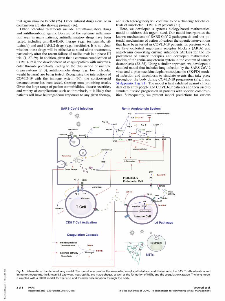
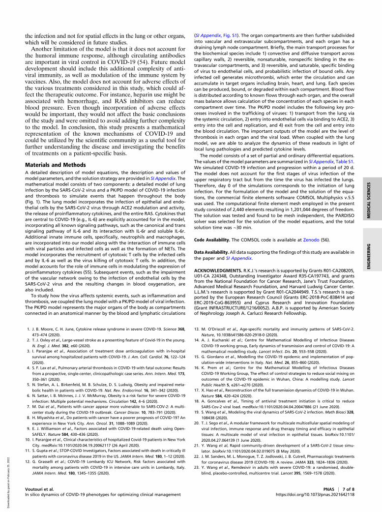
model to address this urgent need. Our model incorporates theknown mechanisms of SARS-CoV-2 pathogenesis and the po-tential mechanisms of action of various therapeutic interventionsthat have been tested in COVID-19 patients. In previous work,we have exploited angiotensin receptor blockers (ARBs) andangiotensin converting enzyme inhibitors (ACEis) for the im-provement of cancer therapies and developed mathematicalmodels of the renin−angiotensin system in the context of cancerdesmoplasia (32–35). Using a similar approach, we developed adetailed model that includes lung infection by the SARS-CoV-2virus and a pharmacokinetic/pharmacodynamic (PK/PD) modelof infection and thrombosis to simulate events that take placethroughout the body during COVID-19 progression (Fig. 1 andSI Appendix, Fig. S1). The model is first validated against clinicaldata of healthy people and COVID-19 patients and then used tosimulate disease progression in patients with specific comorbid-ities. Subsequently, we present model predictions for various
Neutrophil
NETs
Epithelial or Endothelial Cell
SARS-CoV-2
Remdesivir
Tissue Damage
ACE2TMPRSS2
SARS-CoV-2 infection
Adam17
sACE2
Coagulation Cascade
Intrinsic pathway
Extrinsic pathwayTissue Factor
FibrinogenFibrin
Damaged surface heparin
PD-1
T Cell
PD-L1
TCR
MHC I
viral antigen
CD8 T Cell Activation
IL6
sIL6ra
Trans pathwayGP130
canonical
GP130
JAK
STAT
IL6ra
Adam17
Immune Cell
inflammation
anti-IL6
Baricitinib
JAK
STAT
Baricitinib
IL6 Pathways
TLR7
Interferon a/b
NFkB
Interferon γ
Interferon γ
Ang I
Ang II
ACE
ARBs
ACEi
Ang 1-7
AT1R
Angiotensinogenrenin
AT2R
Ang III
AT4RACE2 MASR
Ang 1-9
AT2R
ACEAng 1-7
vasoconstriction
Ang IV
inflammation
sACE2 sACE2
Renin Angiotensin System
ACE2
Fig. 1. Schematic of the detailed lung model. The model incorporates the virus infection of epithelial and endothelial cells, the RAS, T cells activation andimmune checkpoints, the known IL6 pathways, neutrophils, and macrophages, as well as the formation of NETs, and the coagulation cascade. The lung modelis coupled with a PK/PD model for the virus and thrombi dissemination through the body.
2 of 8 | PNAS Voutouri et al.https://doi.org/10.1073/pnas.2021642118 In silico dynamics of COVID-19 phenotypes for optimizing clinical management
Dow
nloa
ded
by g
uest
on
Feb
ruar
y 25
, 202
2
therapies currently employed for treatment of COVID-19 aloneor in combination, and we identify protocols for optimal clinicalmanagement for each of the clinically observed COVID-19phenotypes.
Model DescriptionThe model includes SARS-CoV-2 infection, the renin angio-tensin system (RAS), inflammatory and antiinflammatory cyto-kines, innate and adaptive immune cells, and factors involved inthe coagulation cascade (Fig. 1). SARS-CoV-2 enters the cell bydocking to ACE2, a key component of the RAS. ACE2 can bemembrane bound or soluble, and it regulates inflammation byconverting angiotensin (Ang) II to Ang 1–7 and Ang I to Ang 1–9;as opposed to Ang I and Ang II, which lead to inflammation. Ang1–7 and Ang 1–9 have antiinflammatory effects. Intracellular virusinitiates inflammatory pathways through toll-like receptors andNFκB, which produces interferons and other inflammatory cyto-kines. The viral antigens, along with inflammatory cytokines, causeactivation of naïve T cells, creating virus-specific T effector cells.T cell activation is controlled by viral antigen strength and thepresence of PD-L1/PD-1 inhibition (36). We combine inflamma-tory cytokines into a single variable, but explicitly account for IL6production via the trans pathway in epithelial and endothelial cellsand the canonical pathway in immune cells. In the presence ofinflammatory cytokines and virus, neutrophils can produce neu-trophil extracellular traps (NETs).Because the virus can infect endothelial cells, we also consider
viral dissemination via the blood stream, and the possibility ofsystemic infection and thrombosis. We include the major organsin a PK/PD model, with physiological blood flow patterns ex-plicitly modeled. Infection of endothelial cells, combined withhigh levels of inflammatory cytokines in the plasma, can result inthrombosis. Damage to virally infected endothelial cells and theproduction of NETs can exacerbate the thrombosis, and micro-thrombi can enter the blood stream to accumulate in other or-gans, including the brain, heart, and lung. We use a simplifiedmodel of the coagulation pathways, assuming that formation ofmicrothrombi is proportional to the number of infected endo-thelial cells, the presence of neutrophil NETs, and the level ofinflammatory cytokines. Transport of oxygen from the alveolarspace to the blood vessels in the lung is calculated using amodified diffusion model (37).
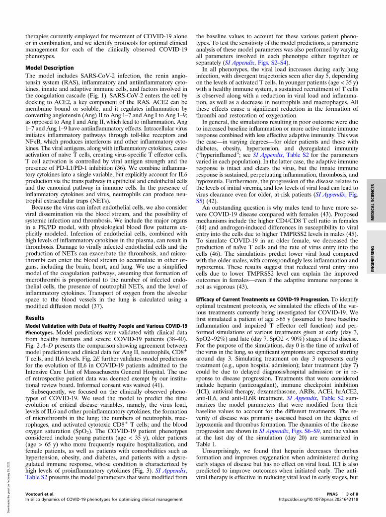
ResultsModel Validation with Data of Healthy People and Various COVID-19Phenotypes. Model predictions were validated with clinical datafrom healthy humans and severe COVID-19 patients (38–40).Fig. 2 A–D presents the comparison showing agreement betweenmodel predictions and clinical data for Ang II, neutrophils, CD8+
T cells, and IL6 levels. Fig. 2E further validates model predictionsfor the evolution of IL6 in COVID-19 patients admitted to theIntensive Care Unit of Massachusetts General Hospital. The useof retrospective patient data was deemed exempt by our institu-tional review board. Informed consent was waived (41).Subsequently, we focused on the clinically observed pheno-
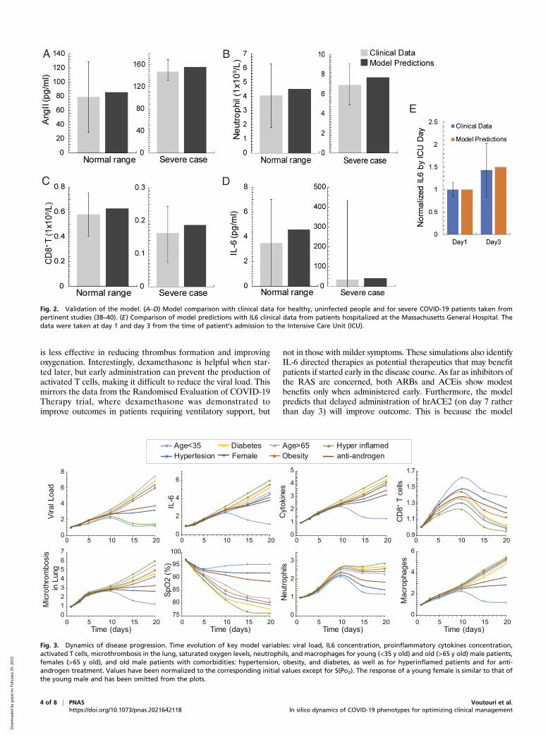
types of COVID-19. We used the model to predict the timeevolution of critical disease variables, namely, the virus load,levels of IL6 and other proinflammatory cytokines, the formationof microthrombi in the lung; the numbers of neutrophils, mac-rophages, and activated cytotoxic CD8+ T cells; and the bloodoxygen saturation (SpO2). The COVID-19 patient phenotypesconsidered include young patients (age < 35 y), older patients(age > 65 y) who more frequently require hospitalization, andfemale patients, as well as patients with comorbidities such ashypertension, obesity, and diabetes, and patients with a dysre-gulated immune response, whose condition is characterized byhigh levels of proinflammatory cytokines (Fig. 3). SI Appendix,Table S2 presents the model parameters that were modified from
the baseline values to account for these various patient pheno-types. To test the sensitivity of the model predictions, a parametricanalysis of these model parameters was also performed by varyingall parameters involved in each phenotype either together orseparately (SI Appendix, Figs. S2–S4).In all phenotypes, the viral load increases during early lung
infection, with divergent trajectories seen after day 5, dependingon the levels of activated T cells. In younger patients (age < 35 y)with a healthy immune system, a sustained recruitment of T cellsis observed along with a reduction in viral load and inflamma-tion, as well as a decrease in neutrophils and macrophages. Allthese effects cause a significant reduction in the formation ofthrombi and restoration of oxygenation.In general, the simulations resulting in poor outcome were due
to increased baseline inflammation or more active innate immuneresponse combined with less effective adaptive immunity. This wasthe case—in varying degrees—for older patients and those withdiabetes, obesity, hypertension, and dysregulated immunity(“hyperinflamed”; see SI Appendix, Table S2 for the parametersvaried in each population). In the latter case, the adaptive immuneresponse is intact and clears the virus, but the innate immuneresponse is sustained, perpetuating inflammation, thrombosis, andhypoxemia. Furthermore, the progression of the disease relates tothe levels of initial viremia, and low levels of viral load can lead tovirus clearance even for older, at-risk patients (SI Appendix, Fig.S5) (42).An outstanding question is why males tend to have more se-
vere COVID-19 disease compared with females (43). Proposedmechanisms include the higher CD4/CD8 T cell ratio in females(44) and androgen-induced differences in susceptibility to viralentry into the cells due to higher TMPRSS2 levels in males (45).To simulate COVID-19 in an older female, we decreased theproduction of naïve T cells and the rate of virus entry into thecells (46). The simulations predict lower viral load comparedwith the older males, with correspondingly less inflammation andhypoxemia. These results suggest that reduced viral entry intocells due to lower TMPRSS2 level can explain the improvedoutcomes in females—even if the adaptive immune response isnot as vigorous (43).
Efficacy of Current Treatments on COVID-19 Progression. To identifyoptimal treatment protocols, we simulated the effects of the var-ious treatments currently being investigated for COVID-19. Wefirst simulated a patient of age >65 y (assumed to have baselineinflammation and impaired T effector cell function) and per-formed simulations of various treatments given at early (day 3,SpO2∼92%) and late (day 7, SpO2 < 90%) stages of the disease.For the purpose of the simulations, day 0 is the time of arrival ofthe virus in the lung, so significant symptoms are expected startingaround day 3. Simulating treatment on day 3 represents earlytreatment (e.g., upon hospital admission); later treatment (day 7)could be due to delayed diagnosis/hospital admission or in re-sponse to disease progression. Treatments that were consideredinclude heparin (anticoagulant), immune checkpoint inhibition(ICI), antiviral therapy, dexamethasone, ARBs, ACEi, hrACE2,anti-IL6, and anti-IL6R treatment. SI Appendix, Table S2 sum-marizes the model parameters that were modified from theirbaseline values to account for the different treatments. The se-verity of disease was primarily assessed based on the degree ofhypoxemia and thrombus formation. The dynamics of the diseaseprogression are shown in SI Appendix, Figs. S6–S9, and the valuesat the last day of the simulation (day 20) are summarized inTable 1.Unsurprisingly, we found that heparin decreases thrombus
formation and improves oxygenation when administered duringearly stages of disease but has no effect on viral load. ICI is alsopredicted to improve outcomes when initiated early. The anti-viral therapy is effective in reducing viral load in early stages, but
Voutouri et al. PNAS | 3 of 8In silico dynamics of COVID-19 phenotypes for optimizing clinical management https://doi.org/10.1073/pnas.2021642118
MED
ICALSC
IENCE
SEN
GINEE
RING
Dow
nloa
ded
by g
uest
on
Feb
ruar
y 25
, 202
2
is less effective in reducing thrombus formation and improvingoxygenation. Interestingly, dexamethasone is helpful when star-ted later, but early administration can prevent the production ofactivated T cells, making it difficult to reduce the viral load. Thismirrors the data from the Randomised Evaluation of COVID-19Therapy trial, where dexamethasone was demonstrated toimprove outcomes in patients requiring ventilatory support, but
not in those with milder symptoms. These simulations also identifyIL-6 directed therapies as potential therapeutics that may benefitpatients if started early in the disease course. As far as inhibitors ofthe RAS are concerned, both ARBs and ACEis show modestbenefits only when administered early. Furthermore, the modelpredicts that delayed administration of hrACE2 (on day 7 ratherthan day 3) will improve outcome. This is because the model
Fig. 2. Validation of the model. (A–D) Model comparison with clinical data for healthy, uninfected people and for severe COVID-19 patients taken frompertinent studies (38–40). (E) Comparison of model predictions with IL6 clinical data from patients hospitalized at the Massachusetts General Hospital. Thedata were taken at day 1 and day 3 from the time of patient’s admission to the Intensive Care Unit (ICU).
0
2
4
6
8
0 5 10 15 20
Vira
l Loa
d
0
2
4
6
0 5 10 15 20
IL-6
Age>65Obesity
Hyper inflamedanti-androgen
Age<35Hypertesion
DiabetesFemale
0
1
2
3
4
5
0 5 10 15 20
Cyt
okin
es
0.9
1.1
1.3
1.5
1.7
0 5 10 15 20
CD
8+ T c
ells
75
80
85
90
95
100
0 5 10 15 20
SpO
2 (%
)
Time (days)
0
1
2
3
0 5 10 15 20
Neut
roph
ils
Time (days)
01234567
0 5 10 15 20
Mic
roth
rom
bosi
s in
Lun
g
Time (days)
0
2
4
6
0 5 10 15 20
Mac
roph
ages
Time (days)
Fig. 3. Dynamics of disease progression. Time evolution of key model variables: viral load, IL6 concentration, proinflammatory cytokines concentration,activated T cells, microthrombosis in the lung, saturated oxygen levels, neutrophils, and macrophages for young (<35 y old) and old (>65 y old) male patients,females (>65 y old), and old male patients with comorbidities: hypertension, obesity, and diabetes, as well as for hyperinflamed patients and for anti-androgen treatment. Values have been normalized to the corresponding initial values except for S(Po2). The response of a young female is similar to that ofthe young male and has been omitted from the plots.
4 of 8 | PNAS Voutouri et al.https://doi.org/10.1073/pnas.2021642118 In silico dynamics of COVID-19 phenotypes for optimizing clinical management
Dow
nloa
ded
by g
uest
on
Feb
ruar
y 25
, 202
2
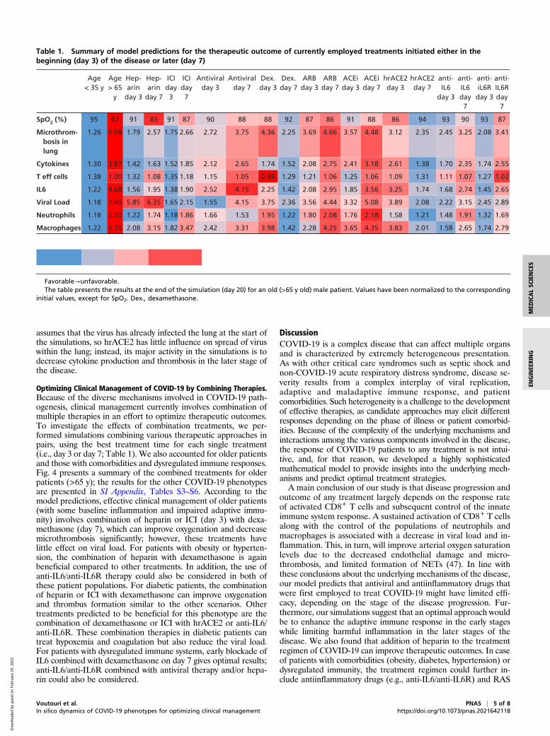
assumes that the virus has already infected the lung at the start ofthe simulations, so hrACE2 has little influence on spread of viruswithin the lung; instead, its major activity in the simulations is todecrease cytokine production and thrombosis in the later stage ofthe disease.
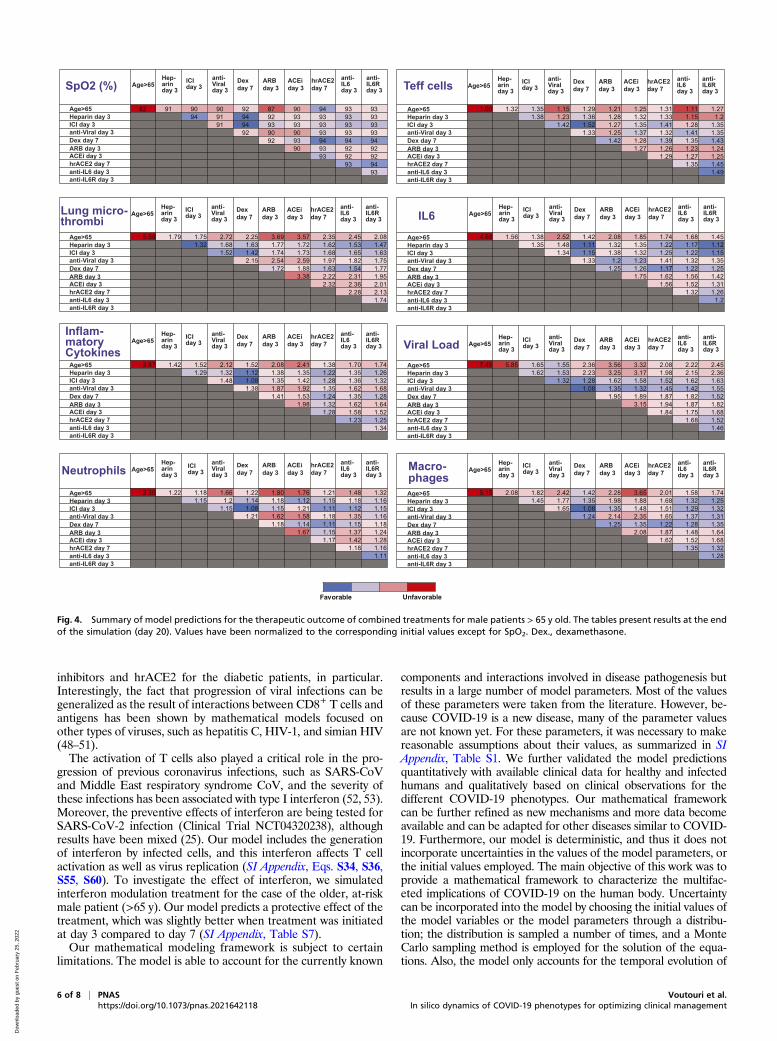
Optimizing Clinical Management of COVID-19 by Combining Therapies.Because of the diverse mechanisms involved in COVID-19 path-ogenesis, clinical management currently involves combination ofmultiple therapies in an effort to optimize therapeutic outcomes.To investigate the effects of combination treatments, we per-formed simulations combining various therapeutic approaches inpairs, using the best treatment time for each single treatment(i.e., day 3 or day 7; Table 1). We also accounted for older patientsand those with comorbidities and dysregulated immune responses.Fig. 4 presents a summary of the combined treatments for olderpatients (>65 y); the results for the other COVID-19 phenotypesare presented in SI Appendix, Tables S3–S6. According to themodel predictions, effective clinical management of older patients(with some baseline inflammation and impaired adaptive immu-nity) involves combination of heparin or ICI (day 3) with dexa-methasone (day 7), which can improve oxygenation and decreasemicrothrombosis significantly; however, these treatments havelittle effect on viral load. For patients with obesity or hyperten-sion, the combination of heparin with dexamethasone is againbeneficial compared to other treatments. In addition, the use ofanti-IL6/anti-IL6R therapy could also be considered in both ofthese patient populations. For diabetic patients, the combinationof heparin or ICI with dexamethasone can improve oxygenationand thrombus formation similar to the other scenarios. Othertreatments predicted to be beneficial for this phenotype are thecombination of dexamethasone or ICI with hrACE2 or anti-IL6/anti-IL6R. These combination therapies in diabetic patients cantreat hypoxemia and coagulation but also reduce the viral load.For patients with dysregulated immune systems, early blockade ofIL6 combined with dexamethasone on day 7 gives optimal results;anti-IL6/anti-IL6R combined with antiviral therapy and/or hepa-rin could also be considered.
DiscussionCOVID-19 is a complex disease that can affect multiple organsand is characterized by extremely heterogeneous presentation.As with other critical care syndromes such as septic shock andnon-COVID-19 acute respiratory distress syndrome, disease se-verity results from a complex interplay of viral replication,adaptive and maladaptive immune response, and patientcomorbidities. Such heterogeneity is a challenge to the developmentof effective therapies, as candidate approaches may elicit differentresponses depending on the phase of illness or patient comorbid-ities. Because of the complexity of the underlying mechanisms andinteractions among the various components involved in the disease,the response of COVID-19 patients to any treatment is not intui-tive, and, for that reason, we developed a highly sophisticatedmathematical model to provide insights into the underlying mech-anisms and predict optimal treatment strategies.A main conclusion of our study is that disease progression and
outcome of any treatment largely depends on the response rateof activated CD8+ T cells and subsequent control of the innateimmune system response. A sustained activation of CD8+ T cellsalong with the control of the populations of neutrophils andmacrophages is associated with a decrease in viral load and in-flammation. This, in turn, will improve arterial oxygen saturationlevels due to the decreased endothelial damage and micro-thrombosis, and limited formation of NETs (47). In line withthese conclusions about the underlying mechanisms of the disease,our model predicts that antiviral and antiinflammatory drugs thatwere first employed to treat COVID-19 might have limited effi-cacy, depending on the stage of the disease progression. Fur-thermore, our simulations suggest that an optimal approach wouldbe to enhance the adaptive immune response in the early stageswhile limiting harmful inflammation in the later stages of thedisease. We also found that addition of heparin to the treatmentregimen of COVID-19 can improve therapeutic outcomes. In caseof patients with comorbidities (obesity, diabetes, hypertension) ordysregulated immunity, the treatment regimen could further in-clude antiinflammatory drugs (e.g., anti-IL6/anti-IL6R) and RAS
Table 1. Summary of model predictions for the therapeutic outcome of currently employed treatments initiated either in thebeginning (day 3) of the disease or later (day 7)
Favorable→unfavorable.The table presents the results at the end of the simulation (day 20) for an old (>65 y old) male patient. Values have been normalized to the corresponding
initial values, except for SpO2. Dex., dexamethasone.
Voutouri et al. PNAS | 5 of 8In silico dynamics of COVID-19 phenotypes for optimizing clinical management https://doi.org/10.1073/pnas.2021642118
MED
ICALSC
IENCE
SEN
GINEE
RING
Dow
nloa
ded
by g
uest
on
Feb
ruar
y 25
, 202
2
inhibitors and hrACE2 for the diabetic patients, in particular.Interestingly, the fact that progression of viral infections can begeneralized as the result of interactions between CD8+ T cells andantigens has been shown by mathematical models focused onother types of viruses, such as hepatitis C, HIV-1, and simian HIV(48–51).The activation of T cells also played a critical role in the pro-
gression of previous coronavirus infections, such as SARS-CoVand Middle East respiratory syndrome CoV, and the severity ofthese infections has been associated with type I interferon (52, 53).Moreover, the preventive effects of interferon are being tested forSARS-CoV-2 infection (Clinical Trial NCT04320238), althoughresults have been mixed (25). Our model includes the generationof interferon by infected cells, and this interferon affects T cellactivation as well as virus replication (SI Appendix, Eqs. S34, S36,S55, S60). To investigate the effect of interferon, we simulatedinterferon modulation treatment for the case of the older, at-riskmale patient (>65 y). Our model predicts a protective effect of thetreatment, which was slightly better when treatment was initiatedat day 3 compared to day 7 (SI Appendix, Table S7).Our mathematical modeling framework is subject to certain
limitations. The model is able to account for the currently known
components and interactions involved in disease pathogenesis butresults in a large number of model parameters. Most of the valuesof these parameters were taken from the literature. However, be-cause COVID-19 is a new disease, many of the parameter valuesare not known yet. For these parameters, it was necessary to makereasonable assumptions about their values, as summarized in SIAppendix, Table S1. We further validated the model predictionsquantitatively with available clinical data for healthy and infectedhumans and qualitatively based on clinical observations for thedifferent COVID-19 phenotypes. Our mathematical frameworkcan be further refined as new mechanisms and more data becomeavailable and can be adapted for other diseases similar to COVID-19. Furthermore, our model is deterministic, and thus it does notincorporate uncertainties in the values of the model parameters, orthe initial values employed. The main objective of this work was toprovide a mathematical framework to characterize the multifac-eted implications of COVID-19 on the human body. Uncertaintycan be incorporated into the model by choosing the initial values ofthe model variables or the model parameters through a distribu-tion; the distribution is sampled a number of times, and a MonteCarlo sampling method is employed for the solution of the equa-tions. Also, the model only accounts for the temporal evolution of
82 91 90 90 92 87 90 94 93 9394 91 94 92 93 93 93 93
91 94 93 93 93 93 9392 90 90 93 93 93
92 93 94 94 9490 93 92 92
93 92 9293 94
93
1.00 1.32 1.35 1.15 1.29 1.21 1.25 1.31 1.11 1.271.38 1.23 1.36 1.28 1.32 1.33 1.15 1.2
1.42 1.52 1.27 1.35 1.41 1.28 1.351.33 1.25 1.37 1.32 1.41 1.35
1.42 1.28 1.39 1.35 1.431.27 1.26 1.23 1.24
1.29 1.27 1.251.35 1.45
1.49
5.58 1.79 1.75 2.72 2.25 3.69 3.57 2.35 2.45 2.081.32 1.68 1.63 1.77 1.72 1.62 1.53 1.47
1.52 1.42 1.74 1.73 1.68 1.65 1.632.15 2.54 2.59 1.97 1.82 1.75
1.72 1.88 1.63 1.54 1.773.38 2.22 2.31 1.95
2.32 2.36 2.012.28 2.13
1.74
4.68 1.56 1.38 2.52 1.42 2.08 1.85 1.74 1.68 1.451.35 1.48 1.11 1.32 1.35 1.22 1.17 1.12
1.34 1.15 1.38 1.32 1.25 1.22 1.151.33 1.2 1.23 1.41 1.32 1.35
1.25 1.26 1.17 1.22 1.251.75 1.62 1.56 1.42
1.56 1.52 1.311.32 1.26
1.2
3.87 1.42 1.52 2.12 1.52 2.08 2.41 1.38 1.70 1.741.29 1.32 1.12 1.38 1.35 1.22 1.35 1.26
1.48 1.08 1.35 1.42 1.28 1.36 1.321.38 1.87 1.92 1.35 1.62 1.68
1.41 1.53 1.24 1.35 1.281.98 1.32 1.62 1.64
1.28 1.58 1.521.23 1.25
1.34
7.45 5.85 1.65 1.55 2.36 3.56 3.32 2.08 2.22 2.451.62 1.53 2.23 3.25 3.17 1.98 2.15 2.36
1.32 1.28 1.62 1.58 1.52 1.62 1.631.08 1.35 1.32 1.45 1.42 1.55
1.95 1.89 1.87 1.82 1.523.15 1.94 1.87 1.82
1.84 1.75 1.681.68 1.52
1.46
2.30 1.22 1.18 1.66 1.22 1.80 1.76 1.21 1.48 1.321.15 1.2 1.14 1.18 1.12 1.15 1.18 1.16
1.15 1.08 1.15 1.21 1.11 1.12 1.151.21 1.62 1.58 1.18 1.35 1.16
1.18 1.14 1.11 1.15 1.181.67 1.15 1.37 1.24
1.17 1.42 1.281.18 1.16
1.11
5.15 2.08 1.82 2.42 1.42 2.28 3.65 2.01 1.58 1.741.45 1.77 1.35 1.98 1.88 1.68 1.32 1.25
1.65 1.08 1.35 1.48 1.51 1.29 1.321.24 2.14 2.35 1.65 1.37 1.31
1.25 1.35 1.22 1.28 1.352.08 1.87 1.48 1.64
1.62 1.52 1.681.35 1.32
1.28
Favorable Unfavorable
Lung micro-thrombi IL6
Viral Load
Macro-phagesNeutrophils
Inflam-matory Cytokines
Age>65Hep-arinday 3
ICIday 3
anti-Viralday 3
ARBday 3
ACEiday 3
anti-IL6day 3
anti-IL6Rday 3
Dexday 7
hrACE2day 7 Age>65
Hep-arinday 3
ICIday 3
anti-Viralday 3
ARBday 3
ACEiday 3
anti-IL6day 3
anti-IL6Rday 3
Dexday 7
hrACE2day 7
Age>65Hep-arinday 3
ICIday 3
anti-Viralday 3
ARBday 3
ACEiday 3
anti-IL6day 3
anti-IL6Rday 3
Dexday 7
hrACE2day 7
Age>65Hep-arinday 3
ICIday 3
anti-Viralday 3
ARBday 3
ACEiday 3
anti-IL6day 3
anti-IL6Rday 3
Dexday 7
hrACE2day 7
Age>65Hep-arinday 3
ICIday 3
anti-Viralday 3
ARBday 3
ACEiday 3
anti-IL6day 3
anti-IL6Rday 3
Dexday 7
hrACE2day 7Age>65
Hep-arinday 3
anti-Viralday 3
ARBday 3
ACEiday 3
anti-IL6day 3
anti-IL6Rday 3
Dexday 7
hrACE2day 7
Age>65Hep-arinday 3
ICIday 3
anti-Viralday 3
ARBday 3
ACEiday 3
anti-IL6day 3
anti-IL6Rday 3
Dexday 7
hrACE2day 7
Age>65Hep-arinday 3
ICIday 3
anti-Viralday 3
ARBday 3
ACEiday 3
anti-IL6day 3
anti-IL6Rday 3
Dexday 7
hrACE2day 7
Age>65Heparin day 3ICI day 3anti-Viral day 3
ARB day 3ACEi day 3
anti-IL6 day 3anti-IL6R day 3
Dex day 7
hrACE2 day 7
Age>65Heparin day 3ICI day 3anti-Viral day 3
ARB day 3ACEi day 3
anti-IL6 day 3anti-IL6R day 3
Dex day 7
hrACE2 day 7
Age>65Heparin day 3ICI day 3anti-Viral day 3
ARB day 3ACEi day 3
anti-IL6 day 3anti-IL6R day 3
Dex day 7
hrACE2 day 7
Age>65Heparin day 3ICI day 3anti-Viral day 3
ARB day 3ACEi day 3
anti-IL6 day 3anti-IL6R day 3
Dex day 7
hrACE2 day 7
Age>65Heparin day 3ICI day 3anti-Viral day 3
ARB day 3ACEi day 3
anti-IL6 day 3anti-IL6R day 3
Dex day 7
hrACE2 day 7
Age>65Heparin day 3ICI day 3anti-Viral day 3
ARB day 3ACEi day 3
anti-IL6 day 3anti-IL6R day 3
Dex day 7
hrACE2 day 7
Age>65Heparin day 3ICI day 3anti-Viral day 3
ARB day 3ACEi day 3
anti-IL6 day 3anti-IL6R day 3
Dex day 7
hrACE2 day 7
Age>65Heparin day 3ICI day 3anti-Viral day 3
ARB day 3ACEi day 3
anti-IL6 day 3anti-IL6R day 3
Dex day 7
hrACE2 day 7
SpO2 (%) Teff cells
ICIday 3
Fig. 4. Summary of model predictions for the therapeutic outcome of combined treatments for male patients > 65 y old. The tables present results at the endof the simulation (day 20). Values have been normalized to the corresponding initial values except for SpO2. Dex., dexamethasone.
6 of 8 | PNAS Voutouri et al.https://doi.org/10.1073/pnas.2021642118 In silico dynamics of COVID-19 phenotypes for optimizing clinical management
Dow
nloa
ded
by g
uest
on
Feb
ruar
y 25
, 202
2
the infection and not for spatial effects in the lung or other organs,which will be considered in future studies.Another limitation of the model is that it does not account for
the humoral immune response, although circulating antibodiesare important in viral control in COVID-19 (54). Future modeldevelopment should include this additional complexity of anti-viral immunity, as well as modulation of the immune system byvaccines. Also, the model does not account for adverse effects ofthe various treatments considered in this study, which could af-fect the therapeutic outcome. For instance, heparin use might beassociated with hemorrhage, and RAS inhibitors can reduceblood pressure. Even though incorporation of adverse effectswould be important, they would not affect the basic conclusionsof the study and were omitted to avoid adding further complexityto the model. In conclusion, this study presents a mathematicalrepresentation of the known mechanisms of COVID-19 andcould be utilized by the scientific community as a useful tool forfurther understanding the disease and investigating the benefitsof treatments on a patient-specific basis.
Materials and MethodsA detailed description of model equations, the description and values ofmodel parameters, and the solution strategy are provided in SI Appendix. Themathematical model consists of two components: a detailed model of lunginfection by the SARS-CoV-2 virus and a PK/PD model of COVID-19 infectionand thrombosis to simulate events that happen throughout the body(Fig. 1). The lung model incorporates the infection of epithelial and endo-thelial cells by the SARS-CoV-2 virus through ACE2 modulation and activity,the release of proinflammatory cytokines, and the entire RAS. Cytokines thatare central to COVID-19 (e.g., IL-6) are explicitly accounted for in the model,incorporating all known signaling pathways, such as the canonical and transsignaling pathway of IL-6 and its interaction with IL-6r and soluble IL-6r.Additional innate immune cells, specifically, neutrophils and macrophages,are incorporated into our model along with the interaction of immune cellswith viral particles and infected cells as well as the formation of NETs. Themodel incorporates the recruitment of cytotoxic T cells by the infected cellsand by IL-6 as well as the virus killing of cytotoxic T cells. In addition, themodel accounts for the role of immune cells in modulating the expression ofproinflammatory cytokines (55). Subsequent events, such as the impairmentof the vascular network owing to the infection of endothelial cells by theSARS-CoV-2 virus and the resulting changes in blood oxygenation, arealso included.
To study how the virus affects systemic events, such as inflammation andthrombosis, we coupled the lungmodel with a PK/PDmodel of viral infection.The PK/PD model represents the major organs of the body as compartmentsconnected in an anatomical manner by the blood and lymphatic circulations
(SI Appendix, Fig. S1). The organ compartments are then further subdividedinto vascular and extravascular subcompartments, and each organ has adraining lymph node compartment. Briefly, the main transport processes forthe biochemical species include 1) convective and diffusive transport acrosscapillary walls, 2) reversible, nonsaturable, nonspecific binding in the ex-travascular compartments, and 3) reversible, and saturable, specific bindingof virus to endothelial cells, and probabilistic infection of bound cells. Anyinfected cell generates microthrombi, which enter the circulation and canaccumulate in target organs including brain, heart, and lung. Each speciescan be produced, bound, or degraded within each compartment. Blood flowis distributed according to known flows through each organ, and the overallmass balance allows calculation of the concentration of each species in eachcompartment over time. The PK/PD model includes the following key pro-cesses involved in the trafficking of viruses: 1) transport from the lung viathe systemic circulation, 2) entry into endothelial cells via binding to ACE2, 3)entry into the cell and replication, and 4) exit from the cell and entry intothe blood circulation. The important outputs of the model are the level ofthrombosis in each organ and the viral load. When coupled with the lungmodel, we are able to analyze the dynamics of these readouts in light oflocal lung pathologies and predicted cytokine levels.
The model consists of a set of partial and ordinary differential equations.The values of themodel parameters are summarized in SI Appendix, Table S1.We simulated COVID-19 infection and progression within a period of 20 d.The model does not account for the first stages of virus infection of theupper respiratory tract but from the time the virus has infected the lungs.Therefore, day 0 of the simulations corresponds to the initiation of lunginfection. For the formulation of the model and the solution of the equa-tions, the commercial finite elements software COMSOL Multiphysics v.5.5was used. The computational finite element mesh employed in the presentstudy consisted of 2,440 elements resulting in 1,201,044 degrees of freedom.The solution was tested and found to be mesh independent, the PARDISOsolver was selected for the solution of the model equations, and the totalsolution time was ∼30 min.
Code Availability. The COMSOL code is available at Zenodo (56).
Data Availability.All data supporting the findings of this study are available inthe paper and SI Appendix.
ACKNOWLEDGMENTS. R.K.J.’s research is supported by Grants R01-CA208205,U01-CA 224348, Outstanding Investigator Award R35-CA197743, and grantsfrom the National Foundation for Cancer Research, Jane’s Trust Foundation,Advanced Medical Research Foundation, and Harvard Ludwig Cancer Center.L.L.M.’s research is supported by Grant R01-CA2044949. T.S.’s research is sup-ported by the European Research Council (Grants ERC-2018-PoC-838414 andERC-2019-CoG-863955) and Cyprus Research and Innovation Foundation(Grant INFRASTRUCTURE/1216/0052). A.B.P. is supported by American Societyof Nephrology Joseph A. Carlucci Research Fellowship.
1. J. B. Moore, C. H. June, Cytokine release syndrome in severe COVID-19. Science 368,
473–474 (2020).2. T. J. Oxley et al., Large-vessel stroke as a presenting feature of Covid-19 in the young.
N. Engl. J. Med. 382, e60 (2020).3. I. Paranjpe et al., Association of treatment dose anticoagulation with in-hospital
survival among hospitalized patients with COVID-19. J. Am. Coll. Cardiol. 76, 122–124
(2020).4. S. F. Lax et al., Pulmonary arterial thrombosis in COVID-19 with fatal outcome: Results
from a prospective, single-center, clinicopathologic case series. Ann. Intern. Med. 173,
350–361 (2020).5. N. Stefan, A. L. Birkenfeld, M. B. Schulze, D. S. Ludwig, Obesity and impaired meta-
bolic health in patients with COVID-19. Nat. Rev. Endocrinol. 16, 341–342 (2020).6. N. Sattar, I. B. McInnes, J. J. V. McMurray, Obesity is a risk factor for severe COVID-19
infection: Multiple potential mechanisms. Circulation 142, 4–6 (2020).7. M. Dai et al., Patients with cancer appear more vulnerable to SARS-COV-2: A multi-
center study during the COVID-19 outbreak. Cancer Discov. 10, 783–791 (2020).8. H. Miyashita et al., Do patients with cancer have a poorer prognosis of COVID-19? An
experience in New York City. Ann. Oncol. 31, 1088–1089 (2020).9. E. J. Williamson et al., Factors associated with COVID-19-related death using Open-
SAFELY. Nature 584, 430–436 (2020).10. I. Paranjpe et al., Clinical characteristics of hospitalized Covid-19 patients in New York
City. medRxiv:10.1101/2020.04.19.20062117 (26 April 2020).11. S. Gupta et al.; STOP-COVID Investigators, Factors associated with death in critically ill
patients with coronavirus disease 2019 in the US. JAMA Intern. Med. 180, 1–12 (2020).12. G. Grasselli et al.; COVID-19 Lombardy ICU Network, Risk factors associated with
mortality among patients with COVID-19 in intensive care units in Lombardy, Italy.
JAMA Intern. Med. 180, 1345–1355 (2020).
13. M. O’Driscoll et al., Age-specific mortality and immunity patterns of SARS-CoV-2.
Nature, 10.1038/s41586-020-2918-0 (2020).14. A. J. Kucharski et al.; Centre for Mathematical Modelling of Infectious Diseases
COVID-19 working group, Early dynamics of transmission and control of COVID-19: A
mathematical modelling study. Lancet Infect. Dis. 20, 553–558 (2020).15. G. Giordano et al., Modelling the COVID-19 epidemic and implementation of pop-
ulation-wide interventions in Italy. Nat. Med. 26, 855–860 (2020).16. K. Prem et al.; Centre for the Mathematical Modelling of Infectious Diseases
COVID-19 Working Group, The effect of control strategies to reduce social mixing on
outcomes of the COVID-19 epidemic in Wuhan, China: A modelling study. Lancet
Public Health 5, e261–e270 (2020).17. X. Hao et al., Reconstruction of the full transmission dynamics of COVID-19 in Wuhan.
Nature 584, 420–424 (2020).18. A. Goncalves et al., Timing of antiviral treatment initiation is critical to reduce
SARS-Cov-2 viral load. medRxiv:10.1101/2020.04.04.20047886 (21 June 2020).19. S. Wang et al., Modeling the viral dynamics of SARS-CoV-2 infection. Math Biosci 328,
108438 (2020).20. T. J. Sego et al., A modular framework for multiscale multicellular spatial modeling of
viral infection, immune response and drug therapy timing and efficacy in epithelial
tissues: A multiscale model of viral infection in epithelial tissues. bioRxiv:10.1101/
2020.04.27.064139 (1 June 2020).21. Y. Wang et al, Rapid community-driven development of a SARS-CoV-2 tissue simu-
lator. bioRxiv:10.1101/2020.04.02.019075 (8 May 2020).22. J. M. Sanders, M. L. Monogue, T. Z. Jodlowski, J. B. Cutrell, Pharmacologic treatments
for coronavirus disease 2019 (COVID-19): A review. JAMA 323, 1824–1836 (2020).23. Y. Wang et al., Remdesivir in adults with severe COVID-19: a randomised, double-
blind, placebo-controlled, multicentre trial. Lancet 395, 1569–1578 (2020).
Voutouri et al. PNAS | 7 of 8In silico dynamics of COVID-19 phenotypes for optimizing clinical management https://doi.org/10.1073/pnas.2021642118
MED
ICALSC
IENCE
SEN
GINEE
RING
Dow
nloa
ded
by g
uest
on
Feb
ruar
y 25
, 202
2
24. J. H. Beigel, K. M. Tomashek, L. E. Dodd, Remdesivir for the treatment of Covid-19—Preliminary report. Reply. N. Engl. J. Med. 383, 994 (2020).
25. H. Pan et al., Repurposed antiviral drugs for COVID-19 –interim WHO SOLIDARITYtrial results. medRxiv:10.1101/2020.10.15.20209817 (15 October 2020).
26. I. F. Hung et al., Triple combination of interferon beta-1b, lopinavir-ritonavir, andribavirin in the treatment of patients admitted to hospital with COVID-19: An open-label, randomised, phase 2 trial. Lancet 395, 1695–1704 (2020).
27. C. Zhang, Z. Wu, J. W. Li, H. Zhao, G. Q. Wang, Cytokine release syndrome in severeCOVID-19: Interleukin-6 receptor antagonist tocilizumab may be the key to reducemortality. Int. J. Antimicrob. Agents 55, 105954 (2020).
28. F. Cantini et al., Baricitinib therapy in COVID-19: A pilot study on safety and clinicalimpact. J. Infect. 81, 318–356 (2020).
29. E. G. Favalli, M. Biggioggero, G. Maioli, R. Caporali, Baricitinib for COVID-19: A suit-able treatment? Lancet Infect. Dis. 20, 1012–1013 (2020).
30. N. Le Bert et al., SARS-CoV-2-specific T cell immunity in cases of COVID-19 and SARS,and uninfected controls. Nature 584, 457–462 (2020).
31. T. J. Iwashyna et al., Implications of heterogeneity of treatment effect for reportingand analysis of randomized trials in critical care. Am. J. Respir. Crit. Care Med. 192,1045–1051 (2015).
32. B. Diop-Frimpong, V. P. Chauhan, S. Krane, Y. Boucher, R. K. Jain, Losartan inhibitscollagen I synthesis and improves the distribution and efficacy of nanotherapeutics intumors. Proc. Natl. Acad. Sci. U.S.A. 108, 2909–2914 (2011).
33. V. P. Chauhan et al., Angiotensin inhibition enhances drug delivery and potentiateschemotherapy by decompressing tumour blood vessels. Nat. Commun. 4, 2516 (2013).
34. V. P. Chauhan et al., Reprogramming the microenvironment with tumor-selectiveangiotensin blockers enhances cancer immunotherapy. Proc. Natl. Acad. Sci. U.S.A.116, 10674–10680 (2019).
35. Y. Zhao et al., Losartan treatment enhances chemotherapy efficacy and reduces as-cites in ovarian cancer models by normalizing the tumor stroma. Proc. Natl. Acad. Sci.U.S.A. 116, 2210–2219 (2019).
36. H. Wang et al., In silico simulation of a clinical trial with anti-CTLA-4 and anti-PD-L1immunotherapies in metastatic breast cancer using a systems pharmacology model. R.Soc. Open Sci. 6, 190366 (2019).
37. T. K. Roy, T. W. Secomb, Theoretical analysis of the determinants of lung oxygendiffusing capacity. J. Theor. Biol. 351, 1–8 (2014).
38. N. Liu, Y. Hong, R.-G. Chen, H.-M. Zhu, High rate of increased level of plasma an-giotensin II and its gender difference in COVID-19: An analysis of 55 hospitalizedpatients with COVID-19 in a single hospital, WuHan, China. MedRxiv:10.1101/2020.04.27.20080432 (1 May 2020).
39. T. Herold et al., Elevated levels of IL-6 and CRP predict the need for mechanicalventilation in COVID-19. J. Allergy Clin. Immunol. 146, 128–136.e4 (2020).
40. X. Chen et al., Detectable serum SARS-CoV-2 viral load (RNAaemia) is closely corre-lated with drastically elevated interleukin 6 (IL-6) level in critically ill COVID-19 pa-tients. Clin. Infect. Dis. 71, 1937–1942 (2020).
41. D. R. Ziehr et al., Respiratory pathophysiology of mechanically ventilated patientswith COVID-19: A cohort study. Am. J. Respir. Crit. Care Med. 201, 1560–1564 (2020).
42. I. M. Zacharioudakis et al., Association of SARS-CoV-2 genomic load with COVID-19patient outcomes. Ann. Am. Thorac. Soc., 10.1513/AnnalsATS.202008-931RL (2020).
43. M. Montopoli et al., Androgen-deprivation therapies for prostate cancer and risk ofinfection by SARS-CoV-2: A population-based study (N = 4532). Ann. Oncol. 31,1040–1045 (2020).
44. S. L. Klein, K. L. Flanagan, Sex differences in immune responses. Nat. Rev. Immunol.16, 626–638 (2016).
45. K. H. Stopsack, L. A. Mucci, E. S. Antonarakis, P. S. Nelson, P. W. Kantoff, TMPRSS2 andCOVID-19: Serendipity or opportunity for intervention? Cancer Discov. 10, 779–782(2020).
46. T. Takahashi et al., Sex differences in immune responses to SARS-CoV-2 that underliedisease outcomes. medRxiv:10.1101/2020.06.06.20123414 (26 June 2020).
47. B. J. Barnes et al., Targeting potential drivers of COVID-19: Neutrophil extracellulartraps. J. Exp. Med. 217, e20200652 (2020).
48. S. Baral, R. Antia, N. M. Dixit, A dynamical motif comprising the interactions betweenantigens and CD8 T cells may underlie the outcomes of viral infections. Proc. Natl.Acad. Sci. U.S.A. 116, 17393–17398 (2019).
49. S. Baral, R. Roy, N. M. Dixit, Modeling how reversal of immune exhaustion elicits cureof chronic hepatitis C after the end of treatment with direct-acting antiviral agents.Immunol. Cell Biol. 96, 969–980 (2018).
50. J. M. Conway, A. S. Perelson, Post-treatment control of HIV infection. Proc. Natl. Acad.Sci. U.S.A. 112, 5467–5472 (2015).
51. R. Desikan, R. Raja, N. M. Dixit, Early exposure to broadly neutralizing antibodies maytrigger a dynamical switch from progressive disease to lasting control of SHIV infec-tion. PLoS Comput. Biol. 16, e1008064 (2020).
52. R. Channappanavar et al., Dysregulated type I interferon and inflammatory mono-cyte-macrophage responses cause lethal pneumonia in SARS-CoV-infected mice. CellHost Microbe 19, 181–193 (2016).
53. R. Channappanavar et al., IFN-I response timing relative to virus replication deter-mines MERS coronavirus infection outcomes. J. Clin. Invest. 129, 3625–3639 (2019).
54. N. Bhardwaj, S. C. Kundu, Electrospinning: A fascinating fiber fabrication technique.Biotechnol. Adv. 28, 325–347 (2010).
55. M. K. Malone et al., Cytokines secreted by stromal cells in TNBC microenvironment aspotential targets for cancer therapy. Cancer Biol. Ther. 21, 560–569 (2020).
56. C. Voutouri, M. R. Nikmaneshi, T. Stylianopoulos, L. L. Munn, R. K. Jain, Code relatedto "In silico dynamics of COVID-19 phenotypes for optimizing clinical management."Zenodo. http://doi.org/10.5281/zenodo.4390805. Deposited 2 September 2020.
8 of 8 | PNAS Voutouri et al.https://doi.org/10.1073/pnas.2021642118 In silico dynamics of COVID-19 phenotypes for optimizing clinical management
Dow
nloa
ded
by g
uest
on
Feb
ruar
y 25
, 202
2