Embed Size (px)
Citation preview

In The Name Of God
Management of Patients With unstable Angina/Non_ST-Elevation Myocardial Infarction


UA/NSTEMI is defined by ECG ST-segment depression or prominent T-wave inversion and/or positive biomarkers of necrosis) in the absence of ST-segment elevation and in an appropriate clinical setting(chest discomfort or anginal equivalent).
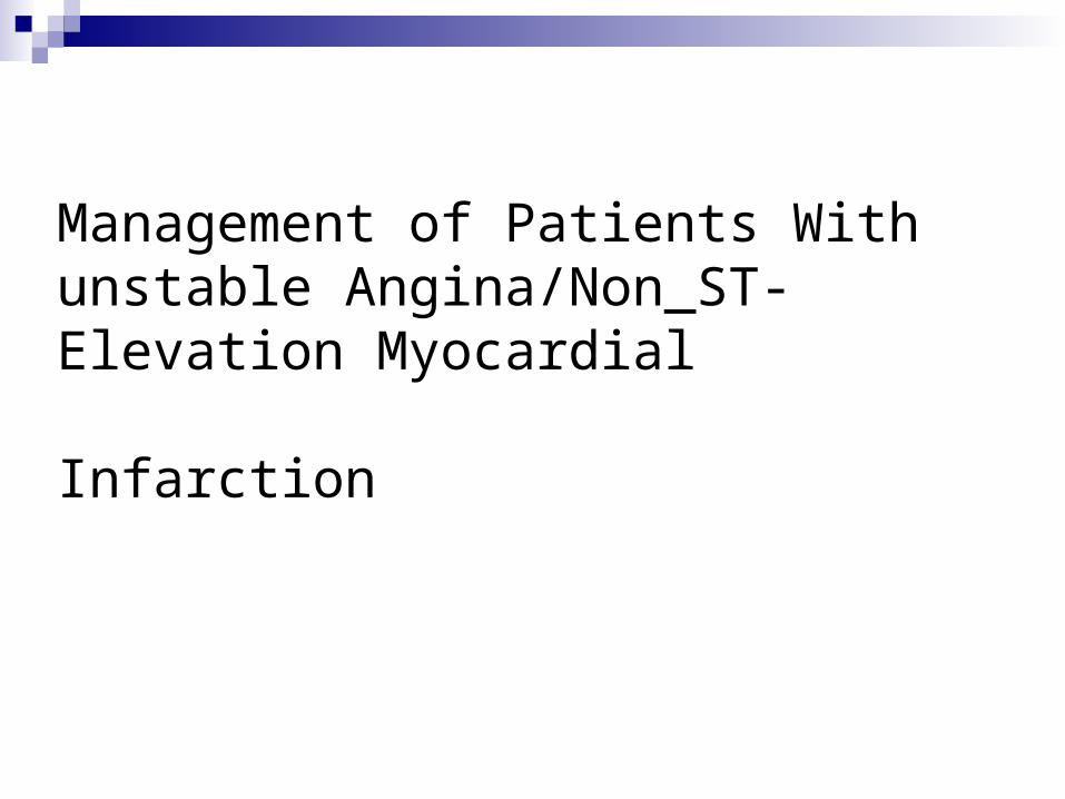
Presentations of UA and NSTEMI
3 principal presentations of UA:
1) rest angina
2) new-onset (less than 2 months) severe angina
3) increasing angina (in intensity,duration, and/or frequency) Non–ST-elevation MI generally presents as prolonged, more intense
rest angina or angina equivalent.
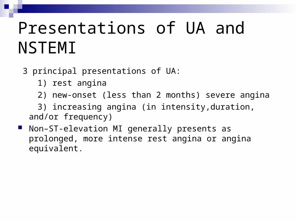
Recognition of Symptoms by Patient Many people are unaware that symptoms besides chest discomfort,
such as shortness of breath , diaphoresis , or extreme fatigue, can represent anginal equivalents
The average UA/NSTEMI patient does not seek medical care for approximately 2 h after symptom onset

Silent and Unrecognized Events
As many as one-half of all AMIs are clinically silent or unrecognized one third present with symptoms other than chest discomfort Patients without chest discomfort are: older women diabetes mellitus have prior HF Unexplained dyspnea, even without angina, is a common and
serious symptom of atypical ischemia/infarction
Clinical Assessment
When the patient makes contact with the medical care system: assess whether the symptoms are potentially a manifestation of an
ACS advise that an evaluation cannot be performed solely via the
telephone they should be instructed to access the EMS system and to be
transported to the hospital by ambulance Patients should be placed on a cardiac monitor, with emergency
resuscitation and defibrillation equipment nearby An ECG should be performed and interpreted as soon as possible,
with a goal of within 10 min of ED arrival.
Clinical Assessment
The recommendation for self-medication has been to encourage earlier contacting of the EMS system, that is, after taking 1 dose of NTG
While awaiting ambulance arrival, patients tolerating NTG can be instructed to take additional NTG every 5 min, up to 3 doses
Patients may be advised to chew ASA (162 to 325 mg) while emergency personnel are en route, may receive ASA en route to the hospital, or may be given ASA on arrival at the hospital.
Patient Transportation
Patients with chest discomfort at rest or other symptoms of ACS for more than 20 min, hemodynamic instability, or recent syncope/presyncope should be referred immediately to an ED.
Patients with less severe symptoms and without high-risk features should be seen initially in an ED or an appropriate outpatient facility.
High-risk patients should seek emergency transportation if available in less than 20 to 30 min.
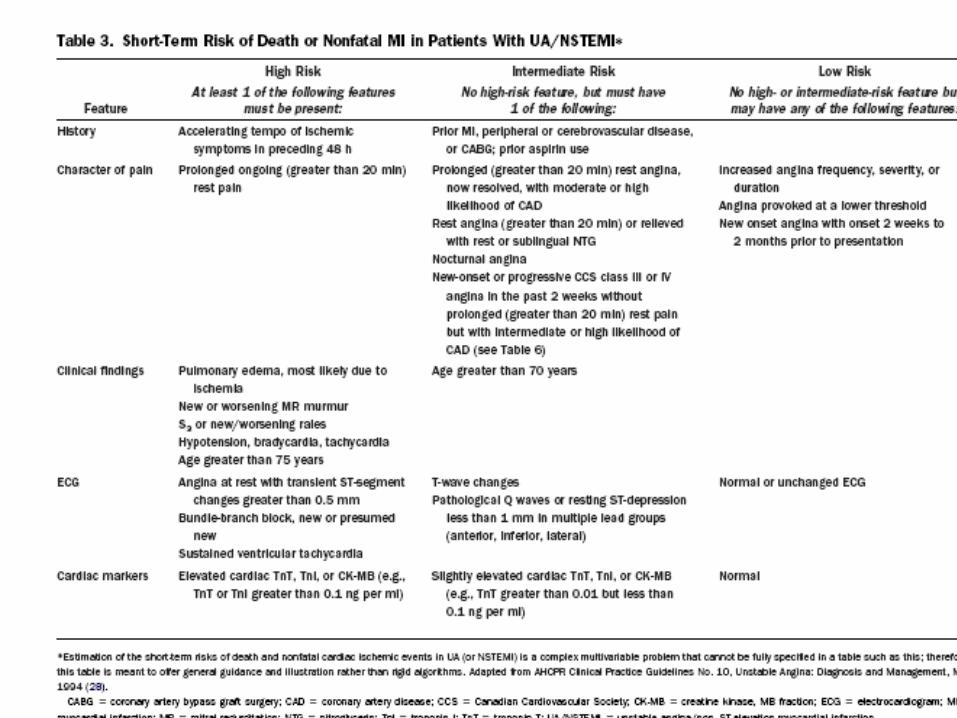
Early Risk Stratification
The initial medical history, physical examination, ECG, assessment of renal function, and cardiac biomarker measurements
Overall, risk is highest at the time of presentation and subsequently declines

History
The 5 most important factors in order of importance: 1) the nature of the anginal symptoms 2)prior history of CAD 3) sex (male) 4) older age 5) an increasing number of traditional risk factors In patients without preexisting clinical CHD, older age is the most
important factor. Some patients have no chest discomfort but present solely with
jaw, neck, arm, shoulder, back, or epigastric discomfort or with unexplained dyspnea without discomfort
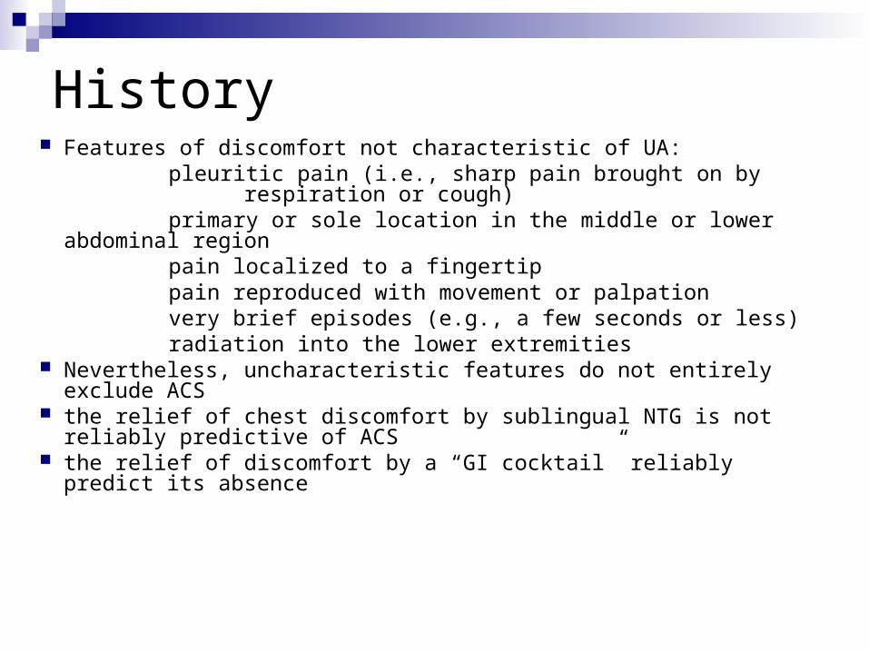
History Features of discomfort not characteristic of UA: pleuritic pain (i.e., sharp pain brought on by
respiration or cough) primary or sole location in the middle or lower abdominal region pain localized to a fingertip pain reproduced with movement or palpation very brief episodes (e.g., a few seconds or less) radiation into the lower extremities Nevertheless, uncharacteristic features do not entirely exclude
ACS the relief of chest discomfort by sublingual NTG is not reliably
predictive of ACS the relief of discomfort by a “GI cocktail” reliably predict its absence
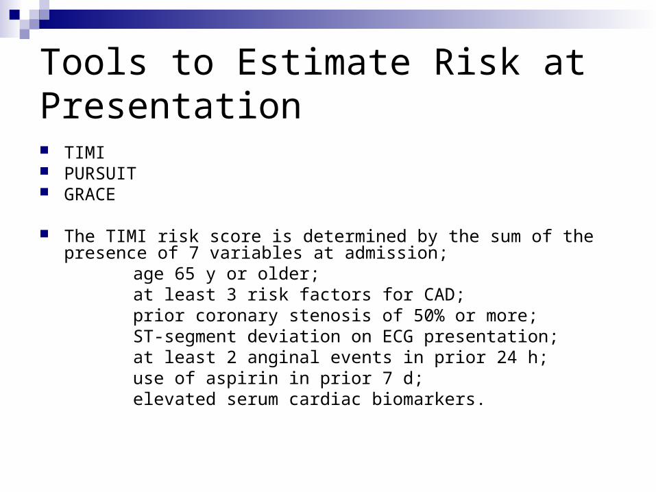
Tools to Estimate Risk at Presentation TIMI PURSUIT GRACE
The TIMI risk score is determined by the sum of the presence of 7 variables at admission;
age 65 y or older; at least 3 risk factors for CAD; prior coronary stenosis of 50% or more; ST-segment deviation on ECG presentation; at least 2 anginal events in prior 24 h; use of aspirin in prior 7 d; elevated serum cardiac biomarkers.

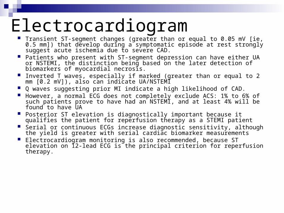
Electrocardiogram Transient ST-segment changes (greater than or equal to 0.05 mV [ie, 0.5 mm])
that develop during a symptomatic episode at rest strongly suggest acute ischemia due to severe CAD.
Patients who present with ST-segment depression can have either UA or NSTEMI, the distinction being based on the later detection of biomarkers of myocardial necrosis.
Inverted T waves, especially if marked (greater than or equal to 2 mm [0.2 mV]), also can indicate UA/NSTEMI
Q waves suggesting prior MI indicate a high likelihood of CAD. However, a normal ECG does not completely exclude ACS: 1% to 6% of such
patients prove to have had an NSTEMI, and at least 4% will be found to have UA Posterior ST elevation is diagnostically important because it qualifies the patient
for reperfusion therapy as a STEMI patient Serial or continuous ECGs increase diagnostic sensitivity, although the yield is
greater with serial cardiac biomarker measurements Electrocardiogram monitoring is also recommended, because ST elevation on 12-
lead ECG is the principal criterion for reperfusion therapy.
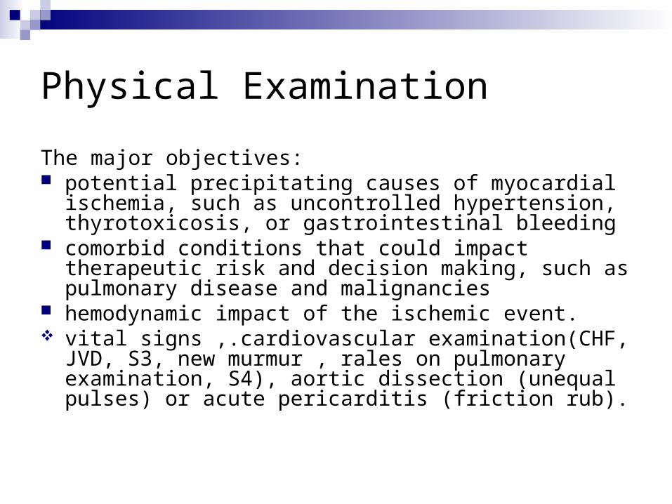
Physical Examination
The major objectives: potential precipitating causes of myocardial ischemia, such
as uncontrolled hypertension, thyrotoxicosis, or gastrointestinal bleeding
comorbid conditions that could impact therapeutic risk and decision making, such as pulmonary disease and malignancies
hemodynamic impact of the ischemic event. vital signs ,.cardiovascular examination(CHF, JVD, S3,
new murmur , rales on pulmonary examination, S4), aortic dissection (unequal pulses) or acute pericarditis (friction rub).
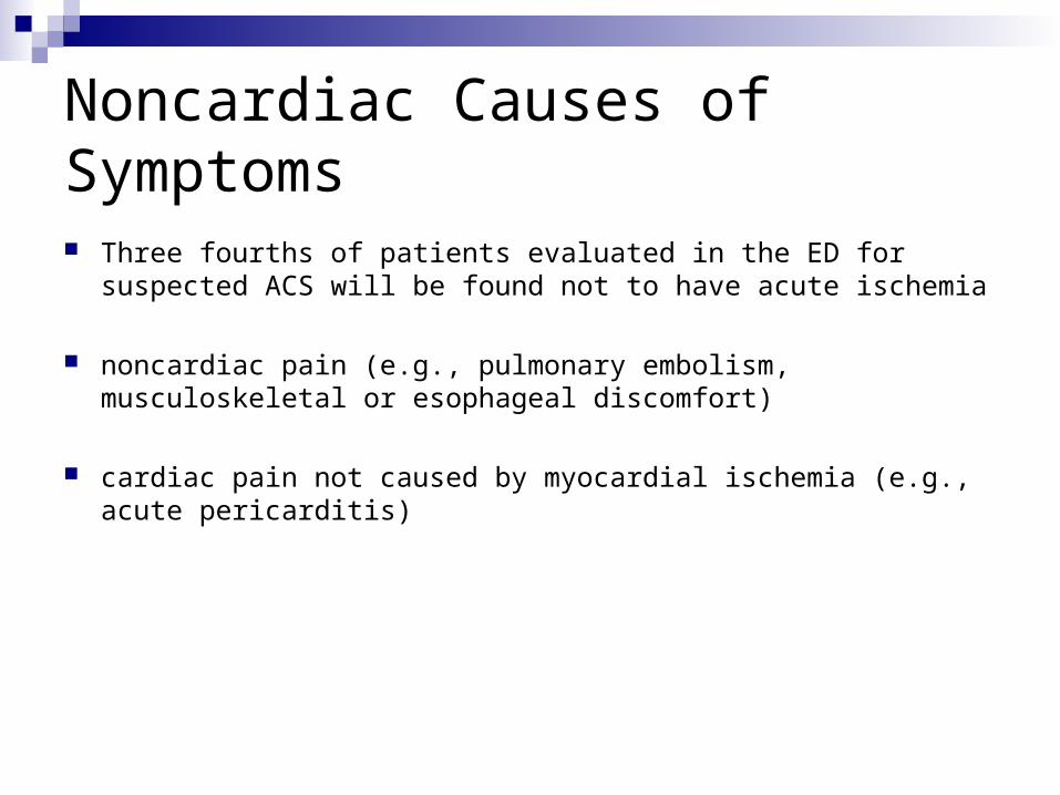
Noncardiac Causes of Symptoms
Three fourths of patients evaluated in the ED for suspected ACS will be found not to have acute ischemia
noncardiac pain (e.g., pulmonary embolism, musculoskeletal or esophageal discomfort)
cardiac pain not caused by myocardial ischemia (e.g., acute pericarditis)
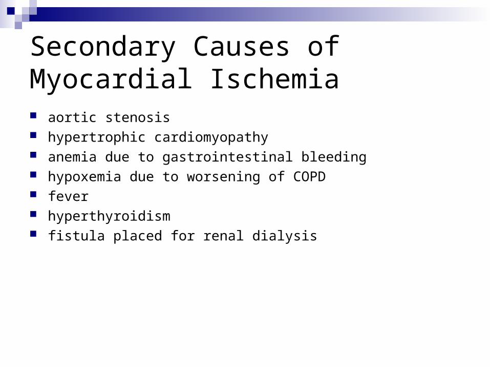
Secondary Causes of Myocardial Ischemia aortic stenosis hypertrophic cardiomyopathy anemia due to gastrointestinal bleeding hypoxemia due to worsening of COPD fever hyperthyroidism fistula placed for renal dialysis
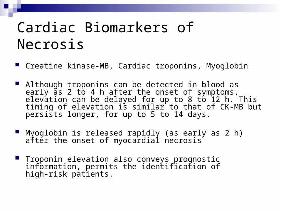
Cardiac Biomarkers of Necrosis
Creatine kinase-MB, Cardiac troponins, Myoglobin
Although troponins can be detected in blood as early as 2 to 4 h after the onset of symptoms, elevation can be delayed for up to 8 to 12 h. This timing of elevation is similar to that of CK-MB but persists longer, for up to 5 to 14 days.
Myoglobin is released rapidly (as early as 2 h) after the onset of myocardial necrosis
Troponin elevation also conveys prognostic information, permits the identification of high-risk patients.

Cardiac Biomarkers of Necrosis
both troponin and CK-MB positive: highest short-term risk
isolated troponin elevation: intermediate risk
isolated CK-MB elevation: lowest risk, equivalent to those with normal marker levels
Equivalent diagnostic and prognostic information is provided by cTnI and cTnT except in patients with renal dysfunction , in whom cTnT is less specific but retains predictive ability
A newer method aims to identify or exclude MI within 6 h of symptoms by relying increasing values while still in their normal ranges
Other lab stuidies:
CBC(anemia)
Chemistry profile: BS, renal function, and electrolytes levels, Potassium and magnesium levels
Creatinine levels must be considered before using an angiotensin-converting enzyme (ACE) inhibitor
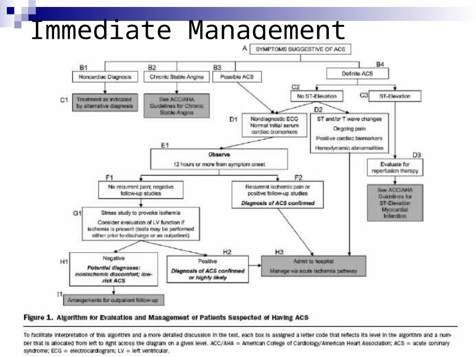
Immediate Management

Early Hospital Care hemodynamically stable: should be admitted to
an inpatient unit High-risk patients, including those with
continuing discomfort and/or hemodynamic instability: should be hospitalized in a CCU and observed for at least 24 h without any major complications.
Shorter periods might be appropriate for patients who are successfully reperfused, have normal LV function, and have minimal or no necrosis
Early Hospital Care
After admission, standard medical therapy is indicated: anti-ischemic therapy anticoagulant therapy ongoing risk stratificationappropriate use of invasive procedures

Early Hospital Care
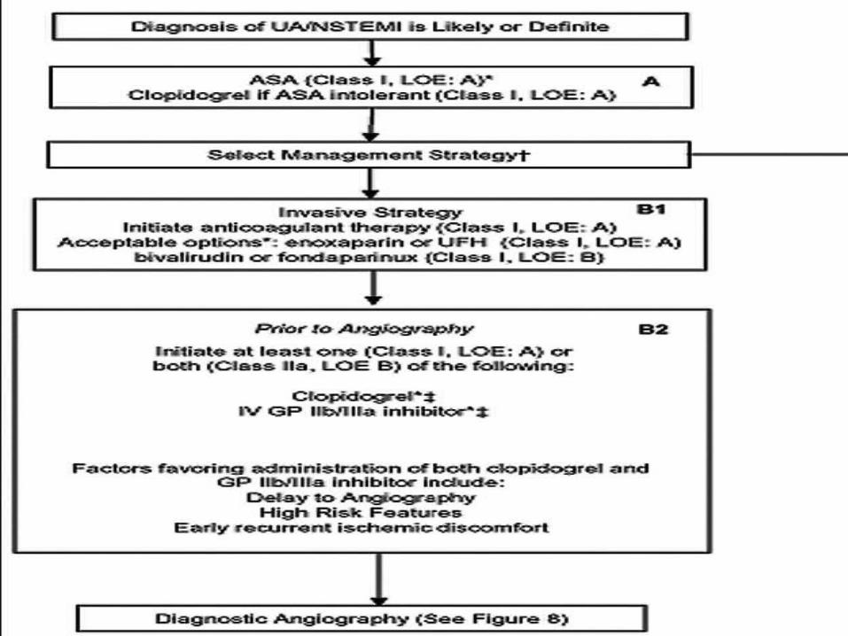
Unless contraindicated, treatment generally should include: ASA beta blocker anticoagulant therapy GP IIb/IIIa receptor antagonist thienopyridine (i.e., clopidogrel; initiation may be deferred
until a revascularization decision is made) A critical early decision is the choice of an angiographic
(invasive) or an initially conservative strategy. Assessment of LV function, which can influence
management, is recommended

General Care
on bed rest initially but can be mobilized to a chair and use a bedside commode when symptom free.
Subsequent activity should be liberalized when response to treatment occurs.
Patients with or at risk for hypoxemia should receive supplemental oxygen.
A short period of initial routine oxygen supplementation is reasonable during stabilization of the patient.
Patients should undergo continuous ECG monitoring during their early hospital phases, because ventricular fibrillation is the major preventable cause of early death.
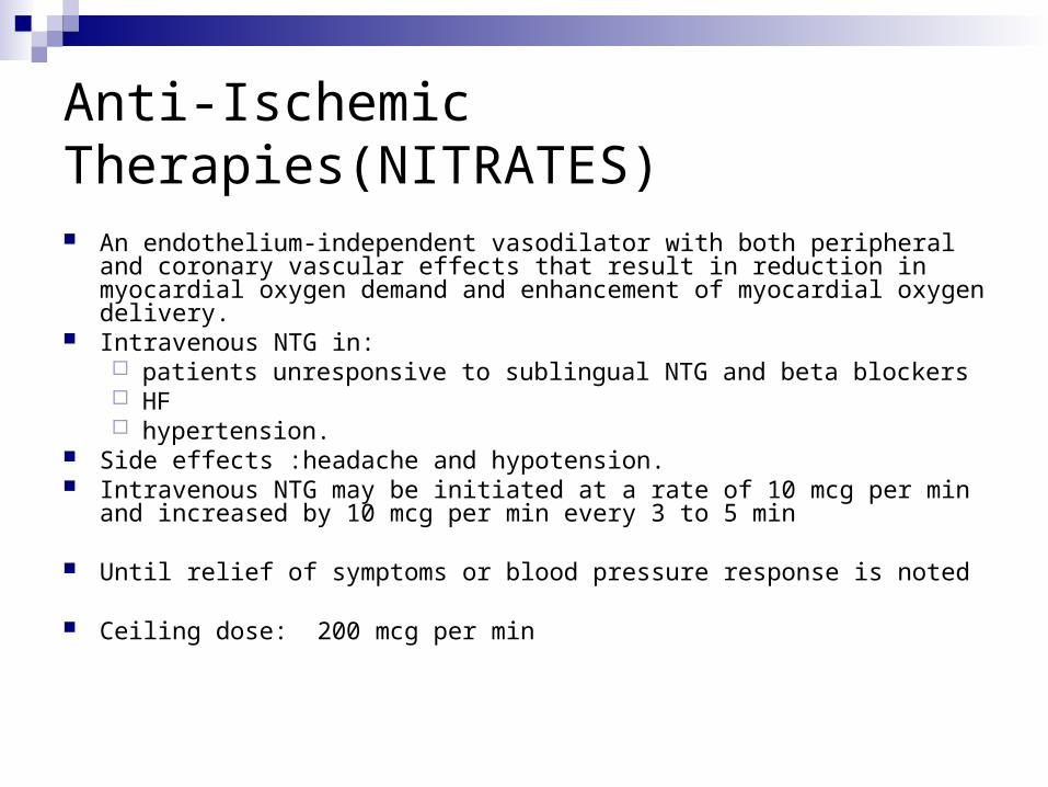
Anti-Ischemic Therapies(NITRATES)
An endothelium-independent vasodilator with both peripheral and coronary vascular effects that result in reduction in myocardial oxygen demand and enhancement of myocardial oxygen delivery.
Intravenous NTG in: patients unresponsive to sublingual NTG and beta blockers HF hypertension.
Side effects :headache and hypotension. Intravenous NTG may be initiated at a rate of 10 mcg per min and increased
by 10 mcg per min every 3 to 5 min
Until relief of symptoms or blood pressure response is noted
Ceiling dose: 200 mcg per min
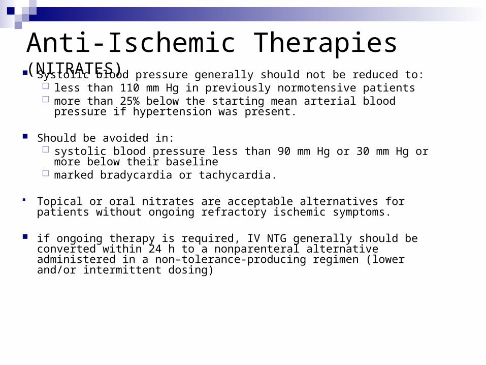
Anti-Ischemic Therapies (NITRATES) Systolic blood pressure generally should not be reduced to:
less than 110 mm Hg in previously normotensive patients more than 25% below the starting mean arterial blood pressure if
hypertension was present.
Should be avoided in: systolic blood pressure less than 90 mm Hg or 30 mm Hg or more below
their baseline marked bradycardia or tachycardia.
Topical or oral nitrates are acceptable alternatives for patients without ongoing refractory ischemic symptoms.
if ongoing therapy is required, IV NTG generally should be converted within 24 h to a nonparenteral alternative administered in a non–tolerance-producing regimen (lower and/or intermittent dosing)
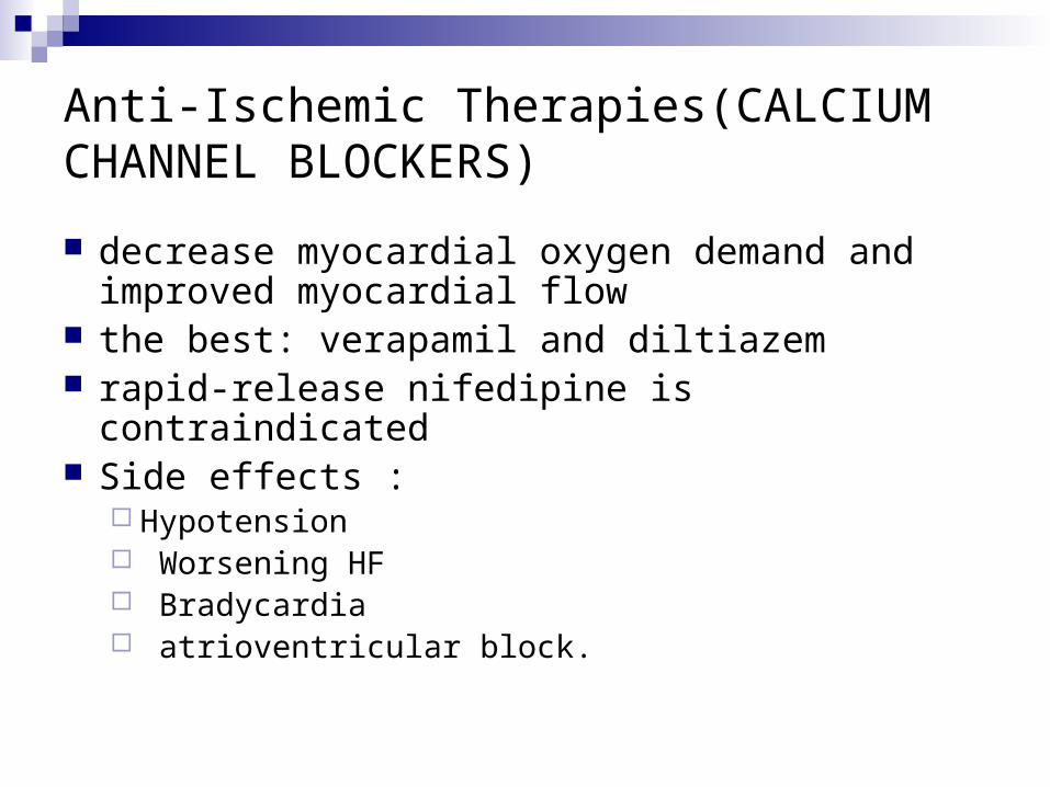
Anti-Ischemic Therapies(CALCIUM CHANNEL BLOCKERS)
decrease myocardial oxygen demand and improved myocardial flow
the best: verapamil and diltiazem rapid-release nifedipine is contraindicated Side effects :
Hypotension Worsening HF Bradycardia atrioventricular block.

Anti-Ischemic Therapies (CALCIUM CHANNEL BLOCKERS)
Indications: in patients unresponsive to or intolerant of nitrates and
beta blockers variant angina.
Contraindications: pulmonary edema or severe LV dysfunction Caution when combined with beta-blocker due to
depress LV function and sinus and AV node conduction.

Anti-Ischemic Therapies(ACEi)
reduce mortality rates in : AMI recently MI LV systolic dysfunction diabetes mellitus with LV dysfunction high-risk chronic CAD, including patients with normal
LV function ARBs may be useful in post-MI and ischemic HF
patients intolerant of ACE inhibitors
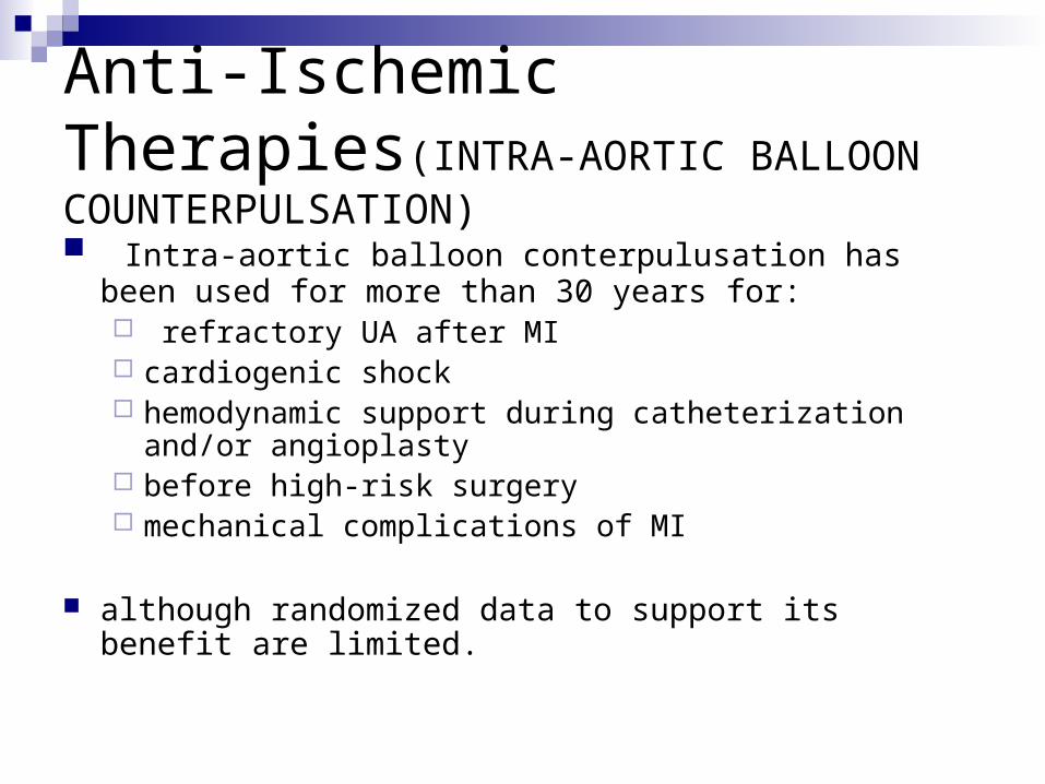
Anti-Ischemic Therapies(INTRA-AORTIC BALLOON COUNTERPULSATION)
Intra-aortic balloon conterpulusation has been used for more than 30 years for: refractory UA after MI cardiogenic shock hemodynamic support during catheterization and/or
angioplasty before high-risk surgery mechanical complications of MI
although randomized data to support its benefit are limited.
Anti-Ischemic Therapies(COX-2 inhibitors and NSAIDs )
discontinue them immediately . Complications among patients who were
taking NSAIDs within 7 d of enrollment: increased risk of death,reinfarctionHFshock

B. Antiplatelet Therapy
(Aspirin, Ticlopidine, Clopidogrel) A. ASPIRIN initial dose : 162 and 325 mg. maintenance dose: 75 to 162 mg (higher initial
maintenance dose After stenting:325 mg per day for 1 month after bare-metal stent implantation and 3 to 6 months after drug-eluting stent implementation)
More rapid buccal absorption occurs with non–entericcoated formulations

Antiplatelet Therapy
B. ADENOSINE DIPHOSPHATE RECEPTOR ANTAGONISTS AND OTHER ANTIPLATELET AGENTS
ticlopidine and clopidogrel are approved irreversible several days to achieve maximal effect in the absence of a loading
dose. Ticlopidine : successful for the secondary prevention of stroke and MI and for the
prevention of stent closure and graft occlusion adverse effects :neutropenia ,TTP Oral loading dose of 500 mg followed by 250 mg BD
Antiplatelet Therapy
Clopidogrel : For secondary prevention, clopidogrel alone was at least as effective as
or modestly more effective than ASA is indicated in patients with UA/NSTEMI who are unable to tolerate ASA Oral loading dose of 300 mg followed by 75 mg Daily for 3 to 12 months A small excess in bleeding patients undergoing CABG surgery within 5 d
of stopping clopidogrel. is recommended in patients who undergo PCI
Sulfinpyrazone, dipyridamole, prostacyclin, and prostacyclin analogs are not recommended.
The thromboxane synthase blockers and thromboxane A2 receptor antagonists have notshown any advantage over ASA.

Anticoagulants (previously referred to as antithrombins)
UFH, enoxaparin, fondaparinux, bivalirudin It is often difficult to conclude that one antithrombotic strategy is
preferred over another
A. UNFRACTIONATED HEPARIN reduction of 33% to 56% in early ischemic events Most of the benefit is short term, with reactivation of the thrombotic
process (“rebound”) after the discontinuation An initial bolus of 60 U per kg (maximum 4,000 U) is followed by an
initial infusion of 12 U per kg per hour (maximum 1,000 U per hour) The duration of therapy in most UA/NSTEMI trials has been 2 to 5
d. Monitoring by INR(1.5 to 2.5) and CBC( for anemia and heparin-
induced thrombocytopenia
Anticoagulants
B. LOW-MOLECULAR-WEIGHT HEPARIN Advantages over UFH :
ease of administration once- or twice-a-day subcutaneous ) eliminating the need for monitoring stimulate platelets less than UFH and less frequently cause heparin-
induced thrombocytopenia lower rates of death and esp. nonfatal MI
In early invasive strategy, outcomes with UFH and LMWH were similar
For patients in whom CABG is planned, it is recommended that LMWH be discontinued and UFH used during the operation.
An initial IV bolus of 30 mg/kg is followed by 1 mg/kg SC BD
Anticoagulants
C. DIRECT THROMBIN INHIBITORS Hirudin, Bivalirudin
D. FACTOR XA INHIBITORS Fondaparinux
decreased binding to plasma proteins and endothelial cells and dose-independent clearance
with a longer half-life, which results in more predictable and sustained anticoagulation and allows fixed-dose, once-daily subcutaneous administration, does not require laboratory monitoring.
A preferred anticoagulant strategy in those at higher risk of bleeding managed with a noninvasive strategy.
Anticoagulants
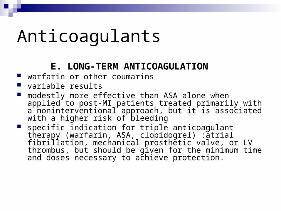
E. LONG-TERM ANTICOAGULATION warfarin or other coumarins variable results modestly more effective than ASA alone when applied to post-MI
patients treated primarily with a noninterventional approach, but it is associated with a higher risk of bleeding
specific indication for triple anticoagulant therapy (warfarin, ASA, clopidogrel) :atrial fibrillation, mechanical prosthetic valve, or LV thrombus, but should be given for the minimum time and doses necessary to achieve protection.

3. Platelet GP IIb/IIIa Receptor Antagonists The binding of fibrinogen molecules to GP IIb/IIIa
receptor on adjacent platelets results in platelet aggregation.
Abciximab, Platelet aggregation gradually returns to normal 24 to 48 h after discontinuation
Eptifibatide Tirofiban platelet aggregation returns to normal 4 to 8 h after
discontinuation of these 2 drugs Treatment effect was greater among higher risk patients
with troponin elevation and ST-segment depression.
Platelet GP IIb/IIIa Receptor Antagonists GP IIb/IIIa inhibitors are of substantial benefit in patients with
UA/NSTEMI who undergo PCI, are of modest benefit in patients who are not routinely scheduled to undergo revascularization (but who may do so), and are of questionable benefit in patients who do not undergo revascularization.
clinical recommendations call for the concomitant use of heparin with GPIIb/IIIa inhibitors(esp. tirofiban).[ more frequent bleeding with enoxaparin than UFH]
Fibrinolytic therapy is not recommended for the management of ACS patients without ST-segment elevation, a posterior-wall MI, or a presumably new left bundle-branch block.
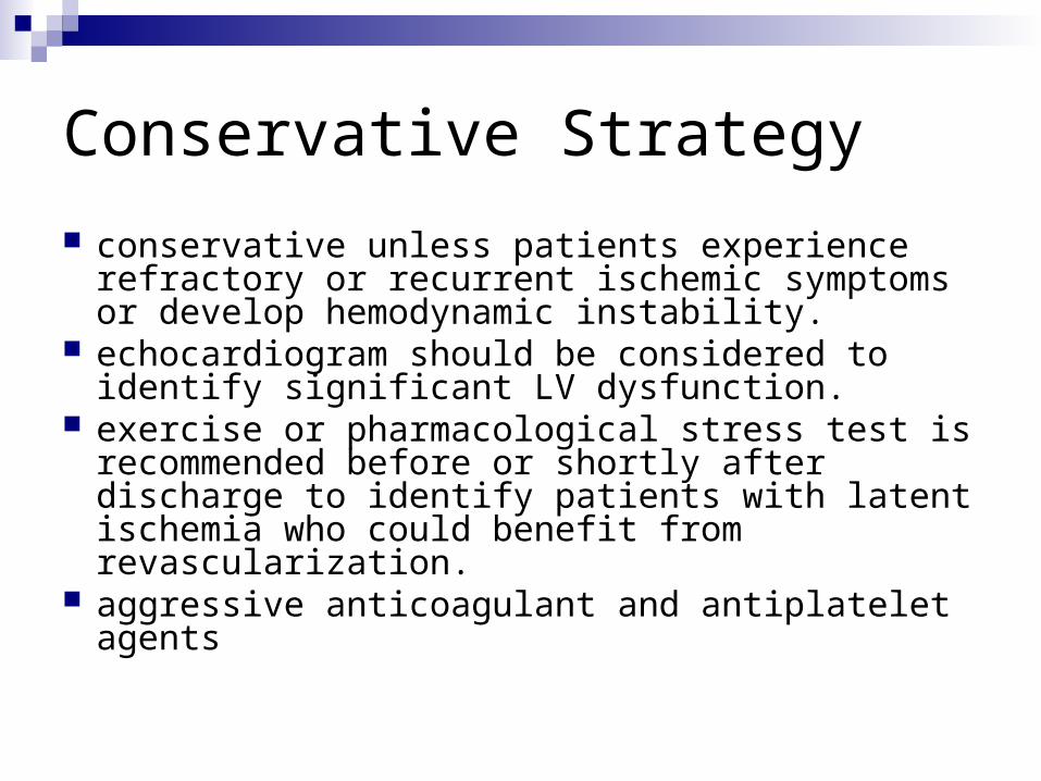
Conservative Strategy
conservative unless patients experience refractory or recurrent ischemic symptoms or develop hemodynamic instability.
echocardiogram should be considered to identify significant LV dysfunction.
exercise or pharmacological stress test is recommended before or shortly after discharge to identify patients with latent ischemia who could benefit from revascularization.
aggressive anticoagulant and antiplatelet agents

Invasive Strategy
Patients treated with an invasive strategy generally will undergo coronary angiography within 4 to 24 h of admission.
subdivided into 2 groups:1. angiography/revascularization urgently because
of ongoing ischemic symptoms or hemodynamic or rhythm instability.
With these patients, GP IIb/IIIa antagonists or clopidogrel may be delayed at the physician’s discretion until the time of angiography
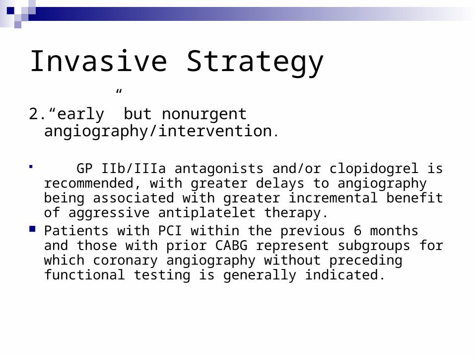
Invasive Strategy
2.“early” but nonurgent angiography/intervention.
GP IIb/IIIa antagonists and/or clopidogrel is recommended, with greater delays to angiography being associated with greater incremental benefit of aggressive antiplatelet therapy.
Patients with PCI within the previous 6 months and those with prior CABG represent subgroups for which coronary angiography without preceding functional testing is generally indicated.
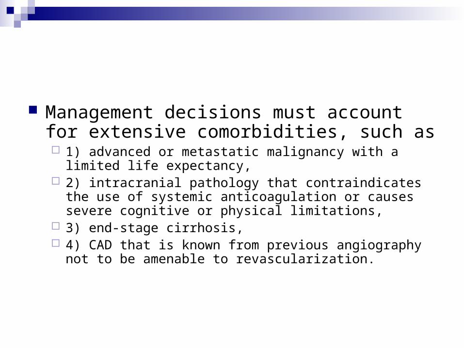
Management decisions must account for extensive comorbidities, such as 1) advanced or metastatic malignancy with a limited life
expectancy, 2) intracranial pathology that contraindicates the use of systemic
anticoagulation or causes severe cognitive or physical limitations,
3) end-stage cirrhosis, 4) CAD that is known from previous angiography not to be
amenable to revascularization.
Risk Stratification Before Discharge A. GENERAL PRINCIPLES AND CARE OBJECTIVES Formal risk assessment tools, such as GRACE and TIMI, can be
useful not only for in-hospital and shortterm assessments but also for longer term (6-month) assessment of risk
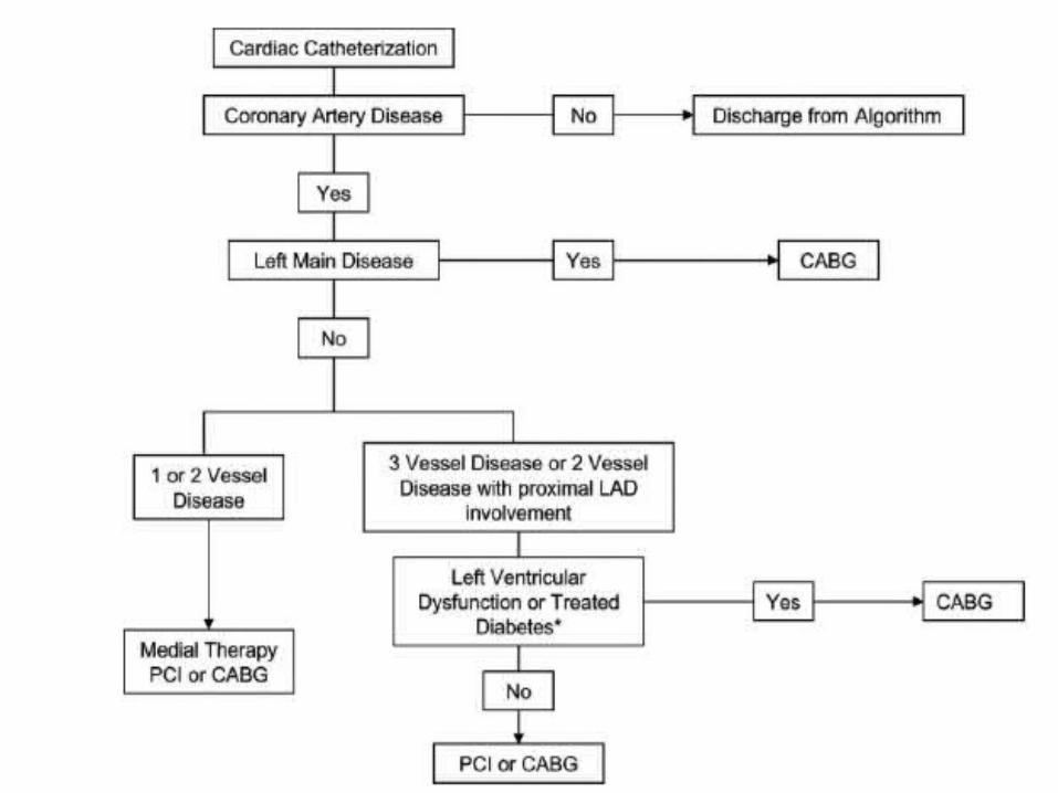
Coronary angiography and revascularization represent powerful modifiers of risk and tools for prognostication.
B. NONINVASIVE TEST SELECTION Low- and intermediaterisk patients may undergo symptom-limited
stress testing if they have been clinically stable for 12 to 24 h. Earlier stress testing (i.e., within 3 to 7 d after UA/NSTEMI) is
superior to later testing (i.e., at 1 month) in that it identifies patients at risk for adverse events within the first month.

SELECTION FOR CORONARY ANGIOGRAPHY
many intermediate- and high-risk patients receive coronary angiography as part of an invasive management strategy.
In addition, coronary angiography is usually indicated in other UA/NSTEMI patients who have either recurrent symptoms or ischemia despite adequate medical therapy or who develop high-risk features clinically
Late Hospital Care Hospital Discharge and Post-Hospital Discharge Care
A.General Principles and Care Objectives Patients who have undergone successful PCI with an
uncomplicated course are usually discharged the next day. Patients who undergo uncomplicated CABG generally are
discharged 4 to 7 d later. Low-risk patients may be discharged soon after noninvasive testing
or coronary angiography. Management of high-risk, unstable patients often requires more
prolonged and vigilant inpatient care.
Long-Term Medical Therapy
Patients with UA/NSTEMI require secondary prevention at discharge. The acute phase of UA/NSTEMI is usually over within 1 to 3 months, after which most patients assume a course of chronic CAD. Therefore, chronic secondary
prevention measures are similar to those for other CAD patients

Postdischarge Risk Assessment and Follow-Up The PURSUIT, TIMI, and GRACE risk models are also useful for
postdischarge risk assessment education on medications, diet, exercise, and smoking cessation (if
appropriate); referral to a cardiac rehabilitation/secondary prevention program (when appropriate); and the scheduling of a timely follow-up appointment.
Low-risk medically treated patients and revascularized patients should return in 2 to 6 weeks,
higher-risk patients should return within 14 d. When stable, typically by 1 to 3 months after discharge, patients
may be followed up as for stable CAD.

Risk Factor Modification
Statin therapy is beneficial regardless of baseline LDL-C levels Data on the utility of ACE inhibitors in stable CAD in the absence of HF or
LV dysfunction have been conflicting , the absolute benefit is proportional to disease-related risk, with those at lowest risk benefiting least
systolic and diastolic blood pressures in the normal range (i.e., less than 140/90 mm Hg; 130/80 mm Hg if the patient has diabetes or chronic kidney disease)
For patients who smoke, tobacco cessation has substantial potential to improve survival
Overweight patients should be instructed in a weight loss Glycemic control The use of NSAIDS and COX-2–selective inhibitors should be minimized
(lowest Cardiovascular risk with naproxen) Folic acid/B-vitamin supplementation is not recommended Antioxidant vitamins (C,E, beta carotene) also not recommended

Physical Activity
Exercise training generally can begin within 1 to 2 weeks after revascularized
UA/NSTEMI Other Activities Daily walking can be encouraged immediately in all patients. In stable patients without complications, sexual activity with the
usual partner can be resumed within 1 week to 10 d. For stable patients, driving can begin 1 week after discharge After complicated MI, driving should be delayed until 2 to 3 weeks
after symptoms have resolved. Air travel within the first 2 weeks of MI should be undertaken

Thank You