Embed Size (px)
Citation preview

1
In vitro activity of Dermaseptins K4S4 and K4K20S4 against 1
Escherichia coli, Staphylococcus aureus and Pseudomonas 2
aeruginosa planktonic growth and biofilm formation 3
4
Amira Zaïri*a, Lionel Ferrièresb, Patricia Latour-Lambertb,d, Christophe Beloinb, 5
Frédéric Tangyc, Jean-Marc Ghigob and Khaled Hania 6 7 a Laboratoire de Biochimie, Faculté de médecine de Sousse, Tunisia 8 b Institut Pasteur, Unité de Génétique des Biofilms. 25 rue du Dr. Roux, 75724 Paris cedex 15 9
France. 10 C Institut Pasteur, Unité de Génomique Virale et Vaccination. 28 rue du Dr. Roux, 75724 11
Paris cedex 15 France 12 d Present address: Institut Pasteur, Unité de Dynamique des Interactions Hôte-Pathogène, 25 13
rue du Dr. Roux, 75724 Paris cedex 15 France 14
15
16 17 18
19
20
Corresponding author: Amira ZAÏRI 21
Address: Department of Biochemistry, Faculty of Medicine Sousse, Tunisia 22
e-mail: [email protected] 23
Phone: (+216) 98 82 53 28 24
Fax: (+216) 73 22 48 99 25
26
27
Running title: antibiofilm activity of dermaseptins 28 29 30
31
32
AAC Accepts, published online ahead of print on 3 February 2014Antimicrob. Agents Chemother. doi:10.1128/AAC.02142-13Copyright © 2014, American Society for Microbiology. All Rights Reserved.
on June 17, 2018 by guesthttp://aac.asm
.org/D
ownloaded from
2
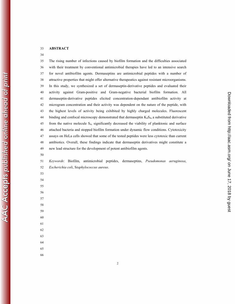
ABSTRACT 33
34
The rising number of infections caused by biofilm formation and the difficulties associated 35
with their treatment by conventional antimicrobial therapies have led to an intensive search 36
for novel antibiofilm agents. Dermaseptins are antimicrobial peptides with a number of 37
attractive properties that might offer alternative therapeutics against resistant microorganisms. 38
In this study, we synthesized a set of dermaseptin-derivative peptides and evaluated their 39
activity against Gram-positive and Gram-negative bacterial biofilm formation. All 40
dermaseptin-derivative peptides elicited concentration-dependant antibiofilm activity at 41
microgram concentration and their activity was dependent on the nature of the peptide, with 42
the highest levels of activity being exhibited by highly charged molecules. Fluorescent 43
binding and confocal microscopy demonstrated that dermaseptin K4S4, a substituted derivative 44
from the native molecule S4, significantly decreased the viability of planktonic and surface 45
attached bacteria and stopped biofilm formation under dynamic flow conditions. Cytotoxicity 46
assays on HeLa cells showed that some of the tested peptides were less cytotoxic than current 47
antibiotics. Overall, these findings indicate that dermaseptin derivatives might constitute a 48
new lead structure for the development of potent antibiofilm agents. 49
50
Keywords: Biofilm, antimicrobial peptides, dermaseptins, Pseudomonas aeruginosa, 51
Escherichia coli, Staphylococcus aureus. 52
53
54
55
56
57
58
59
60
61
62
63
64
65
66
on June 17, 2018 by guesthttp://aac.asm
.org/D
ownloaded from

3
INTRODUCTION 67
68
A biofilm is a microbial community of sessile microorganisms composed of cells that 69
are embedded in a matrix of extracellular polymeric substances, attached to a substratum or 70
interface. Biofilm bacteria are phenotypically and physiologically different from planktonic or 71
suspended cells (1, 2). Bacterial biofilms have been linked to a wide range of infections, 72
particularly in patients requiring indwelling medical devices, such as catheters and prostheses. 73
They have recently been associated with about 80% of all chronic human infections (3) and 74
described as important mediators of healthcare-associated infections (4). 75
Osteomyelitis, infective endocarditis, chronic wounds, and infections related to 76
indwelling devices are examples of infections that are often caused by biofilm-producing 77
strains. Staphylococci and enterobacteriaceae account for a large proportion of these 78
infections, with Staphylococcus aureus and Escherichia coli pathogens representing more 79
than 50 % of the species isolated from patients with medical device-associated infections (6, 80
7). Such infections typically exhibit increased tolerance to antimicrobial, biocidal and 81
immunological challenges, which makes their treatment with conventional chemotherapeutic 82
agents difficult, and sometimes impossible. These concerns have prompted a persistent search 83
for alternative therapies against planktonic microorganisms and biofilms. Of particular 84
interest, dermaseptins, antimicrobial peptides produced by the immune system of frogs, offer 85
a number of properties and attributes that might open promising opportunities for the 86
development of new antibiofilm agents. 87
Antimicrobial peptides are produced by both prokaryotic and eukaryotic cells and 88
have been extensively investigated in recent research because they are involved in several 89
host defense mechanisms and lend themselves to various applications as therapeutic agents. 90
(8–13). Dermaseptins are a family of eight closely related antimicrobial peptides that have 91
originally been isolated from the skin of a tree-dwelling, South American frog (Phyllomedusa 92
sauvagei). They are linear polycationic peptides composed of 28–34 amino acids that are 93
structured in amphipathic -helices in apolar solvents (14, 15). They all have a conserved Trp 94
residue at position 3, an AG(A)KAAL(V/G)G(N/K)AV(A) consensus motif in the mid-95
region, and a positive charge attributable to the presence of Lys residues that punctuate an 96
alternating hydrophobic and hydrophilic sequence (14). New members of the dermaseptin S 97
family (S9–S11) that do not resemble any of the so far characterized natural antimicrobial 98
peptides have recently been identified and cloned from a skin secretion-derived cDNA library 99
(16). In fact, some dermaseptins show marked abilities to inhibit microbial cells efficiently, 100
on June 17, 2018 by guesthttp://aac.asm
.org/D
ownloaded from

4
rapidly, and irreversibly without toxic effects on mammalian cells. They also display cytolytic 101
activity in vitro against a broad spectrum of host-free microorganisms, including bacteria 102
(Gram-positive and Gram-negative) (17–21), protozoa (22–24), yeasts and lamentous fungi 103
(19, 25) and viruses (26-28). 104
Dermaseptin S1, from the skin secretion of Phyllomedusa hypochondrialis frogs, has 105
recently been reported to exhibit electroanalytical activity to dopamine (DA) oxidation. The 106
selectivity in the detection of DA is, in fact, a fundamental aspect for the development of 107
electrochemical sensors with potential applications in the biomedical and pharmaceutical 108
industries (29). Although the precise mechanism of action of antimicrobial peptides is not yet 109
fully understood, the antimicrobial action of dermaseptin is thought to be mediated by the 110
interaction of the amphipathic -helix with the membrane phospholipids, resulting in the 111
permeation of the target cell by destabilizing the plasma membrane via either a ‘barrel-stave’ 112
mechanism or a ‘non-pore carpet-like’ mechanism (30,31). 113
Structure-activity relationship studies performed on native dermaseptin S4 have 114
recently led to the identi cation of synthetic derivatives with improved antimicrobial 115
properties (32-34). When comparing the resistance emergence rates by propagating bacteria 116
under selective antibiotic pressure, both Gram-positive and Gram-negative bacteria were 117
noted to exhibit resistance to commercial antibiotics but not to the L-or D-isomers of the 118
dermaseptin derivatives that were tested (33). Overall, the data obtained from in vitro and in 119
vivo experiments indicate that some dermaseptin derivatives have a variety of potential 120
medical and antimicrobial applications (32). Nevertheless, no previous work has so far been 121
performed on the antibiofilm activity of dermaseptins. Accordingly, the present study was 122
undertaken to investigate the feasibility and gain effects of using dermaseptins against 123
biofilms formed by important biofilm-forming pathogens, including Staphylococcus aureus, 124
Escherichia coli, and Pseudomonas aeruginosa. It particularly focused on the evaluation of 125
the antibiofilm potential of dermaseptin S4 derivatives. A number of chemically-modified 126
peptides were synthesized and investigated for antibiofilm activity. HeLa cultures were used 127
to evaluate their safety and toxicity, and fluorescent binding and confocal microscopy assays 128
were performed to assess the speed and irreversibility of their antibiofilm activity. 129
130
on June 17, 2018 by guesthttp://aac.asm
.org/D
ownloaded from

5
MATERIALS AND METHODS 131
132
Synthesis, purification and preparation of peptides 133
Peptides were prepared by stepwise solid phase synthesis using Fmoc polyamide-134
active ester chemistry on Milligen 9050 pepsynthesizer. All Fmoc-amino acids were from 135
Milligen-Waters (France). 4-(Hydroxymethyl) phenoacetic acid-linked polyamide/kieselguhr 136
resin (pepsin kA), Fmoc-aminoacid penta uorophenyl (Pfp), and 3-hydroxy-2, 3-dehydro-4-137
oxo-benzotriazine (Dhbt) esters were from Milligen/Bioresearch. Cleavage of peptidyl-resin 138
and side chain deprotection were carried out using 5 mg of peptidyl-resin in 1 ml mixture 139
composed of tri uoroacetic acid, para-cresol, thioanisol, water, and ethyl methyl sul de (82.5; 140
5.5; and 2.5% v/v) for 2 h at room temperature. After ltering to remove the resin and ether 141
extraction, the crude peptides were puri ed by a combination of Sephadex gel ltration, ion 142
exchange chromatography, and preparative high performance liquid chromatography (HPLC). 143
The homogeneity of the synthetic peptides was assessed by analytical HPLC, amino acid 144
analysis, solid phase sequence analysis, and mass spectrometry (35). All peptides were stored 145
frozen as stock solutions at 1 mg/ml and -20°C in double-distilled water. Prior to 146
experimentation, fresh solutions were diluted in the appropriate medium. For uorescein-147
labeled peptides, uorescein was introduced at the N-terminus of the peptide using uorescein 148
N-hydroxysuccinimide ester prior to Trifluoroacetic acid (TFA) treatment. 149
150
Bacterial strains and growth media 151
The strains used in this study are listed in Table 1. They were maintained as follows: 152
Pseudomonas aeruginosa was grown in Lysogeny Broth (LB) medium, Escherichia coli was 153
grown in Mueller Hinton (MH) broth (for antimicrobial assays) or in M63B1 medium 154
containing 0.4% glucose medium (for biofilm assay), and Staphylococcus aureus was grown 155
in Tryptic Soy Broth (TSB) containing 0.25 % Glucose. 156
157
Antimicrobial activities against planktonic cultures 158
To investigate the antimicrobial activities of dermaseptins (Table 2), we evaluated the 159
planktonic growth of different strains in the absence or presence of each peptide at different 160
concentrations. Accordingly, an overnight culture of each bacteria grown in the appropriate 161
medium was used to inoculate 100 μl of sterile MH medium in a 96-well plate to an optical 162
density at 600 nm (OD600) of 0.0001. The peptides were added to final concentrations ranging 163
from 0 to 100 μg/ml. Polymyxin used at 6.25 μg/ml served as a positive control. Three 164
on June 17, 2018 by guesthttp://aac.asm
.org/D
ownloaded from
6
replicates were made for each condition. The cultures were incubated at 37°C, with shaking 165
for 24 h. The minimum inhibitory concentration (MIC) was determined from the lowest 166
concentration that induced 100% inhibition. 167
168
In vitro biofilm susceptibility 169
To quantify the biofilm formation of E. coli in the absence and presence of peptides, a 170
microplate-based assay was performed as follows. Strains were grown in M63 media 171
containing 0.4 % glucose for 24 h at 37°C in a shaking bath and then diluted to obtain a 172
suspension with an optical density (OD) at 600 of 0.01. 100 μl of the diluted suspension was 173
added into the wells of a Polyvinyl Chloride (PVC) microtiter plate and incubated for 24 h 174
without shaking to allow biofilm formation. Planktonic cells were carefully removed by 175
pipetting, and biofilms were then treated with 100 μl of different concentrations of each 176
peptide at indicated concentrations and incubated for 24 h at 37°C. Biofilm formation was 177
measured using crystal violet (CV) staining. After treatment, planktonic cells were gently 178
removed; each well was washed three times with water and pat dried with a piece of paper 179
towel in an inverted position. To quantify biofilm formation, each well was stained with 100 180
μl of 1 % crystal violet and incubated for 15 min at room temperature. The plates were then 181
washed three times with water again to remove extra dye. After that, 125 μl of mixed 80% 182
ethanol and 20% acetone were added to each well to dissolve all the absorbed dye. After 30 183
min of incubation at room temperature, OD570 was measured to quantify the total biomass of 184
biofilm formed in each well. The viability of biofilm cells was evaluated using the XTT 185
quantification method. After treatment, planktonic cells were removed by careful pipetting, 186
and the biofilms were washed with 125 μl of PBS 1X. Each well was supplemented with 125 187
μl of 0.05 mg/ml XTT/ 10 mM menadione and incubated at 37°C for 4 h in the dark. The 188
metabolic activity of biofilm cells reflecting their viability was assessed by determining the 189
amount of product resulting from the degradation of XTT into formazan, through OD492 190
measurement. 191
192
Confocal microscopy: Peptides effect on a biofilm in continuous flow 193
Growth of biofilm 194
The bacterial strain used for this experiment was E. coli MG1655 F'Tet traD:apra 195
ATT km-mars. Biofilms were grown at 37°C in M63B1 supplemented with 0.4% glucose as a 196
carbon source, in flow chambers with individual channel dimensions of 1 by 4 by 40 mm. The 197
on June 17, 2018 by guesthttp://aac.asm
.org/D
ownloaded from
7
flow system was assembled and prepared as described previously (36). Individual flow cells 198
were inoculated with fluorescent E. coli (see Table 1 and legend of Fig. 2) and adjusted to an 199
optical density at 620 nm of 0.1. To allow the adherence of bacterial cells to the substratum, 200
flow cells were left without flow for 1 h after inoculation at 37°C. A laminar flow was then 201
activated with a flow rate of 2μl/sec. 202
203
Exposure of biofilm to fluorescent dermaseptins 204
After 5 h of biofilm growth in the flow cell, the flow was stopped, and 300 μl of each 205
fluorescein-tagged peptide sample at the following concentrations (0.78 μg/ml for K4S4 and 206
25 μg/ml for D4D20S4) was added into the chamber by syringe injection. After 20 min, the 207
flow was restarted with a flow rate of 0.4μl/sec per section. Normal flow rate (2μl/sec) per 208
section was set up again 20 min later. Z-stack images were obtained every 1 h for at least 6 h. 209
Flow cells were imaged under confocal microscopy Leica SP5 using the following 210
parameters: Objective 63X oil, 512x512 pixels, line average 2, 1 μm z steps, 60 μm per stack, 211
excitation Filter Wavelengths: 475-490 nanometers and 540-565 nanometers, emission 212
wavenlengths: 500-535 nanometers and 570-620 nanometers. Side-view images of the biofilm 213
were converted into 3D structures using the Software Imaris. All microscopy observations 214
and image acquisitions were performed using methods described above. For quantification of 215
biofilm development with or without peptide, images from each sample were analyzed using 216
the computer program (Imaris MeasurementPro). 217
218
Assay for eukaryotic cell viability 219
The potential cytotoxicity of dermaseptin S4 and its derivatives was measured in 220
normal human cervical HeLa cells using the MTT (3-[4,5-dimethylthiazol-2-yl]-2,5-diphenyl 221
tetrazolium bromide)-based assay. Brie y, cells were seeded into 96-well plates at a density 222
of 2 x 10⁴ cells per well and incubated for 24 h at 37°C prior to drug exposure. On the day of 223
treatment, the culture medium was aspirated from the wells and replaced by the fresh medium 224
containing a drug concentration ranging from 0 to 500 μg/ml. Triplicate wells were used for 225
each treatment. Culture plates were then incubated for 30 min, 3 h or 6 h before the addition 226
of 10 μl of MTT solution (5 μg/ml in PBS) to each well. The wells containing only the 227
medium and MTT were used as controls for each plate. The tetrazolium/formazan reaction 228
was allowed to proceed for 4 h at 37°C, and then 100 μl of the solubilization bu er (10% 229
sodium dodecyl sulfate in 0.1% (v/v) HCL) was added to all wells and mixed thoroughly to 230
on June 17, 2018 by guesthttp://aac.asm
.org/D
ownloaded from
8
dissolve the dark-blue formazan crystals. After overnight incubation at 37°C, the optical 231
density at 540 nm was measured by a 96-well multiscanner auto-reader with the solubilization 232
bu er serving as a blank. To translate the OD540 values into the number of live cells in each 233
well, the OD540 values were compared with those of standard OD540-versus-cell number 234
curves generated for each cell line. The percentage of cell survival was expressed as live cell 235
number in test group/live cell number in control group x 100. 236
237
Statistical analysis 238
Normality in this study was assessed using Proc Univariate in SAS software (37). All data 239
were statistically analyzed using the least-squares method with the procedure GLM for 240
General Linear Model. Effects were treatment, bacteria and the interaction treatment-bacteria. 241
The SNK multivariate test was used to compare means for bacteria and Dunnett's test to 242
compare each experimental mean of treatment (peptide) with the control mean. 243
244
245
246
on June 17, 2018 by guesthttp://aac.asm
.org/D
ownloaded from
9
RESULTS 247
248
In vitro toxicity of dermaseptin and derivatives against human cells 249
We tested the potential in vitro cytotoxicity of dermaseptin S4 and its derivatives 250
(described in Table 2) in confluent monolayers of human HeLa cells using the MTT cell 251
viability assay. Cells were exposed to increasing concentrations of peptides ranging from 0 to 252
128 μg/ml. Cytotoxicity of dermaseptins was concentration-dependent (data not shown). 253
Peptides concentrations that caused 50% cytotoxixity (CC50) were determined (Table 3). The 254
highest cytotoxicity rates observed for all S4 derivatives were recorded at concentrations 255
higher than 24.12 μg/ml (CC50), except for S4(5-28) whose toxicity was slightly lower than 256
the other peptides (CC50 > 100 μg/mL). Interestingly, results showed that shortening the 257
peptide in N- or C-terminal region (S4(5–28) or K4S4(1–16)) and increasing its positive charge 258
by different substitutions (K4K20S4 or K4S4) leads to peptides with low toxicity. 259
260
Antimicrobial activity of dermaseptin S4 derivatives on planktonic bacteria 261
We next evaluated the ability of dermaseptin S4 and its derivatives to inhibit 262
proliferation of Gram-positive S. aureus, and Gram-negative P. aeruginosa and E. coli 263
planktonic cells (Table 3). All peptides under investigation inhibited antimicrobial growth. 264
Their range of activity was, however, dependent on the nature of the peptide and the highly 265
positive charged molecules. In fact, the derivatives in which positive charge was increased 266
without shortening the length of the peptide, i.e. K4K20S4 and K4S4, were the most potent 267
inhibitors, and the derivatives in which positive charge was reduced, such as D4D20S4, were 268
less potent. Likewise, the substituted and deleted peptide K4S4(1–16) and the peptide with N-269
terminal deletions S4(5-28) displayed a nearly homogenous potency in all assays, with a 270
progressive loss of potency as compared to K4S4 particularly against S. aureus. However, the 271
truncation of the N-terminal carboxyl S4(5-28), which potentially resulted in a less toxic 272
peptide (33), did not affect its potency. 273
274
Effect of dermaseptin S4 derivatives on in vitro static biofilm 275
We first assessed the activity of the dermaseptin S4 and its derivatives against mature 276
bacterial biofilms using biofilms formed in microtiter PVC plates. While exposure of bacterial 277
biofilms to the peptides at concentrations of 2 x MIC did not dissolve biofilms as measured by 278
crystal violet staining (data not shown), survival of biofilm cells was reduced (Fig. 1). K4S4 279
on June 17, 2018 by guesthttp://aac.asm
.org/D
ownloaded from
10
and K4K20S4 were the most active peptides against all the strains tested, whereas D4D20S4 was 280
the least active one. However, K4S4(1-16) and S4(5-28), which strongly affected E. coli and P. 281
aeruginosa biofilm, had less activity against S. aureus biofilm. 282
283
284
Effect of dermaseptin S4 derivatives on in vitro E. coli biofilm formed in continuous 285
flow conditions. 286
To further investigate the inhibiting effect of dermaseptin S4 derivatives on pre-formed 287
biofilm, we evaluated their activity on E. coli biofilm developed under continuous flow 288
conditions using confocal imaging to visualize both bacterial cells labeled in red (red 289
fluorescent protein, Rfp) and fluorescent dermaseptins. 290
As mentioned earlier, since K4S4 and K4K20S4 exhibited a similar activity against E. coli 291
biofilm and due to the lack of a fluorescent K4K20S4 peptide, this experiment was performed 292
with fluorescein tagged K4S4 and D4D20S4. Confocal images (Fig 2A and B) showed that, 293
under continuous culture conditions in flow cells for 6 h in minimal media with glucose as 294
carbon source, the fluorescent peptide K4S4 used at 2x MIC (0.78 μg/ml, in green), targeted 295
all biofilm cells, both in the interior part and in the upper layer of the biofilm. The presence of 296
K4S4 appeared to completely stop the growth of the biofilm with just a little detachment, 297
suggesting that this peptide has the ability to inhibit biofilm proliferation (Fig 2A). This was 298
verified by quantitative analysis of bacterial biomass that showed that biofilm proliferation 299
was stopped right after injection of K4S4 (Fig. 3). Co-localization measurements indicated that 300
the peptide penetrated bacterial cells, which confirmed its bacterial cell permeation ability 301
(data not shown). The fact that dermaseptin K4S4 activity was also discernable in the presence 302
of the continuous flow suggests a strong effect of K4S4 despite the dilution factor. In contrast, 303
when the biofilm was exposed to 2x MIC of dermaseptin D4D20S4 (25 μg/ml), the established 304
biofilm continued to grow steadily, and red cells were still visible and continued to multiply 305
(Fig 2B). Interestingly, although D4D20S4 showed both binding cell capacity and penetration, 306
it was not as effective as K4S4 (co-location measures), and appeared to be unable to stop the 307
growth of E. coli biofilm (Fig 3). 308
Among all peptides tested, we found that K4K20S4 and K4S4 were the most potent to 309
inhibit biofilm formation with non toxic concentrations comparing to the others peptides. We 310
also showed that this inhibition of growth was dependent on the nature of the peptide, and the 311
highly charged molecules were the most active. 312
313
on June 17, 2018 by guesthttp://aac.asm
.org/D
ownloaded from

11
DISCUSSION 314
315
Infections in which bacteria are either slow growing, dormant, or within a biofilm, 316
pose a serious clinical challenge for therapy because the cells in these states exhibit tolerance 317
to the activity of antimicrobial agents. Many of these infections, including otitis media, 318
sinusitis, cholesteatoma, and tonsillitis, are caused by biofilm-forming mucosal pathogens, 319
such as Staphylococcus aureus, Escherichia col and Pseudomonas aeruginosa. Accordingly, 320
the discovery of anti-infective agents that are active against both planktonic microorganisms 321
and biofilms is of great public health significance. Antimicrobial peptides represent a 322
potential promising source for the development of new alternative agents to combat resistant 323
bacteria. These molecules are rapidly bactericidal, and since their action relates to physical 324
properties, it is all the more difficult for bacteria to develop resistance to such peptides. 325
Moreover, their structure can be engineered with relative ease, because peptide chemistry 326
allows a multitude of modifications that are relatively time and cost-effective. Nevertheless, 327
most of the agents undergoing clinical trials lack specificity and are examined only for topical 328
uses (31). 329
330
Dermaseptins are a specific class of antimicrobial peptides that have a broad range of 331
antimicrobial activity. Despite their promising properties and attributes, little work has so far 332
been performed to evaluate their effects on bacterial biofilm formation. The present study 333
aims to investigate the antimicrobial activity of five related synthetic peptides derived from 334
the natural peptide dermaseptin S4. It evaluates the effects of dermaseptins and some of their 335
derivatives against a series of Gram-negative and Gram-positive strains, including S. aureus, 336
E. coli and P. aeruginosa, in planktonic and biofilm cells and describes their structure-337
function relationship. 338
339
Our findings revealed that when used at high concentrations, this natural antimicrobial 340
peptide is cytotoxic in vitro for host cells. This toxicity can, however, be reduced through 341
introducing a number of modifications to the native sequence without altering its activity. To 342
increase the antimicrobial activity of S4, four deletions and substitutions were tested based on 343
previous studies performed with Escherichia coli and human red blood cells. Our results 344
revealed that dermaseptins K4S4 and K4K20S4 displayed a hundred times higher increase in 345
antibiofilm potency than the other derivatives. The results also showed that a positive charge 346
on June 17, 2018 by guesthttp://aac.asm
.org/D
ownloaded from

12
substitution led to a lower level of cytotoxicity without altering antibiofilm activity of the 347
peptides. 348
349
Bisubstituted derived peptide K4K20S4 exhibited the best results. This dermaseptin 350
derivative combined two substitutions, the first substitution of methionine by lysine in 351
position 4 and the second asparagine by lysine in position 20. It was previously shown that 352
increasing the number of positive charges (6 versus 4 for dermaseptin S4) and reducing its 353
hydrophobicity index (22.7 versus dermaseptin S4 28.9) resulted in a reduction of hemolytic 354
activity (34). Other studies demonstrated that dermaseptin K4K20S4 had a good antimicrobial 355
activity in vivo with no toxicity in mice (32). The combination of the deletion of the 12 C-356
terminal residues with the substitution of methionine by lysine in position 4 K4S4(1-16), the 357
deletion of 5 N-terminal residues in dermaseptins S4(5-28), or the reduction of positive 358
charges by substituting methionine in position 4 and 20 by aspartic acid were reported to 359
induce a marked decrease in its antimicrobial activity as compared to K4K20S4. It can be 360
concluded from these observations that increasing the net positive charges of the peptide 361
without shortening its sequence would result in analogs that display potent antibiofilm activity 362
and low cytotoxicity. Besides, the selective activity of antimicrobial dermaseptins depends on 363
the membrane lipid composition of the microbe versus the host cell and its electrical potential 364
(33). For example, although the lipid compositions of host cell and P. falciparum membrane 365
are similar, the potential of the parasite membrane is higher than that of the host cell 366
membrane, leading to the discriminating effect of S4 (34). 367
368
Furthermore, the literature shows that several other peptides isolated from different 369
sources have been investigated for their antibiofilm activity against several bacterial strains, 370
including cathelicidin (38, 39), histatin, mucins (40, 41), magainins (42, 445), pleurocidin 371
(43), and lactoferrin (44). Like dermaseptins, these peptides are cationic and unstructured and 372
fold into amphipathic -helices upon contact with membranes. The data presented in this 373
work suggest that the antibiofilm activity of dermaseptins is significantly higher than that of 374
these peptides. For example, magainins II, which are 23-residue -helical peptides isolated 375
from the skin of the frog Xenopus laevis, display antibiofilm activity against P. aeruginosa 376
and E. coli at 246.9 μg/ml and 15.4 μg/ml, respectively (40), whereas 0.38 μg/ml of 377
dermaseptin K4K20S4 was enough to achieve the same effect. In addition, a novel peptide 378
mimetic, meta-phenylene ethylene (mPE), modeled after magainin was tested for its efficacy 379
against several biofilm forming strains, including S. aureus, and the results showed that the 380
on June 17, 2018 by guesthttp://aac.asm
.org/D
ownloaded from

13
eradication of biofilm was observed at 10xMIC (42, 45). Used at 2xMIC, on the other hand, 381
dermaseptin was sufficient to prevent the formation of S. aureus biofilm. Like the archetypal 382
dermaseptins, pyllospetins (PSN), a major family of phyllomedusine skin antimicrobials (45, 383
46), have recently been described to occur in some species in multiple isomeric forms. The 384
evaluation of the activity of these peptides’ (PSN-1) against biofilms is, however, very 385
limited; only few strains have so far been evaluated, and no data have shown their ability to 386
inactivate bacterial strains (46). 387
388
With their broad range of antibacterial activity, dermaseptins may certainly offer new 389
promising candidates for the inhibition of biofilm formation. Although we did not 390
demonstrate their bactericidal activity in this work, it was previously reported that 391
dermaseptin derivatives including K4K20S4, were rapidly bactericidal in vitro and in vivo 392
against clinical isolates of Staphylococcus aureus, Pseudomonas aeruginosa, and Escherichia 393
coli (32). The tested peptides reduced the number of viable CFU of either E. coli or S. aureus 394
by 6 logs in 30 min or less. Another study also described the bactericidal activity of 395
dermaseptins against various oral pathogens, including those immobilized in a biofilm (47). 396
Furthers studies are still needed to determine how dermaseptins kill planktonic bacteria and 397
by which mechanism they inhibit biofilm formation. 398
399
Biofilms are known to constitute a diverse and complex aggregate of bacteria that 400
exhibit over 100-fold resistance to conventional antibiotics (48). Once a biofilm is 401
established, the live cells are typically buried beneath the surface or between layers of dead 402
cells and encased by a glycocalyx, an extracellular matrix of carbohydrates, proteoglycans, 403
DNA, and other cellular constituents. Not only does this complex of biological molecules 404
inhibit diffusion due to steric hindrance, but its constituents are believed to carry charges that 405
have been known to interfere with the diffusion of others antibiotics (42, 45). The confocal 406
microscopy experiments of this study showed that some dermaseptins such as K4S4 penetrate 407
well into the biofilm structure and inhibit biofilm formation. 408
409
The main question that remains is how these dermaseptins can cross a thick layer 410
established by exopolysaccharides, DNA, and proteins, to be able to reach their target cells. In 411
fact, Donlan and his coworkers showed that the matrix of exopolysaccharides also plays a 412
minor role in the antibioresistance properties of the biofilm by being directly bound to the 413
antimicrobial agents and by preventing them from penetrating within the biofilm (1). This 414
on June 17, 2018 by guesthttp://aac.asm
.org/D
ownloaded from
14
suggests that even if the matrix stops certain molecules of antibiotics, other molecules, such 415
as dermaseptins, can cross this barrier and penetrate within the biofilm. The penetration of 416
dermaseptins can stimulate exopolysaccharides production in the matrix and contribute to the 417
increase of its thickness. This increase does not seem to inhibit the peptide which remains 418
active against all the tested biofilm even at low concentrations, suggesting that the matrix may 419
not hold these peptides which are positively charged and that they can reach the target cells. 420
Once in contact with these cells, dermaseptins establish electrostatic interactions with the 421
bacterial membranes, leading to the formation of ionic channels in the lipid bilayers inducing 422
the death of the cell. 423
424
Finally, dermaseptin S4 derivatives present a new lead structure for potent antibiofilm 425
agents. Nevertheless, new studies are needed to correlate the activity of S4 in the in vivo 426
situation. The in vitro biofilm assays employed in this study are essentially preliminary 427
screens. They provide evidence that dermaseptins have a strong effect against biofilm strains, 428
and further studies on their mechanisms of action and activity against other biofilm sources 429
are warranted. 430
431
Acknowledgments 432
The authors would like to express their gratitude to Mr Anouar Smaoui and Mrs Hanen Ben 433
Salem from the English Language Unit at the Sfax Faculty of Science, Sfax, Tunisia for their 434
proofreading and language polishing services. 435
436 437
on June 17, 2018 by guesthttp://aac.asm
.org/D
ownloaded from

15
References 438 439
1. Donlan RM, Costerton JW. 2002. Biofilms: survival mechanisms of clinically relevant 440
microorganisms. Clin Microbiol Rev. 15:167-193. 441
2. Yala JF, Thebault P, Héquet A, Humblot V, Pradier CM, Berjeaud JM. 2011. 442
Elaboration of antibiofilm materials by chemical grafting of an antimicrobial peptide. 443
Appl Microbiol Biotechnol. 89:623-634. 444
3. http://grants.nih.gov/grants/guide/pa-files/PA-03–047.html. 445
4. Dongari-Bagtzoglou A. 2008. Mucosal biofilms: Challenges and future directions. 446
Expert. Rev. Anti.Infect. Ther.6: 141–144. 447
5. Francolini I, Donelli G. 2010. Prevention and control of biofilm-based medical 448
device- related infections. FEMS. Immunol. Med. Microbiol. 59: 227–238. 449
6. Belley A, Neesham-Grenon E, McKay G, Arhin FF, Harris R, Beveridge T, Parr TR 450
Jr, Moeck G. 2009. Oritavancin kills stationary-phase and biofilm Staphylococcus 451
aureus cells in vitro. Antimicrob. Agents Chemother. 53: 918-925. 452
7. Schillaci D, Arizza V, Parrinello N, Di Stefano V, Fanara S, Muccilli V, Cunsolo V, 453
Haagensen JJ, Molin S. 2010. Antimicrobial and antistaphylococcal biofilm activity 454
from the sea urchin Paracentrotus lividus. J. Appl. Microbiol.17-24. 455
8. Salay LC, Nobre TM, Colhone MC, Zaniquelli ME, Ciancaglini P, Stabeli RG, Leite 456
JR, Zucolotto V. 2011. Dermaseptin 01 as antimicrobial peptide with rich 457
biotechnological potential: study of peptide interaction with membranes containing 458
Leishmania amazonensis lipid-rich extract and membrane models. J. Pept. Sci. 459
17:700-707. 460
9. Zampa MF, Araújo IM, Dos Santos Júnior JR, Zucolotto V, Leite JR, Eiras C. 2012. 461
Development of a novel biosensor using cationic antimicrobial Peptide and nickel 462
phthalocyanine ultrathin films for electrochemical detection of dopamine. Int. J. Anal. 463
Chem. 850969. 464
10. Krauson AJ, He J, Wimley WC. 2012. Determining the mechanism of membrane 465
permeabilizing peptides: Identification of potent, equilibrium pore-formers. Biochim. 466
Biophys. Acta. 1818: 1625-1632. 467
11. Nicolas P, El Amri C. 2009. The dermaseptin superfamily: A gene-based 468
combinatorial library of antimicrobial peptides Biochim. Biophys. Acta . 1788: 1537-469
1550 470
on June 17, 2018 by guesthttp://aac.asm
.org/D
ownloaded from

16
12. Rivas L, Luque-Ortega JR, Andreu D. 2009. Amphibian antimicrobial peptides and 471
Protozoa: Lessons from parasites Biochim. Biophys. Acta. 1788: 1570-1581 472
13. Zasloff M. 2002. Antimicrobial peptides of multicellular organisms. Nature 415: 473
389–395 474
14. Mor A, Nguyen VH, Delfour A, Migliore-Samour D, Nicolas P. 1991. Isolation, 475
amino acid sequence and synthesis of dermaseptin, a novel antimicrobial peptide of 476
amphibian skin. Biochemistry. 30: 8824–8830. 477
15. Mor A, Rouffaud MA, Montagne JJ, Nguyen VH, Nicolas P. 1993. Natural and 478
synthetic dermaseptins: in vitro large spectrum antimicrobial peptides. J. Mycol. Med. 479
3: 137–143. 480
16. Lequin O, Ladram A, Chabbert L, Bruston F, Convert O, Vanhoye D, Chassaing G, 481
Nicolas P, Amiche M. 2006. Dermaseptin S9, an alpha-helical antimicrobial peptide 482
with a hydrophobic core and cationic termini. Biochemistry. 17;45(2):468-480 483
17. Zaïri A, Tangy F, Hani K. 2013. Dermaseptin S4 derivative K4K20S4: A potential 484
candidate for development of a new microbicide contraceptive agent - an in vitro 485
study..Eur. J. Contracept. Reprod. Health Care. 18(2):79-87 486
18. . Marynka K, Rotem S, Portnaya I, Cogan U, Mor A. 2007. In vitro discriminative 487
antipseudomonal properties resulting from acyl substitution of N-terminal sequence 488
of dermaseptin S4 derivatives. Chem. Biol. 14(1):75-85. 489
19. Zairi A, Tangy F, Ducos-Galand M, Alonso JM, Hani K. 2007. Susceptibility of 490
Neisseria gonorrhoeae to antimicrobial peptides from amphibian skin, dermaseptin, 491
and derivatives. Diagn. Microbiol. Infect. Dis. 57(3):319-324. 492
20. Porat Y, Marynka K, Tam A, Steinberg D, Mor A. 2006. Acyl-substituted 493
dermaseptin S4 derivatives with improved bactericidal properties, including on oral 494
microflora. Antimicrob. Agents. Chemother. 50(12):4153-4160. 495
21. Dagan A, Efron L, Gaidukov L, Mor A, Ginsburg H. 2002. In vitro antiplasmodium 496
effects of dermaseptins. Antimicrob. Agents. Chemother. 46: 1059–1066. 497
22. Efron L, Dagan A, Gaidukov L, Ginsburg H, Mor A. 2002. Direct interaction 498
of dermaseptin S4 aminoheptanoyl derivative with intraerythrocytic malaria parasite 499
leading to increased specific antiparasitic activityin culture. J. Biol. Chem. 500
277(27):24067-24072 501
23. Salay LC, Nobre TM, Colhone MC, Zaniquelli ME, Ciancaglini P, Stabeli RG, Leite 502
JR, Zucolotto V. 2011. Dermaseptin S1 as antimicrobial peptide with rich 503
biotechnological potential: study of peptide interaction with membranes containing 504
on June 17, 2018 by guesthttp://aac.asm
.org/D
ownloaded from

17
Leishmania amazonensis lipid-rich extract and membrane models. J. Pept. Sci. 505
17(10):700-707 506
24. Pérez-Cordero JJ, Lozano JM, Cortés J, Delgado G. 2011. Leishmanicidal activity of 507
synthetic antimicrobial peptides in an infection model with human dendritic cells. 508
Peptides. 32(4):683-690. 509
25. Morton CO, Dos Santos SC, Coote P. 2007. An amphibian-derived, cationic, alpha-510
helical antimicrobial peptide kills yeast by caspase-independent but AIF-dependent 511
programmed cell death. Mol. Microbiol. 65(2):494-507 512
26. Bergaoui I, Zairi A, Tangy F, Aouni M, Selmi B, Hani K. 2013. In vitro antiviral 513
activity of dermaseptin S4 and derivatives from amphibian skin against herpes 514
simplex virus type 2. J. Med. Virol. 85(2):272-281. 515
27. Lorin C, Saidi H, Belaid A, Zairi A, Baleux F, Hocini H, Bélec L, Hani K, Tangy F. 516
2005. The antimicrobial peptide dermaseptin S4 inhibits HIV-1 infectivity in vitro. 517
Virology. 334(2):264-275. 518
28. Belaid A, Aouni M, Khelifa R, Trabelsi A, Jemmali M, Hani K. 2002. In vitro 519
antiviral activity of dermaseptins against herpes simplex virus type 1. J. Med. Virol. 520
66(2):229-34. 521
29. Zampa MF, Araújo IM, Dos Santos Júnior JR, Zucolotto V, Leite JR, Eiras C. 2012. 522
Development of a novel biosensor using cationic antimicrobial Peptide and nickel 523
phthalocyanine ultrathin films for electrochemical detection of dopamine. Int. J. Anal. 524
Chem. 850969. 525
30. Amiche M, Galanth C. 2011. Dermaseptins as models for the elucidation of 526
membrane-acting helical amphipathic antimicrobial peptides. Curr. Pharm. 527
Biotechnol. 12(8):1184-1193. Review. 528
31. William C. Wimley 2010. Describing the Mechanism of Antimicrobial Peptide 529
Action with the Interfacial Activity Model. ACS. Chem. Biol. 5(10): 905–917 530
32. Navon-Venezia SN, Feder R, Gaidukov L, Carmeli Y, Mor A. 2002. Antimicrobial 531
properties of dermaseptin S4 derivatives with in vivo activity. Antimicrob. Agents. 532
Chemother. 46: 689–694. 533
33. Kustanovich I, Shallev DE, Mikhlin M, Gaidukov L, Mor, A. 2002. Structural 534
requirements for potent versus selective cytotoxicity for antimicrobial dermaseptin S4 535
derivates. J. Biol. Chem. 277: 16941–16951 536
on June 17, 2018 by guesthttp://aac.asm
.org/D
ownloaded from

18
34. Efron L, Dagan A, Gaidukov L, Ginsburg H, Mor A. 2002. Direct interaction of 537
dermaseptin S4 aminoheptanoyl derivate with intraerythrocytic malaria parasite 538
leading to increased specific antiparasitic activity in culture. J. Biol. Chem. 277: 539
24067–24072. 540
35. Mor A, Nguyen VH, Delfour A, Migliore-Samour D, Nicolas P. 1991. Isolation, 541
amino acid sequence, and synthesis of dermaseptin, a novel antimicrobial peptide of 542
amphibian skin. Biochemistry. 30: 8824 – 8830. 543
36. Moller S, Sternberg C, Andersen JB, Christensen BB,. Ramos , Givskov M, and 544
Molin S. 1998. In situ gene expression in mixed-culture biofilms: evidence of 545
metabolic interactions between community members. Appl. Environ. Microbiol. 546
64:721-732. 547
37. SAS/STAT., 2000. User's Guide, Release 6.12 Ed., SAS Institute, Cary, NC. 548
38. Amer LS, Bishop BM, van Hoek ML. 2010. Antimicrobial and antibiofilm activity of 549
cathelicidins and short, synthetic peptides against Francisella. Biochem. Biophys. 550
Res. Commun. 396(2):246-251 551
39. Kanthawong S, Bolscher JG, Veerman EC, Van Marle J, De Soet HJ, Nazmi K, 552
Wongratanacheewin S, Taweechaisupapong S. 2012. Antimicrobial and antibiofilm 553
activity of LL-37 and its truncated variants against Burkholderia pseudomallei. Int. J. 554
Antimicrob. Agents. 39(1):39-44. 555
40. Wei GX, Campagna AN, Bobek LA. 2006. Effect of MUC7 peptides on the growth 556
of bacteria and on Streptococcus mutansbiofilm. J. Antimicrob. Chemother. 557
57(6):1100-1109 558
41. Da Silva BR, De Freitas VA, Nascimento-Neto LG, Carneiro VA, Arruda FV, De 559
Aguiar AS, Cavada BS, Teixeira EH. 2012. Antimicrobial peptide control of 560
pathogenic microorganisms of the oral cavity: a review of the literature. Peptides. 561
36(2):315-321 562
42. Beckloff N, Laube D, Castro T, Furgang D, Park S, Perlin D, Clements D, Tang H, 563
Scott RW, Tew GN, Diamond G. 2007. Activity of an antimicrobial peptide mimetic 564
against planktonic and biofilm cultures of oral pathogens. Antimicrob. Agents. 565
Chemother. 51(11):4125-4132 566
43. .Choi H, Lee DG. 2012. Antimicrobial peptide pleurocidin synergizes with antibiotics 567
through hydroxyl radical formation and membrane damage, and exerts 568
antibiofilm activity. Biochim. Biophys. Acta. 1820(12):1831-1838. 569
on June 17, 2018 by guesthttp://aac.asm
.org/D
ownloaded from

19
44. Dashper SG, Pan Y, Veith PD, Chen YY, Toh EC, Liu SW, Cross KJ, Reynolds EC. 570
2012. Lactoferrin inhibits Porphyromonas gingivalis proteinases and has sustained 571
biofilm inhibitory activity. Antimicrob. Agents. Chemother. 56(3):1548-1556 572
45. Neiva M, Vargas DC, Conceição K, Rádis-Baptista G, Assakura MT, Jared C, 573
Hayashi MA. 2013. Gene expression analysis by ESTs sequencing of the Brazilian 574
frog Phyllomedusa nordestina skin glands. Toxicon. 61:139-150 575
46. Zhang R, Zhou M, Wang L, McGrath S, Chen T, Chen X, Shaw C. 2010. 576
Phylloseptin-1 (PSN-1) from Phyllomedusa sauvagei skin secretion: a novel broad-577
spectrum antimicrobial peptide with antibiofilm activity. Mol. Immunol. 47(11-578
12):2030-2037 579
47. Porat Y, Marynka K, A. Tam, D. Steinberg, Mor A. 2006. Acyl-Substituted 580
Dermaseptin S4 Derivatives with Improved Bactericidal Properties, Including on Oral 581
Microflora. Antimicrob. Agents. Chemother. 50(12): 4153–4160 582
48. Mah TF, O'Toole GA. 2001. Mechanisms of biofilm resistance to antimicrobial 583
agents. Trends. Microbiol. 9 (1):34-39. Review. 584
49. Valle J, Toledo-Arana A, Berasain C , Ghigo JM, Amorena B, Penades JR, Lasa I. 585
2003. SarA and not sigma B is essential for biofilm development by Staphylococcus 586
aureus. Mol. Microbiol. 48:1075–1087 587
588
589
590
591
592
593
594
595
596
597
598
599
600
601
602
603
on June 17, 2018 by guesthttp://aac.asm
.org/D
ownloaded from
20
Legends of figures 604 605 Figure 1. Effects of dermaseptins S4 derivatives on biofilm formation. The viability of biofilm 606 cells was evaluated using the XTT quantification method: Biofilm-forming S. aureus, P. aeruginosa 607 and E. coli strains were grown in 96-well polystyrene plates and biofilms formed exposed to 2 x MIC 608 concentrations of peptides during 24h. Control (negative control wells containing an appropriate 609 medium for each strain only), Polymyxin (positive control wells). Statistical analysis was performed 610 by using SNK multivariate test and Dunnett’s test. K4K20S4 and K4S4 were significantly different from 611 control p<0.001, S4(5-28) and K4S4(1-16) were different from control (p<0.01), and polymyxin is 612 different from control (p< 0.05) (***p<0.001 ;
** p<0.01 ;
* p<0.05) 613
614 615 616 617 618 619 620 621 622 Figure 2 Effect of K4S4 on biofilms. To allow attachment of the bacterial cell to the substratum, flow 623 cells were left without flow for 1h after inoculation at 37°C. Afterwards, a laminar flow with a 624 flowrate of 2μl/sec was activated. After 5h of biofilm growth, the flow was stopped and 300 μl (0.78 625 μg/ml for K4S4 and 25 μg/ml for D4D20S4) of each peptide sample was added. Confocal images 626 acquisition showed that the fluorescent peptide (A): K4S4 (green) has an instant effect on biofilm and 627 stops its formation under dynamic flow conditions. Whereas, D4D20S4, at 25μg/ml, (B), shows a less 628 activity, biofilm cells exhibit high fluorescent signals (red) and survived the dermaseptin treatment. 629 630 631 632 633 634 635 636 637 Figure 3. Confocal assays were confirmed by the quantification of biofilms. Differences in bacterial 638 survival in the biofilms were quantified using the computer program Imaris MeasurementPro. 639 Statistical analysis was performed by using SNK multivariate test and Dunnett’s test. K4S4 was 640 significantly different from control (p<0.001). 641 642
on June 17, 2018 by guesthttp://aac.asm
.org/D
ownloaded from

Table 1. Different stains used in this study. Strains Relevant
characteristics Reference
E. coli MG1655_λatt_Km_mars_F'tet traD
Red fluorescent biofilm forming E. coli strain producing the F pilus, KmR, TetR
Laboratory collection
P. aeruginosa PAO1 Wild type,
prototroph, chl-2 B. Holloway
S. aureus 15981 Biofilm forming
strain (48)
Table 2 Dermaseptin S4 derivatives used in this study .
Dermaseptins Sequence
K4S4 ALWKTLLKKVLKAAAKAALNAVLVGANA
K4K20S4 ALWKTLLKKVLKAAAKAALKAVLVGANA
D4D20S4 ALWDTLLKKVLKAAAKAALDAVLVGANA
K4S4 (1-16) ALWKTLLKKVLKAAAK- - - - - - - - - - - - - -
S4 (5-28) TLLKKVLKAAAKAALNAVLVGANA
on June 17, 2018 by guesthttp://aac.asm
.org/D
ownloaded from
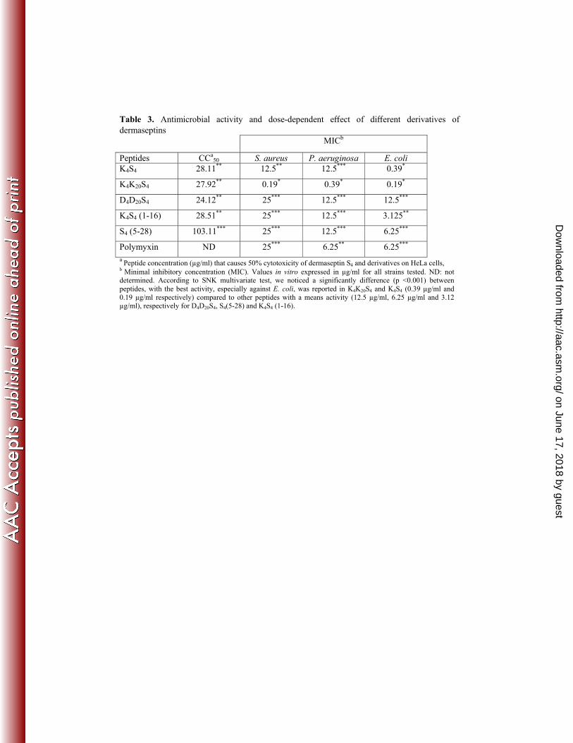
Table 3. Antimicrobial activity and dose-dependent e ect of di erent derivatives of dermaseptins
MICb
Peptides CCa50 S. aureus P. aeruginosa E. coli
K4S4 28.11** 12.5** 12.5*** 0.39*
K4K20S4 27.92** 0.19* 0.39* 0.19*
D4D20S4 24.12** 25*** 12.5*** 12.5***
K4S4 (1-16) 28.51** 25*** 12.5*** 3.125**
S4 (5-28) 103.11*** 25*** 12.5*** 6.25***
Polymyxin ND 25*** 6.25** 6.25*** a Peptide concentration (μg/ml) that causes 50% cytotoxicity of dermaseptin S4 and derivatives on HeLa cells, b Minimal inhibitory concentration (MIC). Values in vitro expressed in μg/ml for all strains tested. ND: not determined. According to SNK multivariate test, we noticed a significantly difference (p <0.001) between peptides, with the best activity, especially against E. coli, was reported in K4K20S4 and K4S4 (0.39 μg/ml and 0.19 μg/ml respectively) compared to other peptides with a means activity (12.5 μg/ml, 6.25 μg/ml and 3.12 μg/ml), respectively for D4D20S4, S4(5-28) and K4S4 (1-16).
on June 17, 2018 by guesthttp://aac.asm
.org/D
ownloaded from