Embed Size (px)
Citation preview
In Vitro Photodynamic Inactivation of Plant-Pathogenic FungiColletotrichum acutatum and Colletotrichum gloeosporioides withNovel Phenothiazinium Photosensitizers
Henrique D. de Menezes,a Gabriela B. Rodrigues,a Simone de Pádua Teixeira,b Nelson S. Massola, Jr.,c Luciano Bachmann,d
Mark Wainwright,e Gilberto U. L. Bragaa,f
Departamento de Análises Clínicas, Toxicológicas e Bromatológicas,a and Departamento de Ciências Farmacêuticas,b Faculdade de Ciências Farmacêuticas de RibeirãoPreto, Universidade de São Paulo, Ribeirão Preto, Brazil; Departamento de Fitopatologia e Nematologia, Escola Superior de Agricultura Luiz de Queiroz, Universidade deSão Paulo, Piracicaba, Brazilc; Departamento de Física, Faculdade de Filosofia, Ciências e Letras de Ribeirão Preto, Universidade de São Paulo, Ribeirão Preto, Brazild; Schoolof Pharmacy and Biomolecular Sciences, Liverpool John Moores University, Liverpool, United Kingdome; Research Support Center in Natural and Synthetic Products,Faculdade de Ciências Farmacêuticas de Ribeirão Preto, Universidade de São Paulo, Ribeirão Preto, Brazilf
The increasing tolerance to currently used fungicides in both clinical and agricultural areas is of great concern. The nonconven-tional light-based approach of antimicrobial photodynamic treatment (APDT) is a promising alternative to conventional fungi-cides. We evaluated the effects of APDT with four phenothiazinium derivatives (methylene blue [MB], new methylene blue N[NMBN], toluidine blue O [TBO], and the novel pentacyclic phenothiazinium photosensitizer [PS] S137) on conidia of threefungal species (Colletotrichum acutatum, Colletotrichum gloeosporioides, and Aspergillus nidulans). The efficacy of APDT witheach PS was determined, initially, based on photosensitizer MICs. Additionally, the effects of APDT with two selected PSs(NMBN and S137) on survival of conidia were evaluated. The subcellular localization of the PS in C. acutatum conidia was deter-mined. The effects of photodynamic treatments on leaves of the plant host Citrus sinensis were also investigated. APDT withS137 showed the lowest MIC. MICs for S137 were 5 �M for the three fungal species when a fluence of 25 J cm�2 was used. APDTwith NMBN (50 �M) and S137 (10 �M) resulted in a reduction in the survival of the conidia of all species of approximately 5 logswith fluences of >15 J cm�2. Washing of the conidia before light exposure did not prevent photodynamic inactivation. BothNMBN and S137 accumulated in cytoplasmic structures, such as lipid bodies, of C. acutatum conidia. No damage to orange treeleaves was observed after APDT.
The control of plant-pathogenic fungal species faces some of theproblems that have been observed in the related clinical area,
including the selection of antifungal-tolerant strains and the rela-tively few classes of currently available and effective fungicides(1–5). Additionally, contamination of agricultural products andthe environment due to overuse and/or inappropriate use of fun-gicides is a matter of major concern.
Colletotrichum is a large genus of ascomycete fungi containingseveral species that are common pathogens of a wide array of cropsand noncultivated plant species (6, 7). Colletotrichum acutatumand Colletotrichum gloeosporioides are among the most pathogenicspecies of this genus and cause economically important losses oftemperate, subtropical, and tropical fruits worldwide (7). Diseasesymptoms range from fruit rot to shoot, leaf, and flower blight.Common hosts include many eudicotyledonous plants such asstrawberry, apple, citrus, and stone fruits (6).
During the asexual stage of their life cycles, C. acutatum and C.gloeosporioides produce abundant unicellular hyaline conidia (6,7). Conidia are specialized structures responsible for the disper-sion, environmental persistence, and/or host infection of manyfungal species (6, 8–10). After being produced, Colletotrichumconidia remain attached to each other by a mucilage and are dis-persed over short distances by rain splash after the mucilage hasbeen dissolved by water, so rates of infection are usually highestduring the wettest periods of the growing season (7).
Current management strategies for these fungi are basedmainly on the intensive use of fungicides. Even so, control failureis common because of the selection of tolerant strains (1–5, 11).
Increasing tolerance to currently used fungicides has stimulatedthe development of new strategies to control pathogenic fungi.Antimicrobial photodynamic treatment (APDT) is an alternativeand promising antifungal discovery platform that can be used tocontrol localized mycoses in animal hosts or to kill fungi in theenvironment (12–22). The approach is based on the use of a pho-tosensitizer (PS) that binds to the surface or preferentially accu-mulates in the target fungal cell (13, 14, 17, 20). Subsequent expo-sure of the PS to light of an appropriate wavelength starts aphotochemical process that produces several reactive oxygen spe-cies (ROS), such as peroxides and singlet oxygen, leading to non-specific oxidative damage and causing the subsequent death of thefungal cells without significant harm to the host (14, 19, 20). Incomparison with currently used fungicides, the multiple and vari-able targets of reactive oxygen species reduce the chance of select-ing tolerant microorganisms. An additional advantage of APDT isthat unlike many conventional fungicides that kill only metabol-
Received 19 August 2013 Accepted 18 December 2013
Published ahead of print 20 December 2013
Address correspondence to Gilberto U. L. Braga, [email protected].
Supplemental material for this article may be found at http://dx.doi.org/10.1128/AEM.02788-13.
Copyright © 2014, American Society for Microbiology. All Rights Reserved.
doi:10.1128/AEM.02788-13
March 2014 Volume 80 Number 5 Applied and Environmental Microbiology p. 1623–1632 aem.asm.org 1623
on January 22, 2020 by guesthttp://aem
.asm.org/
Dow
nloaded from
ically active cells, it is able to kill both metabolically active anddormant or quiescent structures such as conidia (16, 20).
Previous studies have reported the use of APDT with differentPSs to kill fungi of several genera, including Aspergillus, Candida,Cryptococcus, Metarhizium, and Trichophyton (15–17, 20). Dataregarding the photodynamic inactivation of plant-pathogenicfungi are scarce, and most of those studies were performed byusing natural compounds as PSs. Additionally, as far as we know,none of those studies evaluated the effects of APDT on the planthost. DiCosmo et al. (23) reported the in vitro effects of APDT onAlternaria alternata, Aspergillus niger, Cladosporium variable, Col-letotrichum spp., Rhizopus nigricans, Pythium aphanidermatum,and Saprolegnia spp. using naturally occurring thiophene deriva-tives. Similarly, Bourque et al. (24) reported in vitro APDT againstFusarium culmorum and Aspergillus flavus using phenylhep-tatriyne from Bidens pilosa. Interestingly, Lukšiene et al. (25) alsodescribed APDT against Aspergillus flavus, Trichothecium roseum,Fusarium avenaceum, and Rhizopus oryzae using the standard an-ticancer photodynamic treatment (PDT) agent hematoporphyrindimethyl ether as a PS.
Prerequisites for using APDT to control plant-pathogenicfungi include the identification of effective PSs against differentfungal species and the evaluation of side effects of the treatment onthe host plants. The objective of this initial study was to assess theefficacies of APDT with four phenothiazinium derivatives (meth-ylene blue [MB], new methylene blue N zinc chloride double salt[NMBN], toluidine blue O [TBO], and the novel pentacyclic phe-nothiazinium photosensitizer S137) on conidia of the plant-pathogenic fungi C. acutatum and C. gloeosporioides and onconidia of the model ascomycete Aspergillus nidulans. These de-rivatives have already been shown by us to be highly effectivephotosensitizers against Candida and Trichophyton spp. in vitro(15, 16). In an attempt to improve the understanding of the mech-anisms involved in conidial photoinactivation, subcellular photo-sensitizer localization and the effects of treatment on the cellstructures of C. acutatum conidia were investigated. The effects ofAPDT with all PSs on the leaves of sweet orange (Citrus sinensis)were also determined.
MATERIALS AND METHODSFungal species and strains. Colletotrichum acutatum strain CA 142 fromcitrus was obtained from the Plant Pathogenic Fungi Collection of theDepartment of Phytopathology and Nematology (Escola Superior de Ag-ricultura Luiz de Queiroz, University of São Paulo, Piracicaba, Brazil). C.gloeosporioides strain CPC 20935 from avocado was obtained from theCentraalbureau voor Schimmelcultures (CBS) (Utrecht, Netherlands).Aspergillus nidulans strain ATCC 10074 was obtained from the AmericanType Culture Collection (ATCC) (Manassas, VA, USA).
Photosensitizers. MB (catalog number M9140), NMBN (catalognumber 202096), and TBO (catalog number T3260) were purchased fromSigma-Aldrich, Inc. (St. Louis, MO, USA). The novel pentacyclic phe-nothiazinium photosensitizer S137 was synthesized as previously de-scribed (26). Stock solutions of all the PSs were prepared with phosphate-buffered saline (PBS) (pH 7.4) at concentrations 10-fold higher than thehighest concentration used. The solutions were stored in the dark at�20°C for up to 2 weeks. Dilutions were prepared with PBS (pH 7.4).Chemical structures of all the PSs are shown in Fig. S1 in the supplementalmaterial.
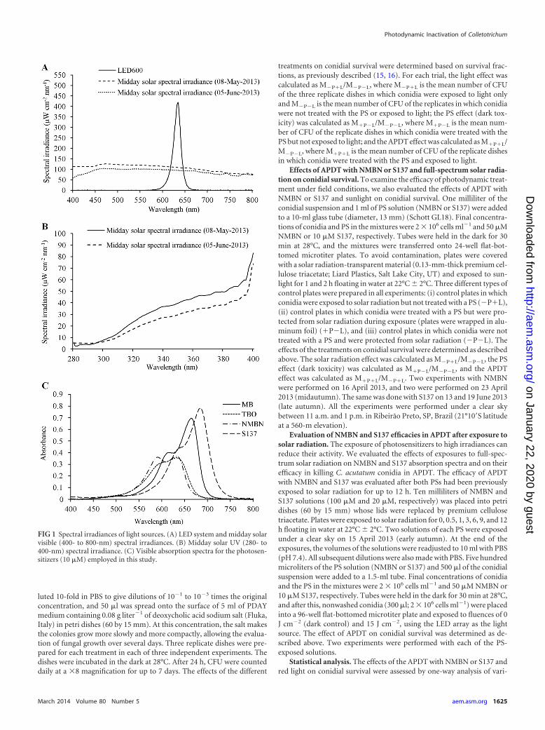
Visible light source and light measurements. All light (light-emittingdiode [LED] and solar radiation) measurements were performed by usinga cosine-corrected irradiance probe (CC-3-UV; Ocean Optics, Dunedin,FL, USA) screwed onto the end of an optical fiber coupled to a USB4000
spectroradiometer (Ocean Optics, Dunedin, FL). Red light was providedby an array of 600 LEDs with an emission peak at 634 nm (LED600). TheLED array was made in-house by using a 5-mm, high-brightness, 7,000- to8,000-mcd LED (ZX Lecomp, São Paulo, Brazil) and allowed homoge-nous illumination of the surface of a 96-well microtiter plate. The inte-grated irradiance in the red region of the visible spectrum (550 to 750 nm)was 9.2 mW cm�2. Light was measured inside the well, at the sample level,to reduce interference.
Solar spectral irradiance was measured under a clear sky at midday on8 May 2013 (midautumn) and 5 June 2013 (late autumn). In midautumn,integrated irradiances in the red region of the visible spectrum and in theUV spectrum (290 to 390 nm) were approximately 20.7 and 3.7 mWcm�2, respectively, and in late autumn, irradiances were approximately18 and 2.9 mW cm�2, respectively. LED and midday solar visible (400- to790-nm) spectral irradiances are shown in Fig. 1A, and solar UV spectralirradiance (280 to 400 nm) is shown in Fig. 1B. Figure 1C shows theabsorption spectra of the dyes (10 �M in PBS [pH 7.4]) measured with anUltraspec 2100 Pro UV-visible spectrophotometer (GE Healthcare, Ger-many).
Fungal growth and production of conidia. All fungi were grown on23 ml Difco potato dextrose agar (Becton, Dickinson and Company,Sparks, MD, USA) supplemented with 1 g liter�1 Bacto yeast extract (BD)(PDAY) in petri dishes (90 by 10 mm) at 28°C for 5 days with 12-h (dark/light) photoperiods. Conidia were carefully scraped from the colonies andsuspended in a 0.01% (vol/vol) Tween 80 (Sigma-Aldrich) solution. Theconcentration of conidia was determined with a hemocytometer, and ap-propriate dilutions were made with the same solution.
Evaluation of APDT efficacies on fungal conidia based on PS MIC.MIC-based experiments were performed, as previously described (15, 16),to determine the most effective PS and the optimized conditions forAPDT. Experiments were performed in 96-well flat-bottomed microtiterplates (TPP, Switzerland). Fifty microliters of the fungal cell suspensionand 50 �l of the PS solution (MB, NMBN, TBO, or S137) were added toeach well. The final concentration of the conidia in the mixture was 4 �104 cells ml�1; the final concentrations of MB, NMBN, and TBO were 0, 1,2.5, 5, 10, 12.5, 25, 50, 75, 100, and 200 �M; and the final concentrationsof S137 were 0, 0.5, 1, 2.5, 5, 10, 12.5, 25, 30, 40, 50, and 75 �M. Plates wereheld in the dark for 30 min at 28°C and exposed to light fluences of 5, 10,15, 20, 25, and 30 J cm�2 using the LED array as a light source or alterna-tively kept in the dark to provide dark controls. Fluences resulted fromexposures of 9, 18, 27, 36, 45, and 54 min, respectively. After the expo-sures, 100 �l of RPMI 1640 culture medium (Gibco, Invitrogen Corpora-tion, NY, USA) (2-fold concentrate) was added to each well, and plateswere incubated at 28°C. Growth was evaluated after 48, 72, and 96 h byvisual inspection when the MICs were determined (see Fig. S2 in thesupplemental material). The MIC was considered the minimal PS concen-tration (for each fluence) in which total growth inhibition was achieved.Three independent experiments were performed.
Effect of APDT with NMBN or S137 and red light on conidial sur-vival. Based on previous MIC experiments, the optimized conditions forAPDT were established, and the effects of the treatments on the survival ofC. acutatum, C. gloeosporioides, and A. nidulans conidia were evaluated.Five hundred microliters of the conidial suspension and 500 �l of the PSsolution (NMBN or S137) were added to a 1.5-ml tube (polypropylene;Axygen Scientific, CA, USA). Final concentrations of conidia and PS in themixtures were 2 � 106 cells ml�1 and 50 �M NMBN or 10 �M S137,respectively. Tubes were held in the dark for 30 min at 28°C, and after that,they were divided into two groups. In one group, conidia were washed toremove unbound PS, and in the other group, cells were not washed beforelight exposure. To remove unbound PS, cells were washed three times(centrifuged at 5,000 � g for 5 min and resuspended in Tween 80 solution[0.01%, vol/vol]). Nonwashed and washed conidia (200 �l; 2 � 106 cellsml�1) were placed into a 96-well flat-bottomed microtiter plate and ex-posed to fluences of 0 J cm�2 (dark control), 15 J cm�2, 20 J cm�2, and 25J cm�2. After light exposure, suspensions were removed and serially di-
de Menezes et al.
1624 aem.asm.org Applied and Environmental Microbiology
on January 22, 2020 by guesthttp://aem
.asm.org/
Dow
nloaded from
luted 10-fold in PBS to give dilutions of 10�1 to 10�3 times the originalconcentration, and 50 �l was spread onto the surface of 5 ml of PDAYmedium containing 0.08 g liter�1 of deoxycholic acid sodium salt (Fluka,Italy) in petri dishes (60 by 15 mm). At this concentration, the salt makesthe colonies grow more slowly and more compactly, allowing the evalua-tion of fungal growth over several days. Three replicate dishes were pre-pared for each treatment in each of three independent experiments. Thedishes were incubated in the dark at 28°C. After 24 h, CFU were counteddaily at a �8 magnification for up to 7 days. The effects of the different
treatments on conidial survival were determined based on survival frac-tions, as previously described (15, 16). For each trial, the light effect wascalculated as M�P�L/M�P�L, where M�P�L is the mean number of CFUof the three replicate dishes in which conidia were exposed to light onlyand M�P�L is the mean number of CFU of the replicates in which conidiawere not treated with the PS or exposed to light; the PS effect (dark tox-icity) was calculated as M�P�L/M�P�L, where M�P�L is the mean num-ber of CFU of the replicate dishes in which conidia were treated with thePS but not exposed to light; and the APDT effect was calculated as M�P�L/M�P�L, where M�P�L is the mean number of CFU of the replicate dishesin which conidia were treated with the PS and exposed to light.
Effects of APDT with NMBN or S137 and full-spectrum solar radia-tion on conidial survival. To examine the efficacy of photodynamic treat-ment under field conditions, we also evaluated the effects of APDT withNMBN or S137 and sunlight on conidial survival. One milliliter of theconidial suspension and 1 ml of PS solution (NMBN or S137) were addedto a 10-ml glass tube (diameter, 13 mm) (Schott GL18). Final concentra-tions of conidia and PS in the mixtures were 2 � 106 cells ml�1 and 50 �MNMBN or 10 �M S137, respectively. Tubes were held in the dark for 30min at 28°C, and the mixtures were transferred onto 24-well flat-bot-tomed microtiter plates. To avoid contamination, plates were coveredwith a solar radiation-transparent material (0.13-mm-thick premium cel-lulose triacetate; Liard Plastics, Salt Lake City, UT) and exposed to sun-light for 1 and 2 h floating in water at 22°C � 2°C. Three different types ofcontrol plates were prepared in all experiments: (i) control plates in whichconidia were exposed to solar radiation but not treated with a PS (�P�L),(ii) control plates in which conidia were treated with a PS but were pro-tected from solar radiation during exposure (plates were wrapped in alu-minum foil) (�P�L), and (iii) control plates in which conidia were nottreated with a PS and were protected from solar radiation (�P�L). Theeffects of the treatments on conidial survival were determined as describedabove. The solar radiation effect was calculated as M�P�L/M�P�L, the PSeffect (dark toxicity) was calculated as M�P�L/M�P�L, and the APDTeffect was calculated as M�P�L/M�P�L. Two experiments with NMBNwere performed on 16 April 2013, and two were performed on 23 April2013 (midautumn). The same was done with S137 on 13 and 19 June 2013(late autumn). All the experiments were performed under a clear skybetween 11 a.m. and 1 p.m. in Ribeirão Preto, SP, Brazil (21°10=S latitudeat a 560-m elevation).
Evaluation of NMBN and S137 efficacies in APDT after exposure tosolar radiation. The exposure of photosensitizers to high irradiances canreduce their activity. We evaluated the effects of exposures to full-spec-trum solar radiation on NMBN and S137 absorption spectra and on theirefficacy in killing C. acutatum conidia in APDT. The efficacy of APDTwith NMBN and S137 was evaluated after both PSs had been previouslyexposed to solar radiation for up to 12 h. Ten milliliters of NMBN andS137 solutions (100 �M and 20 �M, respectively) was placed into petridishes (60 by 15 mm) whose lids were replaced by premium cellulosetriacetate. Plates were exposed to solar radiation for 0, 0.5, 1, 3, 6, 9, and 12h floating in water at 22°C � 2°C. Two solutions of each PS were exposedunder a clear sky on 15 April 2013 (early autumn). At the end of theexposures, the volumes of the solutions were readjusted to 10 ml with PBS(pH 7.4). All subsequent dilutions were also made with PBS. Five hundredmicroliters of the PS solution (NMBN or S137) and 500 �l of the conidialsuspension were added to a 1.5-ml tube. Final concentrations of conidiaand the PS in the mixtures were 2 � 106 cells ml�1 and 50 �M NMBN or10 �M S137, respectively. Tubes were held in the dark for 30 min at 28°C,and after this, nonwashed conidia (300 �l; 2 � 106 cells ml�1) were placedinto a 96-well flat-bottomed microtiter plate and exposed to fluences of 0J cm�2 (dark control) and 15 J cm�2, using the LED array as the lightsource. The effect of APDT on conidial survival was determined as de-scribed above. Two experiments were performed with each of the PS-exposed solutions.
Statistical analysis. The effects of the APDT with NMBN or S137 andred light on conidial survival were assessed by one-way analysis of vari-
FIG 1 Spectral irradiances of light sources. (A) LED system and midday solarvisible (400- to 800-nm) spectral irradiances. (B) Midday solar UV (280- to400-nm) spectral irradiance. (C) Visible absorption spectra for the photosen-sitizers (10 �M) employed in this study.
Photodynamic Inactivation of Colletotrichum
March 2014 Volume 80 Number 5 aem.asm.org 1625
on January 22, 2020 by guesthttp://aem
.asm.org/
Dow
nloaded from
ance (ANOVA). A posttest was performed by using orthogonal contrasts(27). The effects of APDT with NMBN or S137 and solar radiation onconidial survival (field experiments) were compared by using a mixedlinear model. This model is used for the analysis of data on which theresponses are grouped and the assumption of the independence betweenobservations in the same group is not adequate (28). The posttest wasperformed by using orthogonal contrasts. The level of significance was0.05 (P � 0.05). All analyses were carried out by using PROC MIXED inSAS version 9.0 (SAS Institute, Inc., Cary, NC, USA).
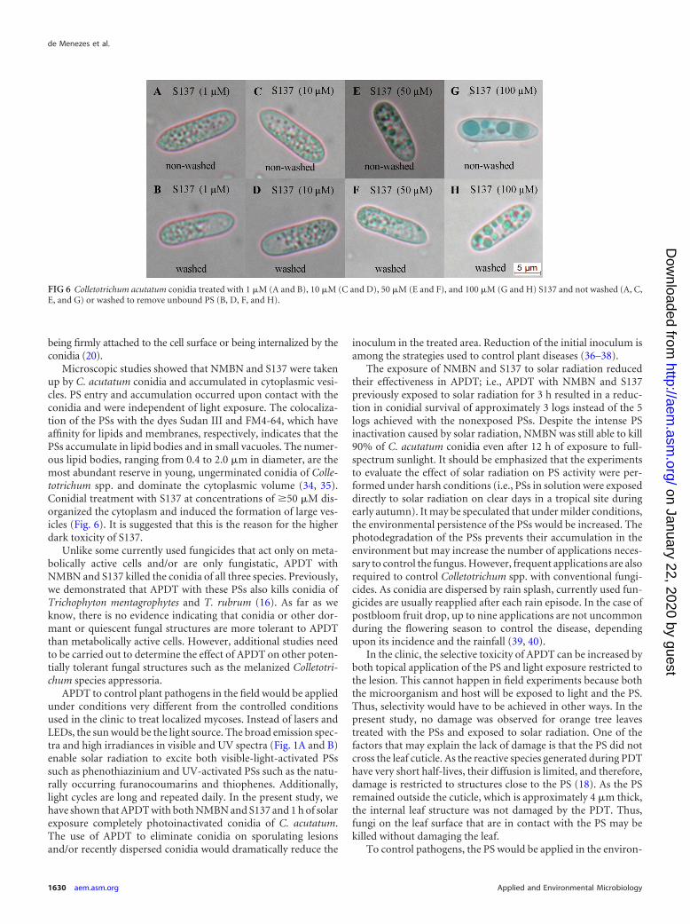
Conidial microscopy studies. Microscopic studies were performed todetermine the subcellular localization of the PS. Conidia of C. acutatum(2 � 106 conidia ml�1) were treated with NMBN (10, 50, and 100 �M) orS137 (1, 10, 50, and 100 �M) and washed or not washed before beingobserved at a �1,000 magnification by using a Leica DMD5000 B fluores-cence microscope (Leica Microsystems GmbH, Wetzlar, Germany). In anattempt to identify the cellular structures in which the PS accumulated,conidia were also stained with the dye Sudan III (0.5% [wt/vol] in 2:8distilled water-ethanol) and the fluorescent dyes 4=,6-diamidino-2-phe-nylindole (DAPI) (Sigma) (3.5 mM in distilled water) and FM4-64 (In-vitrogen) (8 �M in a 1% [vol/vol] dimethyl sulfoxide [DMSO] solution),which have affinity for lipids, genetic material, and cellular membranes,respectively. Conidia were treated with each of the three dyes, kept in thedark for 30 min, and washed three times with PBS (5,000 � g for 5 min)before being observed at a �1,000 magnification.
Effects of PDT on leaves of orange trees (Citrus sinensis). Plants usedwere about 1.5 m tall. Five microliters of MB, TBO, NMBN, and S137 at aconcentration of 50 �M was spotted on the adaxial surface of the leaves.After application of the PS, plants were kept outdoors under a naturalsunlight regimen. Plants were visually evaluated for damage to the leavesdaily for up to 15 days. Experiments were conducted in April and Novem-ber 2012 in Ribeirão Preto, SP, Brazil. Three experiments were performed.
Microscopic studies of orange tree leaves. Microscopic studies wereperformed to determine if the PS penetrated the leaves after being spottedonto the leaf adaxial surface. Ten microliters of a 100 �M solution of thePSs NMBN and S137 was spotted onto the adaxial surface, and the leaveswere kept at 24°C until the PS had dried. After this, leaves were cross-cutin the spotted area by using a table microtome (Roble, Rolemberg &Bhering, Belo Horizonte, Brazil). Histological inspection was carried outat a �200 magnification by using a Leica DM5000 B microscope. Sectionsof the leaves were treated with both NMBN and S137 in order to deter-mine the appearance of the leaf tissues had the PS penetrated and spreadthroughout the structure.
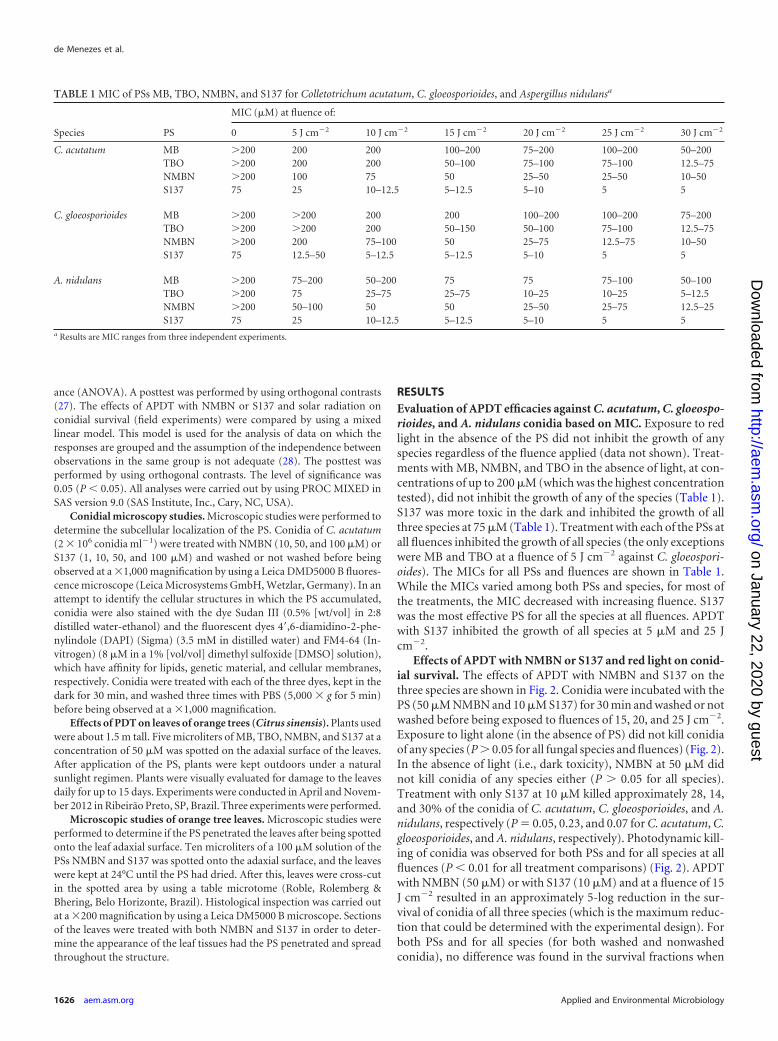
RESULTSEvaluation of APDT efficacies against C. acutatum, C. gloeospo-rioides, and A. nidulans conidia based on MIC. Exposure to redlight in the absence of the PS did not inhibit the growth of anyspecies regardless of the fluence applied (data not shown). Treat-ments with MB, NMBN, and TBO in the absence of light, at con-centrations of up to 200 �M (which was the highest concentrationtested), did not inhibit the growth of any of the species (Table 1).S137 was more toxic in the dark and inhibited the growth of allthree species at 75 �M (Table 1). Treatment with each of the PSs atall fluences inhibited the growth of all species (the only exceptionswere MB and TBO at a fluence of 5 J cm�2 against C. gloeospori-oides). The MICs for all PSs and fluences are shown in Table 1.While the MICs varied among both PSs and species, for most ofthe treatments, the MIC decreased with increasing fluence. S137was the most effective PS for all the species at all fluences. APDTwith S137 inhibited the growth of all species at 5 �M and 25 Jcm�2.
Effects of APDT with NMBN or S137 and red light on conid-ial survival. The effects of APDT with NMBN and S137 on thethree species are shown in Fig. 2. Conidia were incubated with thePS (50 �M NMBN and 10 �M S137) for 30 min and washed or notwashed before being exposed to fluences of 15, 20, and 25 J cm�2.Exposure to light alone (in the absence of PS) did not kill conidiaof any species (P � 0.05 for all fungal species and fluences) (Fig. 2).In the absence of light (i.e., dark toxicity), NMBN at 50 �M didnot kill conidia of any species either (P � 0.05 for all species).Treatment with only S137 at 10 �M killed approximately 28, 14,and 30% of the conidia of C. acutatum, C. gloeosporioides, and A.nidulans, respectively (P 0.05, 0.23, and 0.07 for C. acutatum, C.gloeosporioides, and A. nidulans, respectively). Photodynamic kill-ing of conidia was observed for both PSs and for all species at allfluences (P � 0.01 for all treatment comparisons) (Fig. 2). APDTwith NMBN (50 �M) or with S137 (10 �M) and at a fluence of 15J cm�2 resulted in an approximately 5-log reduction in the sur-vival of conidia of all three species (which is the maximum reduc-tion that could be determined with the experimental design). Forboth PSs and for all species (for both washed and nonwashedconidia), no difference was found in the survival fractions when
TABLE 1 MIC of PSs MB, TBO, NMBN, and S137 for Colletotrichum acutatum, C. gloeosporioides, and Aspergillus nidulansa
Species PS
MIC (�M) at fluence of:
0 5 J cm�2 10 J cm�2 15 J cm�2 20 J cm�2 25 J cm�2 30 J cm�2
C. acutatum MB �200 200 200 100–200 75–200 100–200 50–200TBO �200 200 200 50–100 75–100 75–100 12.5–75NMBN �200 100 75 50 25–50 25–50 10–50S137 75 25 10–12.5 5–12.5 5–10 5 5
C. gloeosporioides MB �200 �200 200 200 100–200 100–200 75–200TBO �200 �200 200 50–150 50–100 75–100 12.5–75NMBN �200 200 75–100 50 25–75 12.5–75 10–50S137 75 12.5–50 5–12.5 5–12.5 5–10 5 5
A. nidulans MB �200 75–200 50–200 75 75 75–100 50–100TBO �200 75 25–75 25–75 10–25 10–25 5–12.5NMBN �200 50–100 50 50 25–50 25–75 12.5–25S137 75 25 10–12.5 5–12.5 5–10 5 5
a Results are MIC ranges from three independent experiments.
de Menezes et al.
1626 aem.asm.org Applied and Environmental Microbiology
on January 22, 2020 by guesthttp://aem
.asm.org/
Dow
nloaded from
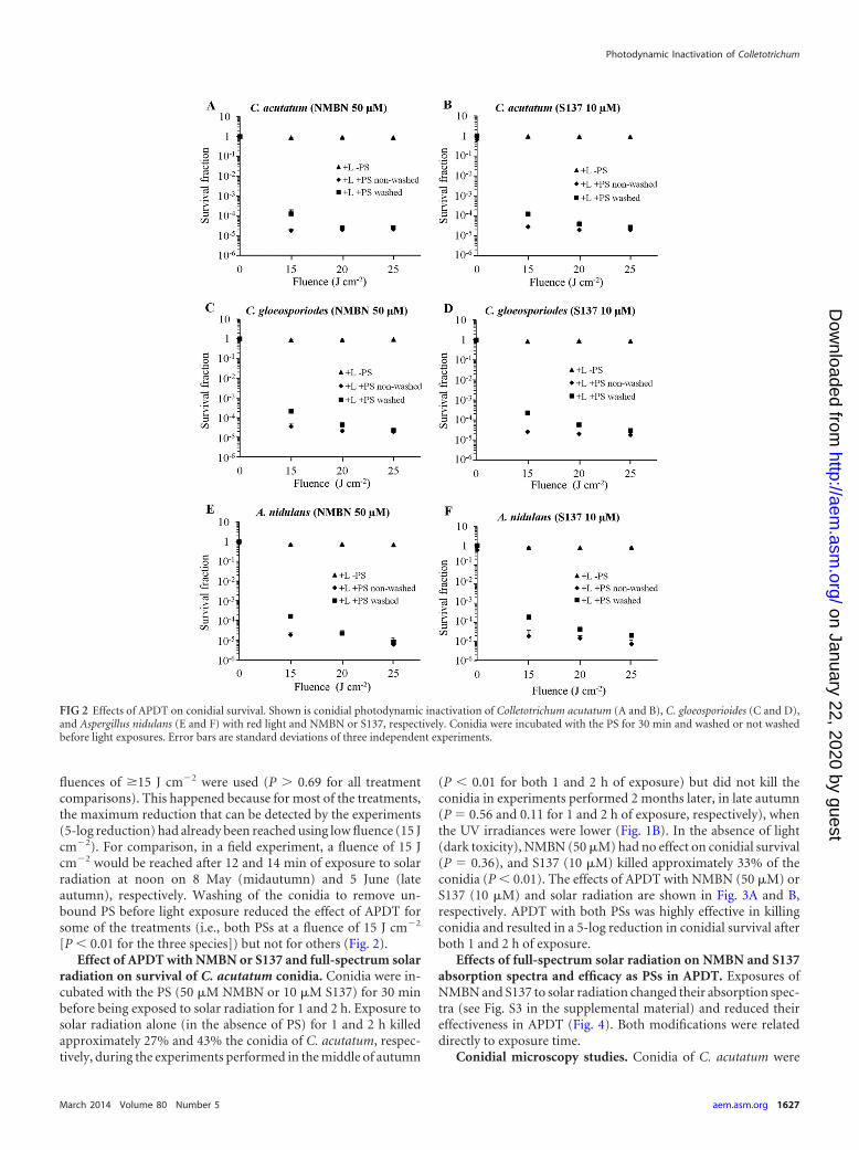
fluences of �15 J cm�2 were used (P � 0.69 for all treatmentcomparisons). This happened because for most of the treatments,the maximum reduction that can be detected by the experiments(5-log reduction) had already been reached using low fluence (15 Jcm�2). For comparison, in a field experiment, a fluence of 15 Jcm�2 would be reached after 12 and 14 min of exposure to solarradiation at noon on 8 May (midautumn) and 5 June (lateautumn), respectively. Washing of the conidia to remove un-bound PS before light exposure reduced the effect of APDT forsome of the treatments (i.e., both PSs at a fluence of 15 J cm�2
[P � 0.01 for the three species]) but not for others (Fig. 2).Effect of APDT with NMBN or S137 and full-spectrum solar
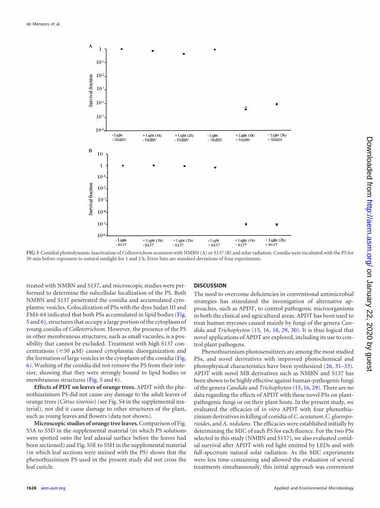
radiation on survival of C. acutatum conidia. Conidia were in-cubated with the PS (50 �M NMBN or 10 �M S137) for 30 minbefore being exposed to solar radiation for 1 and 2 h. Exposure tosolar radiation alone (in the absence of PS) for 1 and 2 h killedapproximately 27% and 43% the conidia of C. acutatum, respec-tively, during the experiments performed in the middle of autumn
(P � 0.01 for both 1 and 2 h of exposure) but did not kill theconidia in experiments performed 2 months later, in late autumn(P 0.56 and 0.11 for 1 and 2 h of exposure, respectively), whenthe UV irradiances were lower (Fig. 1B). In the absence of light(dark toxicity), NMBN (50 �M) had no effect on conidial survival(P 0.36), and S137 (10 �M) killed approximately 33% of theconidia (P � 0.01). The effects of APDT with NMBN (50 �M) orS137 (10 �M) and solar radiation are shown in Fig. 3A and B,respectively. APDT with both PSs was highly effective in killingconidia and resulted in a 5-log reduction in conidial survival afterboth 1 and 2 h of exposure.
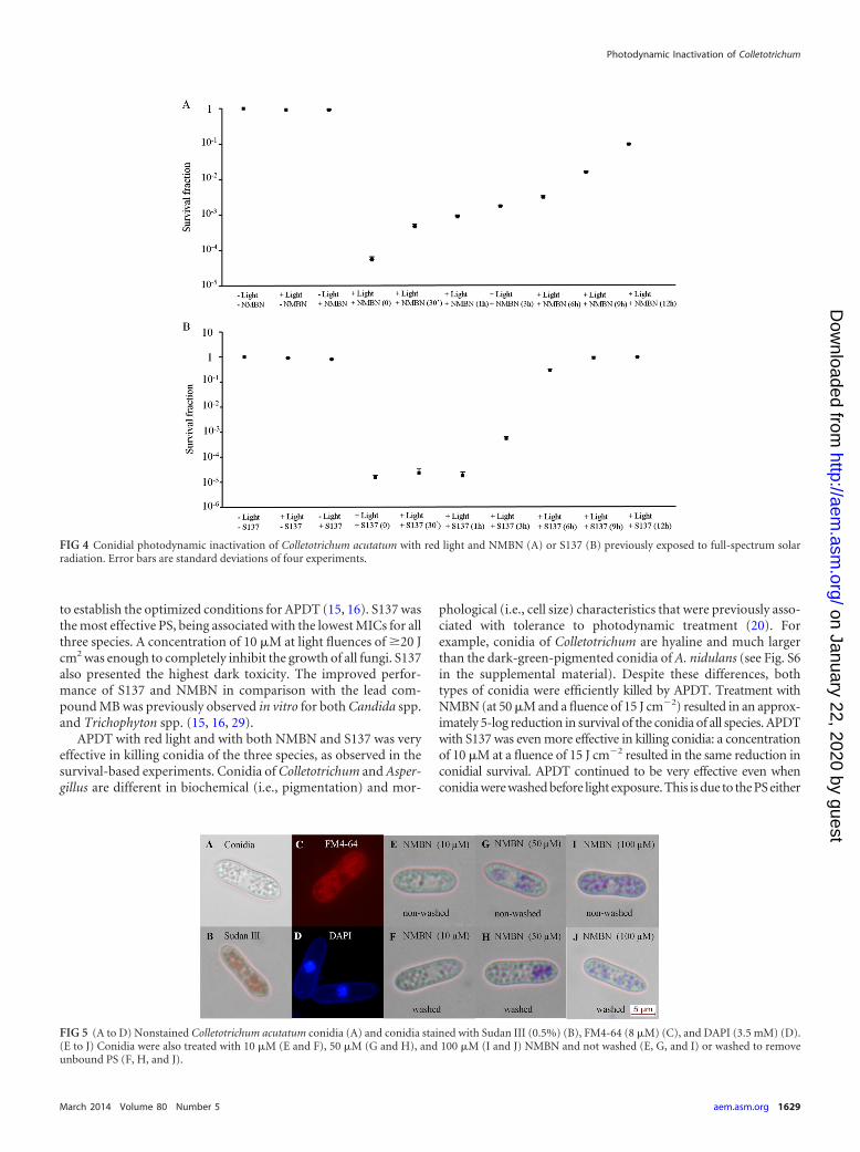
Effects of full-spectrum solar radiation on NMBN and S137absorption spectra and efficacy as PSs in APDT. Exposures ofNMBN and S137 to solar radiation changed their absorption spec-tra (see Fig. S3 in the supplemental material) and reduced theireffectiveness in APDT (Fig. 4). Both modifications were relateddirectly to exposure time.
Conidial microscopy studies. Conidia of C. acutatum were
FIG 2 Effects of APDT on conidial survival. Shown is conidial photodynamic inactivation of Colletotrichum acutatum (A and B), C. gloeosporioides (C and D),and Aspergillus nidulans (E and F) with red light and NMBN or S137, respectively. Conidia were incubated with the PS for 30 min and washed or not washedbefore light exposures. Error bars are standard deviations of three independent experiments.
Photodynamic Inactivation of Colletotrichum
March 2014 Volume 80 Number 5 aem.asm.org 1627
on January 22, 2020 by guesthttp://aem
.asm.org/
Dow
nloaded from
treated with NMBN and S137, and microscopic studies were per-formed to determine the subcellular localization of the PS. BothNMBN and S137 penetrated the conidia and accumulated cyto-plasmic vesicles. Colocalization of PSs with the dyes Sudan III andFM4-64 indicated that both PSs accumulated in lipid bodies (Fig.5 and 6), structures that occupy a large portion of the cytoplasm ofyoung conidia of Colletotrichum. However, the presence of the PSin other membranous structures, such as small vacuoles, is a pos-sibility that cannot be excluded. Treatment with high S137 con-centrations (�50 �M) caused cytoplasmic disorganization andthe formation of large vesicles in the cytoplasm of the conidia (Fig.6). Washing of the conidia did not remove the PS from their inte-rior, showing that they were strongly bound to lipid bodies ormembranous structures (Fig. 5 and 6).
Effects of PDT on leaves of orange trees. APDT with the phe-nothiazinium PS did not cause any damage to the adult leaves oforange trees (Citrus sinensis) (see Fig. S4 in the supplemental ma-terial), nor did it cause damage to other structures of the plant,such as young leaves and flowers (data not shown).
Microscopic studies of orange tree leaves. Comparison of Fig.S5A to S5D in the supplemental material (in which PS solutionswere spotted onto the leaf adaxial surface before the leaves hadbeen sectioned) and Fig. S5E to S5H in the supplemental material(in which leaf sections were stained with the PS) shows that thephenothiazinium PS used in the present study did not cross theleaf cuticle.
DISCUSSION
The need to overcome deficiencies in conventional antimicrobialstrategies has stimulated the investigation of alternative ap-proaches, such as APDT, to control pathogenic microorganismsin both the clinical and agricultural areas. APDT has been used totreat human mycoses caused mainly by fungi of the genera Can-dida and Trichophyton (15, 16, 18, 29, 30). It is thus logical thatnovel applications of APDT are explored, including its use to con-trol plant pathogens.
Phenothiazinium photosensitizers are among the most studiedPSs, and novel derivatives with improved photochemical andphotophysical characteristics have been synthesized (26, 31–33).APDT with novel MB derivatives such as NMBN and S137 hasbeen shown to be highly effective against human-pathogenic fungiof the genera Candida and Trichophyton (15, 16, 29). There are nodata regarding the effects of APDT with these novel PSs on plant-pathogenic fungi or on their plant hosts. In the present study, weevaluated the efficacies of in vitro APDT with four phenothia-zinium derivatives in killing of conidia of C. acutatum, C. gloeospo-rioides, and A. nidulans. The efficacies were established initially bydetermining the MIC of each PS for each fluence. For the two PSsselected in this study (NMBN and S137), we also evaluated conid-ial survival after APDT with red light emitted by LEDs and withfull-spectrum natural solar radiation. As the MIC experimentswere less time-consuming and allowed the evaluation of severaltreatments simultaneously, this initial approach was convenient
FIG 3 Conidial photodynamic inactivation of Colletotrichum acutatum with NMBN (A) or S137 (B) and solar radiation. Conidia were incubated with the PS for30 min before exposures to natural sunlight for 1 and 2 h. Error bars are standard deviations of four experiments.
de Menezes et al.
1628 aem.asm.org Applied and Environmental Microbiology
on January 22, 2020 by guesthttp://aem
.asm.org/
Dow
nloaded from
to establish the optimized conditions for APDT (15, 16). S137 wasthe most effective PS, being associated with the lowest MICs for allthree species. A concentration of 10 �M at light fluences of �20 Jcm2 was enough to completely inhibit the growth of all fungi. S137also presented the highest dark toxicity. The improved perfor-mance of S137 and NMBN in comparison with the lead com-pound MB was previously observed in vitro for both Candida spp.and Trichophyton spp. (15, 16, 29).
APDT with red light and with both NMBN and S137 was veryeffective in killing conidia of the three species, as observed in thesurvival-based experiments. Conidia of Colletotrichum and Asper-gillus are different in biochemical (i.e., pigmentation) and mor-
phological (i.e., cell size) characteristics that were previously asso-ciated with tolerance to photodynamic treatment (20). Forexample, conidia of Colletotrichum are hyaline and much largerthan the dark-green-pigmented conidia of A. nidulans (see Fig. S6in the supplemental material). Despite these differences, bothtypes of conidia were efficiently killed by APDT. Treatment withNMBN (at 50 �M and a fluence of 15 J cm�2) resulted in an approx-imately 5-log reduction in survival of the conidia of all species. APDTwith S137 was even more effective in killing conidia: a concentrationof 10 �M at a fluence of 15 J cm�2 resulted in the same reduction inconidial survival. APDT continued to be very effective even whenconidia were washed before light exposure. This is due to the PS either
FIG 4 Conidial photodynamic inactivation of Colletotrichum acutatum with red light and NMBN (A) or S137 (B) previously exposed to full-spectrum solarradiation. Error bars are standard deviations of four experiments.
FIG 5 (A to D) Nonstained Colletotrichum acutatum conidia (A) and conidia stained with Sudan III (0.5%) (B), FM4-64 (8 �M) (C), and DAPI (3.5 mM) (D).(E to J) Conidia were also treated with 10 �M (E and F), 50 �M (G and H), and 100 �M (I and J) NMBN and not washed (E, G, and I) or washed to removeunbound PS (F, H, and J).
Photodynamic Inactivation of Colletotrichum
March 2014 Volume 80 Number 5 aem.asm.org 1629
on January 22, 2020 by guesthttp://aem
.asm.org/
Dow
nloaded from
being firmly attached to the cell surface or being internalized by theconidia (20).
Microscopic studies showed that NMBN and S137 were takenup by C. acutatum conidia and accumulated in cytoplasmic vesi-cles. PS entry and accumulation occurred upon contact with theconidia and were independent of light exposure. The colocaliza-tion of the PSs with the dyes Sudan III and FM4-64, which haveaffinity for lipids and membranes, respectively, indicates that thePSs accumulate in lipid bodies and in small vacuoles. The numer-ous lipid bodies, ranging from 0.4 to 2.0 �m in diameter, are themost abundant reserve in young, ungerminated conidia of Colle-totrichum spp. and dominate the cytoplasmic volume (34, 35).Conidial treatment with S137 at concentrations of �50 �M dis-organized the cytoplasm and induced the formation of large ves-icles (Fig. 6). It is suggested that this is the reason for the higherdark toxicity of S137.
Unlike some currently used fungicides that act only on meta-bolically active cells and/or are only fungistatic, APDT withNMBN and S137 killed the conidia of all three species. Previously,we demonstrated that APDT with these PSs also kills conidia ofTrichophyton mentagrophytes and T. rubrum (16). As far as weknow, there is no evidence indicating that conidia or other dor-mant or quiescent fungal structures are more tolerant to APDTthan metabolically active cells. However, additional studies needto be carried out to determine the effect of APDT on other poten-tially tolerant fungal structures such as the melanized Colletotri-chum species appressoria.
APDT to control plant pathogens in the field would be appliedunder conditions very different from the controlled conditionsused in the clinic to treat localized mycoses. Instead of lasers andLEDs, the sun would be the light source. The broad emission spec-tra and high irradiances in visible and UV spectra (Fig. 1A and B)enable solar radiation to excite both visible-light-activated PSssuch as phenothiazinium and UV-activated PSs such as the natu-rally occurring furanocoumarins and thiophenes. Additionally,light cycles are long and repeated daily. In the present study, wehave shown that APDT with both NMBN and S137 and 1 h of solarexposure completely photoinactivated conidia of C. acutatum.The use of APDT to eliminate conidia on sporulating lesionsand/or recently dispersed conidia would dramatically reduce the
inoculum in the treated area. Reduction of the initial inoculum isamong the strategies used to control plant diseases (36–38).
The exposure of NMBN and S137 to solar radiation reducedtheir effectiveness in APDT; i.e., APDT with NMBN and S137previously exposed to solar radiation for 3 h resulted in a reduc-tion in conidial survival of approximately 3 logs instead of the 5logs achieved with the nonexposed PSs. Despite the intense PSinactivation caused by solar radiation, NMBN was still able to kill90% of C. acutatum conidia even after 12 h of exposure to full-spectrum sunlight. It should be emphasized that the experimentsto evaluate the effect of solar radiation on PS activity were per-formed under harsh conditions (i.e., PSs in solution were exposeddirectly to solar radiation on clear days in a tropical site duringearly autumn). It may be speculated that under milder conditions,the environmental persistence of the PSs would be increased. Thephotodegradation of the PSs prevents their accumulation in theenvironment but may increase the number of applications neces-sary to control the fungus. However, frequent applications are alsorequired to control Colletotrichum spp. with conventional fungi-cides. As conidia are dispersed by rain splash, currently used fun-gicides are usually reapplied after each rain episode. In the case ofpostbloom fruit drop, up to nine applications are not uncommonduring the flowering season to control the disease, dependingupon its incidence and the rainfall (39, 40).
In the clinic, the selective toxicity of APDT can be increased byboth topical application of the PS and light exposure restricted tothe lesion. This cannot happen in field experiments because boththe microorganism and host will be exposed to light and the PS.Thus, selectivity would have to be achieved in other ways. In thepresent study, no damage was observed for orange tree leavestreated with the PSs and exposed to solar radiation. One of thefactors that may explain the lack of damage is that the PS did notcross the leaf cuticle. As the reactive species generated during PDThave very short half-lives, their diffusion is limited, and therefore,damage is restricted to structures close to the PS (18). As the PSremained outside the cuticle, which is approximately 4 �m thick,the internal leaf structure was not damaged by the PDT. Thus,fungi on the leaf surface that are in contact with the PS may bekilled without damaging the leaf.
To control pathogens, the PS would be applied in the environ-
FIG 6 Colletotrichum acutatum conidia treated with 1 �M (A and B), 10 �M (C and D), 50 �M (E and F), and 100 �M (G and H) S137 and not washed (A, C,E, and G) or washed to remove unbound PS (B, D, F, and H).
de Menezes et al.
1630 aem.asm.org Applied and Environmental Microbiology
on January 22, 2020 by guesthttp://aem
.asm.org/
Dow
nloaded from
ment and in large areas. This would require the use of environ-mentally safe PSs. The phenothiazinium derivative methyleneblue, the lead compound in this work, has a suitable human tox-icity profile, being used routinely for malignancy tracing and thetreatment of methemoglobinemia, in both cases at concentrationshundreds of times higher than those required to kill microorgan-isms (31). Other PSs, such as the above-mentioned furanocou-marins and thiophenes, are produced naturally by plants of severalgenera and may act as protection against infections caused byplant-pathogenic fungi or bacteria (23, 24, 41).
Our initial study demonstrated that in vitro APDT with thephenothiazinium PS in combination with artificial or natural so-lar radiation was highly effective in killing conidia of Colletotri-chum spp. We also demonstrated that APDT did not damage theplant host. In order to explore the real potential of APDT as analternative to the intensive use of conventional fungicides, furtherstudies are necessary to determine the effectiveness of APDT inplanta under field conditions and the environmental impact ofthese new approaches as well as to establish the appropriate for-mulations and delivery systems for the selected PS.
ACKNOWLEDGMENTS
We are grateful to Carlos Alberto de Oliveira for supplying the LED600and to Carlos Artério Sorgi for technical assistance with fluorescence mi-croscopy.
This work was supported by grant 2012/15204-8 from the State of SãoPaulo Research Foundation (FAPESP). We sincerely thank FAPESP for anM.S. fellowship to H.D.D.M.
REFERENCES1. Deising HB, Reimann S, Pascholati SF. 2008. Mechanisms and signifi-
cance of fungicide resistance. Braz. J. Microbiol. 39:288 –295. http://dx.doi.org/10.1590/S1517-838220080002000017.
2. Wong FP, de la Cerda KA, Hernandez-Martinez R, Midland SL. 2008.Detection and characterization of benzimidazole resistance in Californiapopulations of Colletotrichum cereale. Plant Dis. 92:239 –246. http://dx.doi.org/10.1094/PDIS-92-2-0239.
3. Wong FP, Midland SL. 2007. Sensitivity distributions of California pop-ulations of Colletotrichum cereale to the DMI fungicides propiconazole,myclobutanil, tebuconazole, and triadimefon. Plant Dis. 91:1547–1555.http://dx.doi.org/10.1094/PDIS-91-12-1547.
4. Peres NAR, Souza NL, Peever TL, Timmer LW. 2004. Benomyl sensi-tivity of isolates of Colletotrichum acutatum and C. gloeosporioides fromcitrus. Plant Dis. 88:125–130. http://dx.doi.org/10.1094/PDIS.2004.88.2.125.
5. Andrivon D, Ramage K, Guérin C, Lucas JM, Jovan B. 1997. Distribu-tion and fungicide sensitivity of Colletotrichum coccodes in French potato-producing areas. Plant Pathol. 46:722–728. http://dx.doi.org/10.1046/j.1365-3059.1997.d01-60.x.
6. Peres NAR, Timmer LW, Adaskaveg JE, Correll JC. 2005. Lifestylesof Colletotrichum acutatum. Plant Dis. 89:784 –796. http://dx.doi.org/10.1094/PD-89-0784.
7. Wharton PS, Diéguez-Uribeondo J. 2004. The biology of Colletotrichumacutatum. An. Jardin Bot. Madrid 61:3–32. http://rjb.revistas.csic.es/index.php/rjb/article/viewFile/61/60.
8. Nascimento É, da Silva SH, Marques ER, Roberts DW, Braga GUL. 2010.Quantification of cyclobutane pyrimidine dimers induced by UVB radiation inconidia of the fungi Aspergillus fumigatus, Aspergillus nidulans, Metarhiziumanisopliae and Metarhizium robertsii. Photochem. Photobiol. 86:1259 –1266. http://dx.doi.org/10.1111/j.1751-1097.2010.00793.x.
9. Barros BHR, da Silva SH, Marques ER, Rosa JC, Yatsuda AP, RobertsDW, Braga GUL. 2010. A proteomic approach to identifying proteinsdifferentially expressed in conidia and mycelium of the entomopatho-genic fungus Metarhizium anisopliae. Fungal Biol. 114:572–579. http://dx.doi.org/10.1016/j.funbio.2010.04.007.
10. Braga GUL, Destéfano RHR, Messias CL. 1999. Oxygen consumption byMetarhizium anisopliae during germination and growth on different car-
bon sources. J. Invertebr. Pathol. 74:112–119. http://dx.doi.org/10.1006/jipa.1999.4872.
11. Peres NA, Seijo TE, Turechek WW. 2010. Pre- and post-inoculationactivity of a protectant and a systemic fungicide for control of anthracnosefruit rot of strawberry under different wetness durations. Crop Prot. 29:1105–1110. http://dx.doi.org/10.1016/j.cropro.2010.05.010.
12. Vera DMA, Haynes MH, Ball AR, Dai T, Astrakas C, Kelso MJ,Hamblin MR, Tegos GP. 2012. Strategies to potentiate antimicrobialphotoinactivation by overcoming resistant phenotypes. Photochem. Pho-tobiol. 88:499 –511. http://dx.doi.org/10.1111/j.1751-1097.2012.01087.x.
13. Dai T, Fuchs BB, Coleman JJ, Prates RA, Astrakas C, St Denis TG,Ribeiro MS, Mylonakis E, Hamblin MR, Tegos GP. 2012. Concepts andprinciples of photodynamic therapy as an alternative antifungal discoveryplatform. Front. Microbiol. 3:120. http://dx.doi.org/10.3389/fmicb.2012.00120.
14. Calzavara-Pinton P, Rossi MT, Sala R, Venturini M. 2012. Photody-namic antifungal chemotherapy. Photochem. Photobiol. 88:512–522.http://dx.doi.org/10.1111/j.1751-1097.2012.01107.x.
15. Rodrigues GB, Dias-Baruffi M, Holman N, Wainwright M, Braga GUL.2013. In vitro photodynamic inactivation of Candida species and mousefibroblasts with phenothiazinium photosensitizers and red light. Photo-diagnosis Photodyn. Ther. 10:141–149. http://dx.doi.org/10.1016/j.pdpdt.2012.11.004.
16. Rodrigues GB, Ferreira LKS, Wainwright M, Braga GUL. 2012. Suscep-tibilities of the dermatophytes Trichophyton mentagrophytes and T.rubrum microconidia to photodynamic antimicrobial chemotherapy withnovel phenothiazinium photosensitizers and red light. J. Photochem.Photobiol. B 116:89 –94. http://dx.doi.org/10.1016/j.jphotobiol.2012.08.010.
17. Rodrigues GB, Primo FL, Tedesco AC, Braga GUL. 2012. In vitrophotodynamic inactivation of Cryptococcus neoformans melanized cellswith chloroaluminum phthalocyanine nanoemulsion. Photochem. Pho-tobiol. 88:440 – 447. http://dx.doi.org/10.1111/j.1751-1097.2011.01055.x.
18. Smijs TGM, Pavel S. 2011. The susceptibility of dermatophytes to photodynamictreatment with special focus on Trichophyton rubrum. Photochem. Photobiol.80:197–202. http://dx.doi.org/10.1111/j.1751-1097.2004.tb00071.x.
19. St Denis TG, Dai T, Izikson L, Astrakas C, Anderson RR, Hamblin MR,Tegos GP. 2011. All you need is light. Antimicrobial photoinactivation asan evolving and emerging discovery strategy against infectious disease.Virulence 2:509 –520. http://dx.doi.org/10.4161/viru.2.6.17889.
20. Gonzales FP, da Silva SH, Roberts DW, Braga GUL. 2010. Photodynamicinactivation of conidia of the fungi Metarhizium anisopliae and Aspergillus nidu-lans with methylene blue and toluidine blue. Photochem. Photobiol. 86:653– 661. http://dx.doi.org/10.1111/j.1751-1097.2009.00689.x.
21. Donnelly RF, McCarron PA, Tunney MM. 2008. Antifungal photodynamictherapy. Microbiol. Res. 163:1–12. http://dx.doi.org/10.1016/j.micres.2007.08.001.
22. Jori G. 2006. Photodynamic therapy of microbial infections: state of theart and perspectives. J. Environ. Pathol. Toxicol. Oncol. 25:505–519. http://dx.doi.org/10.1615/JEnvironPatholToxicolOncol.v25.i1-2.320.
23. DiCosmo F, Towers GHN, Lam J. 1982. Photo-induced fungicidal ac-tivity elicited by naturally occurring thiophene derivatives. Pest. Manag.Sci. 13:589 –594. http://dx.doi.org/10.1002/ps.2780130604.
24. Bourque G, Arnason JT, Madhosingh C, Orr W. 1985. The photosen-sitization of the plant pathogen Fusarium culmorum by phenylheptatriynefrom Bidens pilosa. Can. J. Bot. 63:899 –902.
25. Lukšiene L, Peciulyte D, Lugauskas A. 2004. Inactivation of fungi in vitroby photosensitization: preliminary results. Ann. Agric. Environ. Med. 11:215–220. http://www.aaem.pl/pdf/11215.pdf.
26. Wainwright M, Meegan K, Loughran C. 2011. Phenothiazine photosensi-tisers. IX. Tetra- and pentacyclic derivatives as photoantimicrobial agents.Dyes Pigm. 91:1–5. http://dx.doi.org/10.1016/j.dyepig.2011.02.001.
27. Montgomery DC. 2000. Design and analysis of experiments, 5th ed. Wi-ley, Hoboken, NJ.
28. Schall R. 1991. Estimation in generalized linear models with randomeffects. Biometrika 78:719 –727. http://dx.doi.org/10.1093/biomet/78.4.719.
29. Dai T, Bil de Arce VJ, Tegos GP, Hamblin MR. 2011. Blue dye and redlight, a dynamic combination for prophylaxis and treatment of cutaneousCandida albicans infections in mice. Antimicrob. Agents Chemother. 55:5710 –5717. http://dx.doi.org/10.1128/AAC.05404-11.
30. Gonzales FP, Maisch T. 2012. Photodynamic inactivation for controlling
Photodynamic Inactivation of Colletotrichum
March 2014 Volume 80 Number 5 aem.asm.org 1631
on January 22, 2020 by guesthttp://aem
.asm.org/
Dow
nloaded from
Candida albicans infections. Fungal Biol. 116:1–10. http://dx.doi.org/10.1016/j.funbio.2011.10.001.
31. Wainwright M, Smalley H, Scully O, Lotfipour E. 2012. Comparativephotodynamic evaluation of new phenothiazinium derivatives againstPropionibacterium acnes. Photochem. Photobiol. 88:523–526. http://dx.doi.org/10.1111/j.1751-1097.2011.01021.x.
32. Wainwright M, Brandt SD, Smith A, Styles A, Meegan K, Loughran C.2010. Phenothiazinium photosensitizers. VII. Novel substituted asym-metric N-benzylphenothiaziniums as photoantimicrobial agents. J. Pho-tochem. Photobiol. B 99:74 –77. http://dx.doi.org/10.1016/j.jphotobiol.2010.02.008.
33. Wainwright M, Meegan K, Loughran C, Giddens RM. 2009. Phenothia-zinium photosensitisers. VI. Photobactericidal asymmetric derivatives.Dyes Pigm. 82:387–391. http://dx.doi.org/10.1016/j.dyepig.2009.02.011.
34. Barbosa AC, do Carmo AE, Graf L, Tomaz R, de Souza CF, Mendes J,Randi MAF, Buchi D, Schadeck RJG. 2006. Morphology and lipid bodyand vacuole dynamics during secondary conidia formation in Colletotri-chum acutatum: laser scanning confocal analysis. Can. J. Microbiol. 52:117–124. http://dx.doi.org/10.1139/w05-104.
35. Mims CW, Richardson EA, Clay RP, Nicholson RL. 1995. Ultrastructure
of conidia and the conidium aging process in the plant pathogenic fungusColletotrichum graminicola. Int. J. Plant Sci. 156:9 –18. http://dx.doi.org/10.1086/297223.
36. Vander Plank JE. 1975. Principles of plant infection. Academic Press,London, United Kingdom.
37. Agrios GN. 2005. Plant pathology, 5th ed. Academic Press, San Diego, CA.38. Zulfiqar M, Brlansky RH, Timmer LW. 1996. Infection of flower and
vegetative tissues of citrus by Colletotrichum acutatum and C. gloeospori-oides. Mycologia 88:121–128. http://dx.doi.org/10.2307/3760791.
39. Timmer LW, Zitko SE. 1992. Timing of fungicide applications for controlof postbloom fruit drop of citrus in Florida. Plant Dis. 76:820 – 823. http://dx.doi.org/10.1094/PD-76-0820.
40. de Goes A, Garrido RBO, Reis RF, Badassari RB, Soares MA. 2008.Evaluation of fungicide applications to sweet orange at different flow-ering stages for control of postbloom fruit drop caused by Colletotri-chum acutatum. Crop Prot. 27:71–76. http://dx.doi.org/10.1016/j.cropro.2007.04.007.
41. Binns SE, Purgina B, Bergeron C, Smith ML, Ball L, Baum BR, ArnasonJT. 2000. Light-mediated antifungal activity of Echinacea extracts. PlantaMed. 66:241–244. http://dx.doi.org/10.1055/s-2000-8573.
de Menezes et al.
1632 aem.asm.org Applied and Environmental Microbiology
on January 22, 2020 by guesthttp://aem
.asm.org/
Dow
nloaded from