Embed Size (px)
Citation preview
In vivo and in vitro studies on drug metabolism and
interactions involving mibefradil, isradipine, lidocaine,
selegiline and metronidazole
Jun-Sheng Wang
Helsinki 2001
Department of Clinical Pharmacology University of Helsinki
Finland
In vivo and in vitro studies on drug metabolism and
interactions involving mibefradil, isradipine, lidocaine,
selegiline and metronidazole
by
Jun-Sheng Wang
ACADEMIC DISSERTATIONTo be presented, with the permission of the Medical Faculty of the University of Helsinki, forpublic examination in the small lecture hall 2 of Biomedicum Helsinki, Haartamaninkatu 8, onJune 13th, 2001, at 12 noon.
Helsinki 2001
Supervised by:
Professor Pertti Neuvonen, MDDepartment of Clinical PharmacologyUniversity of HelsinkiHelsinki, Finland
Docent Kari Kivistö, MDDepartment of Clinical PharmacologyUniversity of HelsinkiHelsinki, Finland
Reviewed by:
Professor Ilari Paakkari, MDDepartment of Pharmacology and ToxicologyUniversity of HelsinkiHelsinki, Finland
Docent Risto Juvonen, PhDDepartment of Pharmacology and ToxicologyUniversity of KuopioKuopio, Finland
Official opponent:
Professor Olavi Pelkonen, MDDepartment of Pharmacology and ToxicologyUniversity of OuluOulu, Finland
ISBN 951-45-9965-9 (nid.)ISBN 951-45-9966-7 (PDF)Helsinki 2001YliopistopainoThis dissertation is available online at http://ethesis.helsinki.fi

To Xia and Stone
4
CONTENTS
ABBREVIATIONS 6
LIST OF ORIGINAL PUBLICATIONS 7
ABSTRACT 8
INTRODUCTION 10
REVIEW OF THE LITERATURE 121 Drug metabolism and CYP 12
1.1 Drug metabolism 121.2 CYP 12
2 Characterization of CYP enzymes involving in drug biotransformation 153 Drug-drug interactions related to inhibition of CYP 17
3.1 The mechanisms of inhibition of CYP 173.2 Characterization of in vitro potency of inhibition 203.3 In vitro-in vivo correlation 233.4 Factors affecting in vitro-in vivo extrapolation 23
4 Drug-drug interactions related to induction of CYP 265 Individual drugs studied 26
5.1 Midazolam 265.2 Triazolam 275.3 Mibefradil 285.4 Isradipine 285.5 Metronidazole 305.6 Lidocaine 315.7 Selegiline 31
AIMS OF THE STUDY 34
MATERIALS AND METHODS 351 In vivo studies 35
1.1 Subjects 351.2 Study protocol 361.3 Blood sampling 371.4 Determination of drug concentrations 381.5 Pharmacokinetic calculations 391.6 Pharmacodynamic measurements 391.7 Statistical analysis 40
2 In vitro studies 412.1 Systems for in vitro studies 412.1.1 Human liver microsomal enzyme-assay system 412.1.2 Recombinant human CYP enzyme-assay system 422.2 Designs of in vitro studies 44
5
2.3 Analysis of metabolite formation by human liver microsomes and recombinantenzymes 44
2.4 Enzyme kinetic studies 452.5 Immunoinhibition studies 462.6 Data analysis 46
RESULTS 471 Mibefradil inhibits CYP3A4 activity in vitro and in vivo (Study I and II) 472 Isradipine inhibits CYP3A4 activity in vitro but not in vivo (Study I and II) 473 Effects of metronidazole on midazolam metabolism in vitro and in vivo (Study III) 484 CYP1A2 and CYP3A4 are involved in lidocaine N-deethylation and hydroxylation (Study IV and V) 49
4.1 Enzyme kinetic results 494.2 Chemical inhibition studies 494.3 Recombinant enzyme studies 504.4 Immunoinhibition studies 50
5 Roles of CYP1A2 and CYP3A4 in selegiline metabolism (Study VI) 515.1 Effects of itraconazole on selegiline metabolism in vitro and in vivo (involvement of
CYP3A4) 515.2 Correlations between caffeine test and pharmacokinetic parameters of selegiline
(involvement of CYP1A2) 51
DISSCUSSION 52 1 Methodological considerations 52 In vitro studies 52 In vivo studies 532 Mibefradil is a potent inhibitor of CYP3A4 543 Isradipine is an inhibitor of CYP3A4 in vitro but not in vivo 554 Metronidazole is not a CYP3A4 inhibitor 565 CYP1A2 and CYP3A4 are involved in lidocaine metabolism 566 Selegiline pharmacokinetics are unaffected by itraconazole 587 General discussion 59
SUMMARY AND CONCLUSIONS 61
ACKNOWLEDGEMENTS 63
REFERENCES 65
6
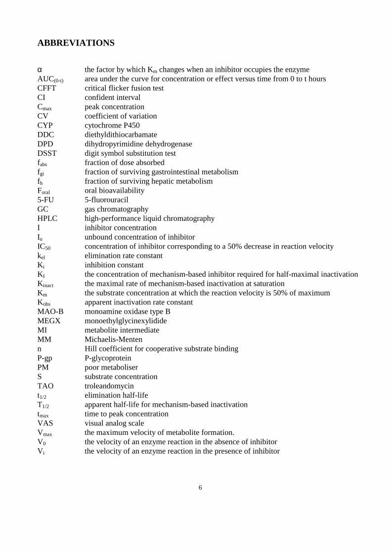
ABBREVIATIONS
α the factor by which Km changes when an inhibitor occupies the enzymeAUC(0-t) area under the curve for concentration or effect versus time from 0 to t hoursCFFT critical flicker fusion testCI confident intervalCmax peak concentrationCV coefficient of variationCYP cytochrome P450DDC diethyldithiocarbamateDPD dihydropyrimidine dehydrogenaseDSST digit symbol substitution testfabs fraction of dose absorbedfgi fraction of surviving gastrointestinal metabolismfh fraction of surviving hepatic metabolismForal oral bioavailability5-FU 5-fluorouracilGC gas chromatographyHPLC high-performance liquid chromatographyI inhibitor concentrationIu unbound concentration of inhibitorIC50 concentration of inhibitor corresponding to a 50% decrease in reaction velocitykel elimination rate constantKi inhibition constantKI the concentration of mechanism-based inhibitor required for half-maximal inactivationKinact the maximal rate of mechanism-based inactivation at saturationKm the substrate concentration at which the reaction velocity is 50% of maximumKobs apparent inactivation rate constantMAO-B monoamine oxidase type BMEGX monoethylglycinexylidideMI metabolite intermediateMM Michaelis-Mentenn Hill coefficient for cooperative substrate bindingP-gp P-glycoproteinPM poor metaboliserS substrate concentrationTAO troleandomycint1/2 elimination half-lifeT1/2 apparent half-life for mechanism-based inactivationtmax time to peak concentrationVAS visual analog scaleVmax the maximum velocity of metabolite formation.V0 the velocity of an enzyme reaction in the absence of inhibitorVi the velocity of an enzyme reaction in the presence of inhibitor
7
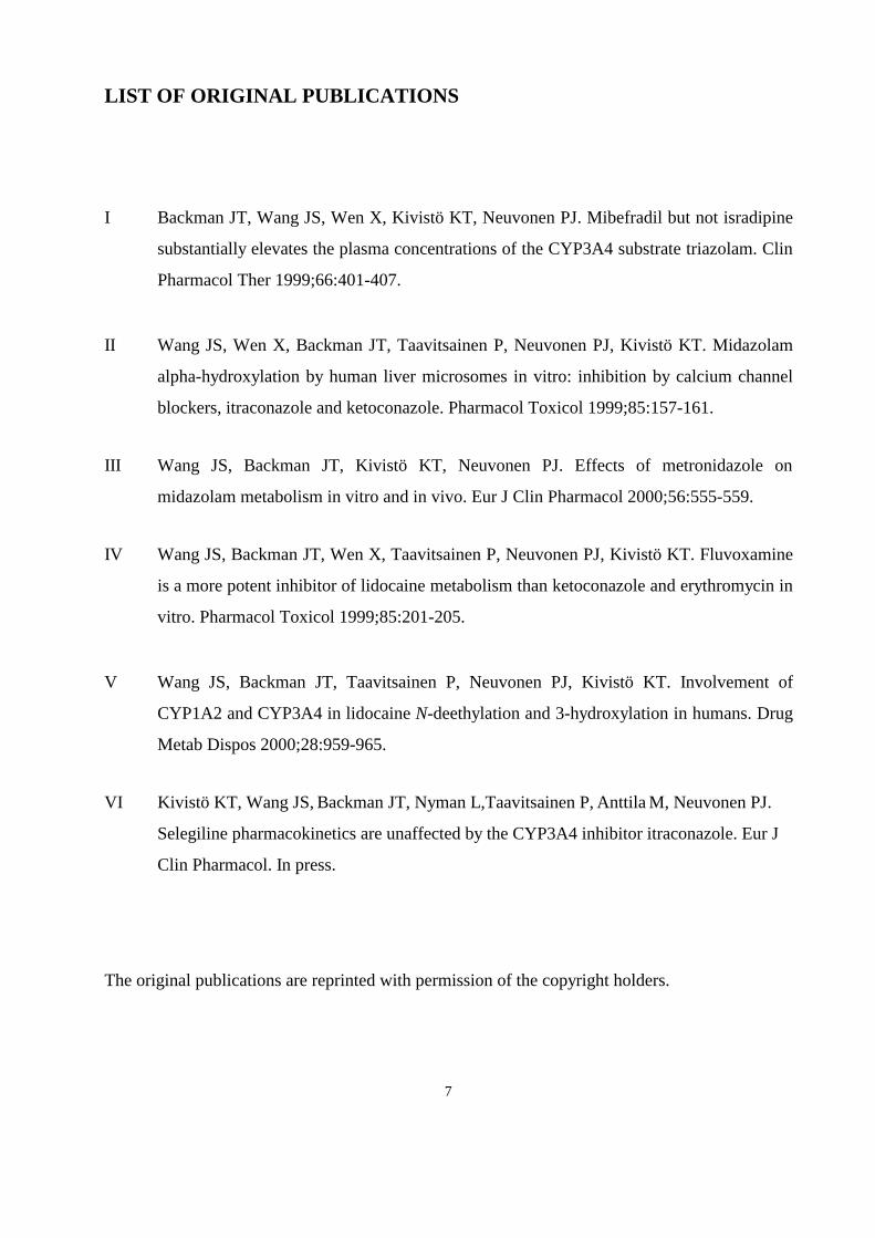
LIST OF ORIGINAL PUBLICATIONS
I Backman JT, Wang JS, Wen X, Kivistö KT, Neuvonen PJ. Mibefradil but not isradipine
substantially elevates the plasma concentrations of the CYP3A4 substrate triazolam. Clin
Pharmacol Ther 1999;66:401-407.
II Wang JS, Wen X, Backman JT, Taavitsainen P, Neuvonen PJ, Kivistö KT. Midazolam
alpha-hydroxylation by human liver microsomes in vitro: inhibition by calcium channel
blockers, itraconazole and ketoconazole. Pharmacol Toxicol 1999;85:157-161.
III Wang JS, Backman JT, Kivistö KT, Neuvonen PJ. Effects of metronidazole on
midazolam metabolism in vitro and in vivo. Eur J Clin Pharmacol 2000;56:555-559.
IV Wang JS, Backman JT, Wen X, Taavitsainen P, Neuvonen PJ, Kivistö KT. Fluvoxamine
is a more potent inhibitor of lidocaine metabolism than ketoconazole and erythromycin in
vitro. Pharmacol Toxicol 1999;85:201-205.
V Wang JS, Backman JT, Taavitsainen P, Neuvonen PJ, Kivistö KT. Involvement of
CYP1A2 and CYP3A4 in lidocaine N-deethylation and 3-hydroxylation in humans. Drug
Metab Dispos 2000;28:959-965.
VI Kivistö KT, Wang JS, Backman JT, Nyman L,Taavitsainen P, Anttila M, Neuvonen PJ.
Selegiline pharmacokinetics are unaffected by the CYP3A4 inhibitor itraconazole. Eur J
Clin Pharmacol. In press.
The original publications are reprinted with permission of the copyright holders.
ABSTRACT
8
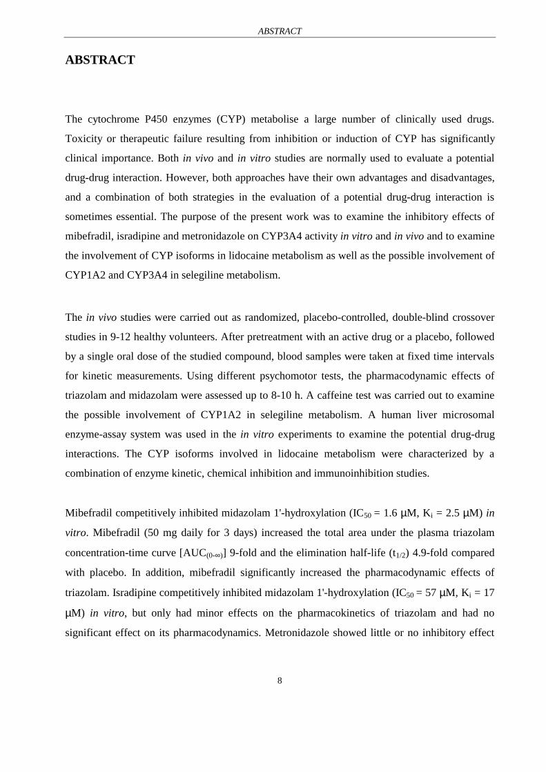
ABSTRACT
The cytochrome P450 enzymes (CYP) metabolise a large number of clinically used drugs.
Toxicity or therapeutic failure resulting from inhibition or induction of CYP has significantly
clinical importance. Both in vivo and in vitro studies are normally used to evaluate a potential
drug-drug interaction. However, both approaches have their own advantages and disadvantages,
and a combination of both strategies in the evaluation of a potential drug-drug interaction is
sometimes essential. The purpose of the present work was to examine the inhibitory effects of
mibefradil, isradipine and metronidazole on CYP3A4 activity in vitro and in vivo and to examine
the involvement of CYP isoforms in lidocaine metabolism as well as the possible involvement of
CYP1A2 and CYP3A4 in selegiline metabolism.
The in vivo studies were carried out as randomized, placebo-controlled, double-blind crossover
studies in 9-12 healthy volunteers. After pretreatment with an active drug or a placebo, followed
by a single oral dose of the studied compound, blood samples were taken at fixed time intervals
for kinetic measurements. Using different psychomotor tests, the pharmacodynamic effects of
triazolam and midazolam were assessed up to 8-10 h. A caffeine test was carried out to examine
the possible involvement of CYP1A2 in selegiline metabolism. A human liver microsomal
enzyme-assay system was used in the in vitro experiments to examine the potential drug-drug
interactions. The CYP isoforms involved in lidocaine metabolism were characterized by a
combination of enzyme kinetic, chemical inhibition and immunoinhibition studies.
Mibefradil competitively inhibited midazolam 1'-hydroxylation (IC50 = 1.6 µM, Ki = 2.5 µM) in
vitro. Mibefradil (50 mg daily for 3 days) increased the total area under the plasma triazolam
concentration-time curve [AUC(0-∞)] 9-fold and the elimination half-life (t1/2) 4.9-fold compared
with placebo. In addition, mibefradil significantly increased the pharmacodynamic effects of
triazolam. Isradipine competitively inhibited midazolam 1'-hydroxylation (IC50 = 57 µM, Ki = 17
µM) in vitro, but only had minor effects on the pharmacokinetics of triazolam and had no
significant effect on its pharmacodynamics. Metronidazole showed little or no inhibitory effect
ABSTRACT
9
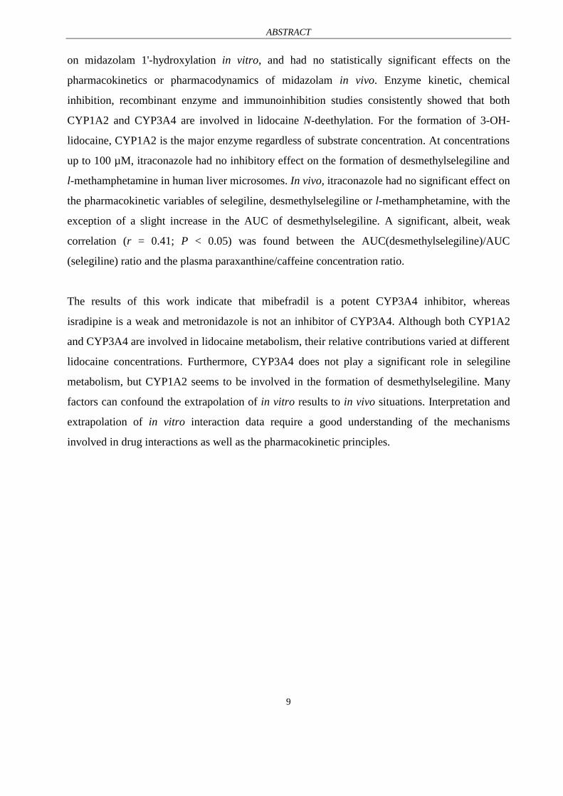
on midazolam 1'-hydroxylation in vitro, and had no statistically significant effects on the
pharmacokinetics or pharmacodynamics of midazolam in vivo. Enzyme kinetic, chemical
inhibition, recombinant enzyme and immunoinhibition studies consistently showed that both
CYP1A2 and CYP3A4 are involved in lidocaine N-deethylation. For the formation of 3-OH-
lidocaine, CYP1A2 is the major enzyme regardless of substrate concentration. At concentrations
up to 100 µM, itraconazole had no inhibitory effect on the formation of desmethylselegiline and
l-methamphetamine in human liver microsomes. In vivo, itraconazole had no significant effect on
the pharmacokinetic variables of selegiline, desmethylselegiline or l-methamphetamine, with the
exception of a slight increase in the AUC of desmethylselegiline. A significant, albeit, weak
correlation (r = 0.41; P < 0.05) was found between the AUC(desmethylselegiline)/AUC
(selegiline) ratio and the plasma paraxanthine/caffeine concentration ratio.
The results of this work indicate that mibefradil is a potent CYP3A4 inhibitor, whereas
isradipine is a weak and metronidazole is not an inhibitor of CYP3A4. Although both CYP1A2
and CYP3A4 are involved in lidocaine metabolism, their relative contributions varied at different
lidocaine concentrations. Furthermore, CYP3A4 does not play a significant role in selegiline
metabolism, but CYP1A2 seems to be involved in the formation of desmethylselegiline. Many
factors can confound the extrapolation of in vitro results to in vivo situations. Interpretation and
extrapolation of in vitro interaction data require a good understanding of the mechanisms
involved in drug interactions as well as the pharmacokinetic principles.
INTRODUCTION
10
INTRODUCTION
The cytochrome P450 (CYP) enzymes constitute a superfamily of isoforms that play an
important role in the oxidative metabolism of drugs, carcinogens and toxic chemicals (Gonzalez
1992). Individual CYP isoforms have considerable but overlapping substrate specificities. The
potential for drug-drug interactions exists whenever there are coadministered two or more drugs
that are metabolized via the same CYP isoform (Lin & Lu 1998). These kinds of drug-drug
interactions may have significant clinical implications, e.g. toxicity or therapeutic failure
resulting from inhibition or induction of the relevant CYP enzymes.
Human studies provide the most definitive data on the probability and magnitude of drug
interactions in clinical therapeutics (von Moltka et al. 1998). However, such in vivo studies also
have limitations and drawbacks. For example, it is difficult to characterize the inhibitory potency
of an agent toward a specific CYP isoform. It is also difficult to characterize the role of a CYP
isoform involved in a metabolic pathway of drug oxidation. Systems for in vitro experimentation
include microsomes, liver slices, hepatocytes, cell lines and expressed enzymes. With this
information, prediction of the likelihood of in vivo drug-drug interactions is possible. Among the
in vitro experimentation, human liver microsomes appear to be the most applicable in vitro
system for the purpose of in vitro to in vivo scaling (Bertz & Granneman 1997).
Although in vitro experiments can provide useful information regarding drug interactions,
extrapolation of in vitro data to in vivo situations is far from straightforward (Lin & Lu 1998).
Factors such as extrahepatic drug metabolism, uncertainty of drug concentrations at the enzyme
catalytic site and the complexity of inhibition mechanisms make it difficult to accurately predict
the in vivo situation from in vitro data. Thus, in view of the advantages and disadvantages of the
in vivo and in vitro systems, a combination of both strategies in the evaluation of potential drug-
drug interactions of a medicine is essential.
Mibefradil and isradipine are novel calcium channel blockers. Metronidazole is a 5-
INTRODUCTION
11
nitroimidazole antimicrobial drug. These drugs are metabolised to some extent by CYP enzymes
and may be potential CYP inhibitors. Lidocaine is a widely used local anaesthetic and
antiarrhythmic drug. The principal metabolic pathway of lidocaine in humans is oxidative N-
deethylation to monoethylglycinexylidide (MEGX) (Hermansson et al. 1980). The MEGX test,
i.e. plasma MEGX concentrations after i.v. lidocaine, has been used to evaluate in vivo activity of
hepatic CYP3A4 (Azoulay et al. 1993, Cakaloglu et al. 1994). However, there are controversial
results regarding the CYP isoforms involved in lidocaine metabolism. Selegiline is an
irreversible inhibitor of monoamine oxidase type B (MAO-B). The specific CYP isoforms
involved in selegiline metabolism have not yet been conclusively identified.
In this study, the effects of mibefradil, isradipine and metronidazole on CYP3A4-mediated in
vitro and in vivo metabolism of midazolam and triazolam were examined. In addition, the
specific CYP enzymes involved in lidocaine and selegiline metabolism were characterized with
in vitro experiments and, in the case of selegiline, with in vivo pharmacokinetic studies in healthy
volunteers. Furthermore, the principles and possibilities for semi-quantitative prediction of drug-
drug interactions in vivo based on in vitro experimental data were also explored in this study.
REVIEW OF THE LITERATURE
12
REVIEW OF THE LITERATURE
1 Drug metabolism and CYP
1.1 Drug metabolism
Most drugs are lipophilic and can not be easily eliminated from the body unless they are
metabolised to more hydrophilic derivatives (Nelson et al. 1996). A large number of enzymes
exist which metabolise foreign compounds. They have been classified as belonging to phase I or
phase II pathways of metabolism. Phase I enzymes include oxidases, hydrolases and reductases,
while the phase II enzymes are all transferases (Gonzalez & Idle 1994). Among the large number
of Phase I enzymes, the CYPs (cytochrome P450s) are the most important superfamily of
enzymes which account for the majority of oxidative biotransformations of xenobiotics and
endogenous biochemicals (Gonzalez 1992). The phase II transferases serve to transfer various
water-soluble molecules such as glucuronic acid, sulfonic acid and glutathione to other chemicals
(Gonzalez & Idle 1994). Often, drugs undergo both phase I and phase II reactions sequentially,
but excretion after only one metabolic reaction is also possible (Krishna & Klotz 1994).
Although liver is the main site of drug metabolism, it has recently become clear that intestinal
drug metabolism has a large influence on presystemic or first-pass drug biotransformation (Hall
et al. 1999). However, both the protein level and catalytic activity of drug-metabolising enzymes
in the small intestine are generally lower than those in the liver, and this is particularly true for
CYP enzymes (Peters & Kremers 1989, Shimada et al. 1994). Therefore, the contribution of the
small intestine to the overall metabolism of a drug is likely to be quantitatively less important
than that of the liver (Lin et al. 1999). In addition to the liver and intestine, drug metabolism
occurs also in organs such as the lung, skin and kidney (Krishna & Klotz 1994).
1.2 CYP
REVIEW OF THE LITERATURE
13
The CYP enzymes represent a superfamily of enzymes that are defined on the basis of
similarities between their amino acid and DNA sequences (Gonzalez 1992, Nelson et al. 1996).
In humans, three CYP gene families (i.e. CYP1, CYP2 and CYP3) are thought to be responsible
for most drug metabolism, and 7 isoforms, i.e. CYP1A2, CYP2A6, CYP2C8/9, CYP2C19,
CYP2D6, CYP2E1 and CYP3A4 account for most of the identified CYP-mediated metabolism.
One of the two enzymes in the CYP1A subfamily is CYP1A2. The other member, CYP1A1, is
expressed in the lungs and placentas of cigarette smokers, and is not believed to contribute to
drug metabolism in vivo (Brosen 1995). There is large inter- and intraindividual variation in
CYP1A2 activity, and cigarette smoking causes marked induction of this enzyme. The
expression of CYP1A2 is not associated with genetic polymorphism in Caucasian populations
(Welfare et al. 1999). In contrast, it is polymorphically expressed in Japanese and Chinese
populations (Chida et al. 1999, Ou-Yang et al. 1999). CYP1A2 is the major enzyme involved in
the metabolism of imipramine, propranolol, clozapine, theophylline, phenacetin, and caffeine
(Pelkonen et al. 1998). It is also the primary enzyme responsible for activating heterocyclic
arylamine food mutagens (Ereshefsky et al. 1995).
CYP2A6 is a quantitatively minor component of the human hepatic CYP, comprising 1 to 5% of
the total CYP protein in liver (Kharasch et al. 2000). CYP2A6 is polymorphically expressed in
humans. Several variant alleles have been identified to contribute to the PM phenotype of
CYP2A6 in populations (Pelkonen et al. 2000, Oscarson 2001). CYP2A6 activity is not only
modified by the polymorphisms in the CYP2A6 gene, but also modified by certain antiepileptic
agents (Sotaniemi et al. 1995) and environmental factors (Pasanen et al. 1997). CYP2A6 is
involved in the metabolism of coumarin, nicotine, SM-12502, chlormethiazole, methoxyflurane,
halothane, valproic acid and disulfiram (Pelkonen et al. 2000, Oscarson 2001). In addition, it
activates a number of procarcinogenes e.g., aflatoxin B1, 1.3-butadiene, 2,6-dichlorobebzonitrile
and several nitrosoamines (Pelkonen et al. 2000, Oscarson 2001).
CYP2C8 and CYP2C9 are the two major members of the human CYP2C subfamily. They
account for 35% and 60%, respectively, of total human CYP2C, whereas CYP2C18 (4%) and
REVIEW OF THE LITERATURE
14
CYP2C19 (1%) are the minor isoforms (Ged et al. 1988, Romkes et al. 1991). CYP2C9 exhibits
genetic polymorphism (Miners & Birkett 1998). Individual homozygous for the Leu359 allele are
associated with PM of CYP2C9. Approximately 1% of individuals in Caucasian populations are
the PM phenotype (Vermeij et al. 1988, Inaba 1990, Miners & Birkett 1998). Another member of
the CYP2C subfamily, CYP2C19, also exhibits genetic polymorphism. The incidence of the PM
phenotype in populations of different racial origin varies, being approximately 2 to 6% of
individuals in Caucasian populations, and 18 to 22% of individuals in Asian populations (Horai
1989, Kalow et al. 1994). Despite the high level of sequence similarity, the human CYP2C
isoforms exhibit relatively little overlap of substrate specificity (Wrighton & Stevens 1992,
Goldstein & de Morais 1994). For example, CYP2C9 is involved in the metabolism of
tolbutamide, phenytoin, S-warfarin, torasemide and fluoxetine (Miners & Birkett 1998), whereas
CYP2C19 is involved in the metabolism of S-mephenytoin, diazepam, propranolol, proguanil,
omeprazole, imipramine, clomipramine and amitriptyline (Flockhart 1995, Pelkonen et al. 1998).
One of the CYP2D subfamily, CYP2D6, is also polymorphically expressed in humans and
therefore it is connected to several clinical implications. Approximately 5 to 10% of individuals
in Caucasian populations but only 1 to 2% of those in Asia populations are of the PM phenotype
(Alvan et al. 1990, Bertilsson et al. 1992). Some ultrarapid metabolisers of CYP2D6 substrates
have been found in Swedish populations (Johansson et al. 1993, Dahl et al. 1995) and with a high
frequency (29%) in an Ethiopian population (Aklillu et al. 1995). This is due to a gene
amplification. More than 50 drugs, including antidepressants, antipsychotics and cardiovascular
drugs, are known to be metabolised primarily by CYP2D6 (Pelkonen et al. 1998).
Another CYP enzyme, CYP2E1, is involved in the metabolic activation of many low-molecular-
weight toxins and carcinogens (Guengerich & Shimada 1991). Furthermore, it oxidises formation
of acetaldehyde from ethanol and metabolises limited number of drugs, i.e. paracetamol,
chlorzoxazone and volatile anaesthetics (Bertz & Granneman 1997).
Three CYPs belong to the CYP3A subfamily: two adult forms designated CYP3A4, CYP3A5,
and a fetal liver form, CYP3A7. CYP3A4 is the most abundant CYP isoform, comprising about
REVIEW OF THE LITERATURE
15
30% and 70% of the total CYP protein in liver (Guengerich et al. 1986, Shimada et al. 1994) and
in the small intestinal epithelial cells (Watkins et al. 1987, Kolars et al. 1992), respectively. It is
thought that CYP3A5 may be subject to a genetic polymorphism since it is found only in about
10 - 30% of liver specimens and at levels 10 - 30% of CYP3A4 (Wrighton et al. 1989, 1990). At
present, CYP3A5 can not be distinguished from the other CYP3A isoforms on the basis of
substrate specificity although some differences in regioselective hydroxylation of cyclosporin has
been demonstrated (Aoyama et al. 1989). The third functional CYP3A isoform is CYP3A7,
which is expressed in fetal liver. However, it may also be selectively expressed in adult livers at
lower levels than CYP3A4 and CYP3A5 (Schuetz et al. 1994). The CYP3A subfamily
metabolises a large number of clinically used drugs. It has been estimated that more than 50% of
drugs used in humans are metabolised to some degree by CYP3A4 (Bertz & Granneman 1997).
2 Characterization of CYP enzymes involved in drug biotransformation
A change in the metabolism of one drug by the coadministration of another is a frequent cause of
clinically important drug interactions. This can result from the induction of a CYP enzyme,
which accelerates drug metabolism and decreases plasma drug concentrations, or from inhibition,
which results in elevated plasma drug concentrations with increased potential for enhanced
effects (Dresser et al. 2000). Characterization of the role of an individual CYP in a given
metabolic pathway is essential in determining and predicting the potential for drug interactions.
Over the past decade, a great deal of information on human CYP and phase II enzymes at the
molecular level has become available (Burchell et al. 1991, Gonzalez et al. 1991, Nelson et al.
1993, 1996). This information, with the availability of antibodies and probe substrates, has made
it possible to determine which CYP isoform(s) is (are) responsible for a specific reaction of a
drug in vivo and in vitro.
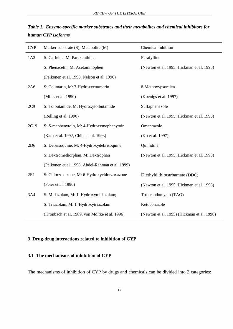
To characterize CYP enzymes involved in drug biotransformation in vitro, enzyme kinetic,
chemical inhibition, and immunoinhibition studies with human liver microsomes are normally
used (Lin & Lu 1997). An enzyme kinetic study is a common mathematical approach to analyse
the relationship between the rate of metabolite formation and the substrate concentration (Segel
REVIEW OF THE LITERATURE
16
1975). In many cases, such data can be analysed by the classical Michaelis-Menten (MM)
equation (equation 1), yielding two kinetic parameters: Vmax (the maximum velocity of
metabolite formation) and Km (the substrate concentration at which the reaction velocity is 50%
of maximum):
v0 = Vmax ⋅ S / (Km + S) (1)
where v0 is the velocity of metabolite formation in the absence of inhibitor and S is the substrate
concentration. However, in some cases, a simple MM equation is not applicable, and multiple
enzyme models are required to fit the kinetic data (Schmider et al. 1996). The chemical
inhibition approach uses a series of chemicals (see table 1) known to have a relatively high
specificity as inhibitors of human CYP isoforms (Newton et al. 1995, Eagling et al. 1998). The
immunoinhibition method uses specific CYP-antibodies that inhibit the activity of a particular
human CYP (Yang et al. 1999).
In addition to the approches mentioned above, several other in vitro approches are also available,
including: demonstration of catalytic activity in recombinant human CYP isoforms, correlation
of a metabolic activity with markers (see table 1) for known CYP enzymes, and studies with
purified enzyme (Lin & Lu 1997). Each in vitro approach has its own advantages and
disadvantages, and a combination of several in vitro approaches is usually used to identify which
CYP isoform(s) is (are) responsible for a metabolic reaction (Lin & Lu 1997).
One important factor which needs to be noted in this context is the substrate concentration used.
It has been reported that contributions of several CYP enzymes toward oxidations of xenobiotics
in vitro are affected by the substrate concentrations used (Yamazaki et al. 1994, 1997,
Venkatakrishnan et al. 1998). Therefore, the use of clinically relevant substrate concentrations in
in vitro studies is important to correctly predict and define the involved CYP isoform(s) (Lin &
Lu 1998, Wang et al. 2000).
REVIEW OF THE LITERATURE
17
Table 1. Enzyme-specific marker substrates and their metabolites and chemical inhibitors for
human CYP isoforms
CYP Marker substrate (S), Metabolite (M) Chemical inhibitor
1A2 S: Caffeine, M: Paraxanthine;
S: Phenacetin, M: Acetaminophen
(Pelkonen et al. 1998, Nelson et al. 1996)
Furafylline
(Newton et al. 1995, Hickman et al. 1998)
2A6 S: Coumarin, M: 7-Hydroxycoumarin
(Miles et al. 1990)
8-Methoxypsoralen
(Koenigs et al. 1997)
2C9 S: Tolbutamide, M: Hydroxytolbutamide
(Relling et al. 1990)
Sulfaphenazole
(Newton et al. 1995, Hickman et al. 1998)
2C19 S: S-mephenytoin, M: 4-Hydroxymephenytoin
(Kato et al. 1992, Chiba et al. 1993)
Omeprazole
(Ko et al. 1997)
2D6 S: Debrisoquine, M: 4-Hydroxydebrisoquine;
S: Dextromethorphan, M: Dextrophan
(Pelkonen et al. 1998, Abdel-Rahman et al. 1999)
Quinidine
(Newton et al. 1995, Hickman et al. 1998)
2E1 S: Chlorzoxazone, M: 6-Hydroxychlorzoxazone
(Peter et al. 1990)
Diethyldithiocarbamate (DDC)
(Newton et al. 1995, Hickman et al. 1998)
3A4 S: Midazolam, M: 1'-Hydroxymidazolam;
S: Triazolam, M: 1'-Hydroxytriazolam
(Kronbach et al. 1989, von Moltke et al. 1996)
Ttroleandomycin (TAO)
Ketoconazole
(Newton et al. 1995) (Hickman et al. 1998)
3 Drug-drug interactions related to inhibition of CYP
3.1 The mechanisms of inhibition of CYP
The mechanisms of inhibition of CYP by drugs and chemicals can be divided into 3 categories:
REVIEW OF THE LITERATURE
18
reversible inhibition, quasi-irreversible inhibition and irreversible inhibition (Halpert 1995, Lin
and Lu 1998, Thummel & Wilkinson 1998). Reversible inhibition is probably the most common
mechanism responsible for the documented drug interactions. Kinetically, reversible inhibition
can be classified as competitive, noncompetitive, uncompetitive and mixed type inhibition (Segel
1975, von Moltke et al. 1996, Lin & Lu 1998). For competitive inhibition, the inhibitor and
substrate compete for the same active site of free enzyme. In the case of uncompetitive
inhibition, the inhibitor does not bind the free enzyme, but binds to the enzyme-substrate
complex, and again the enzyme-substrate-inhibitor complex is nonproductive (Segel 1975, Lin &
Lu 1997, 1998). However, in practice, with two-substrate reactions, it is frequently found that for
a given inhibitor concentration, the inhibition observed is of a mixed type, i.e. inhibitor can bind
both to the free enzyme and to the enzyme-substrate complex. Such behavior is called mixed-
type inhibition (Segel 1975). A special case of mixed-type inhibition is noncompetitive inhibition
in which the inhibitor binds to another site of the enzyme and the inhibitor has no effect on
binding of substrate, but the enzyme-substrate-inhibitor complex is nonproductive.
Mathematically, the velocity of an enzymatic reaction in the absence (v0) of inhibitor can be
described by equation 1 (section 2) and in the presence (vi) of inhibitor it can be described by
equations 2, 3, 4 and 5 for competitive, noncompetitive, uncompetitive and mixed type
inhibition, respectively (Segel 1975).
vi = Vmax ⋅ S / [Km (1 + I / Ki) + S] (2)
vi = Vmax ⋅ S / [Km (1 + I / Ki) + S (1 + I / Ki)] (3)
vi = Vmax ⋅ S / [Km + S (1 + I / Ki)] (4)
vi = Vmax ⋅ S / [Km (1 + I / Ki) + S (1 + I / αKi)] (5)
where Ki is the inhibition constant of the inhibitor, I is the inhibitor concentration and α is the
factor by which Km changes when an inhibitor occupies the enzyme.
Quasi-irreversible inhibitors required at least one cycle of the CYP catalytic process to form
reactive intermediates. These intermediates can form stable complexes with the prosthetic heme
group of CYP, called metabolic intermediate (MI) complex, and sequentially the CYP is
inactivated (Lin & Lu 1998, Thummel & Wilkinson 1998). The formation of the MI complex can
REVIEW OF THE LITERATURE
19
be reversed, and CYP activity can be restored through an in vitro dialysis method (Ma et al.
2000). However, in in vivo conditions, the MI complexes are so stable that it is impossible to
restore the CYP activity, and resynthesis of new enzyme is the only means by which the enzyme
activity can be restored (Lin and Lu 1998, Thummel & Wilkinson 1998). Therefore, this type of
inhibition is called quasi-irreversible.
The clinical relevance of the inhibitory effect of a quasi-irreversible inhibitor may be
underestimated under in vitro conditions. For example, erythromycin, a weak inhibitor of
CYP3A4 in vitro with a high Ki value (16-194 µM) (Pichard et al. 1990, Wrighton et al. 1994,
Lampen et al. 1995, Echizen et al. 1993, Christians et al. 1996), was found to be a potent
inhibitor of CYP3A4 in vivo. Furthermore, it can cause clinically important drug interactions
with CYP3A4 substrates, e.g. tacrolimus (Shaeffer et al. 1994), midazolam (Olkkola et al. 1993),
triazolam (Phillips et al. 1986), cyclosporine (Campana et al. 1996), buspirone (Kivistö et al.
1997), simvastatin (Kantola et al. 1998) and terfenadine (Honig et al. 1992).
Drugs associated with MI complex formation include macrolides like troleadomycin (TAO)
(Pessayre et al. 1981), erythromycin (Babany et al. 1988), clarithromycin (Tinel et al. 1989,
Ohmori et al. 1993), roxithromycin (Tinel et al. 1989) and dirithromycin (Lindstrom et al. 1993);
antidepressants such as fluoxetine, nortriptyline, imipramine and desipramine (Franklin 1995,
Bensoussan et al. 1995, Murray & Field 1992); the anticholinergic agent orphenadrine (Reidy et
al. 1989); and various miscellaneous drugs tiamulin, diltiazem, lidocaine, tamoxifen, and
amiodarone (Larrey et al. 1986, Bensoussan et al. 1995, Witkamp et al. 1995, Thummel &
Wilkinson 1998).
Irreversible inhibition is also called mechanism-based or suicide inhibition. The mechanism of
suicide inhibition involves the formation of CYP-mediated formation of a reactive inhibitor that
covalently binds to the enzyme leading to its inactivation. Several criteria are necessary to prove
that an inhibitor behaves in a mechanism-based fashion. These include: (1) time- and
concentration dependent inactivation, (2) NADPH and inhibitor dependence, (3) substrate
protection, (4) lack of an effect of superoxide dismutase and catalase, (5) lack of an effect of
REVIEW OF THE LITERATURE
20
exogenous nucleophiles, (6) irreversibility of the inactivation, and (7) a 1:1 stoichiometry in the
addition of the reactive species to the apoprotein, the heme, or both (Regal et al. 2000).
Mechanism-based inhibition is potentially harmful, since resynthesis of the new enzyme is the
only approach to restore the enzyme activity, and the inhibitory effects of mechanism-based
inhibitors remain after the elimination of the inhibitor from blood and tissue (Ohyama et al.
2000). The fatal drug-drug interaction between sorivudine, an antiviral drug, and 5-fluorouracil
(5-FU) has been shown to be caused by mechanism-based inhibition of dihydropyrimidine
dehydrogenase (DPD) by sorivudine (Kanamitsu et al. 2000). Therefore, extensive in vitro
studies should be carried out during the drug-developing phase so that harmful in vivo drug-drug
interactions can be prevented (Ohyama et al. 2000).
Numerous chemicals have been identified as mechanism-based inactivators of CYP, among
which furafylline, diethyldithiocarbamate (DDC) and methoxsalen are widely used as in vitro
tools for selective inhibition of CYP1A2, CYP2E1 and CYP2A6 activities, respectively (Newton
et al. 1995, Kharasch et al. 2000). A 15 to 30 minutes preincubation of these inhibitors with
microsomal enzymes in the presence of NADPH is necessary to produce optimal inhibitory
effects in vitro.
3.2 Characterization of in vitro potency of inhibition
Analysis of the relation between inhibitor concentration and reaction velocity decrement may
yield an estimated concentration of inhibitor corresponding to a 50% decrease in reaction
velocity (IC50). The IC50 values are quite useful when comparing the relative inhibitory potency
of different candidate inhibitors. However, an IC50 value is unsuitable to be used in the in vitro-in
vivo scaling, because it only equals the enzyme inhibition constant (Ki) in the case of
noncompetitive inhibition (von Moltke et al. 1998). To any other types of inhibition, an IC50
value is generally higher than the respective Ki value, and therefore may result in an
underestimation of the in vivo inhibitory effect of an inhibitor.
REVIEW OF THE LITERATURE
23
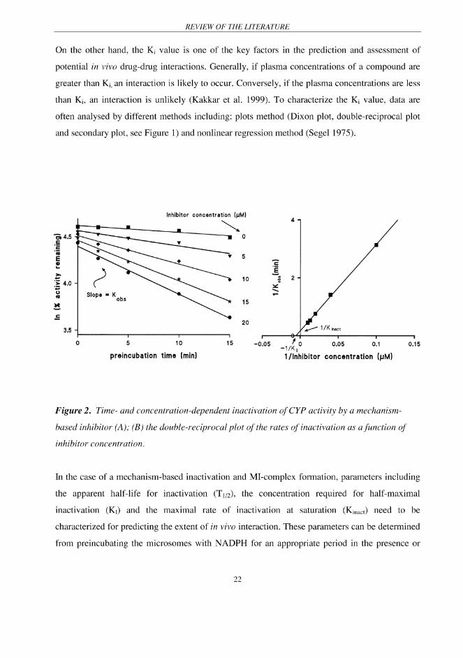
absence of various concentrations of an inhibitor. The apparent inactivation rate constant (Kobs)
can be determined from the slope of log-linear phase of enzymatic activity against preincubation
time. The KI and Kinact can be calculated from double-reciprocal plot of the rate of inactivation of
metabolite formation as a function of inhibitor concentration (Figure 2). The intercept on the
ordinate gives 1/Kinact. If the line is extrapolated to the abscissa, the intercept is -1/KI (Kent et al.
1999, Sinal et al. 1998, Koenigs et al. 1997). The relationship between T1/2, KI and Kinact is
described by the following equation (Lin & Lu 1998):
T1/2 = 0.693 (1 + KI / I) /Kinact (6)
3.3 In vitro-in vivo correlation
One of the main objectives of in vitro drug-drug interaction studies is the quantitative prediction
of in vivo drug metabolism from in vitro data. Different models have been suggested for
estimation of in vivo situations based on in vitro results (Bertz & Granneman 1997, Ito et al.
1998a, 1998b, Lin & Lu 1997, 1998). For in vitro-in vivo extrapolation involving metabolic
inhibition, when the substrate concentration is much lower than the Km value (Km >> S), the
degree of inhibition (R) can be simply expressed, independent of inhibition type, except in the
case of uncompetitive inhibition by the following equation (Tucker 1992, Pelkonen et al. 1998):
R = 1 / (1 + Iu / Ki) (7)
where Iu is the unbound concentration of inhibitor around enzyme site. When the substrate
concentration is similar to or even higher than the Km value, the degree of inhibition can be
calculated from the following equation assuming the mechanism of inhibition is competitive.
R = vi / v0 = (Km + S) / [Km (1 + Iu / Ki ) + S ] (8)
3.4 Factors affecting in vitro-in vivo extrapolation
Although impressive in vitro / in vivo concordance has been reported for individual cases,
quantitative prediction of in vivo situations from in vitro data is far from straightforward (Lin &
Lu 1998). As described above (section 3.3), theoretically, in vivo drug interactions based on the
inhibition of hepatic metabolism can be predicted by the Ki value and the Iu values. However, Iu
REVIEW OF THE LITERATURE
24
values can not be directly measured in vivo. Therefore, analyses have been based on the
assumption that the free drug concentrations in liver and plasma are equal. However, this
hypothesis is not supported by considerable data indicating that the liver concentrations of many
lipophilic compounds greatly exceed unbound plasma concentrations (Lin et al. 1982, Harashima
et al. 1984, von Moltke et al. 1998). In these cases, the extent of the interaction may be
underestimated if plasma unbound concentrations of a drug are used for the prediction.
It should be noted that although an accurate prediction of in vivo drug interaction based on Iu
values has been achieved in several cases, e.g tolbutamide-sulphaphenazole, erythromycin-
cyclosporin, erythromycin-theophylline and quinidine-sparteine interactions (Ito et al. 1998a,
1998b), inaccurate predictions have also been reported between fluoxetine-imipramine,
ciprofloxacin-caffeine and omeprazole-diazepam using Iu values in the prediction model. On the
other hand, when the total plasma concentration of an inhibitor or the total plasma concentration
of an inhibitor multiplied by a liver:plasma partition ratio was used instead of Iu, accurate
predictions of in vivo drug interactions have been achieved using alprazolam, desipramine and
terfenadine as substrates and ketoconazole or selective serotonin reuptake inhibitors as inhibitors
(von Moltka et al. 1994a, 1994b, 1994c, 1995a, 1995b, 1996). In addition to the discordance
between liver and plasma inhibitor concentrations, recent studies also showed that some bound
form of a substrate or a chemical inhibitor is kinetically available for a catalytic reaction or
enzyme inhibition (Baba et al. 2000, Qiu et al. 2000, Li et al. 2000).
Another factor which needs to be considered in in vitro-in vivo extrapolation is the change of
hepatic blood flow. Drugs can be classified as enzyme-limited (low hepatic clearance) or flow-
limited (high-clearance) drugs according to their hepatic clearance (Wilkinson & Shand 1975).
The hepatic clearance of a 'high clearance' drug is highly dependent on the hepatic blood flow
(Lin & Lu 1998), therefore a change in the hepatic blood flow will result in a substantial change
in the in vivo hepatic clearance of a 'high clearance' drug. Conversely, for a 'low clearance' drug,
a change in the hepatic blood flow will have little effect on its hepatic clearance (Bertz &
Granneman 1997).
REVIEW OF THE LITERATURE
25
Extrahepatic drug metabolism may also hinder in vitro-in vivo extrapolation. For drugs with
substantial first-pass metabolism, the systemic bioavailability (F) is the product of the fractions
of the dose absorbed (fabs) and surviving gastrointestinal (fgi) and hepatic (fh) metabolism (Hall et
al. 1999).
F = fabs ⋅ fgi ⋅ fh (9)
It is known that CYP3A4 is highly expressed in the small intestine, and limits the systemic
availability of many drugs. In addition to CYP3A4, CYP2C and CYP2D6 are also expressed in
the duodenum and jejunum of humans (de Waziers et al. 1988, Zhang et al. 1999). Often the
fraction of the dose eliminated by this process is not known. Consequently, the prediction of an
in vivo inhibitory effect is confounded.
Further complications are caused by the P-glycoprotein (P-gp), a transmembrane protein, which
is widely expressed in the gastrointestinal tract, liver, kidney, and the blood-brain barrier (Yu
1999). It acts as a drug efflux pump and plays an important role in drug absorption, distribution
and elimination. Inhibition of P-gp mediated drug transport in the gut can dramatically increase
the systemic availability of an otherwise poorly absorbed drug. In a study conducted by Lown et
al. (1997), the intestinal P-gp rather than CYP3A4 was found to be a primary determinant of oral
cyclosporin bioavailability. Therefore, it is essential to consider the role of P-gp when
extrapolating in vitro results to an in vivo situation. However, this seems to be rather difficult.
In addition, the in vitro incubation conditions used may substantially affect the in vitro results.
Studies with human liver microsomes have revealed that in vitro results are highly dependent on
the assay conditions such as the buffer, ionic strength (Mäenpää et al. 1998), the microsomal
protein concentration and the substrate and inhibitor concentrations used (Lin & Lu 1998).
Further complications come from the design of the in vitro experiment (Bourrie et al. 1996, Bertz
& Richard 1997) and the interindividual variability in drug-metabolising enzyme activities
(Pelkonen & Breimer 1994).
In conclusion, although some researchers have demonstrated that the successful prediction of in
vivo inhibitory drug interactions based on in vitro data is possible in some cases, numerous
REVIEW OF THE LITERATURE
26
factors can foil prospective quantitative prediction. Interpreting in vitro results to in vivo
situations needs a good understanding of the underlying pharmacokinetic principles.
4 Drug-drug interactions related to induction of CYP
The most common mechanism for CYP induction is the de novo synthesis of new enzyme
molecules as a result of a transcriptional activation (Pelkonen et al. 1998). However, with the
exception of induction of CYP1A1, the molecular mechanism involved in CYP induction is not
fully understood. The induction of CYP1A1 is mediated by enhanced transcriptional activation
after the binding of an inducing agent to a cytosolic polycyclic aromatic hydrocarbon (Ah)
receptor (Porter & Coon 1991). However, some inducers appear to increase CYP protein levels
by posttranscriptional regulation (Lin & Lu 1998). For example, troleandomycin produces no
increase in the rate of CYP3A4 protein synthesis, but it decreases the rate of CYP3A4 protein
degradation by forming stable complexes with the enzyme (Thummel & Wilkinson 1998). In
vivo, induction of CYP isoforms may lead to a decrease in toxicity through acceleration of
detoxification, or to an increase in toxicity caused by increased formation of reactive metabolites
(Lin & Lu 1998).
5 Individual drugs studied
5.1 Midazolam
Midazolam is a water-soluble 1,4-benzodiazepine, which is used as a short-acting hypnotic. After
oral administration, midazolam is almost completely absorbed from the gastrointestinal tract
(Thummel et al. 1996). However, the oral bioavailability (Foral) of midazolam is only about 50%
because of extensive first-pass metabolism in the intestinal wall and liver (Allonen et al. 1981,
Olkkola et al. 1994, Gorski et al. 1998). Midazolam is 94-98% bound to plasma proteins and the
degree of binding is independent of the dose (Allonen et al. 1981, Greenblatt et al. 1984).
Midazolam is eliminated almost exclusively via metabolism and less than 1% of an oral dose is
excreted unchanged in the urine (Heizmann & Ziegler 1981, Wandel et al. 1994). Midazolam is
REVIEW OF THE LITERATURE
27
metabolised by CYP3A4 and CYP3A5 (Wandel et al. 1994) to 1'-hydroxymidazolam, 4-
hydroxymidazolam and 1,4-hydroxymidazolam, of which 1'-hydroxymidazolam is the primary
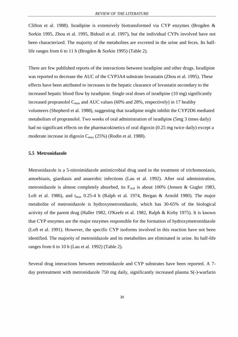
metabolic pathway (Thummel 1996) (Figure 3). The elimination half-life (t1/2) of midazolam
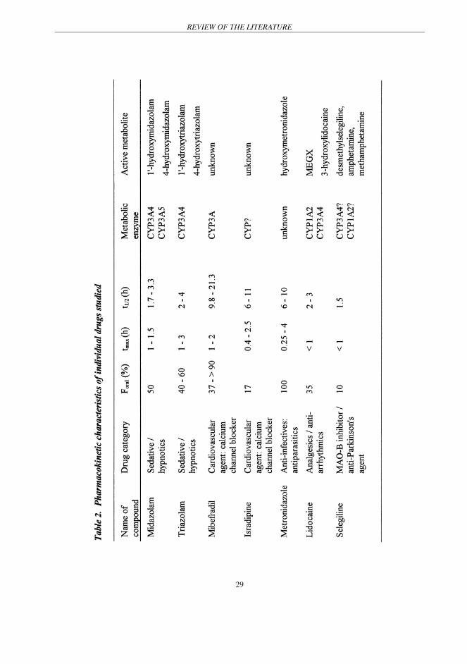
varies between 1.7 and 3.3 h in healthy volunteers (Table 2).
CYP3A4 inhibitors considerably increase midazolam concentrations and effects. The area under
the plasma concentration time curve (AUC) of oral midazolam was increased 16-fold by
ketoconazole and 11-fold by itraconazole (Olkkola et al. 1994). Erythromycin (Olkkola et al.
1993), clarithromycin (Yeates et al. 1996), fluconazole (Olkkola et al. 1996), diltiazem
(Backman et al. 1994), verapamil (Backman et al. 1994) and grapefruit juice (Kupferschmidt et
al. 1995) also significantly increase the AUC of midazolam, although to a lesser extent than
ketoconazole and itraconazole. On the other hand, the concentrations and effects of midazolam
are greatly decreased by CYP3A4 inducers, e.g. rifampicin, carbamazepine and phenytoin
(Backman et al. 1996a, 1996b).
5.2 Triazolam
Triazolam is also a short-acting benzodiazepine hypnotic, and it is closely related to midazolam
in chemical structure (Greenblatt et al. 1983). Like midazolam, triazolam undergoes extensive
first-pass metabolism, resulting in an Foral of 40-60% (Kroboth et al. 1995). Triazolam was found
to be 89% bound to plasma proteins (Eberts et al. 1981). Triazolam is extensively metabolised,
its major metabolites being 1'-hydroxy- and 4-hydroxytriazolam (Eberts et al. 1981) (Figure 3).
These metabolic pathways are mediated by CYP3A4 (Kronbach et al. 1989, von Moltke et al.
1996). The t1/2 of triazolam is 2 to 4 h (Pakes et al. 1981) (Table 2).
Triazolam plasma concentrations and effects are considerably increased by CYP3A4 inhibitors,
e.g. ketoconazole, itraconazole (Varhe et al. 1994), erythromycin (Phillips et al. 1986),
cimetidine (Friedman et al. 1988), diltiazem (Varhe et al. 1996a), fluconazole (Varhe et al.
1996b) and grapefruit juice (Hukkinen et al. 1995, Lilja et al. 2000). On the other hand, CYP3A4
inducers, e.g., rifampicin, markedly reduce triazolam plasma concentrations and its hypnotic
REVIEW OF THE LITERATURE
28
effects (Villikka et al. 1997).
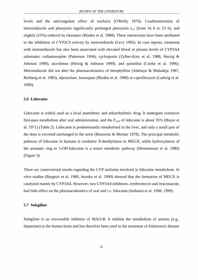
5.3 Mibefradil
Mibefradil selectively blocks T-type calcium channels in contrast to other calcium antagonists
which block only L-type channels (Brogden & Markham 1997). Its Cmax is reached 1 to 2 h after
oral administration (Petrie et al. 1995, Brogden & Markham 1997), and its Foral increases with
increasing oral doses, from 37% after 10mg to >90% after doses ≥160mg (Brogden & Markham
1997). Data regarding the metabolism of mibefradil available from animal studies suggest that it
is extensively metabolised by CYP enzymes in the liver (Skerjanec et al. 1996). Its t1/2 ranges
from 9.8 to 21.3 h (Brogden & Markham 1997) (Table 2).
In vitro studies suggest that CYP3A4, CYP2D6 and CYP1A2 may be competitively inhibited by
mibefradil (Brogden & Markham 1997). Indeed, mibefradil was reported to be associated with
several cases of harmful (sometimes even fatal) drug interactions which led to the withdrawal of
the compound from the market. Furthermore, mibefradil was reported to increase blood
concentrations of cyclosporin 2- to 3- fold in patients undergoing renal transplantation (Brogden
& Markham 1997). Coadministration of mibefradil with terfenadine (metabolised by CYP3A4)
to healthy volunteers led to elevated plasma concentrations of terfenadine, resulting in a mean
prolongation of the QTc interval by 20%. Significant drug interactions have also been reported
between mibefradil and astemizole, cisapride (Brogden & Markham 1997) and simvastatin
(Schmassmann-Suhijar et al. 1998).
5.4 Isradipine
Isradipine is a dihydropyridine derivative with potent calcium channel blocking activity (Hof et
al. 1984) used in the treatment of hypertension (Brogden & Sorkin 1995). Isradipine is subject to
a significant degree of first-pass metabolism, and the Foral of isradipine is only about 17% (Tse &
Jaffe 1987). After oral administration, its Cmax is attained within 0.4 to 2.5 h (Tse & Jaffe 1987,
REVIEW OF THE LITERATURE
30
Clifton et al. 1988). Isradipine is extensively biotransformed via CYP enzymes (Brogden &
Sorkin 1995, Zhou et al. 1995, Bidouil et al. 1997), but the individual CYPs involved have not
been characterized. The majority of the metabolites are excreted in the urine and feces. Its half-
life ranges from 6 to 11 h (Brogden & Sorkin 1995) (Table 2).
There are few published reports of the interactions between isradipine and other drugs. Isradipine
was reported to decrease the AUC of the CYP3A4 substrate lovastatin (Zhou et al. 1995). These
effects have been attributed to increases in the hepatic clearance of lovastatin secondary to the
increased hepatic blood flow by isradipine. Single oral doses of isradipine (10 mg) significantly
increased propranolol Cmax and AUC values (60% and 28%, respectively) in 17 healthy
volunteers (Shepherd et al. 1988), suggesting that isradipine might inhibit the CYP2D6 mediated
metabolism of propranolol. Two weeks of oral administration of isradipine (5mg 3 times daily)
had no significant effects on the pharmacokinetics of oral digoxin (0.25 mg twice daily) except a
moderate increase in digoxin Cmax (25%) (Rodin et al. 1988).
5.5 Metronidazole
Metronidazole is a 5-nitroimidazole antimicrobial drug used in the treatment of trichomoniasis,
amoebiasis, giardiasis and anaerobic infections (Lau et al. 1992). After oral administration,
metronidazole is almost completely absorbed, its Foral is about 100% (Jensen & Gugler 1983,
Loft et al. 1986), and tmax 0.25-4 h (Ralph et al. 1974, Bergan & Arnold 1980). The major
metabolite of metronidazole is hydroxymetronidazole, which has 30-65% of the biological
activity of the parent drug (Haller 1982, O'Keefe et al. 1982, Ralph & Kirby 1975). It is known
that CYP enzymes are the major enzymes responsible for the formation of hydroxymetronidazole
(Loft et al. 1991). However, the specific CYP isoforms involved in this reaction have not been
identified. The majority of metronidazole and its metabolites are eliminated in urine. Its half-life
ranges from 6 to 10 h (Lau et al. 1992) (Table 2).
Several drug interactions between metronidazole and CYP substrates have been reported. A 7-
day pretreatment with metronidazole 750 mg daily, significantly increased plasma S(-)-warfarin
REVIEW OF THE LITERATURE
31
levels and the anticoagulant effect of warfarin (O'Reilly 1976). Coadministration of
metronidazole and phenytoin significantly prolonged phenytoin t1/2 (from 16 h to 23 h), and
slightly (15%) reduced its clearance (Blyden et al. 1988). These interactions have been attributed
to the inhibition of CYP2C9 activity by metronidazole (Levy 1995). In case reports, treatment
with metronidazole has also been associated with elevated blood or plasma levels of CYP3A4
substrates: carbamazepine (Patterson 1994), cyclosporin (Zylber-Katz et al. 1988, Herzig &
Johnson 1999), tacrolimus (Herzig & Johnson 1999), and quinidine (Cooke et al. 1996).
Metronidazole did not alter the pharmacokinetics of theophylline (Adebayo & Mabadeje 1987,
Reitberg et al. 1983), alprazolam, lorazepam (Blyden et al. 1988) or ciprofloxacin (Ludwig et al.
1990).
5.6 Lidocaine
Lidocaine is widely used as a local anaesthetic and antiarrhythmic drug. It undergoes extensive
first-pass metabolism after oral administration, and the Foral of lidocaine is about 35% (Boyes et
al. 1971) (Table 2). Lidocaine is predominantly metabolised in the liver, and only a small part of
the dose is excreted unchanged in the urine (Benowitz & Meister 1978). The principal metabolic
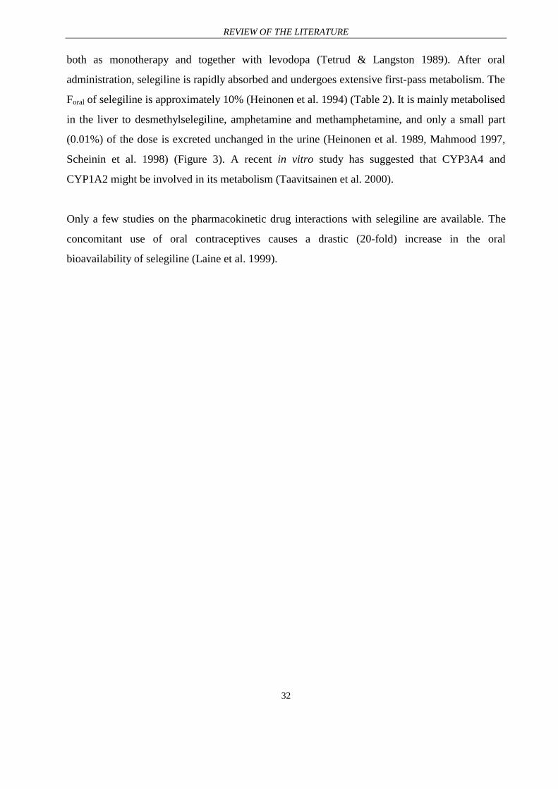
pathway of lidocaine in humans is oxidative N-deethylation to MEGX, while hydroxylation of
the aromatic ring to 3-OH-lidocaine is a minor metabolic pathway (Hermansson et al. 1980)
(Figure 3).
There are controversial results regarding the CYP isoforms involved in lidocaine metabolism. In
vitro studies (Bargetzi et al. 1989, Imaoka et al. 1990) showed that the formation of MEGX is
catalyzed mainly by CYP3A4. However, two CYP3A4 inhibitors, erythromycin and itraconazole,
had little effect on the pharmacokinetics of oral and i.v. lidocaine (Isohanni et al. 1998, 1999).
5.7 Selegiline
Selegiline is an irreversible inhibitor of MAO-B. It inhibits the metabolism of amines (e.g.,
dopamine) in the human brain and has therefore been used in the treatment of Parkinson's disease
REVIEW OF THE LITERATURE
32
both as monotherapy and together with levodopa (Tetrud & Langston 1989). After oral
administration, selegiline is rapidly absorbed and undergoes extensive first-pass metabolism. The
Foral of selegiline is approximately 10% (Heinonen et al. 1994) (Table 2). It is mainly metabolised
in the liver to desmethylselegiline, amphetamine and methamphetamine, and only a small part
(0.01%) of the dose is excreted unchanged in the urine (Heinonen et al. 1989, Mahmood 1997,
Scheinin et al. 1998) (Figure 3). A recent in vitro study has suggested that CYP3A4 and
CYP1A2 might be involved in its metabolism (Taavitsainen et al. 2000).
Only a few studies on the pharmacokinetic drug interactions with selegiline are available. The
concomitant use of oral contraceptives causes a drastic (20-fold) increase in the oral
bioavailability of selegiline (Laine et al. 1999).
AIMS OF THE STUDY
34
AIMS OF THE STUDY
The purpose of the present work was to examine the inhibitory effects of mibefradil, isradipine
and metronidazole on CYP3A4 activity in vivo and in vitro and the involvement of CYP
isoforms in lidocaine and selegiline metabolism.
In detail the aims were:
I To investigate the effects of mibefradil and isradipine on CYP3A4-mediated midazolam 1'-
hydroxylation in vitro (Study II) and on the pharmacokinetics and pharmacodynamics of
triazolam in vivo (Study I).
II To examine the effects of metronidazole on CYP3A4 activity in vitro and in vivo using
midazolam as a probe substrate (Study III).
III To determine the effects of the CYP3A4 inhibitors ketoconazole and erythromycin and the
CYP1A2 inhibitor fluvoxamine on the N-deethylation and 3-hydroxylation of lidocaine using
human liver microsomes (Study IV). To examine the relative contributions of different CYP
isoforms to the N-deethylation and 3-hydroxylation of lidocaine using chemical inhibition,
immunoinhibition and recombinant enzymes studies (Study V).
IV To investigate the effects of itraconazole, a potent CYP3A4 inhibitor, on the metabolism of
selegiline in vitro and in vivo. To study the role of CYP1A2 in selegiline metabolism using
caffeine test as an index of in vivo CYP1A2 activity (Study VI).
MATERIALS AND METHODS
35
MATERIALS AND METHODS
1 In vivo studies
1.1 Subjects
All the subjects were healthy adult (20-32 years) volunteers. A total of 31 volunteers (14 females,
17 males) participated in the studies (Table 3). Two subjects participated in two different studies.
Before entering each study, volunteers were ascertained to be healthy by scrutinizing their
respective medical histories, physical examinations and routine laboratory tests. None of the
subjects were on continuous medication, with the exception of 3 and 5 women using oral
contraceptives in studies I and III, respectively (Table 3). A 12-lead electrocardiographic
recording was taken before study I.
The exclusion criteria for the studies were:
1) a history of intolerance to the study drugs
2) concomitant drug therapy of any kind (excluding contraceptives) for at least 10 days prior to
the study
3) an existing significant disease
4) age under 18 y or over 45 y
5) history of haematological, endocrine, metabolic or gastrointestinal disease
6) pregnancy or nursing
7) participation in other studies concomitantly or within two months prior to entry into the study
8) any significant electrocardiographic abnormalities in Study I
Detailed characteristics of the volunteers are presented in Table 3.
MATERIALS AND METHODS
36
Table 3. Characteristics of volunteers
No. of
study
Subjects
(f/m)
Age, years
(mean with range)
Weight, kg
(mean with range)
Smokers/
non-smokers
No. using
contraceptives
I 9 (5/4) 24 (22-31) 67 (56-82) 4/5 3
III 10 (6/4) 23 (22-27) 66 (57-90) 2/8 5
V 12 (3/9) 23 (20-32) 72 (48-90) 0/12 0
1.2 Study protocol
The studies were carried out between April 1998 and June 1999 in the Department of Clinical
pharmacology, University of Helsinki. The study protocols were approved by the independent
Ethics Committee of the Department of Clinical Pharmacology, University of Helsinki, and by
the Finnish National Agency for Medicines. Before entering the study, all volunteers gave their
written informed consent. All studies were carried out according to a randomized, placebo-
controlled, double-blind crossover design, with a two- to four-week wash-out period between the
phases. Study I had three phases and the others had two (see Table 4 for details).
Each study included pretreatment periods of 3-4 days with an active drug(s) and a placebo,
followed by a single oral dose of triazolam (0.25 mg), midazolam (15 mg) or selegiline (10 mg)
with 150 ml of tap water. Thereafter, the plasma concentration of the drugs were determined for
up to 18 h (triazolam), 32 h (selegiline) and 24 h (midazolam) and the effects of triazolam and
midazolam were measured by 6 and 4 different pharmacodynamic tests for up to 8 and 10 h,
respectively.
To examine the role of CYP1A2 in selegiline metabolism, a caffeine test (a validated index of in
vivo CYP1A2 activity; Fuhr et al. 1996, Spigset et al. 1999) was performed in Study VI by
measuring the plasma paraxanthine/caffeine concentration ratio 6 h after caffeine intake. After

MATERIALS AND METHODS
37
having abstained from intake of caffeine for at least 12 h, the subjects ingested 200 mg caffeine
(two 100 mg Cofi-Tabs tablets, Vitabalans, Hämeenlinna, Finland) on the 3rd day of the
pretreatment during both phases, and a blood sample for the analysis of plasma caffeine and
paraxanthine (1,7-dimethylxanthine) was taken from each subject 6 h after caffeine intake.
The volunteers fasted for 2 h before ingestion of triazolam at 2 p.m., or overnight if the drug (i.e.
midazolam and selegiline) was administered at 9 a.m.. A light standard meal was served 3 and 6
or 7 h after administration of the drug. The subjects were not allowed to smoke or drink alcohol,
coffee, tea or cola during the study days. In addition, the subjects were not allowed to drink
grapefruit juice during the study days and the previous two weeks.
Table 4. Details of the in vivo study designs
Study Pretreatment Study drug Wash-out period
I 50 mg mibefradil, 5 mg isradipine or
matched placebo orally once daily at
3 p.m. for 3 days
0.25 mg oral triazolam
at 2 p.m.
2 weeks
III 400 mg metronidazole or matched
placebo orally twice daily (at 8 a.m.
and 8 p.m.) for 3 days.
15 mg oral midazolam
at 9 a.m.
2 weeks
VI 200 mg itraconazole or matched
placebo orally once daily at 7 to 8
a.m. for 4 days.
10 mg oral selegiline
at 9 a.m.
4 weeks
1.3 Blood sampling
MATERIALS AND METHODS
38
In all studies, a forearm vein was cannulated and blood samples were drawn just before
administration of the study drug and 0.5, 1, 1.5, 2, 3, 4, 5, 6, 8, and 18 hours later (Study I); 0.5,
1, 1.5, 2, 3, 4, 5, 6, 8, 10 and 24 h later (Study III); or 0.5, 1, 1.5, 2, 3, 4, 5, 6, 8, 10, 24 and 32 h
later (Study VI). Plasma or serum samples were separated within 30 minutes and stored at -40º C
until analysis.
1.4 Determination of drug concentrations
Study I. The plasma concentrations of triazolam were determined by gas chromatography (GC)
with electron-capture detection (De Kroon et al. 1989, Gaillard et al. 1993). Methoxydiazepam
was used as an internal standard. The limit of quantification was 0.1 ng/mL, and the interday
coefficient of variation (CV) was 10.6% at 0.35 ng/mL, 7.9% at 0.97 ng/mL, and 5.6% at 1.8
ng/mL (n=10).
Study III. Plasma midazolam and 1'-hydroxymidazolam concentrations were determined by
HPLC assay with UV detector (Ha et al. 1993), using methoxydiazepam as an internal standard.
The limit of quantification was 2 ng/mL and 1 ng/mL for midazolam and 1'-hydroxymidazolam,
respectively, and the interday CV was lower than 7% at relevant concentrations (n = 11). Plasma
metronidazole concentrations were determined by HPLC with UV detector using ornidazole as
an internal standard (Kaye et al. 1980). The limit of quantification was 1 ng/mL and the interday
CV was lower than 1% at relevant concentrations (n = 7).
Study VI. Serum concentrations of selegiline, desmethylselegiline and l-methamphetamine were
determined by a GC method with nitrogen selective detection, using tetrametyl-p-
phenylenediamine, phentermine and pargyline as internal standards for selegiline, l-
methamphetamine and desmethylselegiline, respectively (Taavitsainen et al. 2000). The limit of
detection was 0.05 ng/mL for selegiline and 0.5 ng/mL for desmethylselegiline and l-
methamphetamine. Serum itraconazole and hydroxyitraconazole concentrations were determined
by high-performance liquid chromatography (HPLC) with fluorescence detector using R051012
(Janssen Pharma, Beerse, Belgium) as an internal standard (Allenmark et al. 1990). Furthermore,
MATERIALS AND METHODS
39
HPLC was used to determine plasma caffeine and paraxanthine concentrations using 8-
chlorotheophylline as an internal standard (Miceli et al. 1984). The interday CVs for all these
methods were < 10% (n = 6-10) at relevant concentrations.
1.5 Pharmacokinetic calculations
The pharmacokinetics of the study drugs were characterized by Cmax
, time to Cmax (tmax
), area
under the plasma concentration-time curve [AUC(0-∞) or AUC(0-t)] and t½. Cmax and tmax
were
derived directly from the measured values. The terminal log-linear phase of the plasma drug
concentration-time curve was identified visually for each individual curve. The elimination rate
constant (kel) was determined by linear regression analysis with use of at least the last three
points on the plot of the natural logarithm of the plasma concentration versus time curve. The t½
was calculated from t½ = ln2 / kel. The AUCs were calculated by use of the trapezoidal rule, with
extrapolation to infinity by dividing the last detectable concentration by kel. The pharmacokinetic
parameters were determined by the program MK model, version 5 (Biosoft, Cambridge, UK).
1.6 Pharmacodynamic measurements
The pharmacodynamic effects of triazolam and midazolam (Study I and III) were assessed
immediately after each blood sampling by different psychomotor tests. The volunteers were
trained to perform the tests before the study began. The postural sway test was not used in Study
III.
In the digit symbol substitution test (DSST), the number of digits correctly substituted with the
corresponding symbols in 2 minutes was recorded. DSST measures the processing of sensory
information and includes a motor component in each task (Stone 1984).
To examine the sensitivity of the visual cortex, the discrimination of the fusion of a flickering red
light (Hz) was assessed in constant conditions using the critical flicker fusion test (CFFT) (Smith
MATERIALS AND METHODS
40
& Misiak 1976). The flickering light was looked at from a distance of one meter with special
spectacles to control variation in pupillary diameter. The result of the test was expressed in herz
(i.e. cycles per second).
A 100 mm long horizontal visual analog scale (VAS) was used to measure subjective ratings of
drowsiness (alert - drowsy) and medication effect (no effect - extremely strong effect) (Bond &
Lader 1974). The forms were written in the mother tongue of the volunteers. When taking this
test, the volunteers were unable to see the previous scales they had filled in.
Postural sway was measured by a swaymeter (Erikois-Elektroniikka Ltd., Orimattila, Finland)
during the first 30 seconds with eyes open and thereafter 30 seconds with eyes closed. The mean
speed of the mass center of the subject (in millimeters per second) was used as the result (Swift
1984). Data processing was done by a computer.
For each pharmacodynamic test, the scores were expressed as the increment or decrement
relative to the baseline (0 hours) value in order to control for possible variation in performance
between the different phases. In addition, the area under the effect change score versus time
curve over 0 to 8 h (AUC(0-8h)) was calculated by use of the trapezoidal rule.
1.7 Statistical analysis
The pharmacokinetic and pharmacodynamic results were expressed as mean values ± SD or ±
SEM. In the case of tmax, median and range were used. 95% confidence intervals (CI) on the mean
differences between the placebo and active drug phases were calculated for all variables except
tmax. The data were analyzed by the statistical program Systat for Windows, version 6.0 (Systat,
Evanston, Illinois, USA). The level of statistical significance was P < 0.05.
Study I. The pharmacokinetic variables and the areas under the effect change versus time curve
of triazolam between the study phases were compared using a two-way ANOVA and a posteriori
testing by the Tukey test. Prior to statistical analysis, logarithmic transformations were done for
MATERIALS AND METHODS
41
the pharmacokinetic variables except for tmax. The tmax values were analyzed using Friedman’s
two-way ANOVA and the Wilcoxon signed-rank test.
Study III and VI. Comparisons of the pharmacokinetic variables (except tmax) and the
pharmacodynamic variables between the study phases were carried out using a paired t-test (two-
tailed). The Pearson correlation test was used in Study VI to examine the relationship between
the plasma paraxanthine/caffeine concentration ratio and the following variables: the AUC of
selegiline, the AUC(desmethylselegiline)/AUC(selegiline) ratio and the
AUC(methamphetamine)/AUC (selegiline) ratio.
2 In vitro studies
2.1 Systems for in vitro studies
2.1.1 Human liver microsomal enzyme-assay system
The human liver microsomal samples (see Table 5) used in the in vitro studies (Study II-VI) were
kindly provided by Professor Olavi Pelkonen from University of Oulu (Oulu, Finland). The
collection of the liver samples was approved by the local Ethics Committee. The microsomes
were prepared as described previously (Meier et al. 1983) and suspended in 0.1 M sodium
phosphate buffer (pH 7.4). After the determination of protein concentration (Lowry et al. 1951),
the suspended microsomes were divided into aliquots, frozen and kept at -80°C until used. The
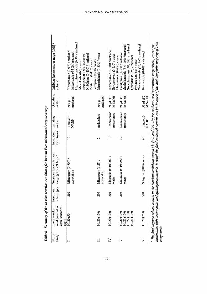
design of the in vitro experiments is summarised below and in Table 6.
The incubation mixture contained 50 (Study II), 100 (Study III, IV and V) or 250 µg (Study VI)
microsomal protein, 0.1 or 0.13 M sodium phosphate buffer (pH 7.4), 5 mM MgCl2, substrate
and 0.1 mM NADPH (Study III - V) or an NADPH regenerating system (Study II and VI) (1 mM
β-NADP, 1mM glucose-6-phosphate, 2 units/mL glucose-6-phosphate dehydrogenase and 5 mM
MgCl2). The reaction was started by the addition of β-NADP, substrate or microsomes. After
incubation in a shaking water bath at 37°C for 10 min, the reaction was terminated by adding
MATERIALS AND METHODS
42
methanol (Study II and III) or NaOH (Study IV-VI) and cooling the samples on ice for 15 min.
All incubations were performed in duplicate and in the linear range with respect to microsomal
protein concentration and incubation time.
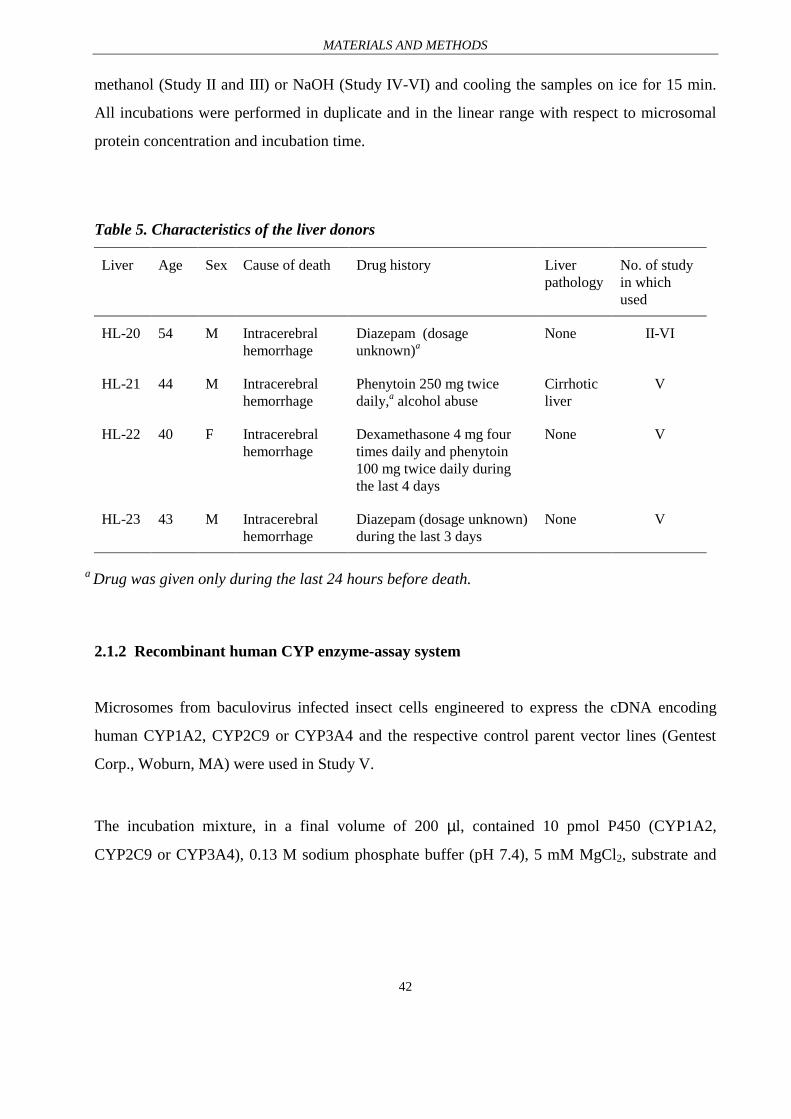
Table 5. Characteristics of the liver donors
Liver Age Sex Cause of death Drug history Liverpathology
No. of studyin whichused
HL-20 54 M Intracerebralhemorrhage
Diazepam (dosageunknown)a
None II-VI
HL-21 44 M Intracerebralhemorrhage
Phenytoin 250 mg twicedaily,a alcohol abuse
Cirrhoticliver
V
HL-22 40 F Intracerebralhemorrhage
Dexamethasone 4 mg fourtimes daily and phenytoin100 mg twice daily duringthe last 4 days
None V
HL-23 43 M Intracerebralhemorrhage
Diazepam (dosage unknown)during the last 3 days
None V
a Drug was given only during the last 24 hours before death.
2.1.2 Recombinant human CYP enzyme-assay system
Microsomes from baculovirus infected insect cells engineered to express the cDNA encoding
human CYP1A2, CYP2C9 or CYP3A4 and the respective control parent vector lines (Gentest
Corp., Woburn, MA) were used in Study V.
The incubation mixture, in a final volume of 200 µl, contained 10 pmol P450 (CYP1A2,
CYP2C9 or CYP3A4), 0.13 M sodium phosphate buffer (pH 7.4), 5 mM MgCl2, substrate and
MATERIALS AND METHODS
44
1.0 mM NADPH. The reaction was started by the addition of microsomes. After incubation in a
shaking water bath at 37°C for 5 min (CYP1A2) or 10 min (CYP2C9 and CYP3A4), the reaction
was terminated by adding 20 µl of 1 M NaOH and cooling the samples on ice for 15 min. All
incubations were performed in duplicate and in the linear range with respect to microsomal
protein concentration and incubation time.
2.2 Designs of in vitro studies
The microsomal enzyme-assay system was used in all the in vitro studies (Study II-VI). The
recombinant human CYP enzyme-assay system was utilized in Study V only. In order to estimate
the inhibitory potency of candidate compounds (Study II, III, IV and VI), a fixed concentration of
substrate (around its corresponding Km value) was coincubated with various concentrations of a
candidate inhibitor. The IC50 and Ki values were characterized when the tested drugs produced
dose dependent inhibitory effects toward the target compounds.
To define the roles of CYP isoforms involved in lidocaine metabolism (Study V), 5 and 20 µM
furafylline, 20 and 100 µM TAO, 3.6 and 100 µM sulfaphenazole, 100 and 300 µM (S)-
mephenytoin, 5 and 25 µM quinidine and 25 and 80 µM pyridine were used as selective
inhibitors for CYP1A2, CYP3A4, CYP2C9, CYP2C19, CYP2D6 and CYP2E1, respectively, and
coincubated with 5 and 800 µM of lidocaine. At these concentrations, the maximum inhibitory
effect of each inhibitor toward the target CYP isoform activity is ≥90% (Hargreaves et al. 1994,
Newton et al. 1995, Eagling et al. 1998, Hickman et al. 1998). The design of the enzyme kinetic
and immunoinhibition studies are described below.
2.3 Analysis of metabolite formation by human liver microsomes and recombinant
enzymes
Study IV and V. Concentrations of MEGX and 3-OH-lidocaine were determined by HPLC, using
procaine hydrochloride (50 µl 10 µM) as the internal standard (Imaoka et al. 1990). The
quantification limit was 2.5 nM (corresponding to a formation rate of 0.5 pmol/min/mg protein)
MATERIALS AND METHODS
45
and 2.0 nM (0.4 pmol/min/mg protein) for MEGX and 3-OH-lidocaine, respectively. The inter-
and intra-day CVs were <8% at relevant concentrations.
Determinations of 1'-hydroxymidazolam, desmethylselegiline and l-methamphetamine
concentrations were carried out using the same methods as described earlier (section 1.4).
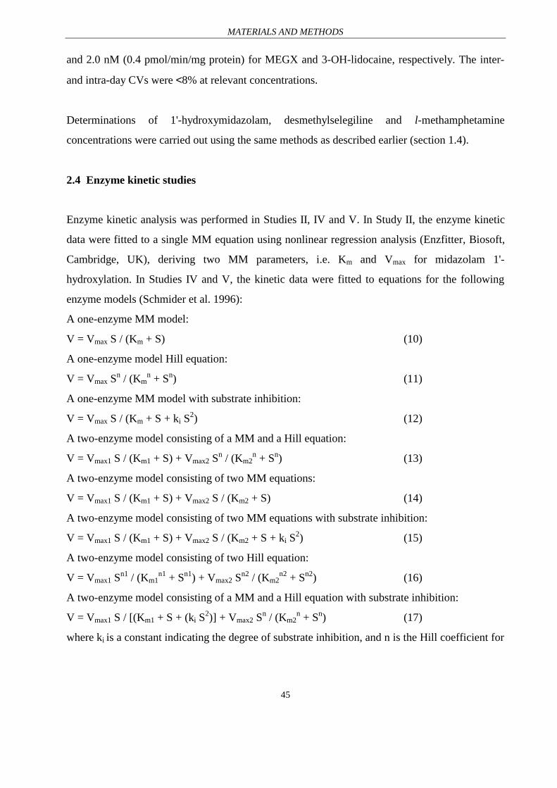
2.4 Enzyme kinetic studies
Enzyme kinetic analysis was performed in Studies II, IV and V. In Study II, the enzyme kinetic
data were fitted to a single MM equation using nonlinear regression analysis (Enzfitter, Biosoft,
Cambridge, UK), deriving two MM parameters, i.e. Km and Vmax for midazolam 1'-
hydroxylation. In Studies IV and V, the kinetic data were fitted to equations for the following
enzyme models (Schmider et al. 1996):
A one-enzyme MM model:
V = Vmax S / (Km + S) (10)
A one-enzyme model Hill equation:
V = Vmax Sn / (Km
n + Sn) (11)
A one-enzyme MM model with substrate inhibition:
V = Vmax S / (Km + S + ki S2) (12)
A two-enzyme model consisting of a MM and a Hill equation:
V = Vmax1 S / (Km1 + S) + Vmax2 Sn / (Km2
n + Sn) (13)
A two-enzyme model consisting of two MM equations:
V = Vmax1 S / (Km1 + S) + Vmax2 S / (Km2 + S) (14)
A two-enzyme model consisting of two MM equations with substrate inhibition:
V = Vmax1 S / (Km1 + S) + Vmax2 S / (Km2 + S + ki S2) (15)
A two-enzyme model consisting of two Hill equation:
V = Vmax1 Sn1 / (Km1
n1 + Sn1) + Vmax2 Sn2 / (Km2
n2 + Sn2) (16)
A two-enzyme model consisting of a MM and a Hill equation with substrate inhibition:
V = Vmax1 S / [(Km1 + S + (ki S2)] + Vmax2 S
n / (Km2n + Sn) (17)
where ki is a constant indicating the degree of substrate inhibition, and n is the Hill coefficient for
MATERIALS AND METHODS
46
cooperative substrate binding. The choice of the best-fitted enzyme model was based on
examination of MM plots, Eadie-Hofstee plots and the residual sum of squares. When necessary,
a statistical analysis (F test) was performed to determine if there was a significant difference in
the size of the residual sum of squares between the models (Motulsky & Ransnas, 1987).
2.5 Immunoinhibition studies
In order to further elucidate the roles of CYP1A2 and CYP3A4 in lidocaine N-deethylation and
3-hydroxylation, immunoinhibition studies were performed (Study V). A polyclonal antibody to
human CYP1A1/2 or a monoclonal antibody to human CYP3A4 (1 mg IgG/mg microsomal
protein) (Gentest Corp., Woburn, MA) was preincubated with human liver microsomes (100 µg)
from HL20, HL21 and HL23 at room temperature for 30 min (anti-CYP1A1/2 and the
combination of anti-CYP1A1/2 and anti-CYP3A4) or at 0°C for 15 min (anti-CYP3A4). The
reaction was initiated by adding 5 or 800 µM lidocaine, and was terminated by 20 µl of 1 M
NaOH after incubation at 37°C for 10 min.
2.6 Data analysis
Reaction velocities were expressed in units of pmol/min/mg protein or pmol/min/pmol protein.
The data represent the mean of duplicate assays for each experiment. The IC50 values were
calculated graphically from the plots of various concentrations of inhibitors versus percentage of
metabolite formation of corresponding control values. The Ki values were derived from fitting
the kinetic data to equations 2-5 by using nonlinear regression analysis (Enzyfitter, Biosoft,
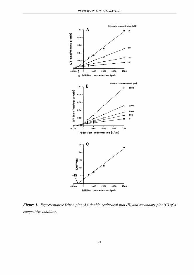
Cambridge, UK). Lineweaver-Burk and Dixon plots were adopted to determine the type of
inhibition.
RESULTS
47
RESULTS
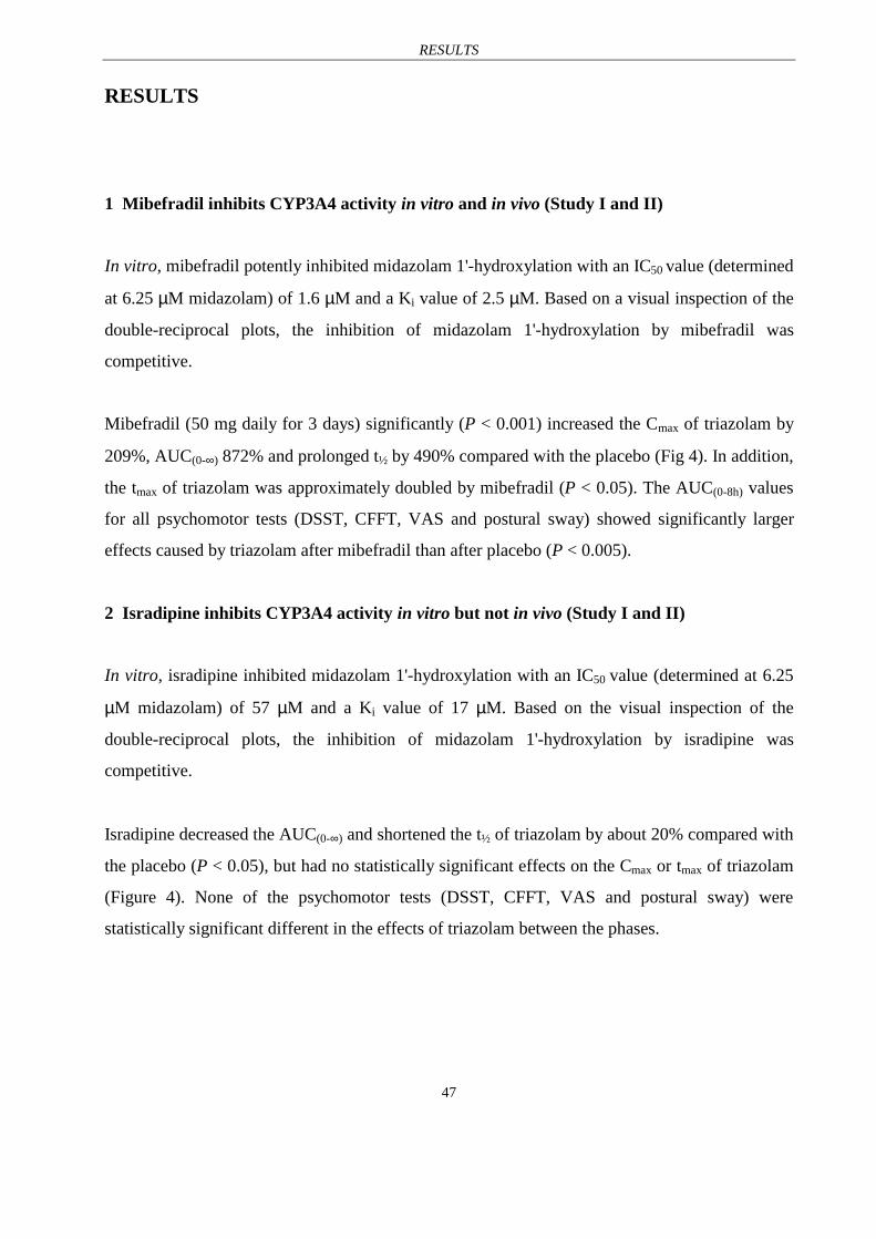
1 Mibefradil inhibits CYP3A4 activity in vitro and in vivo (Study I and II)
In vitro, mibefradil potently inhibited midazolam 1'-hydroxylation with an IC50 value (determined
at 6.25 µM midazolam) of 1.6 µM and a Ki value of 2.5 µM. Based on a visual inspection of the
double-reciprocal plots, the inhibition of midazolam 1'-hydroxylation by mibefradil was
competitive.
Mibefradil (50 mg daily for 3 days) significantly (P < 0.001) increased the Cmax of triazolam by
209%, AUC(0-∞) 872% and prolonged t½ by 490% compared with the placebo (Fig 4). In addition,
the tmax of triazolam was approximately doubled by mibefradil (P < 0.05). The AUC(0-8h) values
for all psychomotor tests (DSST, CFFT, VAS and postural sway) showed significantly larger
effects caused by triazolam after mibefradil than after placebo (P < 0.005).
2 Isradipine inhibits CYP3A4 activity in vitro but not in vivo (Study I and II)
In vitro, isradipine inhibited midazolam 1'-hydroxylation with an IC50 value (determined at 6.25
µM midazolam) of 57 µM and a Ki value of 17 µM. Based on the visual inspection of the
double-reciprocal plots, the inhibition of midazolam 1'-hydroxylation by isradipine was
competitive.
Isradipine decreased the AUC(0-∞) and shortened the t½ of triazolam by about 20% compared with
the placebo (P < 0.05), but had no statistically significant effects on the Cmax or tmax of triazolam
(Figure 4). None of the psychomotor tests (DSST, CFFT, VAS and postural sway) were
statistically significant different in the effects of triazolam between the phases.
RESULTS
49
midazolam AUC0-10 h ratio compared with the placebo and none of the psychomotor tests had a
statistically significant difference in the effects of midazolam between the phases.
4 CYP1A2 and CYP3A4 are involved in lidocaine N-deethylation and hydroxylation
(Study IV and V)
4.1 Enzyme kinetic results
In two human liver microsomal preparations (HL20 HL22), the Eadie-Hofstee plots for both
MEGX and 3-OH-lidocaine formation were nonlinear, suggesting the involvement of multiple
enzymes in both pathways. The MEGX formation was best described by a two enzyme model
consisting of a Michaelis-Menten and a Hill equation and 3-OH-lidocaine formation by a two
enzyme model consisting of two Michaelis-Menten equations with (Study IV) or without (Study
V) substrate inhibition.
In the presence of fixed concentrations of 100 µM TAO or 20 µM furafylline, the formation of
MEGX and 3-OH-lidocaine were consistent with single-enzyme kinetics. Eadie-Hofstee plots for
the formation of both metabolites were linear. Data for the formation of MEGX and 3-OH-
lidocaine were best fitted with a single MM equation. The Km for the formation of both
metabolites was lower in the presence of TAO than in the presence of furafylline, suggesting that
CYP1A2 is the high-affinity component of lidocaine N-deethylation and 3-hydroxylation,
whereas, CYP3A4 is the low-affinity component.
4.2 Chemical inhibition studies
At a concentration of 5 µM lidocaine, fluvoxamine (a potent inhibitor of CYP1A2) was a more
potent inhibitor of the formation of MEGX (IC50 = 1.2 µM) than ketoconazole (IC50 = 8.5 µM)
and erythromycin (IC50 = 200 µM). When the concentration of lidocaine was increased to 800
µM, both fluvoxamine (IC50 = 20.7 µM) and ketoconazole (IC50 = 20.4 µM) caused a modest
inhibition of MEGX formation, erythromycin being a weak inhibitor (IC50 > 250 µM).
RESULTS
50
Fluvoxamine caused a potent inhibition of the formation of 3-OH-lidocaine (IC50 = 0.16 µM and
1.8 µM at 5 and 800 µM, respectively), whereas erythromycin and ketoconazole caused only a
weak inhibition at 5 µM of lidocaine (IC50 = 9.9 µM and =13.9 µM, respectively) and did not
have a consistent effect at 800 µM (IC50 > 250 µM and = 75.0 µM, respectively).
Among the six specific CYP inhibitors used, furafylline (CYP1A2 inhibitor), TAO (CYP3A4
inhibitor) and sulfaphenazole (CYP2C9 inhibitor) inhibited MEGX formation at both lidocaine
concentrations. Furafylline inhibited the formation of 3-OH-lidocaine almost completely
regardless of substrate concentration. However, TAO only slightly inhibited (about 10%) 3-OH-
lidocaine formation at 5 µM lidocaine. Quinidine, (S)-mephenytoin and pyridine did not affect
the metabolism of lidocaine, indicating that CYP2D6, CYP2C19 and CYP2E1, respectively, did
not play a significant role.
4.3 Recombinant enzyme studies
Recombinant CYP1A2 and CYP3A4 catalyzed the formation of MEGX efficiently, with the Km
values of 810 and 1032 µM and Vmax values of 190 and 254 pmol/min/pmol P450, respectively.
On the other hand, both CYP1A2 and CYP3A4 formed 3-OH-lidocaine, CYP1A2 apparently
playing a dominant role. However, CYP2C9 showed negligible activity for both the MEGX and
3-OH-lidocaine formation. The kinetic data for both MEGX and 3-OH-lidocaine formation were
best fitted with a single MM equation.
4.4 Immunoinhibition studies
Anti-CYP1A1/2 had a strong inhibitory effect (>75%) on MEGX formation at 5 µM lidocaine,
but not at 800 µM (about 25%). At both substrate levels, anti-CYP1A1/2 inhibited the formation
of 3-OH-lidocaine almost completely. Anti-CYP3A4 inhibited the formation of MEGX only at
800 µM lidocaine (by 15-30%), and did not inhibit lidocaine 3-hydroxylation. The combination
of anti-CYP1A1/2 and anti-CYP3A4 yielded stronger inhibitory effects on MEGX formation
than anti-CYP1A1/2 alone at both lidocaine concentrations, with no apparent difference in 3-
RESULTS
51
OH-lidocaine formation.
5 Roles of CYP1A2 and CYP3A4 in selegiline metabolism (Study VI)
5.1 Effects of itraconazole on selegiline metabolism in vitro and in vivo (involvement of
CYP3A4)
Concentrations up to 100 µM of itraconazole had no inhibitory effect on the formation of
desmethylselegiline and l-methamphetamine in human liver microsomes. In vivo, itraconazole
(200mg daily for 4 days) caused no significant effects on the pharmacokinetic variables of
selegiline, desmethylselegiline or l-methamphetamine, with the exception that the AUC of
desmethylselegiline was increased by 11% (P < 0.05).
5.2 Correlations between caffeine test and pharmacokinetic parameters of selegiline
(involvement of CYP1A2)
There was a significant correlation (r = 0.41; P < 0.05) between the AUC(desmethylselegiline)/
AUC(selegiline) ratio and the plasma paraxanthine/caffeine concentration ratio. No significant
correlation was found between the AUC of selegiline or the AUC(methamphetamine)/AUC
(selegiline) ratio and the paraxanthine/caffeine ratio (r = -0.33 and 0.20, P = 0.11 and 0.35,
respectively).
DISCUSSION
52
DISCUSSION
1 Methodological considerations
In vitro studies
Human liver microsomal preparations were used in the Studies II-VI. This system appears to be
the most suitable in vitro approach for drug interaction and scaling studies (Parkinson 1996). A
small number of microsomal samples were used in these studies because of the limited
availability of human liver samples. Three liver samples (HL20, HL22, HL23) were obtained
from donors without known liver disease, and one from a patient with cirrhosis caused by heavy
alcohol consumption (HL21). Although CYP activities in microsomes from cirrhotic livers have
been reported to be lower than those from normal livers (George et al. 1995), in our study (Study
V) we did not observe a difference in the rate of formation of MEGX and 3-OH-lidocaine
between the livers. Two potent enzyme inducers, dexamethasone and phenytoin (Meunier et al.
2000, Nation et al. 1990), were given to the donor of HL22 shortly before he died. Consequently,
the CYP3A4-catalyzed 1'-hydroxymidazolam formation rate by microsomes from his liver was
found to be at least 3 times as high as that of the other microsomal preparations. The times of
incubation and concentrations of microsomal protein used in these studies were validated to be
linear with respect to the rate of formation of each metabolite. Such linear conditions are a
necessary prerequisite for extrapolation of the in vitro results to in vivo situations.
The recombinant human CYP enzyme-assay and immunoinhibition studies were used in the
Study V. The recombinant enzyme-assay system, unlike human liver microsomal enzyme-assay
system, expresses only a single CYP isoform activity in a particular recombinant CYP isoform.
Therefore it is a very useful tool for identifying the catalytic capability of a CYP isoform in a
particular metabolic pathway. However, this system also has its own limitations. It does not give
the quantitative input of each isoform into the overall activity within the pathway (Birkett et al.
1993). In addition, the recombinant CYP isoforms expressed in different chimeras produced
DISCUSSION
53
different kinetic parameters (Yamazaki et al. 1997). The system using a polyclonal or a
monoclonal antibody that is specific and highly inhibitory to a single CYP, is applicable to
quantify the catalytic role of the CYP in the metabolism of a substrate in a tissue containing
multiple CYP isoforms (Gelboin 1993). In this study, a polyclonal antibody to human CYP1A1/2
and a monoclonal antibody to human CYP3A4 were used to define the individual contribution of
CYP1A1/2 and CYP3A4 to lidocaine metabolism in human liver microsomes.
The presence of organic solvents can greatly affect the in vitro metabolism of a variety of
enzymatic activities (Chauret et al. 1998, Hickman et al. 1998). In our studies, the stock solutions
of several tested compounds were dissolved in methanol or acetonitrile (Table 6), and
subsequently diluted in the reaction buffer. The final concentrations of the organic solvents in the
incubations were kept minimal and constant, and did not exceed 3% (v/v) and 1% (v/v) for
methanol and acetonitrile, respectively. The one exception to this was for incubations with
itraconazole and hydroxyitraconazole, in which a 5% methanol concentration was used because
of the high lipophility of the compounds. The production of a metabolite formation in
incubations containing a putative inhibitor was compared with that in control incubations to
which only the respective organic solvent was added. In Study II, the studied compounds were
not preincubated with the incubation medium in the presence of NADPH, therefore a possible
mechanism-based inhibition of 1'-hydroxymidazolam formation by these compounds could not
be excluded.
In vivo studies
A randomized, balanced, cross-over protocol for the design was used in all the studies. The
effects of interindividual variability could be minimized with this design, as each subject served
as his or her own control. The wash-out period between the phases of each study was two to four
weeks, which is sufficiently long to minimize possible carry-over effects. The possible effect of
food on the pharmacokinetics of the studied drugs was lessened by using a fasting period before
drug intake and by serving standard meals after drug intake.
DISCUSSION
54
Because all the studies were performed in young, healthy volunteers, it should be careful when
extrapolating the results to patients or elderly populations because pharmacokinetics of drugs can
alter with aging and diseases (Schmucker et al. 1990, Hunt et al. 1992, Le Couteur & McLean
1998). Most of the studies were not balanced with gender since the overall activity of CYP
enzymes shows no significant gender-related differences (Schmucker et al. 1990). Furthermore,
the cross-over design used in the studies ensured that the magnitude of the interactions is not
affected by the imbalanced gender distribution. It was not possible to assess the effects of gender
on the pharmacokinetis and pharmacodynamics of the drugs studied, due to the relatively small
number of subjects (9-12) in each study,
2 Mibefradil is a potent inhibitor of CYP3A4
A significant interaction between mibefradil and the CYP3A4 substrate triazolam was observed
in the in vivo pharmacokinetic study. Compared with placebo, therapeutic doses of mibefradil
almost doubled the Cmax of triazolam, increased the AUC ninefold and t1/2 fivefold.
Consequently, the effects of triazolam were greatly enhanced by mibefradil, consistent with the
changes in the pharmacokinetics. Furthermore, the finding that mibefradil increased the t½ of
triazolam much more than it increased the Cmax indicates that the interaction resulted, in essence,
from the inhibition of the hepatic metabolism of triazolam, while inhibition of the intestinal
metabolism had a smaller contribution.
In good accordance with the in vivo findings, miberfradil showed potent inhibitory effects on
CYP3A4-mediated midazolam 1'-hydroxylation (Ki = 2.3 µM) in human liver microsomes. The
therapeutic plasma concentrations of mibefradil (≈ 0.5-2.0 µM) fall in the range of its effective
inhibitory concentrations. These in vitro results are in reasonable agreement with the recent
findings of Prueksaritanont et al. (1999) and Ma et al. (2000). These authors observed a potent
inhibitory effect of mibefradil on the CYP3A4-mediated testosterone 6β-hydroxylation with IC50
and Ki values of 0.3 µM (Ma et al. 2000) and 2.3 µM (Prueksaritanont et al. 1999), respectively.
In our in vitro experiments, mibefradil was the most potent inhibitor of midazolam 1'-
DISCUSSION
55
hydroxylation among the five calcium channel blockers tested. However, its inhibitory potency
was smaller than that of ketoconazole (Ki = 0.18 µM), though similar to that of itraconazole (Ki =
2.3 µM). These in vitro results conform well with the effects of these agents in vivo. Mibefradil
increased the AUC(0-∞) of triazolam ninefold (Backman et al. 1999), whereas verapamil and
diltiazem only increased it threefold (Varhe et al. 1996a). However, ketoconazole and
itraconazole are more effective CYP3A4 inhibitor than mibefradil. For example, ketoconazole
increased the AUC of triazolam > 20-fold (Varhe et al. 1994, Greeblatt et al. 1998), and
itraconazole increased the AUC of triazolam 14-fold (Varhe et al. 1994), consistent with the high
liver concentrations of itraconazole (von Moltke et al. 1998).
3 Isradipine is an inhibitor of CYP3A4 in vitro but not in vivo
In Study I, isradipine slightly reduced the AUC(0-∞) and shortened the t½ of triazolam by about
20% compared with placebo (P < 0.05), but had no statistically significant effects on the Cmax or
tmax of triazolam. These findings suggested that isradipine does not significantly inhibit CYP3A4
activity in most clinical situations. Furthermore, it is likely that close to steady-state plasma
concentrations of isradipine were reached in this study, since the t½ of isradipine has been
reported to be relatively short (about 8 hours) (Brogden et al. 1995). Consistent with the changes
in its pharmacokinetics, the pharmacodynamics of triazolam were not significantly changed by
isradipine. The only slightly changed pharmacokinetics of triazolam might be explained by a
slight increase in the hepatic clearance of triazolam secondary to the increased hepatic blood flow
by isradipine (Klockowski et al. 1990). Our results are also in good agreement with earlier
studies which found that isradipine had no significant effects on the pharmacokinetics of
cyclosporine (Endresen et al. 1991, Vernillet et al. 1992), and even slightly decreased the AUC
of lovastatin (Zhou et al. 1995).
In vitro, isradipine competitively inhibited midazolam 1'-hydroxylation in human liver
microsomes, with IC50 and Ki values of 57 µM and 17 µM, respectively. However, in humans,
the plasma concentrations of isradipine are usually lower than 0.1 µM (Fitton & Benfield 1990),
which is approximately 100 times lower than its Ki value for CYP3A4-mediated midazolam 1'-
DISCUSSION
56
hydroxylation, and therefore, it will cause negligible inhibitory effects of CYP3A4 activity in
vivo. Thus, despite an apparent discrepancy between the in vivo and in vitro effects of isradipine,
the results of the in vitro experiments are in a good agreement with the in vivo findings.
4 Metronidazole is not a CYP3A4 inhibitor
In Study III, a 3-day pretreatment with metronidazole (400 mg twice daily) had no statistically
significant effects on the pharmacokinetics or pharmacodynamics of midazolam in healthy
volunteers. In agreement with the in vivo results, metronidazole showed little or no inhibitory
effect on midazolam 1'-hydroxylation in vitro with concentrations up to 500 µM (i.e. 5-10 times
higher than the maximum plasma concentrations observed in the in vivo study). These findings
indicate that the activity of CYP3A4 is not affected by metronidazole.
Our results are consistent with the previous reports, in which metronidazole pretreatment had no
significant effect on the plasma concentrations of alprazolam, another substrate of CYP3A4
(Blyden et al. 1988), and was only a weak inhibitor of cyclosporin oxidase (i.e. CYP3A4) in
human liver microsomes (Ki = 223 µM) (Maurice et al. 1992). However, a case report described
that metronidazole increased by 100% plasma concentrations of the CYP3A4 substrates
cyclosporin and tacrolimus (Herzig & Johnson 1999). Because both cyclosporin and tacrolimus
are also P-glycoprotein substrates (Saeki et al. 1993, Yokogawa et al. 1999), the increased
plasma concentrations of cyclosporin and tacrolimus produced by metronidazole might have
been caused by the inhibition of P-glycoprotein by metronidazole.
5 CYP1A2 and CYP3A4 are involved in lidocaine metabolism
The present in vitro studies indicate that both CYP1A2 and CYP3A4 are capable of catalyzing
the formation of MEGX and 3-OH-lidocaine. It appears that CYP1A2 plays the major role in
MEGX formation at clinically relevant lidocaine concentrations. In addition, CYP1A2 is the
major enzyme responsible for the formation of 3-OH-lidocaine regardless of substrate
concentrations. These conclusions were inferred from the following observations. First, enzyme
DISCUSSION
57
kinetic studies with human liver microsomes indicated that at least two distinct enzymes were
involved in MEGX and 3-OH-lidocaine formation. Second, both CYP1A2 inhibitors (furafylline
and fluvoxamine) and CYP3A4 inhibitors (TAO, ketoconazole and erythromycin) inhibited the
formation of MEGX. CYP1A2 inhibitors produced more prominent effects at a therapeutically
relevant 5 µM lidocaine concentration. Third, all the CYP1A2 inhibitors exhibited strong
inhibitory effects on the formation of 3-OH-lidocaine, whereas the CYP3A4 inhibitors exhibited
only weak inhibitory effects at 5 µM lidocaine. Fourth, both recombinant CYP1A2 and CYP3A4
efficiently catalyzed lidocaine N-deethylation. Finally, anti-CYP1A1/2 produced stronger
inhibitory effects on both MEGX and 3-OH-lidocaine formation than anti-CYP3A4 antibodies.
Bargetzi et al. (1989) found that an antiserum against human CYP3A4 dose-dependently
inhibited MEGX formation by human liver microsomes, suggesting that CYP3A4 plays a major
role in the biotransformation of lidocaine to MEGX in humans. The apparent discrepancy
between the present results and those of Bargetzi et al. (1989) seems to be caused by the different
substrate concentration used, i.e. the 5 mM lidocaine concentration used in their study is 1000
times higher than the clinically relevant substrate concentration used in our study.
The present observation that CYP1A2 plays a major role in the N-deethylation of lidocaine at
clinically relevant concentrations is compatible with the findings from two in vivo studies in
humans by Isohanni et al. (1998, 1999). They reported that a 4-day pretreatment with
erythromycin or itraconazole increased the AUC of oral lidocaine by 40-70% compared with
placebo (Isohanni et al. 1999). In addition, this had practically no effects on the pharmacokinetics
of i.v. lidocaine (Isohanni et al. 1998). Considering that the oral bioavailability of lidocaine is
only about 30% due to extensive first-pass metabolism (Tucker & Mather 1979), much greater
effects on the pharmacokinetics of orally administered lidocaine would have been anticipated if
lidocaine were metabolized primarily by CYP3A4.
The MEGX test, i.e. plasma MEGX concentrations after i.v. lidocaine, has been used as a marker
to evaluate the in vivo activity of hepatic CYP3A4 (Azoulay et al. 1993, Cakaloglu et al. 1994).
However, our study indicates that both CYP1A2 and CYP3A4 are involved in the formation of
DISCUSSION
58
MEGX. Therefore, the MEGX test is not a suitable marker of hepatic CYP3A4 activity in vivo.
6 Selegiline pharmacokinetics are unaffected by itraconazole
The potent CYP3A4 inhibitor itraconazole had no significant effects on the pharmacokinetics of
selegiline and its primary metabolites, desmethylselegiline and l-methamphetamine, with the
exception that the mean AUC of desmethylselegiline was increased by 11%. Considering that
selegiline undergoes extensive first-pass metabolism (Heinonen et al. 1994) and CYP3A4 is
abundantly expressed in both the gut wall and liver (Kolars et al. 1992, Kivistö et al. 1996), the
results of our study indicate that CYP3A4 does not play a significant role in selegiline
biotransformation. In agreement with the in vivo results, itraconazole (up to 100 µM) did not
inhibit the formation of desmethylselegiline and l-methamphetamine from selegiline in human
liver microsomes.
Our results appear to be partly at variance with the study of Taavitsainen and co-workers
(Taavitsainen et al. 2000), in which the CYP3A4 inhibitor ketoconazole inhibited the formation
of both desmethylselegiline and l-methamphetamine with apparent Ki values of 15 µM and 18
µM, respectively. However, ketoconazole loses its selectivity to CYP3A4 at concentrations
higher than 10 µM (e.g., ketoconazole inhibited CYP1A2-, CYP2C9-, CYP2C19- and CYP2D6-
mediated activities with IC50 values ranging from 13 to 28 µM; Baldwin et al. 1995).
Consequently, the results of Taavitsainen et al. does not indicate that CYP3A4 is the exclusive
CYP involved in the formation of desmethylselegiline and l-methamphetamine. Furthermore, the
fact that itraconazole is a much less potent inhibitor of CYP3A4 in vitro than ketoconazole (Back
& Tjia 1991, von Moltke et al. 1996) may also be related to the observed discrepancy.
In the in vitro study of Taavitsainen et al. (2000), CYP1A2 was suggested to be involved in the
formation of desmethylselegiline because the CYP1A2 inhibitor furafylline had an inhibitory
effect on the formation of desmethylselegiline with an apparent Ki value of 1.7 µM. In agreement
with this finding, a statistically significant albeit weak relationship (r=0.41; P<0.05) was found
between the paraxanthine/caffeine ratio and the AUC(desmethylselegiline)/AUC(selegiline)
DISCUSSION
59
ratio, supporting the involvement of CYP1A2 in the formation of desmethylselegiline.
7 General discussion
Although quantitative predictions of in vivo situations from in vitro data face many challenges
and controversies, our studies demonstrate that carefully designed in vitro studies can provide
valuable information for predicting potential drug interactions in vivo. With a knowledge of the
in vitro Ki value of a drug for a particular CYP isoform, it is theoretically possible to assess the
likelihood of interactions for a drug cleared through the metabolism by this CYP isoform.
However, several factors need to be considered when the in vitro approach is employed. It is
essential to use a clinically relevant substrate concentration in the in vitro study, as the relative
contributions of different CYP isoforms to the metabolism of a xenobiotic in vitro are affected by
the substrate concentration used (Yamazaki et al. 1994, 1997, Venkatakrishnan et al. 1998). In
agreement with this, the relative contribution of CYP isoforms to lidocaine N-deethylation was
found to be dependent on the substrate concentration (Wang et al. 1999, 2000). This
phenomenon explains the apparent discrepancy between the results of this study and those of
Bargetzi et al. (1989).
Some of the uncertainty in making predictions originate from the evaluation of the concentration
of an inhibitor around the metabolic enzyme site in vivo. A drug may produce drug interactions
in vitro but not necessarily in vivo, e.g. isradipine showed an inhibitory effect on midazolam 1'-
hydroxylation in vitro but caused no inhibition of triazolam metabolism in vivo. It is impossible
to directly measure the unbound concentrations of the inhibitor around the metabolic enzyme in
the liver. Therefore, in many cases the free drug concentration in plasma is used as a substitution.
However, this assumption is not valid for many drugs, e.g. liver concentrations of itraconazole,
diazepam and temazepam are several-fold higher than their corresponding plasma concentrations
(von Moltke et al. 1998). Sometimes, results from human autopsy studies can provide useful
information. However, drug distribution may be modified by postmortem changes (Hilberg et al.
1993, 1994).
DISCUSSION
60
Furthermore, the use of different microsomal protein concentrations can substantially affect in
vitro results. The Ki values of an inhibitor may be overestimated when a high microsomal protein
concentration is used. This may be the result of a depletion of the inhibitor by nonspecific
binding to microsomal proteins. For example, the Ki value for ketoconazole-CYP3A4
interactions in human liver microsomes was estimated to be about 8 µM when a high microsomal
protein concentration (1.5 g/L) was used (Lampen et al. 1995), whereas the Ki value was about
0.18 µM when a low microsomal protein concentration (0.25 g/L) was used in this study (Study
II). A 6-fold increase in the microsomal protein concentration resulted in a 44-fold increase in the
estimated Ki value.
In addition, the role of P-gp in drug disposition may also need to be considered. Because of the
considerable overlap in substrates of P-gp and CYP3A4 (Wandel et al. 1995), the dual inhibition
of both systems may result in a substantial increase in plasma drug concentrations with resultant
toxicity (Wandel et al. 2000). This, in turn, may also foil quantitative in vitro-in vivo scaling. For
example, mibefradil not only inhibits CYP3A4 activity (Backman et al. 1998, Wang et al. 1999),
but also inhibits P-gp-mediated drug transportation (Wandel et al. 2000). Accordingly, the
severity of drug-drug interactions observed during clinical use of mibefradil may have been
caused by the dual inhibition of both P-gp and CYP activities.
Other sources of uncertainty and inaccuracy in predicting the magnitude of in vivo inhibition
includes: the nature and design of the in vitro experiment, presence of active metabolites,
changes in hepatic blood flow and prehepatic metabolism. For all these reasons, caution should
be used in interpreting of the in vitro results to in vivo situations. Qualitative and
semiquantitative predictions are likely to be successful (Bertz & Granneman 1997). However, the
accurate prediction of drug-drug interactions requires careful interpretation of the in vitro results
and a good understanding of the pharmacokinetics and metabolism of the inhibitors and inhibited
drugs (Lin & Lu 1998).
SUMMARY AND CONCLUSIONS
61
SUMMARY AND CONCLUSIONS
In the present series of studies, the effects of mibefradil, isradipine and metronidazole, on
CYP3A4 activity and the CYP isoforms involved in lidocaine metabolism as well as the possible
involvement of CYP1A2 and CYP3A4 in selegiline metabolism were determined and
characterized using in vitro and (or) in vivo pharmacokinetic studies in healthy volunteers. The
major findings and conclusions are as follows:
1 Mibefradil potently inhibited CYP3A4 mediated midazolam 1'-hydroxylation in human liver
microsomes and greatly increased the mean AUC of triazolam by 8.7-fold (P<0.001) in
healthy volunteers. Mibefradil can cause harmful interactions with drugs that are primarily
metabolised by CYP3A4 and may be unsuitable for concomitant administration with them.
2 Isradipine competitively inhibited midazolam 1'-hydroxylation in human liver microsomes
with an apparent Ki value of 17 µM. However, isradipine showed no significant effect on the
pharmacokinetics of the CYP3A4 substrate triazolam in healthy volunteers. It is obvious that
this is because its plasma concentrations are much lower than its Ki value. Isradipine does not
significantly affect CYP3A4 activity in vivo.
3 Metronidazole showed no inhibitory effect on midazolam 1'-hydroxylation in vitro and did
not affect the pharmacokinetics of midazolam in healthy volunteers. Metronidazole does not
significantly affect CYP3A4 activity, but the possibility that it could cause drug interactions
via some other mechanisms can not be ruled out.
4 The results of enzyme kinetic, chemical inhibition, immunoinhibition and recombinant
enzyme studies indicated that both CYP1A2 and CYP3A4 are involved in lidocaine N-
deethylation. At clinically relevant lidocaine concentrations, CYP1A2 plays an important role
in the N-deethylation of lidocaine and it probably is the major isoform involved in lidocaine
metabolism. At higher lidocaine concentrations, CYP1A2 and CYP3A4 seem to be equally
important in lidocaine N-deethylation. On the other hand, CYP1A2 is the major enzyme
SUMMARY AND CONCLUSIONS
62
responsible for the 3-hydroxylation of lidocaine, regardless of substrate concentration.
5 Itraconazole had no significant effect on the pharmacokinetics of selegiline in vivo. It also
had no inhibitory effect on the biotransformation of selegiline to desmethylselegiline and l-
methamphetamine. These results suggest that CYP3A4 does not play a major role in
selegiline biotransformation in vivo. A statistically significant, albeit weak, relationship was
found between the paraxanthine/caffeine ratio and the AUC(desmethylselegiline)/
AUC(selegiline) ratio, suggesting a possible involvement of CYP1A2 in the formation of
desmethylselegiline.
6 A carefully designed in vitro metabolic study can provide valuable information for predicting
drug interactions in vivo. However, interpretation and extrapolation of in vitro interaction
data to in vivo require a good understanding of the mechanisms involved in drug interactions
as well as the pharmacokinetic principles.
ACKNOWLEDGEMENTS
63
ACKNOWLEDGEMENTS
This study was carried out at the Department of Clinical Pharmacology, University of Helsinki,Finland, during 1998-2000.
First and above all I would like to express my deepest gratitude to my supervisor, Prof. Pertti J.Neuvonen, the head of the Department of Clinical Pharmacology, for providing me theopportunity to work in the field of clinical pharmacology and drug metabolism. Without hiscontinuous support, his constructive counsel and broad understanding in sciences, I would neverhave accomplished this work. His care and support in my daily life will be fondly kept in mymemory.
I also feel greatly indebted to my another supervisor, Docent Kari T. Kivistö, for his unfailingsupport, invaluable counsel and skillful scientific guidance. His genuine enthusiasm for scientificwork and broad knowledge of clinical pharmacology have greatly impressed me.
I owe my sincerest thanks to Docent Janne T. Backman, who has kindly helped me to organizethe healthy volunteer studies. His valuable comments and discussions in the fields of statisticsand pharmacokinetics are also highly appreciated.
I sincerely thank Prof. Ilari Paakkari from the Department of Pharmacology and Toxicology,University of Helsinki, and Docent Risto Juvonen from the Department of Pharmacology andToxicology, University of Kuopio, for their critical comments to improve the manuscript in itsfinal stage.
I am deeply grateful to my co-author Päivi Taavitsainen, M.Sc., from the Department ofPharmacology and Toxicology, University of Oulu for her most supportive collaboration worksin the studies. My warmest thanks are due to my good friend Mr. Jouko Laitila for his excellentand skillful technical assistance, and also his kindest help in everyday life. My special gratitudealso extends to my friends and colleagues Mrs. Kerttu Mårtenson, Mrs. Eija Mäkinen-Pulli, Mrs.Lisbet Partanen and Mr. Mikko Neuvonen, for their kind hospitality and helping me indetermination of several drug concentrations.
I am indebted to all my other colleagues at the Department of Clinical Pharmacology, mostnotably Kirsti Villikka, Teemu Kantola, Tommi Lamberg, Jari Lilja, Tiina Varis, Kari Raaska,Harri Luurila, Carl Kyrklund and Mikko Niemi for their collaboration and friendship. I warmlythank Tuija Itkonen for her kind help in numerous practical matters.
I also gratefully acknowledge all the volunteers who took part in my studies.
My warmest thanks are due to Prof. Zhou Hong-Hao and other officials and colleagues in HunanMedical University for their friendship, encouragement and support before and after my arrival toFinland. My warmest thanks are also due to many Chinese friends in Finland for their friendshipand continuous support.
ACKNOWLEDGEMENTS
64
Sincere thanks to Alisdair Mclean for editing of the English language of this thesis.
Financial support by the Helsinki University Central Hospital Research Fund, the NationalTechnology Agency of Finland (TEKES), and the Clinical Drug Research graduate school aregratefully acknowledged.
I am deeply grateful to my parents for their love and kindly taking care of my son during 2000. Ialso gratitude to my wife's parents, my sisters and brothers, and my wife's brother, for theirloving care and encouragement throughout the years of my study.
Finally, my deepest thanks go to my wife, Wen Xia, for her understanding, love andencouragement throughout these years. She is also one of my best supportive collaborators. I alsodeeply thank my little son, Stone, for the happiness he brings to me. With their company, lifebecomes beautiful and enticing.
Helsinki, April 2001
Jun-Sheng Wang
REFERENCES
65
REFERENCES
Abdel-Rahman SM, Marcucci K, Boge T, Gotschall RR, Kearns GL, Leeder JS. Potent inhibition of cytochrome P-450 2D6-mediated dextromethorphan O-demethylation by terbinafine. Drug Metab Dispos 1999;27:770-775.
Adebayo GI, Mabadeje AF. Lack of inhibitory effect of metronidazole on theophylline disposition in healthysubjects. Br J Clin Pharmacol 1987;24:110-113.
Aklillu E, Persson I, Bertilsson L, Johansson I, Rodrigues F, Ingelman-Sundberg M. Frequent distribution ofultrarapid metabolizers of debrisoquine in an ethiopian population carrying duplicated and multiduplicatedfunctional CYP2D6 alleles. J Pharmacol Exp Ther 1996;278:441-446.
Allenmark S, Edebo A, Lindgren K. Determination of itraconazole in serum with high-performance liquidchromatography and fluorescence detection. J Chromatogr 1990;532:203-206.
Allonen H, Ziegler G, Klotz U. Midazolam kinetics. Clin Pharmacol Ther 1981;30:653-661.
Alvan G, Bechtel P, Iselius L, Gundert-Remy U. Hydroxylation polymorphisms of debrisoquine and mephenytoin inEuropean populations. Eur J Clin Pharmacol 1990;39:533-537.
Aoyama T, Yamano S, Waxman DJ, Lapenson DP, Meyer UA, Fischer V, Tyndale R, Inaba T, Kalow W, GelboinHV, et al. Cytochrome P-450 hPCN3, a novel cytochrome P-450 IIIA gene product that is differentially expressed inadult human liver. cDNA and deduced amino acid sequence and distinct specificities of cDNA-expressed hPCN1and hPCN3 for the metabolism of steroid hormones and cyclosporine. J Biol Chem 1989;264:10388-10395.
Azoulay D, Lemoine A, Gries JM, Adam R, Dolizy I, Fredj G, Beaune P and Bismuth H. Lidocaine metaboliteformation in the liver donor as a predictive test for the safe and efficient introduction of cyclosporine in the recipient.Transplant Proc 1993;25:2275-2278.
Baba T, Touchi A, Yamaguchi Y, Ito K, Yamazoe Y, Sugiyama Y, Ohno Y. Can in vitro metabolism and enzymeinhibition be explained by unbound drug concentration? Drug Metab Rev 2000;32 supple 2:222.
Babany G, Larrey D, Pessayre D. 1988. Macrolide antibiotics as inducers and inhibitors of cytochrome-P450 inexperimental animals and man. In Progress in Drug Metabolism, ed. GG Gibson, 11: 61-98. London: Taylor &Francis.
Back DJ, Tjia JF. Comparative effects of the antimycotic drugs ketoconazole, fluconazole, itraconazole andterbinafine on the metabolism of cyclosporin by human liver microsomes. Br J Clin Pharmacol 1991;32:624-626.
Backman JT, Olkkola KT, Aranko K, Himberg JJ, Neuvonen PJ. Dose of midazolam should be reduced duringdiltiazem and verapamil treatments. Br J Clin Pharmacol 1994;37:221-225.
Backman JT, Olkkola KT, Neuvonen PJ. Rifampin drastically reduces plasma concentrations and effects of oralmidazolam. Clin Pharmacol Ther 1996a;59:7-13.
Backman JT, Olkkola KT, Ojala M, Laaksovirta H, Neuvonen PJ. Concentrations and effects of oral midazolam aregreatly reduced in patients treated with carbamazepine or phenytoin. Epilepsia 1996b;37:253-257.
Backman JT, Wang JS, Wen X, Kivistö KT, Neuvonen PJ. Mibefradil but not isradipine substantially elevates theplasma concentrations of the CYP3A4 substrate triazolam. Clin Pharmacol Ther 1999;66:401-407.
Bargetzi MJ, Aoyama T, Gonzalez FJ, Meyer UA. Lidocaine metabolism in human liver microsomes by cytochromeP450IIIA4. Clin Pharmacol Ther 1989;46:521-527.
Benowitz NL, Meister W. Clinical pharmacokinetics of lignocaine. Clin Pharmacokinet 1978;3:177-201.
Bensoussan C, Delaforge M, Mansuy D. Particular ability of cytochromes P450 3A to form inhibitory P450-iron-metabolite complexes upon metabolic oxidation of aminodrugs. Biochem Pharmacol 1995;49:591-602.
Bergan T, Arnold E. Pharmacokinetics of metronidazole in healthy adult volunteers after tablets and suppositories.Chemotherapy 1980;26:231-241.
REFERENCES
66
Bertilsson L, Lou YQ, Du YL, Liu Y, Kuang TY, Liao XM, Wang KY, Reviriego J, Iselius L, Sjoqvist F.Pronounced differences between native Chinese and Swedish populations in the polymorphic hydroxylations ofdebrisoquin and S-mephenytoin. Clin Pharmacol Ther 1992;51:388-397.
Bertz RJ, Granneman GR. Use of in vitro and in vivo data to estimate the likelihood of metabolic pharmacokineticinteractions. Clin Pharmacokinet 1997;32:210-258.
Bidouil S, Dubois J, Hanocq M. Isocratic high-performance liquid chromatographic method for the separation ofisradipine and its main metabolites. Application to in vitro metabolization by h3A4/OR cells. J Chromatogr BBiomed Sci Appl 1997;693:359-366.
Birkett DJ, Mackenzie PI, Veronese ME , Miners JO. In vitro approaches can predict human drug metabolism. TIPS1993;14: 292-294.
Blyden GT, Scavone JM, Greenblatt DJ. Metronidazole impairs clearance of phenytoin but not of alprazolam orlorazepam. J Clin Pharmacol 1988;28:240-245.
Bond A, Lader M. The use of analogue scales in rating subjective feelings. Br J Med Psycol 1974;47:211-218.
Bourrie M, Meunier V, Berger Y, Fabre G. Cytochrome P450 isoform inhibitors as a tool for the investigation ofmetabolic reactions catalyzed by human liver microsomes. J Pharmacol Exp Ther 1996;277:321-332.
Boyes RN, Scott DB, Jebson PJ, Godman MJ, Julian DG. Pharmacokinetics of lidocaine in man. Clin PharmacolTher 1971;12:105-116.
Brogden RN, Markham A. Mibefradil. A review of its pharmacodynamic and pharmacokinetic properties, andtherapeutic efficacy in the management of hypertension and angina pectoris. Drugs 1997;54:774-793.
Brogden RN, Sorkin EM. Isradipine. An update of its pharmacodynamic and pharmacokinetic properties andtherapeutic efficacy in the treatment of mild to moderate hypertension. Drugs 1995;49:618-649.
Brosen K. Drug interactions and the cytochrome P450 system. The role of cytochrome P450 1A2. ClinPharmacokinet 1995;29 Suppl 1:20-25.
Burchell B, Nebert DW, Nelson DR, Bock KW, Iyanagi T, Jansen PL, Lancet D, Mulder GJ, Chowdhury JR, SiestG, Tephly TR, Mackenzie PI. The UDP glucuronosyltransferase gene superfamily: suggested nomenclature based onevolutionary divergence. DNA Cell Biol 1991;10:487-494.
Cakaloglu Y, Tredger JM, Devlin J, Williams R. Importance of cytochrome P-450IIIA activity in determiningdosage and blood levels of FK 506 and cyclosporine in liver transplant recipients. Hepatology 1994;20:309-316.
Campana C, Regazzi MB, Buggia I, Molinaro M. Clinically significant drug interactions with cyclosporin. Anupdate. Clin Pharmacokinet 1996;30:141-179.
Chauret N, Gauthier A, Nicoll-Griffith DA. Effect of common organic solvents on in vitro cytochrome P450-mediated metabolic activities in human liver microsomes. Drug Metab Dispos 1998;26:1-4.
Chiba K, Manabe K, Kobayashi K, Takayama Y, Tani M, Ishizaki T. Development and preliminary application of asimple assay of S-mephenytoin 4-hydroxylase activity in human liver microsomes. Eur J Clin Pharmacol1993;44:559-562.
Chida M, Yokoi T, Fukui T, Kinoshita M, Yokota J, Kamataki T. Detection of three genetic polymorphisms in the5'-flanking region and intron 1 of human CYP1A2 in the Japanese population. Jpn J Cancer Res 1999;90:899-902.
Christians U, Schmidt G, Bader A, Lampen A, Schottmann R, Linck A, Sewing KF. Identification of drugs inhibitingthe in vitro metabolism of tacrolimus by human liver microsomes. Br J Clin Pharmacol 1996;41:187-190.
Clifton GD, Blouin RA, Dilea C, Schran HF, Hassell AE, Gonasun LM, Foster TS. The pharmacokinetics of oralisradipine in normal volunteers. J Clin Pharmacol 1988;28:36-42.
Cooke CE, Sklar GE, Nappi JM. Possible pharmacokinetic interaction with quinidine: ciprofloxacin ormetronidazole? Ann Pharmacother 1996;30:364-366.
Dahl ML, Johansson I, Bertilsson L, Ingelman-Sundberg M, Sjoqvist F. Ultrarapid hydroxylation of debrisoquine ina Swedish population. Analysis of the molecular genetic basis. J Pharmacol Exp Ther 1995;274:516-520.
REFERENCES
67
De Kroon IF, Langendijk PN, De Goede PN. Simultaneous determination of midazolam and its three hydroxymetabolites in human plasma by electron-capture gas chromatography without derivatization. J Chromatogr1989;491:107-116.
de Waziers I, Boisset M, Atteba S. Pre- and postweaning development of drug-metabolizing enzyme activities insmall intestine and liver of rats. Drug Metab Dispos 1988;16:310-315.
Dresser GK, Spence JD, Bailey DG. Pharmacokinetic-Pharmacodynamic consequences and clinical relevance ofcytochrome P450 3A4 inhibition. Clin Pharmacokinet 2000;38:41-57.
Eagling VA, Tjia JF, Back DJ. Differential selectivity of cytochrome P450 inhibitors against probe substrates inhuman and rat liver microsomes. Br J Clin Pharmacol 1998;45:107-114.
Eberts FS Jr, Philopoulos Y, Reineke LM, Vliek RW. Triazolam disposition. Clin Pharmacol Ther 1981;29:81-93.
Echizen H, Kawasaki H, Chiba K, Tani M, Ishizaki T. A potent inhibitory effect of erythromycin and othermacrolide antibiotics on the mono-N-dealkylation metabolism of disopyramide with human liver microsomes. JPharmacol Exp Ther 1993;264:1425-1431.
Endresen L, Bergan S, Holdaas H, Pran T, Sinding-Larsen B, Berg KJ. Lack of effect of the calcium antagonistisradipine on cyclosporine pharmacokinetics in renal transplant patients. Ther Drug Monit 1991;13:490-495.
Ereshefsky L, Riesenman C, Lam YW. Antidepressant drug interactions and the cytochrome P450 system. The roleof cytochrome P450 2D6. Clin Pharmacokinet. 1995;29:10-19.
Fitton A, Benfield P. Isradipine. A review of its pharmacodynamic and pharmacokinetic properties, and therapeuticuse in cardiovascular disease. Drugs 1990;40:31-74.
Flockhart DA. Drug interactions and the cytochrome P450 system. The role of cytochrome P450 2C19. ClinPharmacokinet 1995;29 Suppl 1:45-52.
Franklin MR. Enhanced rates of cytochrome P450 metabolic-intermediate complex formation from nonmacrolideamines in rifampicin-treated rabbit liver microsomes. Drug Metab Dispos 1995;23:1379-1382.
Friedman H, Greenblatt DJ, Burstein ES, Scavone JM, Harmatz JS, Shader RI. Triazolam kinetics: interaction withcimetidine, propranolol, and the combination. J Clin Pharmacol 1988;28:228-233.
Fuhr U, Rost KL, Engelhardt R, Sachs M, Liermann D, Belloc C, Beaune P, Janezic S, Grant D, Meyer UA, StaibAH. Evaluation of caffeine as a test drug for CYP1A2, NAT2 and CYP2E1 phenotyping in man by in vivo versus invitro correlations. Pharmacogenetics 1996;6:159-176.
Gaillard Y, Gay-Montchamp JP, Ollagnier M. Simultaneous screening and quantitation of alpidem, zolpidem,buspirone and benzodiazepines by dual-channel gas chromatography using electron-capture and nitrogen-phosphorusdetection after solid-phase extraction. J Chromatogr 1993;622:197-208.
Ged C, Umbenhauer DR, Bellew TM, Bork RW, Srivastava PK, Shinriki N, Lloyd RS, Guengerich FP.Characterization of cDNAs, mRNAs, and proteins related to human liver microsomal cytochrome P-450 (S)-mephenytoin 4'-hydroxylase. Biochemistry 1988;27:6929-6940.
Gelboin HV. Cytochrome P450 and monoclonal antibodies. Pharmacol Rev 1993;45:413-453.
George J, Murray M, Byth K, Farrell GC. Differential alterations of cytochrome P450 proteins in livers from patientswith severe chronic liver disease. Hepatology 1995;21:120-128.
Goldstein JA, de Morais SM. Biochemistry and molecular biology of the human CYP2C subfamily.Pharmacogenetics 1994;4:285-299.
Gonzalez FJ. Human cytochromes P450: problems and prospects. Trends Pharmacol Sci 1992;13:346-352.
Gonzalez FJ, Crespi CL, Gelboin HV. DNA-expressed human cytochrome P450s: a new age of molecular toxicologyand human risk assessment. Mutat Res 1991;247:113-127.
Gonzalez FJ, Idle JR. Pharmacogenetic phenotyping and genotyping. Present status and future potential. ClinPharmacokinet 1994;26:59-70.
Gorski JC, Jones DR, Haehner-Daniels BD, Hamman MA, O'Mara EM Jr, Hall SD. The contribution of intestinal
REFERENCES
68
and hepatic CYP3A to the interaction between midazolam and clarithromycin. Clin Pharmacol Ther 1998;64:133-143.
Greenblatt DJ, Abernethy DR, Locniskar A, Harmatz JS, Limjuco RA, Shader RI. Effect of age, gender, and obesityon midazolam kinetics. Anesthesiology 1984;61:27-35.
Greenblatt DJ, Divoll M, Abernethy DR, Ochs HR, Shader RI. Clinical pharmacokinetics of the newerbenzodiazepines. Clin Pharmacokinet 1983;8:233-252.
Greenblatt DJ, Wright CE, von Moltke LL, Harmatz JS, Ehrenberg BL, Harrel LM, Corbett K, Counihan M, TobiasS, Shader RI. Ketoconazole inhibition of triazolam and alprazolam clearance: differential kinetic and dynamicconsequences. Clin Pharmacol Ther 1998;64:237-247.
Guengerich FP, Martin MV, Beaune PH, Kremers P, Wolff T, Waxman DJ. Characterization of rat and human livermicrosomal cytochrome P-450 forms involved in nifedipine oxidation, a prototype for genetic polymorphism inoxidative drug metabolism. J Biol Chem 1986;261:5051-5060.
Guengerich FP, Shimada T. Oxidation of toxic and carcinogenic chemicals by human cytochrome P-450 enzymes.Chem Res Toxicol 1991;4:391-407.
Ha HR, Rentsch KM, Kneer J, Vonderschmitt DJ. Determination of midazolam and its alpha-hydroxy metabolite inhuman plasma and urine by high-performance liquid chromatography. Ther Drug Monit 1993;15:338-343.
Hall SD, Thummel KE, Watkins PB, Lown KS, Benet LZ, Paine MF, Mayo RR, Turgeon DK, Bailey DG, FontanaRJ, Wrighton SA. Molecular and physical mechanisms of first-pass extraction. Drug Metab Dispos 1999;27:161-166.
Haller I. In vitro activity of the two principal oxidative metabolites of metronidazole against Bacteroides fragilis andrelated species. Antimicrob Agents Chemother 1982;22:165-166.
Halpert JR. Structural basis of selective cytochrome P450 inhibition. Ann Pharmacol Toxicol 1995;35:29-53.
Harashima H, Sugiyama Y, Sawada Y, Iga T, Hanano M. Comparison between in-vivo and in-vitro tissue-to-plasmaunbound concentration ratios (Kp,f) of quinidine in rats. J Pharm Pharmacol 1984;36:340-342.
Hargreaves MB, Jones BC, Smith DA, Gescher A. nhibition of p-nitrophenol hydroxylase in rat liver microsomes bysmall aromatic and heterocyclic molecules. Drug Metab Dispos 1994;22:806-810.
Heinonen EH, Anttila MI, Lammintausta RA. Pharmacokinetic aspects of l-deprenyl (selegiline) and its metabolites.Clin Pharmacol Ther 1994;56:742-749.
Heinonen EH, Myllyla V, Sotaniemi K, Lamintausta R, Salonen JS, Anttila M, Savijarvi M, Kotila M, Rinne UK.Pharmacokinetics and metabolism of selegiline. Acta Neurol Scand Suppl 1989;126:93-99.
Heizmann P, Ziegler WH. Excretion and metabolism of 14C-midazolam in humans following oral dosing.Arzneimittelforschung 1981;31:2220-2223.
Hermansson J, Glaumann H, Karlen B and von Bahr C. Metabolism of lidocaine in human liver in vitro. ActaPharmacol Toxicol (Copenh) 1980;47:49-52.
Herzig K, Johnson DW. Marked elevation of blood cyclosporin and tacrolimus levels due to concurrentmetronidazole therapy. Nephrol Dial Transplant 1999;14:521-523.
Hickman D, Wang JP, Wang Y, Unadkat JD. Evaluation of the selectivity of In vitro probes and suitability oforganic solvents for the measurement of human cytochrome P450 monooxygenase activities. Drug Metab Dispos1998;26:207-215.
Hilberg T, Bugge A, Beylich KM, Ingum J, Bjorneboe A, Morland J. An animal model of postmortem amitriptylineredistribution. J Forensic Sci 1993;38:81-90.
Hilberg T, Morland J, Bjorneboe A. Postmortem release of amitriptyline from the lungs; a mechanism of postmortemdrug redistribution. Forensic Sci Int 1994;64:47-55.
Hof RP, Scholtysik G, Loutzenhiser R, Vuorela HJ, Neumann P. PN 200-110, a new calcium antagonist:electrophysiological, inotropic, and chronotropic effects on guinea pig myocardial tissue and effects on contraction
REFERENCES
69
and calcium uptake of rabbit aorta. J Cardiovasc Pharmacol 1984;6:399-406.
Honig PK, Woosley RL, Zamani K, Conner DP, Cantilena LR Jr. Changes in the pharmacokinetics andelectrocardiographic pharmacodynamics of terfenadine with concomitant administration of erythromycin. ClinPharmacol Ther 1992;52:231-238.
Horai Y, Nakano M, Ishizaki T, Ishikawa K, Zhou HH, Zhou BI, Liao CL, Zhang LM. Metoprolol and mephenytoinoxidation polymorphisms in Far Eastern Oriental subjects: Japanese versus mainland Chinese. Clin Pharmacol Ther1989;46:198-207.
Hukkinen SK, Varhe A, Olkkola KT, Neuvonen PJ. Plasma concentrations of triazolam are increased byconcomitant ingestion of grapefruit juice. Clin Pharmacol Ther 1995;58:127-131.
Hunt CM, Westerkam WR, Stave GM. Effect of age and gender on the activity of human hepatic CYP3A. BiochemPharmacol 1992;44:275-283.
Imaoka S, Enomoto K, Oda Y, Asada A, Fujimori M, Shimada T, Fujita S, Guengerich FP, Funae Y. Lidocainemetabolism by human cytochrome P-450s purified from hepatic microsomes: comparison of those with rat hepaticcytochrome P-450s. J Pharmacol Exp Ther 1990;255:1385-1391.
Inaba T. Phenytoin: pharmacogenetic polymorphism of 4'-hydroxylation. Pharmacol Ther 1990;46:341-347.
Isohanni MH, Neuvonen PJ, Olkkola KT. Effect of erythromycin and itraconazole on the pharmacokinetics of orallignocaine. Pharmacol Toxicol 1999;84:143-146.
Isohanni MH, Neuvonen PJ, Palkama VJ, Olkkola KT. Effect of erythromycin and itraconazole on thepharmacokinetics of intravenous lignocaine. Eur J Clin Pharmacol 1998;54:561-565.
Ito K, Iwatsubo T, Kanamitsu S, Nakajima Y, Sugiyama Y. Quantitative prediction of in vivo drug clearance anddrug interactions from in vitro data on metabolism, together with binding and transport. Annu Rev PharmacolToxicol 1998a;38:461-499
Ito K, Iwatsubo T, Kanamitsu S, Ueda K, Suzuki H, Sugiyama Y. Prediction of pharmacokinetic alterations causedby drug-drug interactions: metabolic interaction in the liver. Pharmacol Rev 1998b;50:387-412.
Jensen JC, Gugler R. Single- and multiple-dose metronidazole kinetics. Clin Pharmacol Ther 1983;34:481-487.
Johansson I, Lundqvist E, Bertilsson L, Dahl ML, Sjoqvist F, Ingelman-Sundberg M. Inherited amplification of anactive gene in the cytochrome P450 CYP2D locus as a cause of ultrarapid metabolism of debrisoquine. Proc NatlAcad Sci U S A 1993;90:11825-11829.
Kakkar T, Boxenbaum H, Mayersohn M. Estimation of Ki in a competitive enzyme-inhibition model: comparisonsamong three methods of data analysis. Drug Metab Dispos 1999;27:756-762.
Kalow W, Bertilsson L. Interethnic factors affecting drug response. In Testa B, Meyer UA, editor. Advantages indrug research. New York: Academic Press, 1994:1-53.
Kanamitsu SI, Ito K, Okuda H, Ogura K, Watabe T, Muro K, Sugiyama Y. Prediction of in vivo drug-druginteractions based on mechanism-based inhibition from in vitro data: inhibition of 5-fluorouracil metabolism by (E)-5-(2-Bromovinyl)uracil. Drug Metab Dispos 2000;28:467-474.
Kantola T, Kivistö KT, Neuvonen PJ. Erythromycin and verapamil considerably increase serum simvastatin andsimvastatin acid concentrations. Clin Pharmacol Ther 1998;64:177-182.
Kato R, Yamazoe Y, Yasumori T. Polymorphism in stereoselective hydroxylations of mephenytoin and hexobarbitalby Japanese liver samples in relation to cytochrome P-450 human-2 (IIC9). Xenobiotica 1992;22:1083-1092.
Kaye CM, Sankey MG, Thomas LA. A rapid and specific semi-micro method involving high-pressure liquidchromatography for the assay of metronidazole in plasma, saliva, serum, urine and whole blood. Br J Clin Pharmacol1980;9:528-529.
Kent UM, Yanev S, Hollenberg PF. Mechanism-based inactivation of cytochromes P450 2B1 and P450 2B6 by n-propylxanthate. Chem Res Toxicol 1999;12:317-322.
Kharasch ED, Hankins DC, Taraday JK. Single-dose methoxsalen effects on human cytochrome P-450 2A6 activity.
REFERENCES
70
Drug Metab Dispos 2000;28:28-33.
Kivistö KT, Bookjans G, Fromm MF, Griese EU, Münzel P, Kroemer HK. Expression of CYP3A4, CYP3A5 andCYP3A7 in human duodenal tissue. Br J Clin Pharmacol 1996;42:387-389.
Kivistö KT, Kroemer HK. Use of probe drugs as predictors of drug metabolism in humans. J Clin Pharmacol1997;37(1 Suppl):40S-48S.
Kivistö KT, Lamberg TS, Kantola T, Neuvonen PJ. Plasma buspirone concentrations are greatly increased byerythromycin and itraconazole. Clin Pharmacol Ther 1997; 62:348-354.
Klockowski PM, Lener ME, Sirgo MA, Rocci MLJ. Comparative evaluation of the effects of isradipine anddiltiazem on antipyrine and indocyanine green clearances in elderly volunteers. Clin Pharmacol Ther 1990;48:375-380.
Ko JW, Sukhova N, Thacker D, Chen P, Flockhart DA. Evaluation of omeprazole and lansoprazole as inhibitors ofcytochrome P450 isoforms. Drug Metab Dispos 1997;25:853-862.
Koenigs LL, Peter RM, Thompson SJ, Rettie AE, Trager WF. Mechanism-based inactivation of human livercytochrome P450 2A6 by 8-methoxypsoralen. Drug Metab Dispos 1997;25:1407-1415.
Kolars JC, Schmiedlin-Ren P, Schuetz JD, Fang C, Watkins PB. Identification of rifampin-inducible P450IIIA4(CYP3A4) in human small bowel enterocytes. J Clin Invest 1992;90:1871-1878.
Krishna DR, Klotz U. Extrahepatic metabolism of drugs in humans. Clin Pharmacokinet 1994;26:144-160.
Kroboth PD, McAuley JW, Kroboth FJ, Bertz RJ, Smith RB. Triazolam pharmacokinetics after intravenous, oral,and sublingual administration. J Clin Psychopharmacol 1995;15:259-262.
Kronbach T, Mathys D, Umeno M, Gonzalez FJ, Meyer UA. Oxidation of midazolam and triazolam by human livercytochrome P450IIIA4. Mol Pharmacol 1989;36:89-96.
Kupferschmidt HH, Ha HR, Ziegler WH, Meier PJ, Krahenbuhl S. Interaction between grapefruit juice andmidazolam in humans. Clin Pharmacol Ther 1995;58:20-28.
Laine K, Anttila M, Helminen A, Karnani H, Huupponen R. Dose linearity study of selegiline pharmacokinetics afteroral administration: evidence for strong drug interaction with female sex steroids. Br J Clin Pharmacol 1999;47:249-254.
Lampen A, Christians U, Guengerich FP, Watkins PB, Kolars JC, Bader A, Gonschior AK, Dralle H, Hackbarth I,Sewing KF. Metabolism of the immunosuppressant tacrolimus in the small intestine: cytochrome P450, druginteractions, and interindividual variability. Drug Metab Dispos 1995;23:1315-1324.
Larrey D, Tinel M, Letteron P, Geneve J, Descatoire V, Pessayre D. Formation of an inactive cytochrome P-450Fe(II)-metabolite complex after administration of amiodarone in rats, mice and hamsters. Biochem Pharmacol1986;35:2213-2220.
Lau AH, Lam NP, Piscitelli SC, Wilkes L, Danziger LH. Clinical pharmacokinetics of metronidazole and othernitroimidazole anti-infectives. Clin Pharmacokinet 1992;23:328-364.
Le Couteur DG, McLean AJ. The aging liver. Drug clearance and an oxygen diffusion barrier hypothesis. ClinPharmacokinet 1998;34:359-373.
Levy RH. Cytochrome P450 isozymes and antiepileptic drug interactions. Epilepsia 1995;36 (Suppl 5):S8-13.
Li AP, Sakai Y, Lu C, Fackett A, Lin J. Potent binding and drug metanolism: effects of human serum albumin oncoumarin metabolism in human hepatocytes. Drug Metab Rev 2000,23 suppl 2:242.
Lilja JJ, Kivistö KT, Backman JT, Neuvonen PJ. Effect of grapefruit juice dose on grapefruit juice-triazolaminteraction: repeated consumption prolongs triazolam half-life. Eur J Clin Pharmacol 2000;56:411-415.
Lin JH, Chiba M, Baillie TA. Is the role of the small intestine in first-pass metabolism overemphasized? PharmacolRev 1999;51:135-158.
Lin JH, Lu AY. Role of pharmacokinetics and metabolism in drug discovery and development. Pharmacol Rev1997;49:403-449.
REFERENCES
71
Lin JH, Lu AY. Inhibition and induction of cytochrome P450 and the clinical implications. Clin Pharmacokinet1998;35:361-390.
Lin JH, Sugiyama Y, Awazu S, Hanano M. In vitro and in vivo evaluation of the tissue-to-blood partition coefficientfor physiological pharmacokinetic models. J Pharmacokinet Biopharm 1982;10:637-647.
Lin JH, Sugiyama Y, Hanano M, Awazu S. Effect of product inhibition on elimination kinetics of ethoxybenzamidein rabbits. Analysis by physiological pharmacokinetic model. Drug Metab Dispos 1984;12:253-256.
Lindstrom TD, Hanssen BR, Wrighton SA. Cytochrome P-450 complex formation by dirithromycin and othermacrolides in rat and human livers. Antimicrob Agents Chemother 1993;37:265-269.
Loft S, Dossing M, Poulsen HE, Sonne J, Olesen KL, Simonsen K, Andreasen PB. Influence of dose and route ofadministration on disposition of metronidazole and its major metabolites. Eur J Clin Pharmacol 1986;30:467-473.
Loft S, Otton SV, Lennard MS, Tucker GT, Poulsen HE. Characterization of metronidazole metabolism by humanliver microsomes. Biochem Pharmacol 1991;41:1127-1134.
Lown KS, Mayo RR, Leichtman AB, Hsiao HL, Turgeon DK, Schmiedlin-Ren P, Brown MB, Guo W, Rossi SJ,Benet LZ, Watkins PB. Role of intestinal P-glycoprotein (mdr1) in interpatient variation in the oral bioavailability ofcyclosporine. Clin Pharmacol Ther 1997;62:248-260.
Lowry OH, Rosebrough NJ, Farr AL, Randall RJ. Protein measurement with the folin phenol reagent. J Biol Chem1951;193:265-275.
Ludwig E, Graber H, Szekely E, Csiba A. Metabolic interactions of ciprofloxacin. Diagn Microbiol Infect Dis1990;13:135-141.
Ma B, Prueksaritanont T, Lin JH. Drug interactions with calcium channel blockers: possible involvement ofmetabolite-intermediate complexation with CYP3A. Drug Metab Dispos 2000;28:125-130.
Mahmood I. Clinical pharmacokinetics and pharmacodynamics of selegiline. An update. Clin Pharmacokinet1997;3:91-102.
Maurice M, Pichard L, Daujat M, Fabre I, Joyeux H, Domergue J, Maurel P. Effects of imidazole derivatives oncytochromes P450 from human hepatocytes in primary culture. FASEB J 1992;6:752-758.
Meier PJ, Mueller HK, Dick B, Meyer UA. Hepatic monooxygenase activities in subjects with a genetic defect indrug oxidation. Gastroenterology 1983;85:682-692.
Meunier V, Bourrie M, Julian B, Marti E, Guillou F, Berger Y, Fabre G. Expression and induction of CYP1A1/1A2,CYP2A6 and CYP3A4 in primary cultures of human hepatocytes: a 10-year follow-up. Xenobiotica 2000;30:589-607.
Miceli JN, Aravind MK, Ferrell WJ. Analysis of caffeine: comparison of the manual enzyme multipliedimmunoassay (EMIT), automated EMIT, and high-performance liquid chromatography procedures. Ther DrugMonit 1984;6:344-347.
Miles JS, McLaren AW, Forrester LM, Glancey MJ, Lang MA, Wolf CR. Identification of the human livercytochrome P-450 responsible for coumarin 7-hydroxylase activity. Biochem J 1990;267:365-371.
Miners JO, Birkett DJ. Cytochrome P4502C9: an enzyme of major importance in human drug metabolism. Br J ClinPharmacol 1998;45:525-538.
Motulsky HJ, Ransnas LA. Fitting curves to data using nonlinear regression: a practical and nonmathematicalreview. FASEB J 1987;1:365-374.
Murray M, Field SL. Inhibition and metabolite complexation of rat hepatic microsomal cytochrome P450 by tricyclicantidepressants. Biochem Pharmacol 1992;43:2065-2071.
Mäenpää J, Hall SD, Ring BJ, Strom SC, Wrighton SA. Human cytochrome P450 3A (CYP3A) mediatedmidazolam metabolism: the effect of assay conditions and regioselective stimulation by alpha-naphthoflavone,terfenadine and testosterone. Pharmacogenetics 1998;8:137-155.
Nation RL, Evans AM, Milne RW. Pharmacokinetic drug interactions with phenytoin (Part I). Clin Pharmacokinet
REFERENCES
72
1990;18:37-60.
Nelson DR, Kamataki T, Waxman DJ, Guengerich FP, Estabrook RW, Feyereisen R, Gonzalez FJ, Coon MJ,Gunsalus IC, Gotoh O, Okuda K, Nebert DW. The P450 superfamily: update on new sequences, gene mapping,accession numbers, early trivial names of enzymes, and nomenclature. DNA Cell Biol 1993;12:1-51.
Nelson DR, Koymans L, Kamataki T, Stegeman JJ, Feyereisen R, Waxman DJ, Waterman MR, Gotoh O, Coon MJ,Estabrook RW, Gunsalus IC, Nebert DW. P450 superfamily: update on new sequences, gene mapping, accessionnumbers and nomenclature. Pharmacogenetics 1996;6:1-42.
Newton DJ, Wang RW, Lu AY. Cytochrome P450 inhibitors. Evaluation of specificities in the in vitro metabolismof therapeutic agents by human liver microsomes. Drug Metab Dispos 1995;23:54-58.
Ohmori S, Ishii I, Kuriya S, Taniguchi T, Rikihisa T, Hirose S, Kanakubo Y, Kitada M. Effects of clarithromycinand its metabolites on the mixed function oxidase system in hepatic microsomes of rats. Drug Metab Dispos1993;21:358-363.
Ohyama K, Nakajima M, Suzuki M, Shimada N, Yamazaki H, Yokoi T. Inhibitory effects of amiodarone and its N-deethylated metabolite on human cytochrome P450 activities: prediction of in vivo drug interactions. Br J ClinPharmacol 2000;49:244-253.
O'Keefe JP, Troc KA, Thompson KD. Activity of metronidazole and its hydroxy and acid metabolites againstclinical isolates of anaerobic bacteria. Antimicrob Agents Chemother 1982;22:426-430.
Olkkola KT, Ahonen J, Neuvonen PJ. The effects of the systemic antimycotics, itraconazole and fluconazole, on thepharmacokinetics and pharmacodynamics of intravenous and oral midazolam. Anesth Analg 1996;82:511-516.
Olkkola KT, Aranko K, Luurila H, Hiller A, Saarnivaara L, Himberg JJ, Neuvonen PJ. A potentially hazardousinteraction between erythromycin and midazolam. Clin Pharmacol Ther 1993;53:298-305.
Olkkola KT, Backman JT, Neuvonen PJ. Midazolam should be avoided in patients receiving the systemicantimycotics ketoconazole or itraconazole. Clin Pharmacol Ther 1994;55:481-485.
Omar G, Whiting PH, Hawksworth GM, Humphrey MJ, Burke MD. Ketoconazole and fluconazole inhibition of themetabolism of cyclosporin A by human liver in vitro. Ther Drug Monit 1997;19:436-445.
O'Relly RA. The stereoselective interaction of warfarin and metronidazole in man. N Engl J Med 1976;12:354-357.
Oscarson M. Genetic polymorphisms in the cytochrome P450 2A6 (CYP2A6) gene: implications for interindividualdifferences in nicotine metabolism. Drug Metab Dispos 2001;29:91-95.
Ou-Yang DS, Huang SL, Wang W, Xie HG, Xu ZH, Shu Y, Zhou HH. Phenotypic polymorphism and gender-relateddifferences of CYP1A2 activity in a Chinese population. Br J Clin Pharmacol 2000;49:145-151.
Paine MF, Khalighi M, Fisher JM, Shen DD, Kunze KL, Marsh CL, Perkins JD, Thummel KE. Characterization ofinterintestinal and intraintestinal variations in human CYP3A-dependent metabolism. J Pharmacol Exp Ther1997;283:1552-1562.
Pakes GE, Brogden RN, Heel RC, Speight TM, Avery GS. Triazolam: a review of its pharmacological propertiesand therapeutic efficacy in patients with insomnia. Drugs 1981;22:81-110.
Parkinson A. An overview of current cytochrome P450 technology for assessing the safety and efficacy of newmaterials. Toxicol Pathol 1996;24:48-57.
Pasanen M, Rannala Z, Tooming A, Sotaniemi EA, Pelkonen O, Rautio A. Hepatitis A impairs the function ofhuman hepatic CYP2A6 in vivo. Toxicology 1997;123:177-184.
Patterson BD. Possible interaction between metronidazole and carbamazepine. Ann Pharmacother 1994;28:1303-1304.
Pelkonen O, Breimer DD. Role of environmental factors in the pharmacokinetics of drugs: considerations withrespect to animal models, P-450 enzymes, and probe drugs. In: Welling PG, Balant LP, eds: Handbook ofexperimental pharmacology, vol. 110. Berlin: Springer-Verlag, 1994:289-332.
Pelkonen O, Mäenpää J, Taavitsainen P, Rautio A, Raunio H. Inhibition and induction of human cytochrome P450
REFERENCES
73
(CYP) enzymes. Xenobiotica 1998;28:1203-1253.
Pelkonen O, Rautio A, Raunio H, Pasanen M. CYP2A6: a human coumarin 7-hydroxylase. Toxicology2000;144:139-147.
Pessayre D, Descatoire V, Konstantinova-Mitcheva M, Wandscheer JC, Cobert B, Level R, Benhamou PJ, JaouenM, Mansuy D. Self-induction by triacetyloleandomycin of its own transformation into a metabolite forming a stable456 nm-absorbing complex with cytochrome P-450. Biochem Pharmacol 1981;30:553-558.
Peter R, Bocker R, Beaune PH, Iwasaki M, Guengerich FP, Yang CS. Hydroxylation of chlorzoxazone as a specificprobe for human liver cytochrome P-450IIE1. Chem Res Toxicol 1990;3:566-573.
Peters WH, Kremers PG. Cytochrome P450 in the intestinal mucosa of man. Biochem Pharmacol 1989;38:1535-1538.
Petrie JR, Glen SK, MacMahon M, Crome R, Meredith PA, Elliott HL, Reid JL. Haemodynamics, cardiacconduction and pharmacokinetics of mibefradil (Ro 40-5967), a novel calcium antagonist. J Hypertens1995;13:1842-1846.
Phillips JP, Antal EJ, Smith RB. A pharmacokinetic drug interaction between erythromycin and triazolam. J ClinPsychopharmacol 1986;6:297-299.
Pichard L, Fabre I, Fabre G, Domergue J, Saint Aubert B, Mourad G, Maurel P. Cyclosporin A drug interactions.Screening for inducers and inhibitors of cytochrome P-450 (cyclosporin A oxidase) in primary cultures of humanhepatocytes and in liver microsomes. Drug Metab Dispos 1990;18:595-606.
Porter TD, Coon MJ. Cytochrome P-450. Multiplicity of isoforms, substrates, and catalytic and regulatorymechanisms. J Biol Chem 1991;266:13469-13472.
Prueksaritanont T, Ma B, Tang C, Meng Y, Assang C, Lu P, Reider PJ, Lin JH, Baillie TA. Metabolic interactionsbetween mibefradil and HMG-CoA reductase inhibitors: an in vitro investigation with human liver preparations. Br JClin Pharmacol 1999;47:291-298.
Qiu W, Kunze KL, Mather GG, Black C, Levy RH. Phenytoin and tolbutamide metabolism in human livermicrosmes: complex effects of albumin on P450 activity. Drug Metab Rev 2000;23 suppl 2:222.
Ralph ED, Clarke JT, Libke RD, Luthy RP, Kirby WM. Pharmacokinetics of metronidazole as determined bybioassay. Antimicrob Agents Chemother 1974;6:691-696.
Ralph ED, Kirby WM. Bioassay of metronidazole with either anaerobic or aerobic incubation. J Infect Dis1975;132:587-591.
Regal KA, Schrag ML, Kent UM, Wienkers LC, Hollenberg PF. Mechanism-based inactivation of cytochrome P4502B1 by 7-ethynylcoumarin: verification of apo-P450 adduction by electrospray ion trap mass spectrometry. ChemRes Toxicol 2000;13:262-270.
Reidy GF, Mehta I, Murray M. Inhibition of oxidative drug metabolism by orphenadrine: in vitro and in vivoevidence for isozyme-specific complexation of cytochrome P-450 and inhibition kinetics. Mol Pharmacol1989;35:736-743.
Reitberg DP, Klarnet JP, Carlson JK, Schentag JJ. Effect of metronidazole on theophylline pharmacokinetics. ClinPharm 1983;2:441-444.
Relling MV, Aoyama T, Gonzalez FJ, Meyer UA. Tolbutamide and mephenytoin hydroxylation by humancytochrome P450s in the CYP2C subfamily. J Pharmacol Exp Ther 1990;252:442-447.
Rodin SM, Johnson BF, Wilson J, Ritchie P, Johnson J. Comparative effects of verapamil and isradipine on steady-state digoxin kinetics. Clin Pharmacol Ther 1988;43:668-672.
Romkes M, Faletto MB, Blaisdell JA, Raucy JL, Goldstein JA. Cloning and expression of complementary DNAs formultiple members of the human cytochrome P450IIC subfamily. Biochemistry 1991;30:3247-3255.
Saeki T, Ueda K, Tanigawara Y, Hori R, Komano T. Human P-glycoprotein transports cyclosporin A and FK506. JBiol Chem 1993;268:6077-6080.
REFERENCES
74
Scheinin H, Anttila M, Dahl ML, Karnani H, Nyman L, Taavitsainen P, Pelkonen O, Bertilsson L. CYP2D6polymorphism is not crucial for the disposition of selegiline. Clin Pharmacol Ther 1998;64:402-411.
Schmassmann-Suhijar D, Bullingham R, Gasser R, Schmutz J, Haefeli WE. Rhabdomyolysis due to interaction ofsimvastatin with mibefradil. Lancet 1998;351:1929-1930.
Schmider J, Greenblatt DJ, Harmatz JS, Shader RI. Enzyme kinetic modelling as a tool to analyse the behaviour ofcytochrome P450 catalysed reactions: application to amitriptyline N-demethylation. Br J Clin Pharmacol1996;41:593-604.
Schmucker DL, Woodhouse KW, Wang RK, Wynne H, James OF, McManus M, Kremers P. Effects of age andgender on in vitro properties of human liver microsomal monooxygenases. Clin Pharmacol Ther 1990;48:365-374.
Schuetz JD, Beach DL, Guzelian PS. Selective expression of cytochrome P450 CYP3A mRNAs in embryonic andadult human liver. Pharmacogenetics 1994;4:11-20.
Segel RH. Simple inhibition system. In: Segel IH, editor. Enzyme kinetics [chapter 3]. Wiley-Interscience. NewYork: John Wiley & Sons, 1975:100-160.
Shaeffer MS, Collier D, Sorrell MF. Interaction between FK506 and erythromycin. Ann Pharmacother 1994;28:280-281.
Shepherd AMM, Brodie CL, Carrillo DW, Kwan CM. Pharmacokinetic interaction between isradipine andpropranolol. Abstract no. PIID-4, Clin Pharmacol Ther 1988;43:194.
Shimada T, Yamazaki H, Mimura M, Inui Y, Guengerich FP. Interindividual variations in human liver cytochromeP-450 enzymes involved in the oxidation of drugs, carcinogens and toxic chemicals: studies with liver microsomes of30 Japanese and 30 Caucasians. J Pharmacol Exp Ther 1994;270:414-423.
Sinal CJ, Hirst M, Webb CD, Bend JR. Enantioselective, mechanism-based inactivation of guinea pig hepaticcytochrome P450 by N-(alpha-methylbenzyl)-1-aminobenzotriazole. Drug Metab Dispos 1998;26:681-688.
Skerjanec A, Tawfik S, Tam YK. Mechanisms of nonlinear pharmacokinetics of mibefradil in chronicallyinstrumented dogs. J Pharmacol Exp Ther 1996;278:817-825.
Smith JM, Misiak H. Critical flicker frequency (CFF) and psychotropic drugs in normal human subjects-a review.Psychopharmacologia 1976;47:175-182.
Song BJ, Veech RL, Saenger P. Cytochrome P450IIE1 is elevated in lymphocytes from poorly controlled insulin-dependent diabetics. J Clin Endocrinol Metab 1990;71:1036-1040.
Sotaniemi EA, Rautio A, Backstrom M, Arvela P, Pelkonen O. CYP3A4 and CYP2A6 activities marked by themetabolism of lignocaine and coumarin in patients with liver and kidney diseases and epileptic patients. Br J ClinPharmacol 1995;39:71-76.
Spigset O, Hägg S, Söderström E, Dahlqvist R. The paraxanthine:caffeine ratio in serum or in saliva as a measure ofCYP1A2 activity: when should the sample be obtained? Pharmacogenetics 1999;9:409-412.
Stone BM. Pencil and paper tests--sensitivity to psychotropic drugs. Br J Clin Pharmacol 1984;18 Suppl 1:15S-20S.
Swift CG. Postural instability as a measure of sedative drug response. Br J Clin Pharmacol 1984;18 Suppl 1:87S-90S
Taavitsainen P, Anttila M, Nyman L, Karnani H, Salonen JS, Pelkonen O. Selegiline metabolism and cytochromeP450 enzymes: in vitro study in human liver microsomes. Pharmacol Toxicol 2000;86:215-221.
Tetrud JW, Langston JW. The effect of deprenyl (selegiline) on the natural history of Parkinson's disease. Science1989;245:519-522.
Thummel KE, O'Shea D, Paine MF, Shen DD, Kunze KL, Perkins JD, Wilkinson GR. Oral first-pass elimination ofmidazolam involves both gastrointestinal and hepatic CYP3A-mediated metabolism. Clin Pharmacol Ther1996;59:491-502.
Thummel KE, Wilkinson GR. In vitro and in vivo drug interactions involving human CYP3A. Annu Rev PharmacolToxicol 1998;38:389-430.
Tinel M, Descatoire V, Larrey D, Loeper J, Labbe G, Letteron P, Pessayre D. Effects of clarithromycin on
REFERENCES
75
cytochrome P-450. Comparison with other macrolides. J Pharmacol Exp Ther 1989;250:746-751.
Tse FLS, Jaffe JM. Pharmacokinetics of PN 200-110 (isradipine), a new calcium channel antagonist, after oraladministration in man. Eur J Clin Pharmacol 1987;32:361-365.
Tucker GT. The rational selection of drug interaction studies: implications of recent advances in drug metabolism.Int J Clin Pharmacol Ther Toxicol 1992;30:550-553.
Tucker GT and Mather LE. Clinical pharmacokinetics of local anaesthetics. Clin Pharmacokinet 1979;4:241-278.
Varhe A, Olkkola KT, Neuvonen PJ. Oral triazolam is potentially hazardous to patients receiving systemicantimycotics ketoconazole or itraconazole. Clin Pharmacol Ther 1994;56:601-607.
Varhe A, Olkkola KT, Neuvonen PJ. Diltiazem enhances the effects of triazolam by inhibiting its metabolism. ClinPharmacol Ther 1996a;59:369-375.
Varhe A, Olkkola KT, Neuvonen PJ. Effect of fluconazole dose on the extent of fluconazole-triazolam interaction.Br J Clin Pharmacol 1996b;42:465-470.
Venkatakrishnan K, von Moltke LL and Greenblatt DJ. Nortriptyline E-10-hydroxylation in vitro is mediated byhuman CYP2D6 (high affinity) and CYP3A4 (low affinity): implications for interactions with enzyme-inducingdrugs. J Clin Pharmacol 1998;39:567-577.
Vermeij P, Ferrari MD, Buruma OJ, Veenema H, de Wolff FA. Inheritance of poor phenytoin parahydroxylationcapacity in a Dutch family. Clin Pharmacol Ther 1988;44:588-593.
Vernillet L, Bourbigot B, Codet JP, Le Saux L, Moal MC, Morin JF. Lack of effect of isradipine on cyclosporinpharmacokinetics. Fundam Clin Pharmacol 1992;6:367-374.
Villikka K, Kivistö KT, Backman JT, Olkkola KT, Neuvonen PJ. Triazolam is ineffective in patients takingrifampin. Clin Pharmacol Ther 1997;61:8-14.
von Moltke LL, Greenblatt DJ, Cotreau-Bibbo MM, Duan SX, Harmatz JS, Shader RI. Inhibition of desipraminehydroxylation in vitro by serotonin-reuptake-inhibitor antidepressants, and by quinidine and ketoconazole: a modelsystem to predict drug interactions in vivo. J Pharmacol Exp Ther 1994a;268:1278-1283.
von Moltke LL, Greenblatt DJ, Cotreau-Bibbo MM, Harmatz JS, Shader RI. Inhibitors of alprazolam metabolism invitro: effect of serotonin-reuptake-inhibitor antidepressants, ketoconazole and quinidine. Br J Clin Pharmacol1994b;38:23-31.
von Moltke LL, Greenblatt DJ, Duan SX, Harmatz JS, Shader RI. In vitro prediction of the terfenadine-ketoconazolepharmacokinetic interaction. J Clin Pharmacol 1994c;34:1222-1227.
von Moltke LL, Greenblatt DJ, Court MH, Duan SX, Harmatz JS, Shader RI. Inhibition of alprazolam anddesipramine hydroxylation in vitro by paroxetine and fluvoxamine: comparison with other selective serotoninreuptake inhibitor antidepressants. J Clin Psychopharmacol 1995a;15:125-131.
von Moltke LL, Greenblatt DJ, Schmider J, Harmatz JS, Shader RI. Metabolism of drugs by cytochrome P450 3Aisoforms. Implications for drug interactions in psychopharmacology. Clin Pharmacokinet 1995b;29 Suppl 1:33-43.
von Moltke LL, Greenblatt DJ, Schmider J, Duan SX, Wright CE, Harmatz JS, Shader RI. Midazolam hydroxylationby human liver microsomes in vitro: inhibition by fluoxetine, norfluoxetine, and by azole antifungal agents. J ClinPharmacol 1996;36:783-791.
von Moltke LL, Greenblatt DJ, Schmider J, Wright CE, Harmatz JS, Shader RI. In vitro approaches to predictingdrug interactions in vivo. Biochem Pharmacol 1998;55:113-122.
Wacher VJ, Wu CY, Benet LZ. Overlapping substrate specificities and tissue distribution of cytochrome P450 3Aand P-glycoprotein: implications for drug delivery and activity in cancer chemotherapy. Mol Carcinog 1995;13:129-134.
Wandel C, Bocker R, Bohrer H, Browne A, Rugheimer E, Martin E. Midazolam is metabolized by at least threedifferent cytochrome P450 enzymes. Br J Anaesth 1994;73:658-661.
Wandel C, Kim RB, Guengerich FP, Wood AJ. Mibefradil is a P-glycoprotein substrate and a potent inhibitor of
REFERENCES
76
both P-glycoprotein and CYP3A In vitro. Drug Metab Dispos 2000;28:895-898.
Wang JS, Backman JT, Wen X, Taavitsainen P, Neuvonen PJ, Kivistö KT. Fluvoxamine is a more potent inhibitorof lidocaine metabolism than ketoconazole and erythromycin in vitro. Pharmacol Toxicol 1999;85:201-205.
Wang JS, Backman JT, Taavitsainen P, Neuvonen PJ, Kivistö KT. Involvement of CYP1A2 and CYP3A4 inlidocaine N-deethylation and 3-hydroxylation in humans. Drug Metab Dispos 2000;28:959-965.
Watkins PB, Wrighton SA, Schuetz EG, Molowa DT, Guzelian PS. Identification of glucocorticoid-induciblecytochromes P-450 in the intestinal mucosa of rats and man. J Clin Invest 1987;80:1017-1022.
Welfare MR, Aitkin M, Bassendine MF, Daly AK. Detailed modelling of caffeine metabolism and examination ofthe CYP1A2 gene: lack of a polymorphism in CYP1A2 in Caucasians. Pharmacogenetics 1999;9:367-375.
Welker HA, Wiltshire H, Bullingham R. Clinical pharmacokinetics of mibefradil. Clin Pharmacokinet 1998;35:405-423.
White RE. Short- and long-term projections about the use of drug metabolism in drug discovery and development.Drug Metab Dispos 1998;26:1213-1216.
Wilkinson GR, Shand DG. A physiological approach to hepatic drug clearance. Clin Pharmacol Ther 1975;18:377-390.
Witkamp RF, Nijmeijer SM, Monshouwer M, Van Miert AS. The antibiotic tiamulin is a potent inducer andinhibitor of cytochrome P4503A via the formation of a stable metabolic intermediate complex. Studies in primaryhepatocyte cultures and liver microsomes of the pig. Drug Metab Dispos 1995;23:542-547.
Wrighton SA, Brian WR, Sari MA, Iwasaki M, Guengerich FP, Raucy JL, Molowa DT, Vandenbranden M. Studieson the expression and metabolic capabilities of human liver cytochrome P450IIIA5 (HLp3). Mol Pharmacol1990;38:207-213.
Wrighton SA, Ring BJ. Inhibition of human CYP3A catalyzed 1'-hydroxy midazolam formation by ketoconazole,nifedipine, erythromycin, cimetidine, and nizatidine. Pharm Res 1994;11:921-924.
Wrighton SA, Ring BJ, Watkins PB, VandenBranden M. Identification of a polymorphically expressed member ofthe human cytochrome P-450III family. Mol Pharmacol 1989;36:97-105.
Wrighton SA, Stevens JC. The human hepatic cytochromes P450 involved in drug metabolism. Crit Rev Toxicol1992;22:1-21.
Yamazaki H, Guo Z, Persmark M, Mimura M, Inoue K, Guengerich FP and Shimada T. Bufuralol hydroxylation bycytochrome P450 2D6 and 1A2 enzymes in human liver microsomes. Mol Pharmacol 1994;46:568-577.
Yamazaki H, Inoue K, Shaw PM, Checovich WJ, Guengerich FP and Shimada T. Different contributions ofcytochrome P450 2C19 and 3A4 in the oxidation of omeprazole by human liver microsomes: effects of contents ofthese two forms in individual human samples. J Pharmacol Exp Ther 1997;283:434-442.
Yang TJ, Krausz KW, Sai Y, Gonzalez FJ, Gelboin HV. Eight inhibitory monoclonal antibodies define the role ofindividual P-450s in human liver microsomal diazepam, 7-ethoxycoumarin, and imipramine metabolism. DrugMetab Dispos 1999;27:102-109.
Yeates RA, Laufen H, Zimmermann T. Interaction between midazolam and clarithromycin: comparison withazithromycin. Int J Clin Pharmacol Ther 1996;34:400-405.
Yokogawa K, Takahashi M, Tamai I, Konishi H, Nomura M, Moritani S, Miyamoto K, Tsuji A. P-glycoprotein-dependent disposition kinetics of tacrolimus: studies in mdr1a knockout mice. Pharm Res 1999;16:1213-1218.
Yu DK. The contribution of P-glycoprotein to pharmacokinetic drug-drug interactions. J Clin Pharmacol 1999;39:1203-1211.
Zhang QY, Dunbar D, Ostrowska A, Zeisloft S, Yang J, Kaminsky LS. Characterization of human small intestinalcytochromes P-450. Drug Metab Dispos 1999;27:804-809.
Zhou LX, Finley DK, Hassell AE, Holtzman JL. Pharmacokinetic interaction between isradipine and lovastatin innormal, female and male volunteers. J Pharmacol Exp Ther 1995;273:121-127.

REFERENCES
77
Zylber-Katz E, Rubinger D, Berlatzky Y. Cyclosporine interactions with metronidazole and cimetidine. Drug IntellClin Pharm 1988;22:504-505.