Embed Size (px)
Citation preview
The following manuscript was accepted for publication in Pharmaceutical Sciences. It is assigned to an
issue after technical editing, formatting for publication and author proofing.
Citation: Niknam S, Rastegari A, Bozorgi M, Vahedi-Mazdabadi Y, Saeedi M, Akbarzadeh T. In vivo
evaluation of wound healing properties of Platanus orientalis L., Pharm Sci. 2021, doi:
10.34172/PS.2021.50
Pharmaceutical Sciences (Indexed in ISI and Scopus)
https://ps.tbzmed.ac.ir
In vivo evaluation of wound healing properties of Platanus orientalis L.
Somayeh Niknam1, Arezoo Rastegari2, Mahboubeh Bozorgi2, Yasaman Vahedi-Mazdabadi2,
Mina Saeedi3, 2,*, Tahmineh Akbarzadeh4,2,*
1Department of Pharmaceutics, Faculty of Pharmacy, Tehran University of Medical Sciences,
Tehran, Iran
2Persian Medicine and Pharmacy Research center, Tehran University of Medical Sciences,
Tehran, Iran
3Medicinal Plants Research Center, Faculty of Pharmacy, Tehran University of Medical Sciences,
Tehran, Iran
4Department of Medicinal Chemistry, Faculty of Pharmacy, Tehran University of Medical
Sciences, Tehran, Iran
Running title – wound healing effects of P. orientalis
Corresponding authors – Mina Saeedi: Medicinal Plants Research Center, Faculty of Pharmacy,
Tehran University of Medical Sciences, Tehran, Iran, +98-21-64122231, [email protected];
Tahmineh Akbarzadeh: Department of Medicinal Chemistry, Faculty of Pharmacy, Tehran
University of Medical Sciences, Tehran, Iran, +98-21-64122231, [email protected].
Pharmaceutical Sciences (Indexed in ISI and Scopus)
https://ps.tbzmed.ac.ir
Abstract
Background: According to the Iranian Traditional Medicine (ITM) references, Platanus
orientalis L. possesses wound healing properties. Herein, we developed different topical
formulations based on the ethanolic extract of P. orientalis leaves and evaluated its wound
healing effects through an in vivo model.
Methods: Hydroalcoholic extract of the leaves was obtained from ethanol 80% and it was
evaluated for DPPH radical scavenging activity, total phenolic and flavonoid contents as well as
the presence of tannins. Different topical formulations including ointment (D-O) and polymer
film (D-F), were prepared and an in vivo test was run for 14 days in an excision wound model
consisting of 5 groups of 6 rats.
Results: Our results indicated the higher efficacy of D-O comparing with D-F, as wound surface
area remarkably reduced within 14 days post-injury. Also, histological features including
epitheliogenesis score, neovascularization, and collagen density indicated the potential wound
healing effect of D-O.
Conclusion: Wound healing properties of the ethanolic extract of P. orientalis leaves depended
on the type of formulation and D-O was found to be much more potent than D-F, from reducing
wound surface area, maximum epitheliogenesis score, proper neovascularization pattern, and
early type I collagenization points of view.
Keywords: Platanus orientalis L., wound, topical formulation, histopathology
Pharmaceutical Sciences (Indexed in ISI and Scopus)
https://ps.tbzmed.ac.ir
Introduction
Wound is described as a damaged living tissue in a cellular, structural, and functional level. It
can be caused via different routes including physical, thermal, and chemical injuries, divided into
two types; open and closed wounds. An open wound is created when the skin is cut or torn,
whereas a closed wound is defined as a trauma caused by blunt force, creating bruises and
swelling.1,2 Wound healing process consists of several steps: hemostasis or coagulation,
inflammation, proliferation, and maturation or remodeling.3 Wound care provides desired
conditions for the healing process to happen quicker. However, in spite of recent developments,
improvement of wound deformities including scar tissue formation and cosmetic concerns, has
turned out to be considerably limiting.4,5 Bacterial infection is another major obstacle in the
clinical injuries, as it can delay or stop the wound healing process, resulting in further damages.6
One of the progressing fields in modern medicine research is development of wound healing
approaches since it is one of the most concerning issues worldwide.
Plants and their medicinal properties have always been in the center of attention of traditional
and folk medicine. In this respect, phytotherapy is highly preferred by the general population
globally and using medicinal plants and preparations thereof, have been a prehistoric tradition for
wound healing.7,8 Healing properties of the botanical extracts have been fully investigated in
modern medicine and their anti-inflammatory, anti-microbial and anesthetics effects as well as
protective modalities have been proven. It has been demonstrated that herbal extracts can affect
different phases of the healing cascade through their various phytochemicals.9
Platanus orientalis L. (Chinar or Oriental plane) from the family Plantanaceae is a deciduous,
woody, and perennial tree, native to south-western Asia.10 The Platanus genus consists of nine
species that can be found in Asia, Europe, and America and P. orientalis is mainly distributed in
Turkey.11 Chinar (leaves, fruit, seeds, and bark) has been traditionally used in various countries
for medical purposes and non-medical applications such as the removal of heavy metals and
reduction of ozone levels.12-14 Biological activities of Chinar extracts or isolated compounds
have been widely described in the literature. They have shown anti-HIV and anti-cancer,15 anti-
septic and anti-inflammatory properties.12 Also, they have been reported to treat respiratory
difficulties and asthma, ophthalmic disease, dysentery, toothache, dermatological and rheumatic
problems.10,16,17 Furthermore, various anti-inflammatory and analgesic properties of its leaves
have been studied on wound healing.18,19 The hydroalcoholic and polyphenolic extracts of P.
Pharmaceutical Sciences (Indexed in ISI and Scopus)
https://ps.tbzmed.ac.ir
orientalis have also depicted moderate analgesic effects.20,12 It is worth mentioning that the
Iranian Traditional Medicine (ITM) references recommended P. orientalis for the treatment of
wounds.
Phytochemical analysis of the plant has revealed the presence of various second metabolites
including kaempferol 3-O-α-lrhamnopyranoside, kaempferol 3-O-β-d-glucopyranoside, caffeic
acid,21 platanoside, tiliroside,22 flavonol glycosides,23,24 proanthocyanidin glycosides,25 fatty
acids,26 and phytol derivatives.27 It has been demonstrated that polyphenolic flavonoids and
tannins facilitate wound healing process due to their potent anti-oxidant and anti-bacterial
effects. They act synergistically to induce angiogenesis, collagen deposition, epithelization, and
wound contraction in the proliferative stage.28,29
Focusing on various biological activities of P. orientalis involved in wound healing as well as
the use of the plant’s leaves in ITM, we developed topical formulations prepared from the
ethanolic extract of P. orientalis leaves and evaluated their efficacy in an in vivo excision wound
model.
Methods
Chemicals
Ethanol (Merck), double distilled water, deionized water, gallic acid (standard, Sigma), Folin-
Ciocalteu reagent (Merck), sodium carbonate (Merck), HPMC (Merck), chitosan (Sigma), glacial
acetic acid (Merck), sodium hydroxide (Sigma), glycerol (Sigma), formaldehyde (Sigma),
xylazine, ketamine, Comfeel plus (Coloplast UK), eucerin 200 were used in the experimental
section.
Plant material, extraction, and preparation of ethanolic extract
Platanus orientalis leaves were collected from Tehran, Iran. They were identified and voucher
specimen of PMP-456 was deposited in the Herbarium of the Faculty of Pharmacy, Tehran
University of Medical Sciences, Tehran, Iran.
Powdered plant (200 g) was extracted with ethanol 80% at three 72 h rounds at room
temperature, separately. The extract was concentrated under vacuum (Heidolph, Heizbad Hei-VAP,
Germany) at 25 °C and then lyophilized by a laboratory freeze dryer (LTE science LTD, England)
to obtain 63 g of a dry and brittle powder.
Pharmaceutical Sciences (Indexed in ISI and Scopus)
https://ps.tbzmed.ac.ir
Phytochemical analysis
The phytochemicals present in the ethanolic extract of P. orientalis were investigated through the
following assays: measurement of total phenols, total flavonoids, and DPPH radical scavenging
activity as well as qualitative tannin test.
Measurement of total phenolic content
Total phenolic content assay was performed through a colorimetric analysis using the modified
Folin-Ciocalteu method as described in our previous work.30
Measurement of total flavonoid content
The flavonoid content was measured with the aluminum chloride procedure using catechin as a
reference compound according to our previous work.30
DPPH radical scavenging activity (DPPH)
Antioxidant activity of the extract was evaluated using DPPH (1,1-diphenyl-2-picrylhydrazyl)
based on the literature.31
Qualitative tannin test
The presence of tannins was evaluated through two types of qualitative tests as described in the
literature.32,33
Evaluation of wound healing
Experimental animals
Thirty Wistar male rats (200–220 g) of approximately two months of age were studied and
randomly divided into five groups of six rats. The animals were housed in the standard
environmental conditions (temperature: 22 ± 3°C, humidity: 60 ± 5% and a 12 h light/dark
cycle). During the experiment, they were fed a standard pellet diet (Pastor Institute, Iran) and
water ad libitum. All procedures were carried out according to the institutional guidelines for
animal care and use.
Pharmaceutical Sciences (Indexed in ISI and Scopus)
https://ps.tbzmed.ac.ir
Preparation of the topical formulations
To make the extract applicable, two types of topical formulations were prepared for the extract
delivery: ointment and polymer film.
Preparation of ointment formulation
The topical formulation (10%) was accordingly prepared: the ethanolic extract (20 g) was
dissolved in distilled water (20 mL), resulting solution was gradually added to eucerin 200% to
give the final weight of 200 g, and well mixed to afford a homogenous product. Also, an
ointment base with no extract was used as placebo (negative control).
Preparation of polymer film formulation
Hydroxypropyl methylcellulose (HPMC) and chitosan were added to an acetic acid solution in
deionized water (1%) to obtain the final concentrations of 1% and 3%, respectively. The
ethanolic extract and glycerol (to prevent excessive moisture loss) were then added to the
mentioned mixture with final concentrations of 10% and 3%, respectively. The mixture was
slowly stirred at room temperature to afford a homogenous and viscous solution. It should be
noted that a film base with no extract was used as placebo (negative control). All solutions were
kept in the refrigerator to degas and remove any air bubbles. Then, they were poured in petri
dishes and dried for 48 h to form a soft film. The films pH was then adjusted at 6.5-7 using a 1N
NaOH solution.
Excision wound model
The excision wound model was used to evaluate the wound healing property of the ethanolic
extract of P. orientalis leaves. After inducing anesthesia via the intraperitoneal injection of 2%
xylazine (5 mg/kg) and 10% ketamine (90 mg/kg), the rats were fixed in a ventral posture on a
surgery table. Then, the dorsal area from the scapula to the ilium was shaved and prepared for
surgery. One square (2 cm×2 cm), full-thickness wounds (about 2 mm deep), 1 cm away from
both sides of the backbone, was made using a Bistoury surgical knife. The epidermal, dermal,
hypodermal, and panniculus carnosus layers were removed.34
Grouping animals
Pharmaceutical Sciences (Indexed in ISI and Scopus)
https://ps.tbzmed.ac.ir
The animals were numbered, weighed, and randomly divided into five groups of six rats:
O group: Rats treated with ointment base with no extract as placebo (negative control).
D-O group: Rats treated with a topical ointment containing 10% (w/w) ethanolic extract of P.
orientalis leaves.
F group: Rats treated with polymer film base with no extract as placebo (negative control).
D-F group: Rats treated with a polymer film containing 10% (w/w) ethanolic extract of P.
orientalis leaves.
C group: Rats treated with ‘Comfeel® plus’ as positive control.
By creating a wound area in the dorsum of animals in this method, all wounds were dressed by
only sterile gauze on day zero for 24 h to allow completion of expansion phase. From day 1, the
topical formulations (ointments and polymer films) were applied to the surface of the wound.
The ointments, both containing the extract and placebo were applied to the wound using
supportive sterile gauze. And the polymer films were applied directly to the wound surface and
were dressed using supportive sterile gauze. For the positive control group, ‘Comfeel® plus’
sheets were placed on the wounds. The wound dressing was changed every 24 h. All rats were
monitored daily for 14 days and wound healing process was followed. The wound area was
calculated using Adobe Photoshop CC 2017 software (Adobe Systems Inc.).
Histopathology studies
Animals from each group were euthanized on 7 and 14 days post-injury and the skin tissues were
harvested and immediately fixed in the 10% neutral buffered formalin (pH 7.26) for 48 h. Then,
the fixed tissue samples processed, embedded in paraffin, and sectioned to 5 mm thickness.
Finally, the sections were stained with haematoxylin and eosin (H&E). The histological slides
were evaluated by the independent reviewer, using light microscopy (Olympus BX51; Olympus,
Tokyo, Japan). Epithelialization, inflammatory cell infiltration, fibroplasia, and granulation
tissue formation have assessed in different groups, comparatively.35
Epithelialization on day 14 was assessed semiquantitatively on 5 point scale: 0 (without new
epithelialization), 1 (25%), 2 (50%), 3 (75%), and 4 (100%). For these parameters, results were
validated by a comparative analysis of one independent observer blinded to the treatment groups.
Also, neovascularization and collagen density were calculated and analyzed using computer
software Image-Pro Plus® V.6 (Media Cybernetics, Inc., Silver Spring, USA). Masson's
Pharmaceutical Sciences (Indexed in ISI and Scopus)
https://ps.tbzmed.ac.ir
Trichrome (MT) staining was used to recognize the progress of collagen synthesis during the
formation of granulation tissue (GT) and matrix remodeling. Collagen fibers were stained blue in
the MT staining method and the intensity of the color corresponds to the relative amount of
deposited total collagen reflecting the advancement of collagen synthesis and remodeling.
Magnification ×200 was employed for counting the cells and the calculation was repeated for
five fields. The average number of collagen content or collagen density (%) for these fields was
then recorded. The number of blood vessels was also counted in five randomly selected high-
power field (blood vessels/5HPF).36
Statistical analysis
For parametric data, the difference between groups were evaluated by one-way analysis of
variance (ANOVA) followed by Tukey’s post-hoc test for multiple comparisons. P-values less
than 0.05 were considered significant.
Results
Extraction yield and phytochemical analysis
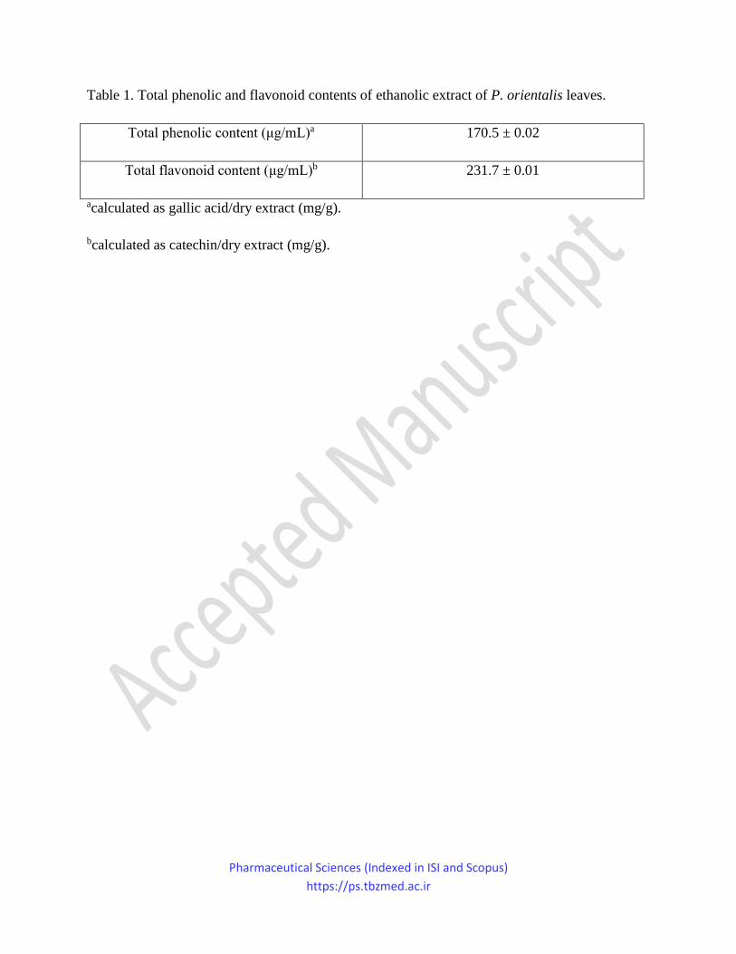
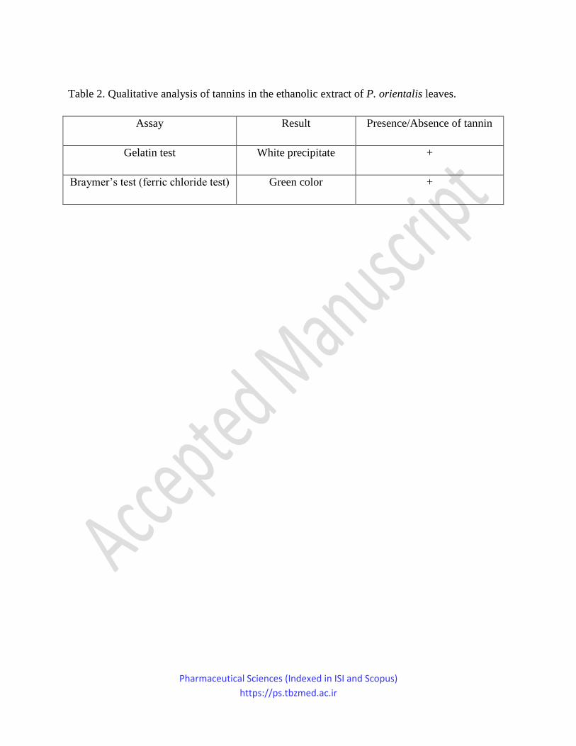
The yield of ethanolic extraction of P. orientalis leaves was calculated as 31.5%. The total
phenolic and flavonoid contents were shown in Table 1 and the results related to the qualitative
tannin tests were portrayed in Table 2.
Table 1
Table 2
1,1-Diphenyl-2-picrylhydrazyl (DPPH) radical scavenging activity
The ethanolic extract of P. orientalis leaves was investigated for its DPPH radical scavenging
activity in comparison to butylated hydroxyanisole (BHA) as the reference antioxidant agent.31
The ethanolic extract showed desirable antioxidant activity with IC50 value of 22.94 ± 0.03
µg/mL comparing with BHA (IC50 = 91.28 ± 0.13 µg/mL).
Wound healing activity
Pharmaceutical Sciences (Indexed in ISI and Scopus)
https://ps.tbzmed.ac.ir
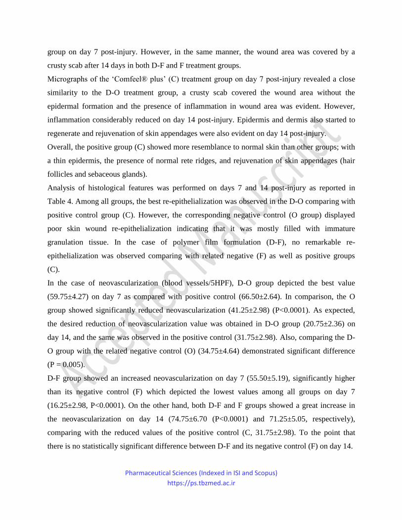
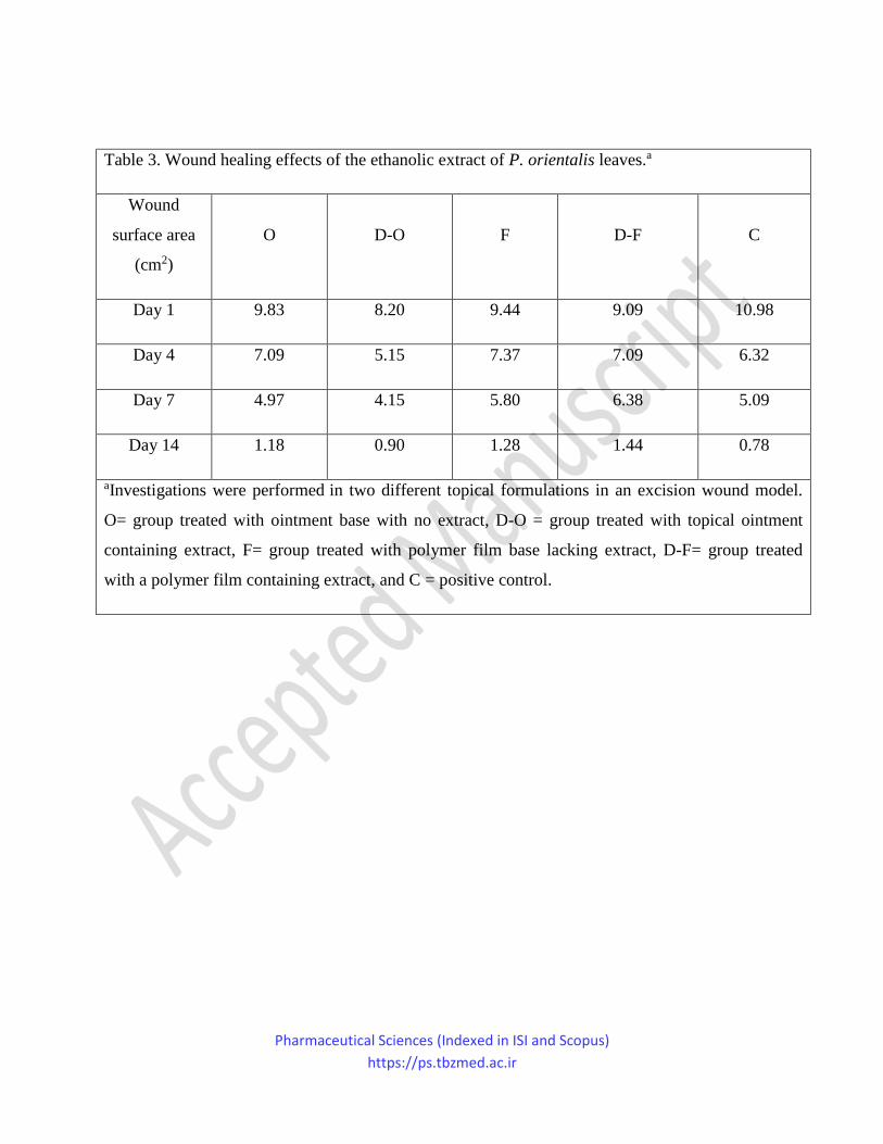
The decrease in the wound surface area was evaluated in different groups during 14 days. The
wound surface area was measured on days 4, 7, and 14 post-injury in all groups (Table 3). The
results were demonstrated in Fig. 1.
Table 3
Figure 1
According to the results shown in Table 3, wound surface area of the D-O group were
remarkably reduced compared to those of the ointment-negative control group (O) on days 4, 7
and 14 post-injury. On the other hand, the D-F group showed no important reduction in the
wound surface area compared to those of the negative control group (F). It could be argued that
the anti-inflammatory and astringent effects of polyphenolic compounds and tannins in the
ethanolic extract of P. orientalis contributed to the obtained results. Poor performance of D-F
group despite containing the same amount of extract can be associated with improper release of
the extract.
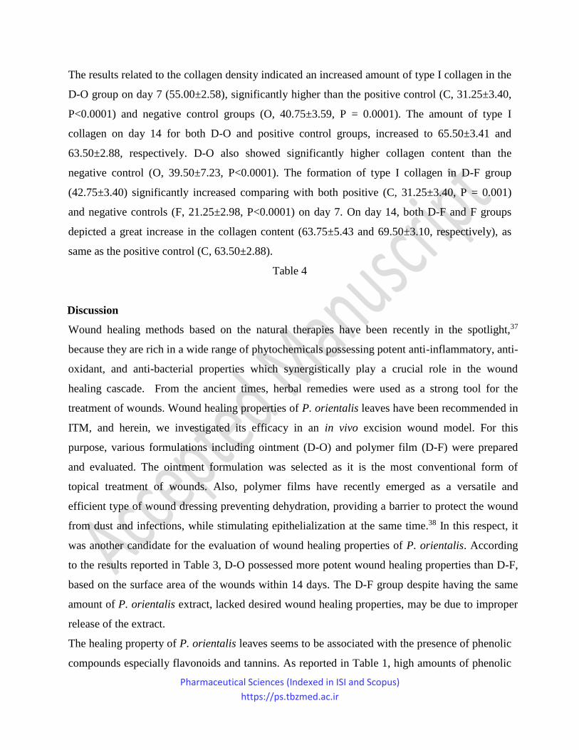
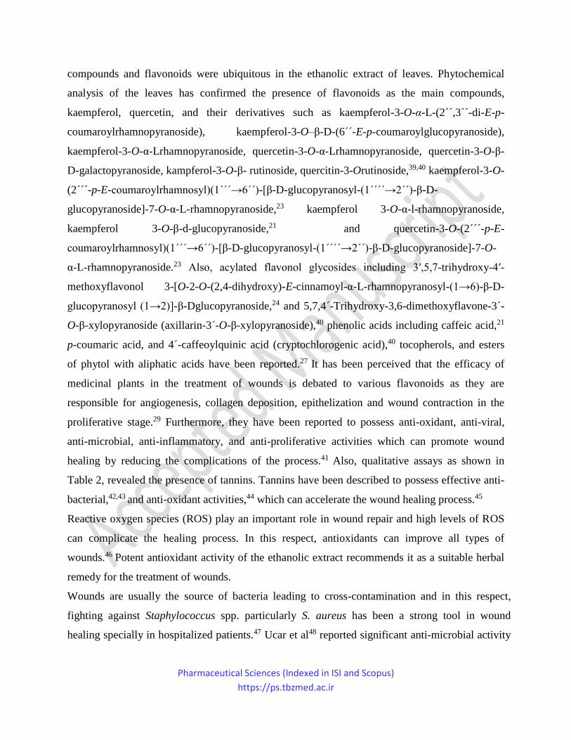
Histopathology study
The study was conducted by the collection of upper layers of treated tissues on days 7 and 14
post-injury, followed by H&E and MT staining as shown in Fig. 2 and 3.
Figure 2
Figure 3
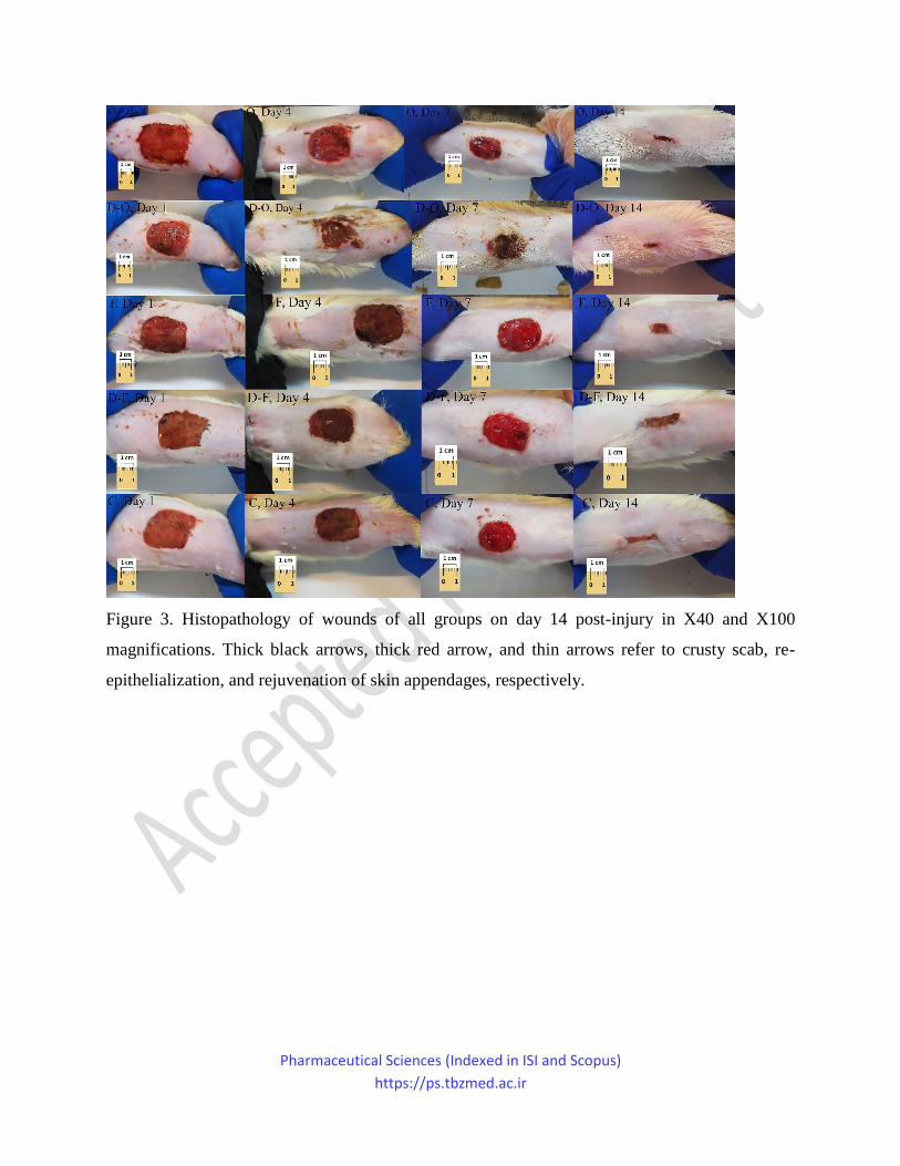
Micrographs of the ointment negative control group (O) on day 7 post-injury showed
inflammatory cells infiltration and the wound covered by a crusty scab. The epidermal layer has
not been formed within 14 days post-injury and the wound area filled by a highly vascularized
granulation tissue. Histopathological evaluation of the group D-O on day 7 showed the
infiltration of inflammatory cells into the defect area. Finally, on day 14 post-injury, the
epithelialization process was started and the inflammatory cells were significantly reduced in
comparison to the O treatment group at the same time point.
Histopathology of wounds treated by the film negative control group (F) showed severe
inflammatory response which significantly decreased within 14 days post-injury. On the other
hand, the inflammatory response in the D-F treatment group was considerably lower than the F
Pharmaceutical Sciences (Indexed in ISI and Scopus)
https://ps.tbzmed.ac.ir
group on day 7 post-injury. However, in the same manner, the wound area was covered by a
crusty scab after 14 days in both D-F and F treatment groups.
Micrographs of the ‘Comfeel® plus’ (C) treatment group on day 7 post-injury revealed a close
similarity to the D-O treatment group, a crusty scab covered the wound area without the
epidermal formation and the presence of inflammation in wound area was evident. However,
inflammation considerably reduced on day 14 post-injury. Epidermis and dermis also started to
regenerate and rejuvenation of skin appendages were also evident on day 14 post-injury.
Overall, the positive group (C) showed more resemblance to normal skin than other groups; with
a thin epidermis, the presence of normal rete ridges, and rejuvenation of skin appendages (hair
follicles and sebaceous glands).
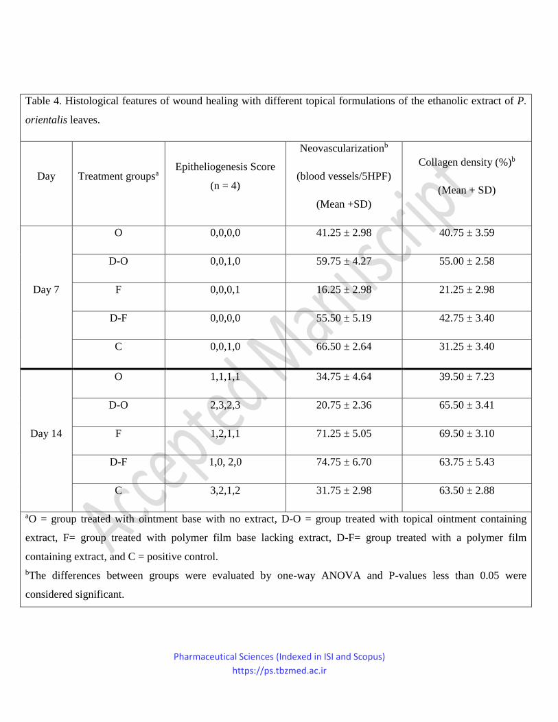
Analysis of histological features was performed on days 7 and 14 post-injury as reported in
Table 4. Among all groups, the best re-epithelialization was observed in the D-O comparing with
positive control group (C). However, the corresponding negative control (O group) displayed
poor skin wound re-epithelialization indicating that it was mostly filled with immature
granulation tissue. In the case of polymer film formulation (D-F), no remarkable re-
epithelialization was observed comparing with related negative (F) as well as positive groups
(C).
In the case of neovascularization (blood vessels/5HPF), D-O group depicted the best value
(59.75±4.27) on day 7 as compared with positive control (66.50±2.64). In comparison, the O
group showed significantly reduced neovascularization (41.25±2.98) (P<0.0001). As expected,
the desired reduction of neovascularization value was obtained in D-O group (20.75±2.36) on
day 14, and the same was observed in the positive control (31.75±2.98). Also, comparing the D-
O group with the related negative control (O) (34.75±4.64) demonstrated significant difference
(P = 0.005).
D-F group showed an increased neovascularization on day 7 (55.50±5.19), significantly higher
than its negative control (F) which depicted the lowest values among all groups on day 7
(16.25±2.98, P<0.0001). On the other hand, both D-F and F groups showed a great increase in
the neovascularization on day 14 (74.75±6.70 (P<0.0001) and 71.25±5.05, respectively),
comparing with the reduced values of the positive control (C, 31.75±2.98). To the point that
there is no statistically significant difference between D-F and its negative control (F) on day 14.
Pharmaceutical Sciences (Indexed in ISI and Scopus)
https://ps.tbzmed.ac.ir
The results related to the collagen density indicated an increased amount of type I collagen in the
D-O group on day 7 (55.00±2.58), significantly higher than the positive control (C, 31.25±3.40,
P<0.0001) and negative control groups (O, 40.75±3.59, P = 0.0001). The amount of type I
collagen on day 14 for both D-O and positive control groups, increased to 65.50±3.41 and
63.50±2.88, respectively. D-O also showed significantly higher collagen content than the
negative control (O, 39.50±7.23, P<0.0001). The formation of type I collagen in D-F group
(42.75±3.40) significantly increased comparing with both positive (C, 31.25±3.40, P = 0.001)
and negative controls (F, 21.25±2.98, P<0.0001) on day 7. On day 14, both D-F and F groups
depicted a great increase in the collagen content (63.75±5.43 and 69.50±3.10, respectively), as
same as the positive control (C, 63.50±2.88).
Table 4
Discussion
Wound healing methods based on the natural therapies have been recently in the spotlight,37
because they are rich in a wide range of phytochemicals possessing potent anti-inflammatory, anti-
oxidant, and anti-bacterial properties which synergistically play a crucial role in the wound
healing cascade. From the ancient times, herbal remedies were used as a strong tool for the
treatment of wounds. Wound healing properties of P. orientalis leaves have been recommended in
ITM, and herein, we investigated its efficacy in an in vivo excision wound model. For this
purpose, various formulations including ointment (D-O) and polymer film (D-F) were prepared
and evaluated. The ointment formulation was selected as it is the most conventional form of
topical treatment of wounds. Also, polymer films have recently emerged as a versatile and
efficient type of wound dressing preventing dehydration, providing a barrier to protect the wound
from dust and infections, while stimulating epithelialization at the same time.38 In this respect, it
was another candidate for the evaluation of wound healing properties of P. orientalis. According
to the results reported in Table 3, D-O possessed more potent wound healing properties than D-F,
based on the surface area of the wounds within 14 days. The D-F group despite having the same
amount of P. orientalis extract, lacked desired wound healing properties, may be due to improper
release of the extract.
The healing property of P. orientalis leaves seems to be associated with the presence of phenolic
compounds especially flavonoids and tannins. As reported in Table 1, high amounts of phenolic
Pharmaceutical Sciences (Indexed in ISI and Scopus)
https://ps.tbzmed.ac.ir
compounds and flavonoids were ubiquitous in the ethanolic extract of leaves. Phytochemical
analysis of the leaves has confirmed the presence of flavonoids as the main compounds,
kaempferol, quercetin, and their derivatives such as kaempferol-3-O-α-L-(2´´,3´´-di-E-p-
coumaroylrhamnopyranoside), kaempferol-3-O–β-D-(6´´-E-p-coumaroylglucopyranoside),
kaempferol-3-O-α-Lrhamnopyranoside, quercetin-3-O-α-Lrhamnopyranoside, quercetin-3-O-β-
D-galactopyranoside, kampferol-3-O-β- rutinoside, quercitin-3-Orutinoside,39,40 kaempferol-3-O-
(2´´´-p-E-coumaroylrhamnosyl)(1´´´→6´´)-[β-D-glucopyranosyl-(1´´´´→2´´)-β-D-
glucopyranoside]-7-O-α-L-rhamnopyranoside,23 kaempferol 3-O-α-l-rhamnopyranoside,
kaempferol 3-O-β-d-glucopyranoside,21 and quercetin-3-O-(2´´´-p-E-
coumaroylrhamnosyl)(1´´´→6´´)-[β-D-glucopyranosyl-(1´´´´→2´´)-β-D-glucopyranoside]-7-O-
α-L-rhamnopyranoside.23 Also, acylated flavonol glycosides including 3′,5,7-trihydroxy-4′-
methoxyflavonol 3-[O-2-O-(2,4-dihydroxy)-E-cinnamoyl-α-L-rhamnopyranosyl-(1→6)-β-D-
glucopyranosyl (1→2)]-β-Dglucopyranoside,24 and 5,7,4´-Trihydroxy-3,6-dimethoxyflavone-3´-
O-β-xylopyranoside (axillarin-3´-O-β-xylopyranoside),40 phenolic acids including caffeic acid,21
p-coumaric acid, and 4´-caffeoylquinic acid (cryptochlorogenic acid),40 tocopherols, and esters
of phytol with aliphatic acids have been reported.27 It has been perceived that the efficacy of
medicinal plants in the treatment of wounds is debated to various flavonoids as they are
responsible for angiogenesis, collagen deposition, epithelization and wound contraction in the
proliferative stage.29 Furthermore, they have been reported to possess anti-oxidant, anti-viral,
anti-microbial, anti-inflammatory, and anti-proliferative activities which can promote wound
healing by reducing the complications of the process.41 Also, qualitative assays as shown in
Table 2, revealed the presence of tannins. Tannins have been described to possess effective anti-
bacterial,42,43 and anti-oxidant activities,44 which can accelerate the wound healing process.45
Reactive oxygen species (ROS) play an important role in wound repair and high levels of ROS
can complicate the healing process. In this respect, antioxidants can improve all types of
wounds.46 Potent antioxidant activity of the ethanolic extract recommends it as a suitable herbal
remedy for the treatment of wounds.
Wounds are usually the source of bacteria leading to cross-contamination and in this respect,
fighting against Staphylococcus spp. particularly S. aureus has been a strong tool in wound
healing specially in hospitalized patients.47 Ucar et al48 reported significant anti-microbial activity
Pharmaceutical Sciences (Indexed in ISI and Scopus)
https://ps.tbzmed.ac.ir
of hydroalcoholic extract of P. orientalis leaves against S. aureus (MIC = 0.018 mg/ml). This can
be considered as a complementary aspect of the plant for inducing wound healing properties.
Using the proper formulation was found to be an important factor playing a significant role in the
wound healing process which was demonstrated by results obtained from histopathological
studies. As reported in Table 4, D-O group showed much better re-epithelialization than D-F
group, even comparable with the positive control group. This may be related to the presence of
flavonoids as they have been documented for their re-epithelialization rate enhancement, in the
proliferative phase for the improvement of cutaneous impairment.49
The neovascularization, formation of new blood vessels, has been found to be a vital process in
the four stages of wound healing including hemostasis, inflammation, proliferation and
remodeling. In the proliferative phase, extra vessels in the wound bed are essential to provide the
metabolic requirements of the cells responsible for repairing the impaired tissue. In the remodeling
phase, the neovascularization value is reduced to that of pre-injury state due to the reduction of
metabolic need.50 Failure of this process conflicts with wound healing leading to chronic ulcers.51
It has been reported that flavonoid glycosides induce neovascularization.52 The high amounts of
flavonoids in the P. orientalis leaves, confirmed the efficacy of D-O group in which the
corresponding value on day 14 (Table 4) reduced to approximately one-third of the value on day 7,
accelerating the wound healing process. The D-F group which showed no significant difference
comparing with F group, showed an increased neovascularization value on day 14 indicating
delayed proliferative phase or disrupted remodeling stage.
To sum up, comparison of D-O and D-F groups revealed no significant difference on day 7,
however, neovascularization in D-O group was significantly reduced comparing with D-F group
on day 14 (P<0.0001).
It has been determined that increase of type I collagen in wound healing process can bring about
contraction and wound closure. Early increase of type I collagen density in D-O group on day 7
was more significant than the positive control (C). The increase of collagen density was also
observed on day 14 in comparison to group C. It may be related to the stimulation of collagen
biosynthesis by flavonoids present in the plants leaves extract.53,54 Although the collagen content
of D-F group increased on days 7 and 14, other histological features did not endorse wound
healing process.

Pharmaceutical Sciences (Indexed in ISI and Scopus)
https://ps.tbzmed.ac.ir
Comparing the collagen density of D-O and D-F groups on day 7 indicated a significant difference
(P<0.001), while it was not statistically important on day 14.
The results reported in Table 4, related to epitheliogenesis score, neovascularization, and collagen
density, demonstrated more potent wound healing property of the ointment formulation (D-O)
than the film polymer type (D-F) which is in good agreement with those results reported in Table
3.
Conclusion
Herein, the wound healing effects of the ethanolic extract of P. orientalis leaves was studied in
two topical formulations including ointment and polymer film. The results indicated the higher
efficacy of ointment (D-O) than polymer film (D-F). Also, anti-bacterial activity of the extract
against S. aureus,44 and antioxidant property of the plant were found to be important factors in
reducing wound complications. Epitheliogenesis score, neovascularization, and collagen density as
significant histological features contributing to wound healing, were also studied. Those results
depicted that the early collagenization and induced neovascularization of the ointment group (D-
O), have greatly contributed to its efficacy comparing to the negative control (O).
Ethical Issues
The study was approved by the Committee of Ethics of the Faculty of Pharmacy of Tehran
University of Medical Sciences with approval number: IR.TUMS.VCR.REC.1397.479.
Acknowledgements
This research was supported by a grant from Tehran university of Medical Sciences with grant
No. 97-02-96-39137.
Conflict of Interest
The authors declare that there is no conflict of interests that could have appeared to influence the
content of this paper.
Author contributions
Pharmaceutical Sciences (Indexed in ISI and Scopus)
https://ps.tbzmed.ac.ir
Somayeh Niknam participated in the suggestion and preparation of formulations. Arezoo
Rastegari performed experimental part and contributed in the preparation of the manuscript.
Mahboubeh Bozorgi and Yasaman Vahedi-Mazdabadi contributed to the selection of the plant
and preparation of extracts. Mina Saeedi and Tahmineh Akbarzadeh designed the project and
supervised all processes.
References:
1. Shuid AN, Anwar MS, Yusof AA. The effects of Carica papaya Linn. latex on the healing of
burn wounds in rats. Malaysian journal of medicine and health sciences. 2005;3(2): 9–47.
2. Jalalpure SS, Agrawal N, Patil MB, Chimkode R, Tripathi A. Antimicrobial and wound
healing activities of leaves of Alternanthera sessilis Linn. International Journal of Green
Pharmacy. 2008; 2(3):141–4. doi: 10.22377/ijgp.v2i3.18.
3. Ayyanar M, Ignacimuthu S. Herbal medicines for wound healing among tribal people in
Southern India: Ethnobotanical and Scientific evidences. International journal of applied
research in natural products. 2009;2(3):29-42.
4. Ruszczak Z, Schwartz RA. Modern aspects of wound healing: An update. Dermatol surg.
2000;26(3):219-29. doi: 10.1046/j.1524-4725.2000.09215.x.
5. Stavrou D. Neovascularisation in wound healing. J wound care. 2008;17(7):298-302. doi:
10.12968/jowc.2008.17.7.30521.
6. Edwards R, Harding KG. Bacteria and wound healing. Curr Opin Infect Dis. 2004;17:91–6.
doi: 10.1097/00001432-200404000-00004.
7. Reuter J, Merfort I, Seelinger G, Wölfle U, Schempp CM. Botanicals in dermatology and skin
health. In: Cooper R, Kronenberg F, editors. Botanical medicine. From bench to beside. New
Rochelle, NY: Mary Ann Liebert Inc; 2009. p. 33–65.
8. Schmidt C, Fronza M, Goettert F, Geller F, Luik S, Flores EMM, et al. Biological studies on
Brazilian plants used in wound healing. J Ethnopharmacol. 2009;122:523–32. doi:
10.1016/j.jep.2009.01.022.
Pharmaceutical Sciences (Indexed in ISI and Scopus)
https://ps.tbzmed.ac.ir
9. Pazyar N, Yaghoobi R, Rafiee E, Mehrabian A, Feily A. Skin wound healing and
phytomedicine: a review. Skin Pharmacol Physiol. 2014;27(6):303-10. doi: 10.1159/000357477.
10. Khan AS. Woody Plants with Possible Anti-HIV Activity. In: Khan AS, editors. Medicinally
important trees. Switzerland: Springer International Publishing; 2017. p. 109–31.
11. Carpenter RJ, Hill RS, Jordan GJ. Leaf cuticular morphology links Platanaceae and
Proteaceae. Int J Plant Sci. 2005;166(5):843–55. doi: 10.1086/431806.
12. Haider S, Nazreen S, Alam MM, Hamid H, Alam MS. Anti-inflammatory and anti-
nociceptive activities of Platanus orientalis Linn. and its ulcerogenic risk evaluation. J
Ethnopharmacol. 2012;143(1):236-40. doi: 10.1016/j.jep.2012.06.029.
13. Khosropour E, Attarod P, Shirvany A, Pypker T, Bayramzadeh V, Hakimi L, et al. Response
of Platanus orientalis leaves to urban pollution by heavy metals. J For Res (Harbin).
2019;30:1437-45. doi: 10.1007/s11676-018-0692-8.
14. Janković B, Dodevski V, Stojmenović M, Krstić S, Popović J. Characterization analysis of
raw and pyrolyzed plane tree seed (Platanus orientalis L.) samples for its application in carbon
capture and storage (CCS) technology. J Therm Anal Calorim. 2018;133(1):465–80. doi:
10.1007/s10973-018-7207-x.
15. Bastos DZ, Pimentel IC, de Jesus DA, de Oliveira BH. Biotransformation of betulinic and
betulonic acids by fungi. Phytochemistry. 2007;68(6):834–9. doi:
10.1016/j.phytochem.2006.12.007.
16. Asadbeigi M, Mohammadi T, Rafieian-Kopaei M, Saki K, Bahmani M, Delfan M.
Traditional effects of medicinal plants in the treatment of respiratory diseases and disorders: an
ethnobotanical study in the Urmia. Asian Pac J Trop Med. 2014;7:364–8. doi: 10.1016/S1995-
7645(14)60259-5.
17. Shende S, Joshi KA, Kulkarni AS, Charolkar C, Shinde VS, Singh-Parihar V, et al. Platanus
orientalis Leaf Mediated Rapid Synthesis of Catalytic Gold and Silver Nanoparticles. J
Nanomed Nanotechnol. 2018;9(2):494. doi: 10.4172/2157-7439.1000494.
18. Nishanbaev SZ, Khidyrova NK, Kuliev ZA. Dimeric Proanthocyanidines from Platanus
orientalis bark. Chem Nat Compd. 2004;40:93. doi:10.1023/B:CONC.0000025479.07578.5d
19. Aliasl J, Khoshzaban F. Traditional Herbal Remedies for Burn Wound Healing in Canon of
Avicenna. Jundishapur J Nat Pharm Prod. 2013;8(4):192–6. doi : 10.17795/jjnpp-11686.
Pharmaceutical Sciences (Indexed in ISI and Scopus)
https://ps.tbzmed.ac.ir
20. Hajhashemi V, Ghannadi A, Mousavi S. Antinociceptive study of extracts of Platanus
orientalis leaves in mice. Res Pharm Sci. 2011;6(2):123-8.
21. Mitrokotsa D, Mitaku S, Demetzos C, Harvala C, Mentis A, Perez S, et al. Bioactive
compounds from the buds of Platanus orientalis and isolation of a new kaempferol glycoside.
Planta Med. 1993;59(06):517–20. doi: 10.1055/s-2006-959751.
22. Dimas K, Demetzos C, Mitaku S, Marselos M, Tzavaras T, Kokkinopoulos D. Cytotoxic
activity of kaempferol glycosides against human leukaemic cell lines in vitro. Pharmacol Res.
2000;41(1):83–6. doi: 10.1006/phrs.1999.0562.
23. El-Alfy TS, El-Gohary HMA, Sokkar NM, Al-Mahdy DA. Two novel acylated flavonol
glycosides from Platanus orientalis L. leaves. Nat Prod Commun. 2008;3:1899–902. doi:
10.1177/1934578X0800301121.
24. Tantry MA, Akbar S, Dar JA, Irtiza S, Galal A, Khuroo MA, et al. Acylated flavonol
glycoside from Platanus orientalis. Fitoterapia. 2012;83(2):281–5. doi:
10.1016/j.fitote.2011.11.004.
25. Nishanbaev SZ, Kuliev ZA, Khidyrova NK, Vdovin AD, Abdullaev ND, Shakhidoyatov
KhM, et al. New oligomeric proanthocyanidin glycosides Platanoside A and Platanoside-B from
Platanus orientalis trunk bark. Chem Nat Compd. 2010;46:357–62. doi: 10.1007/s10600-010-
9616-3.
26. Khidyrova NK, Rashkes YV, Rashkes AM. Abdullaev UA, Khodzhaeva MT, Shakhidoyatov
KhH, et al. Shed plane leaves as a source of α-tocopherol. Chem Nat Compd. 1995;31:312–4.
doi:10.1007/BF01165191.
27. Abdullaev UA, Rashkes YV, Khidyrova NK, Rashkes AM. Mass-spectrometric analysis of
phytol derivatives from the leaves of Platanus orientalis. Chem Nat Compd. 1994;30(3):332–8.
doi: 10.1007/BF00629969.
28. Prabu D, Nappinai M, Ponnudurai K, Prabhu K. Evaluation of woundhealing potential of
Pisonia grandis R. Br: A preclinical study in Wistar rats. Int J Low Extrem Wounds. 2008;7:21.
doi: 10.1177/1534734607314051.
29. Lai HY, Lim YY, Kim KH. Potential dermal wound healing agent in Blechnum orientale
Linn. BMC Complement Altern Med. 2011;12(11):62. doi: 10.1186/1472-6882-11-62.
30. Vahedi-Mazdabadi Y, Karimpour-Razkenari E, Akbarzadeh T, Lotfian H, Toushih M,
Roshanravan N, et al. Anti-cholinesterase and Neuroprotective Activities of Sweet and Bitter
Pharmaceutical Sciences (Indexed in ISI and Scopus)
https://ps.tbzmed.ac.ir
Apricot Kernels (Prunus armeniaca L.). Iran J Pharm Res. 2020;19(4):216-24. doi:
10.22037/ijpr.2019.15514.13139.
31. Rahmani-Nezhad S, Dianat S, Mahdizadeh V, Fooladi Z, Hariri R, Najafi Z, et al.
Investigation of polysaccharide extracts from Iranian and French strains of Agaricus
subrufescens against enzymes involved in Alzheimer’s disease. Bol Latinoam Caribe Plantas
Med Aromat. 2019;18(6):544-54.
32. Pandey A, Tripathi S. Concept of standardization, extraction and pre-phytochemical
screening strategies for herbal drug. J Pharmacogn Phytochem. 2014;2(5):115-9.
33. Singh V, Kumar R. Study of Phytochemical Analysis and Antioxidant Activity of Allium
sativum of Bundelkhand Region. International journal of life-sciences scientific research.
2017;3(6):1451-8.
34. Tanha S, Rafiee-Tehrani M, Abdollahi M, Vakilian S, Esmaili Z, Naraghi ZS, et al. G-CSF
loaded nanofiber/nanoparticle composite coated with collagen promotes wound healing in vivo. J
Biomed Mater Res A. 2017;105(10):2830-42. doi: 10.1002/jbm.a.36135.
35. Valizadeh A, Shirzad M, Pourmand MR, Farahmandfar M, Sereshti H, Amani A. Preparation
and Comparison of Effects of Different Herbal Oil Ointments as Wound-Healing Agents. Cells
Tissues Organs. 2019;207(3-4):177-86. doi: 10.1159/000503624.
36. Almasian A, Najafi F, Eftekhari M, Ardekani MRS, Sharifzadeh M, Khanavi M.
Polyurethane/carboxymethylcellulose nanofibers containing Malva sylvestris extract for healing
diabetic wounds: Preparation, characterization, in vitro and in vivo studies. Mater Sci Eng C
Mater Biol Appl. 2020;114:111039. doi: 10.1016/j.msec.2020.111039.
37. Solati K, Karimi M, Rafieian-Kopaei M, Abbasi N, Abbaszadeh S, Bahmani M.
Phytotherapy for Wound Healing: The Most Important Herbal Plants in Wound Healing Based
on Iranian Ethnobotanical Documents. Mini Rev Med Chem. 2021;21(4):500-19. doi:
10.2174/1389557520666201119122608
38. Leyva-Gómez G, González-Torres M, Alcalá-Alcalá S, Bernal-Chávez SA, Morales-Morfin
JC, González-Del Carmen M, et al. Development of films from natural sources for infections
during wound healing. Cell Mol Biol (Noisy-le-grand). 2021; 67(1):96-100. doi:
10.14715/cmb/2021.67.1.14.
Pharmaceutical Sciences (Indexed in ISI and Scopus)
https://ps.tbzmed.ac.ir
39. Dogan A, Anuk OO. Investigation of the phytochemical composition and antioxidant
properties of chinar (Platanus orientalis L.) leaf infusion against ethanol-induced oxidative stress
in rats. Mol Biol Rep. 2019;46:3049–61. doi: 10.1007/s11033-019-04741-7.
40. El-Alfy TS, El-Gohary HMA, Sokkar NM, Sleem AA, Al-Mahdy DA. Phenolic Constituents
of Platanus Orientalis L. Leaves. Nat Prodt Commun. February 2008;3(2):199-203. doi:
10.1177/1934578X0800300218.
41. Lodhi S, Singhai AK, Wound healing effect of flavonoid rich fraction and luteolin isolated
from Martynia annua Linn. on streptozotocin induced diabetic rats. Asian Pac J Trop Med.
2013;6:253-9. doi: 10.1016/S1995-7645(13)60053-X.
42. Akiyama H, Fujii K, Yamasaki O, Oono T, Iwatsuki K. Antibacterial action of several
tannins against Staphylococcus aureus. J Antimicrob Chemother. 2001;48(4):487-91. doi:
10.1093/jac/48.4.487.
43. Widsten P, Cruz CD, Fletcher GC, Pajak MA, McGhie TK. Tannins and extracts of fruit
byproducts: antibacterial activity against foodborne bacteria and antioxidant capacity. J Agric
Food Chem. 2014;62(46):11146–56. doi: 10.1021/jf503819t.
44. Figueroa-Espinoza MC, Zafimahova A, Alvarado PG, Dubreucq E, Poncet-Legrand C. Grape
seed and apple tannins: emulsifying and antioxidant properties. Food Chem. 2015;178:38-44.
doi: 10.1016/j.foodchem.2015.01.056.
45. Li K, Diao Y, Zhang H, Wang S, Zhang Z, Yu B, et al. Tannin extracts from immature fruits
of Terminalia chebula Fructus Retz. promote cutaneous wound healing in rats. BMC
Complement Altern Med. 2011;11:86. doi: 10.1186/1472-6882-11-86.
46. Fitzmaurice SD, Sivamani RK, Isseroff RR. Antioxidant therapies for wound healing: a
clinical guide to currently commercially available products. Skin Pharmacol Physiol.
2011;24(3):113-26. doi: 10.1159/000322643.
47. Almeida GCM, dos Santos MM, Lima NGM, Cidral TA, Melo MCN, Lima KC. Prevalence
and factors associated with wound colonization by Staphylococcus spp. and Staphylococcus
aureusin hospitalized patients in inland northeastern Brazil: a cross-sectional study. BMC Infect
Dis. 2014;14:328-35. doi: 10.1186/1471-2334-14-328.
48. Ucar E, Eruygur N, Atas M, Ergul M, Ergul M, Sozmen F. Determination of inhibitory
activities of enzymes, related to Alzheimer's disease and diabetes mellitus of plane tree (Platanus
Pharmaceutical Sciences (Indexed in ISI and Scopus)
https://ps.tbzmed.ac.ir
orientalis L.) extracts and their antioxidant, antimicrobial and anticancer activities. Cell Mol Biol
(Noisy-le-grand). 2018;64(11):13-9.
49. Carvalho MTB, Aráujo-Filho HG, Barreto AS, Quintans-Júnior LJ, Quintans JSS, Barreto
RSS. Wound healing properties of flavonoids: a systematic review highlighting the mechanisms
of action. Phytomedicine. 2021;153636. doi:10.1016/j.phymed.2021.153636.
50. Bodna RJ. Chemokine regulation of angiogenesis during wound healing. Adv Wound Care
(New Rochelle). 2015; 4(11): 641-50. doi: 10.1089/wound.2014.0594.
51. Sorg H, Tilkorn DJ, Hager S, Hauser J, Mirastschijski U. Skin Wound Healing: An Update
on the Current Knowledge and Concepts. Eur Surg Res. 2017;58(1-2):81-94. doi:
10.1159/000454919.
52. Xie F, Feng L, Cai W, Qiu Y, Liu Y, Li Y, et al. Vaccarin promotes endothelial cell
proliferation in association with neovascularization in vitro and in vivo. Mol Med Rep.
2015;12(1):1131-6. doi: 10.3892/mmr.2015.3503.
53. Galicka A, Nazaruk J. Stimulation of collagen biosynthesis by flavonoid glycosides in skin
fibroblasts of osteogenesis imperfecta type I and the potential mechanism of their action.
Int J Mol Med. 2007;20(6):889-95. doi: 10.3892/ijmm.20.6.889.
54. Kim YA, Tarahovsky YS, Gaidin SG, Yagolnik EA, Muzafarov EN. Flavonoids determine
the rate of fibrillogenesis and structure of collagen type I fibrils in vitro. Int J Biol Macromol.
2017;104(Pt A):631-7. doi: 10.1016/j.ijbiomac.2017.06.070.
Pharmaceutical Sciences (Indexed in ISI and Scopus)
https://ps.tbzmed.ac.ir
Figure 1. Wound healing process within 14 days.
Pharmaceutical Sciences (Indexed in ISI and Scopus)
https://ps.tbzmed.ac.ir
Figure 2. Histopathology of wounds of all groups on day 7 post-injury in X40 and X100
magnifications. Thick arrows reffer to crusty scab. The microscopic sections were stained using
hematoxylin and eosin (H&E) and Masson's trichrome (MT).
Pharmaceutical Sciences (Indexed in ISI and Scopus)
https://ps.tbzmed.ac.ir
Figure 3. Histopathology of wounds of all groups on day 14 post-injury in X40 and X100
magnifications. Thick black arrows, thick red arrow, and thin arrows refer to crusty scab, re-
epithelialization, and rejuvenation of skin appendages, respectively.
Pharmaceutical Sciences (Indexed in ISI and Scopus)
https://ps.tbzmed.ac.ir
Table 1. Total phenolic and flavonoid contents of ethanolic extract of P. orientalis leaves.
Total phenolic content (μg/mL)a 170.5 ± 0.02
Total flavonoid content (μg/mL)b 231.7 ± 0.01
acalculated as gallic acid/dry extract (mg/g).
bcalculated as catechin/dry extract (mg/g).
Pharmaceutical Sciences (Indexed in ISI and Scopus)
https://ps.tbzmed.ac.ir
Table 2. Qualitative analysis of tannins in the ethanolic extract of P. orientalis leaves.
Assay Result Presence/Absence of tannin
Gelatin test White precipitate +
Braymer’s test (ferric chloride test) Green color +
Pharmaceutical Sciences (Indexed in ISI and Scopus)
https://ps.tbzmed.ac.ir
Table 3. Wound healing effects of the ethanolic extract of P. orientalis leaves.a
Wound
surface area
(cm2)
O D-O F D-F C
Day 1 9.83 8.20 9.44 9.09 10.98
Day 4 7.09 5.15 7.37 7.09 6.32
Day 7 4.97 4.15 5.80 6.38 5.09
Day 14 1.18 0.90 1.28 1.44 0.78
aInvestigations were performed in two different topical formulations in an excision wound model.
O= group treated with ointment base with no extract, D-O = group treated with topical ointment
containing extract, F= group treated with polymer film base lacking extract, D-F= group treated
with a polymer film containing extract, and C = positive control.
Pharmaceutical Sciences (Indexed in ISI and Scopus)
https://ps.tbzmed.ac.ir
Table 4. Histological features of wound healing with different topical formulations of the ethanolic extract of P.
orientalis leaves.
Day Treatment groupsa Epitheliogenesis Score
(n = 4)
Neovascularizationb
(blood vessels/5HPF)
(Mean +SD)
Collagen density (%)b
(Mean + SD)
Day 7
O 0,0,0,0 41.25 ± 2.98 40.75 ± 3.59
D-O 0,0,1,0 59.75 ± 4.27 55.00 ± 2.58
F 0,0,0,1 16.25 ± 2.98 21.25 ± 2.98
D-F 0,0,0,0 55.50 ± 5.19 42.75 ± 3.40
C 0,0,1,0 66.50 ± 2.64 31.25 ± 3.40
Day 14
O 1,1,1,1 34.75 ± 4.64 39.50 ± 7.23
D-O 2,3,2,3 20.75 ± 2.36 65.50 ± 3.41
F 1,2,1,1 71.25 ± 5.05 69.50 ± 3.10
D-F 1,0, 2,0 74.75 ± 6.70 63.75 ± 5.43
C 3,2,1,2 31.75 ± 2.98 63.50 ± 2.88
aO = group treated with ointment base with no extract, D-O = group treated with topical ointment containing
extract, F= group treated with polymer film base lacking extract, D-F= group treated with a polymer film
containing extract, and C = positive control.
bThe differences between groups were evaluated by one-way ANOVA and P-values less than 0.05 were
considered significant.