Embed Size (px)
Citation preview

1
Dissertation on
A STUDY ON
INCIDENCE AND ETIOLOGY OF
VENTILATOR ASSOCIATED PNEUMONIA
Submitted to
THE TAMILNADU DR. M.G.R. MEDICAL UNIVERISTY
CHENNAI – 600 032
In partial fulfillment of the regulations
For the Award of the Degree of
M.D. (GENERAL MEDICINE)
BRANCH -1
MADRAS MEDICAL COLLEGE
CHENNAI – 600 003
MARCH - 2010

2
CERTIFICATE
This is to certify that the dissertation titled “ INCIDENCE AND ETIOLOGY
OF VENTILATOR ASSOCIATED PNEUMONIA ” is the bonafide original work
of Dr. E.RAJALAKSHMI in partial fulfilment of the regulation for M.D. Branch–I
(General Medicine) Examination of the Tamilnadu Dr. M.G.R Medical University to
be held in MARCH 2010. The Period of study was from JANUARY 2008 to JUNE
2009.
Prof.A.RADHAKRISHNAN,M.D. Professor Of Medicine, Madras Medical College and Research Institute, Govt. General Hospital, Chennai – 600 003.
Prof.C.RAJENDIRAN,M.D. Director,& professor Institute of Internal Medicine, Madras Medical College and Research Institute, Govt. General Hospital, Chennai -600 003.
Prof.J.MOHANASUNDARAM,M.D,DNB,PhD
Dean.Madras Medical College and Research
Institute, Govt.GeneralHospital,
Chennai-600 003.

3
DECLARATION
I, Dr.E.RAJALAKSHMI solemnly declare that dissertation titled
“INCIDENCE AND ETIOLOGY OF VENTILATOR ASSOCIATED
PNEUMONIA ” is a bonafide work done by me at Madras Medical College and
Govt. General Hospital from January 2008 to June 2009 under the guidance and
supervision of my unit chief prof.A.RADHAKRISHNAN,MD., Professor of
Medicine.
This dissertation is submitted to Tamilnadu Dr. M.G.R Medical
University, towards partial fulfilment of regulation for the award of M.D. Degree
(Branch – I) in General Medicine.
Dr.E.RAJALAKSHMI Post graduate student,
M.D.General medicine Institute of Internal Medicine,
Madras Medical College Govt. General Hospital, Chennai -600 003
Place: chennai Date :
4
ACKNOWLEDGEMENT
At the beginning I would like to thank my beloved Dean
Prof. J. MOHANASUNDARAM, M.D,DNB,PhD for having given me permission
to conduct this study and allowing me to utilize the resources of Madras Medical
College & Research Institute and Govt. General Hospital, Chennai.
I would like to express my sincere gratitude to my beloved Professor and
Director,Institute of Internal medicine Prof.C.RAJENDIRAN,M.D., for his guidance
and encouragement.
With extreme gratitude, I express my indebtedness to my beloved chief
Prof. A. RADHAKRISHNAN,M.D, for his motivation, advice and valuable criticism
which enabled me to complete this work.
I am extremely thankful to my Assistant Professors
Dr.SUBBURAGHAVALU, M.D. AND Dr.SRIDHAR, MD for their guidance and
encouragement.
I would always remember with extreme sense of thankfulness for the co-
operation and criticism shown by my post graduate colleagues.
5
I am immensely grateful to the generosity shown by the patients who
participated in this study.
CONTENTS
SL.NO TITLE PAGE NO
1 INTRODUCTION 1
2 AIMS AND OBJECTIVES 3
3 REVIEW OF LITERATURE 4
4 MATERIALS AND METHODS 38
5 RESULTS AND OBSERVATION 42
6 DISCUSSION 56
7
8
CONCLUSION
PERFORMA
BIBLIOGRAPHY
MASTER CHART
60
6
ABBREVATIONS
VAP - Ventilator associated pneumonia
TLC - Total leukocyte count
BAL - Bronchoalveolar lavage
ETA - Endotracheal aspiration
EO VAP - Early onset VAP
LO VAP -Late onset VAP
MRSA - Methicillin resistant staph. aureus
HAP - Hospital acquired pneumonia
BBS - Blind bronchial biopsy
CPIS -Clinical pulmonary infection score
ABG - Arterial blood gas
MDR - Multidrug resistance
SSD -Supraglottic secretion draining
CASS - Continuous aspiration of supraglottic secretions
QEA - Quantitative culture of endo tracheal aspirate
PTC -Protected telescopic catheter
MV -Mechanical ventilation
NP -Nosocomial pneumonia
ARDS -Acute respiratory distress syndrome

7
INTRODUCTION
Pneumonia is the second most common nosocomial infection in critically ,
affecting 27% of all critically ill patients. Pneumonia is defined as Nosocomial when
it occurs more than 48 hours after the patient’s admission to the hospital and when it
was not in incubation at the time of hospitalisation.2 Ventilator Associated Pneumonia
(VAP) is a subset of pneumonia and the term refers to nosocomial pneumonia in a
patient on mechanical ventilatory support (by endotracheal tube or tracheostomy) for
greater than or equal to 48 hours.
Ventilator-associated pneumonia (VAP) continues to be a major threat to patients
admitted in intensive care units (ICU) and receiving mechanical ventilation (MV).
Eighty-six percent of nosocomial pneumonias are associated with mechanical
ventilation and are termed ventilator-associated pneumonia (VAP). 3
A VAP arising 48 to 96 hours after tracheal intubation usually is called “early-onset
VAP”, and the one that occurs after this period as the “late-onset VAP” .Generally,
early-onset VAP has a better prognosis and is more likely to be caused by aspiration
of antibiotic-sensitive bacteria colonizing the oropharynx. Late-onset VAP may be

8
caused by more unusual or multidrug-resistant (MDR) pathogens and is associated
with greater morbidity and mortality.
Endotracheal intubation has been identified as a risk factor for developing
VAP.Critically ill patients who are intubated for more than 24 hours were found to be
at 6 to 21 times higher risk of developing ventilator-associated pneumonia and those
patients intubated for less than 24 hours are at 3 times the risk of VAP , compared to
non-intubated patients. Other risk factors for VAP include decreased level of
consciousness, gastric distention, and presence of gastric or small intestine tubes,
trauma, or COPD. VAP is reported to occur at rates of 10 to 35 cases / 1000 ventilator
days, depending on the clinical situation.
Aspiration of oral and /or gastric fluids is recognized to be an essential step in the
development of VAP. Pulmonary aspiration is increased by supine positioning and
pooling of secretions above the ET tube cuff . Estimates of attributable mortality are
variable,but increased duration of ventilation is a consistent finding, along with the
corresponding increase in hospital days and cost.
A major component of the problem is the ineffectiveness of therapy once VAP is
diagnosed. Brun-Buisson et al have demonstrated failure rates of 49 to 62% despite
the use of standard antibiotic combinations. Given the burden of VAP, both physical
and financial, and the difficulties in treatment, prevention strategies would appear to
be of paramount importance.

9
In this study incidence , etiology-the profile of the organisms, percentage of
EOP/LOP, underlying risk factors and their added morbidity & mortality were
analysed.
AIMS AND OBJECTIVES
1. To study the incidence and aetiology of VAP
2. To analyse the underlying risk factors for VAP
3. To study the percentage of early and late onset pneumonia in these patients
4. To study the morbidity and mortality attributed by VAP
10
REVIEW OF LITERATURE
Pneumonia is the second most common nosocomial infection in critically ill patients,
affecting 27% of all critically ill patients1 .Severe hospital acquired pneumonia (HAP)
continues to pose diagnostic and therapeutic challenges to the clinician. It is an
important clinical problem because it is common, causes significant increase in
mortality and increases duration of hospitalisation.
The incidence of HAP has been reported to range from 4 to10 cases per 1000
hospitalisations.3The large variation in the incidence of HAP is in part due to the
different criteria used in the diagnosis of HAP. Previous studies have shown mortality
rates from 8.9% to 70% with higher mortality in surgical patients, ventilated patients
and the type of causative organism.
Definition
NOSOCOMIAL PNEUMONIA

11
Pneumonia, which is parenchymal lung infection, is defined as Nosocomial when it
occurs more than 48 hours after the patient’s admission to the hospital and when
it was not in incubation at the time of hospitalization. Eighty-six percent of
nosocomial pneumonias are associated with mechanical ventilation and are termed
ventilator-associated pneumonia (VAP).VAP occurs in up to 25% of all people who
require mechanical ventilation.
Ventilator-associated pneumonia (VAP)
Sub-type of hospital-acquired pneumonia (HAP) which occurs in people who are on
mechanical ventilation through an endotracheal or tracheostomy tube for at
least 48 hours. VAP is a medical condition that results from infection which floods
the small, air-filled sac (alveoli) in the lung responsible for absorbing oxygen
from the atmosphere. VAP is distinguished from other kinds of infectious
pneumonia because of the different types of microorganisms responsible
antibiotics used to treat, methods of diagnosis, ultimate prognosis, and effective
preventive measures. In the community pneumonia is most often caused by
S.pneumoniae, H. influenzae, or S. aureus. However, in the hospital the organism
associated with pneumonia is most often Pseudomonas, regardless of whether or not
the patient is ventilated.5
The daily hazard rate for first episodes of VAP was high for the first several days
(3.3% per day at Day 5), and then decreased to 1.3% per day after Day 15,
documenting a dramatic decline in pneumonia over time. This is because the
12
intubation process itself contributes to the development of VAP. VAP occurring early
after intubation typically involves fewer resistant organisms and is thus associated
with a more favourable outcome.
Independent predictors of ventilator-associated pneumonia in their Cohort were a
primary admitting diagnosis of burns (risk ratio, 5.09 [95% CI, 1.52 to 17.03]), trauma
(risk ratio, 5.0 [CI, 1.91 to 13.11]), central nervous system disease (risk ratio, 3.40
[CI, 1.31 to 8.81]), respiratory disease (risk ratio, 2.79 [CI, 1.04 to 7.51]), cardiac
disease (risk ratio, 2.72 [CI, 1.05 to 7.01]), mechanical ventilation in the previous 24
hours (risk ratio, 2.28 [CI, 1.11to4.68]), witnessed aspiration (risk ratio, 3.25 [CI,
1.62 to 6.50]), and paralytic agents (risk ratio, 1.57 [CI, 1.03 to 2.39])
VAP is classified in to early-onset VAP (EOP) and late-onset VAP, defined with the
cut-off between 5 to 7 days of mechanical ventilation.
Symptoms and signs
People who are on mechanical ventilation are often sedated and are rarely able to
communicate. As such, many of the typical symptoms of pneumonia will either be
absent or unable to be obtained. The most important symptoms are fever, low body
temp, new purulent sputum, and hypoxia (decreasing amounts of oxygen in the
blood).
Risk factors 6 ,7
• Elderly age
13
• H/o transfer out from IMCU
• Witnessed aspiration
• Diabetes
• Reintubation
• Cerebral diseases
The spectrum of cerebral disease in these patients included meningitis,
meningoencephalitis, Parkinson’s disease, dementia and cerebral infarction. Patients
at risk for staphylococcal VAP include end-stage renal disease, comatose, or
neurosurgical patients,especially if nasal colonization is documented.25
Pathophysiology
VAP primarily occurs because the endotracheal or tracheostomy tube allows free
passage of bacteria into the lower segments of the lung in a person who often has
underlying lung or immune problems. Bacteria travel in small droplets both through
the endotracheal tube and around the cuff.27,28 Bacteria colonize the endotracheal or
tracheostomy tube and are embolized into the lungs with each breath. Bacteria may
also be brought down into the lungs with procedures such as deep suctioning or
bronchoscopy.
Whether bacteria also travel from the sinuses or the stomach into the lungs is,
controversial.26 However spread to the lungs from the blood stream or the gut is
uncommon. Once inside the lungs, bacteria then take advantage of any deficiencies in
the immune system (such as due to malnutrition or chemotherapy) and multiply. A

14
combination of bacterial damage and consequences of the immune response lead to
disruption of gas exchange with resulting symptoms.
The main route of VAP occurrence is aspiration of pathogenic gram-positive and
gram-negative bacteria, and colonization on the oropharynx and gastrointestinal tract.
Under normal conditions, the host defense, including filtration and humidification of
air in the upper airways, epiglottic and cough reflexes, ciliary transport by respiratory
epithelium, phagocytes in distal lung, and systemic cell mediated and humoral
immunity, prevent bacterial invasion.
In intensive care units, the host defenses of patients are usually distorted because of
their underlying diseases and invasive devices that are used. Patients are not able to
cough efficiently due to sedation or underlying disease. Also when they are intubated,
the endotracheal tube holds the vocal cords open and facilitates aspiration. As a
consequence, the endotracheal or tracheostomy tube allows free passage of bacteria
into the lower segments of the lung in a person who often has underlying lung or
immune problems. Bacteria travel in small droplets both through the endotracheal tube
and around the cuff.
Once bacteria reach the distal lung, they multiply and cause invasive disease.
Moreover, bacteria then take advantage of any deficiencies in the immune system of
the host to continue to multiply and worsen the condition. A combination of bacterial
damage and consequences of the immune response lead to disturbances of gas
exchange with resulting symptoms.

15
Morbidity and mortality associated with the development of VAP is high, with
mortality rates ranging from 20 to 41%.8 It has been shown that the development of
VAP increases the length on the mechanical ventilator by 4 days, critical care and
hospital lengths of stay (LOS) by 4 and 9 days, respectively and results in > $40,000
additional costs.9
Microbiology 5
The microbiologic flora responsible for VAP is different from that of the more
common community-acquired pneumonia (CAP). In particular, viruses and fungi are
uncommon causes in people who do not have underlying immune deficiencies.
Though any microorganism that causes CAP can cause VAP, there are several
bacteria which are particularly important causes of VAP because of their resistance to
commonly used antibiotics. These bacteria are referred to as multidrug
resistant(MDR).
Pseudomonas aeruginosa is the most common MDR Gram-negative bacterium
causing VAP. Pseudomonas has natural resistance to many antibiotics and has been
known to acquire resistance to every antibiotic except for polymyxin B. Resistance is
typically acquired through up regulation or mutation of a variety of efflux pumps

16
which pump antibiotics out of the cell. Resistance may also occur through loss of an
outer membrane porin channel (OprD)
Klebsiella pneumoniae has natural resistance to some beta-lactam antibiotics such as
ampicillin. Resistance to cephalosporins and aztreonam may arise through induction
of a plasmid-based extended spectrum beta-lactamase (ESBL) or plasmid-based
ampC-type enzyme.
Serratia marcescens has an ampC gene which can be induced by exposure to
antibiotics such as cephalosporins. Thus, culture sensitivities may initially indicate
appropriate treatment which fails due to bacterial response.
Enterobacter as a group also have an inducible ampC gene. Enterobacter may also
develop resistance by acquiring plasmids.
Citrobacter also has an inducible ampC gene.
Stenotrophomonas maltophilia often colonizes people who have endotracheal tubes or
tracheostomies but can also cause pneumonia. It is often resistant to a wide array of
antibiotics but is usually sensitive to co-trimoxazole.
Acinetobacter are becoming more common and may be resistant to carbapenems such
as imipenem and meropenem.
Burkholderia cepacia is an important organism in people with cystic fibrosis and is
often resistant to multiple antibiotics.
17
Methicillin-resistant Staphylococcus aureus is an increasing cause of VAP. As many
as fifty percent of Staphylococcus aureus isolates in the intensive care setting are
resistant to methicillin. Resistance is conferred by the mecA gene.
Diagnosis 10,11
VAP should be suspected in any person developing fever increasing numbers of
white blood cells on blood testing, and new shadows (infiltrates) on a chest x-ray.
Blood cultures may reveal the microorganism causing VAP.
Diagnostic criteria
A new and persistent (>48-h) infiltrate on chest radiograph 48 hours after admission to
hospital not explained by other pathology such as pulmonary oedema and not deemed
to be incubating at the time of admission into hospital, and Plus two or more of the
three criteria
(i) Fever of >38.3°C,
(ii) Leukocytosis of >12 × 109/ml, and/or
(iii) Purulent tracheobronchial secretions
This criteria has sensitivity of 69% and a specificity of 75% for establishing the
diagnosis of VAP.
Diagnostic strategy
Two strategies exist for diagnosing VAP. One strategy collects cultures from the
trachea of people with symptoms of VAP plus a new or enlarging infiltrate on chest x-

18
ray. The next is more invasive and advocates a bronchoscopy plus bronchoalveolar
lavage (BAL) for people with symptoms of VAP plus a new or enlarging infiltrate on
chest x-ray.In both cases, VAP is not diagnosed when cultures are negative and
another source of the symptoms is sought.
TAs are adequate specimens when strict definitional criteria (organisms on Gram
staining and fewer than 10 squamous epithelial cells per low-power field
[magnification, ×100]) are followed.
Although VAP spreads to the blood or pleural space in <10% of cases, if an organism
known to cause pneumonia is cultured in the setting of clinically suspected
pneumonia, treatment is warranted. Consequently, most experts recommend that two
sets of blood cultures and a thoracocentesis for nonloculated pleural effusions of ≥10
mm in diameter on a lateral decubitus chest radiograph should be part of the
evaluation of suspected VAP. 12 If the effusion is loculated, ultrasound guidance may
be required.
However, it is important to keep in mind not only that the sensitivity of blood cultures
for the diagnosis of VAP is less than 25% but also that when positive, the organisms
may originate from an extrapulmonary site of infection in as many as 64% of cases
and even when VAP is present.

19
Because of the poor specificity of the clinical diagnosis of VAP and of qualitative
evaluation of ETAs, Pugin et al. developed a composite clinical score, called the
clinical pulmonary infection score (CPIS), based on six variables:
(i) Temperature,
(ii) Blood leukocyte count
(iii) volume and purulence of tracheal secretion
(iv) oxygenation,
(v) pulmonary radiography, and
(vi) semi quantitative culture of tracheal aspirate. 13,14.
The score varied from 0 to 12. A CPIS of >6 had a sensitivity of 93% and a
specificity of 100%.
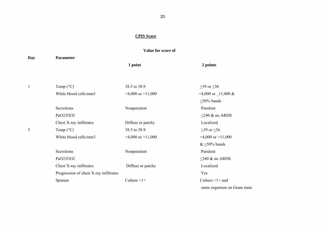
20
CPIS Score
Value for score of
Day Parameter
1 point 2 points
1 Temp (°C) 38.5 to 38.9 >39 or <36
White blood cells/mm3 <4,000 or >11,000 <4,000 or _11,000 &
>50% bands
Secretions Nonpurulent Purulent
PaO2/FiO2 <240 & no ARDS
Chest X-ray infiltrates Diffuse or patchy Localized
3 Temp (°C) 38.5 to 38.9 >39 or <36
White blood cells/mm3 <4,000 or >11,000 <4,000 or >11,000
& >50% bands
Secretions Nonpurulent Purulent
PaO2/FiO2 <240 & no ARDS
Chest X-ray infiltrates Diffuse or patchy Localized
Progression of chest X-ray infiltrates Yes
Sputum Culture >1+ Culture >1+ and
same organism on Gram stain

21
As multiple etiologies may explain why patients develop a fever and pulmonary infiltrates
while receiving mechanical ventilation, we have to search for other infectious and non-
infectious etiologies concurrently with evaluation for VAP. The extent of this investigation is
dictated by the clinical circumstances, including physical examination, laboratory findings,
and the severity of illness.
Evaluation for infectious (other than VAP) and non-infectious causes of fever
Action to be considered
i. Changing and/or culturing intravenous lines
ii. CT scan of sinuses, with fine needle aspirate if abnormal
iii. Evaluation for venous thromboembolism
iv. Clostridium difficile evaluation if diarrhea present
v. Abdominal ultrasound and/or CT scan (especially in the case
Abnormal abdominal physical examination, abnormal liver
function tests, elevated lipase/amylase, or presence of
predisposing factors (abdominal surgery, pancreatitis,
gastrointestinal bleed or malignancy, or high-dose corticosteroids)
vi. Lumbar puncture (especially in the case of a predisposing factor such as head trauma or
neurosurgical procedure)
vii. Drug fever
There is a general consensus that VAP is very likely in certain situations.
These circumstances are outlined below.

22
High probability of VAP
a) Radiographic evidence of cavitation or necrosis of the pulmonary
infiltrate, particularly if rapid and progressive
b) An empyema with an adjacent pulmonary infiltrate
c) Simultaneous recovery of the same microorganism from
respiratory secretions and pleural fluid
d) Simultaneous recovery of the same microorganism from
respiratory secretions and blood, with no other source of the bacteremia
e) Consistent histology on lung biopsy
f) Positive Gram stain/culture on transthoracic needle aspirate
g) Chest X ray demonstrating an air space process abutting a fissure& air bronchogram,
especially if single
Nonquantitative or semi quantitative airway sampling
Gram staining and nonquantitative and semi quantitative cultures of tracheal
secretions have the advantages of reproducibility and of requiring little technical
expertise and no specialized equipment or technique. However, these studies add little
to the sensitivity and specificity the clinical diagnosis of VAP, as the upper respiratory
tract is rapidly, within hours of intubation, colonized by potential pulmonary
pathogens, even when pneumonia is not present. Thus, if an organism is cultured or
noted on Gram stain, one does not know if it is
23
The cause of the pneumonia or simply colonization. In a study of 48
Patients with respiratory failure, concordance between tracheal non quqntitative
cultures and cultures of lung tissue from open lung biopsy was only 40% 96in that study, of
those patients with pneumonia on lung histology, endotracheal aspirate (ETA had a
sensitivity of 82% but a specificity of only 27%.
Only 15% of ETAs are adequate specimens when strict definitional criteria (organisms on
Gram staining and fewer than 10 squamous epithelial cells per low-power field
magnification, ×100) are followed .
Nonquantitative and semi quantitative cultures of ETAs for the diagnosis of VAP are most
useful if the specimen is adequate and antimicrobial therapy has not been added or changed
in the prior 72 h. The negative predictive value of these cultures in this setting is high (94%).
Quantitative cultures of airway specimens
To potentially improve the specificity of the diagnosis of VAP and the consequent
unnecessary antibiotic use and its associated problems, numerous studies have investigated
the role of quantitative cultures of respiratory secretions.
These have included nonbronchoscopic methods such as quantitative cultures of ETAs
(QEAs) and sampling of secretions from distal airways “blindly” via an endobronchial
catheter 15,16,17. Blind bronchial sampling (BBS), PSB, protected telescoping catheter (PTC),
BAL, and protected BAL (mini-BAL) samples can be obtained via the latter method.

24
Bronchoscopic sampling methods permit quantitative cultures of PSB, PTC, and protected
and nonprotected BAL specimens.18-21.
The PSB and PTC are double-sheathed catheters with a biodegradable plug occluding the
distal end of the inner catheter to minimize bacterial contamination.
The PSB and PTC procedures involve placing the tip of the bronchoscope or “blindly
placed” catheter next to an involved bronchial segmental orifice. With bronchoscopy,
direct visualization is possible.22 With a “blind” procedure, the catheter is advanced
until resistance is met and then retracted a few centimetres. The inner catheter is then
advanced 2 or 3 cm beyond the outer catheter, ejecting the plug. With PSB, a brush is further
advanced and rotated several times; with PTC, a 10-ml syringe is used to perform three brief
aspirations of secretions. BAL involves the infusion and aspiration of sterile saline through a
flexible fiber-optic bronchoscope or “blindly placed” catheter wedged into a bronchial
segmental orifice. Protected BAL involves a specialized balloon-tipped catheter with a distal
ejectable plug. When performing a BAL to diagnose VAP, instillation of at least 140 ml of
saline is required to maximize diagnostic yield .
If a bronchoscopically directed quantitative culture is chosen, the patient should receive
adequate sedation, with consideration of a short-acting paralytic agent to prevent coughing
during the procedure. The endotracheal tube must be ≥1.5 mm larger than the external
diameter of the flexible bronchoscope. The patient should receive a fraction of inspired
oxygen (FiO2) of 100%, and positive-end expiratory pressure should be reduced as much as
tolerated. To maximize ventilation and minimize air trapping, the peak inspiratory flow

25
should be decreased to ≤60 litres/min, the respiratory rate set between 10 and 20 breaths/min,
and the peak inspiratory pressure alarm increased.
The patient should be carefully monitored throughout the procedure, with particular attention
to exhaled tidal volume, peak inspiratory pressure, oxygen saturation, the electrocardiogram,
and vital signs. Secondary hypotension should be anticipated, and appropriate intravenous
fluids and vasopressors should be available for immediate administration .
The sampling area should be chosen based on the location of the infiltrate on chest X ray or
CT scan. This typically corresponds to the bronchial segment with purulent secretions and/or
where endobronchial abnormalities are maximal, which can be clues in the setting of diffuse
pulmonary infiltrates or minimal changes in a previously abnormal chest X ray . When in
doubt, sample the posterior right lower lobe, since autopsy studies have indicated that VAP
frequently involves this area.
The presence of more than 1% epithelial cells or 10 epithelial cells per low-power field
(magnification, ×100) in bronchoscopic or “blind” BAL, PSB, PTC, or bronchial sampling
suggests heavy oropharyngeal colonization. Returns of <10% of the instilled BAL fluid are
probably not representative of the lower respiratory tract . For QEAs, the same criteria
mentioned above for nonquantitative and semi quantitative cultures of an ETA should be For
each of the quantitative culturing methods, threshold values have been derived and are
expressed in CFU per millilitre. If the number of CFU/ml is equal to or exceeds the threshold
values for the particular technique, a diagnosis of pneumonia is made. Threshold values often
employed for diagnosing pneumonia by quantitative cultures are ≥105 to 106, ≥104, and ≥103
CFU/ml for QEA, bronchoscopic BAL, and PSB, respectively, with ≥105 CFU/ml being the
26
most widely accepted value for QEA . For “blind” distal sampling, the thresholds are ≥103
CFU/ml for PSB and mini-BAL and ≥104 CFU/ml for cultures obtained with BBS and
unprotected utilized.
Treatment
Treatment must be obtained quickly and treatment initiated without delay .Principles to apply
when choosing appropriate therapy for VAP include knowledge of organisms likely to be
present, local resistance patterns within the ICU, a rational antibiotic regimen, and a rationale
for antibiotic de-escalation or stoppage. Early effective therapy for VAP is associated with
reduced mortality. Luna et al. demonstrated that inadequate therapy during the initial 48 h,
despite provision of adequate therapy after BAL results, was associated with a mortality rate
of 91% . When empirical therapy was appropriate, mortality rates were much lower (38%).
Delays in the administration of appropriate antibiotic therapy for VAP have been associated
with excess mortality . In one study, a delay in appropriate therapy for 24 h or more was
associated with a 69.7% mortality, compared to 28.4% in patients treated without the
delay
(P < 0.001).23,24
A low threshold for suspicion of VAP is needed when a patient's clinical course deteriorates.
The day 1 CPIS can be useful, especially when combined with quantitative cultures. The
choice of which quantitative culture methodology to use is an open debate. However,
diagnostic cost favours QEA, which can also be implemented as a surveillance technique.
27
Antibiotic administration should be promptly initiated when VAP is suspected and
quantitative cultures obtained and should be broad in coverage. Knowledge of local
antibiograms should guide the choice of antibiotics, in addition to likelihood of organisms
(early- or late-onset VAP). For patients already on antibiotics at the time of suspected VAP,
the clinician should choose antibiotics from different classes, as it is likely that resistance to
“in-use” antibiotics has developed.
Assessment of the likelihood of VAP by day 3 is needed to decide whether antibiotics should
be continued. The assessment should include a repeat CPIS, as the change in CPIS can guide
clinical decisions, even stoppage of antibiotics. Assessment of quantitative culture results and
sensitivities at this juncture is prudent, as it may permit early antibiotic de-escalation by
choosing a more narrowly focused agent(s). Monotherapy may be appropriate in many
instances of VAP and should reduce the incidence of drug resistance. A change to
monotherapy may be possible in a responding patient where organism sensitivity results
permit. A short course (6 to 8 days) can be administered to patients with VAP but is
dependent on the patient physiologic response to treatment along with which organisms have
been recovered
Treatment of VAP should be matched to known causative bacteria. However, when VAP is
first suspected, the bacteria causing infection is typically not known and broad-spectrum
antibiotics are given (empiric therapy) until the particular bacterium and its sensitivities are
determined. Empiric antibiotics should take into account both the risk factors a particular
individual has for resistant bacteria as well as the local prevalence of resistant
microorganisms. If a person has previously had episodes of pneumonia, information may be
28
available about prior causative bacteria. The choice of initial therapy is therefore entirely
dependent on knowledge of local flora and will vary from hospital to hospital.
ATS has recently published guidelines to guide empirical antibiotic choices .These guidelines
are divided into those for patients at risk for VAP caused by multidrug-resistant organisms
and those for patients without such risk. Risk factors for multidrug-resistant organisms
include prior antimicrobial therapy in the preceding 90 days, current hospitalization
exceeding 5 days (not necessarily ICU days), high frequency of resistance in the community
or local hospital unit, and immunosuppressive disease and/or therapy and ventilation for
more than five days, Residence in a nursing home, Treatment in a haemodialysis clinic. .
People who do not have risk factors for MDR organisms may be treated differently
depending on local knowledge of prevalent bacteria.
In the absence of risk factors for multidrug-resistant bacteria, the clinician should choose
empirical therapy for Streptococcus pneumoniae, Haemophilus influenzae,
methicillin-sensitive Staphylococcus aureus, and antibiotic-sensitive gram-negative
enteric organisms. Antibiotic choices include ceftriaxone, quinolones (levofloxacin,
moxifloxacin, or ciprofloxacin), ampicillin/sulbactam, or ertapenem.
Antibiotic choice can be tailored to the pathogens’ last sensitivity report should QEA
surveillance cultures be obtained twice weekly and should the growth level exceed 100,000
CFU/ml.
When risk factors for multidrug-resistant organisms are present, the clinician must consider
not only the organisms listed above but also Pseudomonas aeruginosa, Klebsiella,
29
Enterobacter, Serratia, Acinetobacter, Stenotrophomonas maltophilia, Burkholderia cepacia,
and methicillin-resistant S. aureus. Empirical therapy is broadened to include
(i) Either an antipseudomonal cephalosporin (cefepime or ceftazadime), an
antipseudomonal carbepenem (imipenem or meropenem), or a β-lactam/β-
lactamase inhibitor (pipercacillin-tazobactam) plus
(ii) An antipseudomonal fluoroquinolone (ciprofloxacin or levofloxacin) or an
aminoglycoside (amikacin, gentamicin, or tobramycin) plus linezolid or
vancomycin30.
Possible empirical therapy combinations include (but are not limited to):
• Vancomycin/linezolid and ciprofloxacin,
• Cefepime and gentamicin/amikacin/tobramycin
• Vancomycin/linezolid and ceftazidime
• Ureidopenicillin plus β-lactamase inhibitor such as piperacillin/tazobactam or
ticarcillin/clavulanate
• Carbapenem (e.g., imipenem or meropenem)
Therapy is typically changed once the causative bacteria are known and continued until
symptoms resolve (often 7 to 14 days).
There is ongoing research into inhaled antibiotics as an adjunct to conventional therapy.
Tobramycin and polymyxin B are commonly used in certain centers but there is no clinical
evidence to support their use.
30
PREVENTION
General measures
Prevention of VAP involves limiting exposure to resistant bacteria, discontinuing mechanical
ventilation as soon as possible, and a variety of strategies to limit infection while intubated.
Resistant bacteria are spread in much the same ways as any communicable disease. Proper
hand washing, sterile technique for invasive procedures, and isolation of individuals with
known resistant organisms are all mandatory for effective infection control. A variety of
aggressive weaning protocols to limit the amount of time a person spends intubated have
been proposed. One important aspect is limiting the amount of sedation that a ventilated
person receives.
Specific measures
The ability to prevent EOP is clearly greater than that of late-onset VAP. Many of the best-
validated strategies for VAP prevention, including CASS, are for EOP.EOP can also be
prevented by other strategies, such as by simply administering ventilation with patients in the
semirecumbent position,prophylactic short course, high dose antibiotic therapy, and others
Having the ability to prevent late-onset VAP is much more difficult. The pathogenesis is
different than EOP.
Antibiotic selective pressure and cross infection are themes that common to late-onset VAPs.
Many late-onset VAPs, especially Pseudomonas,occurwithout preceding oropharyngeal or
gastric colonization the target of many prevention strategies.
31
No prevention strategy has shown a clear-cut benefit for late-onset VAP. The best prevention
strategies may actually be an accurate diagnosis of EOP and avoidance of antibiotics as much
as possible31.
Potential strategies to prevent VAP
I. Prior to intubation
(i) Address reversible causes of respiratory failure-bronchospasm
,analgesia,sedation
(ii) Non invasive mechanical ventilation
II. Process of intubation
1) Avoid gastric distension
2) Oroendotracheal route
III. After intubation(data supported)
1) Oral route gastric tube
2) Head end elevation 30-45 degrees
3) Good hand hygiene
4) Closed suctioning
5) Continuous sub glottis suctioning
6) Rotational beds
7) Chlorhexidine oral rinse in cardiac patients

32
8) Minimise sedation
9) Weaning protocols
IV. After intubation(controversial)
1) Early vs late enteral nutrion
2) Selective gut decontamination
3) Rotational antibiotic schema
4) Antibiotic impregnated endotracheal tube
Non-invasive mechanical ventilation (NIV) has been associated with more favourable
outcomes (mortality and morbidity) in comparison to endotracheal tube placement in patients
with acute exacerbations of chronic obstructive pulmonary disease or acute pulmonary
oedema .32,-35
The incidence of nosocomial pneumonia was reduced in the group randomized to NIV
Furthermore, immunocompromised patients with bilateral infiltrates also benefited from NIV
over invasive mechanical ventilation (IMV) with regard to both mortality and morbidity. 34
Once the decision to intubate is made, the practice of VAP prevention should be directed at
reducing colonization and aspiration (volume of organisms presented to the lungs). This
begins with choosing the oral route of intubation and focusing on minimizing the duration of
mechanical ventilation (DOMV).

33
Oral intubation is preferred over nasal intubation, as the latter has been associated with both
VAP and sinusitis, with the same bacteria identified in both. Rouby et al., demonstrated a
significant reduction in nosocomial sinusitis when patients are orally cannulated with
endotracheal and gastric tubes.35
Holzapfel et al. have linked the reduction in nosocomial sinusitis to a reduction in VAP.36
Furthermore, the clinician must give careful attention to the mundane and seemingly small
interventions, such as regularly assessing endotracheal cuff pressure, performing
endotracheal suctioning, draining ventilator tube condensate, avoiding gastric overdistention,
avoiding the supine position, avoiding unnecessary ventilator circuit changes, application of
heat and moisture exchangers (HMEs) when appropriate, minimizing out-of-ICU transports,
and regular hand cleaning with soap or alcohol disinfectant.
Maintaining cuff pressure of endotracheal tubes at ≥20 mm Hg reduces nosocomial
pneumonia, presumably by minimizing the passage of oropharyngeal contents into the
trachea. 37
The duration of intubation directly affects the likelihood of VAP, which is more evident in
patients with ICU LOS exceeding 5 days. Fagon et al. suggested that the incidence of VAP
increases by 1% per day of IMV . However, Cook et al. found that the incidence per day
varies over time, with 3% per day during first 5 days of IMV, 2% for the second 5 days, and
1% for the subsequent 5-day period 38. This observation is supported by Ibrahim et al., who
identified an incidence rate of VAP of 11.5%, 56% of which were early onset (≤5 day) 39.
Hence, the greatest attack rates appear to be during the initial days of mechanical ventilation.

34
Additionally, significant risk factors for early-onset VAP include cardiopulmonary
resuscitation and continuous sedation.40
Continuous sedation is more often administered in the acute phase of an illness. In addition to
treating the primary cause of respiratory failure, the DOMV can be reduced through judicious
use of sedatives and analgesics.
Studies by Brook et al. and Kress et al. have demonstrated that protocols for sedative and
analgesic administration with the goal of minimizing constant infusions led to reduced
DOMV.41,42 Furthermore, daily interruption of sedation results in a reduced incidence of
intensive care unit complications, in which VAP was included. 43,44.
Weaning protocols have also resulted in reduced DOMV, whether respiratory therapist
initiated or not.45,46
Patients should be cared for in the semi recumbent position to reduce the extent of aspiration,
especially when receiving enteral feeds.
Radionuclide studies reveal increased tracheal penetration of gastric contents when intubated
patients are supine 47 Drakulovic et al. found that the simple elevation of the head of bed to
45° results in dramatic reductions in VAP incidence and a trend toward reduced mortality 48.
Nonetheless, a recent survey by the University Hospital Consortium revealed that compliance
with the simple and no-cost intervention of elevating the head is woefully low, and a study by
Heyland et al. revealed that the head of bed is on average elevated to 29° and not 45°. 49 .
Compared to supine positioning, studies have shown that simple positioning of the head of
bed to 30° or higher significantly reduces gastric reflux and VAP (8% versus 34%,

35
respectively). Kinetic bed therapy has also led to a reduction in the incidence of VAP.50,51
However, this is costly and has not been directly compared to head-of-bed elevation, a no-
cost option.
Some VAP is contracted from inhalation of bacteria through the ventilator circuit and may be
a result of contaminated aerosols, condensate, or suction catheters. Traditionally, ventilator
circuit changes have been on a regular schedule and often daily. However, the data
examining this practice reveal that there is no benefit to changing the circuit on a regular
basis, and the present recommendations are to change the circuit when soiled .52 Such a
practice would likely reduce the rate of accidental spillage of condensate into the airway.
As heated humidifiers enhance the amount of condensate, attention has been focused on
HMEs. These devices have led to a reduction in VAP, albeit small, and should be used in
patients without significant secretions or concern over the risk of obstruction.53,54. While
changing the HME less frequently than every 48 h may lead to further reductions in VAP,
care must be taken to carefully monitor for trapped secretions and subsequent airway
obstruction or increments in the work of breathing55.
Endotracheal suctioning of intubated patients can be performed through an open or closed
system. In theory the closed system could reduce the incidence of VAP, but in practice this
has not been demonstrated .56,57 Cost analysis favours the closed system, as the enveloped
catheter can be reused for suctioning and needs to be changed only when dysfunctional. 58
However, respiratory therapists have voiced concerns over residue build-up within the lumen
of the endotracheal tube.
36
As most VAP follows from aspiration of oropharyngeal secretions, attention to proper cuff
inflation pressures and endotracheal suctioning can affect the volume presented to the
trachea.
The application of continuous suction of subglottic secretions through specialized
endotracheal tubes will reduce the incidence of VAP .59,60 Surprisingly, this was not
associated a reduction in mortality, ICU LOS, or duration of mechanical ventilation.
While studying the application of continuous subglottic suctioning, Rello et al. noted a trend
of increased VAP in patients with endotracheal cuff pressures of <20 cm H2O61. Hence, it is
recommended not only to assess cuff pressure for tracheal ischemia (which occurs when
pressure exceeds 30 cm H2O) but also to ensure that adequate cuff pressure (>20 mm Hg) is
present.
American and Canadian guidelines strongly recommend the use of supraglottic secretion
drainage (SDD) Special tracheal tubes with an incorporated suction lumen as the EVAC
tracheal tube form Covidien / Mallinckrodt can be used for that reason. studies on the use of
special ET tubes which remove secretions pooled above the cuff with continuous suction
decrease VAP by 45 to 50 % (Cook, De Jonghe, Brochard, & Brun-Buisson, 1998; Valles et
al., 1995). specialized endotracheal tubes that allow continuous aspiration of subglottic
secretions.
The endotracheal tube itself is a reservoir for gram-negative bacteria. The buildup of a
biofilm within endotracheal tubes occurs frequently. One study demonstrated that 84% of
endotracheal tubes examined had a biofilm 62. As documented by Inglis et al., this biofilm is

37
heavily laden with bacteria, usually gram-negative organisms.63,64 At present, ongoing
studies are directed at either eliminating this biofilm or reducing the bacterial load associated
with it.
New cuff technology based on polyurethane material in combination with subglottic drainage
(SealGuard Evac tracheal tube from Covidien/Mallinckrodt) showed significant delay in
early and late onset of VAP.[2]A recent clinical trial indicates that the use of silver-coated
endotracheal tubes may also reduce the incidence of VAP.
Oral decontamination with chlorhexidine has been shown to reduce the incidence of VAP in
patients undergoing cardiac surgery, presumably by reducing oropharyngeal colonization.65
Furthermore, numerous studies with oral decontamination antibiotic pastes alone or co
administered with systemic antibiotics have shown a reduction in early VAP.66-70 Two meta-
analyses have suggested better results with oral decontamination alone than with the
combination of oral and systemic prophylaxis. 71,72 With either approach, however, concern
over the emergence of antibiotic-resistant organisms has tempered use, as has the labor
intensity required to apply these regimens at the bedside. This is particularly true in ICUs
housing organisms with high antibiotic resistance rates. 73-76 While often recommended, it
appears not to be routinely practiced. Two recent studies will further the debate, as they
demonstrated significant reductions in VAP and mortality with selective decontamination of
the digestive tract. 77,78 These two studies were performed under conditions where selective

38
decontamination of the digestive tract is most effective, i.e., surgical intensive care units
housing patients less likely to be colonized with resistant bacteria.
Gastric volumes and acidity affect the incidence of VAP. Reducing the acidity of gastric
secretions and feeding will reduce bacterial overgrowth. However, in high-risk patients
(ventilated for >48 h and coagulopathic), the risk of bleeding outweighs the risk of VAP from
pH-modifying agents.79 Hence, it is difficult to recommend against H2 blockers or proton
pump inhibitors. Sucralafate may indeed be superior from the viewpoint of VAP, but it is less
effective with regard to prophylaxis of gastrointestinal bleeding, and thus it use is not
warranted over H2 blockers or proton pump inhibitors.80-82.
Multiple studies have examined post pyloric versus gastric feedings with regard to incidence
of aspiration and development of VAP. These studies were small and inconclusive. In a
meta-analysis, post pyloric feedings reduce the incidence of VAP and increased the nutrition
delivered. 83However, no single trial demonstrated that post pyloric tube feedings prevent
VAP. The improved delivery of nutrition was likely the result of decreased gastric residual
assessments and consequently fewer stoppages in continuous tube feedings.
A recent publication favoured a delay of greater than 5 days before initiating tube feedings,
as the incidence of VAP was reduced 84. Further data are needed to unconditionally embrace
this practice.
Preventing Multidrug Resistance
Antibiotic cycling remains controversial. Employing a rotational schedule for empirical
antibiotic administration for suspected VAP may indeed lead to a reduced incidence of
39
resistant organisms. 85-87 While such a strategy may not reduce the incidence of VAP,
reductions in mortality may be seen.87 This is likely a result of changes in resistance patterns
resulting in a higher likelihood of choosing appropriate antibiotic regimens 88. Because
rotational schedules have primarily targeted reducing the resistance of gram-negative
organisms, we do not know the impact of rotating antibiotics against gram-positive
organisms, such as methicillin-resistant S. aureus. Furthermore, the frequency with which
to rotate antibiotics remains unclear, as monthly and quarterly regimens have been assessed
with documented successes. 87 Furthermore, the probability of antibiotic cycling leading to a
reduction in antimicrobial resistance is low as determined through mathematical modelling.
89 At this juncture, it is premature to recommend rotating antibiotics or a rotational schema.
Multidrug resistance can also be reduced when patient-antibiotic PK/PD characteristics are
accounted for. Early eradication minimizes the opportunity for a population of organisms to
develop resistance. Peak concentrations for aminoglycosides 10-fold greater than MIC appear
to inhibit the emergence of resistant organisms. 90,91 When choosing fluoroquinolones,
resistant organisms are less likely to be seen when the 24-h area-under-the-curve/MIC levels
are >100 for gram-negative bacteria and >40 for gram-positive bacteria.92 Changes in
medication frequency or infusion rates can increase the time that the antibiotic concentration
exceeds the MIC. For β-lactams, monobactams, glycopeptides, and cabapenems this can be
important in enhancing bactericidal activity, again reducing opportunities for resistant
organisms to emerge.93
In summary, several opportunities to reduce the incidence of VAP are available to the
clinician. Many are no-cost or minimal-cost interventions and should be implemented as part

40
of routine care protocols. Care of the critically ill should be directed at applying interventions
that reduce mortality, minimize morbidity, shorten the length of stay, and reduce cost.
Reducing VAP through the simple measures outlined does exactly that. We recommend that
the clinician's practice include non-invasive mechanical ventilation over intubation when
appropriate, oral intubation when an endotracheal tube is necessary, orogastric over
nasogastric tubes, elevation of the head to at least 30°, minimization of sedation,
administration of a proton pump inhibitor when prophylaxis is indicated, a frequency of
ventilator tubing changes at 7 days or when soiled, avoidance or elimination of endotracheal
tube leak, good technique in removal of condensate, and of course excellent hand hygiene. At
this time we do not support the routine use of endotracheal tubes with subglottic suction
capabilities, rotational beds, in-line suction systems, rotational antibiotic schemes, or
selective gut decontamination.
Strategies and a more thorough discussion on prevention are within the ATS/Infectious
Disease Society of America statement and papers by Kollef and by Dodek et al94 Zack et al.
have demonstrated that a multifaceted and multidisciplinary approach to VAP prevention can
indeed reduce the incidence. 95 Success is dependent upon persistent attention to detail, high
compliance rates, and a champion.
Current standards related to prevention of VAP (The Ventilator Bundle)
O The ventilator bundle is a group of evidence-based practices that, when
implemented together for all patients on mechanical ventilation, result in dramatic
reductions in the incidence of ventilator-associated pneumonia. The ventilator

41
bundle has four key components:
(i) Elevation of the head of the bed to between 30 and 45 degrees,
(ii) Daily “sedation vacation” and daily assessment of readiness to extubate,
(iii) Peptic ulcer disease (PUD) prophylaxis, and
(iv) Deep venous thrombosis (DVT) prophylaxis (unless contraindicated).
Passive humidifiers or heat moisture exchangers are preferred to reduce colonization of the
ventilator circuit. Ventilatory-circuit condensation should be prevented from entering the
endotracheal tubes and any inline nebulizer.
Studies comparing H2 receptor blockers with sucralfate have shown conflicting results, with
a trend toward a reduction of VAP with the use of sucralfate.11,12,13 These benefits were
most notable with late-onset VAP. Use of noninvasiveventilation in the subgroup of
respiratory failure patients with chronic airflow limitation is the prevention of VAP.
Epidemiology and prognosis
It has been shown that VAP prolongs both the duration of mechanical ventilation, the
duration of ICU stay, and hospital length of stay. Moreover, patients who develop VAP have
a higher mortality and crude hospital cost compared to patients without VAP.
Because respiratory failure requiring mechanical ventilation is itself associated with a high
mortality, determination of the exact contribution of VAP to mortality has been difficult. As
of 2006, estimates range from 33% to 50% death in patients who develop VAP. Mortality is
more likely when VAP is associated with certain microorganisms (Pseudomonas,
Acinetobacter, Stenotrophomonas maltophilia), blood stream infections, and
42
ineffective initial antibiotics. VAP is especially common in people who have acute
respiratory distress syndrome (ARDS).
Studies have provided different results when determining attributable mortality, in part
because of very different populations (less-acute trauma patients, acute respiratory distress
syndrome [ARDS] patients, and medical and surgical ICU patients) and in part as a result of
variances in appropriate empirical medical therapy during the initial 2 days. Beyond
mortality, the economics of VAP include increased ICU lengths of stays (LOS) (from 4 to 13
days), and incremental costs associated with VAP have been estimated at between $5,000
and $20,000 per diagnosis.
Limitations
i. The number of patients who had VAP was small
ii. Quantitative cultures of sputum and endotracheal aspirate specimens were not done,
and
iii. The method of obtaining specimens was from the endotracheal aspirate and not by
BAL /PTC.
iv. The systemic signs of pneumonia such as fever, tachycardia, and leukocytosis are
nonspecific; they can be caused by any state that releases the cytokines interleukin-1,
interleukin-6, tumor necrosis factor alpha, and gamma interferon . Examples of such

43
conditions include trauma, surgery, the fibro proliferative phase of ARDS, deep vein
thrombosis, pulmonary embolism, and pulmonary infarction.
v. Although a normal chest radiograph makes VAP unlikely, in one study of surgical
patients, 26% of opacities were detected by computed tomography (CT) scan but not
by portable chest X ray.
In addition, asymmetric pulmonary infiltrates consistent with VAP can be caused by
numerous non-infectious disorders, including atelectasis, chemical pneumonitis, asymmetric
cardiac pulmonary edema, pulmonary embolism, cryptogenic organizing pneumonia,
pulmonary contusion, pulmonary haemorrhage, drug reaction, and asymmetric ARDS. The
overall radiographic specificity of a pulmonary opacity consistent with pneumonia is only
27% to 35%.
vi. The above study based on clinical symptoms and tracheal culture has low sensitivity and
specificity as the upper respiratory tract is rapidly, within hours of intubation, colonized by
potential pulmonary pathogens, even when pneumonia is not present.
Thus, if an organism is cultured or noted on Gram stain, one does not know if it is the cause
of the pneumonia or simply colonization. In a study of 48 patients with respiratory failure,
concordance between tracheal non quantitative cultures and cultures of lung tissue from open
lung biopsy was only 40%.

44
MATERIALS AND METHODS
1. STUDY POPULATION:
A total of 50 patients satisfying all inclusion & exclusion criteria were included for the
study from the population of patients who underwent mechanical ventilation in our medical
intensive care unit & toxicology . written consent was obtained from all patients attenders in
the study after clearly explaining the study procedure .The patients were visited on day 3 of
mechanical ventilation for diagnosis of VAP according to the clinical criteria and also on day
7 for classifying into EOP & LOP.
2. STUDY SETTING:
Patients admitted in imcu and toxicology underwent ventilation > 48 hrs in
MMC govt general hospital.
3. COLLABORATION DEPARTMENT:
IMCU & toxicology
Microbiology dept.
4. ETHICAL APPROVAL:
Institutional ethical committee approved the study
5. STUDY DURATION:
The study was conducted for a period from jan 2008 to jun 2009.

45
6. STUDY DESIGN:
Cross sectional study to evaluate the incidence of ventilation associated pneumonia and also
the percentage of EOP & LOP.
INCLUSION CRITERIA:
Patients who were admitted and underwent mechanical ventilation for 48 hrs in medical
intensive care and toxicology unit age > 12 yrs.
EXCLUSION CRITERIA:
• Age < 12 yrs
• Patients who have got lower respiratory tract infection on admission
Pulmonary tuberculosis
COPD
ARDS
Bronchial asthmatics
METHODS AND MATERIALS:
Patients who were admitted in IMCU & toxicology had underwent mechanical ventilation for
48 hrs were visited and the clinical criteria was applied to diagnose VAP.
46
Those patients who developed new and persistent infiltrates on CXR after 48 hrs of
mechanical ventilation and developing fever > 38.3, purulent tracheoseophageal aspirate and
with total leukocyte count > 10000.
The patients were diagnosed to have VAP based on the following criteria for VAP:
The presence of persistent and new chest x – ray infiltrates after 48 hrs of ventilation plus any
two of the following three
1. fever > 38.3 C
2. total leukocyte count > 10,000
3. Purulent tracheal aspirate
The organism that caused VAP was defined as the organism which was Isolated from the
sputum or endotracheal aspirate which was sent for culture and sensitivity. The day of onset
of VAP was noted to classify into EOP (5 – 7 days) or LOP (>7 days) by revisiting the
patients on day 7.
Death was defined as pneumonia related if the pneumonia was designated as the underlying
or immediate cause of death or was determined to have a major contributing role in the cause
of death.
The underlying risk factors for these patients were noted and revisiting of patients were
done regularly to know the outcome of the patients and their total duration of mechanical
ventilation and their stay in ICU.
47
STATISTICAL ANALYSIS:
Excel and SPSS 12 were used for data analysis
CONFLICT OF INTEREST: none
FINANCIAL SUPPORT : Nil
48
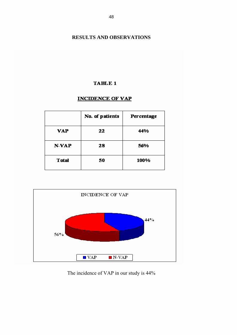
RESULTS AND OBSERVATIONS
The incidence of VAP in our study is 44%
49
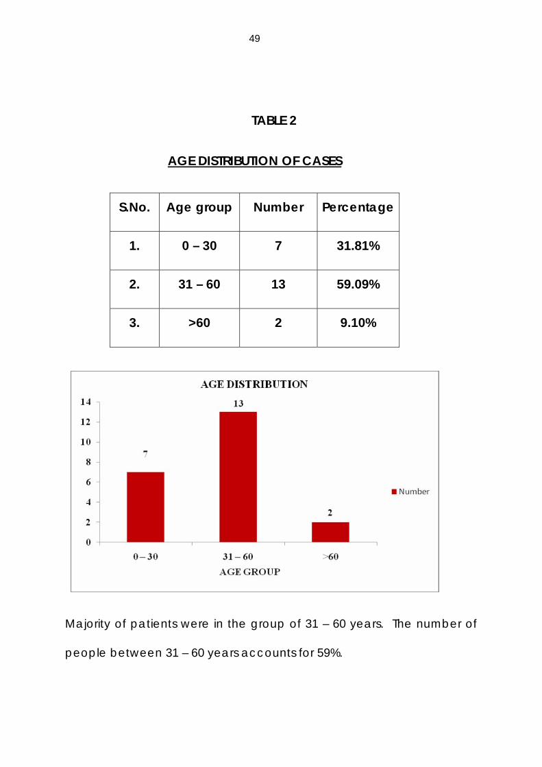
TABLE 2
AGE DISTRIBUTION OF CASES
S.No. Age group Number Percentage
1. 0 – 30 7 31.81%
2. 31 – 60 13 59.09%
3. >60 2 9.10%
Majority of patients were in the group of 31 – 60 years. The number of
people between 31 – 60 years accounts for 59%.
50
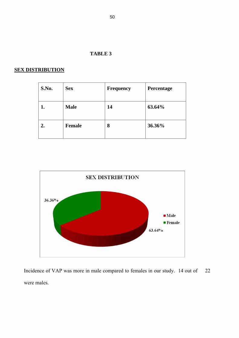
TABLE 3
SEX DISTRIBUTION
S.No. Sex Frequency Percentage
1. Male 14 63.64%
2. Female 8 36.36%
Incidence of VAP was more in male compared to females in our study. 14 out of 22
were males.

51
TABLE 4
METHOD OF INTUBATION
Frequency Percentage
Elective 50 50%
Emergency 50 50%
In our study 50% of patients underwent elective and 50% patients underwent emergency
distribution.

52
TABLE 5
X-RAY FINDING IN THE CASES
S. No. Lobar
distribution
Frequency Percentage
1. Right side 15 68.18%
2. Left side 3 13.63%
3. Bilateral 4 18.18%
In majority of over cases, the CXR infiltrate was in the Right lung which comes
around 68.2%.
18%
53
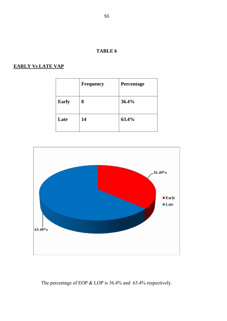
TABLE 6
EARLY Vs LATE VAP
Frequency Percentage
Early 8 36.4%
Late 14 63.4%
The percentage of EOP & LOP is 36.4% and 63.4% respectively.
54
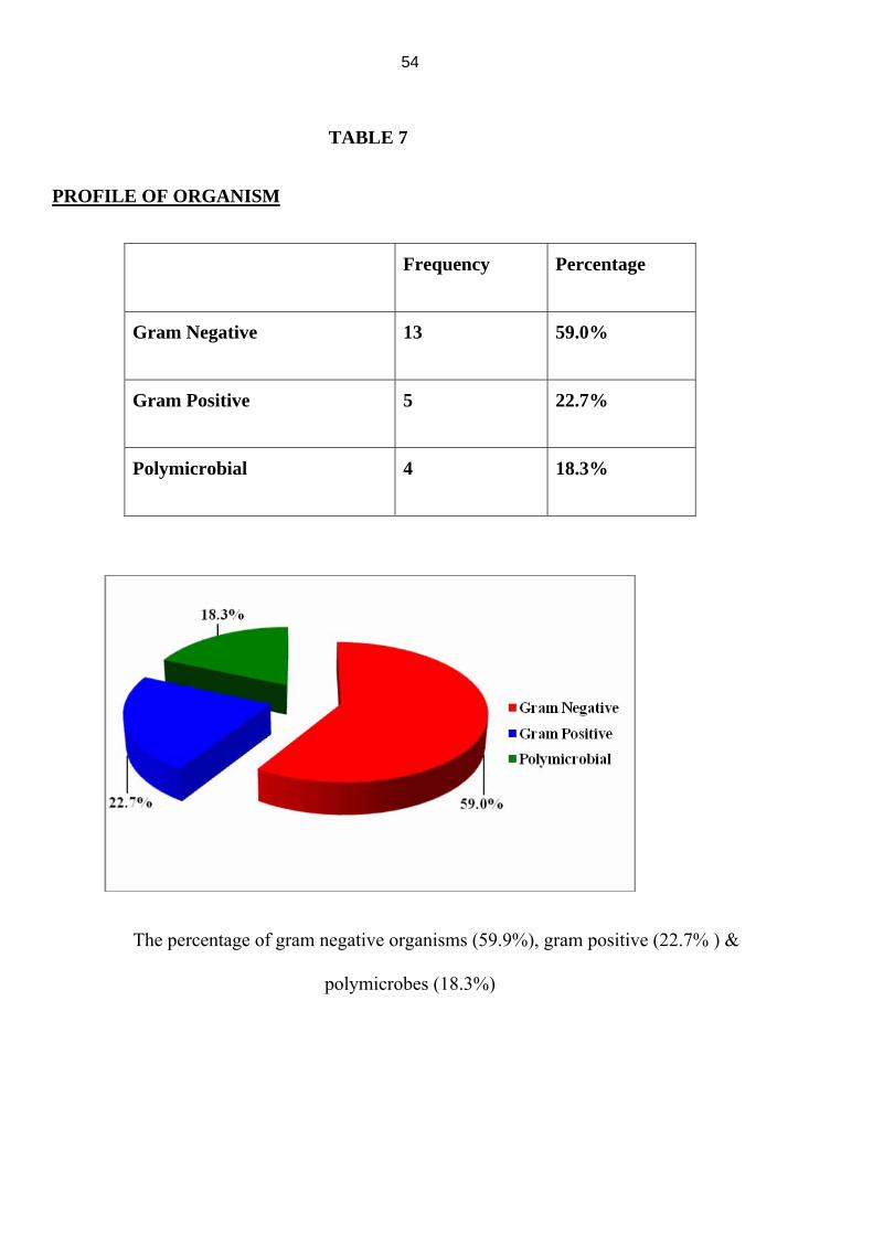
TABLE 7
PROFILE OF ORGANISM
Frequency Percentage
Gram Negative 13 59.0%
Gram Positive 5 22.7%
Polymicrobial 4 18.3%
The percentage of gram negative organisms (59.9%), gram positive (22.7% ) &
polymicrobes (18.3%)

55
TABLE 8
AETIOLOGY
S. No. Micro-organism Frequency Percentage
1. Pseudomonas 7 31.8%
2. MRSA 5 22.7%
3. Polymicrobial 4 18.4%
4. Acinetobacter 3 13.6%
5. Klebsiella 2 9.0%
6. Proteins 1 4.5%
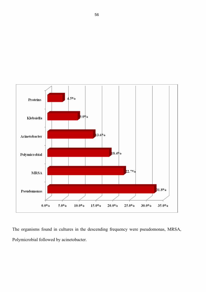
56
The organisms found in cultures in the descending frequency were pseudomonas, MRSA,
Polymicrobial followed by acinetobacter.

57
TABLE 9
AGE DISTRIBUTION AND OUTCOME
Death Recovery Age
Number % Number %
0 – 30 Years 2 14.28% 3 37.5%
30 – 60 years 10 71.42% 5 62.5%
>60 years 2 14.28% 0 0%
The mortality rate was high in patients of age group 30 – 60 years which accounts for 71.4%
of total deaths.
58
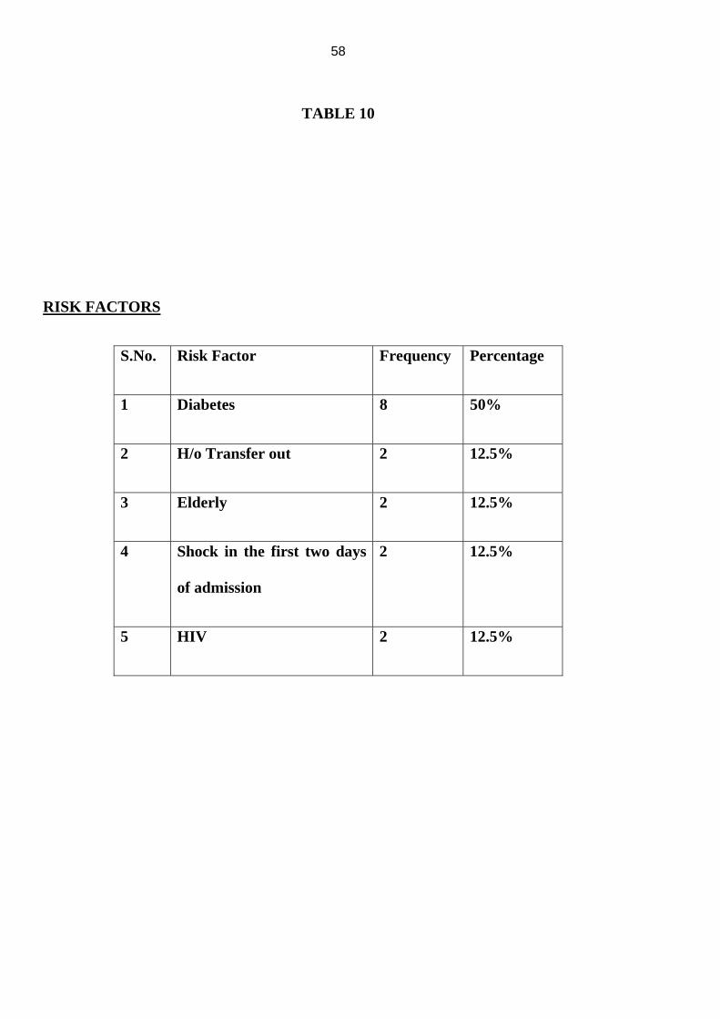
TABLE 10
RISK FACTORS
S.No. Risk Factor Frequency Percentage
1 Diabetes 8 50%
2 H/o Transfer out 2 12.5%
3 Elderly 2 12.5%
4 Shock in the first two days
of admission
2 12.5%
5 HIV 2 12.5%
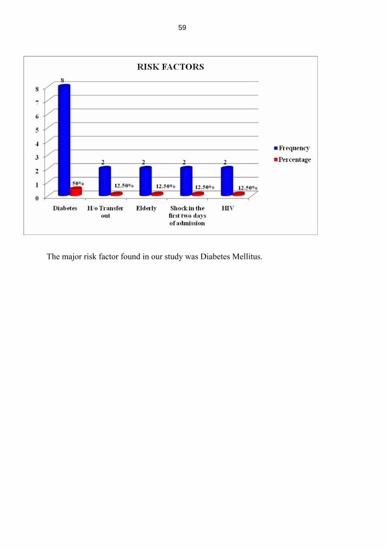
59
The major risk factor found in our study was Diabetes Mellitus.
60
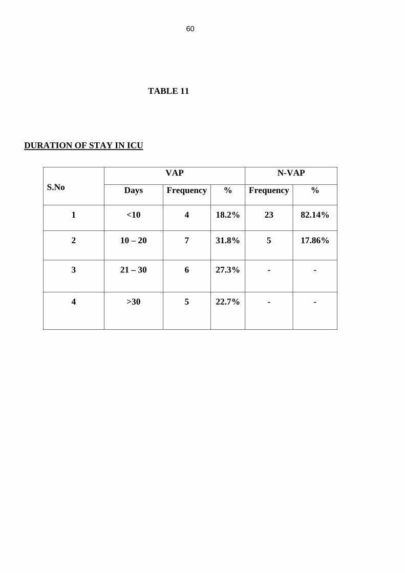
TABLE 11
DURATION OF STAY IN ICU
VAP N-VAP S.No Days Frequency % Frequency %
1 <10 4 18.2% 23 82.14%
2 10 – 20 7 31.8% 5 17.86%
3 21 – 30 6 27.3% - -
4 >30 5 22.7% - -

61
The mean duration of stay in the hospital who developed VAP was 10 – 20 days without VAP gp is < 10 days.

62
DISCUSSION
1. Incidence
Incidence of VAP in our govt.general hospital is 44% ,where as in united states VAP occurs
in up to 25% of all people who require mechanical ventilation.
2. Age distribution
The percentage of patients with VAP in different age groups is as follows
<30years- 31.81%
31-60years- 59.99%
>60years-9.1%
The higher incidence in the age group (31-60) can be attributed to more number of patients
getting admitted &undergoing ventilation in this age group.It may also be due to their
associated co morbid conditions.
3. Sex distribution
In our govt.hospital two third of the cases were males (63.6%) remaining cases one third
were (36.4%) females.
4. Method of intubation
There is equal incidence of VAP in both elective& emergency intubation.

63
5. Chest X-ray infiltrates
In our study 68.2%of patients had infiltrates in right lung.13.6%of patients in the left lung
18.2% bilaterally . The higher percentage of infiltrates in the right lung lower lobe is
because of aspiration being the most common precipitating factor for VAP. Autopsy studies
by Marquette, C. H., M. C. Copin, F. Wallet, R. Neviere, F. Saulnier, D. Mathieu, A.
Durocher, P. Ramon, and A. B. Tonnel. 1995 have indicated that VAP frequently involves
posterior right lower lobe.
6. Types of VAP
The percentage of patients with early onset VAP is 36%, late onset VAP is 63.4%.This is
because probability of getting VAP increases with the duration of mechanical ventilation.
Langer, Mosconi, Cigada, & Mandelli (1989) analyzed the relationship between artificial
ventilatory support and pulmonary infection in 724 critically ill patients who had received
prolonged (greater than 24 h) ventilatory assistance since admission to ICU. They found that
the risk for VAP increased from 5% in patients receiving one day of respiratory assistance to
68.8% in patients receiving more than 30 days.
7. Profile of organism
The profile of organism in our study as follows gram negative 59%, gram positive 22%,
polymicrobial 18.3%.
The organism cultured in our study in descending order were pseudomonas (31.8%)
,MRSA(22.7%), polymicrobials(18.4%), acinetobacter(13.6%), klebsiella(9%) proteus(4.5%)
64
reflecting the higher incidence of MDR organisms in patients with VAP, unlike in
community acquired pneumonia where streptococcus pneumonia is common.
Study conducted by Faisal Wahid, Naveed Masood, Asadullah Jafri. Nosocomial pneumonia
in mechanically ventilated patients Pak Armed Forces Med J Sep showed the following
profile of organisms including Pseudomonas aeruginosa(26%), Staphylococcus aureus(20%),
Acinetobacter spp.(9%), Proteus spp. (6%), Haemophilus spp. (6%), Escherichia coli (6%),
Klebsiella spp. (3%), Streptococcus pneumoniae (3%), Corynebacteria spp. (3%), and
Polymicrobial flora (9%).
8. Mortality
Mortality in our study according to the age distribution is as follows
<30 years-14.28%
31-60 years-71.42%
>60 years-14.28%
The greater mortality in the age group 31-60 years probably attributed to their
associated co morbid conditions.
9. Risk factors

65
The risk factors for VAP found in our study were diabetes mellitus (50%),elderly age group,
transferring out of ICU(for imaging & special tests),shock in the first 2 days of admission &
immune compromised status(HIV).
10 Morbidity
Patients with VAP had prolonged stay in ICU(10-20 days)unlike in patients without
VAP(<10 days). The study conducted by Boyce, J. M., G. Potter-Bynoe, L. Dziobek, and S.
L. Solomon showed an increased ICU lengths of stays (LOS) from 4 to13 days. Bercault &
Boulain, 2001; Rello et al., 2002. showed that the development of VAP increases the length
on the mechanical ventilator by 4 days, critical care and hospital lengths of stay (LOS) by 4
and 9 days, respectively.

66
CONCLUSION
1. Method of intubation emergency or elective did not change the incidence of VAP.
2. The incidence of VAP increases with the duration of mechanical ventilation.
3. Aspiration is major precipitating factor for developing VAP.
4. High incidence of MDR organisms in patients with VAP unlike in community
acquired pneumonia.
5. Diabetes is one of the major risk factor to develop VAP
6. Duration of stay in ICU patients with VAP very much pronged unlike in N- VAP .

67
PERFORMA
Name :
Age:
gender:
Complete diagnosis:
Duration of mechanical ventilation/ hospitalisation:
Indication for intubation:
Emergency/Elective:
Preventive measures undertaken:
Diagnosis of VAP: EARLY / LATE
1 Chest X –ray
2 Temp
3 TLC
4 TBA aspirate
Positioning
Suctioning
Oral care
Antibiotics

68
Others:
ECG-
ABG-
CT Chest-
Treatment:
Outcome:
CBC RFT LFT URINE
Hb Sugar TB Albumin
TC Urea DB Sugar
DC Creatinine SGOT deposits
ESR Na+ SGPT Pus cells
platelets K+ ALP organisms
PCV Blood culture
Total protein/albumin
Urine culture
69
BIBLIOGRAPHY
1. Gallego, M., and J. Rello. 1999. Diagnostic testing for ventilator-associated pneumonia. Clin. Chest Med. 20:671 Kollef, MH. (2005).
2. "What is ventilator-associated pneumonia and why is it important?". Respir Care 50: 714–21; discussion 721-679.
3. Richards, M. J., J. R. Edwards, D. H. Culver, R. P. Gaynes, et al. 1999. Nosocomial infections in medical intensive care units in the United States. Crit. Care Med. 27:887-892.
4. Marquette, C. H., F. Herengt, D. Mathieu, F. Saulnier, R. Courcol, and P. Ramon. 1993. Diagnosis of pneumonia in mechanically ventilated patients. Repeatability of the protected specimen brush. Am. Rev. Respir. Dis. 147:211-214.
5. McEachern, R., and G. D. Campbell, Jr. 1998. Hospital-acquired pneumonia: epidemiology, etiology, and treatment. Infect. Dis. Clin. N. Am. 12:761-779.
6. Craven, D. E., L. M. Kunches, V. Kilinsky, D. A. Lichtenberg, B. J. Make, and W. R. McCabe. 1986. Risk factors for pneumonia and fatality in patients receiving continuous mechanical ventilation. Am. Rev. Respir. Dis. 133:792-796.
7. Cunnion, K. M., D. J. Weber, W. E. B././roadhead, L. C. Hanson, C. F. Pieper, and W. A. Rutala. 1996. Risk factors for nosocomial pneumonia: comparing adult critical-care populations. Am. J. Respir. Crit. Care Med. 153:158-162.
8. Papazian, L., F. Bregeon, X. Thirion, R. Gregoire, P. Saux, J. P. Denis, G. Perin, J. Charrel, J. F. Dumon, J. P. Affray, and F. Gouin. 1996. Effect of ventilator-associated pneumonia on mortality and morbidity. Am. J. Respir. Crit. Care Med. 154:91-97.
9. Kappstein, I., G. Schulgen, U. Beyer, K. Geiger, M. Schumacher, and F. D.Daschner. 1992. Prolongation of hospital stay and extra costs due to ventilator-

70
associated pneumonia in an intensive care unit. Eur. J. Clin. Microbiol. Infect. Dis. 11:504-508.

71
10. American Thoracic Society. 1996. Hospital-acquired pneumonia in adults: diagnosis, assessment of severity, initial antimicrobial therapy, and preventive strategies. A consensus statement, American Thoracic Society, November 1995. Am. J. Respir. Crit. Care Med. 153:1711-1725
11. . Fartoukh, M., B. Maitre, S. Honore, C. Cerf, J. R. Zahar, and C. Brun-Buisson. 2003. Diagnosing pneumonia during mechanical ventilation: the clinical pulmonary infection score revisited. Am. J. Respir. Crit. Care Med. 168:173-179.
12. Bryan, C. S. 1999. Nosocomial pneumonia: blood cultures remain useful. Chest 116:859-860.
13. American Thoracic Society. 1996. Hospital-acquired pneumonia in adults: diagnosis, assessment of severity, initial antimicrobial therapy, and preventive strategies. A consensus statement, American Thoracic Society, November 1995. Am. J. Respir. Crit. Care Med. 153:1711-1725
14. 65. Fartoukh, M., B. Maitre, S. Honore, C. Cerf, J. R. Zahar, and C. Brun-Buisson. 2003. Diagnosing pneumonia during mechanical ventilation: the clinical pulmonary infection score revisited. Am. J. Respir. Crit. Care Med. 168:173-179.
15. Bergmans, D. C., M. J. Bonten, P. W. De Leeuw, and E. E. Stobberingh. 1997. Reproducibility of quantitative cultures of endotracheal aspirates from mechanically ventilated patients. J. Clin. Microbiol. 35:796-798.
16. Cook, D., and L. Mandell. 2000. Endotracheal aspiration in the diagnosis of ventilator-associated pneumonia. Chest 117:195S-197S.
17. Jorda, R., F. Parras, J. Ibanez, J. Reina, J. Bergada, and J. M. Raurich. 1993. Diagnosis of nosocomial pneumonia in mechanically ventilated patients by the blind protected telescoping catheter. Intensive Care Med. 19:377-382.
18. Chastre, J., J. Y. Fagon, M. Bornet-Lecso, S. Calvat, M. C. Dombret, R. al Khani, F. Basset, and C. Gibert. 1995. Evaluation of bronchoscopic techniques for the diagnosis of nosocomial pneumonia. Am. J. Respir. Crit. Care Med. 152:231-240.
19. Gerbeaux, P., V. Ledoray, A. Boussuges, F. Molenat, P. Jean, and J. M. Sainty. 1998. Diagnosis of nosocomial pneumonia in mechanically ventilated patients: repeatability of the bronchoalveolar lavage. Am. J. Respir. Crit. Care Med. 157:76-80.
72
20. Gerbeaux, P., V. Ledoray, A. Boussuges, F. Molenat, P. Jean, and J. M.
Sainty. 1998. Diagnosis of nosocomial pneumonia in mechanically ventilated patients: repeatability of the bronchoalveolar lavage. Am. J. Respir. Crit. Care Med. 157:76-80.
21. Jorda, R., F. Parras, J. Ibanez, J. Reina, J. Bergada, and J. M. Raurich. 1993. Diagnosis of nosocomial pneumonia in mechanically ventilated patients by the blind protected telescoping catheter. Intensive Care Med.
22. Baker, A. M., D. L. Bowton, and E. F. Haponik. 1995. Decision making in nosocomial pneumonia. An analytic approach to the interpretation of quantitative bronchoscopic cultures. Chest 107:85-95.
23. American Thoracic Society. 2005. Guidelines for the management of adults with hospital-acquired, ventilator-associated, and healthcare-associated pneumonia. Am. J. Respir. Crit. Care Med. 171:388-416.
24. Ibrahim, E. H., S. Ward, G. Sherman, R. Schaiff, V. J. Fraser, and M. H. Kollef. 2001. Experience with a clinical guideline for the treatment of ventilator-associated pneumonia. Crit. Care Med. 29:1109-1115.
25. Rello J, Quintana E, Ausina V, et al. Risk factors for Staphylococcus aureus nosocomial pneumonia in critically ill patients. Am Rev Respir Dis 1990; 142:1320–132419 Torres A, Gatell JM, Aznar E, et al.
26. Rouby, J. J., P. Laurent, M. Gosnach, E. Cambau, G. Lamas, A. Zouaoui, J. L. Leguillou, L. Bodin, T. D. Khac, C. Marsault, et al. 1994. Risk factors and clinical relevance of nosocomial maxillary sinusitis in the critically ill. Am. J. Respir. Crit. Care Med. 150:776-783.
27. Feldman, C., M. Kassel, J. Cantrell, S. Kaka, R. Morar, A. Goolam Mahomed, and J. I. Philips. 1999. The presence and sequence of endotracheal tube colonization in patients undergoing mechanical ventilation. Eur. Respir. J. 13:546-551.
28. Inglis, T. J., M. R. Millar, J. G. Jones, and D. A. Robinson. 1989. Tracheal tube biofilm as a source of bacterial colonization of the lung. J. Clin. Microbiol. 27:2014-2018.
29. American Thoracic Society and the Infectious Diseases Society of America. (2005). "ATS/IDSA Guidelines: Guidelines for the management of adults with HAP, VAP, and HCAP". Am J Respir Crit Care Med 171: 388.

73
30. Niederman, MS. (2006). "Use of broad-spectrum antimicrobials for the treatment of
pneumonia in seriously ill patients: maximizing clinical outcomes and minimizing selection of resistant organisms". Clin Infect Dis 42 Supp 2: S72–81. doi:10.1086/499405.
31. Ibrahim, E. H., S. Ward, G. Sherman, and M. H. Kollef. 2000. A comparative analysis of patients with early-onset vs late-onset nosocomial pneumonia in the ICU setting. Chest 117:1434-1442 32. Antonelli, M., G. Conti, M. Rocco, M. Bufi, R. A. De Blasi, G. Vivino, A. Gasparetto, and G. U. Meduri. 1998. A comparison of noninvasive positive-pressure ventilation and conventional mechanical ventilation in patients with acute respiratory failure. N. Engl. J. Med. 339:429-435.
33. Brochard, L., J. Mancebo, M. Wysocki, F. Lofaso, G. Conti, A. Rauss, G. Simonneau, S. Benito, A. Gasparetto, F. Lemaire, et al. 1995. Noninvasive ventilation for acute exacerbations of chronic obstructive pulmonary disease. N. Engl. J. Med. 333:817-822.
34. Hilbert, G., D. Gruson, F. Vargas, R. Valentino, G. Gbikpi-Benissan, M. Dupon, J. Reiffers, and J. P. Cardinaud. 2001. Noninvasive ventilation in immunosuppressed patients with pulmonary infiltrates, fever, and acute respiratory failure. N. Engl. J. Med. 344:481-487.
35. Rouby, J. J., P. Laurent, M. Gosnach, E. Cambau, G. Lamas, A. Zouaoui, J. L. Leguillou, L. Bodin, T. D. Khac, C. Marsault, et al. 1994. Risk factors and clinical relevance of nosocomial maxillary sinusitis in the critically ill. Am. J. Respir. Crit. Care Med. 150:776-783.
36. Holzapfel, L., S. Chevret, G. Madinier, F. Ohen, G. Demingeon, A. Coupry, and M. Chaudet. 1993. Influence of long-term oro- or nasotracheal intubation on nosocomial maxillary sinusitis and pneumonia: results of a prospective, randomized, clinical trial. Crit. Care Med. 21:1132-1138.
37. Tablan, O. C., L. J. Anderson, N. H. Arden, R. F. Breiman, J. C. Butler, M. M. McNeil, et al. 1994. Guideline for prevention of nosocomial pneumonia. Am. J. Infect. Control 22:247-292 Fagon, J. Y., J. Chastre, Y. Domart, J. L. Trouillet, J. Pierre, C. Darne, and C. Gibert. 1989. Nosocomial pneumonia in patients receiving continuous mechanical ventilation. Prospective analysis of 52 episodes with use of a protected specimen brush and quantitative culture techniques. Am. Rev. Respir. Dis. 139:877-884.

74
38. Cook, D. J., S. D. Walter, R. J. Cook, L. E. Griffith, G. H. Guyatt, D. Leasa, R. Z. Jaeschke, and C. Brun-Buisson. 1998. Incidence of and risk factors for ventilator-associated pneumonia in critically ill patients. Ann. Intern. Med. 129:433-440
39. Ibrahim, E. H., S. Ward, G. Sherman, and M. H. Kollef. 2000. A comparative analysis of patients with early-onset vs late-onset nosocomial pneumonia in the ICU setting. Chest 117:1434-1442.
40. Rello, J., J. A. Paiva, J. Baraibar, F. Barcenilla, M. Bodi, D. Castander, H. Correa, E. Diaz, J. Garnacho, M. Llorio, M. Rios, A. Rodriguez, and J. Sole-Violan. 2001. International Conference for the Development of Consensus on the Diagnosis and Treatment of Ventilator-Associated Pneumonia. Chest 120:955-970.
41. Brook, A. D., T. S. Ahrens, R. Schaiff, D. Prentice, G. Sherman, W. Shannon, and M. H. Kollef. 1999. Effect of a nursing-implemented sedation protocol on the duration of mechanical ventilation. Crit. Care Med. 27:2609-2615
42. Kress, J. P., A. S. Pohlman, M. F. O'Connor, and J. B. Hall. 2000. Daily interruption of sedative infusions in critically ill patients undergoing mechanical ventilation. N. Engl. J. Med. 342:1471-1477
43. Marelich, G. P., S. Murin, F. Battistella, J. Inciardi, T. Vierra, and M. Roby. 2000. Protocol weaning of mechanical ventilation in medical and surgical patients by respiratory care practitioners and nurses: effect on weaning time and incidence of ventilator-associated pneumonia. Chest 118:459-467.
44. Schweickert, W. D., B. K. Gehlbach, A. S. Pohlman, J. B. Hall, and J. P. Kress. 2004. Daily interruption of sedative infusions and complications of critical illness in mechanically ventilated patients. Crit. Care Med. 32:1272-1276.
45. Ferrer, M., A. Esquinas, F. Arancibia, T. T. Bauer, G. Gonzalez, A. Carrillo, R. Rodriguez-Roisin, and A. Torres. 2003. Noninvasive ventilation during persistent weaning failure: a randomized controlled trial. Am. J. Respir. Crit. Care Med. 168:70-76.
46. Ely, E. W., M. O. Meade, E. F. Haponik, M. H. Kollef, D. J. Cook, G. H. Guyatt, and J. K. Stoller. 2001. Mechanical ventilator weaning protocols driven by
75
nonphysician health-care professionals: evidence-based clinical practice guidelines. Chest 120:454S-463S.
47. Inglis, T. J., M. R. Millar, J. G. Jones, and D. A. Robinson. 1989. Tracheal tube biofilm as a source of bacterial colonization of the lung. J. Clin. Microbiol. 27:2014-2018
48. Drakulovic, M. B., A. Torres, T. T. Bauer, J. M. Nicolas, S. Nogue, and M. Ferrer. 1999. Supine body position as a risk factor for nosocomial pneumonia in mechanically ventilated patients: a randomised trial. Lancet 354:1851-1858.
49. Heyland, D. K., J. W. Drover, R. Dhaliwal, and J. Greenwood. 2002. Optimizing the benefits and minimizing the risks of enteral nutrition in the critically ill: role of small bowel feeding. J Parenter. Enteral. Nutr. 26:S51-S57.
50. Gentilello, L., D. A. Thompson, A. S. Tonnesen, D. Hernandez, A. S. Kapadia, S. J. Allen, B. A. Houtchens, and M. E. Miner. 1988. Effect of a rotating bed on the incidence of pulmonary complications in critically ill patients. Crit. Care Med. 16:783-786.
51. Fink, M. P., C. M. Helsmoortel, K. L. Stein, P. C. Lee, and S. M. Cohn. 1990. The efficacy of an oscillating bed in the prevention of lower respiratory tract infection in critically ill victims of blunt trauma. A prospective study. Chest 97:132-137.
52. Dreyfuss, D., K. Djedaini, P. Weber, P. Brun, J. J. Lanore, J. Rahmani, Y. Boussougant, and F. Coste. 1991. Prospective study of nosocomial pneumonia and of patient and circuit colonization during mechanical ventilation with circuit changes every 48 hours versus no change. Am. Rev. Respir. Dis. 143:738-743.
53. Dreyfuss, D., K. Djedaini, I. Gros, L. Mier, G. Le Bourdelles, Y. Cohen, P. Estagnasie, F. Coste, and Y. Boussougant. 1995. Mechanical ventilation with heated humidifiers or heat and moisture exchangers: effects on patient colonization and incidence of nosocomial pneumonia. Am. J. Respir. Crit. Care Med. 151:986-992.
54. Kollef, M. H., S. D. Shapiro, V. Boyd, P. Silver, B. Von Harz, E. Trovillion, and D. Prentice. 1998. A randomized clinical trial comparing an extended-use hygroscopic condenser humidifier with heated-water humidification in mechanically ventilated patients. Chest 113:759-767.
76
55. Davis, K., Jr., S. L. Evans, R. S. Campbell, J. A. Johannigman, F. A. Luchette, D. T. Porembka, and R. D. Branson. 2000. Prolonged use of heat and moisture exchangers does not affect device efficiency or frequency rate
56. Deppe, S. A., J. W. Kelly, L. L. Thoi, J. H. Chudy, R. N. Longfield, J. P. Ducey, C. L. Truwit, and M. R. Antopol. 1990. Incidence of colonization, nosocomial pneumonia, and mortality in critically ill patients using a Trach Care closed-suction system versus an open-suction system: prospective, randomized study. Crit. Care Med. 18:1389-1393. of nosocomial pneumonia. Crit. Care Med. 28:1412-1418.
57. Johnson, K. L., P. A. Kearney, S. B. Johnson, J. B. Niblett, N. L. MacMillan, and R. E. McClain. 1994. Closed versus open endotracheal suctioning: costs and physiologic consequences. Crit. Care Med. 22:658-666.
58. Dodek, P., S. Keenan, D. Cook, D. Heyland, M. Jacka, L. Hand, J. Muscedere, D. Foster, N. Mehta, R. Hall, and C. Brun-Buisson. 2004. Evidence-based clinical practice guideline for the prevention of ventilator-associated pneumonia. Ann. Intern. Med. 141:305-313.
59. Kollef, M. H., N. J. Skubas, and T. M. Sundt. 1999. A randomized clinical trial of continuous aspiration of subglottic secretions in cardiac surgery patients. Chest 116:1339-1346
60. Prevention of nosocomial pneumonia in intubated patients: respective role of mechanical subglottic secretions drainage and stress ulcer prophylaxis. Intensive Care Med. 18:20-25.
61. Rello, J., R. Sonora, P. Jubert, A. Artigas, M. Rue, and J. Valles. 1996. Pneumonia in intubated patients: role of respiratory airway care. Am. J. Respir. Crit. Care Med. 154:111-115
62. Sottile, F. D., T. J. Marrie, D. S. Prough, C. D. Hobgood, D. J. Gower, L. X. Webb, J. W. Costerton, and A. G. Gristina. 1986. Nosocomial pulmonary infection: possible etiologic significance of bacterial adhesion to endotracheal tubes. Crit. Care Med. 14:265-270
63. Inglis, T. J., M. R. Millar, J. G. Jones, and D. A. Robinson. 1989. Tracheal tube biofilm as a source of bacterial colonization of the lung. J. Clin. Microbiol. 27:2014-2018173-179.

77
64. Feldman, C., M. Kassel, J. Cantrell, S. Kaka, R. Morar, A. Goolam
Mahomed, and J. I. Philips. 1999. The presence and sequence of endotracheal tube colonization in patients undergoing mechanical ventilation. Eur. Respir. J. 13:546-551.
65. DeRiso, A. J., II, J. S. Ladowski, T. A. Dillon, J. W. Justice, and A. C. Peterson. 1996. Chlorhexidine gluconate 0.12% oral rinse reduces the incidence of total nosocomial respiratory infection and nonprophylactic systemic antibiotic use in patients undergoing heart surgery. Chest 109:1556-1561.
66. Abele-Horn, M., A. Dauber, A. Bauernfeind, W. Russwurm, I. Seyfarth-Metzger, P. Gleich, and G. Ruckdeschel. 1997. Decrease in nosocomial pneumonia in ventilated patients by selective oropharyngeal decontamination (SOD). Intensive Care Med. 23:187-195.
67. Bergmans, D. C., M. J. Bonten, C. A. Gaillard, J. C. Paling, S. van der Geest, F. H. van Tiel, A. J. Beysens, P. W. de Leeuw, and E. E. Stobberingh. 2001. Prevention of ventilator-associated pneumonia by oral decontamination: a prospective, randomized, double-blind, placebo-controlled study. Am. J. Respir. Crit. Care Med. 164:382-388.
68. de Jonge, E., M. J. Schultz, L. Spanjaard, P. M. Bossuyt, M. B. Vroom, J. Dankert, and J. Kesecioglu. 2003. Effects of selective decontamination of digestive tract on mortality and acquisition of resistant bacteria in intensive care: a randomised controlled trial. Lancet 362:1011-1016.
69. Pugin, J., R. Auckenthaler, D. P. Lew, and P. M. Suter. 1991. Oropharyngeal decontamination decreases incidence of ventilator-associated pneumonia. A randomized, placebo-controlled, double-blind clinical trial. JAMA 265:2704-2710
70. Rodriguez-Roldan, J. M., A. Altuna-Cuesta, A. Lopez, A. Carrillo, J. Garcia, J. Leon, and A. J. Martinez-Pellus. 1990. Prevention of nosocomial lung infection in ventilated patients: use of an antimicrobial pharyngeal nonabsorbable paste. Crit. Care Med. 18:1239-1242.
71. Nathens, A. B., and J. C. Marshall. 1999. Selective decontamination of the digestive tract in surgical patients: a systematic review of the evidence. Arch. Surg. 134:170-176

78
72. D'Amico, R., S. Pifferi, C. Leonetti, V. Torri, A. Tinazzi, and A. Liberati. 1998. Effectiveness of antibiotic prophylaxis in critically ill adult patients: systematic review of randomised controlled trials. Br. Med J. 316:1275-1285.
73. Ferrer, M., A. Torres, J. Gonzalez, J. Puig de la Bellacasa, M. el-Ebiary, M. Roca, J. M. Gatell, and R. Rodriguez-Roisin. 1994. Utility of selective digestive decontamination in mechanically ventilated patients. Ann. Intern. Med. 120:389-395.
74. Hammond, J. M., P. D. Potgieter, G. L. Saunders, and A. A. Forder. 1992. Double-blind study of selective decontamination of the digestive tract in intensive care. Lancet 340:5-
75. Misset, B., M. D. Kitzis, G. Conscience, F. Goldstein, A. Fourrier, and J. Carlet. 1994. Mechanisms of failure to decontaminate the gut with polymixin E, gentamicin and amphotericin B in patients in intensive care. Eur. J. Clin. Microbiol. Infect. Dis. 13:165-170.
76. Misset, B., M. D. Kitzis, P. Mahe, G. Conscience, F. W. Goldstein, A. Fourrier, and J. Carlet. 1993. Bacteriological side effects of gut decontamination with polymyxin E, gentamicin, and amphotericin B. Infect. Control Hosp. Epidemiol. 14:62-64.
77. Krueger, W. A., F. P. Lenhart, G. Neeser, G. Ruckdeschel, H. Schreckhase, H. J. Eissner, H. Forst, J. Eckart, K. Peter, and K. E. Unertl. 2002. Influence of combined intravenous and topical antibiotic prophylaxis on the incidence of infections, organ dysfunctions, and mortality in critically ill surgical patients: a prospective, stratified, randomized, double-blind, placebo-controlled clinical trial. Am. J. Respir. Crit. Care Med. 166:1029-1037.
78. de Jonge, E., M. J. Schultz, L. Spanjaard, P. M. Bossuyt, M. B. Vroom, J. Dankert, and J. Kesecioglu. 2003. Effects of selective decontamination of digestive tract on mortality and acquisition of resistant bacteria in intensive care: a randomised controlled trial. Lancet 362:1011-1016
79. Dodek, P., S. Keenan, D. Cook, D. Heyland, M. Jacka, L. Hand, J. Muscedere, D. Foster, N. Mehta, R. Hall, and C. Brun-Buisson. 2004. Evidence-based clinical practice guideline for the prevention of ventilator-associated pneumonia. Ann. Intern. Med. 141:305-313.

79
80. Messori, A., S. Trippoli, M. Vaiani, M. Gorini, and A. Corrado. 2000. Bleeding and pneumonia in intensive care patients given ranitidine and sucralfate for prevention of stress ulcer: meta-analysis of randomised controlled trials. Br. Med. J. 321:1103-1106.
81. Cook, D., G. Guyatt, J. Marshall, D. Leasa, H. Fuller, R. Hall, S. Peters, F. Rutledge, L. Griffith, A. McLellan, G. Wood, A. Kirby, et al. 1998. A comparison of sucralfate and ranitidine for the prevention of upper gastrointestinal bleeding in patients requiring mechanical ventilation. N. Engl. J. Med. 338:791-797
82. Bonten, M. J., C. A. Gaillard, S. van der Geest, F. H. van Tiel, A. J. Beysens, H. G. Smeets, and E. E. Stobberingh. 1995. The role of intragastric acidity and stress ulcus prophylaxis on colonization and infection in mechanically ventilated ICU patients. A stratified, randomized, double-blind study of sucralfate versus antacids. Am. J. Respir. Crit. Care Med. 152:1825-1834
83. Heyland, D. K., J. W. Drover, R. Dhaliwal, and J. Greenwood. 2002. Optimizing the benefits and minimizing the risks of enteral nutrition in the critically ill: role of small bowel feeding. J Parenter. Enteral. Nutr. 26:S51-S57.
84. Ibrahim, E. H., L. Mehringer, D. Prentice, G. Sherman, R. Schaiff, V. Fraser, and M. H. Kollef. 2002. Early versus late enteral feeding of mechanically ventilated patients: results of a clinical trial. J. Parenter. Enteral. Nutr. 26:174-181.
85. Kollef, M. H., J. Vlasnik, L. Sharpless, C. Pasque, D. Murphy, and V. Fraser. 1997. Scheduled change of antibiotic classes: a strategy to decrease the incidence of ventilator-associated pneumonia. Am. J. Respir. Crit. Care Med. 156:1040-1048.
86. Raymond, D. P., S. J. Pelletier, T. D. Crabtree, T. G. Gleason, L. L. Hamm, T. L. Pruett, and R. G. Sawyer. 2001. Impact of a rotating empiric antibiotic schedule on infectious mortality in an intensive care unit. Crit. Care Med. 29:1101-1108.
87. Gruson, D., G. Hilbert, F. Vargas, R. Valentino, C. Bebear, A. Allery, C. Bebear, G. Gbikpi-Benissan, and J. P. Cardinaud. 2000. Rotation and restricted use of antibiotics in a medical intensive care unit. Impact on the incidence of ventilator-associated pneumonia caused by antibiotic-resistant gram-negative bacteria. Am. J. Respir. Crit. Care Med. 162:837-843.
80
88. Kollef, M. H., S. Ward, G. Sherman, D. Prentice, R. Schaiff, W. Huey, and V. J. Fraser. 2000. Inadequate treatment of nosocomial infections is associated with certain empiric antibiotic choices. Crit. Care Med. 28:3456-3464
89. Bergstrom, C. T., M. Lo, and M. Lipsitch. 2004. Ecological theory suggests that antimicrobial cycling will not reduce antimicrobial resistance in hospitals. Proc. Natl. Acad. Sci. USA 101:13285-13290
90. Schentag, J. J., M. C. Birmingham, J. A. Paladino, J. R. Carr, J. M. Hyatt, A. Forrest, G. S. Zimmer, M. H. Adelman, and T. J. Cumbo. 1997. In nosocomial pneumonia, optimizing antibiotics other than aminoglycosides is a more important determinant of successful clinical outcome, and a better means of avoiding resistance. Semin. Respir. Infect. 12:278-293.
91. Verpooten, G. A., R. A. Giuliano, L. Verbist, G. Eestermans, and M. E. De Broe. 1989. Once-daily dosing decreases renal accumulation of gentamicin and netilmicin. Clin. Pharmacol. Ther. 45:22-27
92. Thomas, J. K., A. Forrest, S. M. Bhavnani, J. M. Hyatt, A. Cheng, C. H. Ballow, and J. J. Schentag. 1998. Pharmacodynamic evaluation of factors associated with the development of bacterial resistance in acutely ill patients during therapy. Antimicrob Agents Chemother. 42:521-527.
93. Lipman, J., S. C. Wallis, and C. Rickard. 1999. Low plasma cefepime levels in critically ill septic patients: pharmacokinetic modeling indicates improved troughs with revised dosing. Antimicrob. Agents Chemother. 43:2559-2561.
94. Dodek, P., S. Keenan, D. Cook, D. Heyland, M. Jacka, L. Hand, J. Muscedere, D. Foster, N. Mehta, R. Hall, and C. Brun-Buisson. 2004. Evidence-based clinical practice guideline for the prevention of ventilator-associated pneumonia. Ann. Intern. Med. 141:305-313
95. Zack, J. E., T. Garrison, E. Trovillion, D. Clinkscale, C. M. Coopersmith, V. J. Fraser, and M. H. Kollef. 2002. Effect of an education program aimed at reducing the occurrence of ventilator-associated pneumonia. Crit. Care Med. 30:2407-2412.

81
96. Hill, J. D., J. L. Ratliff, J. C. Parrott, M. Lamy, R. J. Fallat, E. Koeniger, E. M. Yaeger, and G. Whitmer. 1976. Pulmonary pathology in acute respiratory insufficiency: lung biopsy as a diagnostic tool. J. Thoracic Cardiovasc. Surg
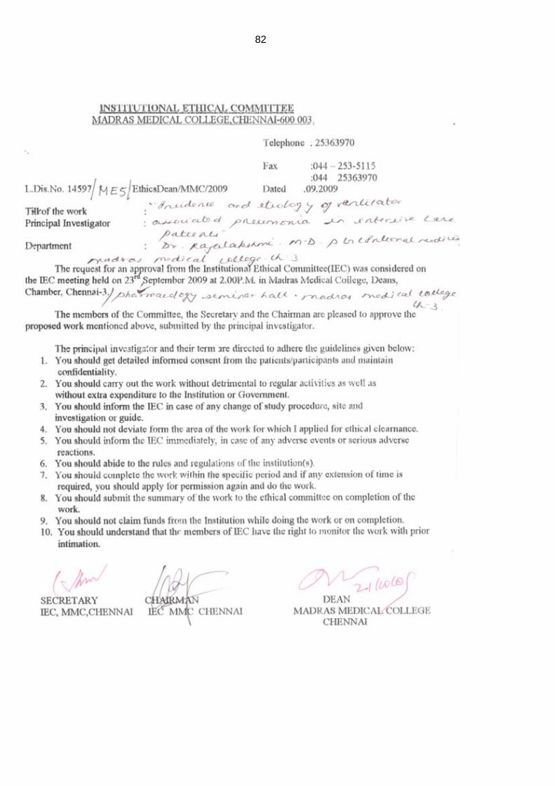
82

83
MASTER CHART
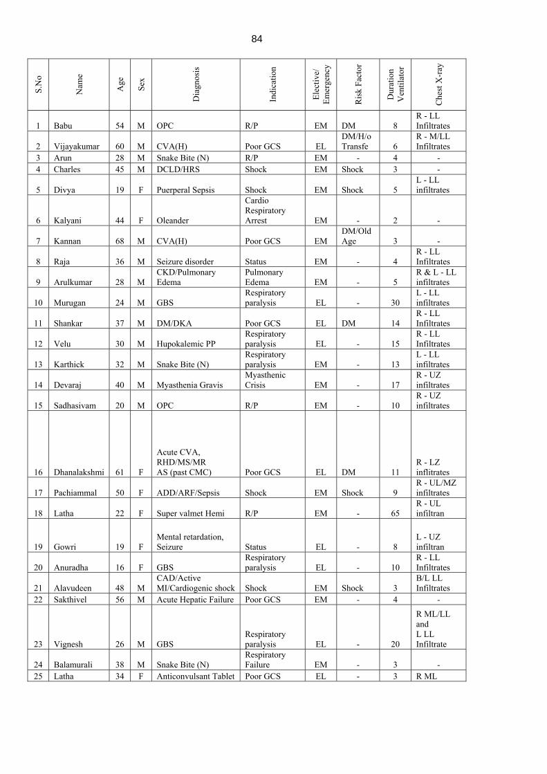
84
S.N
o
Nam
e
Age
Sex
Dia
gnos
is
Indi
catio
n
Elec
tive/
Em
erge
ncy
Ris
k Fa
ctor
Dur
atio
n V
entil
ator
Che
st X
-ray
1 Babu 54 M OPC R/P EM DM 8 R - LL Infiltrates
2 Vijayakumar 60 M CVA(H) Poor GCS EL DM/H/o Transfe 6
R - M/LL Infiltrates
3 Arun 28 M Snake Bite (N) R/P EM - 4 - 4 Charles 45 M DCLD/HRS Shock EM Shock 3 -
5 Divya 19 F Puerperal Sepsis Shock EM Shock 5 L - LL infiltrates
6 Kalyani 44 F Oleander
Cardio Respiratory Arrest EM - 2 -
7 Kannan 68 M CVA(H) Poor GCS EM DM/Old Age 3 -
8 Raja 36 M Seizure disorder Status EM - 4 R - LL Infiltrates
9 Arulkumar 28 M CKD/Pulmonary Edema
Pulmonary Edema EM - 5
R & L - LL infiltrates
10 Murugan 24 M GBS Respiratory paralysis EL - 30
L - LL infiltrates
11 Shankar 37 M DM/DKA Poor GCS EL DM 14 R - LL Infiltrates
12 Velu 30 M Hupokalemic PP Respiratory paralysis EL - 15
R - LL Infiltrates
13 Karthick 32 M Snake Bite (N) Respiratory paralysis EM - 13
L - LL infiltrates
14 Devaraj 40 M Myasthenia Gravis Myasthenic Crisis EM - 17
R - UZ infiltrates
15 Sadhasivam 20 M OPC R/P EM - 10 R - UZ infiltrates
16 Dhanalakshmi 61 F
Acute CVA, RHD/MS/MR AS (past CMC) Poor GCS EL DM 11
R - LZ inflitrates
17 Pachiammal 50 F ADD/ARF/Sepsis Shock EM Shock 9 R - UL/MZ infiltrates
18 Latha 22 F Super valmet Hemi R/P EM - 65 R - UL infiltran
19 Gowri 19 F Mental retardation, Seizure Status EL - 8
L - UZ infiltran
20 Anuradha 16 F GBS Respiratory paralysis EL - 10
R - LL Infiltrates
21 Alavudeen 48 M CAD/Active MI/Cardiogenic shock Shock EM Shock 3
B/L LL Infiltrates
22 Sakthivel 56 M Acute Hepatic Failure Poor GCS EM - 4 -
23 Vignesh 26 M GBS Respiratory paralysis EL - 20
R ML/LL and L LL Infiltrate
24 Balamurali 38 M Snake Bite (N) Respiratory Failure EM - 3 -
25 Latha 34 F Anticonvulsant Tablet Poor GCS EL - 3 R ML

85
S.N
o
Tem
p
TLC
Trac
heal
A
spira
te
Gra
m S
tain
Cul
ture
Inte
rpre
tatio
n
Early
/Lat
e
Suga
r/Ure
a C
reat
inin
e
AB
G
Oth
ers
Dur
atio
n in
IC
U
Out
com
e
1 + 16800 NP N Pseudomonai 1 E 168/46/1.1 Resp.ALK - 20 R 2 + 8900 P - Polymicrobial 1 E 206/54/1.8 Resp.ALK - 18 D 3 + 10000 NP - - 0 - 106/40/0.8 - - 10 R 4 + 11800 NP - - 0 - 66/106/2.0 Met Acid - 5 D 5 - 10800 NP - - 0 - 78/66/2.4 Met Acid - 8 D 6 - 8000 NP - - 0 - 148/48/1.4 - - 3 D 7 - 11800 NP - - 0 - 298/48/3.4 Met Acid - 5 D 8 + - NP - - 0 - 102/36/0.6 Resp.ALK - 8 R 9 + 9600 NP - - 0 - 356/94/3.6 Met Acid - 6 D
10 + 13800 P N Pseudo 1 L 124/36/1.2 Resp.ALK - 30 D 11 + 14240 P P MRSA 1 L 146/76/2.0 Met ALK - 16 R 12 + 14100 NP N - 0 - 446/80/2.1 Resp.ALK - 20 R 13 + 9200 P N Pseudo 1 L 68/26/0.9 Met Acid - 15 R 14 + 12100 P N Klebsicella 1 L 206/19/0.9 - BAL 23 R 15 + 16200 NP N Pseudo 1 L 124/36/1.1 Resp.ALK - 25 D
16 + 10400 P - Polymicrobial 1 L 105/57/1.3 - - 31 D 17 + 10740 P N Acinetobacter 1 E 139/45/1 - - 9 D 18 + 10400 P P MRSA 1 L 123/18/0.6 - - 70 R
19 + 11300 P N Pseudomonas 1 E 62/15/0.5 - - 13 R 20 + 16080 P N Acinetobacter 1 L 98/40/1.2 Resp.ALK - 56 R 21 - 12100 NP - - 0 - 168/48/1.5 Met Acid - 3 D 22 - 8900 NP - - 0 - 86/68/1.4 Met Acid - 5 D
23 + 10800 P N Klebsicella 1 L 106/46/1.2 Resp.ALK - 40 R 24 - 6800 NP - - 0 - 104/32/0.8 - - 7 R 25 - 11800 NP - - 0 - 124/34/0.8 Met Acid - 5 R
overdosage Infiltrate
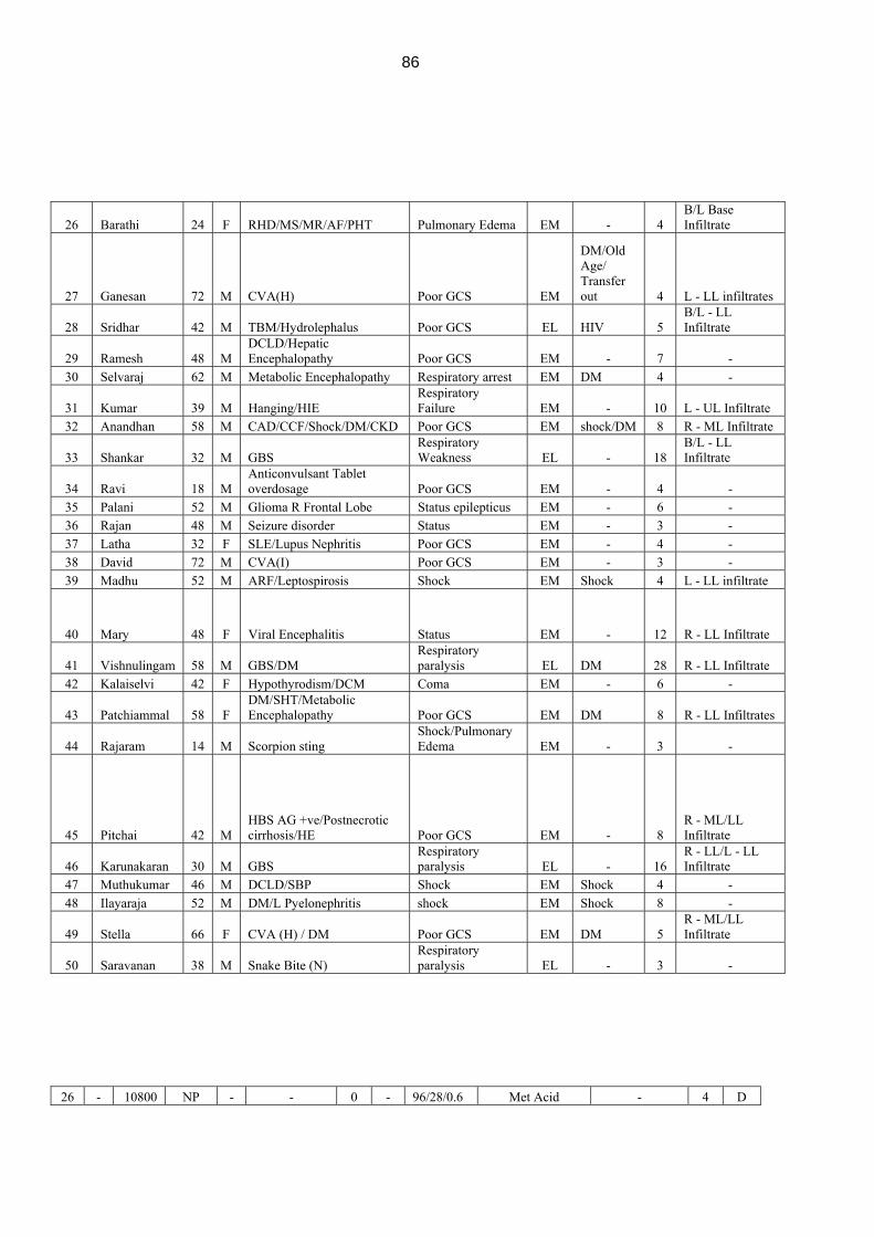
86
26 Barathi 24 F RHD/MS/MR/AF/PHT Pulmonary Edema EM - 4 B/L Base Infiltrate
27 Ganesan 72 M CVA(H) Poor GCS EM
DM/Old Age/ Transfer out 4 L - LL infiltrates
28 Sridhar 42 M TBM/Hydrolephalus Poor GCS EL HIV 5 B/L - LL Infiltrate
29 Ramesh 48 M DCLD/Hepatic Encephalopathy Poor GCS EM - 7 -
30 Selvaraj 62 M Metabolic Encephalopathy Respiratory arrest EM DM 4 -
31 Kumar 39 M Hanging/HIE Respiratory Failure EM - 10 L - UL Infiltrate
32 Anandhan 58 M CAD/CCF/Shock/DM/CKD Poor GCS EM shock/DM 8 R - ML Infiltrate
33 Shankar 32 M GBS Respiratory Weakness EL - 18
B/L - LL Infiltrate
34 Ravi 18 M Anticonvulsant Tablet overdosage Poor GCS EM - 4 -
35 Palani 52 M Glioma R Frontal Lobe Status epilepticus EM - 6 - 36 Rajan 48 M Seizure disorder Status EM - 3 - 37 Latha 32 F SLE/Lupus Nephritis Poor GCS EM - 4 - 38 David 72 M CVA(I) Poor GCS EM - 3 - 39 Madhu 52 M ARF/Leptospirosis Shock EM Shock 4 L - LL infiltrate
40 Mary 48 F Viral Encephalitis Status EM - 12 R - LL Infiltrate
41 Vishnulingam 58 M GBS/DM Respiratory paralysis EL DM 28 R - LL Infiltrate
42 Kalaiselvi 42 F Hypothyrodism/DCM Coma EM - 6 -
43 Patchiammal 58 F DM/SHT/Metabolic Encephalopathy Poor GCS EM DM 8 R - LL Infiltrates
44 Rajaram 14 M Scorpion sting Shock/Pulmonary Edema EM - 3 -
45 Pitchai 42 M HBS AG +ve/Postnecrotic cirrhosis/HE Poor GCS EM - 8
R - ML/LL Infiltrate
46 Karunakaran 30 M GBS Respiratory paralysis EL - 16
R - LL/L - LL Infiltrate
47 Muthukumar 46 M DCLD/SBP Shock EM Shock 4 - 48 Ilayaraja 52 M DM/L Pyelonephritis shock EM Shock 8 -
49 Stella 66 F CVA (H) / DM Poor GCS EM DM 5 R - ML/LL Infiltrate
50 Saravanan 38 M Snake Bite (N) Respiratory paralysis EL - 3 -
26 - 10800 NP - - 0 - 96/28/0.6 Met Acid - 4 D

87
27 - 9200 P - - 0 - 196/56/1.6 - - 4 D 28 + 6800 P N Acinetobacter 1 E 86/32/1.0 Resp.ALK - 5 D 29 - 11800 NP - - 0 - 136/48/1.2 - - 7 D 30 - 12600 NP - - 0 - 192/56/2.4 - - 5 D 31 + 10800 P - Polymicrobial 1 L 118/32/0.8 - - 25 R 32 - 9600 NP - - 0 - 116/56/3.2 - - 8 D 33 + 14600 P N Proteus 1 L 108/42/1.2 Resp.ALK - 30 R 34 + 11400 NP - - 0 - 124/36/0.6 Met Acidosis - 7 R 35 - 7200 NP - - 0 - 108/42/1.2 - - 6 D 36 + 10800 NP - - 0 - 158/32/1.0 Resp.ALK - 7 R 37 + 9600 NP - - 0 - 168/68/5.6 - - 6 D 38 - 10800 NP - - 0 - 108/42/1.2 - - 4 D
39 - 18600 NP - - 0 - 96/48/3.2 Metabolic Alkalaosis - 4 D
40 + 20400 P N Pseudomonas 1 L 112/32/1.2 - LP
Lymphocytosis 18 R 41 + 16200 P P MRSA 1 L 302/48/1.8 - - 42 R
42 - 9600 NP - - 0 - 102/44/0.6 Metabolic Alkalaosis T3 ↓ T4 ↓ 10 D
43 + 12600 P P MRSA 1 E 102/32/1.8 - - 10 D 44 - 10200 NP - - 0 - 142/32/1.0 - - 6 R
45 + 10800 P N Pseudomonas 1 E 86/52/1.8 Metabolic Alkalaosis - 9 D
46 + 12800 P - Polymicrobial 1 L 136/42/0.8 - - 28 D
47 + 12200 NP - - 0 - 108/56/0.8 Metabolic Alkalaosis - 4 D
48 + 14600 NP - - 0 - 288/72/2.8 Metabolic Alkalaosis - 9 D
49 + 13800 P P MRSA 1 E 156/48/1.8 - - 5 D 50 - 9600 NP - - 0 - 124/40/0.8 - - 6 D


![,6729, ÿ(7.( - Lumexlumex.hr/lumex_katalog proizvoda.pdf · .,6729, ÿ(7.(kistovi str. 6 Îhwnh vwu valjci xor #fl ]d ydomnh vwu gu #nh ]d ydomnh vwu vhwryl vwu srvxgh puh @lfh](https://img.pdfslide.net/doc/110x75/5fd0e28818735b7904058ee9/6729-7-proizvodapdf-6729-7kistovi-str-6-hwnh-vwu-valjci-xor.jpg)