Embed Size (px)
Citation preview

Incidental findings in Oncology Imaging
Wan Wan Yap MBChB, FRCR, FRCPC
Diagnostic Imaging , Vancouver Centre BC Cancer Agency

Disclosure
• None

Outline • Background
• Definition • Current problem • Specifically in oncology imaging
• Vascular findings • Pulmonary Embolism
• Organ specific • Thyroid nodules • Pulmonary nodules • Adrenal nodules • Liver lesions • Adnexal lesions
Background
• Incidentaloma (Incidental = by coincidence; oma= tumour Greek) • Unexpected , asymptomatic abnormality that are discovered serendipitously
while searching for other pathology or during screening examination1.
• Incidental radiologic findings are common in clinical practice and research
1. Managing incidental findings on abdominal CT: white paper of the ACR incidental findings committee. Berland LL etal J Am Coll Radiology 2010 Oct;7(10):754-73
Background
• Reasons for the increase • Increase use of imaging from diagnosis to management • Dramatic rise in cross-sectional imaging is the key reason Eg, Use of
• cross sectional imaging ( eg CT simulation) vs plain radiographs, • CT pulmonary angiogram vs VQ scan • Intravenous pyelogram vs CT urography • The list goes on…..
• Aging population • Improve in the quality of imaging
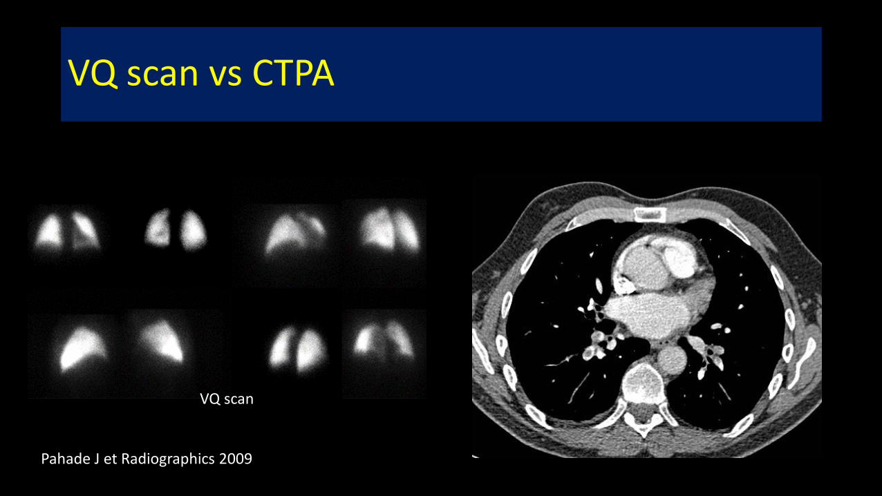
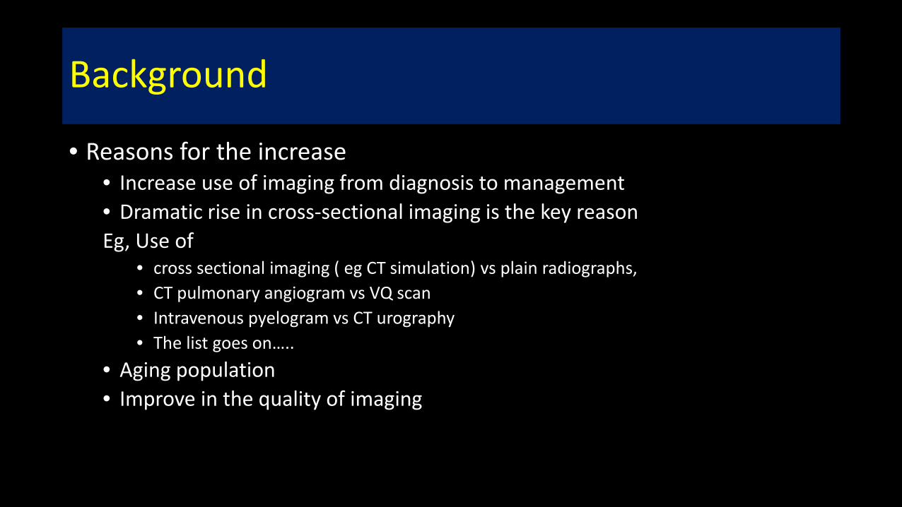
VQ scan vs CTPA
VQ scan
Pahade J et Radiographics 2009

Intravenous urogram CT urogram

‘Incidental’ lesions in oncology could be:
1. Metastases from the pre-existing primary 2. Second primary malignancy 3. Benign lesion
Multidiscliplinary team approach • Further management depends on patient’s comorbidity ,
performance status, life expectancy etc • May impact on treatment planning eg Crohn’s disease and radiation

Specifically for oncology patients
Tests that are potentially problematic • Screening examination
• CT colonography • CT chest for lung screening
• Staging CT chest abdomen and pelvis eg. Prostate cancer • Family history – hereditary screening program • Research • CT simulation for radiation planning • PET-CT

What are the pros and cons? 1. Change incidence of disease.
• Eg CTPA – incidental findings requiring follow up were nearly 3 x more common than emboli.1 • Incidental findings change incidence of disease eg Thyroid cancer double over 30 years2,3, 61%
increase in RCC. 2. Increase patient’s anxiety 3. Dilemma for referring physicians 4. Radiologists are anxious about missing incidental findings 5. Lack of recommendations for management plan for radiologists for indeterminate
findings 6. Detection of early stage synchronous cancer
1. Hall WB, Truitt SG, Scheunemann LP, et al. The prevalence of clinically relevant incidental findings on chest computed tomographic angiograms ordered to diagnose pulmonary embolism. Arch Intern Med 2009;169: 1961-5. 2. Davies L, Welch HG. Increasing incidence of thyroid cancer in the United States 1973-2002. JAMA 2006;295:2164-7. 3. Cronan JJ. Thyroid nodules: is it time to turn off the US machines? Radiology 2008;247:602-4.

Prostate cancer1
• Retrospective evaluate 355 CT AP – patient prostate cancer 5 year period • Incidental Findings (IF) are considered significant if therapeutic intervention,
additional imaging or tissue sampling was advised. • Rate of IF correlated to patient’s age and prostate cancer risk • Result:
• 779 IF in 292 patients • 20.6% were significant • Synchronous malignancy in 5.9% (RCC 1.97%; Lymphoma 1.13%)–
• ALL N0M0 disease • Age >65
• Significant vascular findings – 6 patients
1. Incidental Findings at initial imaging workup of patients with Prostate cancer : Clinical significance and Outcomes. Azadeh et al AJR Dec 2012 Volume 199 Number 6

Vascular findings Pulmonary embolism

Pulmonary embolism
• Malignancy increase risks of thromboembolic disease • Can be detected in CT Abdomen Pelvis ( last few slices), CT Chest ( PV
phase) • Central PE – Generally treat • What about isolated subsegmental PE (ISPE)

CT AbdoPelvis ? Posterior basal
RLL PE
Motion artefact Non diagnostic
Isolated subsegmental PE
right posterobasal No PE
Dedicated CT PA study


Case 1: Melanoma-staging CT
26 May2016
27 May2016 CTPA study

June 2nd 2016 Subsegmental PE resolved
Other history: Recent craniotomy for brain mets Lung nodule
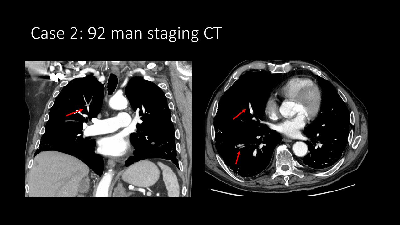
Case 2: 92 man staging CT

Organ specific IF

Thyroid nodules
• Very common in adult population. • Large autopsy study published in 1955 found that 50% of patients
with no clinical history of thyroid disease had thyroid nodules. • Incidental thyroid nodules (ITN)
• 20-67% of ultrasound studies • 25% of contrast-enhanced chest CT scans • 16-18% of CT and MR of the neck • 1-2% FDG PET scans

What we know…
• Small thyroid cancer are indolent • Incidentally detected thyroid cancer are more likely to be papillary
cancer good prognosis even without treatment. • Small thyroid cancers do not benefit from treatment • Subclinical thyroid cancer common
• 36% of 101 autopsies found occult papillary cancers
• Davis et al reported incidence of thyroid cancer tripled from 1975-2009 but mortality stable

Risk of cancer in Incidental thyroid nodules(ITN) • ITN detected on US 1.6% to 12%1 • ITN detected on CT and MRI range from 0-11%2,3 • ITN detected on FDG-PET scan at 33-35%4
1. Smith-Bindman R, Lebda P, Feldstein VA, et al. Risk of thyroid cancer based on thyroid ultrasound imaging characteristics: results of a population-based study.
JAMA Intern Med 2013;173:1788-96
2. Youserm DM, Huang T, Loevner LA, Langlotz CP. Clinical and economic impact of incidental thyroid lesions found with CT and MR. AJNR Am J Neuroradiol 1997;18:1423-8. 13.
3. Nguyen XV, Choudhury KR, Eastwood JD, et al. Incidental thyroid nodules on CT: evaluation of 2 risk-categorization methods for workup of nodules. AJNR Am J Neuroradiol 2013;34:1812-7.
4. Soelberg KK, Bonnema SJ, Brix TH, Hegedus L. Risk of malignancy in thyroid incidentalomas detected by 18F-fluorodeoxyglucose PET: a systematic review. Thyroid 2012;22:918-25

Problems
• Patient’s anxiety • Although FNABs carry minimally risk, cytology difficult to differentiate
adenoma vs carcinoma • repeat bx, • unnecessary surgery (25-41% ITN proceed to surgery- 36-75% = benign) • Only 25% of patients suspicious for malignancy

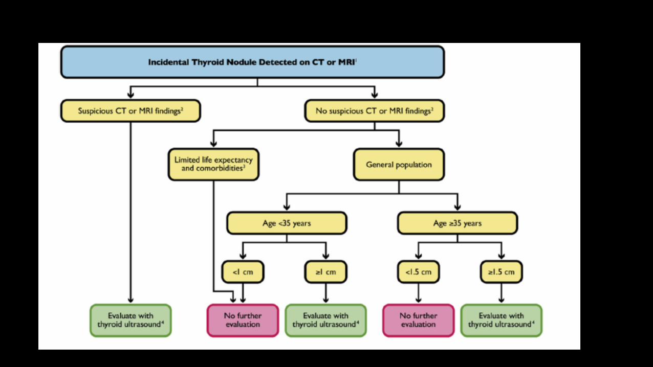
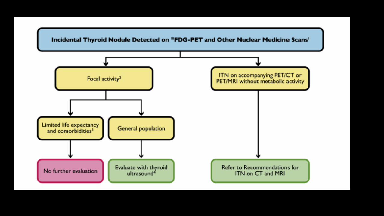
Managing incidental thyroid nodules1
ACR guidelines 3 tiered system • Category 1- Any size with aggressive imaging features • Category 2- <35 years old • Category 3- 1.5cm and not meeting criteria 1 and 2
1. Managing Incidental Thyroid Nodules Detected on Imaging: White Paper of the ACR Incidental Thyroid Findings Committee Jenny K. Hoang, MBBSa , Jill E. Langer, MDb , William D. Middleton, MDc , Carol C. Wu, MDd , Lynwood W. Hammers, DOe , John J. Cronan, MD f , Franklin N. Tessler, MD, CMg , Edward G. Grant, MDh , Lincoln L. Berland, MDg

Clinical input that will be helpful :
HISTORY • Childhood radiation • Endocrine syndromes • Family history

Case 1
• 85 year old, history of diffuse large B cell lymphoma • PET-CT workup show FDG avid right thyroid nodule

1.1cm right thyroid nodule SUV max 5.0

Hurthle cell lesion of undetermined significance • Overall malignancy rate of cytology of follicular lesion of
undetermined significance range from 5-30% • This patient is waiting for surgery

Case 2
• 60 female • Jejunal GIST in the setting of neurofibromatosis • Incidental thyroid nodule found in routine CT


Benign follicular nodule with cystic degeneration

Case 3
• 63 year old T3N2 M1 rectal cancer • PET-CT FDG avid right thyroid nodule
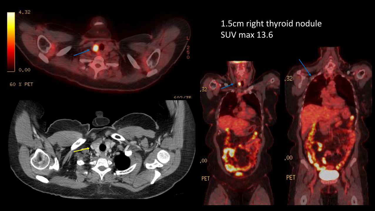
1.5cm right thyroid nodule SUV max 13.6


Papillary thyroid carcinoma

Lung nodules
• Lung nodules • Fleichner’s criteria – follow up small lung nodules incidentally
detected on CT • Perifissural nodules • Triagular intrapulmonary lymph nodes

56 female history of breast cancer

Fleichner’s criteria
Fewer than 1% of very small (<5mm) nodules in patients without a history of cancer will demonstrate malignant behaviours • <5mm ->12 mo FU found NO cancer • 5-9mm 6% malignant detected at 4-8 moths FU scan • 8mm nodules 10-20% risk of malignancy

Solid non calcified nodule (NCPN)
Nodule size (mm)
Low risk High risk
<= 4 No follow up F/U 12 moths If unchanged- no further FU
>4-6 F/U 12 mo Unchanged-Stop
Initial FU CT at 6-12 mo then 18-24 mo if no change
>6-8 Initial 6-12 mo then 18-24 mo Initial FU 3-6 mo 9,12,24 mo if no change
>8 3,9, 24 mo CE CT, PET CT=/- biopsy
Same as low risk patients

History of extrapulmonary malignancy
• Cahan et al (1978) thoracotomy results • 800 with cancer over 35 years • 500 NSCLC (HN, bladder, breast, prostate) • 196 metastastic (melanoma, bone, soft tissue sarcomas and testicular cancer)
• Quint et al(2000) • Non calcified pulmonary nodules >=5mm found on CT chest • HN cancer patients more likely to have NSCLC
• Khokhar et al (2006) • 151 patients – 42% with malignant nodules • 50% lung cancer, 44% metastatic, 3% second primary, 3% unknown primary

NCPN in patients with extrapulmonary cancer
• Shorter interval follow ups • Low threshold for biopsy • Clinical correlation especially history of smoking • Lung cancer is not excluded by the findings of multiple nodules

Liver • Jones et al1 (1992)-
• 1454 consecutive patients • 17%- <= 1.5cm hepatic lesions found • 82% of this patients known to have extrahepatic
malignancy --51% lesions benign, 26% malignant, 23% indeterminant
• 5% with 1 lesion • 19% -2-4 lesions • 74% >5 lesions • Multiple small lesions were more likely to represent
malignant disease than were single small lesions
1.Jones EC, Chezmar JL, Nelson RC, et al. The frequency and significance of small (less than or equal to 15 mm) hepatic lesions detected by CT. AJR 1992; 158: 535539.

• Schwartz et al • Small <=1.cm lesions found in 12.7% patients
• 80.2% benign, • 11.6% malignant • 8.2% indeterminate
• Jang et al
• 1133 colorectal and gastric patents; <=1.5cm hypoattenuating lesions in 25.5% cases
• 94% smooth, <20HU = benign
• Khalil et al • 941 breast cancer patients found 29.4% small liver lesions • 92.7% no change
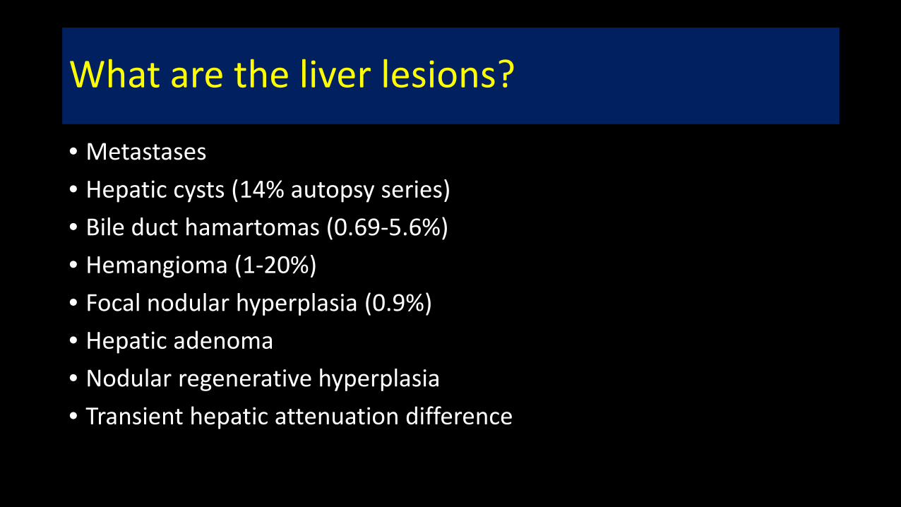
What are the liver lesions?
• Metastases • Hepatic cysts (14% autopsy series) • Bile duct hamartomas (0.69-5.6%) • Hemangioma (1-20%) • Focal nodular hyperplasia (0.9%) • Hepatic adenoma • Nodular regenerative hyperplasia • Transient hepatic attenuation difference

Case 1
• 65 man stage II thymoma resected in 2007 + radiation • Routine Chest CT follow up 14 May 2015

In phase Oppose phase

T2
PV phase
5 min delayed
20 min delayed


Pathology
• Benign, mild steatosis

Case 2
• 66 man hx of papillary thyroid carcinoma and tonsillar SCC with metastatic lymph node
• PET CT indeterminate liver lesion


Angiomyolipoma

Simple hepatic cysts

Adrenal mass
• Common

Pre op GE Junction tumour
Post op gastric pull up

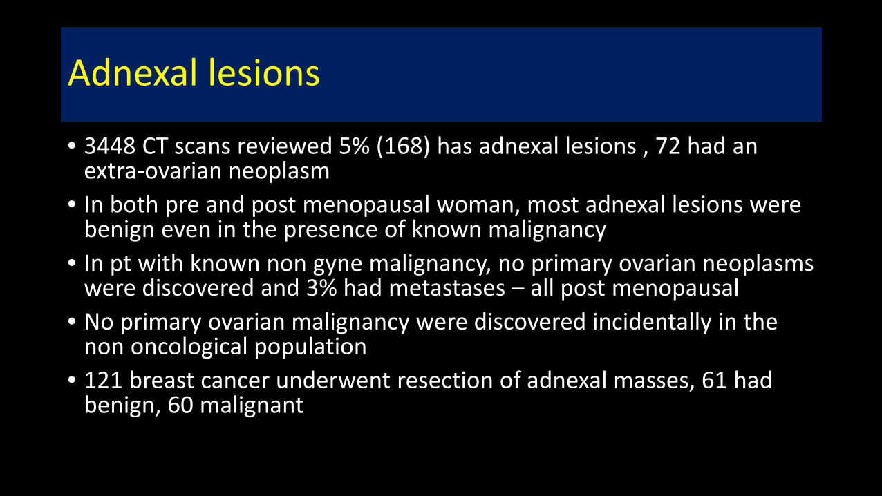
Adnexal lesions
• 3448 CT scans reviewed 5% (168) has adnexal lesions , 72 had an extra-ovarian neoplasm
• In both pre and post menopausal woman, most adnexal lesions were benign even in the presence of known malignancy
• In pt with known non gyne malignancy, no primary ovarian neoplasms were discovered and 3% had metastases – all post menopausal
• No primary ovarian malignancy were discovered incidentally in the non oncological population
• 121 breast cancer underwent resection of adnexal masses, 61 had benign, 60 malignant
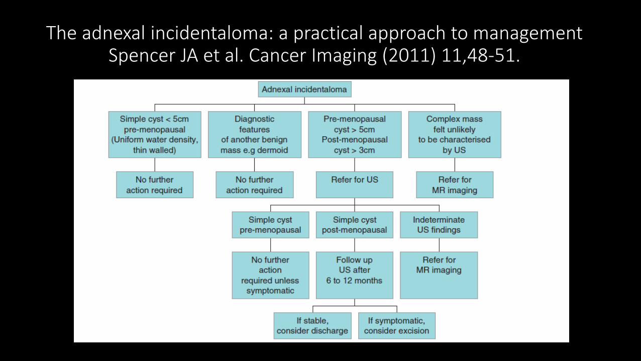
The adnexal incidentaloma: a practical approach to management Spencer JA et al. Cancer Imaging (2011) 11,48-51.

50 year female history of Breast Cancer

38 female abdo pain


• Krukenberg tumour • Metastases

Clinical input
• Risk of malignancy index • Ultrasound score x manopausal state x CA125 • Manopausal state
Conclusion
• Incidental findings are common due to the exponential usage of cross-sectional imaging
• In oncology patients the concerns are mets, second primary or benign • Increase use of guidelines and recommendations to have a more
uniform approach by Radiologists • Multidiscliplinary approach is key to management
• Radiologists- best imaging modality • Clinicians (Med onc, Rad onc, GPO)- co-morbidities, performance status, life
expectancies and patient’s expectations • Pathologists

Thank you [email protected]