Embed Size (px)
Citation preview
INCLUSION BODY MYOSITIS and INCLUSION BODY MYOPATHY.
Bill TillierCalgary, Alberta
August, 2009.
2IBM classification.
Inclusion Body Myositis and Inclusion Body Myopathy are classified as types of muscular dystrophy.
Muscular dystrophy: Idiopathic inflammatory myositis (IIM) disorders:
Dermatomyositis (DM) Polymyositis (PM) Inclusion body myositis and inclusion body myopathies.
Idiopathic means the cause is unknown. There are similarities between PM & IBM (and also big
differences) but DM seems to be quite different.
People often confuse MD with a different disorder; MS: multiple sclerosis, a disorder affecting the nerves.
3Sporadic inclusion body myositis (sIBM).
Called a myositis to emphasize the inflammation of muscle that characterizes it. Sporadic means it just shows up here and there in people (it’s not inherited).
This is the most common form, if you have been diagnosed with "IBM," this is likely what you have.
sIBM is a progressive disorder of skeletal musclecells: as more and more cells are affected and die off, the muscles shrink and become progressively weaker.
sIBM is age-related, as we get older, it gets more and more common, primarily appearing after age 50.
The following diagram outlines the weakness seen.
Symptoms emerge slowly, over months or years.
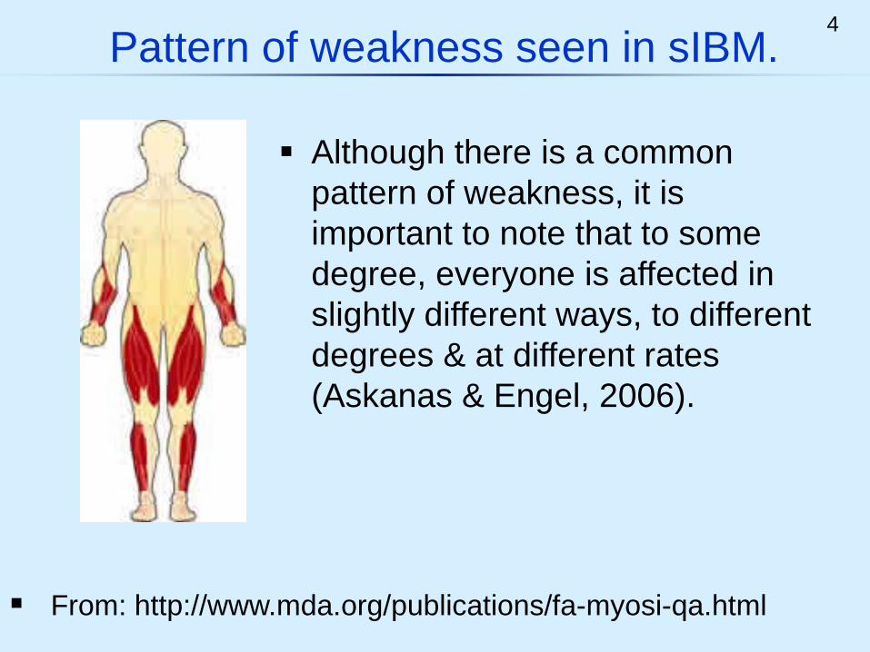
4Pattern of weakness seen in sIBM.
From: http://www.mda.org/publications/fa-myosi-qa.html
Although there is a common pattern of weakness, it is important to note that to some degree, everyone is affected in slightly different ways, to different degrees & at different rates (Askanas & Engel, 2006).
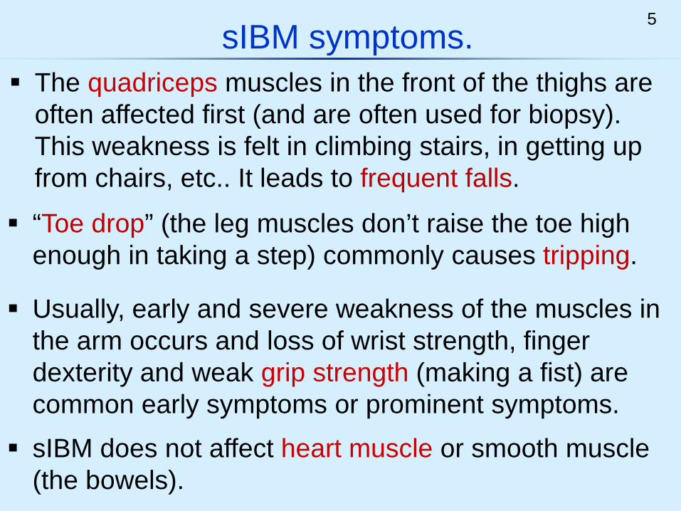
5sIBM symptoms.
The quadriceps muscles in the front of the thighs are often affected first (and are often used for biopsy). This weakness is felt in climbing stairs, in getting up from chairs, etc.. It leads to frequent falls.
sIBM does not affect heart muscle or smooth muscle (the bowels).
“Toe drop” (the leg muscles don’t raise the toe high enough in taking a step) commonly causes tripping.
Usually, early and severe weakness of the muscles in the arm occurs and loss of wrist strength, finger dexterity and weak grip strength (making a fist) are common early symptoms or prominent symptoms.
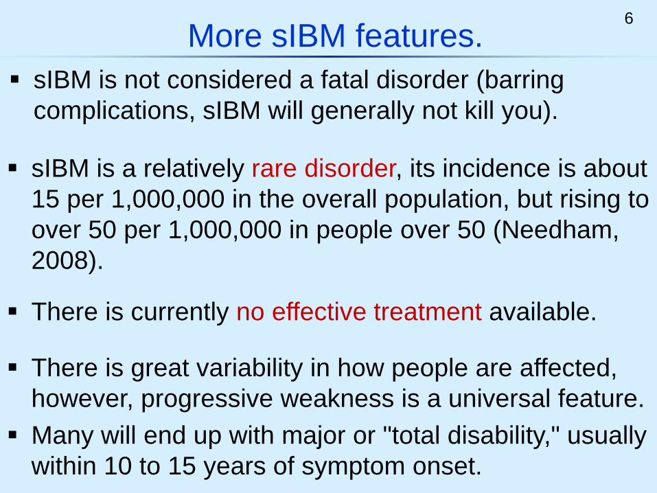
6More sIBM features.
sIBM is not considered a fatal disorder (barring complications, sIBM will generally not kill you).
sIBM is a relatively rare disorder, its incidence is about 15 per 1,000,000 in the overall population, but rising to over 50 per 1,000,000 in people over 50 (Needham, 2008).
There is currently no effective treatment available.
There is great variability in how people are affected, however, progressive weakness is a universal feature.
Many will end up with major or "total disability," usually within 10 to 15 years of symptom onset.
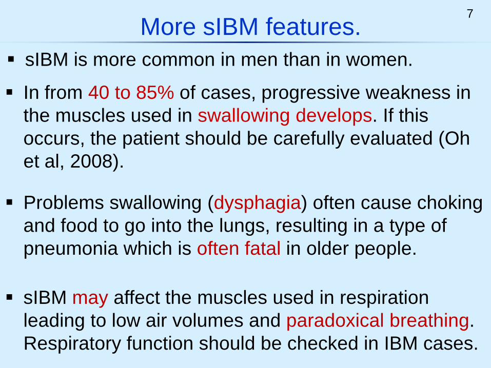
7More sIBM features.
sIBM is more common in men than in women.
sIBM may affect the muscles used in respiration leading to low air volumes and paradoxical breathing. Respiratory function should be checked in IBM cases.
Problems swallowing (dysphagia) often cause choking and food to go into the lungs, resulting in a type of pneumonia which is often fatal in older people.
In from 40 to 85% of cases, progressive weakness in the muscles used in swallowing develops. If this occurs, the patient should be carefully evaluated (Oh et al, 2008).
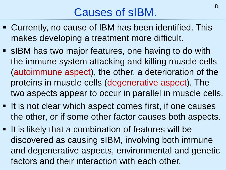
8Causes of sIBM.
Currently, no cause of IBM has been identified. This makes developing a treatment more difficult.
sIBM has two major features, one having to do with the immune system attacking and killing muscle cells (autoimmune aspect), the other, a deterioration of the proteins in muscle cells (degenerative aspect). The two aspects appear to occur in parallel in muscle cells.
It is not clear which aspect comes first, if one causes the other, or if some other factor causes both aspects.
It is likely that a combination of features will be discovered as causing sIBM, involving both immune and degenerative aspects, environmental and genetic factors and their interaction with each other.
9Theories of sIBM.
It appears that a new theory of sIBM may have to emerge before this impasse is resolved. Researchers note that IBM is currently poorly understood and many more discoveries remain to be made.
One current theory is that an undiscovered virustriggers sIBM, setting in motion an ongoing immune response which attacks and kills the muscle cells.
IBM was named for the observation that “inclusions” (inclusion bodies = clumps) and strands of abnormal proteins form in the muscle cells affected by sIBM.
Based upon this, a second theory is that abnormal proteins form in the muscle cells causing sIBM and triggering the immune response that is seen.
10sIBM Research.
Example: The role of beta amyloid proteins in IBM has been questioned by Greenberg (2009). He showed how inappropriate citation of papers has created distortion of the evidence leading to the belief that these proteins are central in causing IBM.
Based upon the description of several unusual cases, the name IBM was suggested in 1971 and since then about 1000 research articles have appeared.
Two main research themes have emerged and become dominant belief systems, one focused on inflammation, the other on amyloid related proteins.
Lately, other ideas have received more attention and questions are being raised over past conclusions.
11Genetics of sIBM.
There is a subset of these MHC genes called the 8.1 ancestral haplotype. A further subset of this group called the Human Leukocyte Antigen (HLA) genes have been linked to patients with sIBM (and fIBM).
sIBM is not an inherited disorder: the disorder is not passed on to the children of patients with sIBM.
sIBM has been linked with a group of genes related to the immune system that are commonly seen in Caucasians from Northern European ancestry.
This group of genes is called the Major Histocompatibility Complex (MHC) and is found on chromosome #6.
12Genetic predispositions in sIBM?
Chemicals controlled by HLA genes play an important role in regulating immune responses to certain environmental factors.
People with certain groups of HLA genes are predisposed to develop autoimmune disorders.
sIBM is associated with HLA-DR3 genes in about 70% of patients (Badrising, 2004).
If you happen to have this particular group of genes, you are likely somehow predisposed to develop IBM.
Conclusion: It appears that some combination of genes interact with each other and with environmental variables to increase the likelihood of sIBM developing in a given individual.
13Diagnosis.
Muscle disorders are generally difficult to diagnose.
Usually a specialist (neurologist or rheumatologist) is required to diagnose the disorder.
Most patients say that diagnosis took a long time and was a frustrating process.
These methods and tests are usually used in diagnosis:
Many say that medications were suggested or tried (generally, no medication is indicated for IBM).
sIBM is often initially misdiagnosed as another muscle disorder called polymyositis.
14Diagnosis of sIBM.
1. Clinical signs are the symptoms the doctor can see when you are examined. The doctor looks for the clinical signs of sIBM; a characteristic pattern of muscle weakness.
2. A blood test is done for creatine kinase (CK) (also known as "phosphocreatine kinase," or CPK), an enzyme in the blood showing muscle damage. CK is at most about 10X normal, although this may vary.
3. Electromyography (EMG): Electrical studies of the muscle done with a computer (an electromyograph) will display abnormalities in these disorders.
4. Muscle Biopsy: Surgical removal of a piece of muscle and its subsequent study in the laboratory.
15Familial Inclusion Body Myositis (fIBM)
fIBM occurs in two or more siblings in a family in the same generation (but it is not passed on to children).
The symptoms and features of fIBM are very similar to those seen in the sporadic form of IB myositis.
fIBM is also linked with similar genes as sIBM, raising the possibility that fIBM and sIBM share similar inherited predispositions.
The familial occurrence of such a rare disorder highlights the importance of genetic predisposition in the causation of sIBM (Needham 2007).
Familial and hereditary forms of IBM existing along with sporadic IBM is challenging to understand – how are they related? What does this say about causes?
16Sporadic versus hereditary forms.
Hereditary inclusion body myopathy (hIBM): “Myopathy” is used here as this type does not show
muscle inflammation (myo: muscle, pathy: disease). Hereditary is used to indicate that it is inherited.
Sporadic inclusion body myositis (sIBM) features inflammation in the muscle and deterioration. It is not directly linked to a genetic mutation (not inherited).
The rest of this presentation will focus on the hereditary forms which are exceedingly rare (in some forms, only a few hundred known cases in the world).
See slide 26 for a general conclusion.
17Sporadic versus hereditary forms.
Various hereditary types follow either autosomal recessive or autosomal dominant patterns of inheritance.
Today, several different hereditary inclusion body myopathies, are recognized. These different forms can display different symptoms but all share similar underlying structural features in common.
As these disorders were discovered, a complex series of names were assigned to them before it was recognized that some shared commonalities or where the same.
18Various names for the recessive form.
Hereditary Inclusion Body Myopathy (hIBM2). Inclusion Body Myopathy 2 (IBM2); Autosomal recessive Inclusion Body Myopathy
(AR-hIBM); Quadriceps-sparing Inclusion Body Myopathy
(QSM); Nonaka distal myopathy with rimmed vacuoles; Distal myopathy with rimmed vacuoles (DMRV).
This is the most common type of inherited IBM.
As the autosomal recessive form was initially described, several different names were given to it:
19Various autosomal dominant forms.
Autosomal dominant forms: IBM 3: was called “AD myopathy with congenital
joint contractures, ophthalmoplegia and rimmed vacuoles” (linked to mutations in a gene on chromosome 17).
IBMPFD: IBM associated with Paget’s disease and frontotemporal dementia (linked to a gene on chromosome 9, located at 9p13-p12).
AD-IBM: Autosomal dominant inclusion body myopathy.
20Features of hIBM 2.
The onset of this disorder usually occurs after the age of 20 but before the middle of the fourth decade. Proximal and distal muscle weakness and wasting of the upper and lower limbs are progressive and result in severe incapacitation within 10 to 20 years.
Argov and Yarom (1984) first described the disorder in Jews of Persian (Iranian) origin.
Sparing of the quadriceps muscles, even in advanced stages of the disorder, is a feature unique to this inherited form of inclusion body myopathy.
hIBM 2 shares several features with sIBM, including “muscle holes” and protein inclusions (but with NO inflammation or quadriceps muscle involvement).
21Mapping to a specific gene location.
All patients of Middle Eastern descent were found to share a single homozygous missense mutation in the GNE gene. This gene produces GNE protein. (GNE stands for: UDP-N-acetylglucosamine 2-epimerase/N-acetylmannosamine kinase).
Eisenberg et al. (1999, 2001) traced the disorder to a gene on an autosome: chromosome #9 (9p12-p11).
GNE is an enzyme involved in the production of sialic acid: a chemical involved in the management of glycoproteins and glycolipids in the cell.
To date, more than 40 different mutations in the GNE gene have been reported that lead to hIBM 2.
22Who is affected?
hIBM 2 is a very rare disorder, affecting about 500 people worldwide. The disorder predominantly affects Iranian Jews, who have a 5 to 10 percent chance of carrying the hIBM 2 (GNE) gene mutation.
Argov et al. (2003) concluded that this mutation first appeared about 1,300 years ago and today, is not limited to those of Jewish descent.
hIBM 2 has also been seen in some Japanese families (first described by Dr. Nonaka in 1981) as well as in culturally diverse families and in various parts of the world.
23Autosomal recessive disorders.
A mutation of the GNE gene on one of the # 9 pair of autosomal chromosomes from each parent is required to cause the disorder. People with only one abnormal gene in the pair will be carriers, but since the gene is recessive they do not exhibit the disorder.
To develop symptoms, recessive traits require that both chromosomes in the pair (one received from mom and the other from dad) carry the mutation.
A child inheriting 1 mutated gene will be a carrier, a child receiving 2 mutated genes will have the disorder.
The following diagram outlines the statistical situation.
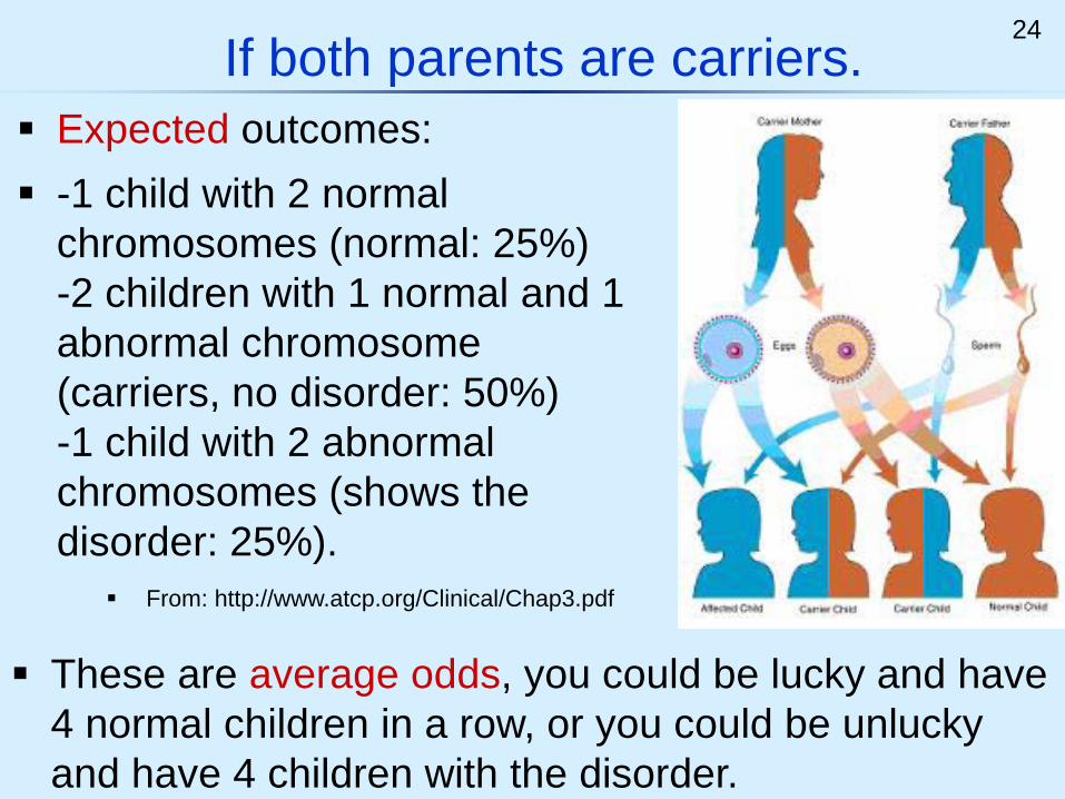
24If both parents are carriers.
These are average odds, you could be lucky and have 4 normal children in a row, or you could be unlucky and have 4 children with the disorder.
Expected outcomes: -1 child with 2 normal
chromosomes (normal: 25%) -2 children with 1 normal and 1 abnormal chromosome (carriers, no disorder: 50%) -1 child with 2 abnormal chromosomes (shows the disorder: 25%).
From: http://www.atcp.org/Clinical/Chap3.pdf
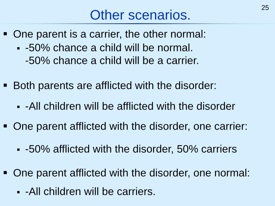
25Other scenarios.
-50% chance a child will be normal. -50% chance a child will be a carrier.
One parent is a carrier, the other normal:
Both parents are afflicted with the disorder:
-All children will be afflicted with the disorder
One parent afflicted with the disorder, one carrier:
One parent afflicted with the disorder, one normal:
-50% afflicted with the disorder, 50% carriers
-All children will be carriers.
26Mutations plus other factors?
GNE mutations have been found in some people who never show symptoms, showing that other factorsmust play a role in the expression of the disorder.
Many genetic conditions show interactions with environmental factors that may speed or slow their expression (the development of symptoms).
These factors also seem to be important in the clinical expression of hIBM 2 in all patients; although the gene defect is present from conception, symptoms may not appear until the 30s or 40s.
Researchers are struggling to discover what these factors may be (Needham, 2007).
27Conclusion.
Inclusion body myositis and myopathy are very complex disorders of the skeletal muscle. It is not clear what causes the sporadic form. The mutations behind the inherited forms are being
described but possible interacting environmental factors remain to be discovered.
These can be frustrating disorders to diagnose and all previous attempted treatments have failed.
Although the slow loss of mobility and arm function is very frustrating, patients must be encouraged to adapt and “make the best of things.”
28Some abbreviations used.
Familial Inclusion Body Myositis (fIBM) Hereditary inclusion body myopathy (hIBM). Polymyositis (PM) Muscular dystrophy (MD) myo = muscle; pathy = disease; itis = inflammation.
Sporadic inclusion body myositis (sIBM).
29References.
Argov Z. & Yarom, R., "Rimmed vacuole myopathy" sparing the quadriceps. A unique disorder in Iranian Jews. J Neurol Sci. 1984 Apr;64(1):33-43.
Argov Z, Eisenberg I, Grabov-Nardini G, Sadeh M, Wirguin I, Soffer D,Mitrani-Rosenbaum S. Hereditary inclusion body myopathy: the Middle Eastern genetic cluster. Neurology. 2003 May 13;60(9):1519-23.
Askanas, V. & Engel, W. K., Inclusion-body myositis: a myodegenerative conformational disorder associated with A-beta, protein misfolding, and proteasome inhibition. Neurology 66 (suppl. 1): S39-S48, 2006.
Badrising UA et al. (2004) Associations with autoimmune disorders and HLA class I and II antigens in inclusion body myositis. Neurology 63: 2396-2398.
Dalakas, MC. August, 2006. Sporadic inclusion body myositis—diagnosis, pathogenesis and therapeutic strategies. Nature Clinical Practice Neurology 2: 437.
Eisenberg I, Thiel C, Levi T, Tiram E, Argov Z, Sadeh M, Jackson CL, Thierfelder L, Mitrani-Rosenbaum S. Fine-structure mapping of the hereditary inclusion body myopathy locus. Genomics. 1999 Jan 1;55(1):43-8.
Eisenberg I, Avidan N, Potikha T, Hochner H, Chen M, Olender T, Barash M, Shemesh M, Sadeh M, Grabov-Nardini G, Shmilevich I, Friedmann A, Karpati G, Bradley WG, Baumbach L, Lancet D, Asher EB, Beckmann JS, Argov Z, Mitrani-Rosenbaum S. The UDP-N-acetylglucosamine 2-epimerase/N-acetylmannosaminekinase gene is mutated in recessive hereditary inclusion body myopathy. Nat Genet. 2001 Sep;29(1):83-7.
Greenberg SA. BMJ. 2009 Jul 20;339:b2680. doi: 10.1136/bmj.b2680. Comment in: BMJ. 2009;339:b2049.How citation distortions create unfounded authority: analysis of a citation network.Needham M, Corbett A, Day T, Christiansen F, Fabian V, Mastaglia FL. (2008). Prevalence of sporadic IBM and factors contributing to delayed diagnosis. J Clin Neurosci. 2008 Dec;15(12):1350-3.
Needham M, Mastaglia FL, Garlepp MJ. (2007) Genetics of inclusion-body myositis. Muscle Nerve. Mar 15; [Epub ahead of print] May, 2007, pps. 549-561.
Nonaka, I.; Sunohara, N.; Ishiura, S.; Satoyoshi, E. Familial distal myopathy with rimmed vacuole and lamellar (myeloid) body formation. J. Neurol. Sci. 51: 141-155, 1981.
Oh TH, Brumfield KA, Hoskin TL, Kasperbauer JL, Basford JR. (2008). Dysphagia in inclusion body myositis: clinical features, management, and clinical outcome. Am J Phys Med Rehabil. Nov;87(11):883-9.


![ISSN 2171-6625 JOURNAL OF NEUROLOGY AND …...Sporadic Inclusion Body Myositis (sIBM) is a progressive muscular disorder developing in the elderly [1]. In Western countries, sIBM is](https://img.pdfslide.net/doc/110x75/608407ff8c7add66d3780c62/issn-2171-6625-journal-of-neurology-and-sporadic-inclusion-body-myositis-sibm.jpg)
![Version 2 Please don’t nore this - Blood · Inflammatory myopathies – inclusion body myositis (IBM) (formerly Inflammatory myopathies: polymyositis [PM], dermatomyositis [DM]](https://img.pdfslide.net/doc/110x75/602246494e545541c973d779/version-2-please-donat-nore-this-blood-inflammatory-myopathies-a-inclusion.jpg)