Embed Size (px)
Citation preview
Morimoto et al. Orphanet Journal of Rare Diseases (2016) 11:149 DOI 10.1186/s13023-016-0519-7
RESEARCH Open Access
Increased Wnt and Notch signaling: a clueto the renal disease in Schimke immuno-osseous dysplasia?
Marie Morimoto1,2, Clara Myung1,2, Kimberly Beirnes1,2, Kunho Choi1,2, Yumi Asakura3, Arend Bokenkamp4,Dominique Bonneau5, Milena Brugnara6, Joel Charrow7, Estelle Colin5, Amira Davis8, Georges Deschenes9,Mattia Gentile10, Mario Giordano11, Andrew K. Gormley12, Rajeshree Govender13, Mark Joseph14, Kory Keller15,Evelyne Lerut16, Elena Levtchenko17, Laura Massella18, Christy Mayfield19, Behzad Najafian20, David Parham21,Jurgen Spranger22, Peter Stenzel23, Uluc Yis24, Zhongxin Yu25, Jonathan Zonana15, Glenda Hendson26and Cornelius F. Boerkoel1,2,27*
Abstract
Background: Schimke immuno-osseous dysplasia (SIOD) is a multisystemic disorder caused by biallelic mutationsin the SWI/SNF-related matrix-associated actin-dependent regulator of chromatin, subfamily A-like 1 (SMARCAL1)gene. Changes in gene expression underlie the arteriosclerosis and T-cell immunodeficiency of SIOD; therefore,we hypothesized that SMARCAL1 deficiency causes the focal segmental glomerulosclerosis (FSGS) of SIOD byaltering renal gene expression. We tested this hypothesis by gene expression analysis of an SIOD patient kidneyand verified these findings through immunofluorescent analysis in additional SIOD patients and a geneticinteraction analysis in Drosophila.
Results: We found increased expression of components and targets of the Wnt and Notch signaling pathways inthe SIOD patient kidney, increased levels of unphosphorylated β-catenin and Notch1 intracellular domain in theglomeruli of most SIOD patient kidneys, and genetic interaction between the Drosophila SMARCAL1 homologueMarcal1 and genes of the Wnt and Notch signaling pathways.
Conclusions: We conclude that increased Wnt and Notch activity result from SMARCAL1 deficiency and, asestablished causes of FSGS, contribute to the renal disease of most SIOD patients. This further clarifies thepathogenesis of SIOD and will hopefully direct potential therapeutic approaches for SIOD patients.
Keywords: Schimke immuno-osseous dysplasia, SMARCAL1 protein, Focal segmental glomerulosclerosis, Wntsignaling pathway, Notch signaling pathway
BackgroundSchimke immuno-osseous dysplasia (SIOD, OMIM242900) is an autosomal recessive disease; its prominentfeatures are facial dysmorphism, hyperpigmented macules,focal segmental glomerulosclerosis (FSGS), spondyloepi-physeal dysplasia, and T-cell immunodeficiency [1–3].Additional features include hypothyroidism, abnormal
* Correspondence: [email protected] of Medical Genetics, University of British Columbia, Vancouver,BC, Canada2Child & Family Research Institute, Vancouver, BC, CanadaFull list of author information is available at the end of the article
© 2016 The Author(s). Open Access This articInternational License (http://creativecommonsreproduction in any medium, provided you gthe Creative Commons license, and indicate if(http://creativecommons.org/publicdomain/ze
dentition, bone marrow failure, thin hair, corneal opacities,arteriosclerosis, cerebral ischemia, and migraine-like head-aches [2–5].The renal disease begins as proteinuria, progresses to
steroid-resistant nephropathy, and ultimately advancesto end-stage renal disease [4, 6]. FSGS is the predomin-ant renal pathology and is refractory to treatment withglucocorticoids, cyclosporine A, and cyclophosphamide[4, 6]. Suggesting a cell autonomous mechanism for therenal disease, renal transplantation is efficacious, and thedisease does not recur in the graft [2, 4, 5].
le is distributed under the terms of the Creative Commons Attribution 4.0.org/licenses/by/4.0/), which permits unrestricted use, distribution, andive appropriate credit to the original author(s) and the source, provide a link tochanges were made. The Creative Commons Public Domain Dedication waiverro/1.0/) applies to the data made available in this article, unless otherwise stated.
Morimoto et al. Orphanet Journal of Rare Diseases (2016) 11:149 Page 2 of 12
Biallelic mutations of the SWI/SNF-related matrix-asso-ciated actin-dependent regulator of chromatin, subfamilyA-like 1 (SMARCAL1) gene cause SIOD [7]. SMARCAL1encodes a DNA annealing helicase that is a distant mem-ber of the SWI/SNF family of ATP-dependent chromatinremodeling proteins [8]. SMARCAL1 recognizes DNAstructure, binds to open chromatin, is involved in theDNA damage response [9, 10] and DNA replication forkrestart [11, 12], and, along with genetic and environmentalfactors, alters gene expression [13].Gene expression changes appear critical to SIOD path-
ology. Full or partial explanations for the vasculardisease and T-cell immunodeficiency of SIOD patientsare respectively decreased expression of elastin (ELN) inthe aorta [14–16] and of interleukin 7 receptor alphachain (IL7R) in the T cells [17–19].Based on these findings, we hypothesized that SMAR-
CAL1 deficiency causes the renal disease of SIOD byaltering gene expression. Studies of other glomerulopa-thies find increased Wnt [20–23] and Notch signaling[24–27] as causes of podocyte dysfunction. CanonicalWnt pathway activation proceeds via inhibition of β-catenin ubiquitination, saturation of the β-catenindestruction complex, cytoplasmic accumulation andnuclear translocation of newly synthesized unpho-sphorylated β-catenin, and subsequent activation oftarget gene transcription through interaction with tran-scription factors and transcriptional co-activators [28].Notch pathway activation involves proteolytic cleavageof the Notch transmembrane receptor by an ADAMmetalloproteinase and the γ-secretase complex, nucleartranslocation of the released Notch1 intracellulardomain (NICD), and subsequent activation of targetgene transcription through interaction of the NICDwith transcription factors and transcriptional co-activators [29]. Wnt and Notch signaling are criticalfor kidney development and become undetectable inthe glomeruli of the postnatal kidney [26, 30].Analyses presented herein showed upregulation of the
Wnt and Notch signaling pathways in the SIOD kidneyand genetic interaction between the Drosophila SMAR-CAL1 homologue and genes encoding components ofthe Wnt and Notch pathways. We suggest therefore thatthe upregulation of the Wnt and/or Notch pathwayscontributes to the renal disease in SIOD.
MethodsPatients and human tissuesThe guardians of the patients referred to this studysigned informed consent approved by the ResearchEthics Board of the University of British Columbia(Vancouver, BC, Canada). Autopsy and biopsy tissueswere obtained according to the protocol approved bythe University of British Columbia (Vancouver, BC,
Canada). The renal parameters and the SMARCAL1mutations of the SIOD patients included in the studyare listed in Table 1 and Additional file 1: Table S1,respectively.In accordance with institutional policies as approved
by the Institutional Review Board (41557) at theUniversity of Washington, human fetal kidney fromsecond trimester elective terminations were provided asde-identified specimens by the Laboratory of Develop-mental Biology at the University of Washington(Seattle, WA), a National Institute of Child Health &Human Development supported program. De-identifiedcontrol specimens provided according to the protocolH06-70283 approved by the Clinical Research EthicsBoard at the University of British Columbia (Vancouver,BC, Canada) included renal biopsy sections from tenpediatric patients with isolated FSGS, postmortem kidneytissue from four pediatric patients, a skin biopsy from a16-year-old female, and adenoma tissue from a 17-year-old female with familial adenomatous polyposis. Samplecharacteristics and use are summarized in Additional file1: Table S2.
Drosophila melanogaster linesThe loss-of-function mutant Marcal1del and the Mar-cal1 overexpression transgenic line pUAST-Marcal1/CyO; tubulin-GAL4/TM3, Sb1 have been previouslydescribed [13] (Additional file 1: Figure S1). The C96-GAL4 UAS-Hrs/MKRS transgenic line, used to controlfor non-specific interactions with the GAL4-UAS sys-tem, was a gift from Dr. Hugo Bellen (Baylor College ofMedicine, Houston, TX, USA). All other Drosophilastocks were obtained from the Bloomington DrosophilaStock Center (Bloomington, IN, USA).
RNA extractionTotal RNA was extracted from flash frozen kidney pul-verized with a Bessman tissue pulverizer (SpectrumLaboratories, Rancho Dominguez, CA, USA) or from 8Drosophila adult female flies of each genotype by usingthe RNeasy Mini Kit (Qiagen, Toronto, ON, Canada).Total RNA from formalin-fixed paraffin-embedded(FFPE) fetal kidney was isolated using the RNeasy FFPEKit (Qiagen, Toronto, ON, Canada). Genomic DNA wasremoved by on-column DNase I digestion (Qiagen,Toronto, ON, Canada).
RNA-seq and KEGG pathway analysisStrand-specific, paired-end RNA-seq on poly(A) RNAwas performed by Macrogen (Seoul, Korea) using theTruSeq Stranded Total RNA Library Prep Kit (Illumina,San Diego, CA) and the HiSeq 2000 System (Illumina,San Diego, CA). This kit depleted the ribosomal RNA(rRNA) using Ribo-Zero rRNA reduction chemistry.
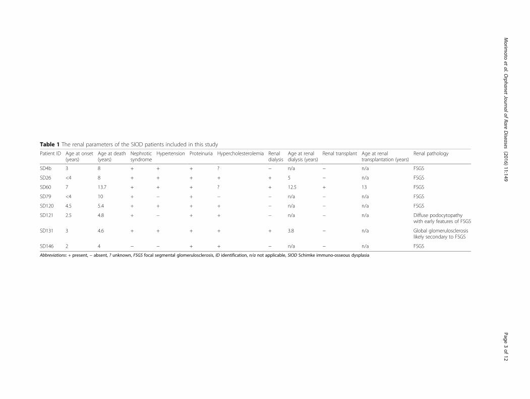
Table 1 The renal parameters of the SIOD patients included in this study
Patient ID Age at onset(years)
Age at death(years)
Nephroticsyndrome
Hypertension Proteinuria Hypercholesterolemia Renaldialysis
Age at renaldialysis (years)
Renal transplant Age at renaltransplantation (years)
Renal pathology
SD4b 3 8 + + + ? − n/a − n/a FSGS
SD26 <4 8 + + + + + 5 − n/a FSGS
SD60 7 13.7 + + + ? + 12.5 + 13 FSGS
SD79 <4 10 + − + − − n/a − n/a FSGS
SD120 4.5 5.4 + + + + − n/a − n/a FSGS
SD121 2.5 4.8 + − + + − n/a − n/a Diffuse podocytopathywith early features of FSGS
SD131 3 4.6 + + + + + 3.8 − n/a Global glomerulosclerosislikely secondary to FSGS
SD146 2 4 − − + + − n/a − n/a FSGS
Abbreviations: + present, − absent, ? unknown, FSGS focal segmental glomerulosclerosis, ID identification, n/a not applicable, SIOD Schimke immuno-osseous dysplasia
Morim
otoet
al.Orphanet
JournalofRare
Diseases
(2016) 11:149 Page
3of
12
Morimoto et al. Orphanet Journal of Rare Diseases (2016) 11:149 Page 4 of 12
Quantification was performed by calculating fragmentsper kilobase per million mapped reads (FPKM). Prior tofold change calculation and log2 transformation, a pseu-docount of 1 was added to each FPKM value to reducethe inherent bias of finding gene expression changes inthose genes where one sample has very little or nodetectable gene expression [31]. The threshold for differ-ential gene expression between the kidney from theSIOD patient and sex-matched unaffected control wasset at log2 fold change (i.e., log2 (FPKMSIOD + 1/FPKMU-
NAFFECTED + 1)) > 1 or < −1. The Kyoto Encyclopedia ofGenes and Genomes (KEGG) pathway analysis was per-formed with the online bioinformatic resource Databasefor Annotation, Visualization, and Integrated Discovery(DAVID) version 6.7 available at https://david.ncifcrf.gov.
Reverse transcriptionFor total RNA extracted from flash frozen kidney, re-verse transcription was performed with the RT2 FirstStrand Kit (Qiagen, Toronto, ON, Canada). For totalRNA extracted from FFPE kidney or adult flies, re-verse transcription was performed with the qScriptcDNA SuperMix (Quanta Biosciences, Gaithersburg,MD, USA).
Gene expression arraysThe Wnt (PAHS-043Y) and Notch (PAHS-059Y) Signal-ing Pathway Plus PCR Arrays (Qiagen, Toronto, ON,Canada) and the RT2 Real-Time SYBR Green/Rox PCRMaster Mix (Qiagen, Toronto, ON, Canada) were usedto assess mRNA levels between the sex-matched un-affected control and the SIOD kidney according to themanufacturer’s specifications. The threshold for callingdifferential mRNA levels was a log2 fold change > 1 or <−1 and a p value of less than 0.05.
Quantitative PCRSsoFast EvaGreen Supermix (Bio-Rad Laboratories,Mississauga, ON, Canada) was used with the StepOne-Plus Real-Time PCR System (Applied Biosystems, ThermoFisher Scientific, Waltham, MA, USA) for quantitativePCR. Human GAPDH and Drosophila Gapdh2 house-keeping genes were used as endogenous controls. The pri-mer sequences used in this study are listed in Additionalfile 1: Table S3.
Indirect immunofluorescenceFFPE sections of tissue or cell pellets were cut at 5 mi-crons. Following deparaffinization and rehydration, heatinduced epitope retrieval was performed with sodiumcitrate buffer (10 mM sodium citrate, 0.05 % Tween 20,pH 6.0). Endogenous peroxidases were inactivated for1 h at room temperature by incubating the sectionswith peroxidase quenching buffer (3 % hydrogen
peroxide in 1× phosphate-buffered saline (PBS), 0.1 %Tween 20, pH 7.4 (PBSTw) for unphosphorylated β-catenin immunofluorescent staining or 1× PBS, 0.2 %Triton X-100, pH 7.4 (PBST) for the Notch1 intracellu-lar domain (NICD) immunofluorescent staining). Non-specific protein binding was blocked by incubating thesections with blocking buffer (20 % normal goat serum,10 % bovine serum albumin, 1× casein (Vector Labora-tories, Burlington, ON, Canada) in PBSTw or PBST)overnight at 4 °C. Endogenous biotin, biotin receptors,and avidin binding sites were blocked with the Avidin/Biotin Blocking Kit (Vector Laboratories, Burlington,ON, Canada).Rabbit anti-unphosphorylated β-catenin (clone D13A1,
Cell Signaling Technology, Danvers, MA, USA) or rabbitanti-NICD (ab8925, Abcam, Toronto, ON, Canada) wereused as primary antibodies. A biotinylated anti-rabbitIgG secondary antibody was used to detect the primaryantibodies. Horseradish peroxidase-conjugated strepta-vidin was then used to detect the biotinylated anti-rabbit IgG secondary antibody. Subsequently, tyramidelabeling was performed using Alexa Fluor 594 tyra-mide (Invitrogen, Thermo Fisher Scientific, Waltham,MA, USA). ProLong Gold Antifade Mountant with4′, 6-diamidino-2-phenylindole (DAPI) (Invitrogen,Thermo Fisher Scientific, Waltham, MA, USA) wasused to mount the sections and counterstain theDNA. Representative images were acquired using a20×/0.75 Plan-APOCHROMAT, 40×/1.3 oil DIC Plan-NEOFLUAR, or 100×/1.30 oil Plan-NEOFLUAR ob-jective lens on an Axiovert 200 inverted microscope,an AxioCam MR microscope camera, and the AxioVi-sion software version 4.8 (Carl Zeiss, Toronto, ON,Canada). The glomerular β-catenin signal was quanti-fied for each sample (see Additional file 1: Methodsfor further details).
Drosophila genetics studiesWe performed an overexpression and loss-of-functiongenetic screen in Drosophila to determine whether theSMARCAL1 homologue Marcal1 genetically interactswith Wnt and Notch pathway genes (see Additional file1: Methods for further details).
StatisticsFor the KEGG pathway analysis, enrichment p valueswere corrected for multiple comparisons by the Bonfer-roni method. A p value of less than 0.05 was consideredstatistically significant. For the PCR expression arrays,data were analyzed by the 2-tailed Student’s t-test. A pvalue of less than 0.05 was considered statisticallysignificant.
Morimoto et al. Orphanet Journal of Rare Diseases (2016) 11:149 Page 5 of 12
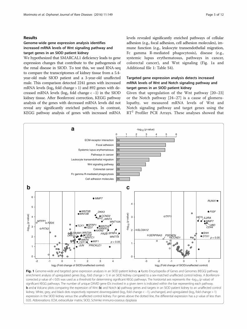
ResultsGenome-wide gene expression analysis identifiesincreased mRNA levels of Wnt signaling pathway andtarget genes in an SIOD patient kidneyWe hypothesized that SMARCAL1 deficiency leads to geneexpression changes that contribute to the pathogenesis ofthe renal disease in SIOD. To test this, we used RNA-seqto compare the transcriptomes of kidney tissue from a 5.4-year-old male SIOD patient and a 3-year-old unaffectedmale. This comparison detected 2241 genes with increasedmRNA levels (log2 fold change > 1) and 892 genes with de-creased mRNA levels (log2 fold change < −1) in the SIODkidney tissue. After Bonferroni correction, KEGG pathwayanalysis of the genes with decreased mRNA levels did notreveal any significantly enriched pathways. In contrast,KEGG pathway analysis of genes with increased mRNA
p = 0.05
40-1-2
0
1
2
3
4
5
6
log2 (Fold change of SIOD/unaffected control)
-log 1
0(p
val
ue)
PITX2
MTFP1FZD5
MYC
LEF1
CCND2
MMP7
WNT5BWNT2B
DKK3
WNT4
TCF7
WIF1
JUN
WNT6
AXIN2VANGL2WNT5ASFRP1
FZD7
WNT7A
WNT10A
FRZB SFRP4
PRICKLE1
FZD2NFATC1
WNT9ADKK1
DAAM1
b
ECM-receptor interaction 34
Focal adhesion 58
Systemic lupus erythematosus 36
Pathways in cancer 81
Leukocyte transendothelial migration 37
Wnt signaling pathway 43
Colorectal cancer 28
Fc gamma R-mediated phagocytosis 30
Cell adhesion molecules 35
a
Fig. 1 Genome-wide and targeted gene expression analyses in an SIOD patienenrichment analysis of upregulated genes (log2 fold change > 1) in an SIOD kidcorrected p value of < 0.05 was used as a threshold for determining significantsignificant KEGG pathways. The number of unique DAVID gene IDs involved inb and c Volcano plots comparing the expression of Wnt (b) and Notch (c) pathkidney. White, grey, and black dots respectively represent downregulated (log2expression in the SIOD kidney versus the unaffected control kidney. For genes0.05. Abbreviations: ECM, extracellular matrix; SIOD, Schimke immuno-osseous d
levels revealed significantly enriched pathways of cellularadhesion (e.g., focal adhesion, cell adhesion molecules), im-mune function (e.g., leukocyte transendothelial migration,Fc gamma R-mediated phagocytosis), disease (e.g.,systemic lupus erythematosus, pathways in cancer,colorectal cancer), and Wnt signaling (Fig. 1a andAdditional file 1: Table S4).
Targeted gene expression analysis detects increasedmRNA levels of Wnt and Notch signaling pathway andtarget genes in an SIOD patient kidneyGiven that upregulation of the Wnt pathway [20–23]or the Notch pathway [24–27] is a cause of glomeru-lopathy, we measured mRNA levels of Wnt andNotch signaling pathway and target genes using theRT2 Profiler PCR Arrays. These analyses showed that
p = 0.05
-1-2
0
1
2
3
4
log2 (Fold change of SIOD/unaffected control)
-log 1
0(p
val
ue)
-3 0
RUNX1
CD44
FIGF
IL2RAMMP7
SHHDTX1CCND1
PSENENSERPINA3
SLC6A12
TFF1 HEYL
UBD
c
61
-log10 (p value)
t kidney. a Kyoto Encyclopedia of Genes and Genomes (KEGG) pathwayney compared to a sex-matched unaffected control kidney. A Bonferroni-KEGG pathways. The horizontal axis represents the -log10 (p value) ofa given term is indicated within the bar representing each pathway.way genes and targets in an SIOD patient kidney to an unaffected controlfold change <−1), unchanged, and upregulated (log2 fold change > 1)above the dotted line, the differential expression has a p value of less thanysplasia
Morimoto et al. Orphanet Journal of Rare Diseases (2016) 11:149 Page 6 of 12
of the 84 Wnt pathway-related genes tested, 30 weredifferentially expressed (Fig. 1b and Additional file 1:Table S5) and that of the 84 Notch pathway-relatedgenes tested, 14 were differentially expressed (Fig. 1cand Additional file 1: Table S6). Wnt pathway-relatedgenes with increased mRNA levels included ligands
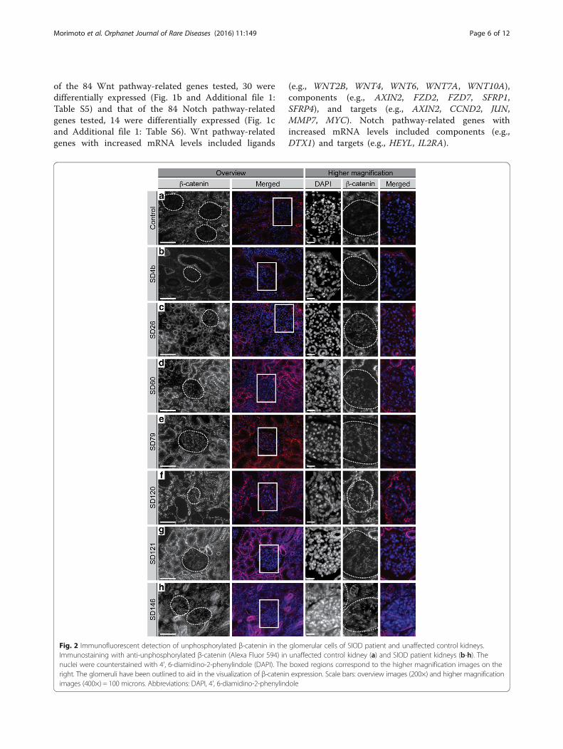
Fig. 2 Immunofluorescent detection of unphosphorylated β-catenin in theImmunostaining with anti-unphosphorylated β-catenin (Alexa Fluor 594) innuclei were counterstained with 4', 6-diamidino-2-phenylindole (DAPI). Theright. The glomeruli have been outlined to aid in the visualization of β-cateninimages (400×) = 100 microns. Abbreviations: DAPI, 4', 6-diamidino-2-phenylind
(e.g., WNT2B, WNT4, WNT6, WNT7A, WNT10A),components (e.g., AXIN2, FZD2, FZD7, SFRP1,SFRP4), and targets (e.g., AXIN2, CCND2, JUN,MMP7, MYC). Notch pathway-related genes withincreased mRNA levels included components (e.g.,DTX1) and targets (e.g., HEYL, IL2RA).
glomerular cells of SIOD patient and unaffected control kidneys.unaffected control kidney (a) and SIOD patient kidneys (b-h). Theboxed regions correspond to the higher magnification images on theexpression. Scale bars: overview images (200×) and higher magnificationole
Morimoto et al. Orphanet Journal of Rare Diseases (2016) 11:149 Page 7 of 12
Markers of Wnt and Notch pathway activation areincreased in the glomerular cells of postnatal SIODpatient kidneys comparable to isolated FSGS controlsHaving established that several Wnt and Notch pathway-related genes and targets have altered expression in anSIOD kidney, we hypothesized that increased Wnt andNotch pathway signaling within the glomeruli contributesto the pathogenesis of FSGS in SIOD. To test this inadditional SIOD patients, we used indirect immunofluor-escence to profile the expression of unphosphorylated β-
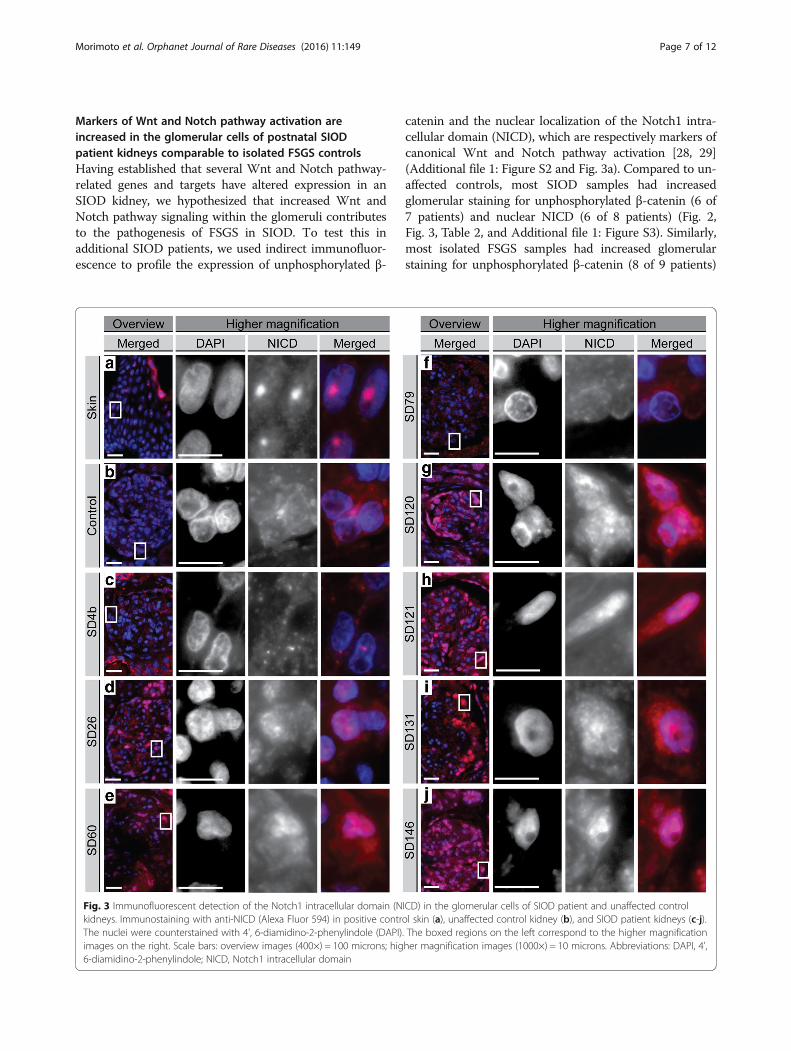
Fig. 3 Immunofluorescent detection of the Notch1 intracellular domain (Nkidneys. Immunostaining with anti-NICD (Alexa Fluor 594) in positive contrThe nuclei were counterstained with 4', 6-diamidino-2-phenylindole (DAPI)images on the right. Scale bars: overview images (400×) = 100 microns; hig6-diamidino-2-phenylindole; NICD, Notch1 intracellular domain
catenin and the nuclear localization of the Notch1 intra-cellular domain (NICD), which are respectively markers ofcanonical Wnt and Notch pathway activation [28, 29](Additional file 1: Figure S2 and Fig. 3a). Compared to un-affected controls, most SIOD samples had increasedglomerular staining for unphosphorylated β-catenin (6 of7 patients) and nuclear NICD (6 of 8 patients) (Fig. 2,Fig. 3, Table 2, and Additional file 1: Figure S3). Similarly,most isolated FSGS samples had increased glomerularstaining for unphosphorylated β-catenin (8 of 9 patients)
ICD) in the glomerular cells of SIOD patient and unaffected controlol skin (a), unaffected control kidney (b), and SIOD patient kidneys (c-j).. The boxed regions on the left correspond to the higher magnificationher magnification images (1000×) = 10 microns. Abbreviations: DAPI, 4',
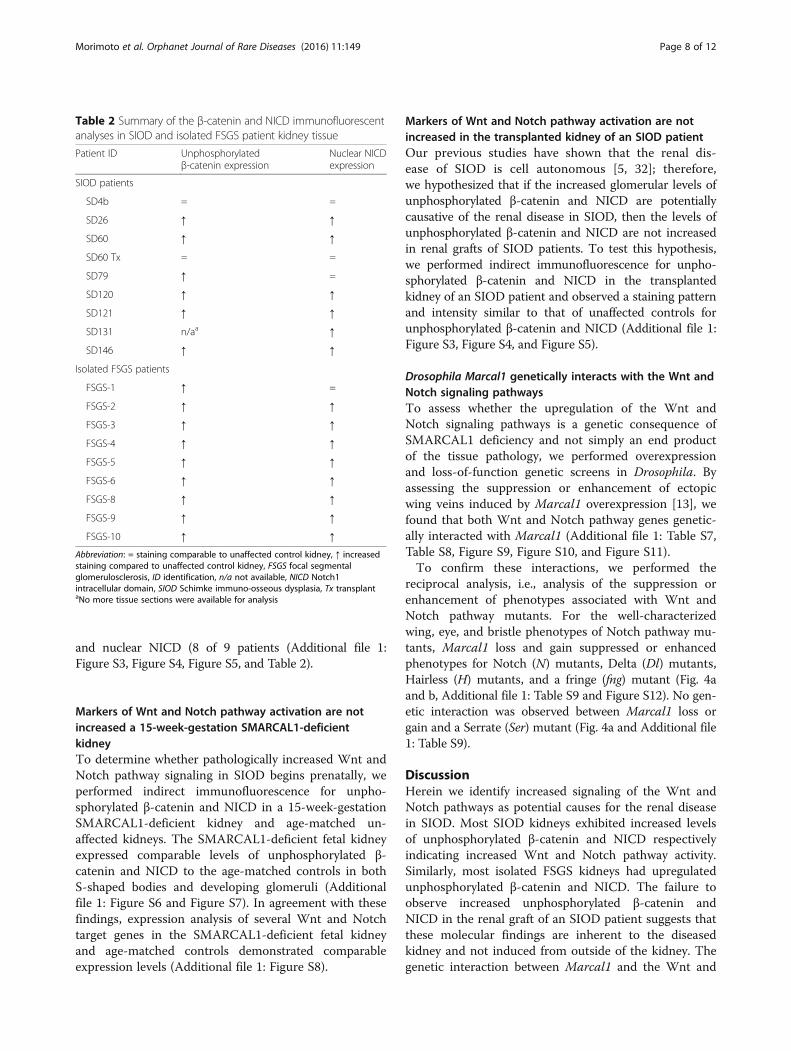
Table 2 Summary of the β-catenin and NICD immunofluorescentanalyses in SIOD and isolated FSGS patient kidney tissue
Patient ID Unphosphorylatedβ-catenin expression
Nuclear NICDexpression
SIOD patients
SD4b = =
SD26 ↑ ↑
SD60 ↑ ↑
SD60 Tx = =
SD79 ↑ =
SD120 ↑ ↑
SD121 ↑ ↑
SD131 n/aa ↑
SD146 ↑ ↑
Isolated FSGS patients
FSGS-1 ↑ =
FSGS-2 ↑ ↑
FSGS-3 ↑ ↑
FSGS-4 ↑ ↑
FSGS-5 ↑ ↑
FSGS-6 ↑ ↑
FSGS-8 ↑ ↑
FSGS-9 ↑ ↑
FSGS-10 ↑ ↑
Abbreviation: = staining comparable to unaffected control kidney, ↑ increasedstaining compared to unaffected control kidney, FSGS focal segmentalglomerulosclerosis, ID identification, n/a not available, NICD Notch1intracellular domain, SIOD Schimke immuno-osseous dysplasia, Tx transplantaNo more tissue sections were available for analysis
Morimoto et al. Orphanet Journal of Rare Diseases (2016) 11:149 Page 8 of 12
and nuclear NICD (8 of 9 patients (Additional file 1:Figure S3, Figure S4, Figure S5, and Table 2).
Markers of Wnt and Notch pathway activation are notincreased a 15-week-gestation SMARCAL1-deficientkidneyTo determine whether pathologically increased Wnt andNotch pathway signaling in SIOD begins prenatally, weperformed indirect immunofluorescence for unpho-sphorylated β-catenin and NICD in a 15-week-gestationSMARCAL1-deficient kidney and age-matched un-affected kidneys. The SMARCAL1-deficient fetal kidneyexpressed comparable levels of unphosphorylated β-catenin and NICD to the age-matched controls in bothS-shaped bodies and developing glomeruli (Additionalfile 1: Figure S6 and Figure S7). In agreement with thesefindings, expression analysis of several Wnt and Notchtarget genes in the SMARCAL1-deficient fetal kidneyand age-matched controls demonstrated comparableexpression levels (Additional file 1: Figure S8).
Markers of Wnt and Notch pathway activation are notincreased in the transplanted kidney of an SIOD patientOur previous studies have shown that the renal dis-ease of SIOD is cell autonomous [5, 32]; therefore,we hypothesized that if the increased glomerular levels ofunphosphorylated β-catenin and NICD are potentiallycausative of the renal disease in SIOD, then the levels ofunphosphorylated β-catenin and NICD are not increasedin renal grafts of SIOD patients. To test this hypothesis,we performed indirect immunofluorescence for unpho-sphorylated β-catenin and NICD in the transplantedkidney of an SIOD patient and observed a staining patternand intensity similar to that of unaffected controls forunphosphorylated β-catenin and NICD (Additional file 1:Figure S3, Figure S4, and Figure S5).
Drosophila Marcal1 genetically interacts with the Wnt andNotch signaling pathwaysTo assess whether the upregulation of the Wnt andNotch signaling pathways is a genetic consequence ofSMARCAL1 deficiency and not simply an end productof the tissue pathology, we performed overexpressionand loss-of-function genetic screens in Drosophila. Byassessing the suppression or enhancement of ectopicwing veins induced by Marcal1 overexpression [13], wefound that both Wnt and Notch pathway genes genetic-ally interacted with Marcal1 (Additional file 1: Table S7,Table S8, Figure S9, Figure S10, and Figure S11).To confirm these interactions, we performed the
reciprocal analysis, i.e., analysis of the suppression orenhancement of phenotypes associated with Wnt andNotch pathway mutants. For the well-characterizedwing, eye, and bristle phenotypes of Notch pathway mu-tants, Marcal1 loss and gain suppressed or enhancedphenotypes for Notch (N) mutants, Delta (Dl) mutants,Hairless (H) mutants, and a fringe (fng) mutant (Fig. 4aand b, Additional file 1: Table S9 and Figure S12). No gen-etic interaction was observed between Marcal1 loss orgain and a Serrate (Ser) mutant (Fig. 4a and Additional file1: Table S9).
DiscussionHerein we identify increased signaling of the Wnt andNotch pathways as potential causes for the renal diseasein SIOD. Most SIOD kidneys exhibited increased levelsof unphosphorylated β-catenin and NICD respectivelyindicating increased Wnt and Notch pathway activity.Similarly, most isolated FSGS kidneys had upregulatedunphosphorylated β-catenin and NICD. The failure toobserve increased unphosphorylated β-catenin andNICD in the renal graft of an SIOD patient suggests thatthese molecular findings are inherent to the diseasedkidney and not induced from outside of the kidney. Thegenetic interaction between Marcal1 and the Wnt and
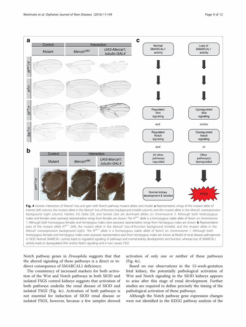
Fig. 4 Genetic interaction of Marcal1 loss and gain with Notch pathway mutant alleles and model. a Representative wings of the mutant allele ofinterest (left column), the mutant allele in the Marcal1 loss-of-function background (middle column), and the mutant allele in the Marcal1 overexpressionbackground (right column). Hairless (H), Delta (Dl), and Serrate (Ser) are dominant alleles on chromosome 3. Although both heterozygousmales and females were assessed, representative wings from females are shown. The Nnd-1 allele is a homozygous viable allele of Notch on chromosome1. Although both homozygous females and hemizygous males were assessed, representative wings from hemizygous males are shown. b Representativeeyes of the mutant allele Nspl-1 (left), the mutant allele in the Marcal1 loss-of-function background (middle), and the mutant allele in theMarcal1 overexpression background (right). The Nspl-1 allele is a homozygous viable allele of Notch on chromosome 1. Although bothhomozygous females and hemizygous males were assessed, representative eyes from hemizygous males are shown. c Model of renal disease pathogenesisin SIOD. Normal SMARCAL1 activity leads to regulated signaling of pathways and normal kidney development and function, whereas loss of SMARCAL1activity leads to dysregulated Wnt and/or Notch signaling and in turn causes FSGS
Morimoto et al. Orphanet Journal of Rare Diseases (2016) 11:149 Page 9 of 12
Notch pathway genes in Drosophila suggests that thatthe altered signaling of these pathways is a direct or in-direct consequence of SMARCAL1 deficiency.The consistency of increased markers for both activa-
tion of the Wnt and Notch pathways in both SIOD andisolated FSGS control kidneys suggests that activation ofboth pathways underlie the renal disease of SIOD andisolated FSGS (Fig. 4c). Activation of both pathways isnot essential for induction of SIOD renal disease orisolated FSGS, however, because a few samples showed
activation of only one or neither of these pathways(Fig. 4c).Based on our observations in the 15-week-gestation
fetal kidney, the potentially pathological activation ofWnt and Notch signaling in the SIOD kidneys appearsto arise after this stage of renal development. Furtherstudies are required to define precisely the timing of thepathological activation of these pathways.Although the Notch pathway gene expression changes
were not identified in the KEGG pathway analysis of the
Morimoto et al. Orphanet Journal of Rare Diseases (2016) 11:149 Page 10 of 12
transcriptome, the high level of crosstalk between theWnt and Notch signaling pathways [33], and their role inkidney development and disease prompted us to alsoinvestigate the upregulation of the Notch pathway as a po-tential cause for the FSGS in SIOD. Possible reasons forthe transcriptome analysis not detecting the upregulationof the Notch pathway include pathway size bias inherentto KEGG pathway analysis (the Wnt signaling pathway in-cludes 141 genes, whereas the Notch signaling pathwayincludes 48 genes) and tissue heterogeneity.The mechanism by which SMARCAL1 deficiency
gives rise to tissue-specific changes in gene expression isincompletely understood. It could arise from a directconsequence of SMARCAL1 deficiency on the DNAstructure of a gene or of the genes encoding the tran-scriptional regulators of that gene. Consistent with this,we previously observed that SMARCAL1 homologuesbind transcriptionally active chromatin and modulategene expression [13]. Sharma et al. (2015) recentlyshowed that the bovine orthologue of SMARCAL1 nega-tively and directly regulates the transcription of MYC byaltering the conformation of its promoter [34]. Alterna-tively, because stalled replication forks induce epigeneticchanges that alter gene expression [35, 36], impedanceof DNA replication fork restart by SMARCAL1 defi-ciency might contribute to the changes in gene expres-sion. Consistent with the latter possibility, we recentlyobserved hypermethylation of the IL7R promoter in theT cells of SIOD patients [19]; reduced IL7R expressionin human CD8+ T cells is associated with hypermethyla-tion of the IL7R promoter [37].A limitation of the study was the use of whole kidney to
profile differential gene expression in an SIOD kidney.Given that the primary lesion is limited to the glomeruli,the affected tissue represents a small fraction of the totaltissue. Although several human gene expression studieson FSGS have used isolated glomeruli [38, 39], others havesuccessfully used renal biopsies [40]. Similar to other hu-man gene expression studies of FSGS [38–40], the expres-sion of podocyte-specific genes including NPHS1, NPHS2,and WT1 were downregulated in the SIOD kidney, andmost of the KEGG pathways that were enriched in our listof upregulated genes were also enriched in the prior stud-ies, including the Wnt signaling pathway [38].A second limitation of the study was that only unpho-
sphorylated β-catenin and nuclear NICD were examinedby immunofluorescence as measures of pathway activa-tion. This constraint arose secondary to limited tissue.We selected these proteins because they are the primaryeffectors of and activation markers for the canonicalWnt and Notch signaling pathways. However, Wnt sig-naling has canonical and non-canonical pathways, andthere is also Wnt-independent β-catenin activation [41].Notch signaling also has canonical and non-canonical
pathways as well as three Notch receptors in addition toNotch1 [42]. Our findings nonetheless set a precedentfor future studies examining the pathogenesis of renaldisease in SIOD.
ConclusionsIn summary, our findings show that the Wnt and Notchpathways are upregulated in the SIOD patient kidney andthat Marcal1, the Drosophila SMARCAL1 homologue,genetically interacts with Wnt and Notch pathway genes.Based on these findings, the renal disease of SIOD is yetanother clinically distinctive feature of SIOD likely arisingthrough alterations of gene expression.
Additional file
Additional file 1: Supplementary Methods, Tables, and Figures.(PDF 45904 kb)
AbbreviationsACV: Anterior crossvein; DAPI: 4', 6-diamidino-2-phenylindole;DAVID: Database for annotation, visualization, and integrated discovery;FFPE: Formalin-fixed paraffin-embedded; FPKM: Fragments per kilobase permillion mapped reads; FSGS: Focal segmental glomerulosclerosis;KEGG: Kyoto Encyclopedia of Genes and Genomes; NICD: Notch1 intracellulardomain; PBS: Phosphate-buffered saline; PCV: Posterior crossvein; qRT-PCR: Quantitative reverse transcription polymerase chain reaction;SIOD: Schimke immuno-osseous dysplasia; SMARCAL1: SWI/SNF-related,matrix-associated, actin-dependent regulator of chromatin, subfamily A-like 1
AcknowledgementsWe are grateful to all of the patients and family members who havecontributed to this study. The authors thank Theresa Sturby (Children’s andWomen’s Health Centre of British Columbia) for her technical expertise, andDrs. Darren Bridgewater and Alireza Barandaran-Heravi for critical review ofthis manuscript.
FundingThis work was supported in part by the New Investigator Award jointlysponsored by the SickKids Foundation and the Canadian Institutes of HealthResearch Institute of Human Development, Child and Youth Health (XG09-025to CFB); the Michael Smith Foundation for Health Research (CI-SCH-O1899(07–1) to CFB); The Little Giants Foundation (CFB); and the Asociacion Española deDisplasias Oseas Minoritarias (CFB). Human fetal tissue was obtained throughthe Laboratory of Developmental Biology project supported by the NationalInstitutes of Health Award Number 5R24HD000836 from the Eunice KennedyShriver National Institute of Child Health & Human Development. MM wassupported by a Four Year Doctoral Fellowship from the University of BritishColumbia. CFB is a scholar of the Michael Smith Foundation for Health Researchand a Clinical Investigator of the Child & Family Research Institute.
Availability of data and materialsThe dataset supporting the conclusions of this article is available in the GeneExpression Omnibus (GEO) repository, Series record GSE75061.
Authors’ contributionsAll authors have made substantial contributions to the article byparticipating in the conception and design (MM, CFB), acquisition of data(MM, CM, KB, KC, YA, AB, DB, MB, JC, EC, AD, GD, M Gentile, M Giordano,AKG, RG, MJ, KK, E Lerut, E Levtchenko, LM, CM, BN, DP, JS, PS, UY, ZY, JZ, GH,CFB), analysis and interpretation of data (MM, CM, KB, KC, BN, CFB), draftingthe manuscript (MM, CFB), or revising it critically for important intellectualcontent (MM, CM, KB, KC, YA, AB, DB, MB, JC, EC, AD, GD, MGentile,MGiordano, AKG, RG, MJ, KK, ELerut, ELevtchenko, LM, CM, BN, DP, JS, PS, UY,ZY, JZ, GH, CFB). All authors read and approved the final manuscript.
Morimoto et al. Orphanet Journal of Rare Diseases (2016) 11:149 Page 11 of 12
Competing interestsThe authors declare that they have no competing interests.
Consent for publicationNot applicable.
Ethics approval and consent to participateThe guardians of the patients referred to this study signed informed consentapproved by the Institutional Review Board of the University of BritishColumbia (Vancouver, BC, Canada). Autopsy and biopsy tissues wereobtained according to the protocol approved by the University of BritishColumbia (Vancouver, BC, Canada).
Author details1Department of Medical Genetics, University of British Columbia, Vancouver,BC, Canada. 2Child & Family Research Institute, Vancouver, BC, Canada.3Department of Endocrinology & Metabolism, Kanagawa Children’s MedicalCenter, Yokohama, Japan. 4Department of Pediatric Nephrology, VUUniversity Medical Center, Amsterdam, The Netherlands. 5Département deBiochimie et Génétique, Centre Hospitalier Universitaire d’Angers, Angers,France. 6Department of Pediatrics, University of Verona, Verona, Italy.7Division of Genetics, Birth Defects and Metabolism, Ann and Robert H. LurieChildren’s Hospital of Chicago, Northwestern University Feinberg School ofMedicine, Chicago, IL, USA. 8Seattle Children’s Hospital, Seattle, WA, USA.9Département de Pédiatrie, Hôpital Robert Debré, Paris, France.10Department of Medical Genetics, Hospital Di Venere – ASL Bari, Bari, Italy.11Pediatric Nephrology and Dialysis Unit, Ospedale Pediatrico Giovanni XXIII,Bari, Italy. 12Department of Pediatrics, University of Oklahoma Health SciencesCenter, Oklahoma City, OK, USA. 13Department of Pediatrics and ChildHealth, Nelson R. Mandela School of Medicine, University of KwaZulu-Natal,Durban, South Africa. 14Department of Pediatric Nephrology, PhoenixChildren’s Hospital, Phoenix, AZ, USA. 15Child Development and RehabiliationCenter, Oregon Institute on Disability & Development, Oregon Health &Science University, Portland, OR, USA. 16Department of Pathology, UniversityHospitals Leuven, Leuven, Belgium. 17Department of Pediatric Nephrology,University Hospitals Leuven, Leuven, Belgium. 18Division of Nephrology,Bambino Gesù Children’s Hospital and Research Institute, Rome, Italy.19Warren Clinic Family Medicine, Tulsa, OK, USA. 20Department of Pathology,University of Washington, Seattle, WA, USA. 21Department of Pathology,Children’s Hospital Los Angeles and Keck School of Medicine, University ofSouthern California, Los Angeles, CA, USA. 22Children’s Hospital, University ofMainz, Mainz, Germany. 23Department of Pathology, Oregon Health andScience University, Portland, OR, USA. 24Department of Pediatrics, Division ofChild Neurology, Dokuz Eylül University, School of Medicine, İzmir, Turkey.25Department of Pathology, University of Oklahoma Health Sciences Center,Oklahoma City, OK, USA. 26Department of Anatomic Pathology, Children’sand Women’s Health Centre of British Columbia, Vancouver, BC, Canada.27Provincial Medical Genetics Program, Department of Medical Genetics,Children’s and Women’s Health Centre of British Columbia, 4500 Oak Street,Room C234, Vancouver, BC V6H 3N1, Canada.
Received: 14 June 2016 Accepted: 23 September 2016
References1. Schimke RN, Horton WA, King CR. Chondroitin-6-sulphaturia, defective
cellular immunity, and nephrotic syndrome. Lancet. 1971;2:1088–9.2. Ehrich JH, Burchert W, Schirg E, Krull F, Offner G, Hoyer PF, et al. Steroid
resistant nephrotic syndrome associated with spondyloepiphyseal dysplasia,transient ischemic attacks and lymphopenia. Clin Nephrol. 1995;43:89–95.
3. Spranger J, Hinkel GK, Stoss H, Thoenes W, Wargowski D, Zepp F. Schimkeimmuno-osseous dysplasia: a newly recognized multisystem disease.J Pediatr. 1991;119:64–72.
4. Boerkoel CF, O’Neill S, Andre JL, Benke PJ, Bogdanovic R, Bulla M, et al.Manifestations and treatment of Schimke immuno-osseous dysplasia: 14new cases and a review of the literature. Eur J Pediatr. 2000;159:1–7.
5. Clewing JM, Antalfy BC, Lücke T, Najafian B, Marwedel KM, Hori A, et al.Schimke immuno-osseous dysplasia: a clinicopathological correlation. J MedGenet. 2007;44:122–30.
6. Sarin S, Javidan A, Boivin F, Alexopoulou I, Lukic D, Svajger B, et al. Insightsinto the renal pathogenesis in schimke immuno-osseous dysplasia: a renal
histological characterization and expression analysis. J Histochem Cytochem.2015;63:32–44.
7. Boerkoel CF, Takashima H, John J, Yan J, Stankiewicz P, Rosenbarker L, et al.Mutant chromatin remodeling protein SMARCAL1 causes Schimke immuno-osseous dysplasia. Nat Genet. 2002;30:215–20.
8. Yusufzai T, Kadonaga JT. HARP is an ATP-driven annealing helicase. Science.2008;322:748–50.
9. Postow L, Woo EM, Chait BT, Funabiki H. Identification of SMARCAL1 as acomponent of the DNA damage response. J Biol Chem. 2009;284:35951–61.
10. Yusufzai T, Kong X, Yokomori K, Kadonaga JT. The annealing helicase HARPis recruited to DNA repair sites via an interaction with RPA. Genes Dev.2009;23:2400–4.
11. Bansbach CE, Betous R, Lovejoy CA, Glick GG, Cortez D. The annealinghelicase SMARCAL1 maintains genome integrity at stalled replication forks.Genes Dev. 2009;23:2405–14.
12. Yuan J, Ghosal G, Chen J. The annealing helicase HARP protects stalledreplication forks. Genes Dev. 2009;23:2394–9.
13. Baradaran-Heravi A, Cho KS, Tolhuis B, Sanyal M, Morozova O, Morimoto M,et al. Penetrance of biallelic SMARCAL1 mutations is associated withenvironmental and genetic disturbances of gene expression. Hum MolGenet. 2012;21:2572–87.
14. Curran ME, Atkinson DL, Ewart AK, Morris CA, Leppert MF, Keating MT. Theelastin gene is disrupted by a translocation associated with supravalvularaortic stenosis. Cell. 1993;73:159–68.
15. Li DY, Brooke B, Davis EC, Mecham RP, Sorensen LK, Boak BB, et al. Elastin is anessential determinant of arterial morphogenesis. Nature. 1998;393:276–80.
16. Morimoto M, Yu Z, Stenzel P, Clewing JM, Najafian B, Mayfield C, et al. Reducedelastogenesis: a clue to the arteriosclerosis and emphysematous changes inschimke immuno-osseous dysplasia? Orphanet J Rare Dis. 2012;7:70.
17. Puel A, Ziegler SF, Buckley RH, Leonard WJ. Defective IL7R expression inT(−)B(+)NK(+) severe combined immunodeficiency. Nat Genet. 1998;20:394–7.
18. Roifman CM, Zhang J, Chitayat D, Sharfe N. A partial deficiency ofinterleukin-7R alpha is sufficient to abrogate T-cell development and causesevere combined immunodeficiency. Blood. 2000;96:2803–7.
19. Sanyal M, Morimoto M, Baradaran-Heravi A, Choi K, Kambham N, Jensen K,et al. Lack of IL7Ralpha expression in T cells is a hallmark of T-cellimmunodeficiency in Schimke immuno-osseous dysplasia (SIOD). ClinImmunol. 2015;161:355–65.
20. Dai C, Stolz DB, Kiss LP, Monga SP, Holzman LB, Liu Y. Wnt/beta-cateninsignaling promotes podocyte dysfunction and albuminuria. J Am SocNephrol. 2009;20:1997–2008.
21. Kato H, Gruenwald A, Suh JH, Miner JH, Barisoni-Thomas L, Taketo MM, et al.Wnt/beta-catenin pathway in podocytes integrates cell adhesion,differentiation, and survival. J Biol Chem. 2011;286:26003–15.
22. Shkreli M, Sarin KY, Pech MF, Papeta N, Chang W, Brockman SA, et al.Reversible cell-cycle entry in adult kidney podocytes through regulatedcontrol of telomerase and Wnt signaling. Nat Med. 2012;18:111–9.
23. He W, Tan RJ, Li Y, Wang D, Nie J, Hou FF, et al. Matrix metalloproteinase-7as a surrogate marker predicts renal Wnt/beta-catenin activity in CKD. J AmSoc Nephrol. 2012;23:294–304.
24. Niranjan T, Bielesz B, Gruenwald A, Ponda MP, Kopp JB, Thomas DB, et al.The Notch pathway in podocytes plays a role in the development ofglomerular disease. Nat Med. 2008;14:290–8.
25. Waters AM, Wu MY, Onay T, Scutaru J, Liu J, Lobe CG, et al. Ectopic notchactivation in developing podocytes causes glomerulosclerosis. J Am SocNephrol. 2008;19:1139–57.
26. Lasagni L, Ballerini L, Angelotti ML, Parente E, Sagrinati C, Mazzinghi B, et al.Notch activation differentially regulates renal progenitors proliferation anddifferentiation toward the podocyte lineage in glomerular disorders. StemCells. 2010;28:1674–85.
27. Murea M, Park JK, Sharma S, Kato H, Gruenwald A, Niranjan T, et al.Expression of Notch pathway proteins correlates with albuminuria,glomerulosclerosis, and renal function. Kidney Int. 2010;78:514–22.
28. Clevers H, Nusse R. Wnt/beta-catenin signaling and disease. Cell. 2012;149:1192–205.
29. Bray SJ. Notch signalling: a simple pathway becomes complex. Nat Rev MolCell Biol. 2006;7:678–89.
30. Kato H, Susztak K. Repair problems in podocytes: Wnt, Notch, andglomerulosclerosis. Semin Nephrol. 2012;32:350–6.
31. Warden CD, Yuan Y-C, Wu X. Optimal calculation of RNA-seq fold-changevalues. Int J Comput Bioinfo In Silico Model. 2013;2:285–92.
Morimoto et al. Orphanet Journal of Rare Diseases (2016) 11:149 Page 12 of 12
32. Lücke T, Marwedel KM, Kanzelmeyer NK, Hori A, Offner G, Kreipe HH, et al.Generalized atherosclerosis sparing the transplanted kidney in Schimkedisease. Pediatr Nephrol. 2004;19:672–5.
33. Collu GM, Hidalgo-Sastre A, Brennan K. Wnt-Notch signalling crosstalk indevelopment and disease. Cell Mol Life Sci. 2014;71:3553–67.
34. Sharma T, Bansal R, Haokip DT, Goel I, Muthuswami R. SMARCAL1 negativelyregulates c-Myc transcription by altering the conformation of the promoterregion. Sci Rep. 2015;5:17910.
35. Schiavone D, Guilbaud G, Murat P, Papadopoulou C, Sarkies P, Prioleau MN,et al. Determinants of G quadruplex-induced epigenetic instability in REV1-deficient cells. EMBO J. 2014;33:2507–20.
36. Khurana S, Oberdoerffer P. Replication stress: a lifetime of epigeneticchange. Genes (Basel). 2015;6:858–77.
37. Kim HR, Hwang KA, Kim KC, Kang I. Down-regulation of IL-7Ralpha expressionin human T cells via DNA methylation. J Immunol. 2007;178:5473–9.
38. Bennett MR, Czech KA, Arend LJ, Witte DP, Devarajan P, Potter SS. Lasercapture microdissection-microarray analysis of focal segmentalglomerulosclerosis glomeruli. Nephron Exp Nephrol. 2007;107:e30–40.
39. Hodgin JB, Borczuk AC, Nasr SH, Markowitz GS, Nair V, Martini S, et al. Amolecular profile of focal segmental glomerulosclerosis from formalin-fixed,paraffin-embedded tissue. Am J Pathol. 2010;177:1674–86.
40. Schwab K, Witte DP, Aronow BJ, Devarajan P, Potter SS, Patterson LT.Microarray analysis of focal segmental glomerulosclerosis. Am J Nephrol.2004;24:438–47.
41. Haq S, Michael A, Andreucci M, Bhattacharya K, Dotto P, Walters B, et al.Stabilization of beta-catenin by a Wnt-independent mechanism regulatescardiomyocyte growth. Proc Natl Acad Sci U S A. 2003;100:4610–5.
42. Ayaz F, Osborne BA. Non-canonical Notch signaling in cancer andimmunity. Front Oncol. 2014;4:345.
• We accept pre-submission inquiries
• Our selector tool helps you to find the most relevant journal
• We provide round the clock customer support
• Convenient online submission
• Thorough peer review
• Inclusion in PubMed and all major indexing services
• Maximum visibility for your research
Submit your manuscript atwww.biomedcentral.com/submit
Submit your next manuscript to BioMed Central and we will help you at every step: