Embed Size (px)
Citation preview
1
Indian Institute of technology Madras
Department of Biotechnology
24th
July, 2012
To
Dean
IC&SR
IIT Madras
Sub: Forwarding a proposal seeking funding from DBT, India.
Dear Sir
I am attaching a proposal that I would like to submit to Department of Biotechnology (GoI),
New Delhi seeking funding.
Request you to kindly forward the same.
Warm regards
Gopala Krishna Aradhyam
Associate Professor
2
Fragment based insights into cross talk between the Ca
2+-binding sites and other domains of Drosophila
melanogaster calnuc (dm-calnuc).
A Research Proposal submitted to the
Department of Biotechnology, New Delhi
by
A. Gopala Krishna, Ph.D.
Department of Biotechnology Indian Institute of Technology Madras, Chennai 600 036, INDIA
3
PROFORMA – I
PROFORMA FOR SUBMISSION OF PROJECT PROPOSALS ON RESEARCH AND
DEVELOPMENT, PROGRAMME SUPPORT
(To be filled by the applicant)
PART I: GENERAL INFORMATION
1. Name of the Institute/University/Organisation submitting the Project Proposal: Indian Institute of Technology Madras.
2. State: Tamilnadu... 3. Status of the Institute: Government
4. Name and designation of the Executive Authority of the Institute/University forwarding the
application : .
The Dean, Industrial Consultancy and Sponsored Research (IC & SR) IIT Madras
5. Project Title: Fragment based insights into cross talk between the Ca2+-binding sites and other domains of Drosophila melanogaster calnuc (dm-calnuc).
6. Category of the Project (Please tick): R&D
7. Specific Area (Please see Annexure - II):
8. Duration: 3 Years
9. Total Cost (Rs.) 52, 92,000
10. Is the project Single Institutional or Multiple-Institutional (S/M) ? : S
11. If the project is multi-institutional, please furnish the following :
Name of Project Coordinator : ...............................................................................
Affiliation: ...............................................................................................................
Address: ..........................................................................................................………………....
12. Scope of application indicating anticipated product and processes
Ca2+ is a very important metal ion that drives normal physiology in cells. Most of the signals arising out of a concentration gradient of this metal ion are transmitted by proteins that bind to it, Ca2+-binding proteins (CABPs). Understanding the biochemistry and structure-function relationship of a novel Ca2+-binding protein is extremely insightful and gives knowledge about its functions in the cell. Here we propose to study one such novel CABP, calnuc, which interacts with many important biomolecules but whose function remains elusive. Structural changes in the protein upon Ca2+-binding and cross-talk between the Ca2+-binding sites are the main focus of this proposal. Towards this goal we propose to make recombinant fragments of the protein and introduce site directed mutations in order to understand the role of each Ca2+-binding domain in the ultimate function of the protein.
4
13. Project Summary.
Ca2+
-binding proteins (CABPs) play a vital role in the physiology of cells. They
regulate processes as varied as transcription, muscle contraction, blood coagulation,
transmission of sensory signals etc., (Heizmann and Hunziker, 1990; Davie et al., 1991;
Baimbridge et al., 1992; Anderssen et al., 1993; Filipek et al., 1993). One set of the family of
CABPs activate signaling pathways upon binding to the metal ion. In all such proteins, the
Ca2+
-sensors, ion binding is accompanied by a large conformational change leading to the
transfer of the signal. Another set of CABPs are those that aid in maintaining the Ca2+
concentration in the cells and are called as Ca2+
buffers. The helix-loop-helix, EF-hand motif
is the most common structure used by proteins to bind Ca2+
ions, the 12 residue loop being
the site of ion binding (Kretsinger and Nockolds, 1973).
In a majority of the CABPs, not only do the EF-hand motif occurs in pairs or higher
order even numbers, but also bind to the metal ion with positive cooperativity: ion binding to
one site enhancing the binding ability of the other site. Gaining insight into the molecular
mechanism that drives cooperativity among the Ca2+
-binding sites is a very interesting,
though elaborate task, one that involves detailed characterization of all ion-binding sites
(Linse and Chazin, 1995). Site directed mutations of important amino acid residues in the
Ca2+
-binding site help immensely in understanding the biochemistry aided ion binding
phenomena.
Calnuc is a novel Ca2+
-binding protein and the only one known to bind to G proteins.
It has also been demonstrated to interact withDNA, cox enzymes in addition to regulating
beta amyloid proteins and unfolded protein response (Aradhyam, et al., 2011). It is a highly
conserved protein from Ciona intestinalis to humans (Kanuru et al, 2010). Especially
interesting is the calnuc from drosophila melanogaster – having three ion binding sites,
unusual for a Ca2+
-binding protein. The functions of this multi-domain protein are still elusive
and we propose a fragment based analysis of several of its physicochemical and
biochemical properties. Fragments will be generated based on the distribution of the Ca2+
-
binding sites, in a manner that one or more of the sites are not part of the fragment. Point
mutants of the important residues in the Ca2+
-binding loops will be generated and various
techniques (ITC, Fluorescence, CD, absorption in addition to protease activity) will be
utilized to assess Ca2+
-binding affinities and other physico-chemical properties.
Cooperativity between the two sites in each fragment will be assessed followed by its
secondary structural features. Complete understanding of its structure-function relationship
and biochemistry will be attempted.
5
PART II: PARTICULARS OF INVESTIGATORS
(One or more co-investigators are preferred in every project. Inclusion of co-investigator(s) is
mandatory for investigators retiring before completion of the project)
Principal Investigator:
14. Name: Dr. A. Gopala Krishna
Date of Birth: .24th July, 1966 Sex (M/F): M
Designation: Associate Professor.
Department: Biotechnology.
Institute/University: Indian Institute of Technology Madras.
Address: Adyar,
Chennai
PIN: 600 036
Telephone: 044- 2257 4112 Fax: 44-22574102 E-mail: [email protected]
Number of research projects being handled at present: None
Co-Investigator
15. Name: NA
Date of Birth : .................................................................Sex (M/F) : .....................................
Designation : ............................................................................................................................
Department : ...........................................................................……………………………….
Institute/University: ................................................................................................................
Address : ...............................................................................……………..............................
....................................…………………………………………………PIN : .......................
Telephone : .......................... Fax:.....................…………E-mail : ……………………........
Number of Research projects being handled at present: .........................................................
Co-Investigator
16. Name : NA
Date of Birth : ................................................................ Sex(M/F) : ......................................
Designation : ...........................................................................................................................
Department : ..........................................................................................................…………..
Institute/University : ...............................................................................................................
Address : ………….................................................................................................................
.........................……………………………………………....... PIN : .........…......................
Telephone : .................…......... Fax ..............…............. E-mail : ..………............................
Number of Research projects being handled at present:..............................................................
Note : Use separate page, if more investigators are involved
6
PART III : TECHNICAL DETAILS OF PROJECT
16. Introduction (not to exceed 2 pages or 1000 words)
16.1 Origin of the proposal
In our extensive studies on human calnuc we have completely characterized its ion
binding properties (Kanuru et al, 2010). We have also demonstrated that the protein has
two exclusive Zn2+-binding sites near its N-terminus. We have demonstrated that one of
the EF-hand motifs is a Ca2+-binding site while the other is a Ca2+/Mg2+ mixed binding
site. In another piece of work we demonstrated that calnuc possess proteolytic activity
and that its enzymatic activity is allosterically regulated by Zn2+-binding (MS is under
revision in J. Biol. Chem.).
These observations have led us to the present proposal wherein we will try to gain
greater insight into the molecular mechanisms of the biochemical properties of calnuc
especially the one from Drosophila melanogaster.
16.2 (a) Rationale of the study supported by cited literature
Calnuc is one of the two, recently discovered Ca2+-binding proteins found in the Golgi
apparatus (the other one being Cab45) (Honore. B, 2009). It is a 63KDa protein having
two EF-hands in most organisms and three in Drosophila melanogaster (Otte, S. et. al.,
1999). No particular function has so far been assigned to calnuc. Presence of three EF-
hand Ca2+-binding sites classifies this protein as a member of the EF-hand super family
of proteins. A group of proteins in the EF-hand containing super family are Ca2+-sensors
that undergo Ca2+-induced conformational changes leading to an exposure of a
hydrophobic surface. The exposed hydrophobic patch serves as a binding surface for
one or more target molecules, which become activated or attenuated upon complex
formation thereby triggering signal transduction. The other group of Ca2+-binding protein
members is the Ca2+-buffers; those that modulate cellular Ca2+ levels in the cell
(Schwaller, B., 2010). We have recently shown that Ca2+-binding ability of calnuc is
enhanced upon interacting with GTP bound G protein alpha subunit (Kanuru et al.,
2010). In all Ca2+-binding proteins, relationship between the Ca2+-binding sites is a
major regulator of its function.
The goal of this study is to find out: (i) if there is any cooperativity observed between the
three Ca2+-binding sites of dm-calnuc (ii) if Ca2+-binding plays a role in the function of
the other domains of the protein, (iii) what is the physiological importance of the other
domains, (iv) what are the thermodynamic aspects of the stabilities of these domains
and the role of the Ca2+-binding sites on these, (v) would the individual domains function
without the ion binding site being present. Structural studies will include experiments
7
using circular dichroism to elucidate the secondary and tertiary structure and the effect
of ion binding. Our goal will also be to individually characterize each of the three Ca2+-
binding sites: whether they are pure Ca2+-binding sites or they exhibit mixed Ca2+/Mg2+
binding. Towards this goal we will generate several, intelligently designed mutant
proteins to firmly characterize the biological role of this protein. The cDNA
corresponding to each of the domains will be separately sub-cloned into expression
vectors, confirmed by sequencing, proteins expressed and their biochemical and
biophysical characterization done.
The project should enhance our understanding the function of calnuc in general and
dm-calnuc in particular and in addition the roles of all its independent domains and most
importantly, cross talk between all of them.
(b) Hypothesis
Ca2+ ions and Ca2+-binding proteins (CaBPs) play a very vital role in the function and
physiology of all cells. Gradients of Ca2+ ions across the cells are used as signals and
transduced across the cells in order to maintain basal/physiologically relevant state of
the cell. The endoplasmic reticulum is the store house of the Ca2+ ions in the cell that is
maintained by CaBPs and Ca2+ channels. Recently, other family members have been
identified whose localization are not only restricted to the ER but found also in the
cytosol as well as extracellularly as free or attached to the membrane.
Calnuc is a novel, Golgi resident, Ca2+-binding protein that has been demonstrated to
interact with important biomolecules in the cell (e.g., DNA, COX, amyloid, G proteins,
etc.,). Despite this knowledge the exact function(s) of calnuc are elusive and intense
research going on in this direction. To better understand the structure-function
relationship of this protein we have ben working on human calnuc and have
demonstrated novel functions. We would like to expand our horizon and propose here
biochemical and biophysical characterization of Drosophila melanogaster calnuc. It has
many unique features that make it an extremely interesting molecule to study. For
example it has three ion binding sties (two EF- hand and another uncharacterized metal
ion binding motif). It also lacks an otherwise well conserved serine that (in human
calnuc) we propose is part of the catalytic site for its proteolytic activity.
8
(c) Key questions.
(1) To define the mode of Ca2+/Zn2+ binding to calnuc. (2) To elucidate the presence (if any) of cooperativity between the ion binding sites of calnuc. (3) To study structural changes in the protein upon binding to various divalent ions (4) Crystallization trials
16.5 Current status of research and development in the subject
Biological role of calcium is a very important area of research for physiologist, biologist,
biochemist, neurobiologist, pharmacologist and structural biologist. This is evident from
the fact that there are specific journals devoted only for calcium research.
Internationally, several groups are studying the Ca2+ binding aspects of many proteins
and elucidating their structure-function relationship. There are groups and societies
such as European Calcium Society and an International Society under which biannual
meeting takes places. It is difficult to make specific mention about the international
status.
In India, there are various groups working on calcium and calcium-binding proteins. To
name a few, Prof K V Chary from TIFR was the first to solve the solution structure of a
calcium-binding protein from Entamaoeba histolytica. His group has been working on
structural aspects of various calcium-binding proteins identified by Prof Alok
Bhattacharya. Prof Alok Bhattacharya of JNU, New Delhi has been working on these
calcium-binding proteins for many years. Dr. Yogendra Sharma has been pioneering
discovery of novel Ca2+-binding proteins and studying their structure-function
relationships. Other aspects of calcium signaling and role of calcium are the focus
subjects of few groups, e.g., Dr. Gaiti Hassan from NCBS is working on the
physiological aspects of Ca2+ signaling using other molecules (e.g., IP3R) as model
systems.
16.6 The relevance and expected outcome of the proposed study
The biochemical role and structure-function relationship of a novel Ca2+-binding
protein, calnuc, will be elucidated. The structural changes attained upon ion binding and
its relation to the function of the protein will be explained. This study will lead to a clea
understanding of the Ca2+-mediated signal transduction processes mediated by calnuc.
Most importantly, the mechanistic aspects of ion binding to the protein and the role of
each binding site will be elucidated clearly. The relationship of these results to the
proteolytic function of the protein will also be answered.
16.7 Preliminary work done so far
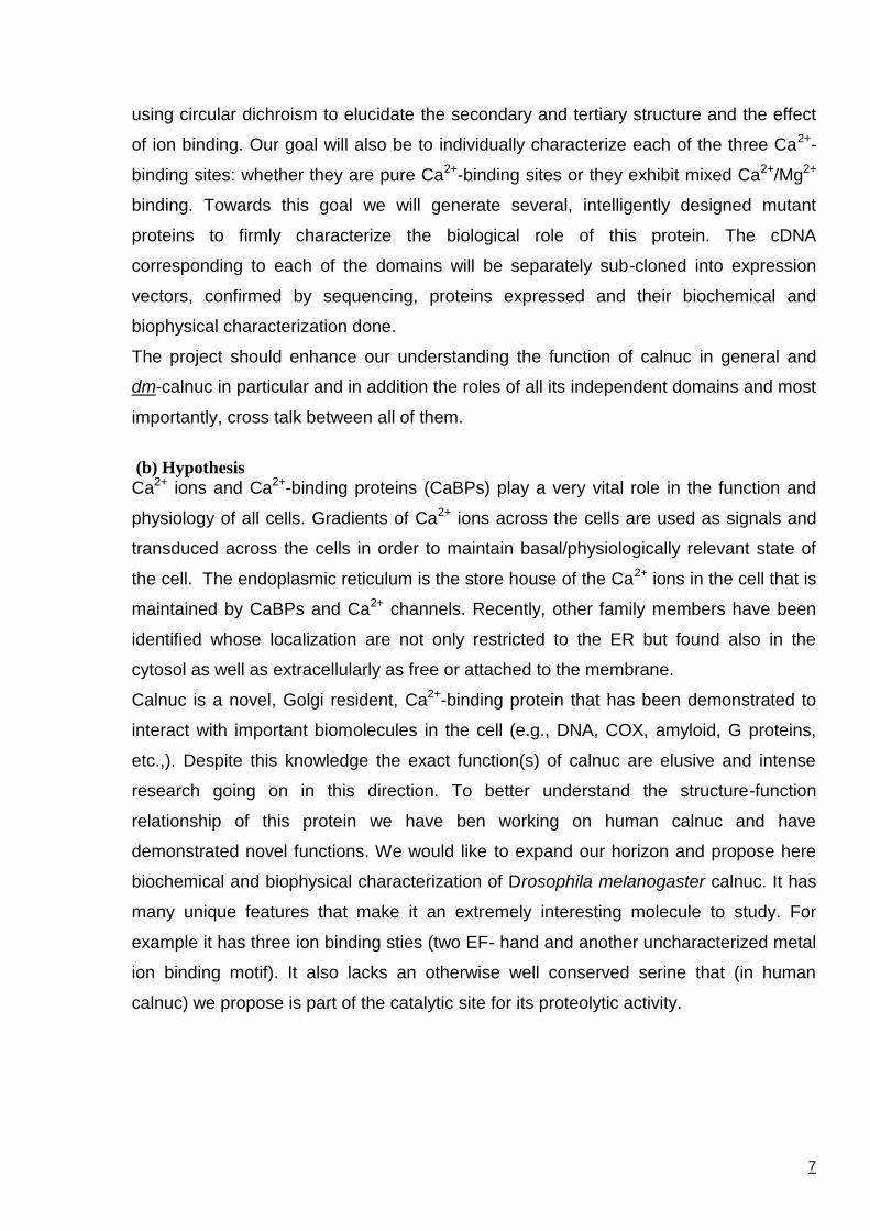
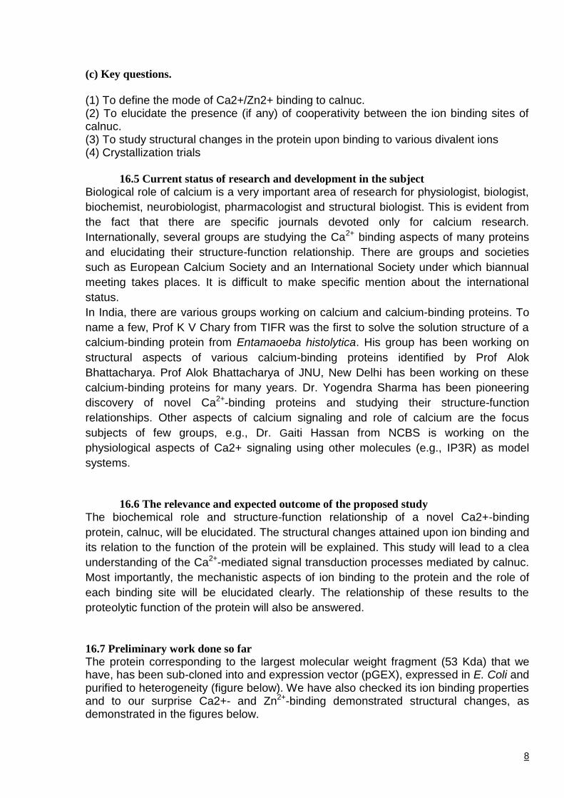
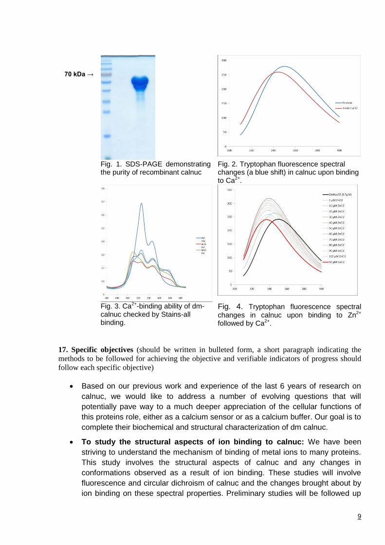
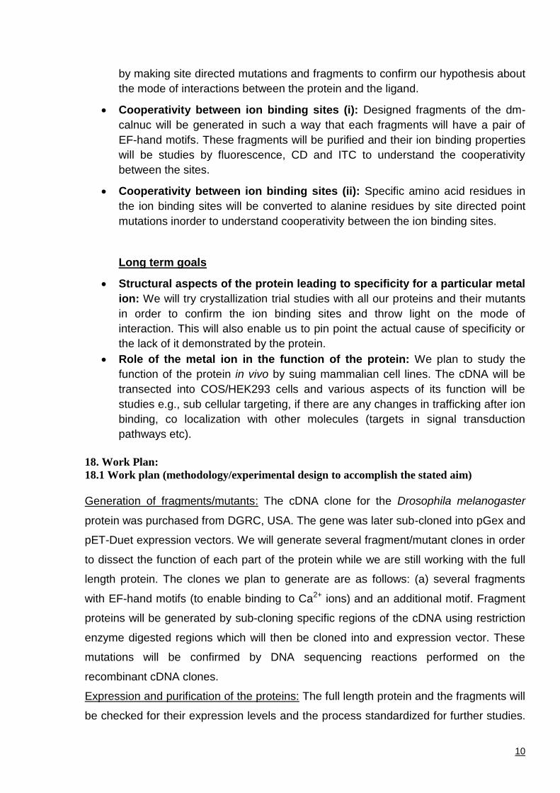
The protein corresponding to the largest molecular weight fragment (53 Kda) that we have, has been sub-cloned into and expression vector (pGEX), expressed in E. Coli and purified to heterogeneity (figure below). We have also checked its ion binding properties and to our surprise Ca2+- and Zn2+-binding demonstrated structural changes, as demonstrated in the figures below.
9
70 kDa →
Fig. 1. SDS-PAGE demonstrating the purity of recombinant calnuc
Fig. 2. Tryptophan fluorescence spectral changes (a blue shift) in calnuc upon binding to Ca2+.
Fig. 3. Ca2+-binding ability of dm-calnuc checked by Stains-all binding.
Fig. 4. Tryptophan fluorescence spectral
changes in calnuc upon binding to Zn2+ followed by Ca2+.
17. Specific objectives (should be written in bulleted form, a short paragraph indicating the
methods to be followed for achieving the objective and verifiable indicators of progress should
follow each specific objective)
Based on our previous work and experience of the last 6 years of research on
calnuc, we would like to address a number of evolving questions that will
potentially pave way to a much deeper appreciation of the cellular functions of
this proteins role, either as a calcium sensor or as a calcium buffer. Our goal is to
complete their biochemical and structural characterization of dm calnuc.
To study the structural aspects of ion binding to calnuc: We have been
striving to understand the mechanism of binding of metal ions to many proteins.
This study involves the structural aspects of calnuc and any changes in
conformations observed as a result of ion binding. These studies will involve
fluorescence and circular dichroism of calnuc and the changes brought about by
ion binding on these spectral properties. Preliminary studies will be followed up
10
by making site directed mutations and fragments to confirm our hypothesis about
the mode of interactions between the protein and the ligand.
Cooperativity between ion binding sites (i): Designed fragments of the dm-
calnuc will be generated in such a way that each fragments will have a pair of
EF-hand motifs. These fragments will be purified and their ion binding properties
will be studies by fluorescence, CD and ITC to understand the cooperativity
between the sites.
Cooperativity between ion binding sites (ii): Specific amino acid residues in
the ion binding sites will be converted to alanine residues by site directed point
mutations inorder to understand cooperativity between the ion binding sites.
Long term goals
Structural aspects of the protein leading to specificity for a particular metal
ion: We will try crystallization trial studies with all our proteins and their mutants
in order to confirm the ion binding sites and throw light on the mode of
interaction. This will also enable us to pin point the actual cause of specificity or
the lack of it demonstrated by the protein.
Role of the metal ion in the function of the protein: We plan to study the
function of the protein in vivo by suing mammalian cell lines. The cDNA will be
transected into COS/HEK293 cells and various aspects of its function will be
studies e.g., sub cellular targeting, if there are any changes in trafficking after ion
binding, co localization with other molecules (targets in signal transduction
pathways etc).
18. Work Plan:
18.1 Work plan (methodology/experimental design to accomplish the stated aim)
Generation of fragments/mutants: The cDNA clone for the Drosophila melanogaster
protein was purchased from DGRC, USA. The gene was later sub-cloned into pGex and
pET-Duet expression vectors. We will generate several fragment/mutant clones in order
to dissect the function of each part of the protein while we are still working with the full
length protein. The clones we plan to generate are as follows: (a) several fragments
with EF-hand motifs (to enable binding to Ca2+ ions) and an additional motif. Fragment
proteins will be generated by sub-cloning specific regions of the cDNA using restriction
enzyme digested regions which will then be cloned into and expression vector. These
mutations will be confirmed by DNA sequencing reactions performed on the
recombinant cDNA clones.
Expression and purification of the proteins: The full length protein and the fragments will
be checked for their expression levels and the process standardized for further studies.
11
Proteins will be expressed in bacterial (BL21DE3) cells by IPTG mediated induction and
later purified by using standard chromatography protocols that have already been
established in the lab (affinity chromatography followed by size exclusion
chromatography). The purity of the protein will be checked by subjecting it to SDS-
PAGE analysis.
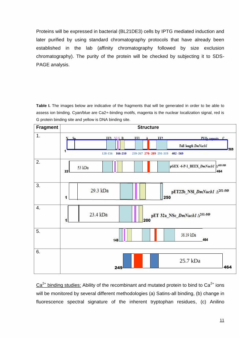
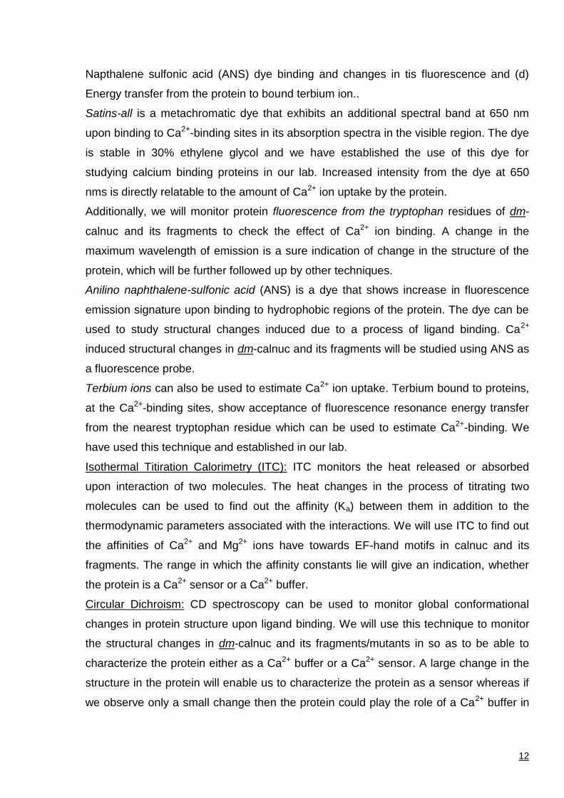
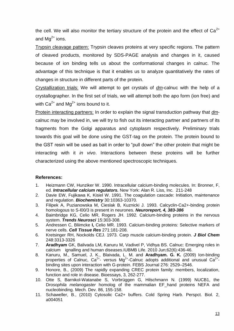
Table I. The images below are indicative of the fragments that will be generated in order to be able to
assess ion binding. Cyan/blue are Ca2+-binding motifs, magenta is the nuclear localization signal, red is
G protein binding site and yellow is DNA binding site.
Fragment Structure
1.
2.
3.
4.
5.
6.
Ca2+ binding studies: Ability of the recombinant and mutated protein to bind to Ca2+ ions
will be monitored by several different methodologies (a) Satins-all binding, (b) change in
fluorescence spectral signature of the inherent tryptophan residues, (c) Anilino
128-156 166-210 239-267 278- 289 291-319 402 -569
464
249
25.7 kDa
12
Napthalene sulfonic acid (ANS) dye binding and changes in tis fluorescence and (d)
Energy transfer from the protein to bound terbium ion..
Satins-all is a metachromatic dye that exhibits an additional spectral band at 650 nm
upon binding to Ca2+-binding sites in its absorption spectra in the visible region. The dye
is stable in 30% ethylene glycol and we have established the use of this dye for
studying calcium binding proteins in our lab. Increased intensity from the dye at 650
nms is directly relatable to the amount of Ca2+ ion uptake by the protein.
Additionally, we will monitor protein fluorescence from the tryptophan residues of dm-
calnuc and its fragments to check the effect of Ca2+ ion binding. A change in the
maximum wavelength of emission is a sure indication of change in the structure of the
protein, which will be further followed up by other techniques.
Anilino naphthalene-sulfonic acid (ANS) is a dye that shows increase in fluorescence
emission signature upon binding to hydrophobic regions of the protein. The dye can be
used to study structural changes induced due to a process of ligand binding. Ca2+
induced structural changes in dm-calnuc and its fragments will be studied using ANS as
a fluorescence probe.
Terbium ions can also be used to estimate Ca2+ ion uptake. Terbium bound to proteins,
at the Ca2+-binding sites, show acceptance of fluorescence resonance energy transfer
from the nearest tryptophan residue which can be used to estimate Ca2+-binding. We
have used this technique and established in our lab.
Isothermal Titiration Calorimetry (ITC): ITC monitors the heat released or absorbed
upon interaction of two molecules. The heat changes in the process of titrating two
molecules can be used to find out the affinity (Ka) between them in addition to the
thermodynamic parameters associated with the interactions. We will use ITC to find out
the affinities of Ca2+ and Mg2+ ions have towards EF-hand motifs in calnuc and its
fragments. The range in which the affinity constants lie will give an indication, whether
the protein is a Ca2+ sensor or a Ca2+ buffer.
Circular Dichroism: CD spectroscopy can be used to monitor global conformational
changes in protein structure upon ligand binding. We will use this technique to monitor
the structural changes in dm-calnuc and its fragments/mutants in so as to be able to
characterize the protein either as a Ca2+ buffer or a Ca2+ sensor. A large change in the
structure in the protein will enable us to characterize the protein as a sensor whereas if
we observe only a small change then the protein could play the role of a Ca2+ buffer in
13
the cell. We will also monitor the tertiary structure of the protein and the effect of Ca2+
and Mg2+ ions.
Trypsin cleavage pattern: Trypsin cleaves proteins at very specific regions. The pattern
of cleaved products, monitored by SDS-PAGE analysis and changes in it, caused
because of ion binding tells us about the conformational changes in calnuc. The
advantage of this technique is that it enables us to analyze quantitatively the rates of
changes in structure in different parts of the protein.
Crystallization trials: We will attempt to get crystals of dm-calnuc with the help of a
crystallographer. In the first set of trials, we will attempt both the apo form (ion free) and
with Ca2+ and Mg2+ ions bound to it.
Protein interacting partners: In order to explain the signal transduction pathway that dm-
calnuc may be involved in, we will try to fish out its interacting partner and partners of its
fragments from the Golgi apparatus and cytoplasm respectively. Preliminary trials
towards this goal will be done using the GST-tag on the protein. The protein bound to
the GST resin will be used as bait in order to “pull down” the other protein that might be
interacting with it in vivo. Interactions between these proteins will be further
characterized using the above mentioned spectroscopic techniques.
References:
1. Heizmann CW, Hunziker W. 1990. Intracellular calcium-binding molecules. In: Bronner, F, ed. Intracellular calcium regulators. New York: Alan R. Liss, Inc. 211-248
2. Davie EW, Fujikawa K, Kisiel W. 1991. The coagulation cascade: Initiation, maintenance and regulation. Biochemistry 30:10363-10370.
3. Filipek A, Puzianowska M, Cieslak B, Kuznicki J. 1993. Calcyclin-Ca2+-binding protein homologous to S-l00/3 is present in neurones. Neuroreport, 4, 383-386
4. Baimbridge KG, Celio MR, Rogers JH. 1992. Calcium-binding proteins in the nervous system. Trends Neurosci 15:303-308.
5. Andressen C, Bliimcke I, Celio MR. 1993. Calcium-binding proteins: Selective markers of nerve cells. Cell Tissue Res 271:181-208.
6. Kretsinger RH, Nockolds CEJ. 1973. Carp muscle calcium-binding protein. J Biol Chem 248:3313-3326
7. Aradhyam GK, Balivada LM, Kanuru M, Vadivel P, Vidhya BS. Calnuc: Emerging roles in calcium ignalling and human diseases.IUBMB Life. 2010 Jun;62(6):436-46.
8. Kanuru, M., Samuel, J. K., Blaivada, L. M. and Aradhyam. G. K. (2009) Ion-binding properties of Calnuc, Ca2+- versus Mg2+-Calnuc adopts additional and unusual Ca2+-binding sites upon interaction with G-protein. FEBS Journal 276: 2529–2546.
9. Honore, B., (2009) The rapidly expanding CREC protein family: members, localization, function and role in disease. Bioessays, 3, 262-277.
10. Otte S, Barnikol-Watanabe S, Vorbrüggen G, Hilschmann N. (1999) NUCB1, the Drosophila melanogaster homolog of the mammalian EF_hand proteins NEFA and nucleobinding. Mech. Dev. 86, 155-158.
11. Schaweller, B., (2010) Cytosolic Ca2+ buffers. Cold Spring Harb. Perspct. Biol. 2, a004051
14
18.2 Connectivity of the participating institutions and investigators (in case of multi-
institutional projects only)
18.3 Alternate strategies (if the proposed experimental design or method does not work what is
the alternate strategy)
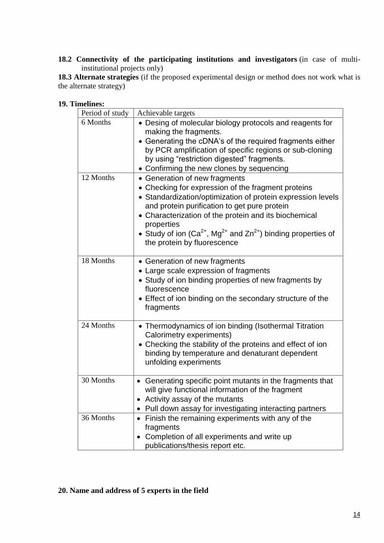
19. Timelines:
Period of study Achievable targets
6 Months
Desing of molecular biology protocols and reagents for
making the fragments.
Generating the cDNA’s of the required fragments either by PCR amplification of specific regions or sub-cloning by using “restriction digested” fragments.
Confirming the new clones by sequencing
12 Months Generation of new fragments
Checking for expression of the fragment proteins
Standardization/optimization of protein expression levels and protein purification to get pure protein
Characterization of the protein and its biochemical properties
Study of ion (Ca2+, Mg2+ and Zn2+) binding properties of the protein by fluorescence
18 Months Generation of new fragments
Large scale expression of fragments
Study of ion binding properties of new fragments by fluorescence
Effect of ion binding on the secondary structure of the fragments
24 Months Thermodynamics of ion binding (Isothermal Titration Calorimetry experiments)
Checking the stability of the proteins and effect of ion binding by temperature and denaturant dependent unfolding experiments
30 Months
Generating specific point mutants in the fragments that
will give functional information of the fragment
Activity assay of the mutants
Pull down assay for investigating interacting partners
36 Months
Finish the remaining experiments with any of the
fragments
Completion of all experiments and write up publications/thesis report etc.
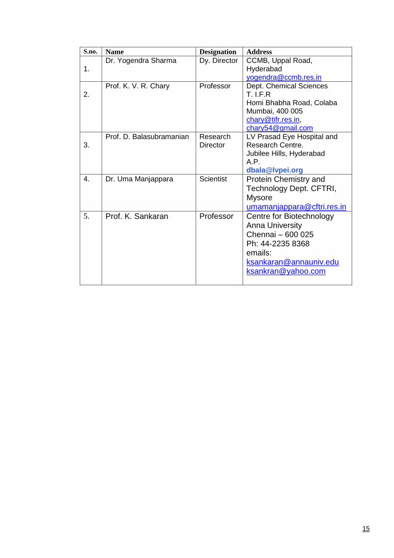
20. Name and address of 5 experts in the field
15
S.no. Name Designation Address
1.
Dr. Yogendra Sharma Dy. Director CCMB, Uppal Road, Hyderabad [email protected]
2.
Prof. K. V. R. Chary Professor Dept. Chemical Sciences T. I.F.R Homi Bhabha Road, Colaba Mumbai, 400 005 [email protected], [email protected]
3.
Prof. D. Balasubramanian Research Director
LV Prasad Eye Hospital and Research Centre. Jubilee Hills, Hyderabad A.P. [email protected]
4.
Dr. Uma Manjappara Scientist Protein Chemistry and Technology Dept. CFTRI, Mysore [email protected]
5. Prof. K. Sankaran
Professor Centre for Biotechnology Anna University Chennai – 600 025 Ph: 44-2235 8368 emails: [email protected] [email protected]
16
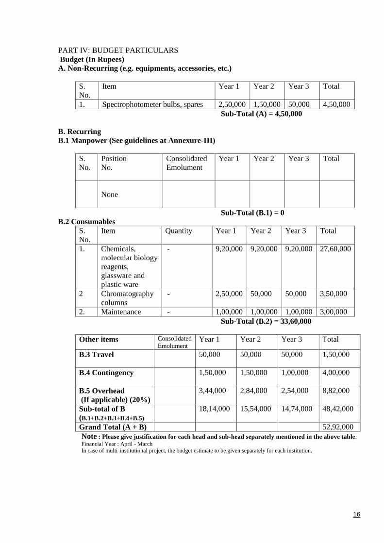
PART IV: BUDGET PARTICULARS
Budget (In Rupees)
A. Non-Recurring (e.g. equipments, accessories, etc.)
S.
No.
Item Year 1 Year 2 Year 3 Total
1. Spectrophotometer bulbs, spares 2,50,000 1,50,000 50,000 4,50,000
Sub-Total (A) = 4,50,000
B. Recurring
B.1 Manpower (See guidelines at Annexure-III)
S.
No.
Position
No.
Consolidated
Emolument
Year 1 Year 2 Year 3 Total
None
Sub-Total (B.1) = 0
B.2 Consumables
S.
No.
Item
Quantity Year 1 Year 2 Year 3 Total
1. Chemicals,
molecular biology
reagents,
glassware and
plastic ware
- 9,20,000 9,20,000 9,20,000 27,60,000
2 Chromatography
columns
- 2,50,000 50,000 50,000 3,50,000
2. Maintenance - 1,00,000 1,00,000 1,00,000 3,00,000
Sub-Total (B.2) = 33,60,000
Other items Consolidated
Emolument Year 1 Year 2 Year 3 Total
B.3 Travel
50,000 50,000 50,000 1,50,000
B.4 Contingency
1,50,000 1,50,000 1,00,000 4,00,000
B.5 Overhead
(If applicable) (20%)
3,44,000 2,84,000 2,54,000 8,82,000
Sub-total of B
(B.1+B.2+B.3+B.4+B.5)
18,14,000 15,54,000 14,74,000 48,42,000
Grand Total (A + B) 52,92,000
Note : Please give justification for each head and sub-head separately mentioned in the above table.
Financial Year : April - March
In case of multi-institutional project, the budget estimate to be given separately for each institution.
17
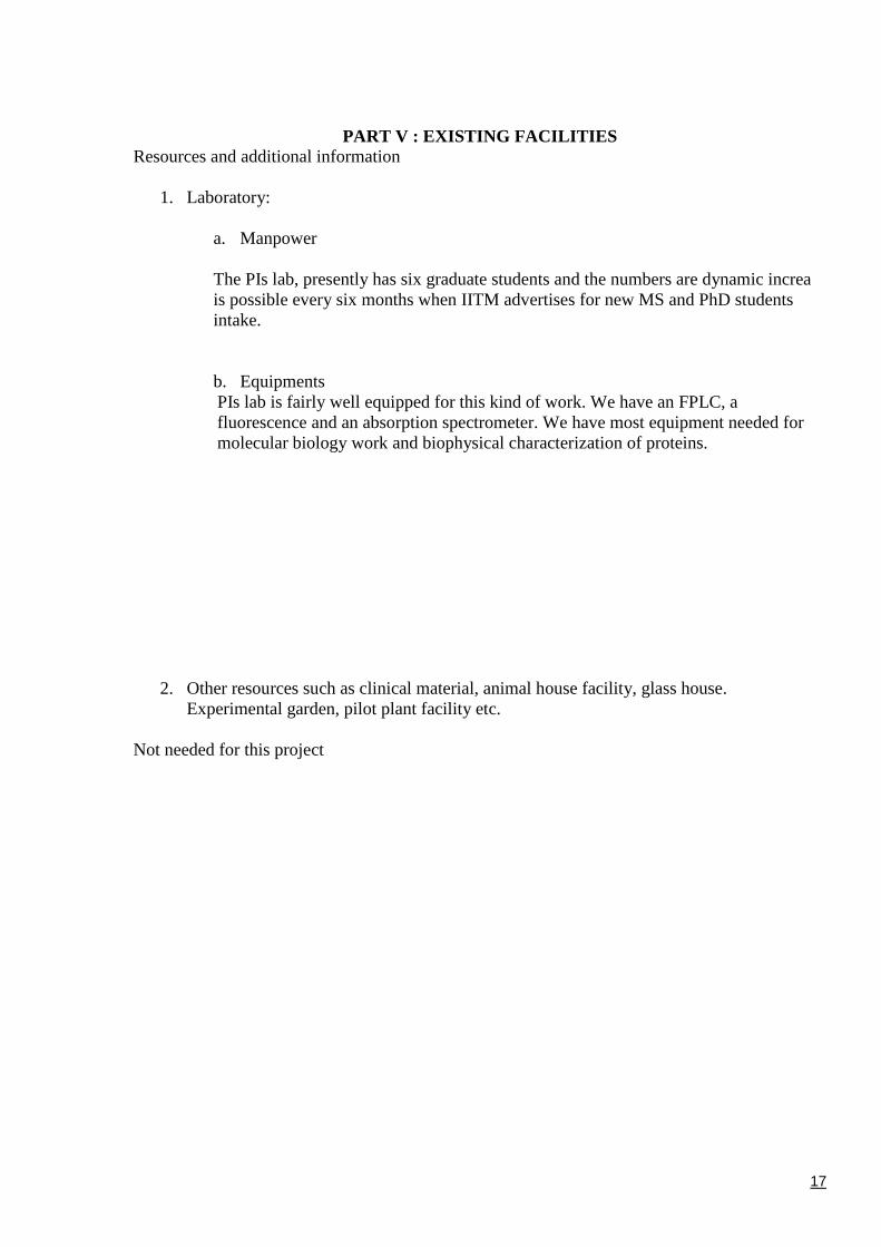
PART V : EXISTING FACILITIES
Resources and additional information
1. Laboratory:
a. Manpower
The PIs lab, presently has six graduate students and the numbers are dynamic increa
is possible every six months when IITM advertises for new MS and PhD students
intake.
b. Equipments
PIs lab is fairly well equipped for this kind of work. We have an FPLC, a
fluorescence and an absorption spectrometer. We have most equipment needed for
molecular biology work and biophysical characterization of proteins.
2. Other resources such as clinical material, animal house facility, glass house.
Experimental garden, pilot plant facility etc.
Not needed for this project
18
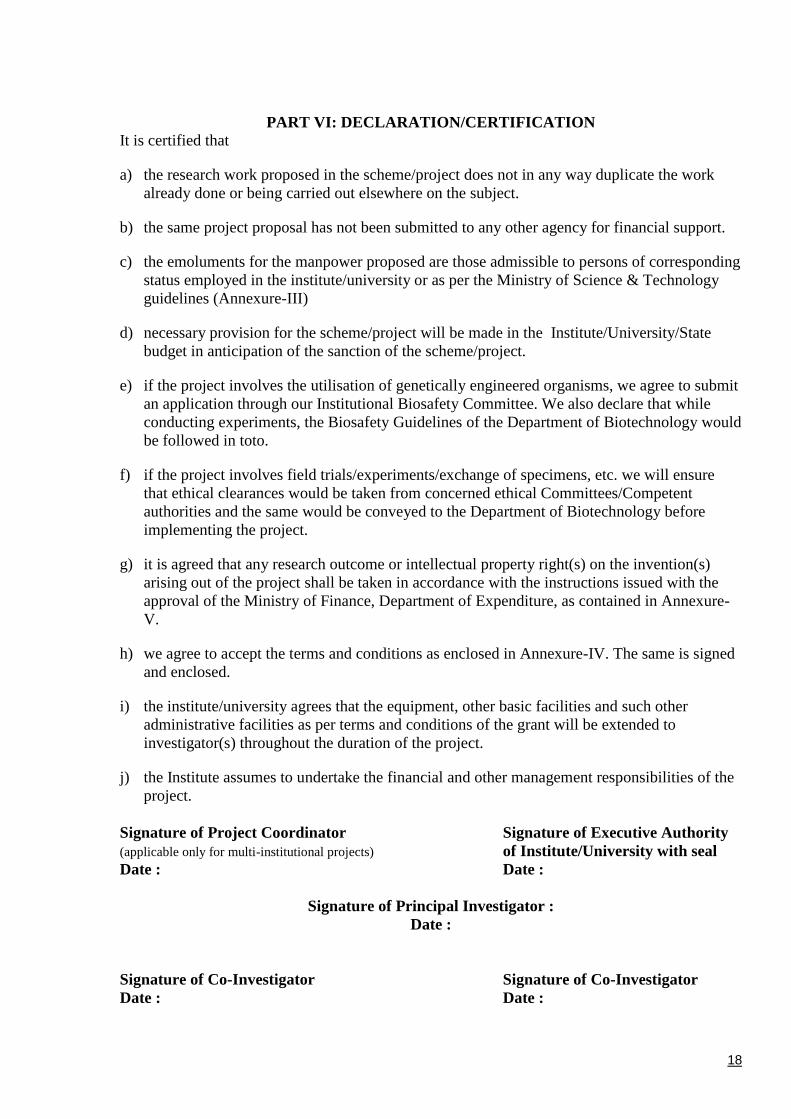
PART VI: DECLARATION/CERTIFICATION
It is certified that
a) the research work proposed in the scheme/project does not in any way duplicate the work
already done or being carried out elsewhere on the subject.
b) the same project proposal has not been submitted to any other agency for financial support.
c) the emoluments for the manpower proposed are those admissible to persons of corresponding
status employed in the institute/university or as per the Ministry of Science & Technology
guidelines (Annexure-III)
d) necessary provision for the scheme/project will be made in the Institute/University/State
budget in anticipation of the sanction of the scheme/project.
e) if the project involves the utilisation of genetically engineered organisms, we agree to submit
an application through our Institutional Biosafety Committee. We also declare that while
conducting experiments, the Biosafety Guidelines of the Department of Biotechnology would
be followed in toto.
f) if the project involves field trials/experiments/exchange of specimens, etc. we will ensure
that ethical clearances would be taken from concerned ethical Committees/Competent
authorities and the same would be conveyed to the Department of Biotechnology before
implementing the project.
g) it is agreed that any research outcome or intellectual property right(s) on the invention(s)
arising out of the project shall be taken in accordance with the instructions issued with the
approval of the Ministry of Finance, Department of Expenditure, as contained in Annexure-
V.
h) we agree to accept the terms and conditions as enclosed in Annexure-IV. The same is signed
and enclosed.
i) the institute/university agrees that the equipment, other basic facilities and such other
administrative facilities as per terms and conditions of the grant will be extended to
investigator(s) throughout the duration of the project.
j) the Institute assumes to undertake the financial and other management responsibilities of the
project.
Signature of Project Coordinator Signature of Executive Authority
(applicable only for multi-institutional projects) of Institute/University with seal
Date : Date :
Signature of Principal Investigator :
Date :
Signature of Co-Investigator Signature of Co-Investigator
Date : Date :
19
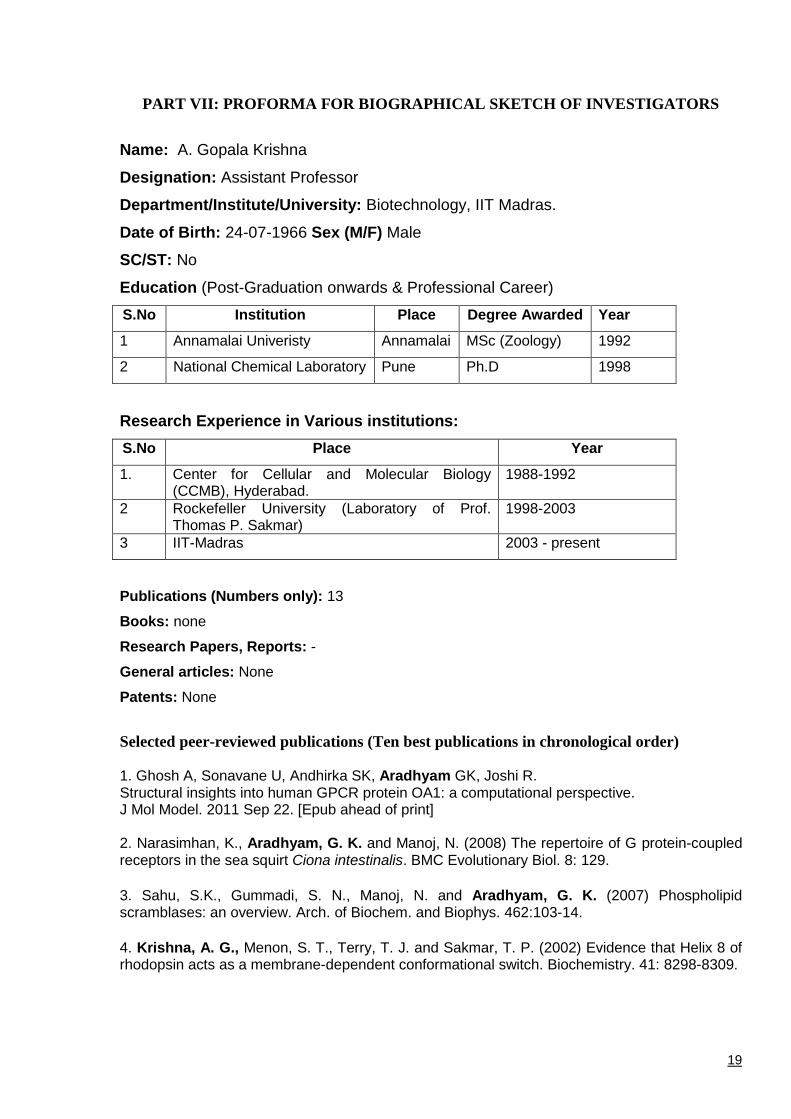
PART VII: PROFORMA FOR BIOGRAPHICAL SKETCH OF INVESTIGATORS
Name: A. Gopala Krishna
Designation: Assistant Professor
Department/Institute/University: Biotechnology, IIT Madras.
Date of Birth: 24-07-1966 Sex (M/F) Male
SC/ST: No
Education (Post-Graduation onwards & Professional Career)
S.No Institution Place Degree Awarded Year
1 Annamalai Univeristy Annamalai MSc (Zoology) 1992
2 National Chemical Laboratory Pune Ph.D 1998
Research Experience in Various institutions:
S.No Place Year
1. Center for Cellular and Molecular Biology (CCMB), Hyderabad.
1988-1992
2 Rockefeller University (Laboratory of Prof. Thomas P. Sakmar)
1998-2003
3 IIT-Madras 2003 - present
Publications (Numbers only): 13
Books: none
Research Papers, Reports: -
General articles: None
Patents: None
Selected peer-reviewed publications (Ten best publications in chronological order)
1. Ghosh A, Sonavane U, Andhirka SK, Aradhyam GK, Joshi R. Structural insights into human GPCR protein OA1: a computational perspective. J Mol Model. 2011 Sep 22. [Epub ahead of print] 2. Narasimhan, K., Aradhyam, G. K. and Manoj, N. (2008) The repertoire of G protein-coupled receptors in the sea squirt Ciona intestinalis. BMC Evolutionary Biol. 8: 129.
3. Sahu, S.K., Gummadi, S. N., Manoj, N. and Aradhyam, G. K. (2007) Phospholipid scramblases: an overview. Arch. of Biochem. and Biophys. 462:103-14.
4. Krishna, A. G., Menon, S. T., Terry, T. J. and Sakmar, T. P. (2002) Evidence that Helix 8 of rhodopsin acts as a membrane-dependent conformational switch. Biochemistry. 41: 8298-8309.
20
5. Marin, E. P., Krishna, A. G. and Sakmar, T. P. (2002) Disruption of the 5 helix of transducin impairs rhodopsin-catalyzed nucleotide exchange. Biochemistry. 41: 6988-6994.
6. Marin, E. P., Krishna, A. G. and Sakmar, T. P. (2001) Rapid activation of transducin by mutations distant from the nucleotide-binding site. J. Biol. Chem. 276: 27400-27405.
7. Marin, E. P., Krishna, A. G., Archambault. V., Simuni. E., Fu. W-Y. and Sakmar, T. P. (2001) The function of interdomain interactions in controlling nucleotide exchange rates in transducin. J. Biol. Chem. 276: 23873-23880.
8. Marin, E. P., Krishna, A. G., Zvyaga, T. A., Isele. J., Seibert, F. and Sakmar, T. P. (2000) The amino terminus of the fourth cytoplasmic loop of rhodopsin modulates rhodopsin-transducin interaction. J. Biol. Chem. 275: 1930-1936.
9. Krishna, A. G.,* Kumar, D. V., Khan, B. M., Rawal, S. K. and Ganesh, K. N. (1998) Taxol-DNA Interaction: fluorescence and CD studies of the groove binding properties of taxol. Biochim. Biophys. Acta 1382: 104-112. * Corresponding Author.
10. Krishna, A. G., Balasubramanian, D. and Ganesh, K. N. (1994) Sugar-DNA molecular recognition: Specific interaction of alpha (1,4) glucopyranose chains with DNA in the minor groove. Biochem. Biophys. Res. Comm. 202: 204-210.
11. Sharma, Y., Krishna, A. G., Balasubramanian, D., Fairwell, T. and Krishna, G. (1993) Studies on the interaction of the dye, Stains-all, with individual calcium binding domains of Calmodulin. FEBS Letters 326: 59-64.
12. Sharma, Y., Krishna, A. G., and Balasubramanian. D. (1993) Alteration of the dynamic
quaternary structure and calcium-binding ability of -crystallin by light. Photochem. Photobiol. 57: 739-743.
13. Sharma, Y., Rao, C. M., Rao, S.C., Krishna, A. G., Somasundaram, T. and Balasubramanian, D. (1989) Binding site conformation dictates the color of the dye Stains-all: A study of the binding of this dye to the eye lens proteins crystallins. J. Biol. Chem. 264: 20923-20927.
14. Sharma, Y., Rao, C. M., Narasu, M.L., Rao, S. C., Somasundaram, T., Krishna, A. G., and
Balasubramanian, D. (1989) Calcium ion binding to and to crystallins: the presence of the
EF-hand motif incrystallin that aids in calcium ion binding. J. Biol. Chem. 264: 12794-12799.
List maximum of five recent publications relevant to the proposed area of work.
1. Aradhyam GK, Balivada LM, Kanuru M, Vadivel P, Vidhya BS.
Calnuc: Emerging roles in calcium signaling and human diseases. IUBMB Life. 2010 Jun;62(6):436-46. Invited Review 2. Sahu, S.K., Aradhyam, G. K.* and Gummadi, S. N. * (2009) Calcium binding studies of peptides of human phospholipid scramblases 1 to 4 suggest that scramblases are new class of calcium binding proteins in the cell. Biochim. Biophys. Acta.. 1790:1274-81. (* Both are corresponding authors)
3. Sahu SK, Aradhyam GK, Gummadi SN. Calcium binding studies of peptides of human phospholipid scramblases 1 to 4 suggest that scramblases are new class of calcium binding proteins in the cell.
21
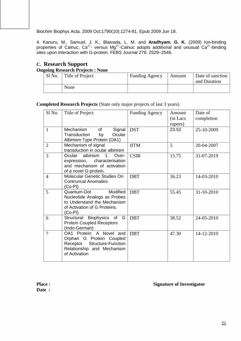
Biochim Biophys Acta. 2009 Oct;1790(10):1274-81. Epub 2009 Jun 18.
4. Kanuru, M., Samuel, J. K., Blaivada, L. M. and Aradhyam. G. K. (2009) Ion-binding properties of Calnuc, Ca2+- versus Mg2+-Calnuc adopts additional and unusual Ca2+-binding sites upon interaction with G-protein. FEBS Journal 276: 2529–2546.
C. Research Support Ongoing Research Projects : None
Sl No. Title of Project Funding Agency Amount Date of sanction
and Duration
None
Completed Research Projects (State only major projects of last 3 years)
Sl No. Title of Project Funding Agency Amount
(in Lacs
rupees)
Date of
completion
1 Mechanism of Signal Transduction by Ocular Albinism Type Protein (OA1)
DST 23.52 25-10-2009
2 Mechanism of signal transduction in ocular albinism
IITM 5 20-04-2007
3 Ocular aibinism 1. Over-expression, characterisation and mechanism of activation of a novel G-protein.
CSIR 15.75 31-07-2019
4 Molecular Genetic Studies On Contruncal Anomalies (Co-PI)
DBT 36.23 14-03-2010
5 Quantum-Dot Modified Nucleotide Analogs as Probes to Understand the Mechanism of Activation of G Proteins. (Co-PI)
DBT 55.45 31-10-2010
6 Structural Biophysics of G Protein Coupled Receptors (Indo-German)
DBT 38.52 24-05-2010
7 OA1 Protein: A Novel and Orphan G Protein Coupled Receptor. Structure-Function Relationship and Mechanism of Activation
DBT 47.30 14-12-2010
Place : Signature of Investigator
Date :