Embed Size (px)
Citation preview
Indicanine A, a New 3-Phenylcoumarin from Root Bark of Erythrina indica1
Augustin E. Nkengfack,*,† Alain K. Waffo,† Guy A. Azebaze,† Zacharias T. Fomum,† Michele Meyer,Bernard Bodo,‡ and Fanie R. van Heerden§
Department of Organic Chemistry, University of Yaounde I, P.O. Box 812 Yaounde, Cameroon, Laboratoire de Chimie desSubstances Naturelles U.R.A. 401, Museum National d’Histoire Naturelle, 63, Rue Buffon, 75005 Paris Cedex 05, France, andDepartment of Chemistry and Biochemistry, Rand Afrikaans University, P.O. Box 524, Auckland Park 2006, South Africa
Received June 18, 1999
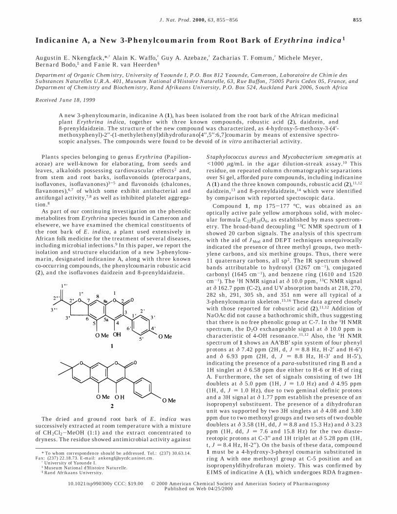
A new 3-phenylcoumarin, indicanine A (1), has been isolated from the root bark of the African medicinalplant Erythrina indica, together with three known compounds, robustic acid (2), daidzein, and8-prenyldaidzein. The structure of the new compound was characterized, as 4-hydroxy-5-methoxy-3-(4′-methoxyphenyl)-2′′-(1-methylethenyl)dihydrofurano[4′′,5′′:6,7]coumarin by means of extensive spectro-scopic analyses. The compounds were found to be devoid of in vitro antibacterial activity.
Plants species belonging to genus Erythrina (Papilion-aceae) are well-known for elaborating, from seeds andleaves, alkaloids possessing cardiovascular effects2 and,from stem and root barks, isoflavonoids (pterocarpans,isoflavones, isoflavanones)3-5 and flavonoids (chalcones,flavanones),6,7 of which some exhibit antibacterial andantifungal activity,7,8 as well as inhibited platelet aggrega-tion.8
As part of our continuing investigation on the phenolicmetabolites from Erythrina species found in Cameroon andelsewere, we have examined the chemical constituents ofthe root bark of E. indica, a plant used extensively inAfrican folk medicine for the treatment of several diseases,including microbial infections.9 In this paper, we report theisolation and structure elucidation of a new 3-phenylcou-marin, designated indicanine A, along with three knownco-occurring compounds, the phenylcoumarin robustic acid(2), and the isoflavones daidzein and 8-prenyldaidzein.
The dried and ground root bark of E. indica wassuccessively extracted at room temperature with a mixtureof CH2Cl2-MeOH (1:1) and the extract concentrated todryness. The residue showed antimicrobial activity against
Staphylococcus aureus and Mycobacterium smegmatis at<1000 µg/mL in the agar dilution-streak assay.10 Thisresidue, on repeated column chromatographic separationsover Si gel, afforded pure compounds, including indicanineA (1) and the three known compounds, robustic acid (2),11,12
daidzein,13 and 8-prenyldaidzein,14 which were identifiedby comparison with reported spectoscopic data.
Compound 1, mp 175-177 °C, was obtained as anoptically active pale yellow amorphous solid, with molec-ular formula C22H20O6, as established by mass spectrom-etry. The broad-band decoupling 13C NMR spectrum of 1showed 20 carbon signals. The analysis of this spectrumwith the aid of JMod and DEPT techniques unequivocallyindicated the presence of three methyl groups, two meth-ylene carbons, and six methine groups. Thus, there were11 quaternary carbons, all sp2. The IR spectrum showedbands attributable to hydroxyl (3267 cm-1), conjugatedcarbonyl (1645 cm-1), and benzene ring (1610 and 1520cm-1). The 1H NMR signal at δ 10.0 ppm, 13C NMR signalat δ 162.7 ppm (C-2), and UV absorption bands at 218, 270,282 sh, 291, 305 sh, and 351 nm were all typical of a3-phenylcoumarin skeleton.15,16 These data agreed closelywith those reported for robustic acid (2).11,12 Addition ofNaOAc did not cause a bathochromic shift, thus suggestingthat there is no free phenolic group at C-7. In the 1H NMRspectrum, the D2O exchangeable signal at δ 10.0 ppm ischaracteristic of 4-OH resonance.11,12 Also, the 1H NMRspectrum of 1 shows an AA′BB′ spin system of four phenylprotons at δ 7.42 ppm (2H, d, J ) 8.8 Hz, H-2′ and H-6′)and δ 6.93 ppm (2H, d, J ) 8.8 Hz, H-3′ and H-5′),indicating the presence of a para-substituted ring B and a1H singlet at δ 6.58 ppm due either to H-6 or H-8 of ringA. Furthermore, the set of signals consisting of two 1Hdoublets at δ 5.0 ppm (1H, J ) 1.0 Hz) and δ 4.95 ppm(1H, d, J ) 1.0 Hz), due to two geminal olefinic protonsand a 3H signal at δ 1.77 ppm establish the presence of anisopropenyl substituent. The presence of a dihydrofuranunit was supported by two 3H singlets at δ 4.08 and 3.80ppm due to two methoxyl groups and two sets of two doubledoublets at δ 3.58 (1H, dd, J ) 8.8 and 15.3 Hz) and δ 3.23ppm (1H, dd, J ) 7.6 and 15.8 Hz) for the two diaste-reotopic protons at C-3′′ and 1H triplet at δ 5.28 ppm (1H,t, J ) 8.4 Hz, H-2′′). On the basis of these data, compound1 must be a 4-hydroxy-3-phenyl coumarin substituted inring A with one methoxyl group at C-5 position and anisopropenyldihydrofuran moiety. This was confirmed byEIMS of indicatine A (1), which undergoes RDA fragmen-
* To whom correspondence should be addressed. Tel.: (237) 30.63.14.Fax: (237) 22.18.73. E-mail: [email protected].
† University of Yaounde I.‡ Museum National d’Histoire Naturelle.§ Rand Afrikaans University.
855J. Nat. Prod. 2000, 63, 855-856
10.1021/np990300y CCC: $19.00 © 2000 American Chemical Society and American Society of PharmacognosyPublished on Web 04/25/2000
tation via its keto tautomer to give two important ionfragments at m/z 233 and 148. The first ion fragment atm/z 233, clearly showed compound 1 possesses one meth-oxyl and an isopropenyldihydrofuran moiety on ring A,while the second ion fragment at m/z 148, indicated thesecond methoxyl group to be located on ring B at C-4′position. Therefore, it remained to be established unam-biguously whether the fusion of the isopropenyl dihydro-furan moiety on ring A is linear or angular. This wasdeduced from NOE difference experiments, which showedno enhancement of the 1H aromatic signal at δ 6.58 ppm(corresponding either to H-6 or H-8) but enhancement ofthe signal at δ 10.0 ppm (4-OH), when the signal at δ 4.08ppm (5-OMe) was irradiated. This finding clearly indicatedthat the single A-ring aromatic proton at δ 6.58 ppm waslocated at C-8. Thus, the isopropenyl dihydrofuran unit wasfused in a linear manner on ring A. From the abovespectroscopic studies, compound 1 was characterized as4-hydroxy-5-methoxy-3-(4′-hydroxyphenyl)-2′′-(1-methyl-ethenyl)dihydrofuran-[4′′,5′′:6,7]coumarin. This compound,which appears to be novel, has been given the trivial nameindicanine A. We were not able to establish the absoluteconfiguration at the C-2′′ stereocenter.
All the isolated compounds were tested in vitro for theirantimicrobial activities against microorganisms, S. aureus209P, M. smegmatis ATCC 607, and Escherichia coli RL65using an agar dilution-streak method.10 None of thecompounds showed any significant activity.
Experimental Section
General Experimental Procedures. All melting pointswere determined on a Kofler hot-stage apparatus and areuncorrected. IR spectra were recorded on a Perkin-Elmer 727Bspectrometer in KBr disks. UV spectra were obtained on aBeckman model 25 spectrophotometer. EIMS (ionization volt-age, 70 eV) were measured with LKB9000S and Nermag/sidarU 3:1 spectometers. 1H and 13C NMR spectra were recordedon a Varian Gemini 2000 and on a Bruker spectrometersequipped with a 5-mm 1H and 13C probe operating at 300 and75 MHz, respectively, with TMS as internal standard. DEPTand JMod were measured with the usual pulse sequence, anddata processing was obtained with standard software.
Plant Material. Root bark of E. indica was collected inJune 1998, at Ibadan, Nigeria. A voucher specimen document-ing the collection is on deposit at the National Herbarium,Yaounde, Cameroon.
Extraction and Isolation. Air-dried, powdered root barkof E. indica (6 kg) was extracted with a mixture of CH2Cl2-MeOH (1:1) and concentrated to dryness on a rotary evaporatorunder reduced pressure to afford a viscous mass of CH2Cl2-MeOH (1:1) extract (200 g). This material was subjected tocolumn chromatography on Si gel (70-230 mesh, ASTM;Merck) packed in n-hexane and eluted with n-hexane-EtOAcmixture. In all, 200 fractions (ca. 250 mL each) were collectedand combined on the basis of TLC analysis, leading to fivemain series (A-E). Fractions 1-50, eluted with a mixture ofhexane-EtOAc (9:1), gave series A, from which robustic acid(2) (4 g) crystallized. Fractions 101-120, eluted with hexane-EtOAc (3:2), gave series C, which was further subjected torepeated column chromatography over Si gel eluted with amixture of hexane-EtOAc (7:3) to yield indicanine A (1) (70mg). Series D, resulting from the combination of fractions 121-181 eluted with a mixture of hexane-EtOAc (1:1), wasrechromatographed with Si gel column chromatography, elut-ing with hexane-EtOAc (3:2) to afford daidzein (60 mg) and8-prenyldaidzein (100 mg).
Indicanine A (1): pale-yellow powder, mp 175-177 °C,yield 0.0012% [R]D -46° (c 1.99, MeOH); UV (MeOH) λmax (logε) 218 (4.54), 270 (3.48), 282 sh (3.86), 291 (4.23), 305 sh (4.17),
351 (4.63) nm; IR νmax (KBr) 3267, 1645, 1610, 1520, 1200,1100 cm-1; 1H NMR (CDCl3, 300 MHz) δ 7.42 ppm (1H, d, J )8.8 Hz, H-2′ and H-6′), 6.93 (2H, d, J ) 8.8 Hz, H-3′and H-5′),6.58 (1H, s, H-8), 5.28 (1H, t, J ) 8.8 Hz, H-2′′), 5.0 (1H, d,J ) 1.0 Hz, dCH), 4.95 (1H, d, J ) 1.0 Hz, dCH), 4.08 (3H, s,5-OMe), 3.80 (3H, s, 4′-OMe), 3.58 (1H, dd, J ) 8.8 and 15.5Hz, H-3′′), 3.23 (1H, dd, J ) 7.6 and 15.8 Hz, H-3′′), δ 1.77(3H, s, CH3-CdC); 13C NMR (75 MHz) δ 164.1 (s, C-4), 162.7(s, C-2), 161.1 (s, C-5), 158 (s, C-7), 155.1 (s, C-4′), 152.4 (s,C-8a), 142.4 (s, C-2′′′), 131.7 (d, C-2′ and C-6′), 123.6 (s, C-1′),113.5 (t, C-1′′′), 113.0 (d, C-3′ and C-5′), 111.0 (s, C-6), 100.0(s, C-5a), 86.1 (d, C-2′′), 60.5 (q, 5-OMe), 55.2 (q, 4′-OMe), 33.2(t, C-3′′), 17.1 (q, 3′′′-Me); EIMS m/z [M]+ 380 (96), 365 (33),337 (19), 233 (100), 217 (33), 190 (44), 175 (28), 148 (98), 135(33), 120 (41), 91 (30), 69 (43), 41 (21), 39 (16); DCI/NH3 [M+1]+
381; HREIMS m/z [M+] 380.1262 (calcd for C22H20O6, 380.1264).Robustic acid (2): white solid, yield 0.066%, mp 212° (lit.12
210°C); HEIMS m/z [M+] 380.1261 (calcd for C22H20O6,380.1260); UV, IR, 1H and 13C NMR were in agreement withthe published data.11,12
Daidzein: amorphous powder, yield 0.0001%, mp 198-200°C; HREIMS m/z 254.0577 (calcd for C15H10O4, 254.O579); IR,1H and 13C NMR spectra data were in agreement withliterature values.13
8-Prenyldaidzein: white amorphous solid, yield 0.0001%,mp 198 °C (lit.14 196-198 °C); HREIMS m/z 322.1206 (calcdfor C20H18O4, 322.1205); IR, 1H and 13C NMR spectral datamatched well with those published in the literature.14
Antimicrobial Activity Screening. Extract and purifiedactive compounds were tested at 1 mg/mL against S. aureus209P, M. smegmatis ATCC 607, and E. coli RL65. The threestrains of bacteria were cultured in Mueller-Hinton agarmedium at 37 °C. After one day, their growth was assessedvisually. The lowest concentration of the test compounds inwhich no visible growth occurred was defined as the minimuminhibitory concentration.
Acknowledgment. The authors thank the InternationalFoundation for Science (I.F.S), Stockholm (Sweden), for finan-cial support of this work under research grant no. F/1392-3F.One of us (A.E.N.) also thanks the Director of “MuseumNational d’Histoire Naturelle,” Paris, France, for three months’fellowship in his institution.
References and Notes(1) Part 35 in the series “Erythrina Studies.” For part 34, see: Nkengfack,
A. E.; Vouffo, T. W.; Vardamides, J. C.; Kouam, J.; Fomum, Z. T.;Meyer, M.; Sterner, O. Phytochemistry 1997, 46, 573-578.
(2) Cordell, G. A. Introduction to Alkaloids: A Biogenetic Approach; JohnWiley & Sons: New York, 1981; pp 450-462.
(3) Nkengfack, A. E.; Vardamides, J. C.; Fomum, Z. T.; Meyer, M.Phytochemistry 1995, 40, 1803-1808.
(4) Nkengfack, A. E.; Vouffo, W. T.; Fomum, Z. T.; Meyer, M.; Ola, B.;Sterner, O. Phytochemistry, 1994, 36, 1047-1051.
(5) Nkengfack, A. E.; Sanson, D. R.; Fomum, Z. T.; Tempesta, M. S. J.Nat. Prod. 1989, 52, 320-324.
(6) Fomum, Z. T.; Ayafor, J. F; Mbafor, J. T.; Mbi, C. M. J. Chem. Soc.,Perkin Trans. 1 1986, 33-37.
(7) Mitscher, L. A.; Gollapudi, S. R.; Gerlach, D. C.; Drake, S. D.; Veliz,E. A.; Ward, J. A. Phytochemistry 1988, 27, 381-385.
(8) Kamat, V. S.; Chuo, F. Y.; Kubo, I.; Nakanishi, K. Heterocycles 1981,15, 1163-1170.
(9) Ayensu, E. S. In Medicinal Plants of West Africa; Reference Publica-tion, Inc.: Algonac, MI, 1978; p 153.
(10) Mitscher, L. A. In Isolation, Separation and Purification of Antibiotics;Weinstein, G., Wagman, G., Eds.; Elsevier: Amsterdam, 1977; p 463.
(11) Khalid, S. A.; Waterman, P. G. Phytochemistry 1983, 22, 1001-1003.(12) Jackson, B.; Owen, P. J.; Scheinmann, F. J. Chem. Soc. C 1971, 3389-
3391.(13) Jha, H. C., Zillizen, F.; Breitmaier, V. Can J. Chem. 1980, 58, 1211-
1213.(14) Munekazu, I.; Toshiyuki, T.; Mizuo, M.; Hirobumi, Y.; Yasuko, K.;
Shigetomo, Y. Chem. Pharm. Bull. 1997, 40, 2749-2752.(15) Olivares, E. M.; Lwande, W.; Monache, F. D.; Bettolo, G. B. M.
Phytochemistry 1982, 21, 1763-1765.(16) East, A. J.; Ollis, W. D.; Wheeler, R. E. J. Chem. Soc. C 1969,
365-367.
NP990300Y
856 Journal of Natural Products, 2000, Vol. 63, No. 6 Notes
![Acaaddemmiicc SSciieenncceess · reduce swellings and pain. Tamarind fruit pulp is applied topically on inflammatory swellings and rheumatism to relieve pain [10, 11]. T. indica bark](https://img.pdfslide.net/doc/110x75/5fbebeffad556d3f9c592699/acaaddemmiicc-ssciieenncceess-reduce-swellings-and-pain-tamarind-fruit-pulp-is.jpg)