Embed Size (px)
Citation preview
Indira Gandhi National Tribal University,
Amarkantak (M.P.)
B.Sc Biotechnology
VIth Semester 2020
Paper name: Animal Diversity II
Reference Notes
Dr. Parikipandla Sridevi
Assistant Professor
Department of Biotechnology
IGNTU, Amarkantak
Unit-I
Protochordates: Outline of classification, General features and important
characters of Herdmania, Branchiostoma
Contents
1. Protochordates
a. Outline of classification
b. General characteristics
c. Classes
2. Herdmania
3. Branchiostoma
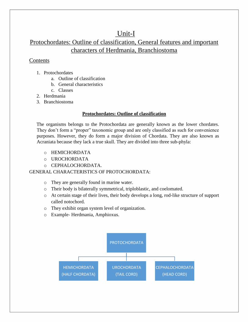
Protochordates: Outline of classification
The organisms belongs to the Protochordata are generally known as the lower chordates.
They don‘t form a ―proper‖ taxonomic group and are only classified as such for convenience
purposes. However, they do form a major division of Chordata. They are also known as
Acraniata because they lack a true skull. They are divided into three sub-phyla:
o HEMICHORDATA
o UROCHORDATA
o CEPHALOCHORDATA.
GENERAL CHARACTERISTICS OF PROTOCHORDATA:
o They are generally found in marine water.
o Their body is bilaterally symmetrical, triploblastic, and coelomated.
o At certain stage of their lives, their body develops a long, rod-like structure of support
called notochord.
o They exhibit organ system level of organization.
o Example- Herdmania, Amphioxus.
PROTOCHORDATA
HEMICHORDATA
(HALF CHORDATA)
UROCHORDATA
(TAIL CORD)
CEPHALOCHORDATA
(HEAD CORD)
HEMICHORDATA:
Marine, Solitarily
Body is divided into proboscis, collar and trunk.
Bilaterally symmetrical
Primitive notochord restricted to proboscis only, thus called stomochord.
Triploblastic
True coelom present
Straight or U shaped gut with anus
Nervous system diffuse and variable
Open circulatory system
Blood has no colour and corpuscles
Excretory organ- Glomerulus
Filter feeding
Four Classes: ENTEROPNEUSTA, PTEROBRANCHIA, PLANCTOSPHAEROIDEA,
GRAPTOLITA.
EXAMPLE: CEPHALODISCUS
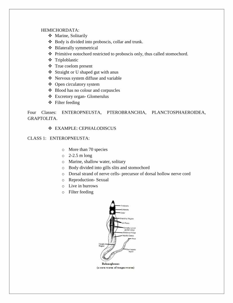
CLASS 1: ENTEROPNEUSTA:
o More than 70 species
o 2-2.5 m long
o Marine, shallow water, solitary
o Body divided into gills slits and stomochord
o Dorsal strand of nerve cells- precursor of dorsal hollow nerve cord
o Reproduction- Sexual
o Live in burrows
o Filter feeding
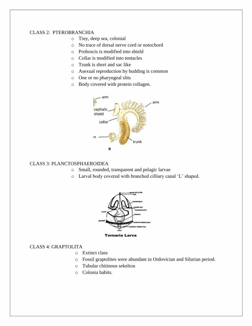
CLASS 2: PTEROBRANCHIA
o Tiny, deep sea, colonial
o No trace of dorsal nerve cord or notochord
o Proboscis is modified into shield
o Collar is modified into tentacles
o Trunk is short and sac like
o Asexual reproduction by budding is common
o One or no pharyngeal slits
o Body covered with protein collagen.
CLASS 3: PLANCTOSPHAEROIDEA
o Small, rounded, transparent and pelagic larvae
o Larval body covered with branched cilliary canal ‗L‘ shaped.
CLASS 4: GRAPTOLITA
o Extinct class
o Fossil graptolites were abundant in Ordovician and Silurian period.
o Tubular chitinous sekelton
o Colonia habits.
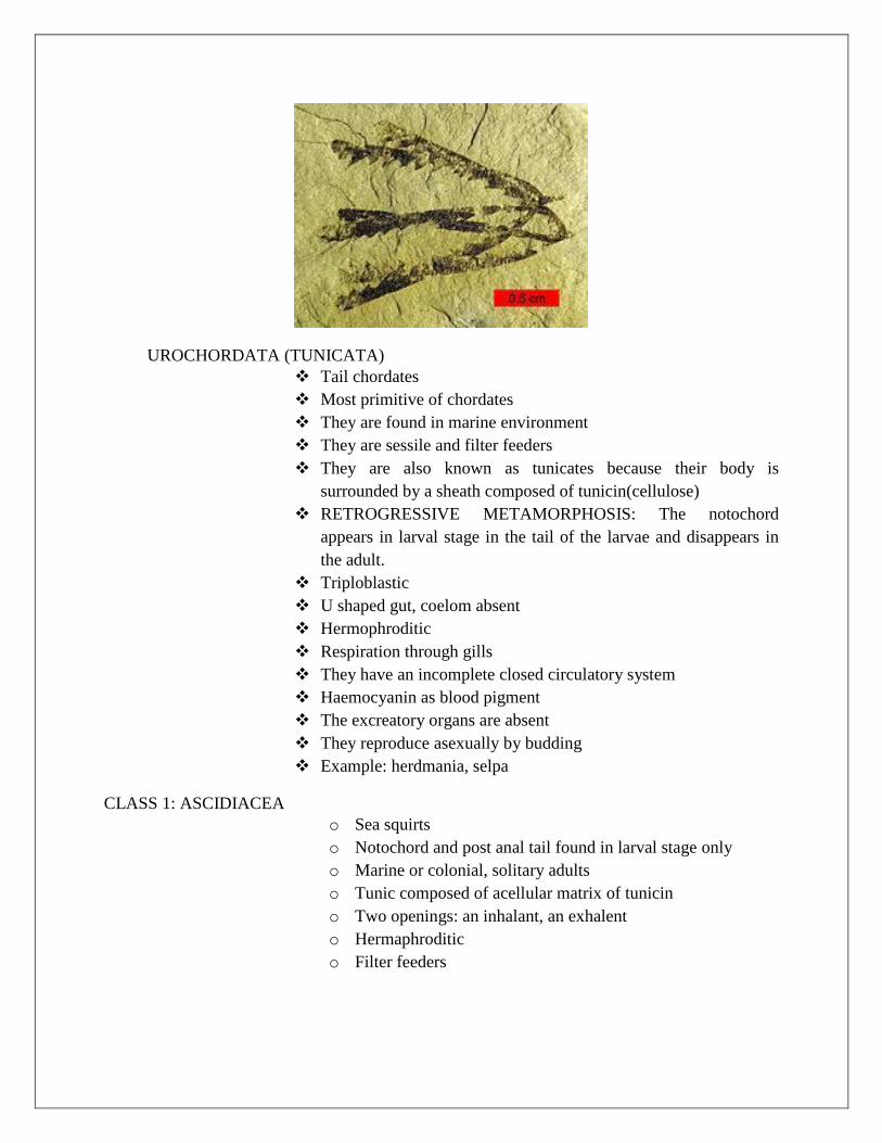
UROCHORDATA (TUNICATA)
Tail chordates
Most primitive of chordates
They are found in marine environment
They are sessile and filter feeders
They are also known as tunicates because their body is
surrounded by a sheath composed of tunicin(cellulose)
RETROGRESSIVE METAMORPHOSIS: The notochord
appears in larval stage in the tail of the larvae and disappears in
the adult.
Triploblastic
U shaped gut, coelom absent
Hermophroditic
Respiration through gills
They have an incomplete closed circulatory system
Haemocyanin as blood pigment
The excreatory organs are absent
They reproduce asexually by budding
Example: herdmania, selpa
CLASS 1: ASCIDIACEA
o Sea squirts
o Notochord and post anal tail found in larval stage only
o Marine or colonial, solitary adults
o Tunic composed of acellular matrix of tunicin
o Two openings: an inhalant, an exhalent
o Hermaphroditic
o Filter feeders
CLASS 2: THALIACEA
o Small barraled shaped animals
o Filter feeders
o Inhalent and exhalent siphon at opposite ends of the body
o Chian like colonies
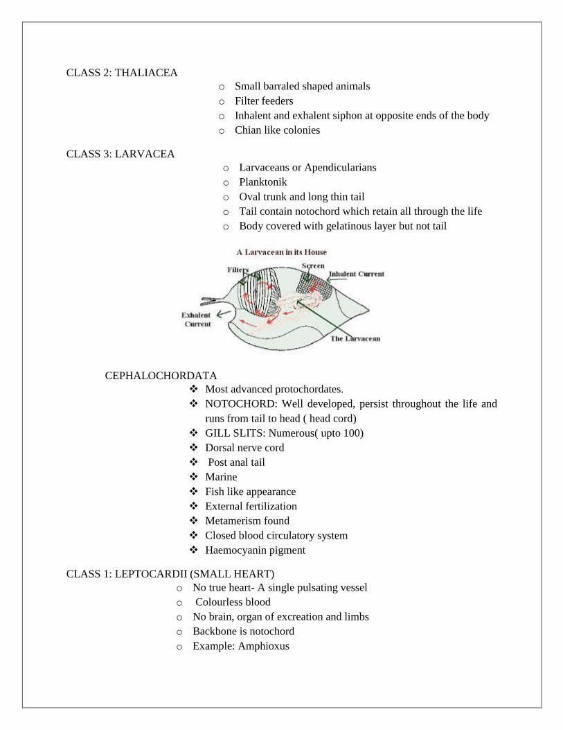
CLASS 3: LARVACEA
o Larvaceans or Apendicularians
o Planktonik
o Oval trunk and long thin tail
o Tail contain notochord which retain all through the life
o Body covered with gelatinous layer but not tail
CEPHALOCHORDATA
Most advanced protochordates.
NOTOCHORD: Well developed, persist throughout the life and
runs from tail to head ( head cord)
GILL SLITS: Numerous( upto 100)
Dorsal nerve cord
Post anal tail
Marine
Fish like appearance
External fertilization
Metamerism found
Closed blood circulatory system
Haemocyanin pigment
CLASS 1: LEPTOCARDII (SMALL HEART)
o No true heart- A single pulsating vessel
o Colourless blood
o No brain, organ of excreation and limbs
o Backbone is notochord
o Example: Amphioxus

HERDMANIA
GENERAL FEATURES AND IMPORTANT CHARACTERS:
PHYLUM-CHORDATA
SUB PHYLUM-UROCHORDATA
CLASS-ASCIDIACEA
ORDER-PLEUROGONA
Herdmania is a marine and sedentary animal. It is fixed to rocky substratum by a flat base.
When it is disturbed it suddenly contracts it‘s body and emmiots inner contents with force
through it‘s apparatures. Hence called Sea squirt.
HABIT AND HABITAT:
They are exclusively marine. The genus is recorded to go to depth of 9m-21.6m of the sea.
It is a macrophages animal which feed on microscopic animals and plants.
The tunic of herdmania provides shelter for many organisms.
A very common occurrence is the growth of a green alga on the tunic which sometimes hide
the whole animal. Mostly gastropods and anemones inhibit in the tunic.
EXTERNAL FEATURES:
It has a potato like shape. It is pink in colour. On the free side body shows two projections, the
branchial and atrial siphons.
The branchial siphon is short and shows a branchial mouth.
The atrial siphon is larger. It bears the atrial aperture. But the openings are bounded by four lips.
The external characters have a Test (Tunic).The body of this animal is covered and protected by
this test. It is a thick leathery covering of the body.
The test has: A clear matrix which is embedded
Cells of various shapes
Interlacing fibers
Calcerous spicules and
Branching vascular vessel
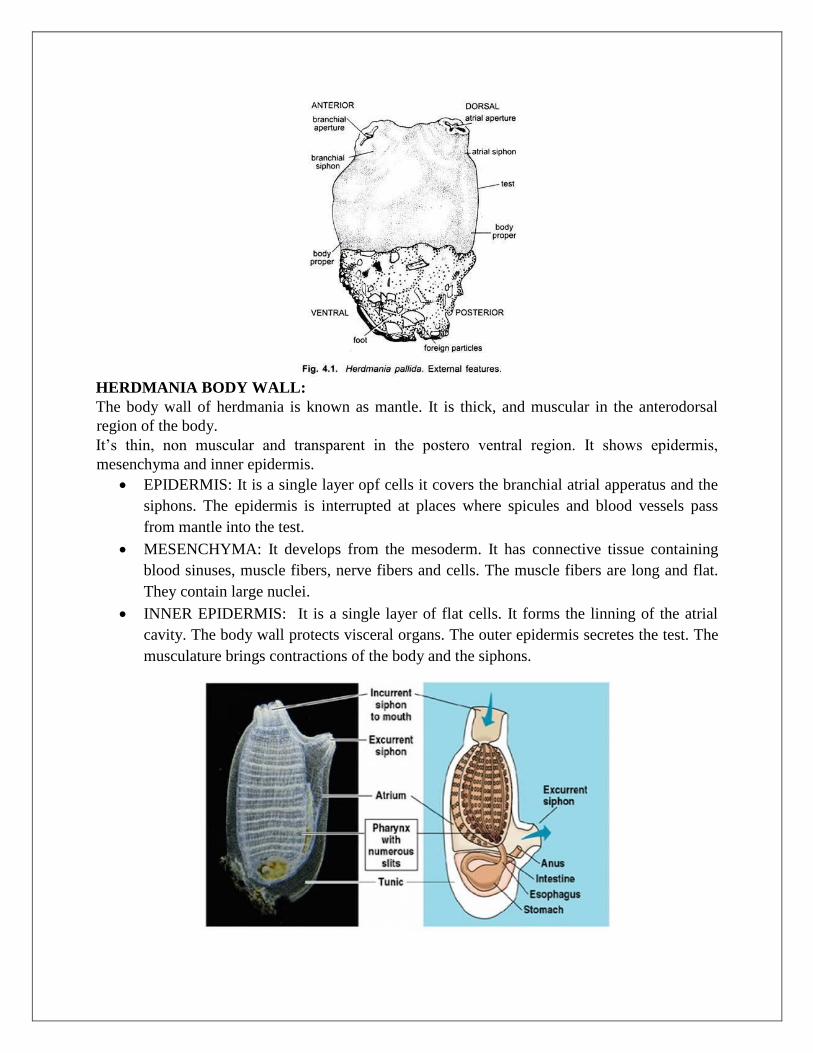
HERDMANIA BODY WALL:
The body wall of herdmania is known as mantle. It is thick, and muscular in the anterodorsal
region of the body.
It‘s thin, non muscular and transparent in the postero ventral region. It shows epidermis,
mesenchyma and inner epidermis.
EPIDERMIS: It is a single layer opf cells it covers the branchial atrial apperatus and the
siphons. The epidermis is interrupted at places where spicules and blood vessels pass
from mantle into the test.
MESENCHYMA: It develops from the mesoderm. It has connective tissue containing
blood sinuses, muscle fibers, nerve fibers and cells. The muscle fibers are long and flat.
They contain large nuclei.
INNER EPIDERMIS: It is a single layer of flat cells. It forms the linning of the atrial
cavity. The body wall protects visceral organs. The outer epidermis secretes the test. The
musculature brings contractions of the body and the siphons.
LOCOMOTION IN HERDMANIA:
Herdmania in adult stage is sessile. The visible movement is observed during the contraction of
the body for squirting out of water through the oral and atrial funnels. This movement is caused
by three sets of special muscles.
-Oral muscle group
- Atrial muscle group
- Trio oral muscle group
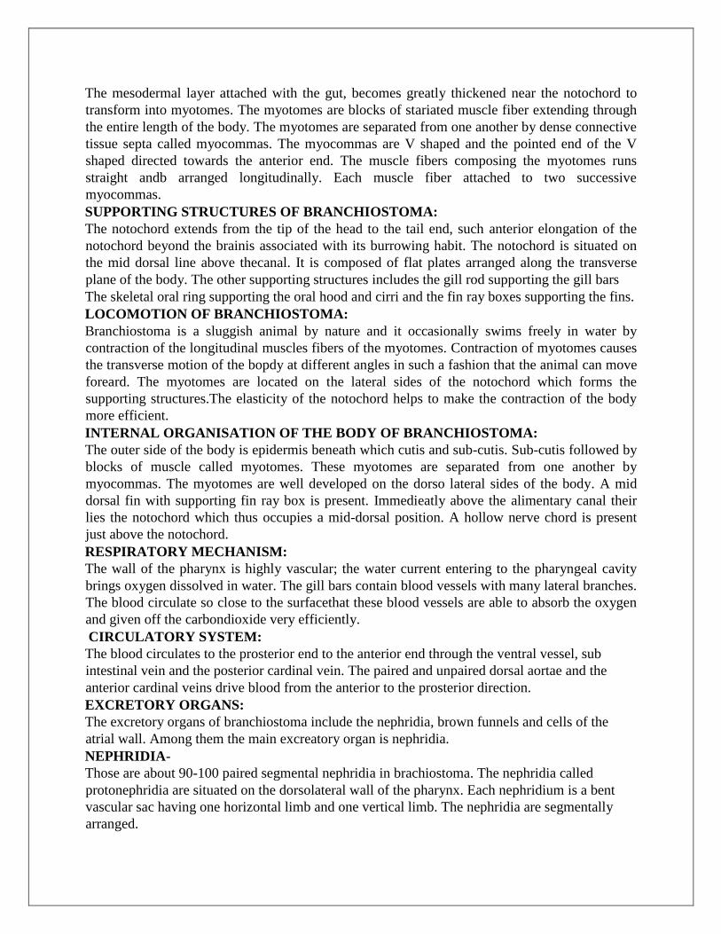
CIRCULATORY SYSTEM:
The circulatory system, is highly developed in Herdmania. Due to extensive vascularization of
the circulatory system is modified. The blood contains afew colourles ameboid and plenty of
pigmented cporpuscles. Six type of corpuscles are claimed to be present in Herdmania.
The heart is cylindrical and tube like structuire enclosed by tubular pericardium. The heart is
attached with one sid eopf the pericardium by a thin connective tissue bridge. It is situated
ventral to right gonad as an obliquely placed tube at about the middle of the endostyle.
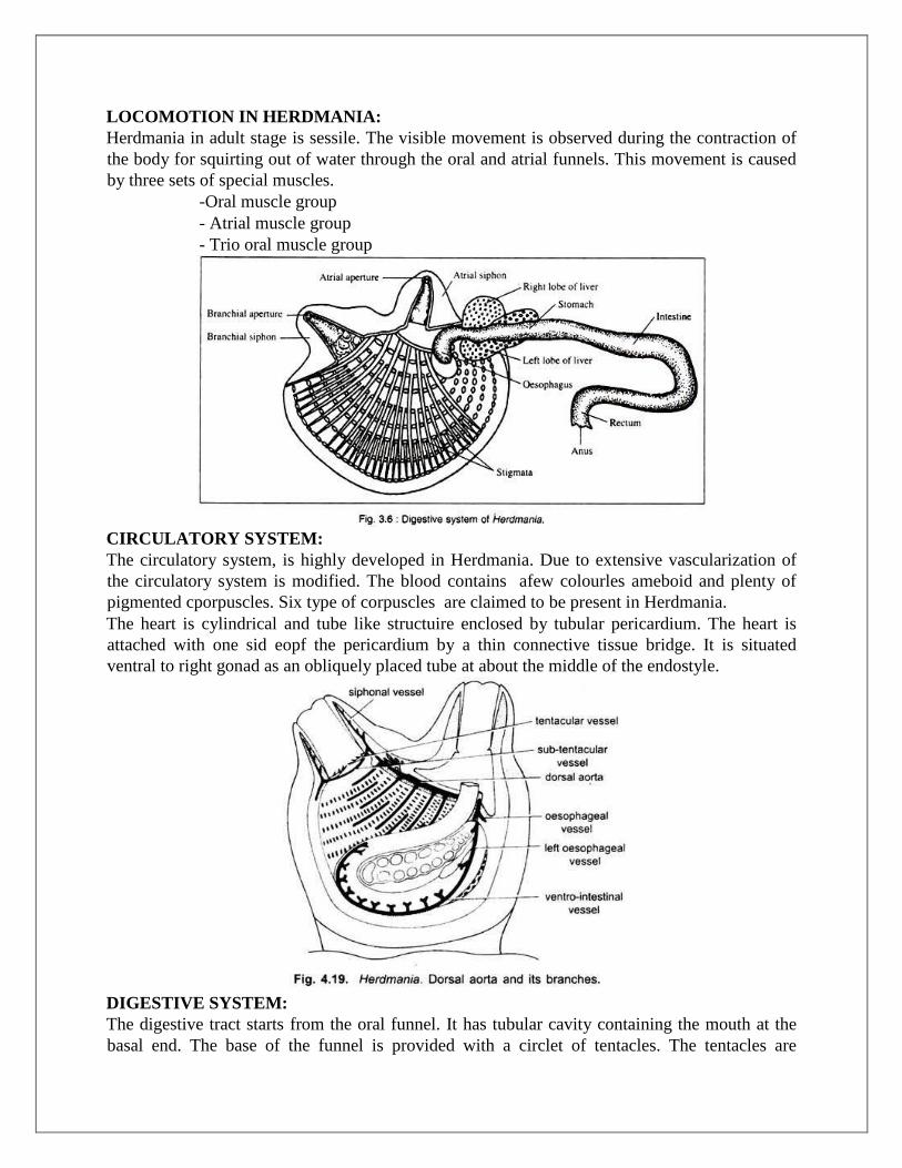
DIGESTIVE SYSTEM:
The digestive tract starts from the oral funnel. It has tubular cavity containing the mouth at the
basal end. The base of the funnel is provided with a circlet of tentacles. The tentacles are

extensively branched. Delicate structures and form a sort of strainer to prevent the entry of larger
particles into pharyngeal cavity.
Tentacles are richly supplied with nerves and thus suggesting as testing organs. Each tentacles
beared numerous paired laterel branches called tentaculets. Which also carry secondary and
tertiary branches.
The mouth leads into a spacious sac like pharynx or branchial sac, which occupies the major part
of the body.
The pharynx leads into a very short curved tube called oesophagus is guarded by two
semicircular fold. The oesophagus opens into the stomach. TYhen the stomach leads into the
intestine which forms a single loop.
The distal limb of the loop is called rectum which opens through the anus into the atrial funnel.
DIGESTIVE GLANDS:
Large digestive glands with two unequal lobes (liver) are present in Herdmania. This gland is
closely associated with the stomach. The liver is composed of an accumulation of a large number
of caeca embedded in the ground substance.
This is made up of connective tissues and blood sinuses. The product of the liver are poured into
the stomach by eleven independent ducts. Each duct is formed by union of many ductules
coming from caeca. The other digestive gland is the pyloric gland. It is an extensively branched
structure and composed of numerous tubules.
RESPIRATORY SYSTEM:
The respiratory system in Herdmania is more efficient than that of Ascidea. The pharynx is
highly vascular structure, and the inter-stigmatic and intra-stigmatic blood vessels help in
exchange of respiratory gases. The presence of internal folding increases the surface area of the
pharynx. Besides the pharynx the test also acts as an accessory respiratory organ. It is richly
supplied with blood vessels and vascular ampullae.
EXCRETORY SYSTEM:
The neural gland in Herdmanis is regarded as the excreatory organ. It is an oval shaped gland
situated dorsal to nerve ganglion.
NERVOUS SYSTEM:
The nervous system in adults is represented by an elongated solid nerve ganglion, called brain or
cerebral ganglion. It is situated ventral to the neural gland.
SENSE ORGAN:
o Several structures function as sensory receptors.

o Receptor cell- Many receptor cells (receive contact stimuli) are present throughout the
test except at the vascular areas.
o The epithelial cell- This clls are covering the vascular ampullae of the test are tactile
receptors.
o The pigmented spots or ocelli as photoreceptors.
o Oral tentacles are able to test water is drawn through the oral funnels.
REPRODUCTIVE SYSTEM:
Herdmania is hermaphrodite. The hermaphroditic gonads consist of two large lobulated bodies.
The right gonad is situated on the right side and just dorsal to the heart while the left gonad is
lodged within the loop of the intestine. Each gonad is formed of 10-25 lobes.
Self fertilization is rare, because the ovarian regions mature prior to the testicular region(
protogynous condition).
When thw gamets become mature, they are expelled out in the sea water. Fertilization is thus
external.
DEVELOPMENT:
o Fertilization is external
o Post gastrular development gives rise to free swimming tadpole larvae.
o Tadpole larvae has a tail with a vacuolated notochord, hollow nerve cord, trunk has a mid
dorsal continuation of the nerve cord in the form of brain.
o Undergoes Retrogressive Metamorphism, to become adult.
BRANCHIOSTOMA
KINGDOM- ANIMALIA
PHYLUM- CHORDATA
SUBPHYLUM= CEPHALOCHORDATA
CLASS- LEPTOCARDII
DISTINCTIVE FEATURES OF BRANCHIOSTOMA:
Branchiostoma is a transparent fish like animal, occouring in the shore, burrowing in rocks.
The body of branchiostoma is narrow, 2.5-5.8 cm long, laterally compressed and pointed at both
the ends.
The anterior two third of the body is triangular incross section.
Along the mid dorsal line a dorsal fix extends the whole body length. It is joined prosteriorly to a
some what broader caudal fin and around the tail.
A ventral fin is situated med ventrally
Three unpaired apparatus are present running from the caudal fin to a median opening, the
atriopore.
The muscle of branchiostoma is arranged in 62 V shaped segments or myotomes:
a) The mouth overreached by the median ventral oral hood, and fringed with tentacle like
cirri.
b) The atriopore in myotome 36 to expel water from the pharynx which enters through the
motuh.
c) The anus, ventral and slightly to the left behind the atriopore but at some distance from
the bprosterior end of the body.
The notochord of branchiostoma is a flexible, unsegmented rod pointed at both ends and runs
from one to other end of the body.
The pharynx supported by gill rods, which border the numerous bgill slits.
The intestine of branchiostoma is straight and without any loop.
The liver diverticulum is simple in branchiostoma.
About 90 pair sof segmentally arranged nephridia are present on the dorsolateral walls of the
pharynx.
The dorsal nerve cord is shorter than the notochord, above which it lies.
A definite brain is absent although anteriorly the central canal of the nerve cord widens to form
the so called cerebral vesicle.
It is almost cosmopolitian distribution and found on the snady shores covering the tidal area to
depth of many meters. It is an inhabitant of the shores of tropical and temperate seas.
HABIT AND HABITAT:
They live in both marine and esturine habitats.
It leads a dual life; It is a sedentary animal although it can swim actively in water. They swim
veritically in water like a fish. The fins don‘t play an important role in swimming.
It burrows in the sand, but remains always at a small depth.
It keeps the anterior end of the body projected along the sandy bed to maintain a constant water
current passing through the mouth and expel through the atriopore.
BODY WALL OF BRANCHIOSTOMA:
The epidermis consists of a single layer of cells. The epithelial cells are columnar type.
The epithelial layer contains cell with sensory hair. But gland and pigment cells are absent.
Beneath the epidermis there are two layer cutis and sub-cutis. The cutis is made up of fibers, and
the sub-cutis has a gelatinous matrix containing fibers. Beneath cutis and sub-cutis there is a
muscle layer.
The mesodermal layer attached with the gut, becomes greatly thickened near the notochord to
transform into myotomes. The myotomes are blocks of stariated muscle fiber extending through
the entire length of the body. The myotomes are separated from one another by dense connective
tissue septa called myocommas. The myocommas are V shaped and the pointed end of the V
shaped directed towards the anterior end. The muscle fibers composing the myotomes runs
straight andb arranged longitudinally. Each muscle fiber attached to two successive
myocommas.
SUPPORTING STRUCTURES OF BRANCHIOSTOMA:
The notochord extends from the tip of the head to the tail end, such anterior elongation of the
notochord beyond the brainis associated with its burrowing habit. The notochord is situated on
the mid dorsal line above thecanal. It is composed of flat plates arranged along the transverse
plane of the body. The other supporting structures includes the gill rod supporting the gill bars
The skeletal oral ring supporting the oral hood and cirri and the fin ray boxes supporting the fins.
LOCOMOTION OF BRANCHIOSTOMA:
Branchiostoma is a sluggish animal by nature and it occasionally swims freely in water by
contraction of the longitudinal muscles fibers of the myotomes. Contraction of myotomes causes
the transverse motion of the bopdy at different angles in such a fashion that the animal can move
foreard. The myotomes are located on the lateral sides of the notochord which forms the
supporting structures.The elasticity of the notochord helps to make the contraction of the body
more efficient.
INTERNAL ORGANISATION OF THE BODY OF BRANCHIOSTOMA:
The outer side of the body is epidermis beneath which cutis and sub-cutis. Sub-cutis followed by
blocks of muscle called myotomes. These myotomes are separated from one another by
myocommas. The myotomes are well developed on the dorso lateral sides of the body. A mid
dorsal fin with supporting fin ray box is present. Immedieatly above the alimentary canal their
lies the notochord which thus occupies a mid-dorsal position. A hollow nerve chord is present
just above the notochord.
RESPIRATORY MECHANISM:
The wall of the pharynx is highly vascular; the water current entering to the pharyngeal cavity
brings oxygen dissolved in water. The gill bars contain blood vessels with many lateral branches.
The blood circulate so close to the surfacethat these blood vessels are able to absorb the oxygen
and given off the carbondioxide very efficiently.
CIRCULATORY SYSTEM:
The blood circulates to the prosterior end to the anterior end through the ventral vessel, sub
intestinal vein and the posterior cardinal vein. The paired and unpaired dorsal aortae and the
anterior cardinal veins drive blood from the anterior to the prosterior direction.
EXCRETORY ORGANS:
The excretory organs of branchiostoma include the nephridia, brown funnels and cells of the
atrial wall. Among them the main excreatory organ is nephridia.
NEPHRIDIA-
Those are about 90-100 paired segmental nephridia in brachiostoma. The nephridia called
protonephridia are situated on the dorsolateral wall of the pharynx. Each nephridium is a bent
vascular sac having one horizontal limb and one vertical limb. The nephridia are segmentally
arranged.
Each sac corresponds to each primary gill barand opens by a nephridiophore to the atrium. A
large number of elongated flame cells or solenocytes opens into the vesicle. The call body gives
off a flagellum thrugh the hollow stalk which helps in eliminating the waste products. Excreation
takes place through the wall of the solenocytes by diffusion and the product pass down into the
cavity of the vesicle through the tubular part.
DIFFERENT RECEPTOR ORGANS:
Pigment spot-
-Referred as cerebral eye
-Photsensetive in nature
-Present in anterior wall of brain.
Eye spots-
-They are photoreceptors.
Kdliker‘s pit-
- It is a chemoreceptor.
Sensory papillae-
-Chemoreceptor and tactile receptor.
Infundibular organs-
- Photoreceptor.
Epidermal sensory cells-
- Present on dorsal surface of the body.
NERVOUS SYSTEM OF BRANCHIOSTOMA:
The nervous system of Branchiostoma consists of a hollow dorsal nerve cord situated just above
the notochord. The anterior part of the nerve tube is slightly dilated to form the brain and the
posterior part remains as the spinal cord. The nerve tube gives paired nerves in each segment of
the body. The paired nerves are actually the dorsal and ventral nerve roots which remain separate
(Fig. 3.20). The ventral nerve root lies opposite to the myotome and the dorsal nerve root passes
out between the myotomes.
REPRODUCTIVE SYSTEM OF BRANCHIOSTOMA:
The sexes are separate in Branchiostoma. The gonads are simple pouch-like segmental organs
situated in the ventro-lateral sides of the pharyngeal region between the 10th and 30th segments.
The gonads are proliferated from the mesodermal cells and the gametes are developed from the
walls of the gonads. It is claimed that each gonad is developed from a single cell. The gonoducts
are absent and the gametes are discharged into the atrium by dehiscence. From the atrium the
gametes escape to the exterior through the atriopore along with the water current. Fertilization
and development occur in sea-water.
Unit II
Reptilia, Aves and Mammalia
Content:
1. RETILIA: Origin and Classification
2. AVES: Origin and classification, Flight adaptation and Migration
3. MAMMALIA: Origin and classification and Dentition
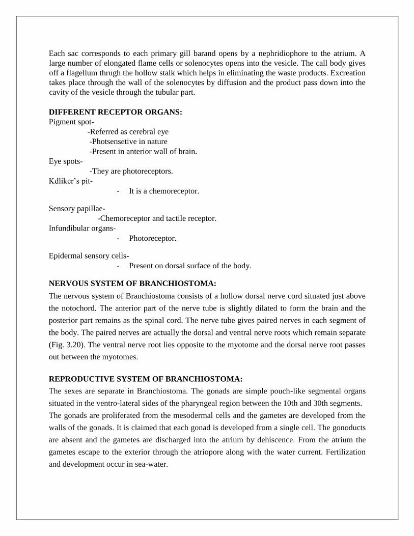
Reptilia
Reptiles are cold-blooded vertebrates, breathe by lungs throughout their existence, and having
the body covered with scales or scutes. A basioccipital bone is present in the skull which
articulates with the vertebral column by a single condyle.
Origin: The first reptiles arose from ancient labyrinthodont amphibians during upper
Carboniferous period, about 270 million years ago and have adapted to terrestrial life. The oldest
known fossils of reptiles are about 350-million-year-old. They were found in deposits from the
early carboniferous period, which occurred between 360 and 286 million years ago.
The earliest reptiles were small, four legged vertebrates that resembles lizards and
had teeth adapted for eating insects present in abundant. They diversified rapidly, and by the
Permian period (286 million to 245 million years ago) they had become the dominant lad
vertebrates.
The Mesozoic era (245 million to 65 million years ago) is often called Age of
Reptiles because nearly all the large vertebrates on earth were reptiles during that time. The
dinosaurs appeared and flourished.
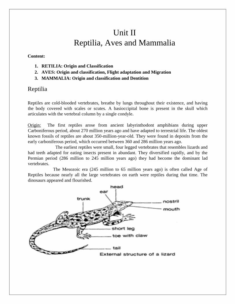
Classification:
Reptilia (Class):
General characteristics:
1. They are inhabitants of terrestrial and aquatic (both marine and freshwaters) environments.
2. Their skin is dry, cornified and usually covered by epidermal scales or scutes. There are a few
integumentary scent glands secreting pheromones during breeding seasons.
3. Single external nasal opening is present on the snout. Ear drums are slightly depressed.
4. Two pairs of pentadactyle limbs are present. The limbs end in clawed digits.
5. The cloacal opening is either transverse or longitudinal.
6. A post-anal tail is present.
7. The heart is composed of two auricles and a partially divided ventricle. There are right and left
systemic arches.
8. The kidney is metanephric type.
9. Mullerian duct persists as oviduct in female and Wolffian duct is retained as vas deference in
male. Males possess copulatory organs.
10. Twelve pairs of cranial nerves are present.
11. Vomero-nasal organ (Organ of Jacobson) is well-developed.
12. Single occipital condyle in the skull is present for the attachment with atlas.
13. Mandible consists usually six pieces of bones.
14. Vertebrae are procoelous. Sternum is greatly developed with ribs.
15. Cleidoic eggs are large. The calcareous shell serves for protection against desiccation and
external injury. The shell is porous for gaseous exchange.
16. Fertilisation is internal.
17. Embryos are provided with extra-embryonic membranes, like amnion, chorion and allantois.
18. These are ectothermic or heliothermic (helios = sun) animals.
Subclass 1. Anapsida [without an arch] [an = without; apsis = an arch]:
1. This subclass is characterised by the skull devoid of fossae in the temporal region.
2. The roof of the skull is solid.
This subclass includes three orders: Cotylosauria, Mesosauria and Chelonia.
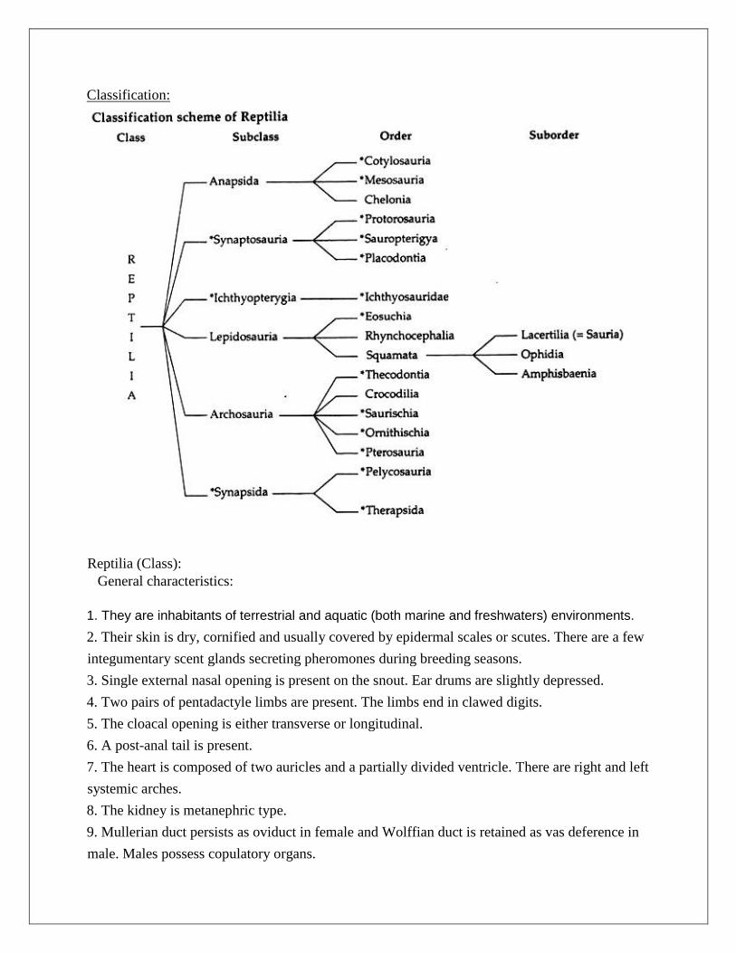
Order 1. Cotylosauria [ kotye = cup shaped hollow; sauros = lizard]: All the members of the
order are extinct and possibly form the ‗stem reptiles‘ from which other reptiles have probably
been evolved. They lived between Upper Carboniferous to Upper Triassic periods.
The characteristic features are:
1. The complete roofing of the skull.
2. Flattened plate-like pelvis.
The order includes two suborders: Captorhinomorpha and Diadectomorpha.
Examples: The earliest known fossil cotylosaur is Romeriscus, collected from the lower
Pennsylvania. The well-known Cotylosaurs are Romeriscus, Diadectes etc.

Order 2. Mesosauria:
(i) They occur in late Carboniferous or early Permian strata,
(ii) They were aquatic in habit and lived in freshwater lakes.
(iii) The body was slender and measured not more than a metre in length,
(iv) The skull was devoid of temporal fenestrae.
(v) The hind legs were much powerful than the front legs. Both the limbs were paddle-shaped.
(vi) The tail was long and laterally compressed, used for swimming.
Example: Mesosaurus.
Order 3. Chelonia (Testudinata) [ Chelone = a tortoise; L. testudines; genitive of
Testudo = a tortoise]:
Chelonians are assumed to be direct descendants of primitive cotylosaurs. In the evolutionary
history of reptiles, the development of a box-like exoskeleton appears extremely peculiar. This
bizarre armour remained unaltered since Triassic period.
The characteristic features of the chelonians are:
1. The body is more or less elliptical and dorsoventrally flattened.
2. Body is encased by a convex dorsal shield, called carapace and a flat ventral plate designated
as plastron which remains, joined at sides.
3. Neck, limbs and tail are retractile into the carapace in most non-marine forms.
4. Limbs are weak, pentadactyle and modified into paddle in marine forms. In aquatic (ponds,
lakes and rivers) forms the limbs are webbed. The chelonians walk slowly on land but swim
fastly by the paddles in marine forms.
5. Body shell is externally protected either with polygonal scutes or leathery scales.
6. The tail is short.
7. The cloacal opening is oval or longitudinal.
8. Male possesses a single grooved copula- tory organ.
9. They are found in terrestrial, aquatic (ponds, lakes, marshes, reservoirs, estuaries and rivers)
and marine environments.
10. Carnivorous forms consume a large number of invertebrates, such as earthworms, crabs,
insects, insect larvae, snails and bivalve molluscs. Larger forms eat fishes, frogs and birds.
Herbivorous forms choose aquatic plants as food.
Subclass 2. Synaptosauria [ synaptas = joined]:
(i) Late Palaeozoic and Mesozoic aquatic reptiles are included under the subclass.
(ii) They possessed a single temporal (parapsid) fossa high in the skull. Synaptosauria includes
three orders: Protorosauria, Sauropterygia and placodontia.
Order 1. Protorosauria :
(i) Existed during lower Permian.

(ii) The reptiles were lizard-like in appearance, not exceeding 30 cm in length.
(iii) They were terrestrial and were very agile.
(iv) The vertebrae were amphicoelous.
Example:
Araeoscelis.
Order 2. Sauropterygia[Gk. sauros = lizard; pterygos = fin]:
(i) Existed between Permian and Triassic strata.
(ii) They were aquatic in habit. They had single temporal vacuity in skull which were bounded
below by a postorbital.
(iii) The squamosal and coracoid were single.
(iv) The feet were webbed in some forms.
Examples:
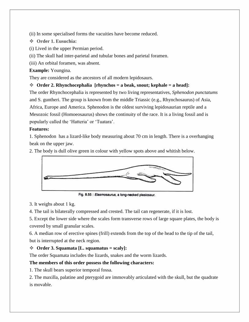
Cyamodus, Plesiosaurus ,Elasmosaurus.
Order 3. Placodontia (Triassic):
More or less related to the plesiosaurs and had grinding teeth on jaws and palate. Example:
Placodus , Henodus .
Subclass 3. Ichthyopterygia (ichthys = fish; pterygos = fin)
(i) The members are all extinct.
(ii) The roof of the skull was provided with an upper opening behind the eye and was bound
below by postfrontal and supra-temporal.
Order 1. Ichthyosauridae :
(i) They occurred between middle Triassic and upper Cretaceous.
(ii) They were marine and fish-like in appearance. Some were of considerable length measuring
about 10-15 metres.
(iii) Their skull had a single lateral vacuity.
(iv) The head was large and produced into an elongated snout.
(v) The tail was long and limbs were in the form of paddles.
(vi) The vertebrae were amphicoelous and cervical ribs were double-headed.
(vii) The sternum was lacking but abdominal ribs were present.
Examples:
Ichthyosaurus, Ophthalmosaurus
Subclass 4. Lepidosauria [ lepis = scale; sauros = lizard]:
(i) Representatives of this subclass have two temporal vacuities in the skull.
(ii) In some specialised forms the vacuities have become reduced.
Order 1. Eusuchia:
(i) Lived in the upper Permian period.
(ii) The skull had inter-parietal and tubular bones and parietal foramen.
(iii) An orbital foramen, was absent.
Example: Youngina.
They are considered as the ancestors of all modern lepidosaurs.
Order 2. Rhynchocephalia [rhynchos = a beak, snout; kephale = a head]:
The order Rhynchocephalia is represented by two living representatives, Sphenodon punctatums
and S. guntheri. The group is known from the middle Triassic (e.g., Rhynchosaurus) of Asia,
Africa, Europe and America. Sphenodon is the oldest surviving lepidosaurian reptile and a
Mesozoic fossil (Homoeosaurus) shows the continuity of the race. It is a living fossil and is
popularly called the ‗Hatteria‘ or ‗Tuatara‘.
Features:
1. Sphenodon has a lizard-like body measuring about 70 cm in length. There is a overhanging
beak on the upper jaw.
2. The body is dull olive green in colour with yellow spots above and whitish below.
3. It weighs about 1 kg.
4. The tail is bilaterally compressed and crested. The tail can regenerate, if it is lost.
5. Except the lower side where the scales form transverse rows of large square plates, the body is
covered by small granular scales.
6. A median row of erective spines (frill) extends from the top of the head to the tip of the tail,
but is interrupted at the neck region.
Order 3. Squamata [L. squamatus = scaly]:
The order Squamata includes the lizards, snakes and the worm lizards.
The members of this order possess the following characters:
1. The skull bears superior temporal fossa.
2. The maxilla, palatine and pterygoid are immovably articulated with the skull, but the quadrate
is movable.

3. Lower jaw is composed of several pieces of bones.
4. The teeth are either acrodont or pleurodont and are borne usually on the maxillae, premaxillae
and palatines.
5. The vertebrae are of procoelous type.
6. Chevron bones are present.
Subclass 5. Archosauria [Ruling reptiles]:
(i) Skull was of diapsid type and lacked inter-parietal and parietal foramina.
(ii) Palatal teeth were lost in some forms. Some forms were toothless.
(iii) The lower jaw was with vacuities between dentary and angular.
(iv) In some forms bipedality (two footed locomotion) was more marked and the girdles were
modified accordingly.
Order 1. Thecodontia [Socketed teeth]:
They were present during Triassic period. Members belonging to the suborder Pseudosuchia
under this order were small in size.
(i) They were carnivorous in nature.
(ii) The teeth were sharp and were lodged in sockets along the jaw edges.
(iii) The hind legs were long and indicated the dawn of bipedality. The members of the other
suborder Phytosauria were aquatic.
(iv) The skull was elongated.
Order 2. Crocodilia or Loricata [L. crocodilus = a crocodile; L. loricatus = clad in mail.]
1. They are carnivorous and freshwater reptiles. They swim by the undulation of their powerful
tail.
2. The limbs are not powerful as the tail and are used in carrying the body on land.
3. The forelimbs are shorter than the hind and have five digits in the forelimbs and four digits in
the hind limbs. The digits of the forelimbs are webbed.
4. The body is elongated and the skin bears epidermal scales which are supported by dermal
bones or scutes. The scales are supported by dermal plates osteoderms.
5. The tail is laterally compressed.
Order 3. Saurischia [ Sauros = lizard; ischion = hip or pelvis]:
(i) The members of this order were characterised by having triradiate pelvic girdle.
(ii) Teeth were borne by the premaxillae.
Examples: Tyrannosaurus, Yaleosaurus, Gorgosaurus.
Order 4. Ornithischia [ Ornithos = bird; ischion = pelvis]:
(i) The order Ornithischia included the ‗bird-like‘ dinosaurs.

(ii) The members of this order possessed a ‗predentary‘ bone in the mandible which supports the
beak.
(iii) They were all herbivorous.
Examples: Iguanodon, Trachodon, Stegosaurus, Nodosaurus.
Order 5. Pterosauria [ Pteron = wing; sauros = lizard]:
(i) This order includes the fossil flying reptiles.
(ii) The bones were pneumatic like that of birds.
(iii) The forelimbs became converted into wings.
(iv) The fourth finger was greatly enlarged (‗wing finger‘) to support the membranous wing.
(v) The fifth finger was lacking and the other fingers were small.
(vi) The sternum was keeled.
Examples:Pteranodon, Pterodactyl us, Rhamphorhynchus .
Subclass 6. Synapsida :
i) They are considered as mammal-like reptiles.
ii) The skull was provided with a single and lateral temporal vacuity lying below the post-orbital
and squamosal.
iii) The supraoccipital was broad.
iv) The lower jaw was flat and the teeth were of heterodont type.
v) The shoulder girdles were with coracoids and pre-coracoids.
Order 1. Pelycosauria [Pelykos = an axe; sauros = lizard]:
i) They existed between upper Carboniferous and lower Permian.
ii) The skull was with temporal vacuity.
iii) In some forms, like Dimetrodon, the neural spines were much elongated and formed a
specialized structure, called a ―sail‖ along the back that consist of an extensive flap of skin
supported internally a row of elongated neural spines.
iv) Some forms were carnivorous but majority were vegetarian.
v) The limbs were short.
Examples:
Dimetrodon , Edaphosaurus.
Order 2. Therapsida [ Therion = mammal; apsida = loop]:
i) They existed between middle Permian and lower Triassic.
ii) They constituted a very important group from the stand-point of evolution. According to
Romer, they bridged the entire evolutionary gap between a primitive reptile and a mammal.
iii) In advanced therapsid reptiles the occipital condyle was double.
iv) The temporal opening in the skull was big.
v) The quadrate and quadratojugal were greatly reduced.
vi) In some forms, a secondary palate was present.
vii) The teeth were distinctly differentiated into the incisors, canines and molars.
viii) The group is regarded as the‘ precursors to mammals.
Examples:
Cynognathus, Lycosaurus, Lystrosaurus.
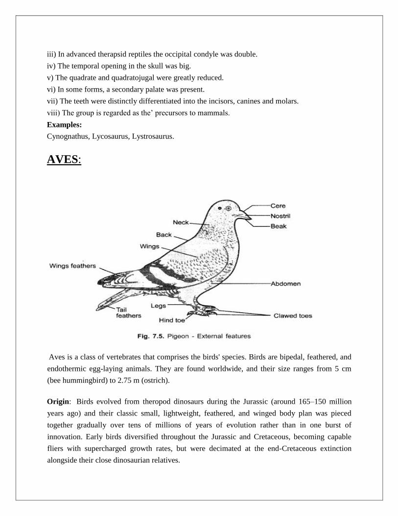
AVES:
Aves is a class of vertebrates that comprises the birds' species. Birds are bipedal, feathered, and
endothermic egg-laying animals. They are found worldwide, and their size ranges from 5 cm
(bee hummingbird) to 2.75 m (ostrich).
Origin: Birds evolved from theropod dinosaurs during the Jurassic (around 165–150 million
years ago) and their classic small, lightweight, feathered, and winged body plan was pieced
together gradually over tens of millions of years of evolution rather than in one burst of
innovation. Early birds diversified throughout the Jurassic and Cretaceous, becoming capable
fliers with supercharged growth rates, but were decimated at the end-Cretaceous extinction
alongside their close dinosaurian relatives.

Characteristic Features of Class Aves:
1. The body of the most bird is spindle-shaped, and it is covered with different types of unique
structures, feathers for the most part.
2. Forelimbs are modified as wings for flight, and the posterior hind limbs are adapted for
walking, perching, wading, or swimming.
3. Only legs are covered with scales, usually with four toes on each foot.
4. The birds' bones are fully ossified and pneumatic or hollow inside, which lower the overall
weight of the body.
5. Uropygial or oil gland is present in the tail region.
6. The mouth is wide, and jaws are covered by horny sheaths that form strong beaks.
7. Teeth are not present in the mouth. In this case, food is swallowed unmasticated.
8. The alimentary canal leads to the cloaca. Alimentary canal often contains additional chambers
like crop and gizzard. In this case, the crop stores and softens food while the gizzard is
muscular to crush and stir up the softened food.
Classification:
Subclass-1: Archiornithes
(Archios = ancient + ornithos = bird)
1. They were the most primitive and extinct fossil birds of the Jurassic period.
2. The jaws did not bear teeth.
3. Archiornithes had a long feathered tail.
4. They had a reptile-like and elongated body.
5. Forelimb had three clawed digits,
6. They had a small brain and eyes.
7. They had non-pneumatic bones and were capable less specialized for flight.
8. They had a well-developed beak.
Order-1: Archaeopterygiformes
1. The tail was long with the distal tapering end.
2. The forelimbs had remiges with three clawed digits.
3. Head was large with proportionately large eyes.
4. They had strong jaws with enameled crowned teeth.
5. Cerebral hemispheres were smooth, long, and narrow with small cerebellum.
Examples: Archaeopteryx lithographica, Archaeornis siemensi.
Subclass-2: Neornithes
All the modern birds belong to the subclass Neornithes. This subclass contains about 10,000
known living birds' species throughout the world. They have a well-developed sternum, which is
usually keeled or carinate.
1. They do not bear long tail with no teeth on both jaws. In this case, teeth are replaced by horny
rhamphotheca over the bill, but extinct forms had teeth.
2. The forelimbs become modified to wings.
Superorder-1: Odontognathae
[Gk. Odontos = teeth + gnathos = jaw]
1. They were the New World toothed birds. They were found in the Upper Cretaceous period of
the Mesozoic Era (100.5 - 66 MYA).
2. They had short tails with a plowshare-shaped pygostyle.
3. They had a well-developed carina for flight muscle.
4. Both jaws had teeth.
5. They were flightless birds but were specialized for swimming.
6. They had intramandibular articulation.
7. Sternum did not bear keel and wings had vestigial humorous only.
Superorder-2: Palaeognathae
1. Superorder Palaeognathae contains 47 species, of which, three cassowaries (Casuarius), five
kiwis (Apteryx), 1 emu (Dromaius), two ostrich (Struthio) and two species of rheas (Rhea).
2. Most of the birds are flightless. In this case, some anatomical features are associated with
flightlessness. These include reduced keel on the sternum, reduced wing bones, etc.
3. All species of tinamous can fly, but they are very terrestrial.
4. During reproduction, the male provides parental care.
5. The males have an erectile penis and the females with the clitoris.
Superorder-3: Impennae
They are flightless aquatic birds. Their forelimbs are modified into paddles, which are used for
both swimming and flight.
Superorder-4: Neognathae
The word "Nognathe" is derived from Greek 'neo' meaning new and 'gnathos' meaning jaw. This
superorder includes all modern living birds with nearly 10,000 species.
1. Neognathae have well-developed wings which are fully adapted for flight.
2. The beak does not contain any teeth.
3. The forelimbs have fused metacarpals with an elongated third finger.
4. Pygostyle is absent, and the tail is composed of 5-6 vertebrae.
5. The sternum is well-developed with the keel.
6. Rectrices are organized in a semicircular manner, which helps in flight.
7. They have a neognathous type skull where the palatines are protruded posteriorly and come in
contact with the base of the cranium.
Flight adaptation in birds:
Effective flight is possible only when two requirements are met, light weight of the body and the
supply of energy. Hence, the organs that are modified for effective flight are:
a. Birds have short body with compact bones. This reduces the weight of the body and
makes it light.
b. There are different kinds of feathers present in birds: outline feather helps to decrease the
drag of the body by making it streamlines; primary feathers helps to provide shape to the
wings and make the flight possible; tail feather are used as rudder for balance of the
body.
c. Bones in birds are either hollow or fused to reduce the weight of the body. The fusion of
caudal bone to form pygostyle and hollow bones of skull are good example of it.
d. Absence of teeth and present of beak is also one of the important adaptations for flight in
birds.
e. Forelimbs are modified into wings and hence aerodynamics in shape. They are also close
to the centre of gravity.
f. Different muscles present in birds helps in the movement of legs, wings, neck and tail.
Mainly wings are made of strong muscle so as to provide energy for flying which are
called pectoral and breast muscles.
g. Birds generally eat foods that are small and light but which high source of energy. They
have gizzard that helps to grind the eaten food and absorb it. Beside this, some birds also
have crop that aids in softening and absorbing of eaten food.
h. The most developed system in birds is the respiratory system. It is highly developed
because of the energy required for flying mechanism. Though the lungs are relatively
small but there is a great interconnection with the inner organs which generates the
required energy. The air that is taken in passes to the air sacs on the posterior side then
reaches the lungs. Similarly, when on second breathing; the air that is already present in
the posterior side rushes to the anterior side thus leaving the space for next round of
breathing, and then the inhaled air enters the posterior side. This way exchange of gases
takes place. Generally most of the birds have only one way of air current but there are
also some that possess bi- directional air current. Birds lac alveoli, instead many small
sacs called parabronchi are present that are connected to the capillaries.
i. The circulation of blood uses four chambered heart with two atria and two ventricular.
j. Blood with high blood pressure and high amount of sugar is present in the avian heart.
k. Eyes are well developed having binocular vision in birds whereas the olfactory organs
have reduced function. It is because good sight is necessary for safe flight.
l. Ovaries and testis are present but the size is reduced, in some only one ovary is
functional.
m. In most of the female birds liver is pushed towards right side in order to balance the
weight of the body.
Migration: It is change of habitat periodically recurring and alternating in direction, which
tend to secure optimum environmental conditions at all times. Bird migration is a more or
less regular, extensive movement between their breeding regions and their wintering regions.
Causes of Migration:
Most species of birds migrate more or less on schedule and follow the routes in a regular
fashion. The actual causative factors determining the course and direction of migration are
not clearly known.
The following factors may be related to the problems of migration:
i. Instinct and Gonadal changes.
ii. Scarcity of food and day length.
iii. Photoperiodism: The increase of day length (Photoperiodism) induces bird‘s
migration. The day length affects pituitary and pineal glands and also caused
growth of gonads which secret sex hormones that are the stimulus for
migration. In India, Siberian crane, geese, swan those come from central Asia,
Himalayas, begin to return from March and onwards with the increase of day
length.
iv. Seasonal variation.
v. Light: The experiments of Rowan with Juncos (summer visitor to Canada)
have established that light plays an important role in the development of
gonads, which has indirect role on migration. If the gonads undergo
regression, the urge for migration is not felt. So the seasonal changes in
illumination appear to be a crucial factor for determining migration.
Types of migration:
(i) Latitudinal migration:
The latitudinal migration usually means the movement from north to south, and vice versa. Most
birds live in the land masses of the northern temperate and subarctic zones where they get
facilities for nesting and feeding during summer. They move towards south during winter.

(ii) Longitudinal migration: The longitudinal migration occurs when the birds migrate from east to west and vice- versa. Starlings (Sturnus vulgaris), a resident of east Europe and west Asia migrate towards the Atlantic coast. California gulls, a resident and breed in Utah, migrate westward to winter in the Pacific coast.
(iii) Altitudinal migration: The altitudinal migration occurs in mountainous regions. Many birds inhabiting the mountain peaks migrate to low lands during winter. Golden plover (Pluvialis) starts from Arctic tundra and goes up to the plains of Argentina covering a distance of 11 250 km .
(iv) Partial migration: All the members of a group of birds do not take part in migration. Only several members of a group take part in migration. Blue Jays of Canada and northern part of United States travel southwards to blend with the sedentary populations of the Southern States of U.S.A. Coots and spoon bills (Platalea) of our country may be example of partial migration.
(v) Total migration: When all the members of a species take part in the migration, it is called total migration.
(vi) Vagrant or irregular migration: When some of the birds disperse to a short or long distance for safety and food, it is called vagrant or irregular migration. Herons may be the example of vagrant or irregular migration. Other examples are black stork (Ciconia nigra), Glossy ibis (Plegadis falcinellus), spotted eagle (Aquila clanga), and bee eater (Merops apiaster).
(vii) Daily migration: Some birds make daily journey from their nests by the influence of environmental factors such as temperature, light, and humidity also. Examples are crows, herons and starlings.
(viii) Seasonal migration: Some birds migrates at different seasons of the year for food or
breeding, called seasonal migration, e.g., cuckoos, swifts, swallows
etc. They migrate from the south to the north during summer.
These birds are called summer visitors. Again there are some birds
like snow bunting, red wing, shore lark, grey plover etc. which
migrate from north to south during winter

Mammalia:
Mammals are vertebrate animals constituting the class Mammalia, and characterized by the
presence of mammary glands which in females produce milk for feeding their young, a
neocortex, fur or hair, and three middle ear bones.
Origin: Mammals originated from reptiles but the fossils show that the reptiles that gave rise
to mammals were Synapsids that diverged from the main reptilian stock almost at the base of
phylogeny during Permian period. Hence, mammalian relationship with the extant reptiles is
remote.
General Characters of Mammals:
1. Body of mammals is covered by epidermal hair.
2. Integumentary glands are — sweat (sudoriferous), sebaceous (oil), scent (odoriferous) glands.
3. Mammary glands are present to supply milk for the nourishment of suckling young.
4. External fleshy pinna is present in mammals.
5. Eyes with upper and lower eyelids and often with eyelashes.
6. Nictitating membrane is translucent and hairless; it is vestigial in higher mammals.
7. A muscular diaphragm is present in between the thoracic and abdominal cavities.
8. Endo-thermal homoeotherm animals.
9. RBCs are non-nucleated, biconcave and usually circular in form.
10. The four-chambered heart is highly powerful.
11. Only left aortic arch is present in the arterial system.
Subclass — Prototheria (Greek: protos = first, therion = beast):
General Characters:
1. The females lay eggs.
2. The testes are abdominal.
3. The cloaca receives the openings of urinary bladder, vas deferens and ureters.
4. Ribs possess single head.
5. The mammary glands lack teats.
6. External pinna absent.
7. In childhood, teeth are present but adults lack teeth.
Examples: Ornithorhynchus, Tachyglossus, Zaglossus, Echidna.
Subclass — Theria (Greek: therion = beast):
General Characters:
1. Female members of this subclass do not lay eggs but give birth to young ones.
2. Mammary glands are provided with nipples or teats.
3. Pinna or external ear is present.

Classification:
4. The ureters open directly into the urinary bladder.
5. At the end of the digestive tract an anus is present.
6. Teeth are present throughout the life period.
7. Testes are situated in the scrotum.
8. Ribs possess two heads for articulation with vertebrae.
This subclass includes three infraclasses, of which Pantotheria is extinct.
Examples: Macropus (Kangaroo), Didelphis (Opossum), Thylacinus (Tasmanian wolf – the
mystery marsupials), Talpa (Mole), Tupaia (Tree- shrew).

Dentition: The arrangement of teeth in the upper and lower jaws, mainly on the premaxilla,
maxilla and dentary bones, is called dentition.
Origin and Structure of Teeth in Mammals:
Teeth have evolved from denticles which are released from armour near the margins of
the mouth as ossification in the integument. A typical mammalian tooth can be distinguished
mainly into two regions — crown and root. The crown is the exposed part of the tooth and
situated above the root and in the old age it is generally subject to wear.
The root is the hidden part in the gum which is
anchored in the socket or alveolus of the jaw bone.
The tooth encloses a pulp cavity that contains blood
vessels, nerves, and connective tissue. The junction
of crown and root is called neck.
Types of Dentition in Mammals
A. Classification According to the Shape and Size of the Teeth:
Homodont:
Homodont or isodont type of teeth is a condition where the teeth are all alike in their shape and
size, e.g., the toothed whales (Odontoceti). Pinnipedians show a tendency towards homodont
condition. Fishes amphibians reptiles and in the extinct toothed birds, the homodont or isodont
condition is observed.
Heterodont:
Heterodont condition is the usual feature in mammals, i.e. the teeth are distinguished according
to their shape, size and function. The function is also different at different parts of the tooth row.
Except mammals heterodont condition is found in Port Jackson Shark (Heterodontus), in several
reptiles, especially among mammal-like reptiles.
B. According to the Mode of Attachment of Teeth:
Thecodont: Thecodont type dentition is the rule among mammals. In this condition, the
teeth are lodged in bony sockets or alveoli of of the jaw bone and capillaries and nerves
enter the pulp cavity through the open tips of the hollow roots. Except mammals, the
codont type of teeth is found in crocodiles and in some fishes (Haddock, Garpike and
Barracuda).
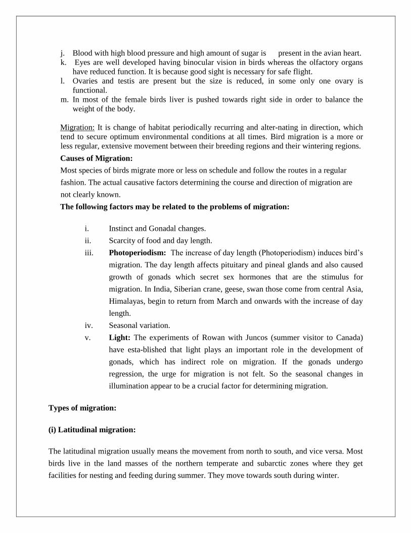
Acrodont: The teeth are fused to the surface of the underlying jawbone. They have no
roots and are attached to the edge of the jawbone by fibrous membrane e.g., fishes,
amphibians and some reptiles.
Pleurodont: Here the teeth are attached to the inner-side of the jawbone. The tooth
touches the bone only with the outer surface of its root. Iguanidae (Iguana), Xenosauridae
(Xenosaurus, Mexico).
According to the Succession or Replacement of Teeth:
The teeth can be divided into three categories:
(i) Monophyodont
(ii) Diphyodont and
(iii) Polyphyodont.
Among mammals the first two categories are found.
(i)Monophyodont: In some mammals, only one set of teeth develops in their life time and this
condition is called Monophyodont, e.g., Marsupials retain all their milk teeth except last
premolars, the toothed whales (Odontoceti), some rodents (e.g., squirrels), certain insectivores
(e.g., moles). Among platypus, sirenians and toothless whales develop only one set of teeth
(monophyodont dentition). These teeth may not erupt (some whales) or, if they develop are
usually shed shortly afterward.
(ii) Diphyodont: In most mammals two sets of teeth are found. The first temporary set of teeth,
called deciduous teeth, milk teeth or lacteal teeth, are lost or replaced by a second set of teeth,
termed permanent teeth. In bats and guinea-pigs the milk teeth are lost even before birth. In milk
teeth the molars are absent.
(iii) Polyphyodont: In this condition, the teeth are replaced continuously throughout life, e.g.,
lower vertebrates replace their teeth, generation following generation (Dogfish, snakes).
References:
Reptiles - http://www.notesonzoology.com/reptilia/reptilia-characters-and-classification-
zoology/3887
http://www.biologydiscussion.com/zoology/reptiles/reptiles-origin-history-and-
classification/41033
Aves - https://biologyeducare.com/aves/
http://www.notesonzoology.com/birds/birds-general-characters-and-its-classification-
zoology/3800
https://www.assignmenthelp.net/flight-adaptation-in-aves
http://www.biologydiscussion.com/zoology/birds/bird-migration-definition-types-causes-
and-guiding-mechanisms/41286
Mammalia - http://www.notesonzoology.com/phylum-chordata/mammals-general-
characters-and-its-classification-zoology/3920
http://www.iaszoology.com/origin-of-mammals/
http://www.biologydiscussion.com/zoology/mammals/dentition-in-mammals-definition-
origin-types-and-unusual-teeth-in-mammals/41558
Comparitive anatomy of vertebrates: Integument system,
respiratory , digestive system
The study of structure, of the function of structure, & of the range of variation in structure &
function among vertebrates:
Kingdom: Animal
Phylum: Chordata
Subphylum: Vertebrata
Vertebrate characteristics: 1 - notochord (at least in the embryo)
2 - pharynx with pouches or slits in wall (at least in the embryo)
3 - dorsal, hollow nervous system
4 - vertebral column
THE GENERAL BODY PLAN OF VERTEBRATES
• Head: Cephalization and development of sensory organs, and
protective covering
• Trunk Somites: Mesenchymal masses on both sides of the notochord
• Trunk Coelom: Body cavity b/t the gut and the body wall
• Trunk Visceral Organs: Heart-Pericardium, Lung-Pleura, and
Abdomen-Peritoneum.• Tail: Somites, notochord,innervation, dorsal and ventral aorta
INTEGUMENTARY SYSTEM
The skin and associated structures make up the integumentary system. The skin protects land-
dwelling organisms from desiccation and from loss of heat. Skin is a mammal's largest organ. Its
protects the body against physical, chemical, and biological attacks, it helps to regulate body
temperature, it is used to communicate to other individuals, and a skin derivative provides
nourishment for the young. Like the integuments of other vertebrates, mammalian skin is
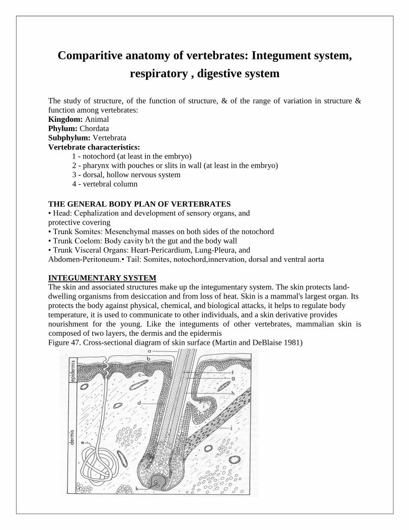
composed of two layers, the dermis and the epidermis
Figure 47. Cross-sectional diagram of skin surface (Martin and DeBlaise 1981)

Epidermis.--The epidermis consists of several layers, representing successive stages of
development. The oldest part of the skin is the outer layer of tough, protective, cells. The cells,
which are dead, are continually worn off at the surface and replaced from below. As the cells age
and mature, they eventually lose their nuclei and most of the cell contents are converted to
keratin.
Dermis.--The dermis lies below the epidermis. It is a thick layer of connective tissue with
associated muscles, nerves, and blood vessels. The connective tissue consists largely of collagen.
Collagen may be up to 6 per cent of body weight in humans, and is the most abundant protein in
the body, being present in skin, bones, tendons, cartilage, and ligaments.
Hair
General.--Hair is a uniquely mammalian feature. The developing epidermis invaginates
into the
dermis to form a follicle. At the deepest point of the follicle, the dermis pushes back and
forms a
small structure called the papilla. The papilla is well supplied with blood vessels.
Epidermal cells
on top of the papilla multiply and are pushed towards the surface by those growing
beneath
them, keratinizing (as epidermal cells also do to form the outer layer of skin) and forming
the hair.
Glands.--Sebaceous glands open into each follicle. They secrete oily substances (sebum)
that continually lubricate and condition skin and hair. Cells inside these glands gradually
fill with grease and then break away, becoming part of the secretion themselves. Sebum
makes beavers waterproof and prevents undue drying of the pelage of terrestrial
mammals. Glands that secrete cellular debris as well as molecular products are termed
apocrine glands. These glands empty into or near a hair follicle.
Digestive System
Digestive tract - ‗tube‘ from mouth to vent or anus
that functions in:
ingestion
digestion
absorption
egestion
Major subdivisions include the oral cavity, pharynx,
esophagus, stomach, small & large intestines, and
cloaca. Accessory organs include the tongue, teeth,
oral glands, pancreas, liver & gall bladder.

Differences in the anatomy of vertebrate digestive tracts is often correlated with the nature &
abundance of food:
readily absorbed (e.g., hummingbirds) vs. requiring extensive enzymatic activity (e.g.,
carnivores)
constant food supply (e.g., herbivores) vs. scattered supply (e.g., carnivores)
The embryonic digestive tract of vertebrates consists of 3 regions:
1 - midgut - contains yolk or attached yolk sac
2 - foregut - oral cavity, pharynx, esophagus, stomach, & small intestine
3 - hindgut - large intestine & cloaca
Mouth & oral cavity: The oral cavity begins at the mouth & ends at the pharynx. Fish have a
very short oral cavity, while tetrapods typically have longer oral cavities. The mammalian mouth
is specialized to serve as a suckling and masticatory organ (with muscular cheeks).
Palate = roof of the oral cavity. Primary palate - internal nares lead into the oral cavity anteriorly
secondary palate - nasal passages are located above the secondary palate and open at the end of
the oral cavity
Teeth are derivations of dermal armor.
Placoid scales - show gradual transition to
teeth at the edge of the jaw
Composition of teeth - primarily dentin
surrounded by enamel
Vary among vertebrates in number,
distribution in the oral cavity, degree of
permanence, mode of attachment, & shape
TEETH:
1 - have tended toward reduced numbers

& distribution
2 - most vertebrates (through reptiles) have succession of teeth
3 - most vertebrates (except mammals) replace teeth in ‗waves‘ (back to front; every
other tooth)
4 - mammals generally develop 2 sets of teeth: milk (deciduous) teeth & permanent
teeth
Tongue:
Gnathostome fish & primitive amphibians - tongue is a
simple crescent-shaped elevation in the floor of the oral
cavity caused by the underlying hyoid skeleton & is called
the primary tongue
Most amphibians - primary tongue (or hypobranchial eminence)
+ glandular field (or tuberculum impar) ('stuffed' with hypobranchial musculature)
Reptiles & mammals - primary tongue + glandular field (or tuberculum impar) + lateral
lingual swellings (more hypobranchial muscle)
Birds - lateral lingual swellings are suppressed & intrinsic muscle is usually lacking
Oral glands - secrete a variety of substances including:
saliva
o Lubrication and binding: the mucus in saliva is extremely effective in binding
masticated food into a slippery bolus that (usually) slides easily through the
esophagus without inflicting damage to the mucosa. Saliva also coats the oral
cavity and esophagus, and food basically never directly touches the epithelial cells
of those tissues.
o
o Initiates starch digestion: in most species, amylase is present in saliva and begins
to digest dietary starch into maltose. Amylase does not occur in the saliva of
carnivores.
Pharynx - part of digestive tract exhibiting pharyngeal pouches (at least in the embryo) that may
give rise to slits
Fish - pharynx is respiratory organ
Tetrapods:
o pharynx is the part of the foregut preceeding the esophagus & includes:
glottis (slit leading into the larynx)
openings of auditory (eustachian) tubes

opening into esophagus
Esophagus:
a distensible muscular tube connecting the pharynx & the stomach
may have diverticulum called the crop (see diagram of pigeon below)
Stomach = muscular chamber(s) at end of esophagus
serves as storage & macerating site for ingested solids & secretes digestive enzymes
Vertebrate stomachs:
o Cyclostomes - weakly developed; similar to esophagus
o Fish, amphibians, & reptiles - increasing specialization (more differentiated from
the esophagus)
o Birds - proventriculus (glandular stomach) and ventriculus (muscular stomach, or
gizzard)
Mammals - well-developed stomach; ruminants have multichambered stomachs:
Reticulo-rumen (reticulum and rumen)
Reticulum and rumen are often discussed together since each compartment is separated
by a low partition. Eighty percent of the capacity of the stomach is related to the reticulo-
rumen. The contents of the reticulum and rumen intermix freely. The rumen is the main
fermentation vat where billions of microorganisms attack and break down the relatively
indigestible feed components of the ruminant's diet.
Omasum After fermentation in the reticulum and rumen, food passes to the omasum. The omasum
acts as a filter pump to sort liquid and fine food particles. Coarse fibre particles are not
allowed to enter the omasum. Also, the omasum may be the site for absorption of water,
minerals and nitrogen.
Abomasum The abomasum is the true stomach and the only site on the digestive tract that produces
gastric juices (HCl and the enzymes, pepsin and rennin). Ingesta only remains here for 1
to 2 hours.
Accessory organs - Liver, gall bladder, & pancreas
Liver & gall bladder
o liver produces bile which is stored in the gall bladder (cyclostomes, most birds,
and some mammals, including cervids, have no gall bladder)
o bile aids in digestion by emulsifying fats (breaking fats down into tiny particles
that permits more efficient digestion by enzymes)
Pancreas - secretes pancreatic juice (bicarbonate solution to neutralize acids coming from
the stomach plus enzymes to help digest carbohydrates, fats, and proteins) into the
intestine.
Ceca - blind diverticula that serve to increase the surface area of the vertebrate digestive tract
Fishes - pyloric & duodenal ceca are common in teleosts; these are primary areas for
digestion and absorption (not fermentation chambers)
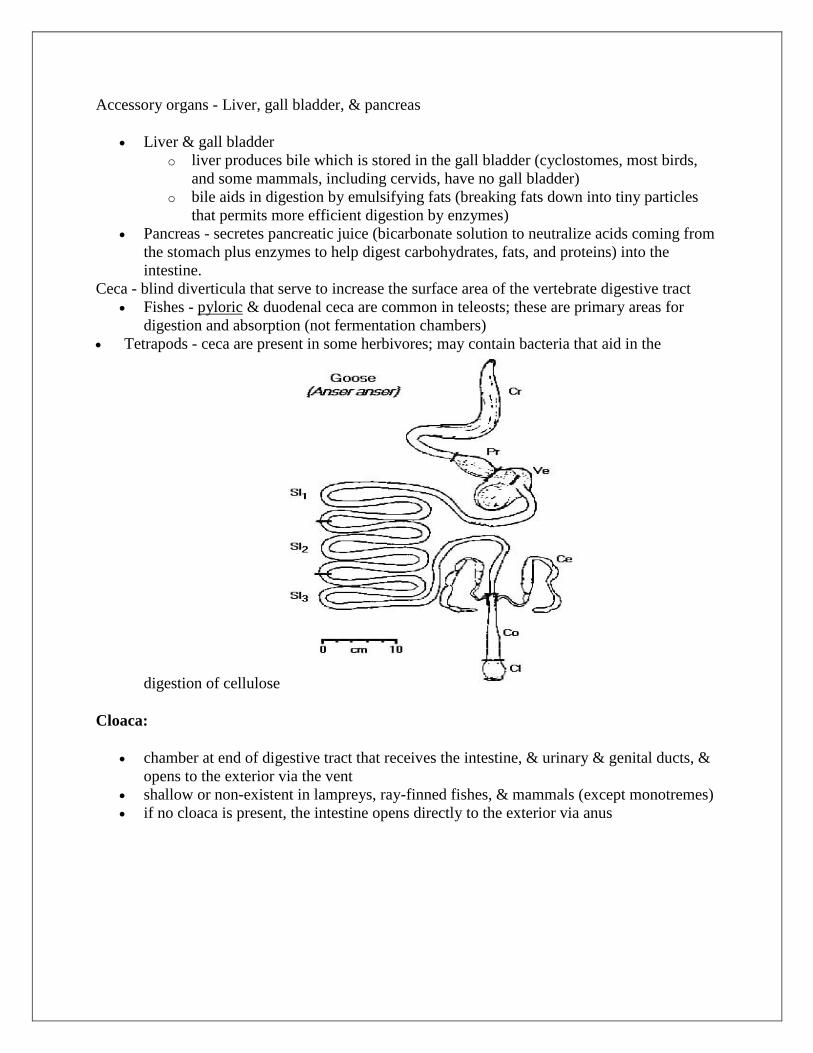
Tetrapods - ceca are present in some herbivores; may contain bacteria that aid in the
digestion of cellulose
Cloaca:
chamber at end of digestive tract that receives the intestine, & urinary & genital ducts, &
opens to the exterior via the vent
shallow or non-existent in lampreys, ray-finned fishes, & mammals (except monotremes)
if no cloaca is present, the intestine opens directly to the exterior via anus
Respiratory System
Respiration is the process of obtaining oxygen from the external environment & eliminating
CO2.
External respiration - oxygen and carbon dioxide exchanged between the external
environment & the body cells
Internal respiration - cells use oxygen for ATP production (& produce carbon dioxide in
the process)
Adaptations for external respiration:
1 - Primary organs in adult vertebrates are external & internal gills, swim bladders or lungs,
skin, & the buccopharyngeal mucosa
2 - Less common respiratory devices include filamentous outgrowths of the posterior trunk &
thigh (African hairy frog), lining of the cloaca, & lining of esophagus
Respiratory organs:
Cutaneous respiration
o respiration through the skin can take place in air, water, or both
o most important among amphibians (especially the family Plethodontidae)
Gills (see Respiration in Fishes)
o Cartilaginous fishes:
5 ‗naked‘ gill slits
Anterior & posterior walls of the 1st 4 gill chambers have a gill surface
(demibranch). Posterior wall of last (5th) chamber has no demibranch.
Interbranchial septum lies between 2 demibranchs of a gill arch
Gill rakers protrude from gill cartilage & ‗guard‘ entrance into gill
chamber
2 demibranchs + septum & associated cartilage, blood vessels, muscles, &
nerves = holobranch.
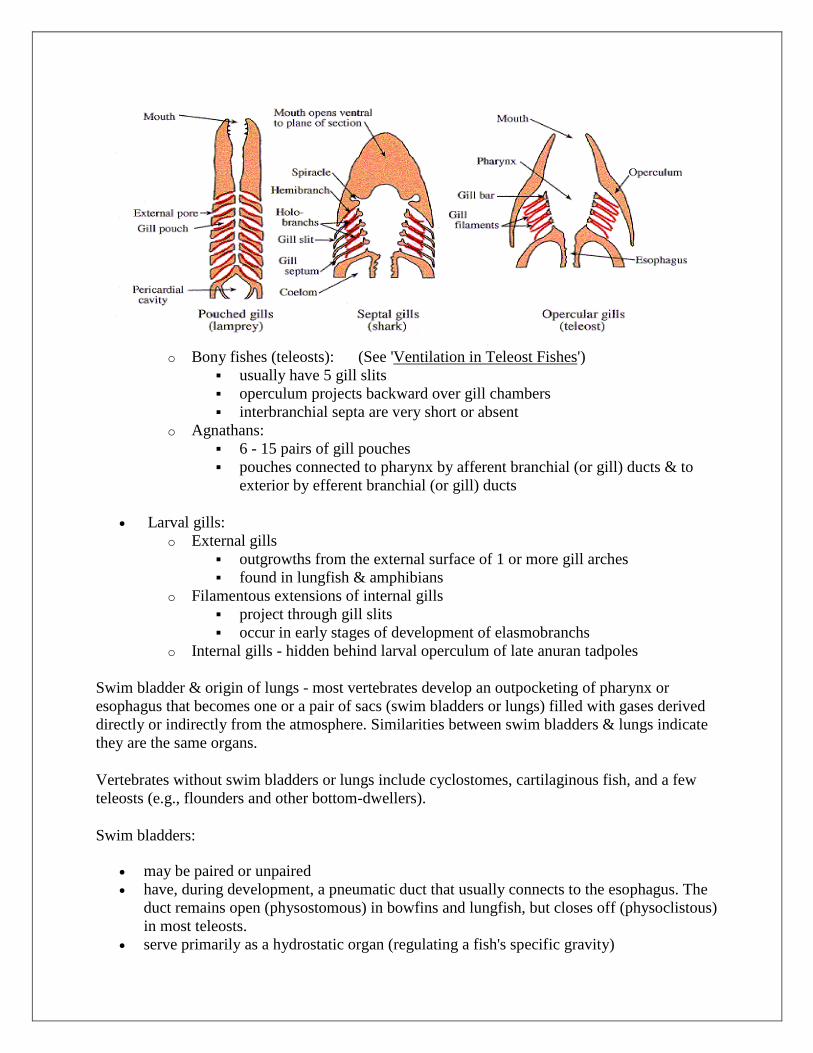
o Bony fishes (teleosts): (See 'Ventilation in Teleost Fishes')
usually have 5 gill slits
operculum projects backward over gill chambers
interbranchial septa are very short or absent
o Agnathans:
6 - 15 pairs of gill pouches
pouches connected to pharynx by afferent branchial (or gill) ducts & to
exterior by efferent branchial (or gill) ducts
Larval gills:
o External gills
outgrowths from the external surface of 1 or more gill arches
found in lungfish & amphibians
o Filamentous extensions of internal gills
project through gill slits
occur in early stages of development of elasmobranchs
o Internal gills - hidden behind larval operculum of late anuran tadpoles
Swim bladder & origin of lungs - most vertebrates develop an outpocketing of pharynx or
esophagus that becomes one or a pair of sacs (swim bladders or lungs) filled with gases derived
directly or indirectly from the atmosphere. Similarities between swim bladders & lungs indicate
they are the same organs.
Vertebrates without swim bladders or lungs include cyclostomes, cartilaginous fish, and a few
teleosts (e.g., flounders and other bottom-dwellers).
Swim bladders:
may be paired or unpaired
have, during development, a pneumatic duct that usually connects to the esophagus. The
duct remains open (physostomous) in bowfins and lungfish, but closes off (physoclistous)
in most teleosts.
serve primarily as a hydrostatic organ (regulating a fish's specific gravity)
gain gas by way of a 'red body' (or red gland); gas is resorbed via the oval body on
posterior part of bladder.
Lungs & associated structures
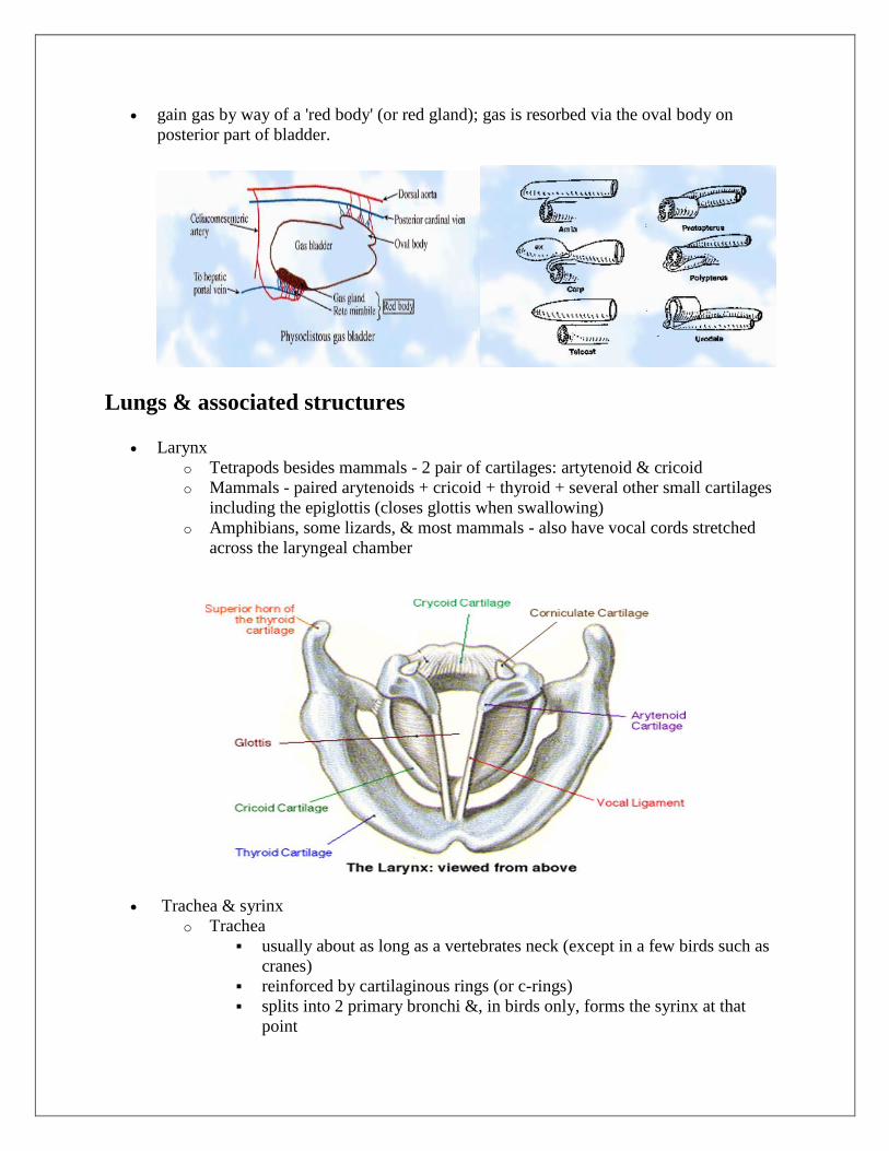
Larynx
o Tetrapods besides mammals - 2 pair of cartilages: artytenoid & cricoid
o Mammals - paired arytenoids + cricoid + thyroid + several other small cartilages
including the epiglottis (closes glottis when swallowing)
o Amphibians, some lizards, & most mammals - also have vocal cords stretched
across the laryngeal chamber
Trachea & syrinx
o Trachea
usually about as long as a vertebrates neck (except in a few birds such as
cranes)
reinforced by cartilaginous rings (or c-rings)
splits into 2 primary bronchi &, in birds only, forms the syrinx at that
point

Lungs
o Amphibian lungs
2 simple sacs
internal lining may be smooth or have simple sacculations or pockets
air exchanged via positive-pressure ventilation
Reptilian lungs
simple sacs in Sphenodon & snakes
Lizards, crocodilians, & turtles - lining is septate, with lots of chambers &
subchambers
air exchanged via positive-pressure ventilation
Avian lungs - modified from those of reptiles:
air sacs (diverticula of lungs) extensively distributed throughout most of
the body
arrangement of air ducts in lungs ----> no passageway is a dead-end
air flow through lungs (parabronchi) is unidirectional
o Mammalian lungs:
multichambered & usually divided into lobes
air flow is bidirectional:

Trachea <---> primary bronchi <---> secondary bronchi <---> tertiary bronchi <---> bronchioles
<---> alveoli

VERTEBRATES
CONTENTS
Characteristics of vertebrates
• Body structure
• Heart
• Branchial arches
• Notochord
• Renal structure
• Urogenital system
CHARACTERISTICS OF VERTEBRATES
• Bilateral symmetry
• Presence of notochord
• Pharynx with slits
• Dorsal tubular nervous system
• Well-developed vertebrate
• Post-anal tail
BODY STRUCTURE
• Head, trunk and tail
• Head: Cephalization and development of sensory organs, and protective covering.
• Trunk somites: Mesenchymal masses on both sides of the notochord
• Trunk coelom: body cavity between the gut and the body wall
• Trunk visceral organs: Heart-pericardium, Lung- pleura, and Abdomen-Peritoneum
• Tail: Somites, notochord, innervation, dorsal and ventral aorta
HEART
The heart is a highly modified tube that functions as a pump to drive blood circulation to the body in all vertebrates and, in animals with lungs, also to the pulmonary system. Proper heart functions are not only essential for the life of vertebrates‘ animals, but also for the development of vertebrate embryos.
COMPARATIVE ANATOMY OF HEART
BRANCHIAL ARCHES
• In tetrapod's pharynx, the slit becomes the auditory tube and the cavity of the
middle ear.
• The aortic arches are housed by the pharyngeal arches.
• Most vertebrates have 6 pairs of arches
• One vestigial pair of arches
• Each arch has four structural components: skeletal element, cartilage,
mesodermal- mesenchymal component, nerve and a vessel.
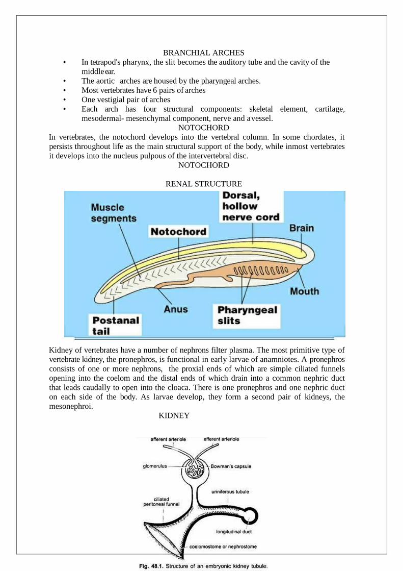
NOTOCHORD
In vertebrates, the notochord develops into the vertebral column. In some chordates, it
persists throughout life as the main structural support of the body, while inmost vertebrates
it develops into the nucleus pulpous of the intervertebral disc.
NOTOCHORD
RENAL STRUCTURE
Kidney of vertebrates have a number of nephrons filter plasma. The most primitive type of
vertebrate kidney, the pronephros, is functional in early larvae of anamniotes. A pronephros
consists of one or more nephrons, the proxial ends of which are simple ciliated funnels
opening into the coelom and the distal ends of which drain into a common nephric duct
that leads caudally to open into the cloaca. There is one pronephros and one nephric duct
on each side of the body. As larvae develop, they form a second pair of kidneys, the
mesonephroi.
KIDNEY
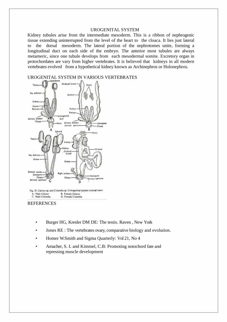
UROGENITAL SYSTEM
Kidney tubules arise from the intermediate mesoderm. This is a ribbon of nephrogenic
tissue extending uninterrupted from the level of the heart to the cloaca. It lies just lateral
to the dorsal mesoderm. The lateral portion of the nephrotomes unite, forming a
longitudinal duct on each side of the embryo. The anterior most tubules are always
metameric, since one tubule develops from each mesodermal somite. Excretory organ in
protochordates are vary from higher vertebrates. It is believed that kidneys in all modern
vertebrates evolved from a hypothetical kidney known as Archinephros or Holonephros.
UROGENITAL SYSTEM IN VARIOUS VERTEBRATES
REFERENCES
• Burger HG, Kresler DM DE: The testis. Raven , New York
• Jones RE : The vertebrates ovary, comparative biology and evolution.
• Homer W. Smith and Sigma Quarterly: Vol 21, No 4
• Amacher, S. L and Kimmel, C.B: Promoting notochord fate and
repressing muscle development
The CNS consists of the brain and spinal cord.
The brain is the most complex organ in the body and uses 20 percent of the total oxygen we
breathe in.
The brain consists of an estimated 100 billion neurons, with each connected to thousands more.
The brain can be divided into four main lobes: temporal, parietal, occipital and frontal.
CONTENTS
1. Anatomy of brain
2. Anatomy of eye
3. Anatomy of ear
4. Autonomic nervous system in mammals
5. Conclusion
6. References
ANATOMY OF BRAIN
INTRODUCTION
It is one of the largest organs in the body, and coordinates most body activities. It is the center for all
thought, memory, judgment, and emotions. Each part of the brain is responsible for controlling different
body functions, such as temperature regulation and breathing. The brain is contained in skull and weights
1300-1400g.It is made up of about 1000 billion neurons 7 each neurons is surrounded by about 10 glial
cells (neuroglia). Neurons cannot multiply and many neurons are last everyday in life but glial cells can
multiply throughout the life. Brain is also covered by ‗Meninges‘ like spinal cord, outer duramater,
middle arachnoidmater and inner piameter.
CENTRAL NERVOUS SYSTEM
The CNS consists of the brain and spinal cord. The brain is an important organ that controls thought,
memory, emotion, touch, motor skills, vision, breathing, temperature, hunger, and every process that
regulates our body.
FACTS OF CENTRAL NERVOUS SYSTEM

FIG- Different parts of the brain.
DIFFERENT PARTS OF BRAIN
The brain can be divided into the cerebrum, brainstem, and cerebellum:
Cerebrum. The cerebrum (front of brain) is composed of the right and left hemispheres,
which are joined by the corpus callosum. Functions of the cerebrum include: initiation of movement, coordination of movement, temperature, touch, vision, hearing, judgment, reasoning, problem solving, emotions, and learning.
Brainstem. The brainstem (middle of brain) includes the midbrain, the pons, and the medulla. Functions of this area include: movement of the eyes and mouth, relaying sensory messages (such as hot, pain, and loud).
Cerebellum. The cerebellum (back of brain) is located at the back of the head. Its function is to coordinate voluntary muscle movements and to maintain posture, balance, and equilibrium.
Pons. A deep part of the brain, located in the brainstem, the pons contains many of the control areas for eye and face movements.
Medulla. The lowest part of the brainstem, the medulla is the most vital part of the entire brain and contains important control centers for the heart.
Spinal cord. A large bundle of nerve fibers located in the back that extends from the base of the brain to the lower back, the spinal cord carries messages to and from the brain and the rest of the body.
Frontal lobe. The largest section of the brain located in the front of the head, the frontal lobe is involved in personality characteristics and movement. Recognition of smell usually involves parts of the frontal lobe.
Parietal lobe. The middle part of the brain, the parietal lobe helps a person to identify objects and understand spatial relationships (where one's body is compared to objects around the person). The parietal lobe is also involved in interpreting pain and touch in the body.
Occipital lobe. The occipital lobe is the back part of the brain that is involved with vision. Temporal lobe. The sides of the brain, these temporal lobes are involved in short-term
memory, speech, musical rhythm, and some degree of smell recognition.
ANATOMY OF EYE
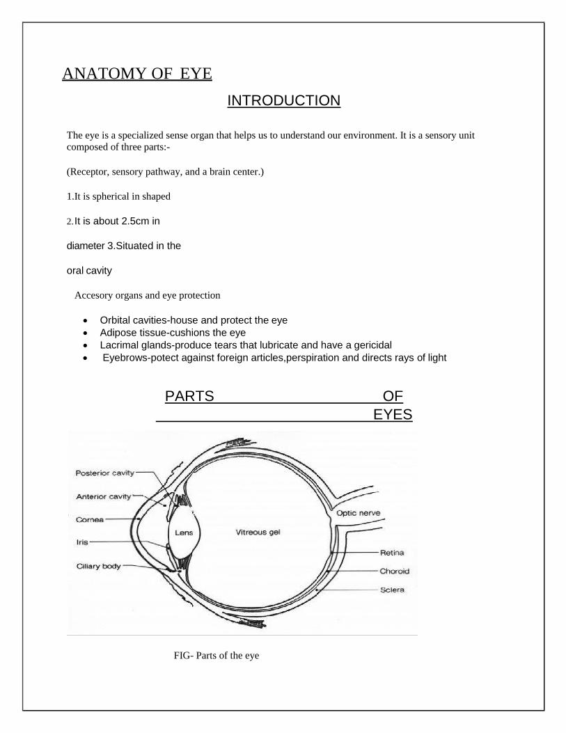
INTRODUCTION
The eye is a specialized sense organ that helps us to understand our environment. It is a sensory unit
composed of three parts:-
(Receptor, sensory pathway, and a brain center.)
1.It is spherical in shaped
2. It is about 2.5cm in
diameter 3.Situated in the
oral cavity
Accesory organs and eye protection
Orbital cavities-house and protect the eye
Adipose tissue-cushions the eye
Lacrimal glands-produce tears that lubricate and have a gericidal
Eyebrows-potect against foreign articles,perspiration and directs rays of light
PARTS OF
EYES
FIG- Parts of the eye

Sclera- The sclera is outermost layer of the eyeball. It is the white (and opaque) part of the eyeball. Muscles responsible for moving the eyeball are attached to the eyeball at the sclera
Cornea- At the front of the eyeball, the sclera becomes the cornea. The cornea is the transparent dome-shaped part of the eyeball. Light rays from the outside world first pass through the cornea before reaching the lens. Together with the lens, cornea is responsible for focusing light on retina
Choroid- The choroid is the middle layer of the eyeball located between the sclera and the retina. It provides nutrients and oxygen to the outer surface of the retina.
Anterior Chamber- The space between the cornea and the lens is known as the anterior chamber. It is filled with fluid called aqueous humor. The anterior chamber is also known as anterior cavity.
Aqueous humor- The Aqueous humor is a transparent watery fluid that circulates in the anterior chamber. It provides oxygen and nutrients to the inner eye and exerts fluid pressure that helps maintain the shape of the eye. The aqueous humor is produced by the ciliary body.
Posterior Chamber- The posterior chamber is a larger area than the anterior chamber. It is located opposite to the anterior chamber at the back of the lens. It is filled with a fluid called vitreous humor.
Vitreous humor- The vitreous humor is a transparent jelly-like fluid that fills the posterior chamber. It exerts fluid pressure that keeps the retina layers pressed together to maintain the shape of the eye and to maintain sharp focus of images on the retina.
Iris- The choroid continues at the front of the eyeball to form the Iris. The iris is a flat, thin, ring- shaped structure sticking in to the anterior chamber. This is the part that identifies a person’s eye colour. The iris contains circular muscles which go around the pupil and radial muscles that radiate toward the pupil. When the circular muscles contract they make the pupil smaller, when the radial muscles contract, they makes the pupil wider.
Ciliary muscles- The cilliary muscles are located inside the ciliary body. These are the muscles that continuously change the shape of the lens for near and distant vision.
Ciliary Body- The choroid continues at the front of the eyeball to form the ciliary body. It produces the aqueous humour. The ciliary body also contains the ciliary muscles that contract or relax to change the shape of the lens.
Zonules- The zonule also known as suspensory ligaments is a ring of small fibres that hold the lens suspended in place. It connects the lens to the ciliary body and allows the lens to change shape.
Lens- The lens is a biconvex transparent disc made of proteins called crystallines. It is located directly behind the iris and focuses light on to the retina. In humans, the lens changes shape for near and for distant vision.

ANATOMY OF EAR
INTRODUCTION
Human ear is an organ for hearing and equilibrium that detects and analyzes sound by transduction or the
conversion of sound waves into electrochemical impulses and maintains the sense of balance
(equilibrium). The human ear, like that of other Mammals contains sense organs that serve two quite
different functions: that of hearing and that of postur equilibrium and coordination of head and eye
movements. Anatomically, the ear has three distinguishable parts: the outer, middle, and inner ear. The
outer ear consists of the visible portion called the auricle, or pinna, which projects from the side of the
head, and the short external auditory canal, the inner end of which is closed by the tempous membrane,
commonly called as eardrum.
PARTS OF EAR
FIG- parts of ear
The ear is made up of three parts: the outer, middle, and inner ear. All three parts of the ear are important
for detecting sound by working together to move sound from the outer part through the middle and into
the inner part of the ear. Ears also help to maintain balance.
The Outer Ear- The outer ear includes:
Auricle (cartilage covered by skin placed on opposite sides of the head)
Auditory canal (also called the ear canal)
Eardrum outer layer (also called the tympanic membrane)The outer part of the ear collects sound. Sound travels through the auricle and the auditory canal, a short tube that ends at the eardrum.
The Middle Ear- The middle ear includes:
Eardrum
Cavity (also called the tympanic cavity) Ossicles (3 tiny bones that are attached)
o Malleus (or hammer) - long handle attached to the eardrum, o Incus (or anvil) - the bridge bone between the malleus and the stapes
o Sapes (or stirrup) - the footplate; the smallest bone in the body
The Inner Ear- The inner ear includes:
Oval window - connects the middle ear with the inner ear Semicircular ducts - filled with fluid; attached to cochlea and nerves; send information on
balance and head position to the brain
Cochlea - spiral-shaped organ of hearing; transforms sound into signals that get sent to the brain
Auditory tube - drains fluid from the middle ear into the throat behind the nose
AUTONOMIC NERVOUS SYSTEMS IN MAMMALS
ORGANIZATION OF NERVOUS SYSTEM
The nervous system is a network of cells called neurons that coordinate actions and transmit signals
between different parts of the body.
The nervous system is an organ system that coordinates voluntary and involuntary actions and responses
by transmitting signals between different parts of our bodies.
DIVISIONS OF NERVOUS SYSTEM
In most animals, including humans, the nervous system consists of two parts: central and peripheral. The
central nervous system (CNS) is composed of the brain, spinal cord, and cerebellum. The peripheral
nervous system (PNS) consists of sensory neurons, motor neurons, and neurons that communicate either
between subdivisions of the PNS or connect the PNS to the CNS
The nervous system has three broad functions: sensory input, information processing, and motor output.
In the PNS, sensory receptor neurons respond to physical stimuli in our environment, like touch or
temperature, and send signals that inform the CNS of the state of the body and the external environment.
This sensory information is then processed by the CNS, predominantly by the brain.
After information is processed, motor neurons return signals to the muscles and glands of the PNS, which
responds with motor output. Central neurons, which in humans greatly outnumber the sensory and motor
neurons, make all of their input and output connections with other neurons. The connections of these
neurons form neural circuits that are responsible for our perceptions of the world and determine our
behavior. Along with neurons, the nervous system relies on the function of other specialized cells called
glial cells, or glia, that provides structural and metabolic support to the nervous system.
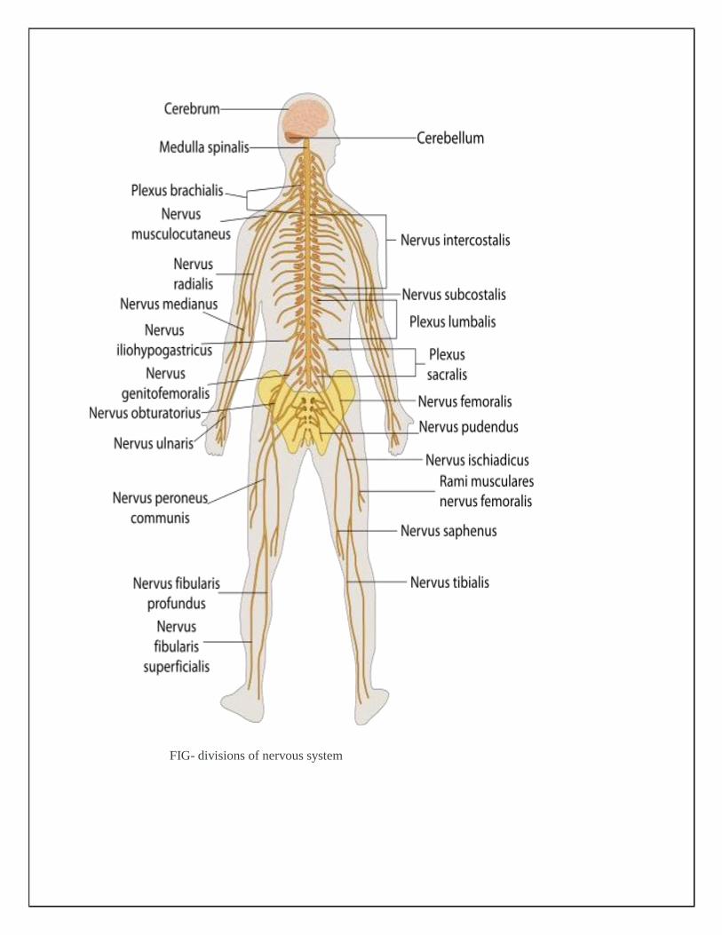
FIG- divisions of nervous system
FUNCTIONS OF NERVOUS SYSTEM
The primary function of the nervous system is to coordinate and control the various body functions. The
nervous system has three overlapping functions based on the sensory input, integration, and motor output.
The nervous system is a highly integrated system.
Sensory Input- Sensory input comes from the many sensory receptors that monitor
changes occurring both inside and outside the body. The total sum of the information
gathered by these receptors is called sensory input. The nervous system processes and
interprets sensory input and decides what actions should be taken. The nervous system
activates effector organs such as muscles and glands to cause a response called motor
output.
Integration- At a more integrative level, the primary function of the nervous system is to
control and communicate information throughout the body. It does this by extracting
information from the environment using sensory receptors. This sensory input is sent to
the central nervous system, which determines an appropriate response.
Motor Response- Once the response is activated, the nervous system sends signals
via motor output to muscles or glands to initiate the response. In humans, the
sophistication of the nervous system allows for language, abstract representation of c
culture, and many other features of society that would not otherwise exist.
SUB -DIVISIONS OF NERVOUS SYSTEM
Central nervous system
The CNS includes the brain and spinal cord along with various centers that integrate all the sensory and
motor information in the body. These centers can be broadly subdivided into lower centers, including the
spinal cord and brain stem, that carry out essential body and organ-control functions and higher centers
within the brain that control more sophisticated information processing, including our thoughts and
perceptions.
Peripheral Nervous System
The PNS is a vast network of nerves consisting of bundles of axons that link the body to the brain and the
spinal cord. Sensory nerves of the PNS contain sensory receptors that detect changes in the internal and
external environment. This information is sent to the CNS via afferent sensory nerves. Following
information processing in the CNS, signals are relayed back to the PNS by way of efferent peripheral
nerve.
Autonomic and Somatic Nervous Systems
The PNS is further subdivided into the autonomic nervous system (ANS) and the somatic nervous
system. The autonomic system has involuntary control of internal organs, blood vessels, and
smooth and cardiac muscles. The somatic system has voluntary control of our movements via
skeletal muscle.
As mentioned, the autonomic nervous system acts as a control system and most functions occur
without conscious thought. The ANS affects heart rate, digestion, respiratory rate, salivation,
perspiration, pupil diameter, urination, and sexual arousal. While most of its actions are
involuntary, some, such as breathing, work in tandem with the conscious mind. The ANS is
classically divided into two subsystems: the parasympathetic nervous system (PSNS) and
sympathetic nervous system (SNS).
Parasympathetic and Sympathetic Nervous system
Broadly, the parasympathetic system is responsible for stimulation of ―rest-and-digest‖ activities
that occur when the body is at rest, including sexual arousal, salivation, lacrimation (tears),
urination, digestion, and defecation. The sympathetic nervous system is responsible for stimulating
activities associated with the ―fight-or-flight‖ response: mobilizing the systems of the body for
escape or attacking sources of danger. In truth, the functions of both the parasympathetic and
sympathetic nervous systems are not so straightforward, but this division is a useful rule of thumb.
The enteric nervous system (ENS) controls the gastrointestinal system and is sometimes considered
part of the autonomic nervous system. However, it is sometimes considered an independent system
because it can operate independently of the brain and the spinal cord.
Conclusions
Our bodies consist of a number of biological systems that carry out specific functions which are
necessary for normal biody functions. Human body consists of the physical substance of living cells
and extracellular materials and is organized into tissues, organs, and systems. Brain is one of the
largest and most complex organs in the human body. It is made up of more than 100 billion nerves
that communicate in trillions of connections called synapses. The eye is our organ of sight. The eye
has a number of components which includes cornea, iris, pupil, retina lens, and macula, optic nerve,
choroid and vitreous. The ear is the organ of hearing. In mammals, the ear is usually three part-the
outer ear,the middle ear and the inner ear. The autonomic nervous system regulates a variety of
body process that takes place without conscious effort.
References
1. www.webmd.com 2. www.wikipedia.com
3. www.helpline.com 4. www.medicinenet.com