Embed Size (px)
Citation preview
J. clin. Path., 1973, 26, 268-272
Indirect cutaneous immunofluorescence1II Clinical significance
THOMAS K. BURNHAM
From the Department ofDermatology, Henry Ford Hospital, Detroit, Michigan, USA
SYNOPSIS Sera of 532 patients with bullous diseases, connective tissue diseases and malignancieswere tested for pemphigus epidermal intercellular fluorescence (ICF) and for the bullous pem-
phigoid 'tubular' band by the indirect fluorescent antibody technique. Human normal skin cryostatsections were used.The band and ICF were seen primarily only in bullous pemphigoid and pemphigus respectively,
Some indirect band and ICF-negative patients demonstrated positive direct results in involved skin.suggesting that direct tests should be performed in indirect negative patients clinically thought tohave pemphigus or bullous pemphigoid.No close correlation was found between disease activity and positive or negative indirect tests in
bullous pemphigoid and pemphigus. Steroids did not interfere with positive results of thisdiagnostically extremely valuable test.
Direct and indirect immunofluorescence has becomea useful diagnostic tool in confirming the clinicaldiagnoses of bullous pemphigoid and pemphigus.Beutner and Jordon (1964) first demonstratedantibodies in the sera of patients with pemphigusvulgaris to an intercellular substance of stratifiedsquamous epithelium by indirect immunofluores-cence. Epidermal intercellular fluorescence (ICF)consisting of smooth polygonal lines of fluorescencearound the epidermal cells was noted. Intercellularfluorescence was also demonstrated by the directtechnique in involved and uninvolved skin of pem-phigus patients (Beutner, Lever, Witebsky, Jordon,and Chertock, 1965). These investigators also foundthat sera of patients with bullous pemphigoid reactedwith the basal zone of stratified squamous epith-elium producing a 'band' of fluorescence at this sitewith the indirect technique (Beutner et al, 1965).Jordon first demonstrated a similar band in involvedand uninvolvedbullous pemphigoid skin bythe directtechnique (Jordon, Beutner, Witebsky, Blumenthal,Hale, and Lever, 1967). This band, althoughoccurringat the same site, the dermal-epidermal junction, as'Presented at the 92nd annual meeeting of the American Dermato-logical Association, Dorado Beach, Puerto Rico, 14 April 1972.
This investigation was supported in,part by Public Health ServiceResearch grant number 5RO1 CA 10999-03 IMB from the NationalCancer Institute and in part by the Elizabeth and Charles PattersonFund for Dermatology Research in lupus erythematosus.
Received for publication 10 January 1973.
that first reported by us in LE skin lesions (BurnhamNeblett, and Fine, 1963) differed morphologicallyfrom it in being 'tubular' due'to a dark centre inplaces producing a hollow tubular appearance bothby the direct (Burnham, Fine, and Neblett, 1970) andindirect techniques (Burnham and Fine, 1972).Chorzelski, von Weiss, and Lever (1966) and Beutner,Jordon, and Chorzelski(1968a) reported that indirectICF titres were related to disease activity in pem-phigus with higher titres in patients with extensivedisease. Fluctuations in serialtitreswere also found inindividual patients related to activity of the disease(Chorzelski et al, 1966) with ICF titres falling inremission and rising with relapse of the disease in aparticular patient. Conversely, in bullous pemphigoidno such relationship between indirect band titres anddisease activity was found by Chorzelski, Jablon'ska,Blaszczyk, and Jarzabek (1968) although Jordon andhis coworkers (1969) found a closer relationshipbetween the extent of disease and the complement-fixing bullous pemphigoid antibody titre. However,Katz, Halprin, and Inderbitzin (1969) reported thatthe titres of both ICF and the band-inducing anti-bodies were related to activity of the disease in bothpemphigus and bullous pemphigoid, respectively,while Beutner et al (1968a) found that positiveindirect band tests were more common in bullouspemphigoid patients with active lesions.The aims of this report are (1) to confirm that
268
copyright. on F
ebruary 17, 2020 by guest. Protected by
http://jcp.bmj.com
/J C
lin Pathol: first published as 10.1136/jcp.26.4.268 on 1 A
pril 1973. Dow
nloaded from
Clinical significance of the indirect cutaneous immunofluorescence
indirect cutaneous immunofluorescence is a valuablediagnostic confirmatory test for the clinical diag-noses of bullous pemphigoid and pemphigus; (2)to emphasize that direct tests should be performedin patients strongly suspected of having bullouspemphigoid or pemphigus if the indirect test isnegative; and (3) to evaluate the relationshipbetween the duration and activity of the disease andthe effects of drugs with the results of the in-direct technique.
Materials and Methods
CLINICAL MATERIAL
Sera of 532 patients with bullous diseases, malig-nancies, and connective tissue diseases were tested(table I).
STAINING TECHNIQUEIndirect techniqueThe sera, stored at - 20°C, were tested within twoweeks by the indirect fluorescent antibody technique.Four micron-thick human normal skin cryostatsections were employed as described previously(Burnham and Fine, 1972).
Direct techniqueThe cryostat cut skin sections were prewashed inveronal-buffered saline (VBS) pH 7-2-7'3 and dis-tilled water to remove any unbound globulins. Theywere then dried in air and incubated with the variousconjugates diluted as for the indirect technique(Burnham and Fine, 1972). They were washed againas above, air-dried, and stored at 4°C until examined.
EXAMINATIONSThe slides were examined without coverslips. Theequipment consisted of a Leitz Ortholux microscopewith an Osram HBO 200 watt mercury lamp, a BG12excitation filter, paired Schott OG4 and GG4 barrier
filters, and a 95 x fluorite oil immersion objective(Burnham and.-Fine, 1971, 1972).
ANTINUCLEAR FACTOR (ANF) TESTS
Antinuclear factor determinations were performedwith the immunofluorescent tumour imprint tech-nique (Burnhain, Neblett, and Fine, 1966; Burnham,Neblett, Fine, anld Bank, 1969) modified by mainlyemploying touch imprints of human normal andlymphoma spleen imprints as nuclear substrate forindirect immunofluorescence.
Results
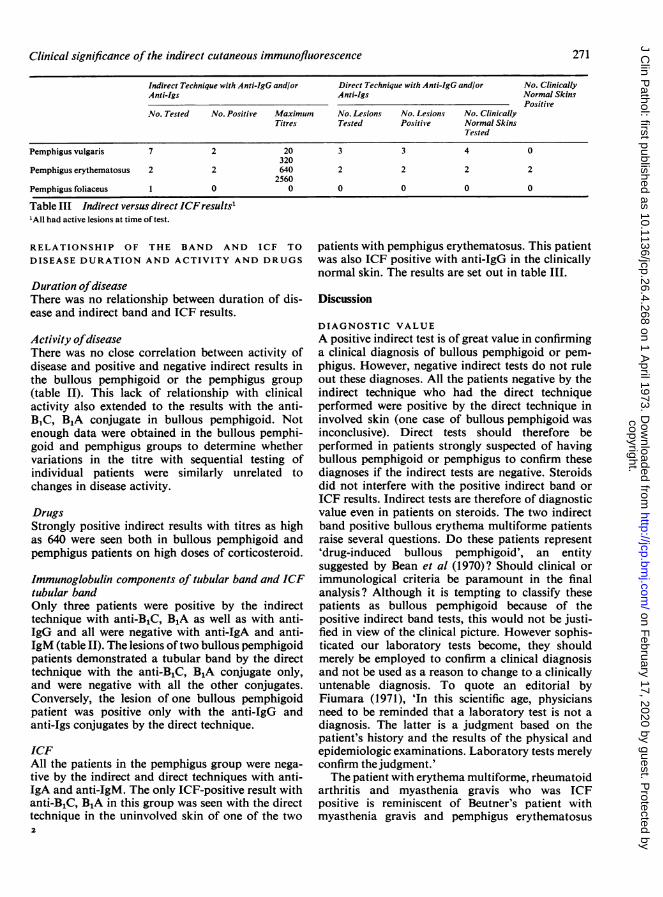
MORPHOLOGYThe bullous pemphigoid 'tubular' band (fig. 1)(Burnham and Fine, 1972) and epidermal inter-cellular fluorescence (ICF) (fig. 2) were seen in thebullous pemphigoid and the pemphigus grouprespectively.
INCIDENCE OF THE BULLOUS PEMPHIGOIDTUBULAR BAND AND ICFThe band was seen essentially only in bullouspemphigoid. Only two patients, clinically not bullouspemphigoid, demonstrated a band by the indirecttechnique out of the 532 patients studied (table I).Bullous erythema multiforme remained the preferredclinical diagnosis in both these indirect band-positive patients. These two patients possibly rep-resented 'drug-induced bullous pemphigoid' asdescribed by Bean, Good, and Windhorst (1970).Only three patients not in the pemphigus group
demonstrated ICF out of the 532 tested (table I).One patient with bronchogenic carcinoma demon-strated ICF with the initial test only and was negativeon repeated retesting. One patient with erythemamultiforme had coexisting myasthenia gravis andrheumatoid arthritis. This patient also demonstratedICF in involved and uninvolved skin by the directtechnique.
Diagnosis Number Tested Number Positivefor NumberPositiveforBand Epidermal Intercellular Fluorescence
Pemphigus vulgaris 7 0 2Pemphigus erythematosus 2 0 2Pemphigus foliaceus I 0 0Bullouspemphigoid 20 12 0Bullous erythema multiforme 14 2 0Erythema multiforme 15 0 1Erythema annulare centrifugum 1 0 1Tumours 108 0 1
Table I Resultsfrom 532 patients tested by the indirect technique'
Systemic lupus erythematosus (74), discoid lupus erythematosus (14), scleroderma (38), suspected connective tissue disease (53), polymorphous;ight eruption (34), Jessner's lymphocytic infiltrate (8), rheumatoid arthritis (33), miscellaneous medical diseases and dermatoses (88), cicatricialpemphigoid (2), Hailey-Hailey (4), dermatitis herpetiformis (9), herpes zoster (2), toxic epidermal necrolysis (2), pressure bullae (1), subcornealpustular dermatosis (1), dysplastic epidermolysis bullosa (1).Numbers in parentheses represent number of patients tested and negative by indirect technique.
269
copyright. on F
ebruary 17, 2020 by guest. Protected by
http://jcp.bmj.com
/J C
lin Pathol: first published as 10.1136/jcp.26.4.268 on 1 A
pril 1973. Dow
nloaded from

Thomas K. Btirnham
Fig. 1 Fig. 2Fig. 1 Bullouspemphigoid tubular bandNormal skin incubated with bullous pemphigoid serum diluted 1:0lfollowed by incubation withfluorescein conjugated
goat antihuman IgG. A convoluted sharply demarcated band with a dark centre in places producing a hollow tubularappearance ispresent at the dermal-epidermaljunction (arrow). Epidermis in upperpart ofphotograph.Fig. 2 Pemphigus epidermal intercellular fluorescence (ICF)Normal skin incubated withpemphigus erythematosus serum diluted 1 :lOfollowed by incubation withfluorescein
conjugatedgoat antihuman IgG. Smooth polygonalICF is seen in the intercellular space. Note the diminishedfluorescenceintensity of ICF in the basal cell layer (lower part ofphotograph) with complete absence offluorescence at the basal celldermal border.
Thephotographs were taken with a 95 x fluorite oil immersion objective. 10 x ocular, Leica MD body, magnified x 3.
RELATIONSHIP OF INDIRECT VERSUS DIRECT
RESULTS IN BULLOUS PEMPHIGOID AND IN
THE PEMPHIGUS GROUPThe bullous pemphigoid tubular band was seen inonly 12 out of 20 bullous pemphigoid patients bythe indirect technique (table II). However, the directtest was positive in the involved skin in nearly allthe patients tested (one inconclusive) with at leastone of the conjugates. The tendency for positivedirect results in involved skin, despite negativeindirect tests, was also seen with ICF in the pem-phigus group (table II).
Indirect Direct TechniqueTechnique
Lesions Normal Skin
No. positive ofno. tested 12/20 10/11 7/10Maximum titres withanti-IgG and/or anti-Igs 1280 (active)
640 (remission)Anti-IgA 0/12 0/11 0/10Anti-IgM 0/12 2/11 0/10Anti-BIC, 9,A 3/12 9/11 5/10Anti-IgG and/or anti-Igs 12/20 8/11 7/10
Table LI Comparison of results of tests with indirectand direct techniques for tubular band.
270
copyright. on F
ebruary 17, 2020 by guest. Protected by
http://jcp.bmj.com
/J C
lin Pathol: first published as 10.1136/jcp.26.4.268 on 1 A
pril 1973. Dow
nloaded from
Clinical significance of the indirect cutaneous immunofluorescence
Indirect Technique with Anti-IgG and/or Direct Technique with Anti-IgG and/or No. ClinicallyAnti-Igs Anti-Igs Normal Skins
PositiveNo. Tested No. Positive Maximum No. Lesions No. Lesions No. Clinically
Titres Tested Positive Normal SkinsTested
Pemphigus vulgaris 7 2 20 3 3 4 0320
Pemphigus erythematosus 2 2 640 2 2 2 22560
Pemphigus foliaceus 1 0 0 0 0 0 0
Table III Indirect versus direct ICF results''All had active lesions at time of test.
RELATIONSHIP OF THE BAND AND ICF TO
DISEASE DURATION AND ACTIVITY AND DRUGS
Duration ofdiseaseThere was no relationship between duration of dis-ease and indirect band and ICF results.
Activity ofdiseaseThere was no close correlation between activity ofdisease and positive and negative indirect results inthe bullous pemphigoid or the pemphigus group(table II). This lack of relationship with clinicalactivity also extended to the results with the anti-B1C, B1A conjugate in bullous pemphigoid. Notenough data were obtained in the bullous pemphi-goid and pemphigus groups to determine whethervariations in the titre with sequential testing ofindividual patients were similarly unrelated tochanges in disease activity.
DrugsStrongly positive indirect results with titres as highas 640 were seen both in bullous pemphigoid andpemphigus patients on high doses of corticosteroid.
Immunoglobulin components of tubular band and ICFtubular bandOnly three patients were positive by the indirecttechnique with anti-B,C, B1,A as well as with anti-IgG and all were negative with anti-IgA and anti-IgM (table II). The lesions oftwo bullous pemphigoidpatients demonstrated a tubular band by the directtechnique with the anti-B,C, B1A conjugate only,and were negative with all the other conjugates.Conversely, the lesion of one bullous pemphigoidpatient was positive only with the anti-IgG andanti-Igs conjugates by the direct technique.
ICFAll the patients in the pemphigus group were nega-tive by the indirect and direct techniques with anti-IgA and anti-IgM. The only ICF-positive result withanti-B,C, B1A in this group was seen with the directtechnique in the uninvolved skin of one of the two
patients with pemphigus erythematosus. This patientwas also ICF positive with anti-IgG in the clinicallynormal skin. The results are set out in table III.
Discussion
DIAGNOSTIC VALUEA positive indirect test is of great value in confirminga clinical diagnosis of bullous pemphigoid or pem-phigus. However, negative indirect tests do not ruleout these diagnoses. All the patients negative by theindirect technique who had the direct techniqueperformed were positive by the direct technique ininvolved skin (one case of bullous pemphigoid wasinconclusive). Direct tests should therefore beperformed in patients strongly suspected of havingbullous pemphigoid or pemphigus to confirm thesediagnoses if the indirect tests are negative. Steroidsdid not interfere with the positive indirect band orICF results. Indirect tests are therefore of diagnosticvalue even in patients on steroids. The two indirectband positive bullous erythema multiforme patientsraise several questions. Do these patients represent'drug-induced bullous pemphigoid', an entitysuggested by Bean et al (1970)? Should clinical orimmunological criteria be paramount in the finalanalysis? Although it is tempting to classify thesepatients as bullous pemphigoid because of thepositive indirect band tests, this would not be justi-fied in view of the clinical picture. However sophis-ticated our laboratory tests become, they shouldmerely be employed to confirm a clinical diagnosisand not be used as a reason to change to a clinicallyuntenable diagnosis. To quote an editorial byFiumara (1971), 'In this scientific age, physiciansneed to be reminded that a laboratory test is not adiagnosis. The latter is a judgment based on thepatient's history and the results of the physical andepidemiologic examinations. Laboratory tests merelyconfirm thejudgment.'The patient with erythema multiforme, rheumatoid
arthritis and myasthenia gravis who was ICFpositive is reminiscent of Beutner's patient withmyasthenia gravis and pemphigus erythematosus
271
copyright. on F
ebruary 17, 2020 by guest. Protected by
http://jcp.bmj.com
/J C
lin Pathol: first published as 10.1136/jcp.26.4.268 on 1 A
pril 1973. Dow
nloaded from
Thomas K. Burnham
(Beutner, Chorzelski, Hale, and Hausmanowa-Petnusewitz, 1968b). However, a diagnosis of pem-phigus in our patient could not be confirmed clinic-ally or histopathologically.
LACK OF RELATIONSHIP BETWEEN POSITIVEINDIRECT RESULTS AND DISEASE ACTIVITYThe lack of close correlation between activity ofdisease and indirect results in bullous pemphigoidhas been reported by some authors (Chorzelski et al,1968; Sams, 1970), but other investigators found acorrelation (Beutner et al, 1968a; Katz et al, 1969).Absence of correlation extended to the findings withthe anti-BIC, B,A conjugate in our series. This castssome doubt on the possibility of using titres of thecomplement-binding antibody as a therapeuticguide in bullous pemphigoid as suggested by thefindings of Jordon et al (1969). However, it is stillpossible that those patients who are positive withanti-B,C by the indirect technique may be found tohave a correlation between disease activity andtitre of the complement-binding antibody withsequential testing.There was also no close correlation between
disease activity and positive and negative indirectresults in the pemphigus group which has not beenthe finding of other authors (Chorzelski et al, 1966;Beutner et al, 1968a).We did not obtain sufficient data to determine
whether there is a relationship between serialfluctuations of the ICF titre and changes in diseaseactivity in following an individual patient as noneof the patients were in remission at the time of anyof their indirect tests.
PATHOGENESISThe question whether these band and ICF-inducingantibodies are pathogenetic is still unresolved.Katz et al (1969) and Schroeter, Sams, and Jordon(1969) noted that the drop in ICF titre lagged byseveral weeks behind clinical remission in pemphigus.This favours the idea that these antibodies may be aresult rather than the cause of the lesions. However,against this theory, Katz et al (1969) noted that arise in ICF titre preceded exacerbation of the skindisease in one pemphigus patient. Schroeter et al(1969) even suggested that relapses might be pre-dicted by a rise in ICF titre in pemphigus.
Despite our ignorance regarding the pathogeneticrole, if any, of these antibodies, their demonstrationby both the direct and indirect techniques providesan extremely valuable diagnostic aid in confirmingthe clinical diagnoses of bullous pemphigoid andpemphigus.
I should like to thank our colleagues at the Henry
Ford Hospital for their help in obtaining the seraand biopsy specimen, and especially Edward A.Krull MD, for providing the normal skin.
I am also extremely grateful to Mr Arthur Bowdenand Mr Walter Harlan for their invaluable help withthe photographs.
Paula W. Bank MS and Judy Jaquillard BAprepared and screened the antinuclear factor slides.
Colleen Aman BS, Judith Nolish BA, and DonnaTymensky BA prepared the slides for cutaneousimmunofluorescence.
References
Bean, S. F., Good, R. A., and Windhorst D. B. (1970), Bullouspemphigoid in an 11-year-old boy. Arch. Derni., 102, 205-208.
Beutner, E. H., Chorzelski, T. P., Hale, W. L., and Hausmanowa-Petnusewitz, I. (1968b). Autoimmunity in concurrent myas-thenia gravis and pemphigus erythematosus. J. Amer. nied.Ass., 203, 845-849
Beutner, E. H., and Jordon, R. E. (1964). Demonstration of skinantibodies in sera of pemphigus vulgaris patients by indirectimmunofluorescent staining. Proc. Soc. exp. Biol. (N. Y.)117, 505-510.
Beutner, E. H., Jordon, R. E., and Chorzelski, T. P. (1968a). Theimmunopathology of pemphigus and bullous pemphigoid.J. Invest. Derm., 51, 63-80.
Beutner, E. H., Lever, W. F., Witebsky, E., Jordon, R., and Chertock,B. (1965). Autoantibodies in pemphigus vulgaris: Response toan intercellular substance of epidermis. J. Amer. med. Ass., 192,682-688.
Burnham, T. K., and Fine, G. (1971). The immunofluorescent 'band'test for lupus erythematosus. 111. Employing clinically normalskin. Arch. Derm., 103, 24-32.
Burnham, T. K. and Fine, G. (1972). Indirect cutaneous immuno-fluorescence. 1. Morphologic observations in bullous diseases,malignancies and connective tissue diseases. Arch. Dermi.,105, 52-58.
Burnham, T. K., Fine, G., and Neblett, T. R. (1970). Immunofluores-cent 'band' test for lupus erythematosus. 11. Employing skinlesions. Arc/i. Derni., 102, 42-50.
Burnham, T. K., Neblett, T. R., and Fine, G. (1963). The applicationof the fluorescent antibody technic to the investigation oflupus erythematosus and various dermatoses. J. Invest. Derm7.,41, 451-456.
Burnham, T. K., Neblett, T. R., and Fine, G. (1966). The immuno-fluorescent tumor imprint technic. 1. Description and evalUa-tion. Anier. J. clin. Pat/i., 45, 714-721.
Burnham, T. K., Neblett, T. R., Fine, G., and Bank, P. (1969). Theimmunofluorescent tumor imprint technique. IV. The signifi-cance of 'thready' nuclear immunofluorescence. Arch. Deryii.,99, 611-616.
Chorzelski, T. P., Jabloniska, S., Blaszczyk, M., and Jarzabek, M.(1968). Autoantibodies in pemphigoid. Derniatologica (Basel),136, 325-334.
Chorzelski, T. P., von Weiss, J. F., and Lever, W. F. (1966). Clinicalsignificance of autoantibodies in pemphigus. Arc/i. Derin.,93, 570-576.
Fiumara, N. J. (1971). A laboratory test is not a diagnosis. (Editorial).J. Amer. ined. Ass., 217, 71.
Jordon, R. E., Beutner, E. H., Witebsky, E., Blumenthal, G., Hale,W. L., and Lever, W. F. (1967). Basement zone antibodies inbullous pemphigoid. J. Anier. med. Ass., 200, 751-756.
Jordon, R. E., Sams, W. M., Jr., and Beutner, E. H. (1969). Comple-ment immunofluorescent staining in bullous pemphigoid.J. Lab. clin. Med., 74, 548-556.
Katz, S. I., Halprin, K. M., and Inderbitzin, T. M. (1969). The use ofhuman skin for the detection of antiepithelial autoantibodies.J. Invest. Derni., 53, 390-399.
Sams, W. M., Jr. (1970). Bullous pemphigoid: Is it an immunologicdisease? Arc/i. Dernm., 102, 485-497.
Schroeter, A., Sams, W. M., Jr., and Jorden, R. E. (1969). Immuno-fluorescent studies of pemphiguLs foliaceus in a child. Arc/i.Derni., 100, 736-740.
272
copyright. on F
ebruary 17, 2020 by guest. Protected by
http://jcp.bmj.com
/J C
lin Pathol: first published as 10.1136/jcp.26.4.268 on 1 A
pril 1973. Dow
nloaded from