Embed Size (px)
Citation preview
INDIUM-ZINC-OXIDE NANOCRYSTALLITES: PREPARATION,
PROPERTIES AND VISIBLE-LIGHT-GENERATED
PHOTOCATALYTIC EFFICIENCY IN DEGRADATION
OF PSYCHOACTIVE DRUGS FROM WATER SYSTEMS
T. IVETIĆ1, N. FINČUR2, B. MILJEVIĆ3, LJ. ĐAČANIN FAR1, S. LUKIĆ-PETROVIĆ1,
B. ABRAMOVIĆ2
1University of Novi Sad, Faculty of Sciences, Department of Physics,
Trg Dositeja Obradovića 4, 21000 Novi Sad, Serbia
E-mails: [email protected]; [email protected]; [email protected] 2University of Novi Sad, Faculty of Science, Department of Chemistry,
Biochemistry and Environmental Protection,
Trg Dositeja Obradovića 3, 21000 Novi Sad, Serbia
E-mails: [email protected]; [email protected] 3University of Novi Sad, Faculty of Technology, Department of Materials Engineering,
Bulevar Cara Lazara 1, 21000 Novi Sad, Serbia
E-mail: [email protected]
Received September 27, 2017
Abstract. In this work, we studied the structure, band gap, particle size, and
morphology of the indium-zinc-oxide (IZO) nanocrystalline powders (NCPs) prepared
via solid-state method. Materials characterization was performed by using a variety of
techniques (XRD, Raman, SEM-EDS, and UV-Vis). The estimated values of the
optical band gaps of the obtained IZO NCPs indicated a possibility of their
photocatalytic activity under solar irradiation. According to this, the efficiency of the
obtained NCPs as photocatalysts in the degradation of alprazolam and amitriptyline,
the active components one of the most prescribed psychoactive drugs nowadays,
under simulated solar irradiation, was explored.
Key words: Oxide material, semiconductor, photocatalysis, psychoactive drugs.
1. INTRODUCTION
Heterogeneous photocatalysis (HPC) belongs to the well-established
wastewater treatment technology known as advanced oxidation processes (AOPs).
Besides, AOPs include photolysis, ozonization, Fenton’s oxidation, electro-
chemical oxidation, ultrasound irradiation and wet air oxidation [1, 2]. HPC
method degrades or otherwise transforms the organic pollutants in water to usually
less toxic chemicals or a completely harmless carbon dioxide and water, and
inorganic ions depending on the structure of molecules.
Romanian Journal of Physics 63, 608 (2018)
Article no. 608 T. Ivetić et al. 2
HPC requires the presence of a metal oxide semiconductor in contaminated
water and its illumination with either UV or visible light source. The effectiveness
of HPC depends on the physicochemical properties (mostly electronic structure,
size, surface area, morphology, and composition) of the used metal oxide material
made in the form of powder or thin film. Therefore, the preparation procedure
employed for the fabrication of these semiconductor materials is crucial for
obtaining the best photocatalytic performance. The latest industrial trends for HPC
are encouraging the preparation of ecologically affordable photocatalysts via
inexpensive routes such as it is mechanochemical processing, with desired
characteristics to induce the oxidation of the organic pollutants under visible light
irradiation [3, 4].
In the pursuit of the most effective and suitable aforementioned
semiconductor material that supports the photodegradation of the organic
pollutants, zinc oxide (ZnO) was found to be one of the most promising candidates
to replace the commonly used material for this purpose, titanium dioxide (TiO2)
known as "the gold standard" photocatalyst [5]. Major drawbacks in ZnO
photocatalytic performance are related to its UV light requirements for the band
gap excitation, fast recombination of the charge carriers that diminishes the
effectiveness of the degradation processes, and photo-corrosion vulnerability [5].
These barriers could be overcome by tailoring the ZnO electronic and surface-bulk
structure and related photogenerated charge carrier transfer pathways. In this
regard, the substitutional ZnO doping with various metal cations like Mn, Ga, Ag,
Cd, Mg, and/or ZnO coupling with other metal oxide semiconductors with different
band gaps to form heterojunctions like ZnO/TiO2, ZnO/SnO2, ZnO/CuO,
ZnO/Fe2O3, have been studied so far [5, 6]. Furthermore, it was already reported
that modification of ZnO with In2O3 greatly improves its stability, reduces its size
and enables better control of the charge carrier’s recombination for enhancement of
ZnO photocatalytic efficiency [6, 7]. Progress in making a better ZnO
photocatalytic performance with In2O3 could be achieved firstly by changing the
coordination environment of the Zn2+
ions in the ZnO lattice via defect engineering
and/or indium doping. The group-III impurities like In act as shallow donors when
substituting Zn site in ZnO [8]. Donor impurities introduce new electronic energy
levels within the band gap states and modify the electronic band structure by
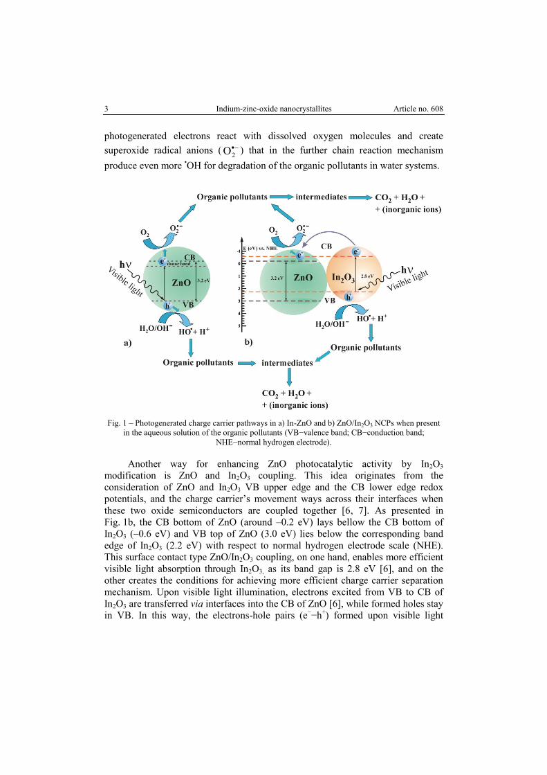
shifting the band gap towards the visible region [3−5, 9] (illustrated in Fig. 1a).
In3+
-doping, then makes possible for ZnO to absorb more visible light photons,
excites the electrons (e−) from valence band (VB) to the conduction band (CB) and
consequently leaves holes (h+) in VB. Then, holes react with surrounding water
molecules (H2O) and produce strong oxidizing agents, hydroxyl radicals (•OH),
species mostly responsible for the transformation of the organic substances via
HPC into CO2 and H2O and other less harmful compounds. Simultaneously,
3 Indium-zinc-oxide nanocrystallites Article no. 608
photogenerated electrons react with dissolved oxygen molecules and create
superoxide radical anions (
2O ) that in the further chain reaction mechanism
produce even more •OH for degradation of the organic pollutants in water systems.
Fig. 1 – Photogenerated charge carrier pathways in a) In-ZnO and b) ZnO/In2O3 NCPs when present
in the aqueous solution of the organic pollutants (VB−valence band; CB−conduction band;
NHE−normal hydrogen electrode).
Another way for enhancing ZnO photocatalytic activity by In2O3
modification is ZnO and In2O3 coupling. This idea originates from the
consideration of ZnO and In2O3 VB upper edge and the CB lower edge redox
potentials, and the charge carrier’s movement ways across their interfaces when
these two oxide semiconductors are coupled together [6, 7]. As presented in
Fig. 1b, the CB bottom of ZnO (around –0.2 eV) lays bellow the CB bottom of
In2O3 (–0.6 eV) and VB top of ZnO (3.0 eV) lies below the corresponding band
edge of In2O3 (2.2 eV) with respect to normal hydrogen electrode scale (NHE).
This surface contact type ZnO/In2O3 coupling, on one hand, enables more efficient
visible light absorption through In2O3, as its band gap is 2.8 eV [6], and on the
other creates the conditions for achieving more efficient charge carrier separation
mechanism. Upon visible light illumination, electrons excited from VB to CB of
In2O3 are transferred via interfaces into the CB of ZnO [6], while formed holes stay
in VB. In this way, the electrons-hole pairs (e−−h
+) formed upon visible light
Article no. 608 T. Ivetić et al. 4
excitation are effectively separated which gives more time for their efficient impact
on the surrounding aqueous solution through the oxidation/reduction reactions and
improves the degradation rate of the organic pollutants under visible light
irradiation.
Alprazolam and amitriptyline are representatives of a large group of
psychoactive drugs that include narcoleptics, ataractics/tranquilizers, hypnotics,
sedatives, and antidepressants. Alprazolam is a new generation benzodiazepine that
possesses anxiolytic, sedative and hypnotic properties [10−13], while amitriptyline
is one of the most used tricyclic antidepressants for treating disorders like
depression, anxiety, and a variety of chronic pain syndromes [14, 15].
Pharmaceuticals used for treating mental disorders and diseases i.e. their active
components (ACs) are continuously introduced into the environment during their
manufacturing, disposal or after use by human and animal excretions, and are
increasingly becoming an ecological problem [16−19]. Namely, ACs are found in
drinking and environmental waters in many countries, and it was established that
the main water pollution with these ACs originates from the wastewater treatment
plants [16]. Apparently, ACs are not completely removed by conventional water
purification procedures and hence, supplementary methods like HPC are more than
needed. Furthermore, HPC using semiconductor nanoparticles has already shown
most effectiveness in removing pharmaceutical contaminants from wastewaters [1].
This work explores the possibility of using the simple, sustainable and eco-
friendly solid-state mechanochemical processing for the preparation of indium-
zinc-oxide nanocrystallites (NCs), and their application as photocatalyst in the
HPC of selected active components of psychoactive drugs under simulated solar
irradiation (SSI). The obtained NCs structure, morphology, and optical properties
were examined in detail using a variety of techniques for materials
characterization. The solid-state mechanochemical method for NCs preparation
used in this study is a good alternative to a solution-based method for
manufacturing the functionally tailored nanomaterials [10, 11, 14, 20, 21].
2. EXPERIMENTAL PROCEDURE
2.1. SAMPLE PREPARATION
Two different mechanochemical procedures were employed for obtaining
indium-zinc-oxide NCs. In the first procedure starting precursors, ZnO (Sigma-
Aldrich; purity 99.9% powder and particle size 1 m) and In2O3 (Alfa Aesar;
purity 99.99% and metal basis 325 mesh powder), were stoichiometrically mixed
to achieve about 5% (w/w) of indium doping in an agate mortar for 10 min, pressed
under 50 kg/cm2 load, annealed at two different temperatures (700 and 950 °C for
5 Indium-zinc-oxide nanocrystallites Article no. 608
1 h) in air atmosphere, and ground again for 10 min (marked as In-ZnO-700 and
In-ZnO-950 samples, respectively). The second preparation procedure was
designed for preparing the contact-type ZnO/In2O3 NCs mixture and it was based
on the recent applied mechanochemical route for obtaining ZnO/SnO2 NCs
photocatalyst mixture [14]. The starting precursors, in the molar ratio of
ZnO:In2O3=2:1 (37 wt.% of ZnO and 63 wt.% of In2O3), were ground for 10 min
in an agate mortar, annealed at 700 °C for 2 h, and additionally ground for 10 min
(marked as ZnO/In2O3 sample).
2.2. MATERIALS CHARACTERIZATION
X-ray diffraction (XRD) was carried out using Philips PW 1050 instrument,
with Cu Kα1,2 radiation, and a step scan mode of 0.02°/s in angular range
2θ = 1090° which enabled good profile fitting using PDXL and HighScore Plus
(PANanalytical) software. Scanning electron microscope, SEM (JEOL JSM-
6460LV) equipped with an energy-dispersive spectrometer (EDS) was used to
investigate the morphology, microstructure and elemental concentration of the
obtained samples. The Raman spectra of the In-ZnO samples were measured using
the Centice MMS Raman spectrometer equipped with charge-coupled device
(CCD) as a detector. A diode laser operating at 785 nm (1.58 eV) was used as the
excitation source. The diffuse reflectance spectra (DRS) were obtained using the
Ocean Optics QE65000 High-sensitivity Fiber Optic Spectrometer, and in
accordance with it, the Kubelka-Munk function was estimated using Spectra Suite
Ocean Optics operating software. All measurements were carried out at room
temperature.
2.3. MEASUREMENTS OF PHOTOCATALYTIC ACTIVITY
The photocatalytic activities of In-ZnO and ZnO/In2O3 samples were
evaluated by degradation of the aqueous alprazolam solution (8-chloro-1-methyl-6-
phenyl-4H-[1,2,4]triazole[4,3,-α]-[1,4]-benzodiazepine, CAS No. 28981-97-7,
C17H13ClN4, Mr = 308.765, 98%, Sigma-Aldrich). Besides, the photocatalytic
activity of ZnO/In2O3 sample was evaluated by degradation of the aqueous
amitriptyline solution (3-(10,11-dihydro-5H-dibenzo[a,d][7]annulen-5-ylidene)-
N,N-dimethylpropan-1-amine hydrochloride, C20H24ClN, Mr = 313.9, CAS No.
549-18-8, 98%, Sigma-Aldrich). In both cases, the initial concentration of
investigated pharmaceutically active components (alprazolam/amitriptyline) was
0.03 mmol/L, while the catalyst loading was 1.0 mg/mL.
Article no. 608 T. Ivetić et al. 6
A cell made of Pyrex glass (total volume of ca. 40 mL) with a plain window
for the light beam focus, magnetic stirring bar, and the water-circulating jacket was
used in the photocatalytic degradation experiments. Uniform dispersion with the
photocatalyst NCs and adsorption equilibrium was made by sonication (50 Hz) of
the suspension, prior to the illumination, in dark for 30 min. From the beginning,
the suspension was thermostated at 250.5 °C in O2 stream (3.0 mL/min). The
obtained suspension was then illuminated by simulated solar irradiation using a 50
W halogen lamp (Philips). During irradiation, the stirring and streaming with O2
constant rate flow were continued. Kinetic studies of alprazolam/amitriptyline
photodegradation were monitored by ultra fast liquid chromatography with UV/Vis
diode array detector (UFLC−DAD) set at 222 nm (wavelength of alprazolam
maximum absorption) and at 206 nm (wavelength of amitriptyline maximum
absorption). The aliquots of 0.5 mL of each reaction mixtures were taken at the
beginning of the experiment and then at certain time intervals up to 60 min, filtered
through a Millipore (Millex-GV, 0.22 m) membrane filter to remove NCs, and
samples were injected and analyzed with UFLC-Shimadzu. Procedures for
UFLC−DAD analysis were described previously in the case of alprazolam [10],
and in the case of amitriptyline [14].
3. RESULTS AND DISCUSSION
3.1. STRUCTURE AND MORPHOLOGY
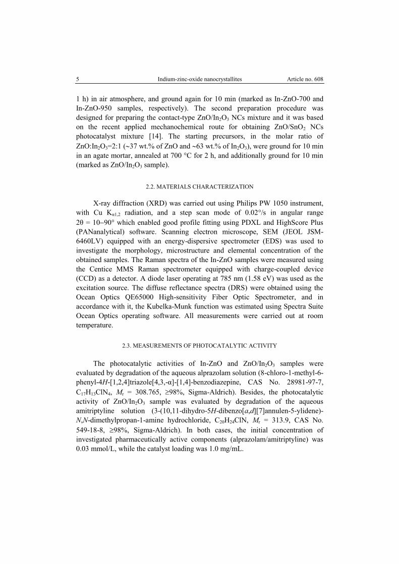
Figure 2 shows XRD patterns of the obtained In-ZnO samples. The main
diffraction peaks were indexed as hexagonal wurtzite ZnO (JCPDS #36-1451)
phase. Few diffraction peaks (marked with an asterisk in Fig. 2) were found to
belong to the cubic In2O3 (JCPDS #44-1087) phase.
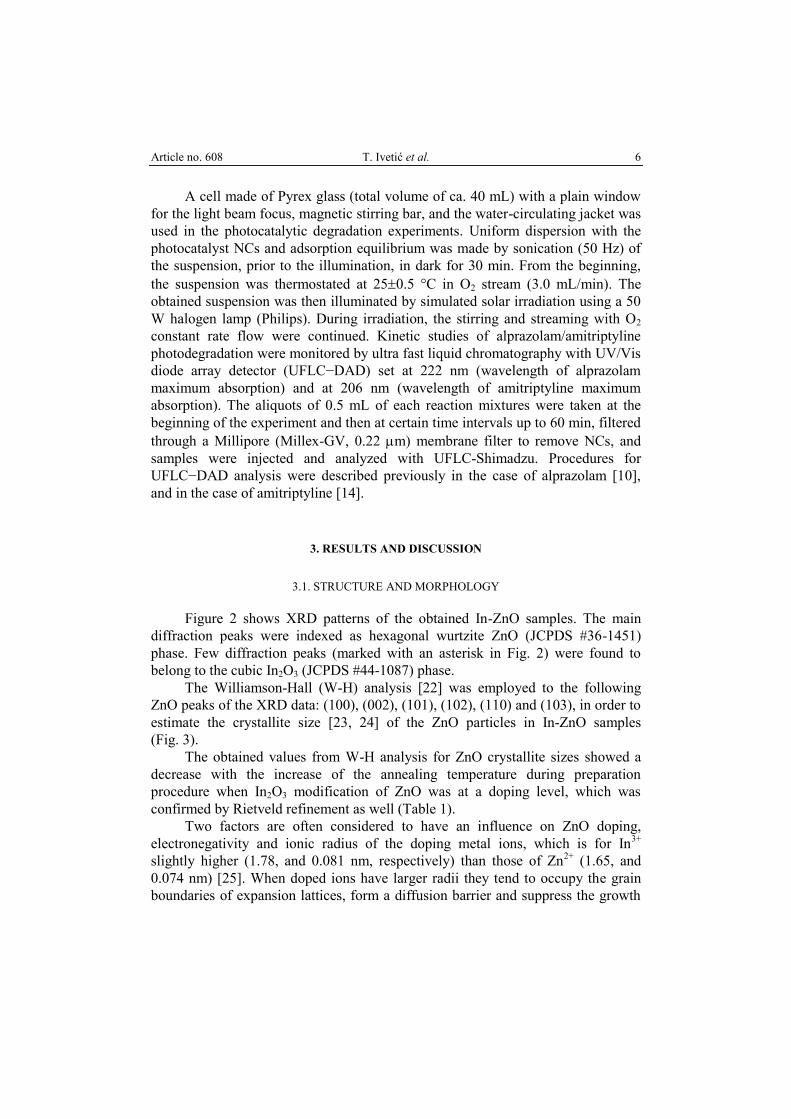
The Williamson-Hall (W-H) analysis [22] was employed to the following
ZnO peaks of the XRD data: (100), (002), (101), (102), (110) and (103), in order to
estimate the crystallite size [23, 24] of the ZnO particles in In-ZnO samples
(Fig. 3).
The obtained values from W-H analysis for ZnO crystallite sizes showed a
decrease with the increase of the annealing temperature during preparation
procedure when In2O3 modification of ZnO was at a doping level, which was
confirmed by Rietveld refinement as well (Table 1).
Two factors are often considered to have an influence on ZnO doping,
electronegativity and ionic radius of the doping metal ions, which is for In3+
slightly higher (1.78, and 0.081 nm, respectively) than those of Zn2+
(1.65, and
0.074 nm) [25]. When doped ions have larger radii they tend to occupy the grain
boundaries of expansion lattices, form a diffusion barrier and suppress the growth
7 Indium-zinc-oxide nanocrystallites Article no. 608
of the ZnO crystal. The difference in oxidation states of In3+
and Zn2+
ions further
enhances the lattice distortion. There is a possibility of In2O3 decomposition when
heated above 850 °C, and a creation of In2+
ion that is chemically even more
similar to the Zn2+
ion [26]. According to this, as the temperature rises there is a
chance for more indium ions (both In2+
and In3+
) to enter the ZnO lattice and
replace Zn2+
ions. ZnO lattice parameters change can be used to study the doping
effects (Table 1) (ZnO reference values from JCPDS #36-1451 are
a = b = 3.24982; c = 5.20661). For example, the c-axis lattice constant usually
decreases in the case of doped ions with smaller ionic radii (In-ZnO-950) as they
tend to occupy the grain boundaries of the compression lattices, and usually
increases for doped ions with larger ionic radii (In-ZnO-700) as they tend to
occupy grain boundaries of the expansion lattices [25].
Table 1
The results of the structural analysis for In-ZnO samples
Structural properties Rietveld refinement W-H analysis
In-ZnO-700 In-ZnO-950 In-ZnO-700 In-ZnO-950
Crystallite size, nm 66.5(6) 41.5(3) 96.29±16.05 66.34±18.09
Microstrain (%) 0.102(7) 0.128(13) 0.117±0.15 1.02±0.37
Unit cell parameter a = b = 3.2517(3)
c = 5.2090(5)
a = b = 3.2527(4)
c = 5.2046(7)
Weight % (ZnO) 96.9 97.2
Weight % (In2O3) 3.1 2.8
Fig. 2 – XRD patterns of a) In-ZnO-700 and b) In-ZnO-950 samples.
Article no. 608 T. Ivetić et al. 8
Fig. 3 – The W-H analysis (left) of a) In-ZnO-700, and b) In-ZnO-950 samples. Fit to the data,
the crystalline size (D) is extracted from the y-intercept of the fit as D = Kλ/(y-intercept); K = 0.9
and λ = 0.1540598 nm. SEM images (right) of a) In-ZnO-700 and b) In-ZnO-950 samples.
a) b)
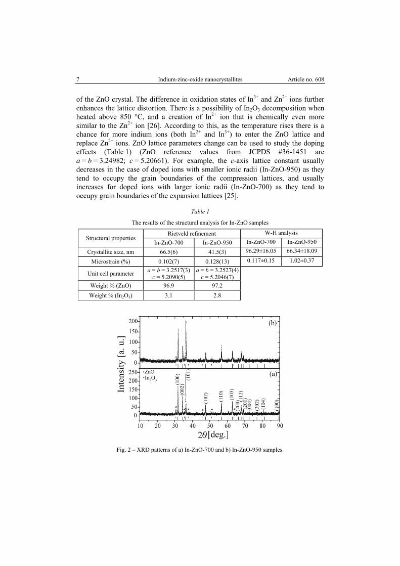
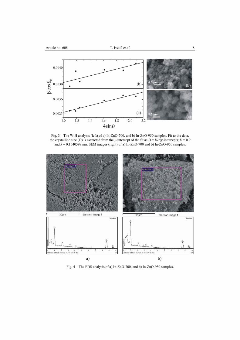
Fig. 4 – The EDS analysis of a) In-ZnO-700, and b) In-ZnO-950 samples.
9 Indium-zinc-oxide nanocrystallites Article no. 608
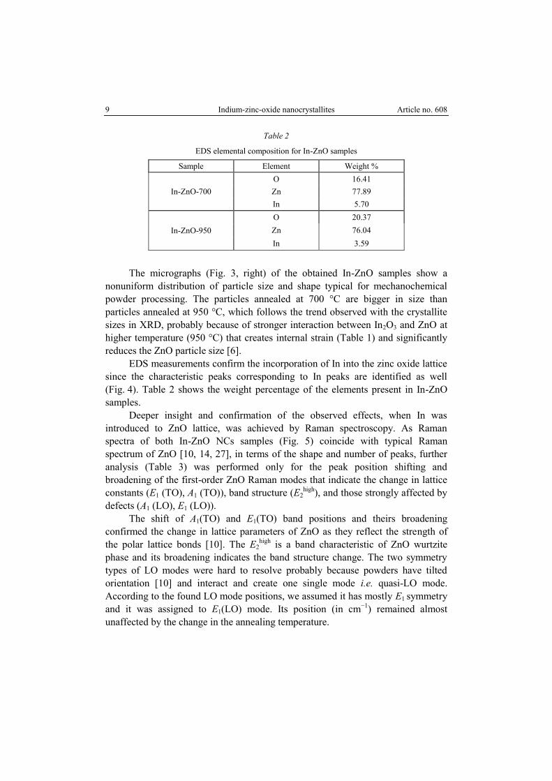
Table 2
EDS elemental composition for In-ZnO samples
Sample Element Weight %
In-ZnO-700
O 16.41
Zn 77.89
In 5.70
In-ZnO-950
O 20.37
Zn 76.04
In 3.59
The micrographs (Fig. 3, right) of the obtained In-ZnO samples show a
nonuniform distribution of particle size and shape typical for mechanochemical
powder processing. The particles annealed at 700 °C are bigger in size than
particles annealed at 950 °C, which follows the trend observed with the crystallite
sizes in XRD, probably because of stronger interaction between In2O3 and ZnO at
higher temperature (950 °C) that creates internal strain (Table 1) and significantly
reduces the ZnO particle size [6].
EDS measurements confirm the incorporation of In into the zinc oxide lattice
since the characteristic peaks corresponding to In peaks are identified as well
(Fig. 4). Table 2 shows the weight percentage of the elements present in In-ZnO
samples.
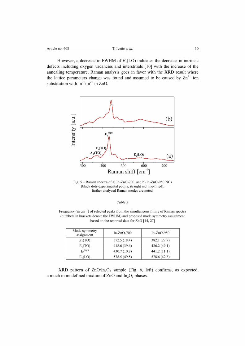
Deeper insight and confirmation of the observed effects, when In was
introduced to ZnO lattice, was achieved by Raman spectroscopy. As Raman
spectra of both In-ZnO NCs samples (Fig. 5) coincide with typical Raman
spectrum of ZnO [10, 14, 27], in terms of the shape and number of peaks, further
analysis (Table 3) was performed only for the peak position shifting and
broadening of the first-order ZnO Raman modes that indicate the change in lattice
constants (E1 (TO), A1 (TO)), band structure (E2high
), and those strongly affected by
defects (A1 (LO), E1 (LO)).
The shift of A1(TO) and E1(TO) band positions and theirs broadening
confirmed the change in lattice parameters of ZnO as they reflect the strength of
the polar lattice bonds [10]. The E2high
is a band characteristic of ZnO wurtzite
phase and its broadening indicates the band structure change. The two symmetry
types of LO modes were hard to resolve probably because powders have tilted
orientation [10] and interact and create one single mode i.e. quasi-LO mode.
According to the found LO mode positions, we assumed it has mostly E1 symmetry
and it was assigned to E1(LO) mode. Its position (in cm−1
) remained almost
unaffected by the change in the annealing temperature.
Article no. 608 T. Ivetić et al. 10
However, a decrease in FWHM of E1(LO) indicates the decrease in intrinsic
defects including oxygen vacancies and interstitials [10] with the increase of the
annealing temperature. Raman analysis goes in favor with the XRD result where
the lattice parameters change was found and assumed to be caused by Zn2+
ion
substitution with In3+
/In2+
in ZnO.
Fig. 5 – Raman spectra of a) In-ZnO-700, and b) In-ZnO-950 NCs
(black dots-experimental points, straight red line-fitted),
further analyzed Raman modes are noted.
Table 3
Frequency (in cm−1) of selected peaks from the simultaneous fitting of Raman spectra
(numbers in brackets denote the FWHM) and proposed mode symmetry assignment
based on the reported data for ZnO [14, 27]
Mode symmetry
assignment In-ZnO-700 In-ZnO-950
A1(TO) 372.5 (18.4) 382.1 (27.9)
E1(TO) 418.6 (39.6) 426.2 (49.1)
E2high 430.7 (10.8) 441.2 (11.1)
E1(LO) 578.5 (49.5) 578.6 (42.8)
XRD pattern of ZnO/In2O3 sample (Fig. 6, left) confirms, as expected,
a much more defined mixture of ZnO and In2O3 phases.
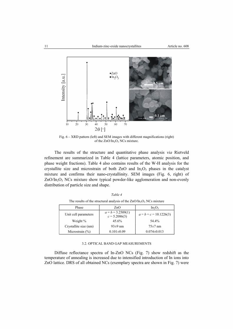
11 Indium-zinc-oxide nanocrystallites Article no. 608
Fig. 6 – XRD pattern (left) and SEM images with different magnifications (right)
of the ZnO/In2O3 NCs mixture.
The results of the structure and quantitative phase analysis via Rietveld
refinement are summarized in Table 4 (lattice parameters, atomic position, and
phase weight fractions). Table 4 also contains results of the W-H analysis for the
crystallite size and microstrain of both ZnO and In2O3 phases in the catalyst
mixture and confirms their nano-crystallinity. SEM images (Fig. 6, right) of
ZnO/In2O3 NCs mixture show typical powder-like agglomeration and non-evenly
distribution of particle size and shape.
Table 4
The results of the structural analysis of the ZnO/In2O3 NCs mixture
Phase ZnO In2O3
Unit cell parameters a = b = 3.2509(1)
c = 5.2086(3) a = b = c = 10.1226(3)
Weight % 45.6% 54.4%
Crystallite size (nm) 93±9 nm 75±7 nm
Microstrain (%) 0.101±0.09 0.074±0.013
3.2. OPTICAL BAND GAP MEASUREMENTS
Diffuse reflectance spectra of In-ZnO NCs (Fig. 7) show redshift as the
temperature of annealing is increased due to intensified introduction of In ions into
ZnO lattice. DRS of all obtained NCs (exemplary spectra are shown in Fig. 7) were
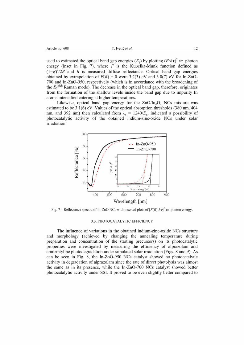
Article no. 608 T. Ivetić et al. 12
used to estimated the optical band gap energies (Eg) by plotting (F·h)2 vs. photon
energy (inset in Fig. 7), where F is the Kubelka-Munk function defined as
(1R)2/2R and R is measured diffuse reflectance. Optical band gap energies
obtained by extrapolation of F(R) = 0 were 3.2(3) eV and 3.0(7) eV for In-ZnO-
700 and In-ZnO-950, respectively (which is in accordance with the broadening of
the E2high
Raman mode). The decrease in the optical band gap, therefore, originates
from the formation of the shallow levels inside the band gap due to impurity In
atoms intensified entering at higher temperatures.
Likewise, optical band gap energy for the ZnO/In2O3 NCs mixture was
estimated to be 3.1(6) eV. Values of the optical absorption thresholds (380 nm, 404
nm, and 392 nm) then calculated from λg = 1240/Eg, indicated a possibility of
photocatalytic activity of the obtained indium-zinc-oxide NCs under solar
irradiation.
Fig. 7 – Reflectance spectra of In-ZnO NCs with inserted plots of [F(R)·h]2 vs. photon energy.
3.3. PHOTOCATALYTIC EFFICIENCY
The influence of variations in the obtained indium-zinc-oxide NCs structure
and morphology (achieved by changing the annealing temperature during
preparation and concentration of the starting precursors) on its photocatalytic
properties were investigated by measuring the efficiency of alprazolam and
amitriptyline photodegradation under simulated solar irradiation (Figs. 8 and 9). As
can be seen in Fig. 8, the In-ZnO-950 NCs catalyst showed no photocatalytic
activity in degradation of alprazolam since the rate of direct photolysis was almost
the same as in its presence, while the In-ZnO-700 NCs catalyst showed better
photocatalytic activity under SSI. It proved to be even slightly better compared to
13 Indium-zinc-oxide nanocrystallites Article no. 608
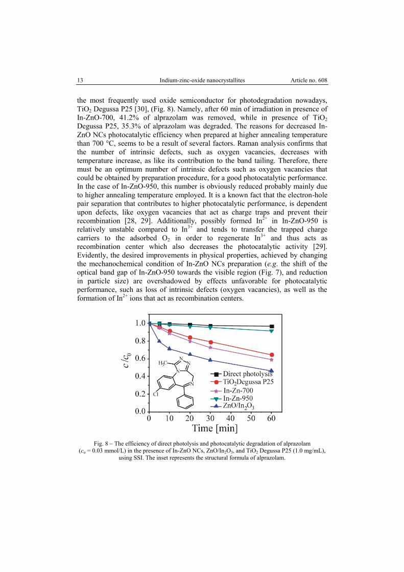
the most frequently used oxide semiconductor for photodegradation nowadays,
TiO2 Degussa P25 [30], (Fig. 8). Namely, after 60 min of irradiation in presence of
In-ZnO-700, 41.2% of alprazolam was removed, while in presence of TiO2
Degussa P25, 35.3% of alprazolam was degraded. The reasons for decreased In-
ZnO NCs photocatalytic efficiency when prepared at higher annealing temperature
than 700 °C, seems to be a result of several factors. Raman analysis confirms that
the number of intrinsic defects, such as oxygen vacancies, decreases with
temperature increase, as like its contribution to the band tailing. Therefore, there
must be an optimum number of intrinsic defects such as oxygen vacancies that
could be obtained by preparation procedure, for a good photocatalytic performance.
In the case of In-ZnO-950, this number is obviously reduced probably mainly due
to higher annealing temperature employed. It is a known fact that the electron-hole
pair separation that contributes to higher photocatalytic performance, is dependent
upon defects, like oxygen vacancies that act as charge traps and prevent their
recombination [28, 29]. Additionally, possibly formed In2+
in In-ZnO-950 is
relatively unstable compared to In3+
and tends to transfer the trapped charge
carriers to the adsorbed O2 in order to regenerate In3+
and thus acts as
recombination center which also decreases the photocatalytic activity [29].
Evidently, the desired improvements in physical properties, achieved by changing
the mechanochemical condition of In-ZnO NCs preparation (e.g. the shift of the
optical band gap of In-ZnO-950 towards the visible region (Fig. 7), and reduction
in particle size) are overshadowed by effects unfavorable for photocatalytic
performance, such as loss of intrinsic defects (oxygen vacancies), as well as the
formation of In2+
ions that act as recombination centers.
Fig. 8 – The efficiency of direct photolysis and photocatalytic degradation of alprazolam
(co = 0.03 mmol/L) in the presence of In-ZnO NCs, ZnO/In2O3, and TiO2 Degussa P25 (1.0 mg/mL),
using SSI. The inset represents the structural formula of alprazolam.
Article no. 608 T. Ivetić et al. 14
The significantly better result was obtained when ZnO/In2O3 NCs mixture
was used as a photocatalyst in photocatalytic degradation of alprazolam. Namely,
by using ZnO/In2O3, 53.7% of alprazolam was removed after 60 min of irradiation.
Bearing in mind that ZnO/In2O3 showed the highest efficiency in degradation of
alprazolam and in order to investigate the influence of substrate type on the process
of photocatalytic degradation, efficiency of mentioned photocatalyst was
investigated in degradation of amitriptyline under simulated solar irradiation.
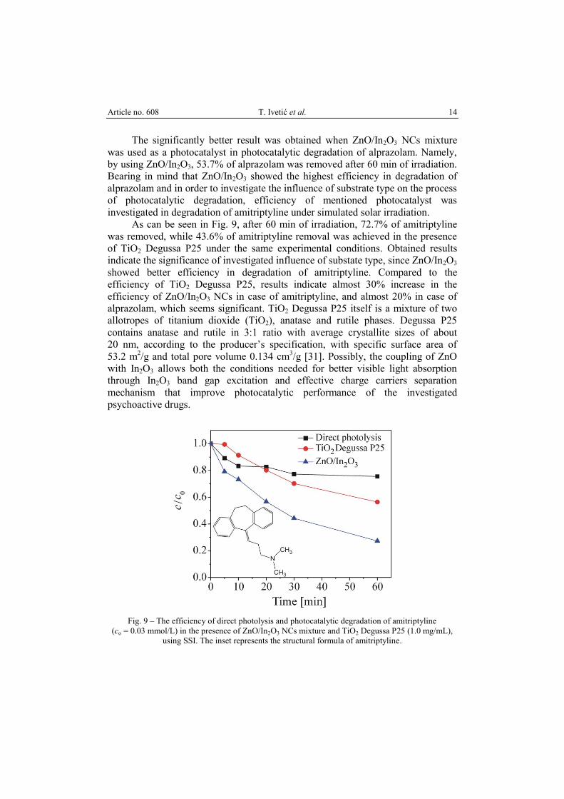
As can be seen in Fig. 9, after 60 min of irradiation, 72.7% of amitriptyline
was removed, while 43.6% of amitriptyline removal was achieved in the presence
of TiO2 Degussa P25 under the same experimental conditions. Obtained results
indicate the significance of investigated influence of substate type, since ZnO/In2O3
showed better efficiency in degradation of amitriptyline. Compared to the
efficiency of TiO2 Degussa P25, results indicate almost 30% increase in the
efficiency of ZnO/In2O3 NCs in case of amitriptyline, and almost 20% in case of
alprazolam, which seems significant. TiO2 Degussa P25 itself is a mixture of two
allotropes of titanium dioxide (TiO2), anatase and rutile phases. Degussa P25
contains anatase and rutile in 3:1 ratio with average crystallite sizes of about
20 nm, according to the producerʼs specification, with specific surface area of
53.2 m2/g and total pore volume 0.134 cm
3/g [31]. Possibly, the coupling of ZnO
with In2O3 allows both the conditions needed for better visible light absorption
through In2O3 band gap excitation and effective charge carriers separation
mechanism that improve photocatalytic performance of the investigated
psychoactive drugs.
Fig. 9 – The efficiency of direct photolysis and photocatalytic degradation of amitriptyline
(co = 0.03 mmol/L) in the presence of ZnO/In2O3 NCs mixture and TiO2 Degussa P25 (1.0 mg/mL),
using SSI. The inset represents the structural formula of amitriptyline.
15 Indium-zinc-oxide nanocrystallites Article no. 608
4. CONCLUSIONS
To summarize, this paper reports the structure and optical properties of
mechanochemically prepared indium-zinc-oxide NCs. XRD, Raman, SEM-EDS,
and UV-Vis spectroscopy were employed to characterize the obtained NCs. The
potential application of the obtained NCs in photocatalytic processes was examined
by determining the photocatalytic degradation kinetics of alprazolam and
amitriptyline in the water solutions, for the first time to the best of our knowledge
by using the obtained indium-zinc-oxide NCs and under simulated solar irradiation.
The results show, in the case of In-ZnO samples, that the annealing temperature
should be not higher than 700 °C. Even though the particle sizes are significantly
reduced at the higher temperature and optical band gap set for better visible light
absorption, it badly affects the photocatalytic activity as unstable In2+
that
accelerates the recombination rate and diminishes the photocatalytic efficiency is
formed. The higher photocatalytic efficiency was obtained in the case of forming
the contact-type structure between ZnO and In2O3 (ZnO/In2O3 NCs mixture),
which seems to be a result of a synergetic effect of both visible light activation
through In2O3 absorption and charge separation mechanism.
Acknowledgments. The authors are grateful to the APV Provincial Secretariat for Higher
Education and Scientific Research for partly financing this work, and acknowledge the support of the
Ministry of Education, Science and Technological Development of the Republic of Serbia (Project
numbers: ON 172042, ON 171022 and III 45020).
REFERENCES
1. S. Sarkar, R. Das, H. Choi, C. Bhattacharjee, RSC Adv. 4, 57250–57266 (2014).
2. A. Gajović, A.M.T. Silva, R.A. Segundo, S. Šturm, B. Jančar, M. Čeh, Appl. Catal. B Environ.
103, 351–361 (2011).
3. A.B. Djurišić, Y.H. Leung, A.M. Ching Ng, Mater. Horiz. 1, 400–410 (2014).
4. M.M. Khan, S.F. Adil, A. Al-Mayouf, J. Saudi Chem. Soc. 19, 462–464 (2015).
5. S.G. Kumar, K.S.R.K. Rao, RSC Adv. 5, 3306–3351 (2015).
6. S. Martha, K.H. Reddy, K.M. Parida, J. Mater. Chem. A 2, 3621–3631 (2014).
7. Z. Wang, B. Huang, Y. Dai, X. Qin, X. Zhang, P. Wang, H. Liu, J. Yu, J. Phys. Chem. C 113,
4612–4617 (2009).
8. A. Janotti, C.G. Van de Walle, Rep. Prog. Phys. 72, 126501 (2009).
9. X. Zhang, D. Xu, D. Huang, F. Liu, K. Xu, H. Wang, S. Zhang, J. Am. Ceram. Soc. 100, 2781–
2789 (2017).
10. T.B. Ivetić, M.R. Dimitrievska, N.L. Finčur, Lj.R. Đačanin, I.O. Guth, B.F. Abramović,
S.R. Lukić-Petrović, Ceram. Int. 40, 1545–1552 (2014).
11. T.B. Ivetić, N.L. Finčur, Lj.R. Đačanin, B.F. Abramović, S.R. Lukić-Petrović, Mater. Res. Bull.
62, 114–121 (2015).
12. P. Pérez-Lozano, E. García-Montoya, A. Orriols, M. Miñarro, J.R. Ticó, J.M. Suñé-Negre,
J. Pharmaceut. Biomed. Anal. 34, 979–987 (2004).
13. M.R. Ganjali, H. Haji-Hashemi, F. Faridbod, P. Norouzi, M. Qomi, Int. J. Electrochem. Sci. 7,
1470–1481 (2012).
Article no. 608 T. Ivetić et al. 16
14. T.B. Ivetić, N.L. Finčur, B.F. Abramović, M.R. Dimitrievska, G.R. Štrbac, K.O. Čajko,
B.B. Miljević, Lj.R. Đačanin, S.R. Lukić-Petrović, Ceram. Int. 42, 3575–3583 (2016).
15. H. Li, M.W. Sumarah, E. Topp, Environ. Toxicol. Chem. 32, 509–516 (2013).
16. M. Wu, J. Xiang, C. Que, F. Chen, G. Xu, Chemosphere 138, 486–493 (2015).
17. P. Nagarnaik, A. Batt, B. Boulanger, J. Environ. Manage. 92, 872–877 (2011).
18. P. Bottoni, S. Caroli, A. Barra Caracciolo, Toxicol. Environ. Chem. 92, 549–565 (2010).
19. Y. Vystavna, F. Huneau, V. Grynenko, Y. Vergeles, H. Celle-Jeanton, N. Tapie, H. Budzinski,
P. Le Coustumer, Water Air Soil Poll. 223, 2111–2124 (2012).
20. K. Ralphs, C. Hardacre, S.L. James, Chem. Soc. Rev. 42, 7701–7718 (2013).
21. M. Dimitrievska, T.B. Ivetić, A.P. Litvinchuk, A. Fairbrother, B.B. Miljević, G.R. Štrbac,
A. Pérez Rodríguez, S.R. Lukić-Petrović, J. Phys. Chem. C 120, 18887–18894 (2016).
22. G.K. Williamson, W. Hall, Acta Metall. 1, 22–31 (1953).
23. V.D. Mote, Y. Purushotham, B.N. Dole, J. Theor. Appl. Phys. 6, 6 (2012).
24. T. Ungár, Proc. of the Denver X-ray Conference. Advances in X-ray Analysis 40, 612 (1996).
25. X. Yu-Jing, G. Zi-Sheng, H. Tao, Chin. Phys. B 23, 087701 (2014).
26. Z. Ruiong, C. Jianxun, J. Hanying, J. Cent. South Univ. Technol. 4, 13–15 (1997).
27. T.B. Ivetić, M.R. Dimitrievska, I.O. Gúth, Lj.R. Đačanin, S.R. Lukić-Petrović, J. Res. Phys. 36,
43–51 (2012).
28. J. Xu, Y. Teng, F. Teng, Sci. Rep. 6, 32457 (2016).
29. A. Younis, D. Chu, Y.V. Kaneti, S. Li, Nanoscale 8, 378–387 (2016).
30. T. Ohno, K. Sarukawa, K. Tokieda, M. Matsumura, J. Catal. 203, 82–86 (2001).
31. N. Tomić, M. Grujić-Brojčin, N. Finčur, B. Abramović, B. Simović, J. Krstić, B. Matović,
M. Šćepanović, Mater. Chem. Phys. 163, 518–528 (2015).