Embed Size (px)
Citation preview
Ic
Ha
b
c
a
ARRAA
KIPTMT
1
btatAcetIBy�
m
mf
r
h0l
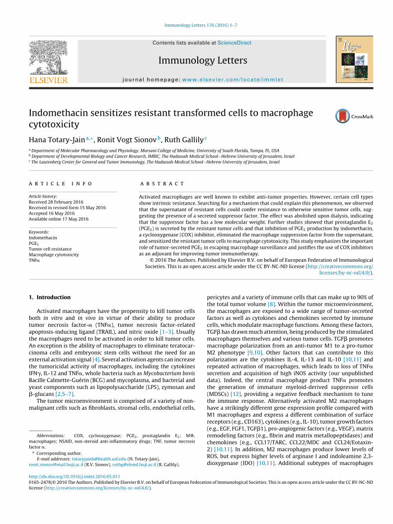
Immunology Letters 176 (2016) 1–7
Contents lists available at ScienceDirect
Immunology Letters
j ourna l ho me page: www.elsev ier .com/ locate / immlet
ndomethacin sensitizes resistant transformed cells to macrophageytotoxicity
ana Totary-Jaina,∗, Ronit Vogt Sionovb, Ruth Gallilyc
Department of Molecular Pharmacology and Physiology, Morsani College of Medicine, University of South Florida, Tampa, FL, USADepartment of Developmental Biology and Cancer Research, IMRIC, The Hadassah Medical School−Hebrew University of Jerusalem, IsraelThe Lautenberg Center for General and Tumor Immunology, The Hadassah Medical School−Hebrew University of Jerusalem, Israel
r t i c l e i n f o
rticle history:eceived 28 February 2016eceived in revised form 15 May 2016ccepted 16 May 2016vailable online 17 May 2016
eywords:
a b s t r a c t
Activated macrophages are well known to exhibit anti-tumor properties. However, certain cell typesshow intrinsic resistance. Searching for a mechanism that could explain this phenomenon, we observedthat the supernatant of resistant cells could confer resistance to otherwise sensitive tumor cells, sug-gesting the presence of a secreted suppressor factor. The effect was abolished upon dialysis, indicatingthat the suppressor factor has a low molecular weight. Further studies showed that prostaglandin E2
(PGE2) is secreted by the resistant tumor cells and that inhibition of PGE2 production by indomethacin,
ndomethacinGE2
umor cell resistanceacrophage cytotoxicity
NF�
a cyclooxygenase (COX) inhibitor, eliminated the macrophage suppression factor from the supernatant,and sensitized the resistant tumor cells to macrophage cytotoxicity. This study emphasizes the importantrole of tumor-secreted PGE2 in escaping macrophage surveillance and justifies the use of COX inhibitorsas an adjuvant for improving tumor immunotherapy.
© 2016 The Authors. Published by Elsevier B.V. on behalf of European Federation of ImmunologicalSocieties. This is an open access article under the CC BY-NC-ND license (http://creativecommons.org/
. Introduction
Activated macrophages have the propensity to kill tumor cellsoth in vitro and in vivo in virtue of their ability to produceumor necrosis factor-� (TNF�), tumor necrosis factor-relatedpoptosis-inducing ligand (TRAIL), and nitric oxide [1–3]. Usuallyhe macrophages need to be activated in order to kill tumor cells.n exception is the ability of macrophages to eliminate teratocar-inoma cells and embryonic stem cells without the need for anxternal activation signal [4]. Several activation agents can increasehe tumoricidal activity of macrophages, including the cytokinesFN�, IL-12 and TNF�, whole bacteria such as Mycobacterium bovisacille Calmette-Guérin (BCG) and mycoplasma, and bacterial andeast components such as lipopolysaccharide (LPS), zymosan and
-glucans [2,5–7].The tumor microenvironment is comprised of a variety of non-alignant cells such as fibroblasts, stromal cells, endothelial cells,
Abbreviations: COX, cyclooxygenase; PGE2, prostaglandin E2; M�,acrophages; NSAID, non-steroid anti-inflammatory drugs; TNF, tumor necrosis
actor �.∗ Corresponding author.
E-mail addresses: [email protected] (H. Totary-Jain),[email protected] (R.V. Sionov), [email protected] (R. Gallily).
ttp://dx.doi.org/10.1016/j.imlet.2016.05.011165-2478/© 2016 The Authors. Published by Elsevier B.V. on behalf of European Federatio
icense (http://creativecommons.org/licenses/by-nc-nd/4.0/).
licenses/by-nc-nd/4.0/).
pericytes and a variety of immune cells that can make up to 90% ofthe total tumor volume [8]. Within the tumor microenvironment,the macrophages are exposed to a wide range of tumor-secretedfactors as well as cytokines and chemokines secreted by immunecells, which modulate macrophage functions. Among these factors,TGF� has drawn much attention, being produced by the stimulatedmacrophages themselves and various tumor cells. TGF� promotesmacrophage polarization from an anti-tumor M1 to a pro-tumorM2 phenotype [9,10]. Other factors that can contribute to thispolarization are the cytokines IL-4, IL-13 and IL-10 [10,11] andrepeated activation of macrophages, which leads to loss of TNF�secretion and acquisition of high iNOS activity (our unpublisheddata). Indeed, the central macrophage product TNF� promotesthe generation of immature myeloid-derived suppressor cells(MDSCs) [12], providing a negative feedback mechanism to tunethe immune response. Alternatively activated M2 macrophageshave a strikingly different gene expression profile compared withM1 macrophages and express a different combination of surfacereceptors (e.g., CD163), cytokines (e.g., IL-10), tumor growth factors(e.g., EGF, FGF1, TGF�1), pro-angiogenic factors (e.g., VEGF), matrixremodeling factors (e.g., fibrin and matrix metallopeptidases) and
chemokines (e.g., CCL17/TARC, CCL22/MDC and CCL24/Eotaxin-2) [10,11]. In addition, M2 macrophages produce lower levels ofROS, but express higher levels of arginase I and indoleamine 2,3-dioxygenase (IDO) [10,11]. Additional subtypes of macrophagesn of Immunological Societies. This is an open access article under the CC BY-NC-ND
2 unolog
hm
tsmlpfnoo
2
2
Hev
2
fhC1hlsbaLpDb
Lfiglsccm
2
B1brHisplmnflwc
H. Totary-Jain et al. / Imm
ave also been identified [13], suggesting a broad spectrum ofacrophage activation stages [6].We have observed that while some tumor cells are susceptible
o macrophage cytotoxicity, others are resistant. The aim of ourtudy was to characterize the mechanisms involved in conferringacrophage resistance upon tumor cells. This study shows that a
ow molecular weight factor secreted by tumor cells, defined asrostaglandin E2 (PGE2), prevents macrophage activation requiredor tumor cytotoxicity. Inhibition of PGE2 production, using theon-steroid anti-inflammatory drug (NSAID) indomethacin, notnly restored macrophage activation, but also conferred sensitivityf the otherwise resistant tumor cells to macrophage cytotoxicity.
. Material and methods
.1. Mice
C57BL/6 mice were obtained from the Animal Breeding Farm,ebrew University-Hadassah Medical School of Jerusalem. Allxperiments involving animals were approved by the Hebrew Uni-ersity’s Institutional Animal Care and Use Committee.
.2. Cell cultures
Bone-marrow-derived macrophages (BMM�) were obtainedrom bone marrow cells (usually ∼30 × 106 cells per mouse)arvested from the femur and tibia of 6–8-week old female57BL/6 mice, which were cultivated in DMEM supplemented with5% heat-inactivated fetal calf serum (FCS), 5% heat-inactivatedorse serum, 30% L929 cell conditioned medium (LCM), 2 mM-glutamine, 10 mM HEPES, 100 U/ml penicillin and 100 �g/mltreptomycin. The macrophages were cultivated on 9 cm diameteracteriological grade culture dishes (Miniplast, Ein Shemer, Israel)nd were used as effectors 10–21 days after bone marrow seeding.CM was prepared by seeding 106 L929 cells in 20 ml DMEM sup-lemented with 10% FCS in a 75 cm2 tissue culture flask (Nunclon,enmark). Following 4–5 days incubation, when a monolayer hadeen reached, the supernatant was collected and sterile filtered.
A9 fibrosarcoma cells (a C3H fibrosarcoma derived from929 cells), L929 fibrosarcoma cells, NIH3T3 mouse embryonicbroblast-like cells and M109 Madison lung carcinoma cells wererown in DMEM supplemented with 5% heat-inactivated FCS, 2 mM-glutamine, 10 mM HEPES and antibiotics. The FCS used waselected from batches that did not pre-activate macrophages. Allell cultures were incubated at 37◦C in a humidified incubatorontaining 5% CO2. All cultures were routinely tested for beingycoplasma-free.
.3. Cytotoxicity assay
Cytotoxicity assay was performed as described previously [4].riefly, target cells in the log phase of growth were pulsed with
�Ci/ml of [3H]thymidine (sp. Act 5 Ci/mM; American Radiola-eled Chemicals, Inc.) for 24 h, washed in PBS, trypsinized andesuspended in DMEM with 10% FCS, 2 mM l-glutamine, and 10 mMEPES. Ten thousand target cells were added to 1 × 10⁵ BMM�
n 96 flat-bottomed microwells (Nunc, Denmark) in 300 �l DMEMupplemented with 10% heat-inactivated FCS in the absence orresence of 1 �g/ml LPS (Escherichia coli o55:B5, Bacto®, Difco). Fol-
owing 72 h, the supernatants (300 �l) were harvested from theicrowells, diluted with Insta-Gel (Packard, Downers Grove, Illi-
ois) and the radioactivity counted. The samples were kept at 4◦C
or 24 h prior to counting. Percentage specific cytolysis was calcu-ated by the following formula: % Cytolysis = [(E-SR)/(T-SR)] × 100%here E is the d.p.m. of the supernatant from co-culture of targetells and macrophages, SR the spontaneous release in d.p.m. of an
y Letters 176 (2016) 1–7
equal number of target cells in medium without macrophages, andT the total d.p.m. uptake of target cells.
For determination of target cell sensitivity to TNF�, 5000[3H]thymidine-labeled cells were seeded in 100 �l DMEM with 5%FCS in each well of a 96-well plate. At the following day, 50 �l of var-ious concentrations of TNF� (Genentech Inc., San Francisco) wereadded, followed by a 3-day incubation at 37◦C. The extent of celldeath was determined by measuring the released radioactivity asdescribed above. Control wells got 50 �l of medium. Alternatively,cells were incubated with TNF� in the presence of 2 �g/ml Acti-nomycin D (Sigma), and the extent of cell death determined 18 hlater.
2.4. Production of TNF ̨ by activated macrophages anddetermination of TNF ̨ titer
One hundred thousand BMM� were added to each of the 96 flat-bottomed microwells in 100 �l DMEM with 10% heat-inactivatedFCS, 18 h prior to activation. The activation step was performed bychanging the medium to DMEM without FCS or cell culture super-natant in the same medium, either in the absence or presence of1 �g/ml LPS, followed by incubation for 24 h. Macrophage super-natants were assayed for TNF� by bioassay as described previously[4]. Briefly, 4 × 10⁴ Cl-7 cells were plated per 96 flat-bottomedmicrowell in 100 �l DMEM with 5% FCS. On the following day, 3-fold dilutions of test supernatants and control media were madein the wells, followed by immediate addition of actinomycin D(Sigma; 2 �g/ml, final concentration). The cultures were incubatedfor 20 h at 37◦C, and the survived Cl-7 cells were stained for 10 minwith crystal violet (0.2% in 2% ethanol), washed with running tapwater and allowed to dry. The destruction of the Cl-7 monolayerwas determined by the amount of light (at 550 nm) absorbed bythe residual stained cells in the wells using a Dynatech MicroElisaReader (Artek, Farmingdale, NY). The S50 titer of TNF� was definedas the reciprocal of the dilution of the test solution required todestroy 50% of the target cell monolayer, as compared to controlsamples.
2.5. Conditioned medium of cultured cells
2 × 10⁵ cells were seeded per well in a 24-well culture plate(Nunclon, Denmark) in 1.5 ml DMEM with 5% FCS. After 24 h, themedium was exchanged to fresh medium, and the supernatant col-lected 24 h later. The supernatants were centrifuged at 1200 rpm for15 min, and kept at 4◦C until use.
2.6. Surface TNF ̨ receptor binding assay
The assay was performed in accordance to Holtmann & Wallach[14]. TNF� was labeled with 125I by the chloramine-T method toa specific radioactivity of 1500 Ci/mmol. One million target cellswere seeded in growth medium in tissue culture plates the daybefore assay. On the following day, the cells were washes andincubated on ice for 2 h with 0.5 nM 125I-TNF� in the absenceor presence of excess unlabeled TNF� (20 �M) in PBS containing140 mM NaCl, 1.5 mM KH2PO4, 8 mM Na2HPO4, 2.7 mM KCl, 0.5 mMMgCl2, 0.9 mM CaCl2, 0.5% bovine serum albumin (BSA) and 15 mMsodium azide. Thereafter, the cells were washed three times in thebinding buffer, detached in Ca2+- and Mg2+-free PBS containing
5 mM EDTA and transferred to vials for radioactivity measure-ments. Specific binding of TNF� was calculated by subtracting thevalues of binding observed in the presence of an excess of unlabeledTNF� from the value of binding observed with 127I-TNF� alone.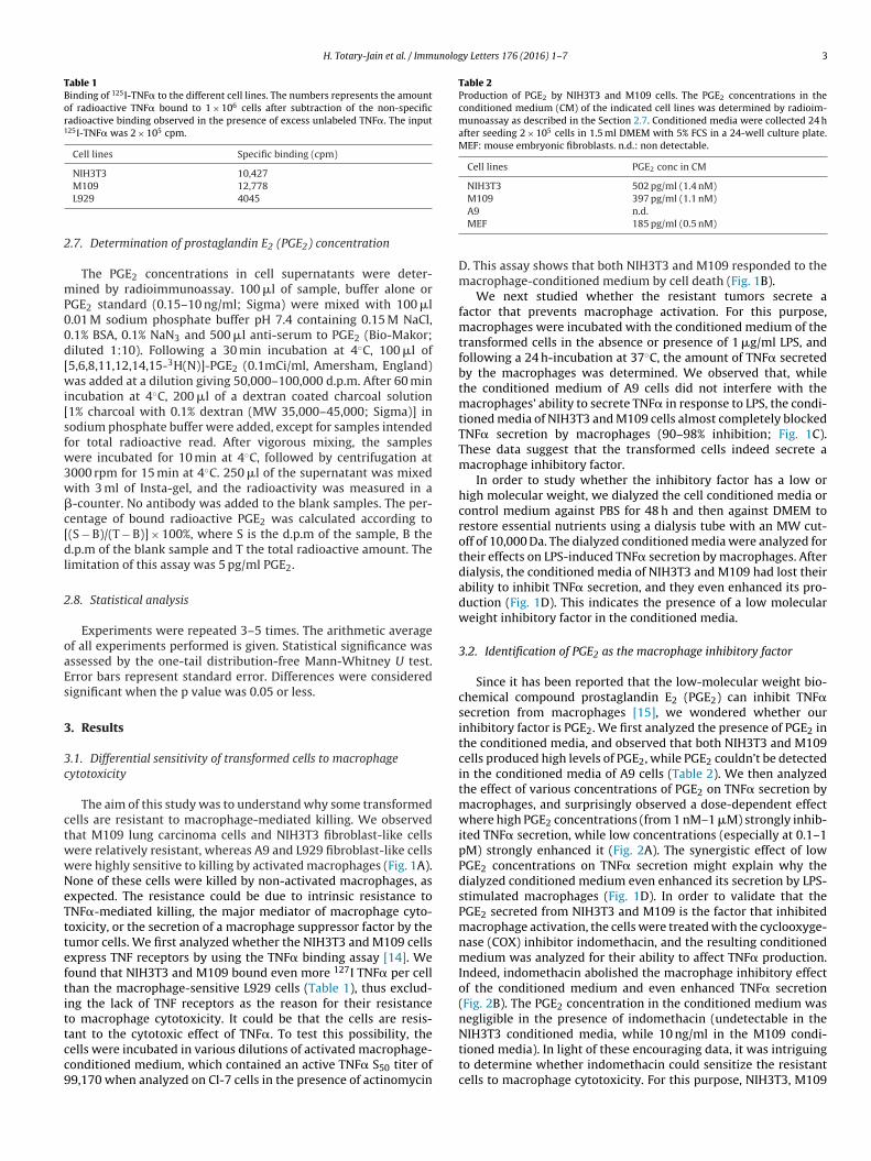
H. Totary-Jain et al. / Immunology Letters 176 (2016) 1–7 3
Table 1Binding of 125I-TNF� to the different cell lines. The numbers represents the amountof radioactive TNF� bound to 1 × 10⁶ cells after subtraction of the non-specificradioactive binding observed in the presence of excess unlabeled TNF�. The input125I-TNF� was 2 × 10⁵ cpm.
Cell lines Specific binding (cpm)
NIH3T3 10,427
2
mP00d[wi[sfw3w�c[dl
2
oaEs
3
3c
ctwwNeTtteftittcc9
Table 2Production of PGE2 by NIH3T3 and M109 cells. The PGE2 concentrations in theconditioned medium (CM) of the indicated cell lines was determined by radioim-munoassay as described in the Section 2.7. Conditioned media were collected 24 hafter seeding 2 × 10⁵ cells in 1.5 ml DMEM with 5% FCS in a 24-well culture plate.MEF: mouse embryonic fibroblasts. n.d.: non detectable.
Cell lines PGE2 conc in CM
NIH3T3 502 pg/ml (1.4 nM)M109 397 pg/ml (1.1 nM)
M109 12,778L929 4045
.7. Determination of prostaglandin E2 (PGE2) concentration
The PGE2 concentrations in cell supernatants were deter-ined by radioimmunoassay. 100 �l of sample, buffer alone or
GE2 standard (0.15–10 ng/ml; Sigma) were mixed with 100 �l.01 M sodium phosphate buffer pH 7.4 containing 0.15 M NaCl,.1% BSA, 0.1% NaN3 and 500 �l anti-serum to PGE2 (Bio-Makor;iluted 1:10). Following a 30 min incubation at 4◦C, 100 �l of5,6,8,11,12,14,15-3H(N)]-PGE2 (0.1mCi/ml, Amersham, England)as added at a dilution giving 50,000–100,000 d.p.m. After 60 min
ncubation at 4◦C, 200 �l of a dextran coated charcoal solution1% charcoal with 0.1% dextran (MW 35,000–45,000; Sigma)] inodium phosphate buffer were added, except for samples intendedor total radioactive read. After vigorous mixing, the samplesere incubated for 10 min at 4◦C, followed by centrifugation at
000 rpm for 15 min at 4◦C. 250 �l of the supernatant was mixedith 3 ml of Insta-gel, and the radioactivity was measured in a-counter. No antibody was added to the blank samples. The per-entage of bound radioactive PGE2 was calculated according to(S − B)/(T − B)] × 100%, where S is the d.p.m of the sample, B the.p.m of the blank sample and T the total radioactive amount. The
imitation of this assay was 5 pg/ml PGE2.
.8. Statistical analysis
Experiments were repeated 3–5 times. The arithmetic averagef all experiments performed is given. Statistical significance wasssessed by the one-tail distribution-free Mann-Whitney U test.rror bars represent standard error. Differences were consideredignificant when the p value was 0.05 or less.
. Results
.1. Differential sensitivity of transformed cells to macrophageytotoxicity
The aim of this study was to understand why some transformedells are resistant to macrophage-mediated killing. We observedhat M109 lung carcinoma cells and NIH3T3 fibroblast-like cellsere relatively resistant, whereas A9 and L929 fibroblast-like cellsere highly sensitive to killing by activated macrophages (Fig. 1A).one of these cells were killed by non-activated macrophages, asxpected. The resistance could be due to intrinsic resistance toNF�-mediated killing, the major mediator of macrophage cyto-oxicity, or the secretion of a macrophage suppressor factor by theumor cells. We first analyzed whether the NIH3T3 and M109 cellsxpress TNF receptors by using the TNF� binding assay [14]. Weound that NIH3T3 and M109 bound even more 127I TNF� per cellhan the macrophage-sensitive L929 cells (Table 1), thus exclud-ng the lack of TNF receptors as the reason for their resistanceo macrophage cytotoxicity. It could be that the cells are resis-
ant to the cytotoxic effect of TNF�. To test this possibility, theells were incubated in various dilutions of activated macrophage-onditioned medium, which contained an active TNF� S50 titer of9,170 when analyzed on Cl-7 cells in the presence of actinomycinA9 n.d.MEF 185 pg/ml (0.5 nM)
D. This assay shows that both NIH3T3 and M109 responded to themacrophage-conditioned medium by cell death (Fig. 1B).
We next studied whether the resistant tumors secrete afactor that prevents macrophage activation. For this purpose,macrophages were incubated with the conditioned medium of thetransformed cells in the absence or presence of 1 �g/ml LPS, andfollowing a 24 h-incubation at 37◦C, the amount of TNF� secretedby the macrophages was determined. We observed that, whilethe conditioned medium of A9 cells did not interfere with themacrophages’ ability to secrete TNF� in response to LPS, the condi-tioned media of NIH3T3 and M109 cells almost completely blockedTNF� secretion by macrophages (90–98% inhibition; Fig. 1C).These data suggest that the transformed cells indeed secrete amacrophage inhibitory factor.
In order to study whether the inhibitory factor has a low orhigh molecular weight, we dialyzed the cell conditioned media orcontrol medium against PBS for 48 h and then against DMEM torestore essential nutrients using a dialysis tube with an MW cut-off of 10,000 Da. The dialyzed conditioned media were analyzed fortheir effects on LPS-induced TNF� secretion by macrophages. Afterdialysis, the conditioned media of NIH3T3 and M109 had lost theirability to inhibit TNF� secretion, and they even enhanced its pro-duction (Fig. 1D). This indicates the presence of a low molecularweight inhibitory factor in the conditioned media.
3.2. Identification of PGE2 as the macrophage inhibitory factor
Since it has been reported that the low-molecular weight bio-chemical compound prostaglandin E2 (PGE2) can inhibit TNF�secretion from macrophages [15], we wondered whether ourinhibitory factor is PGE2. We first analyzed the presence of PGE2 inthe conditioned media, and observed that both NIH3T3 and M109cells produced high levels of PGE2, while PGE2 couldn’t be detectedin the conditioned media of A9 cells (Table 2). We then analyzedthe effect of various concentrations of PGE2 on TNF� secretion bymacrophages, and surprisingly observed a dose-dependent effectwhere high PGE2 concentrations (from 1 nM–1 �M) strongly inhib-ited TNF� secretion, while low concentrations (especially at 0.1–1pM) strongly enhanced it (Fig. 2A). The synergistic effect of lowPGE2 concentrations on TNF� secretion might explain why thedialyzed conditioned medium even enhanced its secretion by LPS-stimulated macrophages (Fig. 1D). In order to validate that thePGE2 secreted from NIH3T3 and M109 is the factor that inhibitedmacrophage activation, the cells were treated with the cyclooxyge-nase (COX) inhibitor indomethacin, and the resulting conditionedmedium was analyzed for their ability to affect TNF� production.Indeed, indomethacin abolished the macrophage inhibitory effectof the conditioned medium and even enhanced TNF� secretion(Fig. 2B). The PGE2 concentration in the conditioned medium wasnegligible in the presence of indomethacin (undetectable in the
NIH3T3 conditioned media, while 10 ng/ml in the M109 condi-tioned media). In light of these encouraging data, it was intriguingto determine whether indomethacin could sensitize the resistantcells to macrophage cytotoxicity. For this purpose, NIH3T3, M109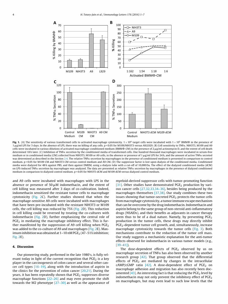
4 H. Totary-Jain et al. / Immunology Letters 176 (2016) 1–7
Fig. 1. (A) The sensitivity of various transformed cells to activated macrophage cytotoxicity. 1 × 10⁴ target cells were incubated with 1 × 10⁵ BMM� in the presence of1 �g/ml LPS for 3 days. In the absence of LPS, there was no killing of any cells. p < 0.05 for M109/NIH3T3 versus A9/L929. (B) Cell sensitivity to TNF�. NIH3T3, M109 and A9cells were incubated in various dilutions of activated macrophage-conditioned medium (BMM�-CM) in the presence of 2 �g/ml actinomycin D, and the extent of cell deathdetermined 18 h later. (C) Inhibition of TNF� secretion by the conditioned media of transformed cells. One hundred thousand macrophages were incubated in serum-freemedium or in conditioned media (CM) collected from NIH3T3, M109 or A9 cells, in the absence or presence of 1 �g/ml LPS for 24 h, and the amount of active TNF� secretedwas determined as described in the Section 2.4. The relative TNF� secretion by macrophages in the presence of conditioned medium is presented in comparison to controlmedium. p < 0.05 for M109 CM and NIH3T3 CM versus control medium and A9 CM. (D) The suppressor factor is lost upon dialysis of the conditioned media. Conditionedm is tubo ted am 109 d
aacicmtciiPtwiF
4
epctymt
edia were dialyzed for 48 h against PBS, and then against DMEM, using a dialysn LPS-induced TNF� secretion by macrophages was analyzed. The data are presenedium in comparison to dialyzed control medium. p < 0.05 for NIH3T3 dCM and M
nd A9 cells were incubated with macrophages with LPS in thebsence or presence of 50 �M indomethacin, and the extent ofell killing was measured after 3 days of co-cultivation. Indeed,ndomethacin sensitized the resistant tumor cells to macrophageytotoxicity (Fig. 2C). Further studies showed that when theacrophage-sensitive A9 cells were incubated with macrophages
hat have been pre-incubated with the resistant NIH3T3 or M109ells, the cell killing was reduced by 75% (Fig. 2D). This reductionn cell killing could be reversed by treating the co-cultures withndomethacin (Fig. 2D), further emphasizing the central role ofGE2 in mediating the macrophage inhibitory effect. This is fur-her manifested by the suppression of A9 cell killing when PGE2as added to the co-culture of A9 and macrophages (Fig. 2E). Max-
mum inhibition was obtained at 1–10 nM PGE2 (47–51% inhibition;ig. 2E).
. Discussion
Our pioneering study, performed in the late 1980′s, is fully rel-vant today in light of the current recognition that PGE2 is a keylayer in the carcinogenesis of colon cancer and several other can-er cell types [16–19], along with the introduction of aspirin in
he clinics for the prevention of colon cancer [20,21]. During theears, it has been repeatedly shown that PGE2 suppresses diverseacrophage functions [22–26] and may even promote the shiftowards the M2 phenotype [27–30] as well as the appearance of
e with a cut-off of 10,000 Da. The effect of the dialyzed conditioned media (dCM)s relative TNF� secretion by macrophages in the presence of dialyzed conditionedCM versus dialyzed control medium.
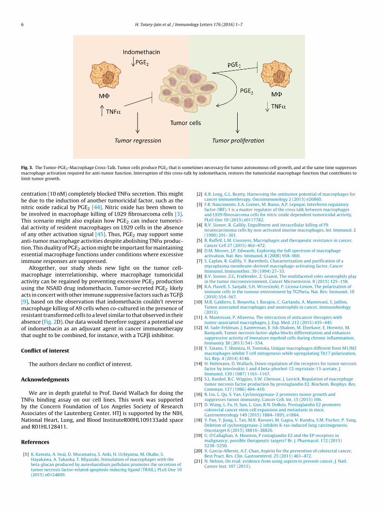
myeloid-derived suppressor cells with tumor-promoting function[31]. Other studies have demonstrated PGE2 production by vari-ous cancer cells [27,32,33,34–36], besides being produced by themacrophages themselves [37,38]. Our study combines these twoissues showing that tumor-secreted PGE2 protects the tumor cellsfrom macrophage cytotoxicity, a tumor immune escape mechanismthat can be overcome by the drug indomethacin. Indomethacin andaspirin belong to the same group of non-steroid anti-inflammatorydrugs (NSAIDs), and their benefits as adjuvants in cancer therapyseem thus to be of a dual nature. Namely, by preventing PGE2production in the tumor cells, these drugs may directly inhibitPGE2-dependent tumor cell growth, and, simultaneously, increasemacrophage cytotoxicity towards the tumor cells (Fig. 3). Bothmechanisms contribute to the reduction of the tumor cell mass.Our study suggests a mechanistic explanation for the anti-tumoreffects observed for indomethacin in various tumor models (e.g.,[39–41]).
The dose-dependent effects of PGE2 observed by us onmacrophage secretion of TNF� has also been observed by anotherresearch group [42]. That group observed that the differentialeffects of PGE2 are mediated by changes in the intracellularcAMP/cGMP ratio [42]. A dose-dependent effect of PGE2 on
macrophage adhesion and migration has also recently been doc-umented [43]. An interesting fact is that reducing the PGE2 level byindomethacin may not only prevent the inhibitory effect of PGE2on macrophages, but may even lead to such low levels that the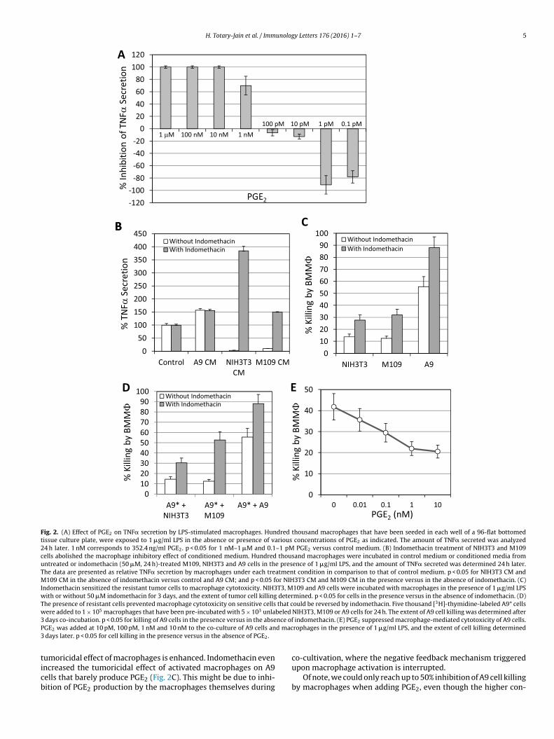
H. Totary-Jain et al. / Immunology Letters 176 (2016) 1–7 5
Fig. 2. (A) Effect of PGE2 on TNF� secretion by LPS-stimulated macrophages. Hundred thousand macrophages that have been seeded in each well of a 96-flat bottomedtissue culture plate, were exposed to 1 �g/ml LPS in the absence or presence of various concentrations of PGE2 as indicated. The amount of TNF� secreted was analyzed24 h later. 1 nM corresponds to 352.4 ng/ml PGE2. p < 0.05 for 1 nM–1 �M and 0.1–1 pM PGE2 versus control medium. (B) Indomethacin treatment of NIH3T3 and M109cells abolished the macrophage inhibitory effect of conditioned medium. Hundred thousand macrophages were incubated in control medium or conditioned media fromuntreated or indomethacin (50 �M, 24 h)-treated M109, NIH3T3 and A9 cells in the presence of 1 �g/ml LPS, and the amount of TNF� secreted was determined 24 h later.The data are presented as relative TNF� secretion by macrophages under each treatment condition in comparison to that of control medium. p < 0.05 for NIH3T3 CM andM109 CM in the absence of indomethacin versus control and A9 CM; and p < 0.05 for NIH3T3 CM and M109 CM in the presence versus in the absence of indomethacin. (C)Indomethacin sensitized the resistant tumor cells to macrophage cytotoxicity. NIH3T3, M109 and A9 cells were incubated with macrophages in the presence of 1 �g/ml LPSwith or without 50 �M indomethacin for 3 days, and the extent of tumor cell killing determined. p < 0.05 for cells in the presence versus in the absence of indomethacin. (D)The presence of resistant cells prevented macrophage cytotoxicity on sensitive cells that could be reversed by indomethacin. Five thousand [3H]-thymidine-labeled A9* cellswere added to 1 × 10⁵ macrophages that have been pre-incubated with 5 × 10³ unlabeled NIH3T3, M109 or A9 cells for 24 h. The extent of A9 cell killing was determined after3 nce ofP d mac3
ticb
days co-incubation. p < 0.05 for killing of A9 cells in the presence versus in the abseGE2 was added at 10 pM, 100 pM, 1 nM and 10 nM to the co-culture of A9 cells an
days later. p < 0.05 for cell killing in the presence versus in the absence of PGE2.
umoricidal effect of macrophages is enhanced. Indomethacin evenncreased the tumoricidal effect of activated macrophages on A9ells that barely produce PGE2 (Fig. 2C). This might be due to inhi-ition of PGE2 production by the macrophages themselves during
indomethacin. (E) PGE2 suppressed macrophage-mediated cytotoxicity of A9 cells.rophages in the presence of 1 �g/ml LPS, and the extent of cell killing determined
co-cultivation, where the negative feedback mechanism triggeredupon macrophage activation is interrupted.
Of note, we could only reach up to 50% inhibition of A9 cell killingby macrophages when adding PGE2, even though the higher con-
6 H. Totary-Jain et al. / Immunology Letters 176 (2016) 1–7
F ometim talk bl
cbnbTdoatei
maua[mraot
C
A
TbANa
R
[
[
[
[
[
[
[
[
[
[
[
ig. 3. The Tumor-PGE2-Macrophage Cross-Talk. Tumor cells produce PGE2 that is sacrophage activation required for anti-tumor function. Interruption of this cross-
imit tumor growth.
entration (10 nM) completely blocked TNF� secretion. This mighte due to the induction of another tumoricidal factor, such as theitric oxide radical by PGE2 [44]. Nitric oxide has been shown toe involved in macrophage killing of L929 fibrosarcoma cells [3].his scenario might also explain how PGE2 can induce tumorici-al activity of resident macrophages on L929 cells in the absencef any other activation signal [45]. Thus, PGE2 may support somenti-tumor macrophage activities despite abolishing TNF� produc-ion. This duality of PGE2 action might be important for maintainingssential macrophage functions under conditions where excessivemmune responses are suppressed.
Altogether, our study sheds new light on the tumor cell-acrophage interrelationship, where macrophage tumoricidal
ctivity can be regained by preventing excessive PGE2 productionsing the NSAID drug indomethacin. Tumor-secreted PGE2 likelycts in concert with other immune suppressive factors such as TGF�9], based on the observation that indomethacin couldn’t reverse
acrophage killing of A9 cells when co-cultured in the presence ofesistant transformed cells to a level similar to that observed in theirbsence (Fig. 2D). Our data would therefore suggest a potential usef indomethacin as an adjuvant agent in cancer immunotherapyhat ought to be combined, for instance, with a TGF� inhibitor.
onflict of interest
The authors declare no conflict of interest.
cknowledgments
We are in depth grateful to Prof. David Wallach for doing theNF� binding assay on our cell lines. This work was supportedy the Concern Foundation of Los Angeles Society of Researchssociates of the Lautenberg Center. HTJ is supported by the NIH,ational Heart, Lung, and Blood InstituteR00HL109133add spacend R01HL128411.
eferences
[1] K. Kawata, A. Iwai, D. Muramatsu, S. Aoki, H. Uchiyama, M. Okabe, S.Hayakawa, A. Takaoka, T. Miyazaki, Stimulation of macrophages with thebeta-glucan produced by aureobasidium pullulans promotes the secretion oftumor necrosis factor-related apoptosis inducing ligand (TRAIL), PLoS One 10(2015) e0124809.
[
mes necessary for tumor autonomous cell growth, and at the same time suppressesy indomethacin, restores the tumoricidal macrophage function that contributes to
[2] K.B. Long, G.L. Beatty, Harnessing the antitumor potential of macrophages forcancer immunotherapy, Oncoimmunology 2 (2013) e26860.
[3] F.R. Nascimento, E.A. Gomes, M. Russo, A.P. Lepique, Interferon regulatoryfactor (IRF)-1 is a master regulator of the cross talk between macrophagesand L929 fibrosarcoma cells for nitric oxide dependent tumoricidal activity,PLoS One 10 (2015) e0117782.
[4] R.V. Sionov, R. Gallily, Engulfment and intracellular killing of F9teratocarcinoma cells by non-activated murine macrophages, Int. Immunol. 2(1990) 291–301.
[5] B. Ruffell, L.M. Coussens, Macrophages and therapeutic resistance in cancer,Cancer Cell 27 (2015) 462–472.
[6] D.M. Mosser, J.P. Edwards, Exploring the full spectrum of macrophageactivation, Nat. Rev. Immunol. 8 (2008) 958–969.
[7] S. Caplan, R. Gallily, Y. Barenholz, Characterization and purification of amycoplasma membrane-derived macrophage-activating factor, CancerImmunol. Immunother. 39 (1994) 27–33.
[8] R.V. Sionov, Z.G. Fridlender, Z. Granot, The multifaceted roles neutrophils playin the tumor microenvironment, Cancer Microenviron. 8 (2015) 125–158.
[9] R.A. Flavell, S. Sanjabi, S.H. Wrzesinski, P. Licona-Limon, The polarization ofimmune cells in the tumour environment by TGFbeta, Nat. Rev. Immunol. 10(2010) 554–567.
10] M.R. Galdiero, E. Bonavita, I. Barajon, C. Garlanda, A. Mantovani, S. Jaillon,Tumor associated macrophages and neutrophils in cancer, Immunobiology(2013).
11] A. Mantovani, P. Allavena, The interaction of anticancer therapies withtumor-associated macrophages, J. Exp. Med. 212 (2015) 435–445.
12] M. Sade-Feldman, J. Kanterman, E. Ish-Shalom, M. Elnekave, E. Horwitz, M.Baniyash, Tumor necrosis factor-alpha blocks differentiation and enhancessuppressive activity of immature myeloid cells during chronic inflammation,Immunity 38 (2013) 541–554.
13] Y. Tatano, T. Shimizu, H. Tomioka, Unique macrophages different from M1/M2macrophages inhibit T cell mitogenesis while upregulating Th17 polarization,Sci. Rep. 4 (2014) 4146.
14] H. Holtmann, D. Wallach, Down regulation of the receptors for tumor necrosisfactor by interleukin 1 and 4 beta-phorbol-12-myristate-13-acetate, J.Immunol. 139 (1987) 1161–1167.
15] S.L. Kunkel, R.C. Wiggins, S.W. Chensue, J. Larrick, Regulation of macrophagetumor necrosis factor production by prostaglandin E2, Biochem. Biophys. Res.Commun. 137 (1986) 404–410.
16] B. Liu, L. Qu, S. Yan, Cyclooxygenase-2 promotes tumor growth andsuppresses tumor immunity, Cancer Cell. Int. 15 (2015) 106.
17] D. Wang, L. Fu, H. Sun, L. Guo, R.N. DuBois, Prostaglandin E2 promotescolorectal cancer stem cell expansion and metastasis in mice,Gastroenterology 149 (2015) 1884–1895, e1884.
18] Y. Pan, Y. Jiang, L. Tan, M.K. Ravoori, M. Gagea, V. Kundra, S.M. Fischer, P. Yang,Deletion of cyclooxygenase-2 inhibits K-ras-induced lung carcinogenesis,Oncotarget 6 (2015) 38816–38826.
19] G. O’Callaghan, A. Houston, P rostaglandin E2 and the EP receptors inmalignancy: possible therapeutic targets? Br. J. Pharmacol. 172 (2015)5239–5250.
20] X. Garcia-Albeniz, A.T. Chan, Aspirin for the prevention of colorectal cancer,
Best Pract. Res. Clin. Gastroenterol. 25 (2011) 461–472.21] N. Nelson, On trial: evidence from using aspirin to prevent cancer, J. Natl.Cancer Inst. 107 (2015).
unolog
[
[
[
[
[
[
[
[
[
[
[
[
[
[
[
[
[
[
[
[
[
[
[
H. Totary-Jain et al. / Imm
22] J.G. Kim, Y.S. Hahn, IFN-gamma inhibits the suppressive effects of PGE2 on theproduction of tumor necrosis factor-alpha by mouse macrophages, Immunol.Invest. 29 (2000) 257–269.
23] X. Qian, J. Zhang, J. Liu, Tumor-secreted PGE2 inhibits CCL5 production inactivated macrophages through cAMP/PKA signaling pathway, J. Biol. Chem.286 (2011) 2111–2120.
24] Z. Konopski, R. Seljelid, T. Eskeland, Cytokines and PGE2 modulate thephagocytic function of the beta-glucan receptor in macrophages, Scand. J.Immunol. 37 (1993) 587–592.
25] G. Strassmann, V. Patil-Koota, F. Finkelman, M. Fong, T. Kambayashi, Evidencefor the involvement of interleukin 10 in the differential deactivation ofmurine peritoneal macrophages by prostaglandin E2, J. Exp. Med. 180 (1994)2365–2370.
26] M. Sokolowska, L.Y. Chen, Y. Liu, A. Martinez-Anton, H.Y. Qi, C. Logun, S.Alsaaty, Y.H. Park, D.L. Kastner, J.J. Chae, J.H. Shelhamer, Prostaglandin E2inhibits NLRP3 inflammasome activation through EP4 receptor andintracellular cyclic AMP in human macrophages, J. Immunol. 194 (2015)5472–5487.
27] M. Heusinkveld, P.J. de Vos van Steenwijk, R. Goedemans, T.H.Ramwadhdoebe, A. Gorter, M.J. Welters, T. van Hall, S.H. van der Burg, M2macrophages induced by prostaglandin E2 and IL-6 from cervical carcinomaare switched to activated M1 macrophages by CD4+ Th1 cells, J. Immunol. 187(2011) 1157–1165.
28] L. Liu, D. Ge, L. Ma, J. Mei, S. Liu, Q. Zhang, F. Ren, H. Liao, Q. Pu, T. Wang, Z.You, Interleukin-17 and prostaglandin E2 are involved in formation of an M2macrophage-dominant microenvironment in lung cancer, J. Thorac. Oncol. 7(2012) 1091–1100.
29] B. Luan, Y.S. Yoon, J. Le Lay, K.H. Kaestner, S. Hedrick, M. Montminy, CREBpathway links PGE2 signaling with macrophage polarization, Proc. Natl. Acad.Sci. U. S. A. 112 (2015) 15642–15647.
30] Q. Zhang, D.J. Cai, B. Li, Ovarian cancer stem-like cells elicit the polarization ofM2 macrophages, Mol. Med. Rep. 11 (2015) 4685–4693.
31] J. Chang, J. Vacher, B. Yao, X. Fan, B. Zhang, R.C. Harris, M.Z. Zhang,Prostaglandin E receptor 4 (EP4) promotes colonic tumorigenesis, Oncotarget6 (2015) 33500–33511.
32] Y. Sasaki, Y. Nakatani, S. Hara, Role of microsomal prostaglandin E synthase-1
(mPGES-1)-derived prostaglandin E2 in colon carcinogenesis, Prostag. OtherLipid Mediat. 121 (2015) 42–45.33] E. Half, X.M. Tang, K. Gwyn, A. Sahin, K. Wathen, F.A. Sinicrope,Cyclooxygenase-2 expression in human breast cancers and adjacent ductalcarcinoma in situ, Cancer Res. 62 (2002) 1676–1681.
[
y Letters 176 (2016) 1–7 7
34] A.C. Abrahao, R.M. Castilho, C.H. Squarize, A.A. Molinolo, D. dos Santos-PintoJr., J.S. Gutkind, A role for COX2-derived PGE2 and PGE2-receptor subtypes inhead and neck squamous carcinoma cell proliferation, Oral Oncol. 46 (2010)880–887.
35] J.E. Rundhaug, M.S. Simper, I. Surh, S.M. Fischer, The role of the EP receptorsfor prostaglandin E2 in skin and skin cancer, Cancer Metastasis Rev. 30 (2011)465–480.
36] K. Kaminska, C. Szczylik, F. Lian, A.M. Czarnecka, The role of prostaglandin E2in renal cell cancer development: future implications for prognosis andtherapy, Future Oncol. 10 (2014) 2177–2187.
37] E. Kuroda, U. Yamashita, Mechanisms of enhanced macrophage-mediatedprostaglandin E2 production and its suppressive role in Th1 activation inTh2-dominant BALB/c mice, J. Immunol. 170 (2003) 757–764.
38] Y. Endo, K. Blinova, T. Romantseva, H. Golding, M. Zaitseva, Differences inPGE2 production between primary human monocytes and differentiatedmacrophages: role of IL-1beta and TRIF/IRF3, PLoS One 9 (2014) e98517.
39] C.M. Moon, J.H. Kwon, J.S. Kim, S.H. Oh, K. Jin Lee, J.J. Park, S. Pil Hong, J.H.Cheon, T.I. Kim, W.H. Kim, Nonsteroidal anti-inflammatory drugs suppresscancer stem cells via inhibiting PTGS2 (cyclooxygenase 2) and NOTCH/HES1and activating PPARG in colorectal cancer, Int. J. Cancer 134 (2014) 519–529.
40] S. Santander, C. Cebrian, P. Esquivias, B. Conde, F. Esteva, P. Jimenez, J. Ortego,A. Lanas, E. Piazuelo, Cyclooxygenase inhibitors decrease the growth andinduce regression of human esophageal adenocarcinoma xenografts in nudemice, Int. J. Oncol. 40 (2012) 527–534.
41] E.M. Connolly, J.H. Harmey, T. O’Grady, D. Foley, G. Roche-Nagle, E. Kay, D.J.Bouchier-Hayes, Cyclo-oxygenase inhibition reduces tumour growth andmetastasis in an orthotopic model of breast cancer, Br. J. Cancer 87 (2002)231–237.
42] H. Renz, J.H. Gong, A. Schmidt, M. Nain, D. Gemsa, Release of tumor necrosisfactor-alpha from macrophages. Enhancement and suppression aredose-dependently regulated by prostaglandin E2 and cyclic nucleotides, J.Immunol. 141 (1988) 2388–2393.
43] I.C. Osma-Garcia, C. Punzon, M. Fresno, M.D. Diaz-Munoz, Dose-dependenteffects of prostaglandin E2 in macrophage adhesion and migration, Eur. J.Immunol. (2015).
44] S. Milano, F. Arcoleo, M. Dieli, R. D’Agostino, P. D’Agostino, G. De Nucci, E.
Cillari, Prostaglandin E2 regulates inducible nitric oxide synthase in themurine macrophage cell line J774, Prostaglandins 49 (1995) 105–115.45] M.E. Snider, R.H. Fertel, B.S. Zwilling, Prostaglandin regulation of macrophagefunction: effect of endogenous and exogenous prostaglandins, Cell. Immunol.74 (1982) 234–242.