Embed Size (px)
Citation preview

8/20/2019 Infant Cholestasis Guidelines
http://slidepdf.com/reader/full/infant-cholestasis-guidelines 1/14
Guideline for the Evaluation of Cholestatic Jaundice in Infants:
Recommendations of the North American Society for Pediatric
Gastroenterology, Hepatology and NutritionAbstract
For the primary care provider, cholestatic jaundice ininfancy, defined as jaundice caused by an elevated con- jugated bilirubin, is an uncommon but potentially seriousproblem that indicates hepatobiliary dysfunction. Earlydetection of cholestatic jaundice by the primary carephysician and timely, accurate diagnosis by the pediatricgastroenterologist are important for successful treatmentand a favorable prognosis. The Cholestasis GuidelineCommittee of the North American Society for PediatricGastroenterology, Hepatology and Nutrition has formu-
lated a clinical practice guideline for the diagnosticevaluation of cholestatic jaundice in the infant. The Cho-lestasis Guideline Committee, consisting of a primarycare pediatrician, a clinical epidemiologist (who alsopractices primary care pediatrics), and five pediatric gas-troenterologists, based its recommendations on a com-prehensive and systematic review of the medical litera-ture integrated with expert opinion. Consensus wasachieved through the Nominal Group Technique, a struc-tured quantitative method.
The Committee examined the value of diagnostic testscommonly used for the evaluation of cholestatic jaundiceand how those interventions can be applied to clinicalsituations in the infant. The guideline provides recom-
mendations for management by the primary care pro-vider, indications for consultation by a pediatric gastro-enterologist, and recommendations for management bythe pediatric gastroenterologist.
The Cholestasis Guideline Committee recommendsthat any infant noted to be jaundiced at 2 weeks of age beevaluated for cholestasis with measurement of total anddirect serum bilirubin. However, breast-fed infants whocan be reliably monitored and who have an otherwisenormal history (no dark urine or light stools) and physi-cal examination may be asked to return at 3 weeks of ageand, if jaundice persists, have measurement of total anddirect serum bilirubin at that time.
This document represents the official recommenda-
tions of the North American Society for Pediatric Gas-troenterology, Hepatology and Nutrition on the evalua-tion of cholestatic jaundice in infants. The AmericanAcademy of Pediatrics has also endorsed these recom-mendations. These recommendations are a generalguideline and are not intended as a substitute for clinical judgment or as a protocol for the care of all patients withthis problem.
BACKGROUND
Cholestatic jaundice, characterized by elevation of se-rum conjugated bilirubin, is an uncommon but poten-tially serious condition that indicates hepatobiliary dys-function. Early detection of cholestatic jaundice by theprimary care provider and timely, accurate diagnosis bythe pediatric gastroenterologist are important for suc-cessful treatment and a favorable prognosis. In contrast, physiologic jaundice and breast milk jaundice, commoncauses of jaundice in the first weeks of life, are caused byan elevation of serum unconjugated bilirubin. Both areself-limited maturational disorders observed in many in-
fants in the first weeks of life.Cholestatic jaundice affects approximately 1 in every
2,500 infants (1,2), and is thus infrequently seen by mostproviders of medical care to infants. However, distin-guishing jaundice caused by cholestasis from nonchole-static conditions is critical because cholestatic jaundice ismuch more likely to have a serious etiology that needsprompt diagnosis and therapy. The most common causesof cholestatic jaundice in the first months of life arebiliary atresia and neonatal hepatitis, which account formost cases. Neonatal hepatitis has referred to a histologicappearance of widespread giant cell transformation. Al-though giant cell transformation is recognized to be non-specific and may be associated with infectious, meta-
bolic, and syndromic disorders, this term is used to beconsistent with the older literature reviewed for thisguideline. Alpha-1 antitrypsin deficiency causes another5% to 15% of cases (1,2). The remaining cases arecaused by a variety of other disorders, including extra-hepatic obstruction from common duct gallstone or cho-ledochal cyst; metabolic disorders such as tyrosinemia,galactosemia, and hypothyroidism; inborn errors of bileacid metabolism; Alagille syndrome; infection; and otherrare disorders (Table 1).
Infants with cholestatic jaundice caused by bacterialsepsis, galactosemia, hypopituitarism, or gallstone oftenappear acutely ill. These disorders require early diagno-sis and urgent treatment. However, many infants with
cholestatic jaundice appear otherwise healthy and grownormally. The benign appearance of such an infant maylull the parents or physician into believing that the jaun-dice is physiologic or caused by breast-feeding, when infact it may be caused by biliary atresia. Biliary atresiaoccurs in 1 in 10,000 to 19,000 infants (3–6) (Elliott EJ,Australian Pediatric Surveillance Unit, Extra-hepatic Bil-iary Atresia Study Group, personal communication,
Journal of Pediatric Gastroenterology and Nutrition39:115–128 © August 2004 Lippincott Williams & Wilkins, Philadelphia
115

8/20/2019 Infant Cholestasis Guidelines
http://slidepdf.com/reader/full/infant-cholestasis-guidelines 2/14
2001). Substantial observational evidence suggests thatearlier diagnosis and surgical repair lead to better out-comes for this disorder (7). The Kasai portoenterostomyappears to have the greatest likelihood of re-establish-ment of bile flow and the longest term survival of theinfant’s native liver when performed before the age of 45to 60 days (7–10). Early diagnosis of many of the other
conditions that cause cholestasis may also lead to betteroutcomes because better support of the infant may avoidcomplications of liver disease (11).
Despite these data showing that early diagnosis is po-tentially life saving, referral for evaluation of cholestatic jaundice frequently occurs after 45 to 60 days of age(12). In recognition of this, and noting that no evidence-based guideline for its evaluation currently exists, theCholestasis Guideline Committee was formed by theNorth American Society for Pediatric Gastroenterology,Hepatology and Nutrition (NASPGHAN) to develop aclinical practice guideline for the diagnostic evaluationof cholestasis in infants.
METHODS
The Cholestasis Guideline Committee consisted of ageneral pediatrician, a clinical epidemiologist who alsopractices general pediatrics, and five pediatric gastroen-terologists. The team addressed the problem of jaundicecaused by conjugated hyperbilirubinemia in the 2- to8-week-old infant. The guideline is intended to assist
primary care pediatricians, neonatologists, family prac-titioners, nurse practitioners, physician’s assistants, pe-diatric gastroenterologists, and pediatric surgeons withthe process of diagnosis of infants with cholestatic jaun-dice, and is specifically intended to decrease the timefrom initial presentation to diagnosis because of the im-portance of early diagnosis and treatment of biliary atre-sia. The targeted settings include primary care outpatientsettings, as well as specialty referral facilities. Thisguideline is not intended for the care of the ill prematureinfant in the intensive care setting.
Cholestasis was defined as reduced bile formation orflow resulting in the retention of substances normallyexcreted into bile. Conjugated hyperbilirubinemia is themost common marker; issues regarding the specifics of bilirubin measurement are detailed below. A desirableoutcome was defined as optimal treatment of the under-lying conditions. Optimal treatment requires timely di-agnosis of the cause of the jaundice, minimizing unnec-essary diagnostic testing and consultation, and minimiz-ing delay in diagnosis of time-sensitive conditions, suchas biliary atresia.
The guideline development process was intended toincorporate the best available evidence from the medicalliterature, in combination with expert consensus whenavailable evidence was limited. Using expert opinion, aninitial diagnostic algorithm was developed, including aset of potentially useful diagnostic tests for infants withcholestatic jaundice. To develop evidence-based esti-mates of the sensitivity, specificity, and accuracy of thediagnostic tests under consideration, Medline (1966–2002) was searched without language restriction usingthe terms listed in Table 2. Unpublished literature wasnot sought.
Abstracts for articles found in each search were re-viewed for relevance. After editorials, letters, and reviewarticles were eliminated, original studies that appeared toaddress the accuracy and reliability of the tests in ques-tion for the diagnosis of biliary atresia were retrieved infull and independently reviewed by two committee mem-bers, using previously published criteria for the validityof studies of diagnostic tests (13) (Table 3). The keycriteria include an independent, blind comparison of thediagnostic test to a criterion standard, performed in non-selected, consecutive patients at risk for the condition of
TABLE 2. Search strategies
Biopsy: biliary atresia/ti,ab,sh and (biopsy or biopsy, needle)GGT: biliary atresia/ti,ab,sh and gamma glutamyl transferaseRadionuclide scan: biliary atresia/RIERCP: biliary atresia/ti,ab,sh and explode
cholangiopancreatography, endoscopic retrogradeLipoprotein X: biliary atresia/ti,ab,sh and lipoprotein XUltrasound: biliary atresia/ti,ab,sh and (ultrasound or sonography or
ultrasonography)Duodenal aspirate: biliary atresia/ti,ab,sh and (intestinal
secretions/sh or intubation, gastrointestinal)
TABLE 1. Most likely causes of cholestasis in the younger-than 2-month-old infant
Obstructive cholestasisBiliary atresiaCholedochal cystGallstones or biliary sludgeAlagille syndrome
Inspissated bileCystic fibrosisNeonatal sclerosing cholangitisCongenital hepatic fibrosis/Caroli’s disease
Hepatocellular cholestasisIdiopathic neonatal hepatitisViral infection
CytomegalovirusHIV
Bacterial infectionUrinary tract infectionSepsisSyphilis
Genetic/metabolic disorders1-antitrypsin deficiencyTyrosinemiaGalactosemia
HypothyroidismProgressive familial intrahepatic cholestasis (PFIC)Cystic fibrosisPanhypopituitarism
Toxic/secondaryParenteral nutrition-associated cholestasis
NASPGHAN 116
J Pediatr Gastroenterol Nutr, Vol. 39, No. 2, August 2004

8/20/2019 Infant Cholestasis Guidelines
http://slidepdf.com/reader/full/infant-cholestasis-guidelines 3/14
interest. Studies were considered to meet minimum cri-teria for validity if the test was performed in consecutivepatients and did not appear to influence the determina-tion of the final diagnosis. Each article was assigned ascore based on these criteria, and the quality of evidenceunderlying each of the recommendations made by theCholestasis Guideline Committee was determined ac-cording to the scheme shown in Table 4.
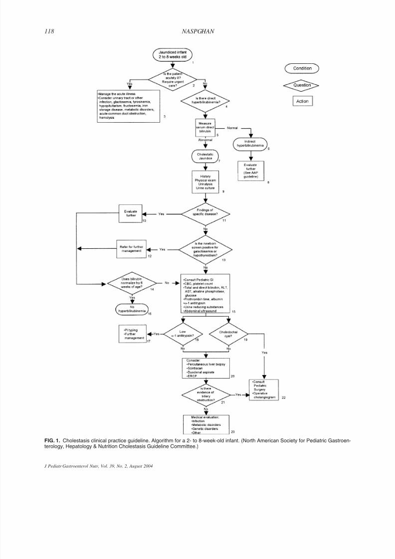
The final algorithm (Fig. 1) is based on the evidence
found for each test, combined with expert opinion andconsensus when the published evidence was insufficient.Recommendations are summarized in Table 5. Consensuswas achieved through the nominal group technique (14).
The guidelines were then reviewed by primary carephysicians in community and academic practices. In ad-dition, the guidelines were distributed to the NASPGHANmembership for review and comment and were officiallyendorsed by the NASPGHAN Executive Council. Thisdocument represents the official recommendations of NASPHGAN on the evaluation of cholestatic jaundice of the infant. The American Academy of Pediatrics has alsoendorsed these recommendations. This review and rec-ommendations are a general guideline and are not in-
tended as a substitute for clinical judgment or as a pro-tocol for the care of all patients with this problem.
INITIAL EVALUATION OF THE
JAUNDICED INFANT
The initial recognition and evaluation of any infantwith possible cholestasis depends upon physicians andother providers of medical care to infants. The age limitsof 2 weeks to 8 weeks of age used in this guideline wereselected because they represent times when primary careproviders typically examine healthy infants. As theevaluation proceeds, the tests to accurately diagnose cho-lestasis fall increasingly in the province of the referralcenter and pediatric gastroenterologist. Different caremodels or differing relationships between primary phy-sicians and referral centers may alter the point at whichthe responsibility for the evaluation shifts from primarycare to specialist. The goal remains the early detectionand efficient diagnostic evaluation of cholestasis in in-fants.
No screening test can predict which infant will expe-rience cholestasis (15). Thus, the detection of cholestasisrests on the clinical recognition of jaundice, pale stools,and/or dark urine by the parent or primary care provider.Each of these findings is an imperfect method of detect-ing cholestasis. Jaundice at 2 weeks of age is a relativelycommon finding, observed in 2.4% to 15% of newborns(16,17). Most such infants have unconjugated hyperbil-irubinemia because of breast milk jaundice, a benigncondition, although a recent report documents occasionalkernicterus in otherwise healthy-appearing infants (18).In one study, jaundice was found in 9% of breast-fedinfants at 4 weeks of age, but in fewer than 1 in 1,000
TABLE 3. Validity criteria for studies of diagnostic tests
1. Was the diagnostic standard adequate?2. Was the test compared independently and blindly to the
diagnostic (“criterion”) standard?3. Were the referral pattern and patient population described?4. Was there an adequate spectrum of disease in tested patients?5. Was the test clearly described?
6. Was observer variation in test results addressed?
TABLE 4. Coding scheme for quality of evidence
Level of evidence Criterion for this level
Level A Recommendation based on 2 or more studies thatcompared the test to a criterion standard in anindependent, blind manner in an unselectedpopulation of infants similar to those addressed inthe guideline.
Level B Recommendation based on a single study that
compared the test to a criterion standard in anindependent, blind manner in an unselectedpopulation of infants similar to those addressed inthe guideline.
Level C Recommendation based on lower quality studies orstudies for which inadequate information isprovided to assess quality, together with expertopinion and consensus of the committee.
Level D No studies available; recommendation based on expertopinion and consensus of the committee.
Table 5. Recommendations
RecommendationLevel of evidence
It is recommended that any infant noted to be jaundicedat 2 weeks of age be clinically evaluated for cholestasiswith measurement of total and direct serum bilirubin.However, breast-fed infants who can be reliablymonitored and who have an otherwise normal history(no dark urine or light stools) and physical examinationmay be asked to return at 3 weeks of age and, if
jaundice persists, have measurement of total and directserum bilirubin at that time.
C
Retest any infant with an acute condition or otherexplanation for jaundice whose jaundice does notresolve with appropriate management of the diagnosedcondition.
D
Ultrasound is recommended for infants with cholestasis of unknown etiology.
A
Liver biopsy is recommended for most infants withcholestasis of unknown etiology.
A
GGTP and lipoprotein X are not routinely recommendedin the evaluation of cholestasis in young infants.
C
Scintigraphy and duodenal aspirate are not routinelyrecommended but may be useful in situations in whichother tests are not readily available.
A
MRCP and ERCP are not routinely recommended,although ERCP may be useful in experienced hands.
C
GUIDELINES FOR CHOLESTATIC JAUNDICE IN INFANTS 117
J Pediatr Gastroenterol Nutr, Vol. 39, No. 2, August 2004
8/20/2019 Infant Cholestasis Guidelines
http://slidepdf.com/reader/full/infant-cholestasis-guidelines 4/14
FIG. 1. Cholestasis clinical practice guideline. Algorithm for a 2- to 8-week-old infant. (North American Society for Pediatric Gastroen-terology, Hepatology & Nutrition Cholestasis Guideline Committee.)
NASPGHAN 118
J Pediatr Gastroenterol Nutr, Vol. 39, No. 2, August 2004

8/20/2019 Infant Cholestasis Guidelines
http://slidepdf.com/reader/full/infant-cholestasis-guidelines 5/14
bottle-fed infants (19). Testing of all jaundiced newbornsat the 2-week visit will detect cholestasis in relativelyfew infants (for every 60 to 375 infants with visible jaundice, 1 will have cholestasis). However, the nextscheduled health maintenance visit generally does notoccur until 2 months of age, beyond the optimal time forintervention in biliary atresia.
The report of pale stools by the parent or observationof clay colored stool by the physician raises the suspicionof cholestasis. In one study of 3,629 infants, 4 passedpale stools during the first 4 weeks of life, none morethan three times. None of these infants had liver disease.The high specificity of persistent pale stools means thatthis finding almost certainly indicates disease when it ispresent. However, infants with biliary atresia have beennoted to have pigmented stools at presentation, and stoolcolor is variable in other conditions causing cholestasis(20). Parents do not appear to be reliable observers of stool color: even when the specific question “What coloris your baby’s stool?” was added to a booklet received byall parents for well care, the average age for definitivecare for infants with biliary atresia was not improved (4).A scoring card displaying color photographs of normaland abnormal stools modestly improved case finding in asubsequent program (21).
Dark urine is also a nonspecific indicator of increasedconjugated bilirubin. No studies could be found that ad-dressed the utility of this sign for case finding. Literaturereview did not identify any studies that defined specific-ity or sensitivity for the findings of jaundice, pale stools,or dark urine in combination. Thus, the clinician mustcontinue to test for cholestasis when these findings arepresent, despite the low yield. The American Academyof Pediatrics practice parameter on the management of hyperbilirubinemia in the healthy term newborn recom-mends testing for conjugated hyperbilirubinemia if jaun-dice is accompanied by dark urine or light stool or if itpersists beyond 3 weeks (22).
The Cholestasis Guideline Committee recommendsthat any infant noted to be jaundiced at 2 weeks of age beevaluated for cholestasis with measurement of total anddirect serum bilirubin. However, breast-fed infants whocan be reliably monitored and who have an otherwisenormal history (no dark urine or light stools) and physi-cal examination (Table 6) may be asked to return at 3weeks of age and, if jaundice persists, have measurementof total and direct serum bilirubin at that time.
Bacterial infections, including urinary tract infection,
are a well known cause of concomitant conjugated hy-perbilirubinemia, probably because of the effect of bac-terial endotoxin on bile formation (23,24). Otherwiseasymptomatic jaundiced infants may have urinary tractinfection, particularly if the onset of jaundice occurs after8 days of age (25). Jaundice caused by an acute infectionresolves when the acute illness has been addressed. Be-cause jaundice is more likely to be related to the under-lying illness, rather than primary liver disease, in the
acutely ill infant, management is oriented to the under-lying acute illness.
Because either conjugated or unconjugated hyperbili-rubinemia may be present in a jaundiced infant who doesnot appear acutely ill, measurement of serum bilirubinthat includes both total and direct (conjugated) bilirubinis recommended. The most commonly used laboratorydetermination (the diazo or van den Bergh method) doesnot specifically measure conjugated bilirubin but reportsdirect bilirubin. For methodological reasons, the higherthe total bilirubin (even if it is all unconjugated), thehigher the reported direct bilirubin (26–28). Measure-ments of direct bilirubin may vary significantly bothwithin and between laboratories (29–32). The methodsused may also influence the measurement of conjugatedbilirubin (33–35). A specific measurement of conjugatedbilirubin, such as that obtained with the Ektachem sys-tem, is optimal. Because canalicular excretion of biliru-bin can be rate limiting to overall clearance, infants withhigh nonconjugated bilirubin may retain some conju-gated bilirubin. Therefore, in the presence of elevatedtotal bilirubin, conjugated bilirubin levels are consideredabnormal when values greater than 1.0 mg/dL are re-ported (36). Practitioners whose laboratories use the Ek-tachem system could use a value of conjugated bilirubingreater than 1.0 to define cholestasis, regardless of totalbilirubin. For this guideline, we defined an abnormaldirect bilirubin as a value greater than 1.0 mg/dL if thetotal bilirubin is less than 5 mg/dL, or a value of directbilirubin that represents more than 20% of the total bil-irubin if the total bilirubin is greater than 5 mg/dL. It isimportant to be aware of the potential for error in thesemeasurements and consult with the laboratory if the mea-surements are inconsistent with the appearance of thepatient. Urine testing has also been used to diagnosecholestasis: assay of sulfated bile acids in urine wasfound in a single study to distinguish control infants fromthose with cholestasis, but this test is not generally avail-able (37).
INITIAL EVALUATION OF THE INFANT WITH
CONJUGATED HYPERBILIRUBINEMIA
For infants with only unconjugated hyperbilirubin-emia, practitioners can refer to the previously publishedguideline on management of unconjugated hyperbiliru-binemia from the American Academy of Pediatrics (22).
If the conjugated serum bilirubin is elevated, cholestasisis present. The further evaluation of an infant with cho-lestasis is a matter of some urgency. Because biliaryatresia is one of the possible causes of cholestasis, thegoal of management is to complete the diagnostic evalu-ation, or at least to exclude biliary atresia, by 45 to 60days of age. History and physical examination can helpguide the diagnostic process for the infant with cholesta-sis. Some findings on history or physical examination
GUIDELINES FOR CHOLESTATIC JAUNDICE IN INFANTS 119
J Pediatr Gastroenterol Nutr, Vol. 39, No. 2, August 2004

8/20/2019 Infant Cholestasis Guidelines
http://slidepdf.com/reader/full/infant-cholestasis-guidelines 6/14

8/20/2019 Infant Cholestasis Guidelines
http://slidepdf.com/reader/full/infant-cholestasis-guidelines 7/14
to more focused diagnostic evaluation, rather than furtherinvestigation of cholestasis. It is important to evaluate orrepeat the newborn screen for galactosemia and hypo-thyroidism because these conditions can present withconjugated hyperbilirubinemia and require urgent man-agement to prevent serious sequelae (39–42). Keeping inmind that the diagnosis of one disease does not precludethe presence of another, additional evaluation is neces-sary if the jaundice does not resolve with treatment of these specific conditions.
Consultation with a pediatric gastroenterologist is es-sential for infants with conjugated hyperbilirubinemia of unknown cause. The nature of this consultation may varyamong practice settings. A telephone consultation maybe particularly useful for practitioners at some distancefrom specialty care. Of the tests listed in Box 15 of thealgorithm (Fig. 1), which should be ordered and wherethey should be performed is likely to vary, depending onwhich of the tests are readily available to the referringphysician, and on whether examination of the patient by
the specialist will be delayed. If the level of serumalpha-1 antitrypsin is found to be low, Pi typing is indi-cated. If an obvious extrinsic obstruction (such as cho-ledochal cyst) is present, referral for surgery is war-ranted. Of the tests listed at this decision point, the ul-trasound is the most operator dependent, and thus may bemost appropriately performed at the referral center bymore experienced personnel. In many practice settings,all of the listed tests would be best obtained by imme-diate referral of the patient for evaluation by the pediatricgastroenterologist. In the evaluation for infants with pos-sible biliary atresia, it is optimal for an experienced pe-diatric surgeon to also be involved.
Ultrasonography is useful to identify anatomic abnor-
malities such as choledochal cyst. The finding of a smallor absent gallbladder may suggest extrahepatic biliaryatresia, but reported sensitivities as low as 73% indicatethat ultrasound cannot be used to rule out this diagnosis.Several reports of high sensitivity and specificity of the“triangular cord” sign on ultrasound suggest that this testmay be useful in the diagnosis of extrahepatic biliaryatresia.(43–45) Again, this appears to be operator depen-dent. Despite these limitations, we recommend ultraso-nography for the evaluation of the infant with cholestasisof unknown etiology.
Gamma glutamyl transpeptidase (GGT) has been usedin the past to distinguish biliary atresia from neonatal
hepatitis, but wide variability in levels makes interpreta-tion of test results difficult. Especially in the older infantwith cholestasis, a very low GGT level may be useful toexclude obstruction and, in conjunction with an elevatedalkaline phosphatase level, suggests genetic and meta-bolic causes of intracellular cholestasis. The degree of elevation of GGT is not useful in discriminating the eti-ology of the cholestasis. The evidence supporting the useof ultrasound and GGT is summarized in Table 7.
FURTHER EVALUATION OF THE INFANT
WITH CHOLESTASIS
Once it is established that cholestasis is present, theprincipal diagnostic concern is the differentiation of he-patocellular from obstructive cholestasis, of disorders of physiology from disorders of anatomy, and of diseasethat is managed medically from disease that is managedsurgically. The tests that have been used to make thisdifferentiation and about which we were able to find atleast some evidence regarding their value were percuta-neous liver biopsy, hepatobiliary radionuclide scan, andduodenal aspirate. Evidence and citations for the use of these tests are summarized in Table 7.
Percutaneous Liver Biopsy
Most studies of percutaneous liver biopsy are retro-spective analyses using as a gold standard the clinicalcourse or surgical or autopsy results. Biopsy interpreta-tion is pathologist dependent, and requires experiencethat many general pathologists lack. Review of all of thestudies of biopsy revealed that 50% to 99% of patientswith biliary atresia are correctly identified with biopsy.Biliary atresia is incorrectly suspected from the biopsy in0% to 46%.
In 1974, Brough and Bernstein (46) demonstrated thediagnostic usefulness of the percutaneous liver biopsyand established the diagnostic criteria that are in currentuse. They compared the original pathologic diagnosis in181 consecutive patients to the ultimate diagnosis basedon surgical findings and long-term follow-up. The origi-nal diagnosis was correct in 148 patients (93.7%), a highlevel of accuracy. More importantly, the type of errorseen in the 10 patients with incorrect diagnoses wouldnot lead to missed diagnosis of biliary atresia. In onlyone patient with biliary atresia was the original biopsyinterpreted as hepatitis, which would lead to delay in thediagnosis and loss of important time before surgical cor-rection. The remaining nine were suspected of havingbiliary atresia but actually had hepatitis. In this report,liver biopsy had a very high sensitivity (99%) and speci-ficity (92%) for the diagnosis of biliary atresia, withsomewhat less specificity for the diagnosis of neonatalhepatitis. Unfortunately, 23 patients were lost to follow-up, which reduces the value of this study. The highestquality subsequent studies performed using the Brough
and Bernstein criteria demonstrate very good sensitivityand specificity for biliary atresia.The interpretation of a single liver biopsy in a child
with neonatal cholestasis is also limited by the dynamicsof disease. Many cholestatic conditions express them-selves differently with time. Liver biopsy specimens ob-tained early in the course of biliary atresia may be indis-tinguishable from hepatitis (47). In addition to being ableto visualize the hepatocanalicular cholestasis and injury,
GUIDELINES FOR CHOLESTATIC JAUNDICE IN INFANTS 121
J Pediatr Gastroenterol Nutr, Vol. 39, No. 2, August 2004

8/20/2019 Infant Cholestasis Guidelines
http://slidepdf.com/reader/full/infant-cholestasis-guidelines 8/14
T A B L E
7 .
D i a g n o s t i c t e s t s
t o d
i s t i n g u i s h E H B A f r o m o t h e r
c a u s e s
o f
c h o l e s t a s i s ( B o x 2 1 o f
F i g u r e
1 )
P r o c e d u r e
Q u
a l i t y o
f
e v
i d e n c e
S e n s i t i v i t y *
f o r
o b s t r u c t i o n
S p e c i f i c i t y *
f o r
o b s t r u c t i o n
L i k e l i h o o d
r a t i o *
f o r
o b s t r u c t i o n
L i k e l i h o o d
r a t i o *
f o r
n o n o b s t r u c t i v e
c h o l e s t a s i s
T i m e
d e l a y
R i s k
C o m m e
n t s — o t h e r
d i a g n o s e s ,
i s s u e s
r e l e v a n t
t o t h
i s t e s t
U l t r a s o u n d
4 4 – 4
6 , 5
1 – 7
1
A
7 3 % – 1
0 0 %
f o r
s m a l l
o r
a b s e n t
G B
8 3 % – 1
0 0 %
f o r
“ t r i a n g u l a r
c o r d
” s i g n
6 7 % – 1
0 0 % f o r
s m a l l
o r
a b s
e n t
G B
9 8 % – 1
0 0 % f o r
“ t r i a n g u l a r
c o r d
” s i g
n
2 . 2
– i n f i n i t e
4 1
. 5 – i n
f i n i t e
2 . 5
– i n f i n i t e
5 . 8
– i n f i n i t e
M i n i m a l
M i n i m a l
P r o c e d u r e
i s o
p e r a t o r
d e p e n d e n t ;
p r i m a r i l
y u
s e f u l
t o r u
l e o
u t
a n a t o m i c
a b n o r m a l i t i e s
s u c h
a s
c h o l e d o c h a l
c y s t ,
r a t h e r
t h a n
m a k e a
d i a g n o s i s
o f
E H B A
.
G G T P
4 7
, 5
6 , 7
2 – 8
1
C
7 8 % – 8
6 %
6 7 % – 1
0 0 %
2 . 4
– i n f i n i t e
3 . 0
– 7 . 1
M i n i m a l
M i n i m a l
V a r i e t y
o f
c u t - o f f
v a l u e s
u s e d ,
n o
s t u d y w
i t h c l e a r l y i n
d e p e n d e n t
c o m p a r i s o n ;
m o s t
s t u d i e s
s h o w
c o n s i d e r a b l e
o v e r l a p .
P e r c u t a n e o u s
b i o p s y
4 7
, 5
6 , 6
2 , 6
9 ,
7 4
, 8
2 – 8
9
A
8 9 % – 9
9 %
8 2
. 5 % – 9
8 %
5 . 2
– 4 9
. 5
7 . 5
– 9 8
1 – 3
d a y s
V e r y
l o w
P a u c i t y
o f
i n t r a h e p a t i c
b i l e
d u c t s ,
g i a n t c e
l l t r a n
s f o r m a t i o n ,
m e t a b o l i c
a n d
s t o r a g e
d i s e a s e s ,
P F I C
, i n f e c t i o n , n
e o n a t a l
s c l e r o s i n g
c h o l a n g i t i s
c a n b
e
d i a g n o s e d
w i t h
t h i s
t e s t . M
o s t
p a t i e n t s
w i l l
r e q u i r e
t h i s
t e s t ;
s h o u l d
b e
i n t e r p r e t e d
b y
a
p a t h o l o g i s t
e x p e r i e n c e d
w i t h
p e d i a t r i c
l i v e r
d i s e a s e .
S e n s i t i v
i t y m
a y b
e h
i g h e r
i n
e x p e r t h
a n d s .
R a d i o n u c l i d e
s c a n n i n g
5 3
, 5
4 , 5
6 , 6
2 ,
6 5
, 6
9 , 7
4 , 8
2 ,
8 6
, 8
9 – 1
0 9
A
8 3 % – 1
0 0 %
3 3 % – 1
0 0 %
1 . 3
– i n f i n i t e
2 – i n
f i n i t e
3 – 5
d a y s
f o r
p h e n o b a r b i t a l
p r i m i n g
M i n i m a l
M o s t
s t u d i e s
w i t h
s e n s i t i v i t y
o f
1 0 0 %
, s
o o
b s t r u c t i o n
e x t r e m e l y
u n l i k e l y
i f e x
c r e t i o n
f o u n d
.
D i s a d v a
n t a g e
o f
t e s t i s t i m
e
d e l a y a n
d c o
s t .
D u o d e n a l
a s p i r a t e
6 2
, 8
2 , 8
8 , 9
7 ,
1 1 0 – 1
1 4
A
9 1 % – 1
0 0 %
4 3 % – 1
0 0 %
1 . 6
– i n f i n i t e
4 . 8
– i n f i n i t e
n o n e
M i n i m a l
O b s t r u c t i o n
e x t r e m e l y
u n l i k e l y
i f
b i l e
f o u n d
i n d
u o d e n a l
f l u i d
.
M a y
r e q u i r e
f l u o r o s c o p y
f o r
t u b e
p l a
c e m e n t ;
t e s t i s i n
v a s i v e .
M R C P
7 1
, 1
1 5
– 1 1 9
C
1 0 0 %
2 s t u
d i e s
6 0 %
1 s t u
d y
2 . 5
( i m p r e c i s e )
I n f i n i t e
( i m p r e c i s e )
M i n i m a l
R e q u i r e s d
e e p s e d
a t i o n
o r
g e n e r a l
a n e s t h e s i a ;
v e r y
f e w a d e q u a t e
s t u d i e s .
E R C P
8 8
, 1
2 0 – 1
2 7
C
1 0 0 %
1 s t u
d y
1 0 0 %
1 s t u
d y
I n f i n i t e
I n f i n i t e
?
U n k n o w n
R e q u i r e s s
o p h i s t i c a t e d
i n s t r u m e n t a t i o n
a n d
e x p e r t i s e
n o t
c u r r
e n t l y
w i d e l y
a v a i l a b l e ;
n u m b e r s
b a s e d
o n
a s i n
g l e
s t u d y w
i t h a s i n
g l e
e n d o s c o
p i s t .
L i p o p r o t e i n
X
7 9
, 1
2 8 – 1
3 6
N o t
d i s c u s s e d
3 0 % – 8
8 %
8 1 % – 1
0 0 %
1 . 5
– i n f i n i t e
2 . 7
– 8 . 3
M i n i m a l
T e s t
n o
l o n g e r
a v a i l a b l e ,
m o s t
s t u d i e s
v e r y
s m a l l
w i t h
p o o r
d e s c r i p t i o n
o f
m e t h o d s .
S e n s i t i v i t y
, r a
n g e
o f
v a
l u e s
f r o m
h i g h e s t
q u a l i t y
s t u d i e s .
S p e c i f i c i t y
, r a
n g e
o f
v a
l u e s
f r o m
h i g h e s t
q u a l i t y
s t u d i e s .
L R
, t h
e r a
t i o o
f t h
e l i k e
l i h o o d
o f
a t e
s t r e
s u l t
i n p
a t i e n t s w h o
h a v e
a d i
s e a s e
c o m p a r e d
w i t h
t h e
l i k e l i h o o d
o f
t h e
s a m e
t e s t
r e s u l t
i n t h
o s e
w i t h o u t
t h e
d i s e a s e ;
c a n
b e c
a l c u l a t e d
a s
s e n s i t i v i t y
( 1 - s p e c i f i c i t y ) .
S e e
J a e s c h
k e
e t
a l (
1 4 )
f o r
d e t a i l s .
I n t h
i s t a
b l e
, t h
e r a
n g e
i s b
a s e d
o n
t h e
s e n s i t i v i t y a n d
s p e c i f i c i t y
f r o m
h i g h e s t
q u a l i t y
s t u d i e s .
NASPGHAN 122
J Pediatr Gastroenterol Nutr, Vol. 39, No. 2, August 2004

8/20/2019 Infant Cholestasis Guidelines
http://slidepdf.com/reader/full/infant-cholestasis-guidelines 9/14
the liver biopsy also can provide disease-specific find-ings. Examples include PAS-positive granules in alpha-1antitrypsin deficiency, ductal paucity in Alagille syn-drome, necroinflammatory duct lesions in sclerosingcholangitis, and other findings that are relatively specificfor metabolic and storage diseases.
The evidence indicates that liver biopsy can be per-formed safely and expeditiously in young infants and isuseful in establishing specific diagnoses (48). The Cho-lestasis Guideline Committee recommends that a liverbiopsy be performed in most infants with undiagnosedcholestasis, to be interpreted by a pathologist with ex-pertise in pediatric liver disease. A percutaneous liverbiopsy is recommended before performing a surgicalprocedure to diagnose biliary atresia. If the biopsy isdone early in the course of the disease (before 6 weeks of age), the biopsy may have to be repeated if the results areequivocal.
Scintigraphy
Injected radioactive material is normally excreted intothe intestine within a predictable period of time. Nonvi-sualization of radioactivity within the intestine (in thescanning field comprising the intestines) 24 hours afterinjection is considered to be an abnormal result, indicat-ing biliary obstruction or hepatocellular dysfunction. Inthe studies reviewed, a variety of radiolabeled scinti-graphic agents were used, the diagnostic criteria variedgreatly, and no blinded comparisons were found betweenscintigraphy and a gold standard for diagnosis. Althoughit is thought that the precision of the test can be improvedby administering phenobarbital for several days beforeimaging, no studies are available to confirm or refute thishypothesis. In the available studies, the sensitivity of scintigraphy for the diagnosis of biliary atresia is high;virtually all patients with complete biliary obstructionshowed no excretion on scintiscan. A few patients with anegative test (tracer evident in the intestine) later expe-rienced biliary atresia, presumably because of diseaseevolving from incomplete to complete obstruction.Specificity of scintigraphy for biliary atresia or otherobstructive processes is low; many patients without ana-tomic obstruction will not excrete tracer. Although thehigh sensitivity for biliary atresia makes this a fairlygood single test for detecting disease, it is time consum-ing and expensive and does have significant false-positive and false-negative results. The Cholestasis
Guideline Committee concludes that hepatobiliary scin-tigraphy generally adds little to the routine evaluation of the cholestatic infant but may be of value if other meansfor excluding biliary obstruction are not available.
Duodenal Aspirate
Limited data indicate that duodenal aspirate analysisfor bilirubin concentration can identify patients with bil-
iary obstruction with a sensitivity similar to that of scin-tiscan. In this test, fluid is obtained from the duodenum,either by placing a tube or a string in the duodenum, andanalyzed for bilirubin concentration. A positive test forobstruction is one in which the bilirubin concentration of the aspirate is no greater than serum concentration. Thiswould appear to be a low-tech, inexpensive alternative toscintiscan, yet it is not commonly used, probably becauseit is time and labor intensive, invasive, and inconvenient.The Cholestasis Guideline Committee concludes that theduodenal aspirate test may be useful in situations inwhich other tests to detect biliary obstruction are notavailable.
Magnetic Resonance Cholangiopancreatography
The few reports available to date have studied a verysmall number of patients, and although the results areencouraging, firm conclusions are not possible. Magneticresonance cholangiopancreatography (MRCP) requires
deep sedation or general anesthesia. Additional technicaladvancement and clinical experience are necessary be-fore MRCP can be used in the evaluation of cholestaticinfants. The Cholestasis Guideline Committee concludesthat this test cannot be routinely recommended based onthe currently available data.
Endoscopic Retrograde Cholangiopancreatography
Endoscopic retrograde cholangiopancreatography(ERCP) is an evolving technology that has been increas-ingly used in some tertiary referral centers to diagnosethe cause of cholestasis in young infants. ERCP involvesendoscopic intubation of the biliary (and pancreatic)ducts via the ampulla of Vater with a small tapered cath-eter and injection of contrast material to facilitate radio-logic visualization of the ductal systems. A small pedi-atric side-viewing duodenoscope has enhanced the capa-bility to perform this procedure in young infants. Mostendoscopists prefer to perform this procedure with thepatient under general anesthesia.
The data supporting the use of ERCP for the evalua-tion of the cholestatic infant are sparse. Many of thereports simply document the use of the prototype smallerside-viewing instrument for these patients. Operators re-port mixed success with the procedure to distinguish bil-iary atresia from neonatal hepatitis and other forms of
nonoperative cholestasis. Most tertiary centers use ERCPto sort as surgical or nonsurgical cases that remain equiv-ocal after liver biopsy. No controlled studies have beenconducted to compare endoscopic techniques. In largerstudies, sensitivity and specificity are excellent, butfailed cannulation and/or visualization are reported inmore than 10%. Patient populations vary from younginfants to older infants (before or after 60 to 90 days of age). No controlled study has been done to demonstrate
GUIDELINES FOR CHOLESTATIC JAUNDICE IN INFANTS 123
J Pediatr Gastroenterol Nutr, Vol. 39, No. 2, August 2004

8/20/2019 Infant Cholestasis Guidelines
http://slidepdf.com/reader/full/infant-cholestasis-guidelines 10/14
that ERCP will alter the final diagnosis. A cost-benefitanalysis of ERCP has not been performed, but it is pos-sible that the procedure could obviate the need for sur-gical exploration in some infants. ERCP in pediatric pa-tients has been recommended only at facilities with ap-propriate support staff and specialists with expertise inthis procedure in young infants (49).
The Cholestasis Guideline Committee concludes thatERCP is not frequently used because of the cost of in-strumentation and the need for technical expertise. Theusefulness of ERCP appears to be center and operatordependent. Clinicians with the appropriate expertise andinstrumentation will find the test a useful adjunct in theevaluation of the difficult-to-diagnose cause of cholesta-sis in an infant in whom laparotomy is being contem-plated. The Committee recommends that a liver biopsybe obtained before subjecting an infant to ERCP. Underselected circumstances, ERCP can clarify the cause of neonatal cholestasis and obviate the need for laparotomy.ERCP can be recommended in selected cases at facilitieswith appropriate support staff and specialists with exper-tise in this procedure in young infants.
Lipoprotein X
This test is no longer widely available; the few studieson this subject are of poor quality and suggest poor sen-sitivity and specificity for differentiating biliary atresiafrom neonatal hepatitis. The Cholestasis Guideline Com-mittee recommends that this test not be performed.
CONCLUSION
The rapid and effective diagnosis of the cause of cho-lestasis in an infant is challenging. The initial detectionof these infants remains in the domain of the primarycare provider and depends on the recognition of jaundicepast the age of 2 weeks or recognition of abnormal stoolor urine color. Laboratory testing for cholestasis shouldinclude direct (conjugated) bilirubin. A direct bilirubinvalue greater than 1.0 mg/dL if the total bilirubin is lessthan 5 mg/dL or a direct bilirubin more than 20% of thetotal bilirubin if the total bilirubin is greater than 5mg/dL is considered abnormal. The relative rarity of cho-lestatic jaundice in contrast to unconjugated hyperbiliru-binemia in this age range dictates that many jaundicedinfants will be tested to find a few with elevated direct
bilirubin. It is recommended that primary care providerscontinue to screen for cholestasis, despite its rarity, be-cause of the gravity of the consequences of missed di-agnosis.
The Cholestasis Guideline Committee recommendsthat any infant noted to be jaundiced at 2 weeks of age beevaluated for cholestasis with measurement of total anddirect serum bilirubin. However, breast-fed infants whocan be reliably monitored and who have an otherwise
normal history (no dark urine or light stools) and physi-cal examination may be asked to return at 3 weeks of ageand, if jaundice persists, have measurement of total anddirect serum bilirubin at that time.
Identification of cholestasis warrants a prompt effortto accurately diagnose the cause. In an ill appearing in-fant, the primary cause of the acute problem should besought and treated. Cholestasis that does not resolve withresolution of the acute problem requires thorough evalu-ation. In a well appearing infant, cholestasis should beevaluated without delay because this condition has eti-ologies that are both serious and treatable. Patients withextrahepatic biliary atresia often appear well at initialpresentation, and in these patients there is evidence thatearly diagnosis and surgical bile drainage is associatedwith longer survival of the native liver.
Most of the diagnostic tests that are used to determinethe etiology of cholestasis in infants are operator depen-dent and variable in utility, and most are not commonlyperformed outside of referral centers. Although specific
circumstances may support the performance of diagnos-tic testing at a referring hospital, most practitioners willneed to use the diagnostic expertise of a referral center.Diagnostic workup by referring hospitals may be mostuseful to quickly exclude some of the many causes of cholestasis.
For all diagnostic tests reviewed, the sensitivity andspecificity varied widely. Percutaneous liver biopsy hadthe greatest diagnostic accuracy in published studies.However, because of the dynamic and progressive natureof extrahepatic biliary atresia, even this test can be mis-leading. Scintigraphy appears to be primarily useful inexcluding extrahepatic obstruction, consistent with thediagnosis of extrahepatic biliary atresia. Sonographic
evaluation is currently useful for excluding anatomic ab-normalities but may have a greater value in the future if descriptions of new findings (such as the “triangularcord” sign) are corroborated. ERCP may have a role incenters with pediatric gastroenterologists experienced inits use. Too little experience with MRCP has been re-ported to analyze its utility. Duodenal aspirate or stringtest may be useful in remote sites or if other testing is notreadily available.
In summary, the diagnosis of neonatal cholestasis is anurgent matter. This guideline and algorithm have beendeveloped to assist in this process. No single pathwayappears to be clearly superior for the diagnosis of con-
ditions leading to cholestasis. A guide is provided herefor clinicians who are challenged with the prospect of detecting and evaluating the rare infant with cholestasis.Vigilance is crucial in detecting these patients, selectingappropriate and timely diagnostic testing based on theresources available in their own institutions, and refer-ring the infant to a pediatric gastroenterologist who canprovide the essential diagnostic and treatment modalitiesfor optimal outcome. The scarcity and relatively low
NASPGHAN 124
J Pediatr Gastroenterol Nutr, Vol. 39, No. 2, August 2004

8/20/2019 Infant Cholestasis Guidelines
http://slidepdf.com/reader/full/infant-cholestasis-guidelines 11/14
quality of research found in this area indicate that furtherresearch is needed.
Authors
Virginia Moyer, MD, MPHHouston, Texas
Deborah K. Freese, MDRochester, Minnesota
Peter F. Whitington, MDChicago, Illinois
Alan D. Olson, MDMalvern, Pennsylvania
Fred Brewer, MDSanta Rosa, California
Richard B. Colletti, MDBurlington, Vermont
Melvin B. Heyman, MD, MPHSan Francisco, California
REFERENCES
1. Dick MC, Mowat AP. Hepatitis syndrome in infancy: an epide-miologic survey with 10-year follow up. Arch Dis Child 1985;60:512–6.
2. Balistreri WF. Neonatal cholestasis. J Pediatr 1985;106:171–84.3. Yoon PW, Bresee JS, Olney RS, et al. Epidemiology of biliary
atresia: a population based study. Pediatrics 1997;99:376–82.4. Matsui A, Ishikawa T. Identification of infants with biliary atresia
in Japan. Lancet 1994;343:925.5. McKiernan PJ, Baker AJ, Kelly DA. The frequency and outcome
of biliary atresia in the UK and Ireland. Lancet 2000;355:25–9.6. Danks DM, Campbell PE, Jack I, et al. Studies of the aetiology of
neonatal hepatitis and biliary atresia. Arch Dis Child 1977;52:360–7.
7. Chardot C, Carton M, Spire-Benelac N, et al. Is the Kasai opera-tion still indicated in children older than 3 months diagnosed withbiliary atresia? J Pediatr 2001;138:224–8.
8. Mieli-Vergani G, Howard ER, Portman B, et al. Late referral forbiliary atresia–missed opportunities for effective surgery. Lancet 1989;1:421–3.
9. Altman RP, Lilly JR, Greenfeld J, et al. A multivariable risk factor analysis of the portoenterostomy (Kasai) procedure forbiliary atresia. Ann Surg 1997;226:348–53.
10. Mowat AP, Davidson LL, Dick MC. Earlier identification of biliary atresia and hepatobiliary disease: selective screening in thethird week of life. Arch Dis Child 1995;72:90–2.
11. Psacharopoulos HT, Mowat AP, Cook PJL, et al. Outcome of liver disease associated with a-1-antirypsin deficiency (PiZ): im-plications for genetic counseling and antenatal diagnosis. Arch
Dis Child 1983;58:882–7.12. Hussein M, Howard ER, Mieli-Vergani G, et al. Jaundice at 14
days of age: exclude biliary atresia. Arch Dis Child 1991;66:1177–9.
13. Jaeschke R, Guyatt G, Sackett DL. Users’ guides to the medicalliterature III. How to use an article about a diagnostic test. JAMA
1994;271:389–91.
14. McMurray AR. Three decision-making aids: brainstorming,nominal group, and delphi technique. J Nursing Staff Dev 1994;10:62–5.
15. Mushtaq I, Logan S, Morris M, et al. Screening of newborninfants for cholestatic hepatobiliary disease with tandem massspectrometry. BMJ 1999;319:471–7.
16. Winfield CR, MacFaul R. Clinical study of jaundice in breast andbottle fed babies. Arch Dis Child 1978;53:506–7.
17. Kelly DA, Stanton A. Jaundice in babies: implications for com-munity screening for biliary atresia. BMJ 1995;310:1172–3.
18. Johnson LH, Bhutani VK, Brown AK. System-based approach to
management of neonatal jaundice and prevention of kernicterus. J Pediatr 2002;140;396–403.
19. Crofts DJ, Michel VJ-M, Rigby AS, et al. Assessment of stoolcolour in community management of prolonged jaundice in in-fancy. Acta Paediatr 1999;88:969–74.
20. Burton EM, Babcock DS, Heubi JE, et al. Neonatal jaundice:clinical and ultrasonographic findings. South Med J 1990;83:294–302.
21. Matsui A, Dodoriki M. Screening for biliary atresia. Lancet 1995;345:1181.
22. American Academy of Pediatrics Provisional Committee forQuality Improvement and Subcommittee on Hyperbilirubinemia.Management of hyperbilirubinemia in the healthy term newborn.Pediatrics 1994;94:558–65.
23. Utili R, Abernathy CO, Zimmerman HJ. Cholestatic effects of Escherichia coli endotoxin on the isolated perfused rat liver. Gas-troenterology 1976;70:248–53.
24. Utili R, Abernathy CO, Zimmerman HJ. Studies on the effects of E. coli endotoxin on canalicular bile formation in the isolatedperfused rat liver. J Lab Clin Med 1977;89;471–82.
25. Garcia FJ, Nager AL. Jaundice as an early diagnostic sign of urinary tract infection in infancy. Pediatrics 2002;109:846–51.
26. Heirwegh KP, Fevery J, Meuwissen JA, et al. Recent advances inthe separation and analysis of diazo-positive bile pigments. Meth-ods Biochem Anal 1974;22:205–50.
27. Tisdale WA, Klatskin G, Kinsella ED. The significance of thedirect-reacting fraction of serum bilirubin in hemolytic jaundice.
Am J Med 1959;26:214–27.
28. Fevery J, Claes J, Heirwegh K, et al. Hyperbilirubinemia: signifi-cance of the ratio between direct-reacting and total bilirubin. ClinChem Acta 1967;17:73–9.
29. Pleasure JR. Bilirubin measurement problems. Pediatrics 1988;82:808–9.
30. Lott JA, Doumas BT. ‘Direct’ and total bilirubin tests: contem-
porary problems. Clin Chem 1993;39:641–7.31. Vreman HJ, Verter J, Oh W, et al. Interlaboratory variability of
bilirubin measurements. Clin Chem 1996;42:869–73.
32. Doumas BT, Eckfeldt JH. Errors in measurement of total biliru-bin: a perennial problem. Clin Chem 1996;42:845–8.
33. Gulian JM, Dalmasso C, Millet V, et al. Influence of photoiso-mers in bilirubin determinations on Kodak Ektachem and Hitachianalysers in neonatal specimens: study of the contribution of structural and configurational isomers. Eur J Clin Chem Clin
Biochem 1995;33:503–12.
34. Doumas BT, Wu TW. The measurement of bilirubin fractions inserum. Crit Rev Clin Lab Sci 1991;28:415–45.
35. Francoual J, Myara A, Benattar C, et al. Investigation of total andconjugated bilirubin determination during the neonatal period.
Eur J Clin Chem Clin Biochem 1993;31:499–502.
36. Rosenthal P, Blanckaert N, Kabra PM, et al. Formation of bili-rubin conjugates in human newborns. Pediatr Res 1986;20:
947–50.37. Matsui A, Kasano Y, Yamaushi Y, et al. Direct enzymatic assay
of urinary sulfated bile acids to replace serum bilirubin testing forselective screening of neonatal cholestasis. J Pediatr 1996;129:306–8.
38. Sivan Y, Merlob P, Nutman J, et al. Direct hyperbilirubinemiacomplicating ABO hemolytic disease of the newborn. Clin Pedi-atr (Phila) 1983;22:537–8.
39. Kaufman FR, Costin G, Thomas DW, et al. Neonatal cholestasisand hypopituitarism. Arch Dis Child 1984;59:787–9.
GUIDELINES FOR CHOLESTATIC JAUNDICE IN INFANTS 125
J Pediatr Gastroenterol Nutr, Vol. 39, No. 2, August 2004
8/20/2019 Infant Cholestasis Guidelines
http://slidepdf.com/reader/full/infant-cholestasis-guidelines 12/14
40. Ellaway CJ, Silinik M, Cowell CT, et al. Cholestatic jaundice andcongenital hypopituitarism. J Paediatr Child Health 1995;31:51–3.
41. Sheehan AG, Martin SR, Stephure D, et al. Neonatal cholestasis,hypoglycemia, and congenital hypopituitarism. J Pediatr Gastro-enterol Nutr 1992;14:426–30.
42. Choo-Kang LR, Sun CC, Counts DR. Cholestasis and hypogly-cemia: manifestations of congenital anterior hypopituitarism.
J Clin Endocrinol Metab 1996;81:2786–9.43. Choi SO, Park WH, Lee HJ. Ultrasonographic ‘triangular cord’:
the most definitive finding for noninvasive diagnosis of extrahe-patic biliary atresia. Eur J Pediatr Surg 1998;8:12–6.
44. Tan Kendrick AP, Phua KB, Ooi BC, et al. Making the diagnosisof biliary atresia using the triangular cord sign and gallbladderlength. Pediatr Radiol 2000;30:69–73.
45. Kotb MA, Kotb A, Sheba MF, et al. Evaluation of the triangularcord sign in the diagnosis of biliary atresia. Pediatrics 2001;108:416–20.
46. Brough AJ, Bernstein J. Conjugated hyperbilirubinemia in earlyinfancy: a reassessment of liver biopsy. Hum Pathol 1974;5:507–16.
47. Landing BH, Wells TR, Ramicone E. Time course of the intra-hepatic lesion of extrahepatic biliary atresia: a morphometricstudy. Pediatr Pathol 1985;4:309–19.
48. Fox VF, Cohen MB, Whitington PF, et al. Outpatient liver biopsy
in children. J Pediatr Gastroenterol Nutr 1996;23:213–6.49. Fox VL, Werlin SL, Heyman MB. Endoscopic retrograde chol-
angiopancreatography in children. Subcommittee on Endoscopyand Procedures of the Patient Care Committee of the NorthAmerican Society for Pediatric Gastroenterology and Nutrition.
J Pediatr Gastroenterol Nutr 2000;30:335–42.
50. Gates GF, Sinatra FR, Thomas DW. Cholestatic syndromes ininfancy and childhood. AJR 1980;134:1141–8.
51. Davidson S, Frand M, Itzchak Y. Hepatobiliary ultrasonographyas a diagnostic aid in neonatal jaundice. Isr J Med Sci 1982;18:947–51.
52. Abramson SJ, Treves S, Teele RL. The infant with possible bil-iary atresia: evaluation by ultrasound and nuclear medicine. Pe-diatr Radiol 1982;12:1–5.
53. Kirks DR, Coleman RE, Filston HC, Rosenberg ER, Merten DF.An imaging approach to persistent neonatal jaundice. ARJ 1984;142:461–5.
54. Green D, Carroll BA. Ultrasonography in the jaundiced infant: anew approach. J Ultrasound Med 1986;5:323–9.
55. Cox KL, Stadalnik RC, McGahan JP, Sanders K, Cannon RA,Ruebner BH. Hepatobiliary scintigraphy with technetium-99mdisofenin in the evaluation of neonatal cholestasis. J Pediatr Gas-troenterol Nutr 1987;6:885–91.
56. Ikeda S, Sera Y, Akagi M. Serial ultrasonic examination to dif-ferentiate biliary atresia from neonatal hepatitis—special refer-ence to changes in size of the gallbladder. Eur J Pediatr 1989;148:396–400.
57. Gopi VK, Joseph TP, Varma KK. Ultrasonography in conjugatedhyperbilirubinemia. Ind Pedtr 1989;26:723–5.
58. Zhou P, Qin R. B mode ultrasound in the diagnosis of biliaryatresia [Chinese]. Chung-hua I Hsueh Tsa Chih 1990;8:461–2.
59. Burton EM, Babcock DS, Heubi JE, Gelfand MJ. Neonatal jaun-dice: clinical and ultrasonographic findings. So Med J 1990;83:24–302.
60. Emblem R, Stake G, Monclair T. Progress in the treatment of biliary atresia: a plea for surgical intervention within the first twomonths of life in infants with persistent cholestasis. Acta Pediatr 1993;82:971–4.
61. Lai MW, Chang MH, Hsu SC, et al. Differential diagnosis of extrahepatic biliary atresia from neonatal hepatitis: a prospectivestudy. J Pediatr Gastronenterol Nutr 1994;18:121–7.
62. Ikeda S, Sera Y, Yamamoto H, Ogawa M. Effect of phenobarbitalon serial ultrasonic examination in the evaluation of neonatal
jaundice. Clin Imag 1994;18:146–8.
63. Hessel G, Yamada RM, Escanhowla CAF, Butofff-Silva JM, To-ledo RJ. Valor de ultra-sonografia abdominal e da biopsia he-patica percutanea no diagnostico diferencial da colestase nwona-tal. [Portuguese] Arg Gastronentero 1994;31:75–82.
64. Lin WY, Lin CC, Changlai SP, Shen YY. Comparison of Tc-99mdisofenin cholescintigraphy with ultrasonography in the differen-tiation of biliary atresia from other forms of neonatal jaundice.Pediatr Surg Int 1997;12:30–3.
65. Ikeda S, Yoshihisa S, Ohshiro H, Uchino S, Akizuki M, Kondo Y.Gallbladder contraction in biliary atresia: a pitfall of ultrasounddiagnosis. Pediatr Radiol 1998;28:451–3.
66. Farrant P, Meire HB, Vergani-Mieli G. Ultrasound features of thegall bladder in infants presenting with conjugated hyperbilirubi-naemia. Brit J Radiol 2000;73:1154–8.
67. Choi SO, Park WH, Lee HJ, Woo SK. ‘Triangular cord’: a sono-graphic finding applicable in the diagnosis of biliary atresia.
J Pediatr Surg 1996;31:363–6.
68. Park WH, Choi SO, Lee HJ, Kim SP, Zeon SK, Lee SL. A newdiagnostic approach to biliary atresia with emphasis on the ultra-sonographic triangular cord sign: comparison of ultrasonography,hepatobiliary scintigraphy, and liver needle biopsy in the evalu-ation of infantile cholestasis. J Pediatr Surg 1997;32:1555–9.
69. Park WH, Choi SO, Lee HJ. The ultrasonographic ‘triangularcord’ coupled with gallbladder images in the diagnostic predic-tion of biliary atresia from infantile intrahepatic cholestasis.
J Pediatr Surg 1999;34:1706–10.70. Kim MJ, Park YN, Han SJ, et al. Biliary atresia in neonates and
infants: triangular area of high signal intensity in the porta hepatisat T2-weighted MR cholangiography with US and histopatholog-ic correlation. Radiology 2000;215:315–401.
71. Platt MS, Potter JL, Boeckman CR, Jaberg, C. ElevatedGGTP/SGOT ratio. An early indicator of infantile obstructivecholangiopathy. Am J Dis Child 1981;135:834–36.
72. Wright K, Christie DL. Use of g-glutamyl transpeptidase in thediagnosis of biliary atresia. Am J Dis Child 1981;135:134–6.
73. Manolaki AG, Larcher VF, Mowat AP, Barrett JJ, Portmann BB,Howard ER. The prelaparotomy diagnosis of extrahepatic biliaryatresia. Arch Dis Child 1983;58:591–4.
74. Fung KP, Lau SP. g-Glutamyl transpeptidase activity and its se-rial measurement in differentiation between extrahepatic biliaryatresia and neonatal hepatitis. J Pediatr Gastroenterol Nutr 1985;4:208–13.
75. Tazawa Y, Yamada M, Nakagawa M, et al. Significance of serumlipoprotein-X gammaglutamyltranspeptidase in the diagnosis of biliary atresia. A preliminary study in 27 cholestatic young in-fants. Eur J Ped 1986;145:54–7.
76. Deutsch J, Kurz R, Muller WD, Becker H. Lipoprotein X,gamma-glutamyltranspeptidase and biliary atresia. Eur J Pediatr
1987;146:313–4.
77. Fung KP, Lau SP. Differentiation between extrahepatic and in-trahepatic cholestasis by discriminant analysis. J Paediatr Child
Health 1990;26:132–5.
78. Tazawa Y, Nakagawa M, Abukawa D, et al. Fall and rise varia-tions of serum GGTP in preoperative infants with biliary atresia.
J Pediatr Gastroenterol Nutr 1990;4:555–8.
79. Maggiore O, Hadchouel BM, Lemonnier A, Alagille D. Diagnos-tic value of serum g-glutamyl transpeptidase activity in liver dis-eases in children. J Pediatr Gastroenterol Nutr 1993;12:21–6.
80. Yamagiwa I, Iwafuchi M, Obata K, Saito H. Pre-operative time
course changes in liver function tests in biliary atresia: Its use-fulness in the discrimination of biliary atresia in early infancy.
Acta Paediatrica Japonica 1996;38:506–12.
81. Sanz CR, Castilla EN. Papel de la biopsia hepatica en el diag-nostico de la colestasis prolongada en lactantes. La Revista de
Investigacion Clinica 1992;44:193–202.
82. Zerbini MCN, Gallucci SDD, Maezono R, et al. Liver biopsy inneonatal cholestasis: a review on statistical grounds. Mod Pathol1997;10:793–9.
83. Hays DM, Woolley MM, Snyder WH, et al. Diagnosis of biliary
NASPGHAN 126
J Pediatr Gastroenterol Nutr, Vol. 39, No. 2, August 2004

8/20/2019 Infant Cholestasis Guidelines
http://slidepdf.com/reader/full/infant-cholestasis-guidelines 13/14
atresia: relative accuracy of percutaneous liver biopsy, open liverbiopsy, and operative cholangiography. J Pediatr 1967;71:598–607.
84. Ferry GD, Selby ML, Udall J, Finegold M, Nichols B. Guide toearly diagnosis of biliary obstruction in infancy. Clin Pediatr 1985;24:305–11.
85. Tolia V, Dubois RS, Kagalwalla A, Fleming S, Dua V. Compari-son of radionuclear scintigraphy and liver biopsy in the evaluation
of neonatal cholestasis. J Pediatr Gastroenterol Nutr 1986;8:30–4.
86. Deutsch J. Value of percutaneous blind liver biopsy in preopera-tive diagnosis of atresia of the extrahepatic bile ducts. Monatsschr Kinderheilkd 1987;11:763–9.
87. Guelrud M, Jaen D, Mendoza S, Plaz J, Torres P. ERCP in thediagnosis of extrahepatic biliary atresia. Gastrointestinal Endosc1991;37:522–6.
88. Yachha SK, Khanduri A, Kumar M, et al. Neonatal cholestasissyndrome: an appraisal at a tertiary center. Indian Pedr 1996;33:729–34.
89. Collier BD, Treves S, Davis MA, Heyman S, Subramantian G,McAfee JG. Simultaneous 99mTc-P-Buty-IDA and 131l-Rosebengal scintigraphy in neonatal jaundice. Radiology 1980;134:719–22.
90. Majd M, Reba RC, Altman RP. Effect of phenobarbital on99mTC-IDA scintigraphy in the evaluation of neonatal jaundice.
Sem Nuc Med 1981;11:194–204.91. Majd M, Reba RC, Altman RP. Hepatobiliary scintigraphy with
99mTc-PIPIDA in the evaluation of neonatal jaundice. Pediatrics1981;67:140–5.
92. Ohi R, Klingensmith WC, Lilly JR. diagnosis of hepatobiliarydisease in infants and children with Tc-99m-Diethyl-IDA imag-ing. Clin Nuc Med 1981;6:297–302.
93. Hitch DC, Leonard JC, Pysher TJ, Manion CV, Smith EI. Dif-ferentiation of cholestatic jaundice in infants. Am J Surg 1981;142:671–7.
94. Leonard JC, Hitch DC, Manion CV. The use of Diethyl-IDA Tc99m clearance curves in the differentiation of biliary atresia fromother forms of neonatal jaundice. Radiology 1982;142:773–6.
95. Gerhold JP, Kilingensmith WC, Kuni CC, et al. Diagnosis of biliary atresia with radionuclide hepatobiliary imaging. Radiology1983;146:499–504.
96. Wynchank S, Guillet J, Leccia F, Soubiran G, Blanquet P. Biliary
atresia and neonatal hepatobiliary scintigraphy. Clin Nuc Med 1984;9:121–4.
97. Jaw TS, Wu CC, Ho YH, Huang BL, Lu CC. Diagnosis of ob-structive jaundice in infants: Tc-99m DISIDA in duodenal juice.
J Nucl Med 1984;25:360–3.
98. Johnston GS, Rosenbaum RC, Hill JL, Diaconis JN. Differentia-tion of jaundice in infancy: an application of radionuclide biliarystudies. J Surg Onc 1985;30:206–8.
99. Ang ES, Goh ASW, Quak SH, Phua KB, Sundram FX. Hepato-biliary scintigraphy in the diagnosis of biliary atresia—a Sin-gapore experience. Ann Acad Med 1986;15:502–6.
100. Dick MC, Mowat AP. Biliary scintigraphy with DISIDA—a sim-pler way of showing bile duct patency in suspected biliary atresia.
Arch Dis Child 1986;61:191–2.
101. Ben-Haim S, Seabold JE, Kao SCS, Johnson J, Tran D, BrownBP. Utility of Tc-99m mebrofenin scintigraphy in the assessmentof infantile jaundice. Clin Nuc Med 1995;20:153–63.
102. Spivak W, Sarkar S, Winter D, Glassman M, Donlon E, TuckerKJ. Diagnostic utility of hepatobiliary scintigraphy with 99mTc-DISIDA in neonatal cholestasis. J Pediatr 1987;110:855–61.
103. El Tumi MA, Clarke MB, Barrett JJ, Mowat AP. Ten minuteradiopharmaceutical test in biliary atresia. Arch Did Child 1987;62:180–4.
104. Verreault J, Danais S, Blanchard H, Lamoureux F, Soucy JP,Lamoureux J. Scintigraphie hepato-bilaire au disida-99mTc etcholangiopathies obstructives infantiles. [French] Chir Pediatr 1987;28:1–7.
105. Rosenthal P, Miller JH, Sinatra FR. Hepatobiliary scintigraphyand the string test in the evaluation of neonatal cholestasis.
J Pediatr Gastroenterol Nutr 1989;8:292–6.106. Portnoy O, Granot E, Katz S. Neonatal hepatitis and biliary atre-
sia [Hebrew] Harefuah 1994;126:443–7.107. Ferretti-Cisneros MC, Fernandes MIM, Galvao LC, Maciel LMZ,
Sawamura R. Excrecao de 99mTc-DISIDA pelo trato biliar nodiagnostico etiologico da colestase neonatal.[Portugese] Arg Gas-
troenterol 1995;32:85–9.108. Gilmour SM, Hershkop M, Reifen R, Gilday D, Roberts EA.
Outcome of hepatobiliary scanning in neonatal hepatitis syn-drome. J Nucl Med 1997;38:1279–82.
109. Halim AJ, Yakin F. Prolonged conjugated hyperbilirubinaemia—evaluation using duodenal fluid inspection for bile. Med J Ma-laysia 1983;38:327–30.
110. Rosenthal P, Liebman WM, Sinatra FR, Perman Ja, Thaler MM.String test in evaluation of cholestatic jaundice in infancy. J Pe-diatr 1985;107:253–4.
111. Greene HL, Helinek GL. Letter to the editor. J Pediatr 1980;97:512.
112. Penna FJ, Leao E. Duodenal intubation in the differential diag-nosis of obstructive jaundice in infants. Arq Gastroenterol 1982;19:143–6.
113. Yamashiro Y, Robinson PG, Lari J, Dodge JA. Duodenal bileacids in diagnosis of congenital biliary atresia. J Ped Surg 1983;
18:278–9.114. Guibaud L, Lachaud A, Touraine R, Guibal AL, Pelizzari M,
et.al. MR Cholangiography in Neonates and Infants: Feasibilityand Preliminary Applications. AJA 1998;170:27–31.
115. Miyazaki T, Yamashita Y, Tang Y, et al. Single-shot MR chol-angiopancreatography of neonates, infants, and young children.
AJA 1998;170:33–7.
116. Jaw TW, Kuo YT, Liu GC, Chen SH, Wang CK. MR cholangi-ography in the evaluation of neonatal cholestasis. Radiology1999;212:249–56.
117. Ng KK, Wan YL, Lui KW, et al. Three-dimensional magneticresonance cholangiopancreatography for evaluation of obstruc-tive jaundice. J Formos Med Assoc 2000;96:167–71.
118. Betz B, Bisset GS, Johnson ND, Daugherty CC, Balistren WF.MR imaging of biliary cysts in children with biliary atresia: clini-cal associations and pathologic correlation. AJR 1994;162:167–71.
119. Guelrud M, Jaen D, Torres P, et al. Endoscopic cholangiopan-creatography in the infant: evaluation of a new prototype pediatricduodenoscope. Gastrointestinal Endo 1987;33:4–8.
120. Heyman MB, Shapiro HA, Thaler MM. Endoscopic retrogradecholangiography in the diagnosis of biliary malformations in in-fants. Gastrointestinal Endo 1988;34:449–53.
121. Wilkinson ML, Vergani GM, Ball C, Portmann B, Mowat AP.Endoscopic retrograde cholangiopancreatography in infantilecholestasis. Arch Dis Child 1991;66:121–3.
122. Shirai Z, Toriya H, Maeshiro K, Ikeda S. The usefulness of en-doscopic retrograde cholangiopancreatography in infants andsmall children. Am J Gastroenterol 1993;88:536–41.
123. Derks HHF, Huibregtse K, Taminiau JAJM. The role of endo-scopic retrograde cholangiopancreatography in cholestatic in-fants. Endoscopy 1994;26:724–8.
124. Ohnuma N, Takahashi H, Tanabe M, Yoshida H, Iwai J. The roleERCP in biliary atresia. Gastrointest Endosc 1997;45:365–70.
125. Ohnuma N, Takahashi H, Tanabe M, Yoshida H, Iwai J. Endo-scopic retrograde cholangiopancreatography (ERCP) in biliarytract disease of infants less than one year old. J Exp Med 1997;181:67–74.
126. Iinuma Y, Narisawa R, Iwafuchi M, et al. The role of endoscopicretrograde cholangiopancreatography in infants with cholestasis.
J Pediatr Surg 2000;35:545–9.
127. Campbell DP, Poley JR, Alaupovic P. Determination of serumlipoprotein-X for the early differentiation between neonatal hepa-titis and biliary atresia. J Surg Res 1975;18:385–90.
GUIDELINES FOR CHOLESTATIC JAUNDICE IN INFANTS 127
J Pediatr Gastroenterol Nutr, Vol. 39, No. 2, August 2004
8/20/2019 Infant Cholestasis Guidelines
http://slidepdf.com/reader/full/infant-cholestasis-guidelines 14/14
128. Witt IV, Ober M. Lipoprotein-X bei neugeborenen: gehauftesauftreten ohne nachweisbare cholestase. [German] J Clin Chem
Clin Biochem 1976;14:197–202.
129. Poley JR, Caplan DB, Magnani HN, et al. Quantitative changes of serum lipoprotein-X after cholestyramine administration in in-fants with cholestatic biliary tract and liver disease. Eur J Clin
Invest 1978;8:397–404.
130. Kruiswijk T, Holtkamp HC, Diaz D, Seidel B, Szakaly M. Therelevance of some laboratory tests in the diagnosis of cholestasisand the differentiation between intra- and extrahepatic obstruc-tion. Clin Chim Acta 1981;112:285–91.
131. Yakabe S, Ikeda K, Ohgami H, Nakagawara A, Matsuo S, Yae Y.
Clinical significance of lipoprotein-X in congenital biliary atresia. RZ Kinderchir 1984;39:168–70.
132. Tawawa Y. Lipoprotein-X and diagnosis of biliary atresia. Eur J Pediatr 1987;46:312–313.
133. Detysch J. Lipoprotein X, gamma-glutamyltranspeptidase andbiliary atresia. Eur J Pediatr 1987;46:313–4.
134. Chiou SS, Huang BL, Jaw TS. Serum lipoprotein profiles ininfantile cholestas. [Chinese] Chung-135. Hua Min Kuo Hsiao
ErH K O I Hsueh Hui Tsa Chih 1988;3:174–9.135. Bojanovski M, Lukermann R, Schultz-Falthen J, Sturm E, Bur-
delski M, Bojanovski D. Parameters of lipoprotein metabolismand cholestatic infants and children. Prog Lipid Res 1991;30:295–300.
NASPGHAN 128
J Pediatr Gastroenterol Nutr, Vol. 39, No. 2, August 2004