Embed Size (px)
Citation preview

i
Infection Control & Occupational SafetyRecommendations for Oral Health Professionals
in India
2007
COORDINATORS:
Padma Bhushan Awardee Honorary Brigadier Dr. Anil Kohli MDS, FDS RCS (Eng),DNBE (USA) FACD, D. Litt (Hon)Consultant to the Indian Armed ForcesPresident –Dental Council of India, New Delhi, India
Dr. Raghunath Puttaiah, BDS, MPHAssociate Professor – Diagnostic Science, Director – Infection Control, Baylor College ofDentistry, Texas A&M University Health Science Center, Dallas, Texas, United States of America
ADDITIONAL CONTRIBUTORS
Dr. Raman Bedi, BDS, DDS, FDS RCS (Edin, Eng)Professor & DirectorKing’s College London, London, United Kingdom
Dr. K. Sadashiva Shetty, BDS, MDSPrincipal & HOD OrthodonticsBapuji Dental College, Davangere, India
Dr. Malika Kohli, BDS, MSDepartment of PeriodontologyGoldman Dental School, Boston University, Boston, USA
Dr. B. Sureshchandra, BDS, MDSProfessor and Head of Department, Conservative Dentistry and EndodonticsA.J. Institute of Dental Sciences, Mangalore, India
Dr. Jay Shulman, DMD, MSPHProfessor – Public Health SciencesBaylor College of Dentistry, TAMUS HSC, Dallas, USA
01Prelims- NEW.p65 10/20/2007, 9:41 AM1
ii
Dr. Usha Mohan Das, BDS, MDSPrincipalProfessor and Head, Department of Pedodontics and Preventive DentistryV.S. Dental College, Bangalore, India
Dr. Vishal Gupta, MDS(Gold Medalist), MOrth RCS (Edin)Consultant Orthodontics
Dr. Ajoy Roychowdhury, BDS, MPHAssociate Professor – Oral SurgeryAll India Institute of Medical Sciences, New Delhi, India
Dr. Vimal Arora, BDS, MDSProfessor – ProsthodonticsBrigadier and Command Dental Advisor – Southern CommandCommand Dental Center, Pune, India
Dr. Dennis Youngblood, DDSOral & Maxillofacial Surgeon, North Texas Dental ConsultantsDallas, USA
Dr. Robert Cederberg, DDSAssociate Dean, Clinical AffairsATSU Dental School, Mesa, Arizona, USA
Dr. Hui Liang, DDS, PHDAssociate Professor, RadiologyBaylor College of Dentistry, TAMUS HSC, Dallas, USA
Mr. Shih-Ming Lin, BSGraduate Student, Biomedical Sciences & Research Assistant – Diagnostic ScienceBaylor College of Dentistry, TAMUS HSC, Dallas, USA
01Prelims- NEW.p65 10/20/2007, 9:41 AM2

iii
SflÊSâÿ ∞fl¢ ¬Á⁄UflÊ⁄U ∑§ÀÿÊáÊ ◊¢òÊË
÷Ê⁄Uà ‚⁄U∑§Ê⁄U
ÁŸ◊ʸáÊ ÷flŸ, Ÿß¸ ÁŒÀ‹Ë - vvÆÆvv
Minister for Health & Family WelfareGovernment of India
Nirman Bhavan, New Delhi - 110011
«UÊÚ. •ã’È◊ÁáÊ ⁄UÊ◊ŒÊ‚
DR. ANBUMANI RAMADOSS
Foreword
As we move forward in the twenty first century, new specializations are coming to the forefrontin the field of medicine. Medicine today, is no longer limited to its traditional contours of studyand application. Globalization has given birth to new concepts which have integrated nationsand professionals together. Time and again medicine has proved to be the catalyst in breakinggeographical barriers across the globe.
One of the positive outcomes of this dynamic configuration has been the birth of healthprofessionals across different specialities in the medical arena. India has seen a tremendousgrowth in the potential and ability of its medical professionals. Our doctors today rub shoulderswith the best professionals across the world and they are brand managers for their ability andapplication. Dentistry has been one of the key areas of growth as far as technology, treatmentand medical professionalism is concerned. There are now over 250 registered dental schools inour country with the enrolment from across the globe. We have over one lakh dental professionalsin the country and this is second to the United States of America.
In the last few years there has been a clear focus on specialization within the field of dentistry.This has been facilitated by lifestyle habits and practices and the emergence of new form ofdiseases. Dental safety is a key area of concern and needs to be addressed on top priority. Manycountries in the world have strong guidelines and recommendations for dental safety whichincludes labour regulations for employee safety. In a country like India, the concept is new andneeds to be advocated on the highest priority.
This publication is just the beginning of a new era in Dental Safety and has been crafted byDr. Raghunath Puttaiah, a tenured faculty at Baylor College of Densitry, Texas A&M UniversityHealth Science Center, Dallas and Dr. Anil Kohli, President, Dental Council of India. This
01Prelims- NEW.p65 10/20/2007, 9:41 AM3

iv
(Dr. Anbumani Ramadoss)
document is based on standards and regulations applicable to and being implemented in othermedically advanced countries for the benefit of the patients and care providers. This documentwould be a good starting point for infection control and safety curriculum development in allIndian dental schools. I am sure practitioners would take the document earnestly in mainstreaminginfection control and occupational safety in their profession in India.
My very best wishes to the authors in their endeavour
01Prelims- NEW.p65 10/20/2007, 9:41 AM4
v
Dr. Raghu Puttaiah received his Degree in Dentistry from BapujiDental College, Davangere, University of Mysore, India in 1983.He completed his MPH in Epidemiology and International Health,and a Residency in Dental Public Health at the University ofAlabama at Birmingham, USA in 1992 and was awarded the firstJohnson & Johnson Medical Inc. Post-Doctoral Fellowship inInfectious Disease Control in Dentistry at UTHSC-SA DentalSchool under the guidance of Dr. James A. Cottone. Currently,he is a tenured Associate Professor in Diagnostic Science and theDirector of Infection Control at Baylor College of Dentistry, Texas
A&M University System Health Science Center in Dallas, Texas, USA. Prior to his entry intothe field of Infection Control, Raghu was extensively involved in Oral Cancer EpidemiologicalResearch. He has been an adjunct faculty at the Eastman Dental Institute, University CollegeLondon. He has conducted numerous research projects in the field of Dental Infection Controland Occupational Safety, dental materials/devices development and testing. He has presentedover 90 original research abstracts at international level meetings in dental research. He haspublished book chapters, research papers, review papers, and columns on Dental InfectionControl. His papers are cited by institutions such as the Centers for Disease Control andPrevention, and the National Cancer Institute in their documents. He is a reviewer for the Journalof the American Dental Association, BioMed Central and Quintessence. He has been a consultantfor over 25 dental industries and has received multiple product development grants from theNational Institute for Dental & Craniofacial Research-NIH in the field of dental infection control,specifically on biofouling in dental water systems. He serves on national committees and theISO Committee on Dental Units. He is also a grant reviewer for the United States HealthResources and Service Administration. Raghu has been an invited speaker on dental infectioncontrol and safety in United States, India, Mexico, United Kingdom, Taiwan, Philippines,Malaysia, Thailand, India and Italy. He has developed a complete program/course on DentalInfection Control and Occupational Safety and has developed a compact disc/DVD/OnlineProgram on this subject. His interest is to develop a complete training program/curriculum forIndia, China and other developing countries on Dental Infection Control, to provide safe dentalcare for patients and occupational safety for dental health care providers. He has been a memberof the Organization for Safety and Asepsis Procedures since 1993. His ambition in life is to helpdevelop public health educational and model systems for emerging economies and developingcountries and introduce new infection control devices and methodologies into India and China.Dr. Puttaiah is the President of “Innovative Devices & Educational Solutions (D-IDEAS), LLC,Plano, Texas, USA. This document has been adapted from the In-Office Dental Infection ControlManual of D-IDEAS.
Dr. Raghunath (Raghu) Puttaiah
01Prelims- NEW.p65 10/20/2007, 9:41 AM5

vi
Dr. Anil Kohli
Dr. Anil Kohli is the President of the Dental Council of India. Hewas the Founder Member and President of the Indian EndodonticSociety, President of the Federation of Operative Dentistry,President of the International College of Dentists (India Section)and the Advisor to the Ministry of Health & Family Welfare.Presently, he is the Chairman of Pierre Fauchard Academy,Member of the King George’s Dental University, Lucknow, India.At the international level, he serves as the Chairman of theCommission for Education, Asia Pacific Dental Federation. Hereceived the Merit Award for Professional Excellence by the Pierre
Fauchard Academy, USA. He has been conferred the Padma Awards twice; The Padmashree in1992 and the Padmabhushan in 2005. He received the much coveted B.C. Roy Award in 2005for his contributions to the profession. Recently, he has been conferred the Presidential GoldMedal at the 94th Indian Science Conference by the Honourable Prime Minister of India,Dr. Manmohan Singh. He is presently functioning in the capacity of Consultant in the PresidentEstate Clinic at the Rashtrapati Bhawan and also the Personal Dental Surgeon to the PrimeMinister of India. Dr. Kohli has the rare distinction of being the first ever dental surgeon to beconferred the Honorary Rank of “Brigadier” by the President of India in 2007. Dr. Kohli recentlypublished a handbook for dental practitioners on “HIV and AIDS in Dental Practice. He attendsinternational meetings regularly and keeps in touch with scientists and educators around theworld in developing alliances; He also keeps up with scientific, research, policy and educationalupdates in dentistry from around the world so that the same advances be initiated and implementedin India in about 250 dental colleges and educational institutions. He has to his credit more than30 publications in national and international dental journals.
01Prelims- NEW.p65 10/20/2007, 9:41 AM6
vii
Acknowledgements
I am extremely grateful to my family and friends who have constantly been a source of inspirationand encouragement. My sincere gratitude to Raghu for having his collaborative efforts to bringthis manuscript into the present form. I take this opportunity to express my reverence to the“Almighty” with whose blessing I have been able to realize my dreams. My sincere thanks toall who provided the encouragement and support, and the same from my colleagues at theDental Council of India and the entire dental fraternity in the country. I would like to express thetremendous pride I have about Malika, my cherished daughter, who has been instrumental inmy journey towards accomplishment. My friends Vimal Arora and Vishal Gupta have put in alot of effort and time in bringing out this document. My special thanks to Dr. Usha Mohan Dasfor conceptualizing and designing the cover, and contributing to the scientific contents of themanuscript in molding it into completion.
Finally I acknowledge every person who in some way or the other has motivated me towardsaccomplishing my objectives and dreams…
……..Anil Kohli
01Prelims- NEW.p65 10/20/2007, 9:41 AM7
viii
Acknowledgements
I thank my wife Dr. Radha Holavanahalli, my daughter Aditi and my son Vishnu for beingloving and patient while I was away from my family. I am grateful to the patients that havemade me understand the gravity of infectious diseases. I thank Dr. James A. Cottone, Mr. WalterBond, Dr. Chris Miller, Dr. Jim Crawford and Johnson & Johnson Medical Inc., for providingme the Socratic learning experiences in Dental Infection Control & Safety. I thank OSAP forgiving me the opportunity to be a part of the infection control group, and Dr. Shannon E. Millsfor being a good friend and facilitating use of the state-of-the-art facilities at the USAF DIS,Brooks AFB, San Antonio during my Fellowship. I thank Dr. Robert Langlais and Dr. WilliamH. Binnie who helped me find my first and current position as a faculty. I also thank all mycolleagues at the Baylor College of Dentistry for all the support in my research & clinical work,affording me the time to concentrate on this project. I appreciate the help of Dr. Anil KumarReddy and Dr. Bobby Jivnani allowing me to use their clinic for developing the visual aids. I amlucky to have good childhood friends whose company I cherished, whose intellect and scienceI admired, and who steered me towards academics and research rather than purely clinicalpractice. I am fortunate and grateful to have research support from the dental industry in the US,Canada and Italy. I sincerely thank Dr. Kohli and the Dental Council of India for providing anopportunity in utilizing my expertise to develop and implement Infection Control & Safety byIndian students and practitioners. Lastly, I thank all who have helped me in their own ways evenwithout me realizing the worth of their efforts. My sincere thanks, love and respect to all ofyou…
……….. Raghu Puttaiah
01Prelims- NEW.p65 10/20/2007, 9:41 AM8

ix
Table of Contents
Introduction 1
1. The Rationale for Dental Infection Control and Occupational Safety 2
2. Common Infectious Diseases Encountered in Dentistry 9
3. Infectious Disease in India – Epidemiology, Impact and Perceptions 13
4. The Status of Dental Infection Control & Safety in India – A Study 17
5. Medical History and Dental Safety 22
6. Immunization of Personnel Involved in Dental Care 25
7. Handwashing and Handcare 27
8. Personal Protective Equipment 30
9. Surface Barriers Versus Disinfection 34
10. Single-use-disposables 37
11. Chemical Germicide Use 40
12. Instrument Reprocessing and Sterilization Monitoring 46
13. Dental Unit Water System Contamination Control 52
14. Infection Control in Dental Radiology 58
15. Considerations for Dental Laboratories 63
16. Special Topics 65
- Extracted Teeth, Biopsy Specimens and Tissues 65
- Oral and Maxillofacial Surgery and Laser/Electrosurgery Plumes 66
- CJD & Prion Related Diseases 66
17. Classification & Management of Dental Wastes and Sharps 67
18. Management of Exposure to Blood and Body Fluids 68
- Aseptic and Clinical Requirements During Patient Care 69
19. Occupational Safety Directives For Dentistry 70
- Bloodborne Pathogens 70
- Chemical Hazard Control 75
- Facility And Equipment Safety 77
20. Selected Definitions 80
21. Sources of Information, List of Readings Including Internet Resources 84
01Prelims- NEW.p65 10/20/2007, 9:41 AM9

x
01Prelims- NEW.p65 10/20/2007, 9:41 AM10

INTRODUCTIONKohli & Puttaiah
Scope of these recommendationsThis “first” document addresses the Dental Infection Control & Occupational Safety for practices inIndia. These recommendations are applicable to all levels and fields of dental practice and all personsinvolved in providing dental care directly or indirectly including dentists, dental assistants, dental nurses,dental technicians, students, trainees and volunteers.
This document is applicable to dental care provided within dental clinics, hospitals, dental colleges,dental auxiliary institutions, mobile dental units, dental laboratories, clinical laboratories and dental campsor outreach services providing dental care. This document will be updated regularly based on new risksand possible control measures for the risks.
Objectives of these recommendationsThe objectives of this document when implemented, is to control patient-to-patient infectious diseasetransmission, and occupational exposure of dental health care personnel (DHCP) to infectious, chemicaland other hazards present/encountered during the practice of dentistry.
This document provides a framework for developing a concise yet practical curriculum in dental safety tobe implemented in the didactic and clinical curriculum of undergraduate dentistry, post-graduate dentistry,and dental auxiliary programs as an integral part of educational requirements.
This document provides the framework in initial and regular periodic continuing dental educationrequirements and documentation of training for all active dental practitioners, including dental auxiliariesinvolved in patient care, as well as supervisory and educational duties as in clinical faculty.
Sources of informationThe sources of information for crafting this document is from published papers, reports, white-paperdocuments and governmental publications of recommendations and standards that are being followed incountries that have achieved success. While evidence-based information is used in developing thisdocument, a pragmatic and common sense approach is used in the absence of evidence-based information.
A majority of this document has been adapted from “Dental Infection Control & Safety Manual, © ofDental Innovative Devices & Educational Solution LLC, Dallas, TX, USA” and is being used as an in-office safety manual in conjunction with an Audio-Visual CD/DVD by dental practitioners in the UnitedStates.
Chapter 01 -NEW.p65 10/20/2007, 9:53 AM1
Infection Control in Dentistry2
Chapter 1
The Rationale for Dental Infection Control andOccupational Safety
Puttaiah, Bedi, Youngblood, Shulman & Kohli
IntroductionMany countries currently follow acceptable standards in Dental Infection Control & Safety dictated by ahigher level of practice standards. These standards are formulated by regulatory agencies in their respectivecountries or regions to improve the level of patient safety and personnel safety. Many patients wereinfected with Hepatitis-B virus by dentists and dental surgeons in the United States in the nineteen sixtiesand seventies, still, infection control did not gain importance, possibly due to the advent of vaccines tocombat the Hepatitis-B virus. Although concepts in dental infection control were developed in the 1960s(due to Hepatitis-B viral infections), this field only gained priority and was implemented after HumanImmunodeficiency Viral (HIV) infections reached epidemic proportions. Infection control gained furthermomentum in the United States of America after patients treated by a Human Immunodeficiency Virusdentist later tested positive for the HIV virus, and also after health care workers became infected whileinvolved in patient care activities. While this disease has been ravaging the African subcontinent since thelate eighties, and today Asia and South-Asia in particular, it is now being controlled in the United Statesof America and Western Europe, where dentists have improved their practice of infection control eithervoluntarily or involuntarily. The number of individuals infected with HIV and developing severe disease(i.e. AIDS) continues to rise worldwide. There is an annual increase world-wide, each with high morbiditylevels within the populations, but with dramatic regional variations. While the caseload in the Americasand Europe is increasing, it is not as much as in Asia, with India having about 5.7 million cases, andChina about 650,000 cases of HIV infections. Apart from HIV and AIDS there is a plethora of bloodborneand other common diseases encountered in the dental clinic that may pose a risk. Based on the evidence,information, and rules, local to either the country or region, high standards of Dental Infection Control &Occupational Safety must be followed by the dental team for the safety of the patients and Dental HealthcareWorkers. Disease transfer to the dentist and dental staff during dental care is considered an “occupationalexposure” to a given pathogen, while disease transfer from one patient to another in the dental clinics isconsidered “cross-infection”. Therefore, the dental health care provider must be knowledgeable aboutthe diseases commonly encountered during dental care and must responsibly provide care to patientswithout getting infected, or without infecting patients.
Rationale:The rationale for infection control is to “control” iatrogenic, nosocomial infections among patients, andpotential occupational exposure of care providers to disease causing microbes during provision of care.
Chapter 01 -NEW.p65 10/20/2007, 9:53 AM2

The Rationale for Dental Infectiion Control and Occupational Safety 3
The term “disease control or infection control” does not mean total prevention of iatrogenic, nosocomialinfections or occupational exposures to blood and other potentially infectious material (BOPIM), it onlymeans reducing the risks of disease transmission. Although the goals are oriented towards diseaseprevention, reduction in potential risks of disease spread is only practical.
Routes of Disease TransmissionRoutes of disease transmission can be specific to various fields of healthcare. In dentistry, diseases can betransmitted from patient-to-patient, dentist to patient, and patient to dentist, when adequate precautionsare not followed. Dental Healthcare Workers and patients can further transmit the diseases to their respectivefamilies and friends. Common modes of disease transmission in the order of severity are—
Percutaneous (high risk)• Inoculation of microbes from blood and saliva transmitted through needles and sharps
Contact (high risk)• Touching or exposing non-intact skin to infective oral lesions, infected tissue surfaces or infected
fluids, splash and spatter of infected fluids
Inhalation of Aerosols or droplets containing pathogens (moderate risk)• Breathing bioaerosols suspended in the clinics ambient air laden with infective material while
using handpieces and scalers or droplet nucleii from coughing
Indirect contact through fomites (low risk)• Touching contaminated inanimate surfaces in the dental treatment room or operatory
The risks of disease transmission may vary depending upon host susceptibility, virulence and infectivityof the organism, the dose of or number of organisms, period of exposure (time-span) and finally the modeof transmission. Controlling virulence of all pathogenic organisms or trying to reduce inherent patientsusceptibility is next to impossible. A practical approach would be to understand the disease processes,routes of transmission, methods for controlling transmission, and implementing adequate infection controland safety measures during practice to break the chain of infection. Immunization against diseases, use ofpractical barrier techniques, use of personal protective equipment, engineering and work practice controls,disinfection of contaminated surfaces/equipment, sterilization of critical and semi-critical instruments,and use of aseptic protocols during treatment, broadly encompass the realm of Dental Infection Control& Safety.
Chapter 01 -NEW.p65 10/20/2007, 9:53 AM3

Infection Control in Dentistry4
Decontamination & Spaulding’s Classification:• The first level of decontamination is called sanitization, a process of thorough physical cleaning
to reduce the quantity of microbes and bioburden (normally a solution containing a detergent isused). "Sanitization or thorough cleaning is carried out prior to disinfection or sterilization".This can be achieved by thoroughly cleaning the surfaces with soap and water or initially withdisinfectants that have a detergent.
• The next level is disinfection, a process that kills all vegetative microorganisms, fungi and someviruses but not necessarily bacterial endospores using chemical germicides, radiation, ultravioletrays or heat.
• The third level is sterilization, a process that kills all bacteria, fungi, viruses, and bacterialendospores using chemical methods such as liquids and gases, chemical methods in combinationwith heat and pressure, physical methods such as dry heat, steam under pressure, or radiation.
Before one uses any infection control measure, it is necessary to understand the criticality of surfaces.Earle H. Spaulding in 1968 categorized medical devices based on risk of disease transmission and theirreprocessing methods prior to their use in patient care. The same principles were modified by Favero &Bond to include 4 categories (expansion to include Environmental Surfaces as a category). Instrumentand operatory surfaces can be classified as critical, semicritical, non-critical, & environmental surfacesbased on potential for disease transmission. All materials being used in dentistry should be approved forpatient care in the respective countries. Items which are considered single-use-disposable must be discardedafter one use and not be reprocessed.
Chapter 01 -NEW.p65 10/20/2007, 9:53 AM4
The Rationale for Dental Infectiion Control and Occupational Safety 5
Universal Precautions/Standard Precautions for Dentistry:Some infectious diseases have symptoms and signs which are readily recognizable in a clinical situation,while, others conditions are clinically unidentifiable without further laboratory tests. Therefore, it isrecommended by the Centers for Disease Control & Prevention that all patients be treated as potentiallyinfectious. One should not discriminate the patient based on their appearance, medical history only orbased on other possible tell-tale signs of disease. Appropriate level of infection control measures such asuse of personal protective equipment or other levels of control should be the same for all patients. Forexample the clinician should not double glove for only known HBV infected patients as only 20 -30% ofthe HBV patients know that they are infected. If one needs to double glove it should be done for allpatients and not only for known infectious disease patients. The level of infection control should be basedupon the anticipated clinical procedures to be carried out and not on the knowledge of the patient’sinfectious disease status. The level of PPE use should be based upon reasonably anticipated risks andbased during the procedure to be conducted. If there is no use of aerosol/splash/spatter during a procedure,only exam gloves would suffice as PPE. If a handpiece, air/water syring or ultrasonic scaler is to be used,then Protective Eyewear, Mask, Gown and Gloves must be used. In dentistry, both universal and standardprecautions means the same. Additional precautions may need to be used in other adverse events or whenthere is a public health crisis or disaster situations where the risks may be different.
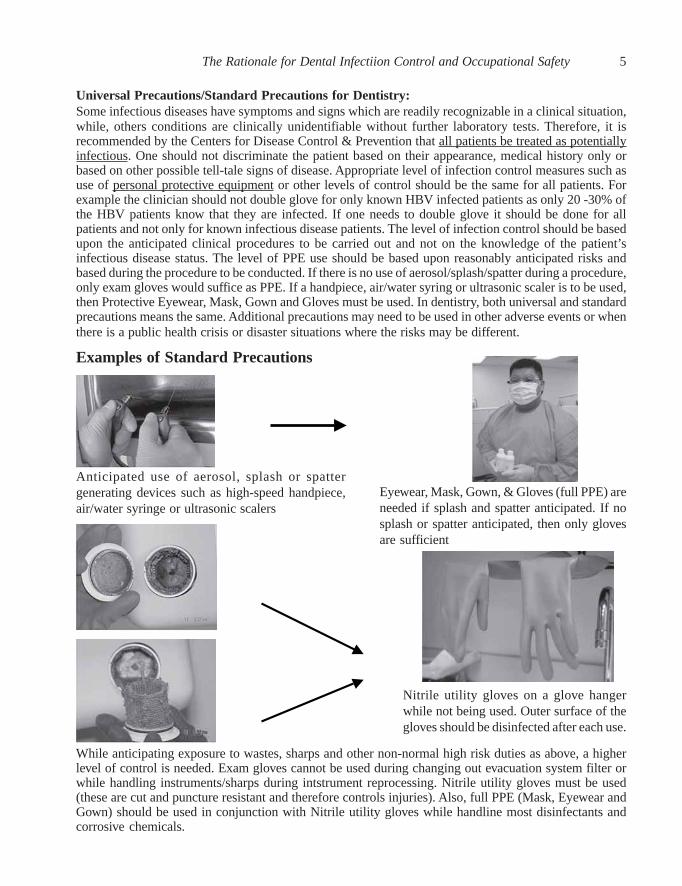
Examples of Standard Precautions
Anticipated use of aerosol, splash or spattergenerating devices such as high-speed handpiece,air/water syringe or ultrasonic scalers
Eyewear, Mask, Gown, & Gloves (full PPE) areneeded if splash and spatter anticipated. If nosplash or spatter anticipated, then only glovesare sufficient
Nitrile utility gloves on a glove hangerwhile not being used. Outer surface of thegloves should be disinfected after each use.
While anticipating exposure to wastes, sharps and other non-normal high risk duties as above, a higherlevel of control is needed. Exam gloves cannot be used during changing out evacuation system filter orwhile handling instruments/sharps during intstrument reprocessing. Nitrile utility gloves must be used(these are cut and puncture resistant and therefore controls injuries). Also, full PPE (Mask, Eyewear andGown) should be used in conjunction with Nitrile utility gloves while handline most disinfectants andcorrosive chemicals.
Chapter 01 -NEW.p65 10/20/2007, 9:53 AM5
Infection Control in Dentistry6
Not
e: C
ondi
tions
add
ress
ed in
the
tabl
e ar
e fr
eque
ntly
see
n in
den
tal p
atie
nts
and
ther
efor
e ne
ed to
be
cons
ider
ed in
pro
tect
ing
patie
nts
and
the
dent
al h
ealth
car
e w
orke
rs.
The
mod
es o
f tr
ansm
issi
on in
den
tistr
y ar
e co
mm
only
dir
ect c
onta
ct w
ith le
sion
s, s
aliv
a, b
lood
, ora
l muc
osa,
and
dro
plet
s or
aer
osol
s co
ntai
ning
infe
ctio
us a
gent
s.
Con
tact
-les
ion
exud
ate,
sal
iva,
sex
ual
cont
act,
bloo
dC
onta
ct-l
esio
n ex
udat
e, s
aliv
a, b
lood
Con
tact
-les
ion
exud
ate,
sal
iva,
blo
odC
onta
ct-l
esio
n ex
udat
e, s
aliv
a, b
lood
, nas
opha
ryng
eal
secr
etio
nsC
onta
ct-l
esio
n ex
udat
e, g
enita
l se
cret
ions
, sec
retio
ns f
rom
eye
Con
tact
-les
ion
exud
ate,
muc
osa,
sal
iva,
blo
od, b
ody
flui
dsC
onta
ct-l
esio
n, m
ucos
a, b
lood
Con
tact
-les
ion,
muc
osa,
sal
iva,
blo
od, b
ody
flui
dsC
onta
ct-m
ucos
a, s
aliv
a, l
esio
n ex
udat
eC
onta
ct-b
lood
, sal
iva,
bod
y fl
uids
Con
tact
-blo
od, s
aliv
a, b
ody
flui
dsC
onta
ct-b
lood
, sal
iva,
bod
y fl
uids
Con
tact
-blo
od,
sem
en,
non-
inta
ct s
kin
Aer
osol
, co
ntac
tA
eros
ol,
drop
let
Aer
osol
, dr
ople
tA
eros
ol,
drop
let
Aer
osol
, dr
ople
tA
eros
ol, d
ropl
et, i
ntim
ate
cont
act
Aer
osol
, dro
plet
, int
imat
e co
ntac
t
Dro
plet
, co
ntac
tD
ropl
et,
cont
act
Dro
plet
, co
ntac
t, in
gest
ion
Dro
plet
, con
tact
, sal
iva,
blo
od, e
xuda
teD
ropl
et, c
onta
ct, s
aliv
aD
ropl
et, c
onta
ct, s
aliv
a, b
lood
Inge
stio
n, r
arel
y bl
ood
Inge
stio
n, r
arel
y bl
ood
Tab
le 3
. Inf
ecti
ous
Dis
ease
s co
mm
only
enc
ount
ered
in d
enti
stry
Con
diti
onH
abit
atR
oute
s of
Tra
nsm
issi
on
Sexu
ally
tra
nsm
itte
d di
seas
es
1.H
erpe
tic I
nfec
tions
2.A
cute
her
petic
gin
givo
stom
atiti
s3.
Her
petic
Whi
tlow
4.G
onco
ccal
Inf
ectio
ns5.
Chl
amyd
ial
Infe
ctio
ns6.
Tri
chom
onal
Inf
ectio
ns7.
Con
dylo
ma
Acu
min
atum
8.Sy
phili
s9.
Infe
ctio
us M
onon
ucle
osis
10.
Hep
atiti
s B
Vir
us I
nfec
tion
11.
Hep
atiti
s D
Vir
us I
nfec
tion
12.
Hep
atiti
s C
Vir
us I
nfec
tion
13.
Hum
an I
mm
unod
efic
ienc
y
V
irus
Inf
ectio
n
Res
pira
tory
Dis
ease
s1.
Com
mon
Col
d2.
Sinu
sitis
3.Ph
aryn
gitis
4.Pn
eum
onia
5.T
uber
culo
sis
6.SA
RS
7.A
vian
Inf
luen
za (
H5N
1 Fl
u)
Chi
ldho
od D
isea
ses
1.C
hick
enpo
x2.
Her
pang
ina
3.H
and,
foo
t and
mou
th d
isea
se4.
Rub
ella
& R
ubeo
la5.
Mum
ps6.
Cyt
omeg
alov
irus
inf
ectio
n
Oth
er C
omm
on C
ondi
tion
s1.
Hep
atiti
s A
Vir
us I
nfec
tion
2.H
epat
itis
E V
irus
Inf
ectio
n
Ora
l, ph
aryn
x, a
no-g
enita
l, sk
in, v
isce
ra, e
yeO
ral,
ging
iva,
pha
rynx
Fing
ers,
han
dsO
ral,
phar
ynx,
gen
itals
Gen
itals
, ey
es, o
roph
aryn
xG
enita
ls,
orop
hary
nx,
oral
, ga
stro
inte
stin
alA
no-g
enita
l sk
in, o
ral,
muc
osal
are
asG
enita
ls, s
kin,
ora
l m
ucos
a, o
roph
aryn
xSk
in, o
ral
muc
osa,
gen
itals
, par
otid
s, s
aliv
aL
iver
, blo
od, b
ody
flui
dsL
iver
, blo
odL
iver
, blo
odB
lood
, ora
l m
ucos
a, s
kin
Upp
er R
espi
rato
ry T
ract
Upp
er R
espi
rato
ry T
ract
Upp
er R
espi
rato
ry T
ract
Res
pira
tory
Tra
ctR
espi
rato
ry T
ract
Res
pira
tory
Tra
ctR
espi
rato
ry T
ract
, G
astr
oint
estin
al T
ract
Ora
l, sk
inO
ral,
orop
hary
nxO
ral,
hand
s, f
eet
Res
pira
tory
tra
ct, o
ral
skin
Paro
tids,
pan
crea
s, t
estis
, CN
SSa
livar
y gl
ands
Liv
er,
gast
roin
test
inal
tra
ctL
iver
, ga
stro
inte
stin
al t
ract
Chapter 01 -NEW.p65 10/20/2007, 9:53 AM6

The Rationale for Dental Infectiion Control and Occupational Safety 7T
able
4. A
dapt
atio
n of
Spa
uldi
ng’s
Cla
ssif
icat
ion:
Lev
elR
isks
Con
trol
Met
hods
Mat
eria
ls/D
evic
esC
ritic
al
Sem
icri
tical
Non
criti
cal
Env
iron
men
tal
Hig
h
Hig
h
Mod
erat
e to
low
Low
Ster
iliza
tion
by:
-Aut
ocla
ve-C
hem
icla
ve-D
ry H
eat
-Im
mer
sion
in f
ull s
tren
gth
Glu
tara
ldeh
yde
(8 h
ours
for
ster
iliza
tion
and
rins
ed w
ith s
teri
le w
ater
)or -S
teri
le s
ingl
e-us
e-di
spos
able
s
Ster
iliza
tion
by:
-Aut
ocla
ve-C
hem
icla
ve-D
ry H
eat
-Im
mer
sion
in f
ull s
tren
gth
Glu
tara
ldeh
yde
(8 h
ours
for
ster
iliza
tion
and
rins
ed u
sing
ste
rile
wat
er)
or -Ste
rile
sin
gle-
use-
disp
osab
les
-Cle
an b
ut n
on-s
teri
le si
ngle
-use
-dis
posa
bles
supp
lies
Su
rfac
e D
isin
fect
ion
wit
h i
nte
rmed
iate
lev
elho
spit
al d
isin
fect
ants
:-H
ydro
gen
pero
xide
bas
ed-P
heno
ls-I
odop
hors
-Qua
tern
ary
Am
mon
ia C
ompo
unds
or -Dis
posa
ble
Bar
rier
s
Dis
infe
ctio
n w
ith
In
term
edia
te t
o lo
w l
evel
disi
nfec
tant
s:-P
heno
ls-I
odop
hors
-Qua
tern
ary
Am
mon
ia C
ompo
unds
Sani
tiza
tion
:-S
crub
was
h w
ith s
oap
and
wat
er
Item
s th
at a
re u
sed
in s
urge
ry w
hich
pie
rce
soft
and
hard
tis
sue—
Sca
lpel
bla
des,
bur
s, e
xtra
ctio
n fo
rcep
s, e
leva
tors
,ne
edle
s, f
iles,
bon
e-ro
nger
s, p
erio
dont
al i
nstr
umen
tsus
ed in
pro
phyl
axis
, sur
gica
l dra
ins
for
absc
esse
s, a
ndan
y ot
her
inst
rum
ent u
sed
in s
urge
ry, d
enta
l exp
lore
rs,
peri
odon
tal
prob
es,
biop
sy p
unch
, su
rgic
al d
rain
s,en
dodo
ntic
file
s an
d re
amer
s, a
nd i
mpl
ants
Item
s th
at d
o no
t ne
cess
arily
pen
etra
te s
oft
and
hard
tissu
es b
ut w
hich
cro
ss t
he v
erm
ilion
bor
der
(lip
) in
toth
e or
al c
avity
—M
outh
mir
rors
, ha
ndpi
ece,
ane
sthe
tic
syri
nges
, ch
ipsy
ring
es,
amal
gam
con
dens
ers,
im
pres
sion
tra
ys,
air/
wat
er s
yrin
ge t
ips,
hig
h-vo
lum
e ev
acua
tor
tips
Item
s us
ed in
den
tistr
y w
hich
do
not c
ross
the
verm
ilion
bord
er o
r pen
etra
te th
e so
ft ti
ssue
s— c
hair
ligh
t han
dles
,in
stru
men
t tra
ys, h
igh
touc
h w
ork
surf
aces
, bra
cket
tabl
es,
chai
r co
ntro
ls,
Air
/wat
er s
yrin
ges,
hos
esan
d de
ntal
cha
irs
Floo
rs, w
alls
and
doo
r ha
ndle
s th
at a
re n
ot c
onsi
dere
dhi
gh to
uch
surf
aces
. Gen
eral
hou
seke
epin
g ru
le a
pplie
sto
the
se s
urfa
ces
Chapter 01 -NEW.p65 10/20/2007, 9:53 AM7

Infection Control in Dentistry8
Examples for Spaulding’s Classification
Critical Items:Items that pierce the skin or mucosamust be sterilized or sterile signle-use-disposables
Semi-Critical Items:Non-sharp items that enter the oralcavity must be sterilized, sterile signle-use-disposables or single-single-usedisposable supplies
Semi-Critical Items:Non-sharp items that enter the oralcavity must be sterilized, sterile signle-use-disposables or single-single-usedisposable supplies
Environmental Surfaces:Walls, floors and non-hightouch or non-intimate surfacesshould be maintained throughhouse-keeping methods
Chapter 01 -NEW.p65 10/20/2007, 9:53 AM8

Chapter 2
Common infectious diseases encountered in Dentistry
Puttaiah & Roychowdhury
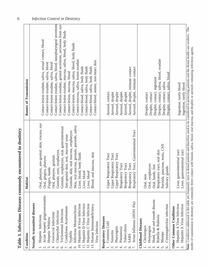
Many diseases are encountered in dental practice. Sometimes it is the patient that is infected who comesin to seek care, and sometimes it could be the clinician or clinical staff affected by the disease condition.
Hepatitis
Hepatitis AHepatitis A Virus (HAV) belongs to the picornoviridae family and is an RNA virus. HAV infection causesjaundice and rarely causes death. Among otherwise healthy adults the death rate is about 1 in 1000, inpeople >50 years of age the rate is 27 in 1000. Incubation period is about 4 to 6 weeks. Once the personrecovers from Hepatitis A infection, the person is protected for life. A vaccine against Hepatitis A viralinfection is now available. If one has not been exposed to HAV, a one time vaccination may provide lifelong immunity.
Hepatitis EHepatitis E Viral (HEV) infection is similar in nature to the HAV infection epidemiologically but for thehigher rate of infection among pregnant women in the third trimester (20% infection rate). Outbreaks arecommonly seen in the South Asia, Southeast Asia, Africa, Central and South American regions amongother geographic regions in the world. As of today, there is no vaccine available against Hepatitis E Virus.
Hepatitis BHepatitis B Viral (HBV) infection is caused by a DNA virus which is a hepadnavirus. Most patients withHBV infections cannot be clinically identified as being infected. About 2-7% of the population in theSouth Asian, Middle-Eastern, Mediterranean region, East European, Russian, Parts of Central and SouthAmerican region are infected with this virus. Certain Alaskan and Canadian regions (Tundra), SouthAmerican Region, Africa, Southeast Asian region including China are considered high in prevalence(>8% of the population). Most of the North American, parts of the South American, Australian, and WestEuropean regions are considered low in prevalence (<2% of the population). The incubation period lastsfrom 45-160 days therefore it is also called chronic hepatitis infection. Transmission can be bothpercutaneous and non-percutaneous, but, primarily bloodborne. This variety of hepatitis is very contagiousand has been occupationally acquired by dentists in the past. Outcomes of HBV infection are — about90% of the infected become healthy again; about 9-10% become asymptomatic carriers or suffers fromchronic persistent hepatitis or develop active hepatitis leading to hepatocellular carcinoma and death;about 1% develops fulminant disease right after infection and die. Vaccines against HBV infections areavailable. The rate of infection among dentists (general practitioners and specialists included) rangedfrom 13.6% to 38.5%. Therefore it is not an uncommon disease affecting dentists. There have been cases
Chapter 02 -NEW.p65 10/20/2007, 9:57 AM9
Infection Control in Dentistry10
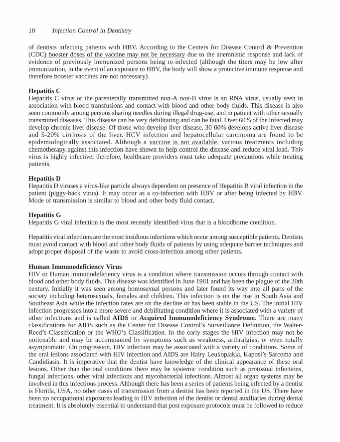
of dentists infecting patients with HBV. According to the Centers for Disease Control & Prevention(CDC) booster doses of the vaccine may not be necessary due to the anemnistic response and lack ofevidence of previously immunized persons being re-infected (although the titers may be low afterimmunization, in the event of an exposure to HBV, the body will show a protective immune response andtherefore booster vaccines are not necessary).
Hepatitis CHepatitis C virus or the parenterally transmitted non-A non-B virus is an RNA virus, usually seen inassociation with blood transfusions and contact with blood and other body fluids. This disease is alsoseen commonly among persons sharing needles during illegal drug-use, and in patient with other sexuallytransmitted diseases. This disease can be very debilitating and can be fatal. Over 60% of the infected maydevelop chronic liver disease. Of those who develop liver disease, 30-60% develops active liver diseaseand 5-20% cirrhosis of the liver. HCV infection and hepatocellular carcinoma are found to beepidemiologically associated. Although a vaccine is not available, various treatments includingchemotherapy against this infection have shown to help control the disease and reduce viral load. Thisvirus is highly infective; therefore, healthcare providers must take adequate precautions while treatingpatients.
Hepatitis DHepatitis D viruses a virus-like particle always dependent on presence of Hepatitis B viral infection in thepatient (piggy-back virus). It may occur as a co-infection with HBV or after being infected by HBV.Mode of transmission is similar to blood and other body fluid contact.
Hepatitis GHepatitis G viral infection is the most recently identified virus that is a bloodborne condition.
Hepatitis viral infections are the most insidious infections which occur among susceptible patients. Dentistsmust avoid contact with blood and other body fluids of patients by using adequate barrier techniques andadopt proper disposal of the waste to avoid cross-infection among other patients.
Human Immunodeficiency VirusHIV or Human immunodeficiency virus is a condition where transmission occurs through contact withblood and other body fluids. This disease was identified in June 1981 and has been the plague of the 20thcentury. Initially it was seen among homosexual persons and later found its way into all parts of thesociety including heterosexuals, females and children. This infection is on the rise in South Asia andSoutheast Asia while the infection rates are on the decline or has been stable in the US. The initial HIVinfection progresses into a more severe and debilitating condition where it is associated with a variety ofother infections and is called AIDS or Acquired Immunodeficiency Syndrome. There are manyclassifications for AIDS such as the Center for Disease Control’s Surveillance Definition, the Walter-Reed’s Classification or the WHO’s Classification. In the early stages the HIV infection may not benoticeable and may be accompanied by symptoms such as weakness, arthralgias, or even totallyasymptomatic. On progression, HIV infection may be associated with a variety of conditions. Some ofthe oral lesions associated with HIV infection and AIDS are Hairy Leukoplakia, Kaposi’s Sarcoma andCandidiasis. It is imperative that the dentist have knowledge of the clinical appearance of these orallesions. Other than the oral conditions there may be systemic condition such as protozoal infections,fungal infections, other viral infections and mycobacterial infections. Almost all organ systems may beinvolved in this infectious process. Although there has been a series of patients being infected by a dentistis Florida, USA, no other cases of transmission from a dentist has been reported in the US. There havebeen no occupational exposures leading to HIV infection of the dentist or dental auxiliaries during dentaltreatment. It is absolutely essential to understand that post exposure protocols must be followed to reduce
Chapter 02 -NEW.p65 10/20/2007, 9:57 AM10

Common infectious diseases commonly seen in Dentistry 11
the probability of sero-conversion, by taking antiviral drugs immediately after exposure to a patientinfected with HIV.
TuberculosisTuberculosis is one of the oldest infectious diseases known to humans. In the past most countries had thisdisease under control. But now this disease has reemerged in both prevalence and with a new type of amulti-drug-resistant-strains. Mycobacterium tuberculosis is the organism which commonly affects thelungs but may involve most organs in the body. Each year about 8 million people develop TB and 3million die. TB mimics many respiratory conditions, therefore when the practitioner observes a cough ofmore than 3 weeks of duration, sputum possibly tinged with blood, unexplained weight loss, and nightsweats, the patient should be referred for a TB skin test and treatment. If diagnosed with active infectionthe patient must be treated till pronounced non-infectious and then may access dental care. It is pragmaticto defer care for patients with active TB till such time the disease is controlled. In the United States,dentists can defer elective dental care till such time the patient is pronounced non-infectious, and allemergency dental treatments may be provided in institutions that are equipped to deal with the control ofcross contamination or occupational exposure. Such facilities should include negative air pressure treatmentrooms with the air vented to the outside of the building. The air conditioning and ventilation system mustalso be equipped with HEPA filters and the personnel must use masks that have a HEPA filter during thecontact with infected patients. Dentists and staff must undergo testing for the disease on a periodic basis,especially if living in endemic areas where the prevalence is high. Many healthcare institutions in theUnited States have made annual TB testing mandatory for their personnel and have effective TB controlplans. In endemic areas, the testing may be done every six months. Similar control plans may be adoptedby individual clinics for the benefit of the personnel and patients.
Spread of infection Diseases–HIV in the USA
This series of images shows clusters of HIV cases (each yellow dot = 30 cases) being spread in the USA over a periodof 11 years. In 1983 there were about 1000 registered cases, in 1985 about 10,000 cases, in 1989 about 100,000 cases,and in 1994 about 440,000 HIV cases (The Centers for Disease Control & Prevention, Atlanta, Georgia, USA).
Chapter 02 -NEW.p65 10/20/2007, 9:57 AM11

Infection Control in Dentistry12
In intial exposure, there are two possibilities, infection or no infection. If there is no infection the personis lucky. If infected after exposure the outcomes could be immediate or acute HIV infection leading toAIDS, or asymptomatic persistent generalized lymphadenopathy that may be in a chronic state for manyyears and then go into the AIDS state. On the other hand asymptomatic PGL or asymptomatic patientscan remain so for many years (over 20 years) without advancing into the AIDS stage.
Outcomes of Exposure to HIV
In a dental setting, the dentist and the dental staff may be infected by a variety of conditions. Followingare the conditions and possible return to work approach—Table 5: When can an infected clinician return to work?
Condition When should one return to work OR what is to be doneConjunctivitis After discharge ceasesStaph. Aureus (active) After lesions have healedStrep. Group A 24 hours after starting effective antimicrobial treatmentViral Respiratory Infection After resolution of Acute symptomsActive Tuberculosis After treatment with antimicrobials and deemed non-infectiousPositive Skin Test for TB After evaluation for infectious status, chest x-ray, and treatment if needed till
deemed non-infectiousInfluenza After symptoms resolvePediculosis (hair lice) After treatment provided and no liceHerpetic Whitlow After lesions healOrofacial Herpes After lesions heal, need to be regularly on anti-herpes medicines for the rest
of the lifeChicken Pox (Varicella) After lesions dry and crust outShingles (Herpes Zoster) After lesions dry and crust outHepatitis B (HBe antigen +ve) After deemed HBe antigen –ve, UP/SP and expert panel /Infectious Diseases
MD to monitor clinicianHepatitis C Seropositive Need to use UP/SP, Proper Aseptic Techniques to protect patients, Anti-viral
Medication, MonitoringHIV/AIDS After anti-retroviral therapy started, UP/SP and expert panel /Infectious
Diseases MD to monitor clinicianMeasles After 7 days from the appearance of rashMumps After 9 day from start of parotitisRubella After 5 days from the appearance of rashPertussis After 5 days from start of effective antimicrobial therapyDiarrhea After symptoms resolveAmoebiasis After starting effective antimicrobial therapy and symptoms resolveEnteroviral Infections After symptoms resolveHepatitis A After 7 day from the onset of Jaundice
Although it is difficult to implement in most clinical situation, we should try to implement these guidelines to the bestof efforts. These conditions range from ow risks of disease transmission to very high risks of transmission within theclinics. These conditions may be passed on to the patient or be spread in the clinical area between clinicians.
Chapter 02 -NEW.p65 10/20/2007, 9:57 AM12

Chapter 3
Infectious Disease in India – Epidemiology, Impact andPerceptions
Kohli, Mohan Das, Arora & Puttaiah
HIV & AIDSAlmost 5 million people were infected by HIV globally in 2005 – the highest jump since the first reported case in1981 – taking the number living with the virus to a record 40.3 million, the United Nations announced in November2005. They further announced 4.9 million new infections was fueled by the epidemic’s continuing rampage in SubSaharan Africa and a spike in the former Soviet Union and Eastern Europe, Central Asia and East Asia. More than 3.1million people died this year from AIDS, including 570,000 children – far more than the toll from all natural disasterssince last December’s tsunami or natural disasters in Asia, particularly Indian and Pakistan. Southern Africa, includingSouth Africa, which has the world’s most cases at more than 5.1 million, continues to be worst hit. Sub-SaharanAfrica is home to 25.8 million HIV-positive people, or 64% of the world’s total. In Asia, a total of 1.2 million newcases since 2003 pushed total cases to 8.3 million, with conditions in countries such as Vietnam and Pakistan ripe fora rapid spread.
UNAIDS said the number of HIV-positive women reached 17.5 million this year, one million more than in 2003.“UNAIDS and governments should “wake up and smell the coffee”, said “Anjali Gopalan, director of the NaazFoundation, India, (a well established organization working on the control of HIV in India). The outgoing chief ofIndia’s official National Aids Control Organization, S.Y. Quraishi, said 70 percent of Indian sex workers either didnot know what a condom was or how to use one. “If the situation remains unchanged, India could have an estimated50 million HIV cases by 2025.” For nearly two decades India is one of the leading countries as far as AIDS infectionsare concerned. Since 1994 almost every country in Southeast Asia and Africa has seen its HIV prevalence ratesdouble and India is no exception. India with a population of more than 1 billion is a melting pot in the globalmarketplace. About half the population is in the sexually active age. The first HIV case was detected in 1986 by Dr.S. Solomon (YRG Care, Chennai, India) and as of today, HIV has been reported in all the individual States and UnionTerritories. As of today, it is in almost all segments of the society, gender, age and social class and sexual orientation.The spread of HIV in India has been diverse, with much of India having a low rate of infection and the epidemicbeing most extreme in the southern half of the country and in the far north-east. The highest HIV prevalence rates arefound in Maharashtra in the west; Andhra Pradesh and Karnataka in the south; and Manipur and Nagaland in thenorth-east. As of August 2006, 90% of all nationally reported AIDS cases have been found in 10 of the 38 StatesUnion Territories. The greatest numbers were in Maharashtra and Gujarat in the west; Tamil Nadu, Andhra Pradeshand Karnataka in the south; and Manipur and West Bengal in the north-east.
In the southern States, the infections are mostly due to heterosexual contact, while infections are mainly foundamongst injecting drug users in Manipur and Nagaland. The Indian National AIDS Control Organization (NACO)estimates that 5.21 million people were living with HIV in 2005, giving an adult prevalence of 0.91%. This representsa slight increase from the 2004 estimate, and a substantial increase from 4.58 million in 2002. These rates are stillbeing reviewed and updated with some reports stating a much lower prevalence. However, even low prevalence ratesin such a large population still means a huge caseload, impacting both the public health, primary and tertiary caresystems with respect to prevention, control, stabilization, education and intervention modalities. Even if there is avery low increase of about 0.1% in the HIV prevalence rate in within this huge population, the caseload increasestremendously further burdening the already stretched healthcare system.
Chapter 03 - NEW.p65 10/20/2007, 9:59 AM13

Infection Control in Dentistry14
The national HIV prevalence has risen dramatically since the start of the epidemic, but a study released at the beginningof 2006 suggests that the HIV infection rate has fallen in southern India, the region that has been hit hardest by AIDS.In addition, NACO has released figures suggesting that the overall rate of new HIV infections in the country isslowing. Researchers claim that this decline is the result of successful prevention campaigns, which have led to anincrease in condom use. “Could this also have been to attrition within the HIV population?” is a question we maywant to address.
Today it is globally known and evidence-based that HIV viral particle have been isolated from saliva, and the latter isone of the primary screening methods for HIV infection. Saliva is normally contaminated with blood from gingivalinflammatory tissue and therefore it is possible that HIV and HBV could spread from one individual to another eventhrough saliva. Therefore, saliva must be treated as potentially infectious as blood or other body fluids with respect toHIV and other bloodborne diseases.
The need for an effective infection control program has always been an essential and integral part of the dental practice.Over the years dentistry, with rare documented exceptions, dental infection control has been successful in limiting thetransmission of infectious diseases during patient care. Exceptions have involved the transmission of hepatitis B virus(HBV) and herpes simplex virus (HSV) between practitioners and patients during dental treatment and are beinginvestigated. Unfortunately, early reports of occurrences of disease transmission between dentists and patients were nottaken seriously and were treated by the profession as isolated incidences. However, when acquired immunodeficiencysyndrome (AIDS) a disease with a social stigma of initially being more prevalent among homosexuals was identified asan infectious disease, health care providers involved with close patient contact began to review and re-evaluate thealready existing control measures developed for HBV. The HIV/AIDS epidemic precipitated rethinking the implementationof infection control measures. In 1983, the Centers for Disease Control (CDC) made the first recommendations for theprevention of exposure to blood and body fluids through the use of universal precautions. In 1986, recommendedInfection-Control Practices for Dentistry was published and later updated in 1993. The CDC published recommendationfor prevention of HIV transmission in health-care settings in 1987, which recommended that blood and body fluidprecautions be consistently used for all patients regardless of their bloodborne infection status. Under universal precautions,blood and certain body fluids of all patients are considered to be potentially infectious for human immunodeficiencyvirus (HIV), hepatitis B virus (HBV), hepatitis c virus (HCV) and other bloodborne pathogens. The rationale for treatingall patients as potentially infectious is due to the fact that most patients are unaware of their infectious disease status.This concept was intended to use protective measures to prevent parenteral, mucous membrane and non-intact skinexposures of healthcare workers to bloodborne pathogens based on the clinical procedure rather than the known infectiousdisease status of the patient or the appearance of the patient. In addition, the recommendation for health care workers tobe immunized against hepatitis B was an additional measure.
Infection Control & Occupational Safety is all measures taken to control infections during clinical care, be it patient-to-patient transmission or between the patient and care provider. In dentistry due to multiple procedures and specializedfields, the techniques that are used today have to be specifically adapted multiple idiosyncrasies yet simplified toaccommodate diversity in the risks. Although basic principles about the spread of infections in dental clinics andhospital set-ups have not changed, new issues have emerged with underlying diseases being more compromising,invasive procedures being more common, newly identified varieties of microorganisms responsible for a broaderspectrum of infection, bacterial isolates becoming more resistant to common and conventional antibiotic therapies,and patients being treated by greater variety of health care providers. Taken cumulatively these factors have presentedan impressive array of new challenges for the most sophisticated clinician and the infection control practitioner.
About 20 years after exposure to infection control concepts in this country as well as in other advanced countries of theworld, control and prevention of infectious diseases in dental patients and health care providers remains a conundrum.There has been a lot of effort placed on the practice of infection control without acceptance (positive attitude) towardsthe acquired knowledge. A survey on Infection Control Needs Assessment of Practitioners in Eight Countries by Puttaiahet al, AADR, 2005 showed that even in advanced countries, dentists practice adequate infection control but still did notunderstand the real concept of Universal or Standard Precautions. About 17% to 64% of practitioners felt that allpatients were not to be treated as potentially infectious, 50% to 86% felt that medical history and appearance of thepatient dictated the level of infection control to be practiced, 53% to 75% felt it was appropriate to double-glove whiletreating infectious patients, 12% to 64% felt that the infectious disease status of the patient was always known, and lastly18% to 65% felt that it was alright to refuse care to infectious patients. Could this be due to Stigma? These numbers arevery discouraging as even practitioners in advanced countries have not understood the concepts but still practice by rote.It is our duty as teachers in Infection Control and Safety to make the practitioners have the knowledge, make them
Chapter 03 - NEW.p65 10/20/2007, 9:59 AM14

Infectious Disease on India – Epidemiology, Impact and Perception 15
develop a positive attitude and then practice in a safe manner. The “why, when and what”, need to be explained aboutany new concept for safer practice. While anticipating risks, clinical application of personal safety techniques as prescribedby U.S. Occupational Safety and Health Administration addresses procedure specific aseptic techniques, barrier usesuch as gloves, masks, protective eyewear, clinical attire and use of disposable inanimate surface barriers in the absenceof decontamination. Further use of automated instrument decontamination devices, time efficient and effective heatsterilization methods, chemical disinfectants, waste management procedures and single use disposable items have createda safer environment for dental personnel and patients alike.
TuberculosisTuberculosis has been and continues to be taking a terrible toll on the Indian population. According to the WHO GlobalTB Report 2006, India has more new TB cases annually than any other country, ranking first among the 22 high-burdencountries worldwide. TB remains one of the leading infectious causes of mortality in India, resulting in 364,000 deathsannually. There were more than 1.8 million new TB cases in India in 2004, representing over one-fifth of all TB casesworldwide. The estimated incidence rate in 2004 was 168 per 100,000 people. (Global Tuberculosis control WHOreport 2006). However, the Indian Scientific Community has developed many of the basic principles necessary to fightthis epidemic. These methods are now being practiced in many countries around the world. [GUEST Editorial 2004Journal of the Indian Medical Assoc., New Delhi, India].
TB is able, to produce acute to latent-chronic disease that mostly affects lungs but can virtually affect every organsystem in the body. The main concern for dental professionals and health care workers is transmission of the diseaseby inhalation of infective droplets expelled through cough by an infectious patient with active pulmonary disease.Authors around the world have emphasized strict adherence to infection control guidelines and for prompt initiationof treatment for patients with confirmed or suspected tuberculosis among this patient population. Obviously thesepatients present a serious risk for us as dentists and health care workers. We need to emphasize the critical importanceof deferring routine care, isolation of active cases (infectious) till treated and deemed non-infectious. As in otherparts of the world, multi-drug resistant TB is seen in patients with HIV. Therefore, one should not only understandthat additional precautions beyond universal/standard precautions may be needed. As India is a high risk country forTB, dental health care professional must undergo annual tests/chest x-rays to rule out active TB. TB skin test - PPDor purified protein derivatives skin test will show positive reaction to TB because most Indians have been immunizedwith the BCG vaccine. Therefore, chest x-rays as well as other microbiological/immunological methods need to beused for testing the care provider. If found with active lesions and deemed infectious, they should be isolated includingfrom patient care and treatment started immediately. After successful initiation and when deemed non-infectious, thecare provider can be allowed to return to active clinical practice. However, periodic monitoring needs to be conductedon the sustained efficacy of treatment.
HIV and TBHIV and TB form a lethal combination, each speeding the other’s progress. HIV weakens the system. An HIV-positive patient has a greater likelihood of acquiring TB, more so multi-drug resistant TB. A leading cause of death inHIV patients is TB. It accounts for about 13% of AIDS deaths worldwide. In Africa. WHO and its internationalpartners have formed the TB/HIV Working group, which develops policy on the control of HIV-related TB andadvises on how to tackle this lethal combination.
HEPATITIS – B VirusHBV is found in blood and blood products, saliva, sputum, breast milk, tears, wound fluid, sperm, sweat, vaginaldischarges. Minute quantities of fluid may be sufficient for parenteral transmission of disease. Only half of thoseinfected with HBV have a clinically diagnosed illness, therefore about 50% does not report disease status, of thesepatients 15% could become carriers. Hepatitis B is a major cause worldwide of acute and chronic hepatitis, cirrhosisand primary hepatocellular carcinoma. Hepatitis B virus (HBV) is probably the most important chronic viral infectionaffecting humans and HBV is a leading killer among all infectious agents. Approximately 400 million people arechronic HBV carriers worldwide. HBV infection and its complications are global health problems. The spectrum ofchronic HBV infection ranges from asymptomatic Hepatitis B surface Antigen (HBsAg) carrier state to chronichepatitis with progression to cirrhosis and end-stage liver disease. Despite the development of an effective vaccineagainst HBV, this infection remains a serious threat to public health in India. Several studies from India have reporteda HBV prevalence rate of 3% to 6%. However, these data are known to underestimate the prevalence of chronic HBVInfection. India with a population of approximately one billion and assuming a lower prevalence rate of 3%, Indiastill harbors approximately 30 million HBV carriers. A modest estimate would put the number of deaths occurringdue to HBV infection per year in India to around 100,000. HBV is responsible for about 68% of cirrhosis of the liver,
Chapter 03 - NEW.p65 10/20/2007, 9:59 AM15

Infection Control in Dentistry16
and 80% of hepatocellular carcinoma in India. In spite of the fact that HBV is a major killer in India, and this is easilypreventable, the new infections are on the rise. A decision-analytical model estimated that in India, vaccinationshould save 25 lives per 100,000 people/year; hepatitis B immunization is not yet available freely to infants andchildren as part of the state sponsored immunization program as in the United States. Incorporation of HBV vaccinationinto the Expanded Program of Immunization for infants could bring down the rates over time. One of the successesin the control of bloodborne diseases among dental professionals is the reducing rates of HBV due to mandatoryimmunization.
On the global perspective, one third of the world’s total population (2 billion) has been infected with hepatitis Bvirus. There are approximately 400 million people with chronic HBV infection (lifelong infection). Near to home(India) about 67% (275 million) reside in Asia and the Pacific Islands. By comparison, there are 170 million peoplewith chronic hepatitis C and 47 million people with HIV/AIDS in the world. In many Asian countries, 10% (5-20%)of the population are HBV carriers. China has the greatest burden of chronic hepatitis B and liver cancer. An estimated130 million (10% of the population) has chronic hepatitis B. About 0.5 million die each year from liver cancer or endstage liver disease caused associated with hepatitis B. About 1 million people die each year (equivalent to 2800deaths/day, 115 deaths/hour, or 1-2 deaths/minute) from liver cancer or liver failure caused by HBV.
HBV Facts - How Does It Spread?HBV is fifty to hundred times more infectious than HIV. The highest concentrations of the virus are found in blood(as high as ten billion viruses per mL); ten to a hundred times lower concentrations are found in semen and vaginalfluid. Hepatitis B virus is not spread by air, food, water, breastfeeding, casual contact in an office setting, kissing,hugging, coughing, sneezing, and sharing eating utensils or drinking glasses. Spread of HBV from mother to babyusually happens at the time of birth (still horizontal transmission). Child to-child spread most likely happens as aresult of contact with skin sores, small breaks in the skin, or mucous membranes with blood. Spread within thehousehold from sharing toothbrushes or razors may also occur because HBV can survive for at least seven daysoutside the body. 90% of infants infected at birth with HBV will become chronically infected. About 30-60% ofchildren aged 1-5 years infected with HBV will become chronically infected. Up to 10% of older children and adultsinfected with HBV will become chronically infected. Most people from Asia including the Pacific Region and Africabecome infected with HBV during childhood: from infected mother to child at birth, from child to child contact inhousehold settings, and from reuse of non-sterilized needles and syringes healthcare facilities.
The Relationship between HBV and Liver Cancer - A Silent KillerOne out of four people with chronic hepatitis B virus infection who become chronically infected during childhood(in other words, approximately 100 million of the 400 million chronic HBV infected people in the world) will die ofHBV-related liver cancer or cirrhosis. Liver cancer is often fatal because of its insidious nature of being practicallyasymptomatic in the initial stages. Thus, the diagnosis is incidentally made quite late while the patient is beingtreated at a tertiary center for other health problems. Liver cancer can occur in those with chronic HBV infectionwithout cirrhosis; the risk is higher with cirrhosis, in men (3:1 male to female ratio), and with a family history of livercancer. Liver cancer usually develops between thirty five to sixty five years of age. An estimated 600,000 people eachyear die of liver cancer; 380,000 deaths each year are from countries in Eastern Asia alone (China, Hong Kong,Japan, and Korea). China alone accounts for 54% of the liver cancer deaths worldwide. Liver cancer is one of the topthree causes of death by cancer in most of Asia, the Pacific Region, and sub-Saharan Africa. At least 80% of livercancer is caused by HBV. Worldwide, liver cancer is the fourth leading cause of cancer death in men, althoughuncommon in North America and Europe.
HBV infection, especially during infancy and early childhood, is easy to prevent with the hepatitis B vaccine. Since80% of liver cancer is HBV-related, the vaccine is considered the first ‘anti-cancer vaccine.’ Hepatitis B vaccine issafe and has been given to over 500 million people in the world. When given to infants immediately after birth and at1 month and at six months, completion of the three-dose vaccine series induces a protective antibody response in95% to 99% of vaccinated infants, even when the mother is a hepatitis B carrier. In addition, a direct reduction in livercancer in cohorts of immunized children has already been demonstrated in Taiwan. Post-vaccination testing forimmunity is not necessary after routine vaccination of infants, children, adolescents, or adults. Testing for immunity(anti-HBs) at nine to fifteen months of age after completing the series at six months of age is advised for infants bornto mothers who are infected with HBV. Testing one to two months after completing the series is advised for healthcareworkers, and persons with HIV infection. No booster shot is necessary after completion of the three-dose series dueto the anemnistic response in successfully immunized patients.
Chapter 03 - NEW.p65 10/20/2007, 9:59 AM16

Chapter 4
The Status of Dental Infection Control & Safety inIndia – A Study
Puttaiah, Bedi & Shetty
Introduction: This Dental Infection Control needs assessment study was conducted by investigatorsfrom India (Dr. Sadashiva Shetty, Davangere), the United States (Dr. Raghu Puttaiah, Dallas) and UnitedKingdom (Dr. Raman Bedi) using a self-administered needs assessment survey in India. The data collectedwere from a convenience sample in South India and could be used by educational institutions, governmentsand industries interested in improving the quality of infection control in South Asia. This study couldhelp understand the needs and help develop a basic need-based educational curriculum in dental infectioncontrol and safety for India. Similar studies have been conducted in Pakistan, South Korea, China,Philippines, Thailand, Taiwan and the United States of America.
Background: Dentistry is predominantly a field of surgery, involving exposure to blood and otherpotentially infectious materials,1 and therefore, requires a high standard of Infection Control and Safetypractice in controlling cross contamination and occupational exposures to bloodborne diseases. Apartform bloodborne diseases such as Hepatitis B infection and HIV infections, the dental health care workersare potentially at risk of acquiring respiratory diseases, childhood diseases, sexually transmitted diseasescommonly encountered in dentistry.2, 3 Although most principles of dental infection control and safetywere formulated in the 1960s, this field only gained importance over the past two decades due to theAIDS Epidemic in the Americas and Europe.2 Currently, adequate control of communicable diseaseshave been achieved in the developed countries through education, public health measures, regulations aswell as practice of dental infection control & safety.4, 5, 6, 7, 8, 9, 10, 11 As the incidence of AIDS andother associated bloodborne diseases is on the rise in South Asia (with the case load in India about 5.7million) and is predicted to reach epidemic proportions similar to that in Africa and the United States,10,11 it is essential to improve the knowledge, attitude and practice of Dental Infection Control and Safetyin the South Asian region. The population at risk of being infected through dental care would be all ages,gender, social class and occupation if strict infection control and safety procedures are not followed bythe practitioners. Anyone who seeks dental care at a place that does not utilize strict aseptic and protectivemeasures could possibly be a victim of infectious disease.
Materials & Methods: A preliminary data collection instrument with about 100 variables on theKnowledge, Attitude and Practice regarding Dental IC & Safety was developed by the investigators.About 500 of this self-administered instrument was distributed in South India (Karnataka and TamilNadu States) among faculty members of dental schools, dentists in private clinical practice, and dentistsworking for the Governments. The completed instruments were edited for content and context and datawere entered and analyzed using SPSS for Windows
Chapter 04 - NEW.p65 10/20/2007, 10:00 AM17

Infection Control in Dentistry18
Results: Of the 500 instruments distributed 456 (91.2%) returned completed instruments. About 63.5%were male and 36.5 female. The mean age of the respondents was 30.92 years. The mean amount of timeinvolved in practice among these respondents was 6 days per week, 11.7 months per year, 6 years inpractice treating about 12 patients per day. About 57.9% were graduates without post-graduate training,42.1% with at least a post-graduation or advanced training. About 21% were in General Practice, 10.4%had specialized in Oral Surgery, 7% in Orthodontics, 5.8% in Restorative Dentistry, and 55.4% in otherspecialty fields. The respondents represented about 20 schools from the South and South Central regionsof India but were practicing in the states of Karnataka and Tamil Nadu in South India. About 82% wereinvolved in private clinical practice and about 16% were faculty in various dental schools (Tables 1 and 2provide details on the Knowledge, Attitude, Practice and Needs of the respondents).
Table 1. Knowledge, Attitude, Practice and Needs Regarding Infection ControlVariable Percent
1 Epidemiological trend of “HIV on the rise in India” versus Perceived Levelof TrainingHIV on the rise in India 98.2Have adequate training in Dental Infection Control & Safety 70.0
2 Knowledge versus Attitude regarding HBV and HIV among respondentsPerception of knowledge on HBV as a disease 76.0Tested knowledge HBV as a disease 96.0Perception of knowledge on HIV as a disease 83.0Tested knowledge on HIV as a disease 99.0
3 Knowledge versus Attitude regarding HBV and HIV disease transmissionHIV & HBV transmission possible in the dental office 94.8Potential for disease transmission through splash or spatter 73.0Infectious diseases transmitted through sharp injuries only 56.3
4 Universal Precautions – Knowledge, Attitudes and PracticeHIV & HBV status of patients is always known 15.2All patients should be treated as potentially infectious irrespective of appearance 69.3Medical History patient dictates the level of infection control practice 54.1Double-gloving practiced while treating a patient with known infectious disease status 77.7Right to refuse care for patients with infectious diseases 20.8
5 HBV infections in dentistry:Risk of transmission in dentistry 98.2Need for immunization for Dental Practitioners 99.6
6 Immunization against infectious diseasesHBV 80.7Influenza 58.1Polio 96.4Diphtheria 94.7Tetanus 96.1Rubella 79.3Measles 87.0Mumps 83.0Chicken pox 82.6
7 HIV & AIDS in Dentistry:Risk of transmission in dentistry 94.9Need for improved infection control measures 99.1Practicing adequate infection control 61.5Having access to the needed infection control materials and equipment 38.3
8 Perceptions of Needs regarding Dental Infection Control & SafetyNeed more training 97.3Local agencies, organizations, associations and bodies to be more involved 98.2Can afford the costs for implementing infection control measures(equipment and supplies) 80.3Need for local financial institutions to provide loans for upgrading the equipment 98.6Need for local manufacture of infection control equipment and materials 95.9Mandatory infection control & safety curriculum in dental schools 99.3
Chapter 04 - NEW.p65 10/20/2007, 10:00 AM18

The Status of Dental Infection Control & Safety in India – a Study 19
Table 2. Practice measures being implementedVariable Percent
1 Antimicrobial HandsoapsCarbolic acid-based 71.4Chlorhexidine gluconate-based 70.0Triclosan-based 36.0Parachlorometaxylenol-based 22.6
2 Use of Personal Protective Equipment – PPE-Single-use-disposable exam gloves 87.3-Sterile single-use-disposable for surgical procedures 84.2-Face masks 92.6-Protective Eyewear 46.6-Protective Gowns 48.7
3 Safety on SharpsUse of single-use-disposable syringe/needles 95.0Use of Sharps Containers for disposal of needles and other sharps 35.0
4 Use of Chemical GermicidesPreprocedural antimicrobial mouthrinse 71.0Immersion disinfectant--Glutaraldehyde 90.7Surface disinfectant--Bleach (sodium hypochlorite) 79.1-Phenolic disinfectant 43.0-Quaternary Ammonia compounds/disinfectants 45.5-Iodophor disinfectant 27.8
5 Use of surface barriers 74.86 Control of Aerosols
Rubber-dam 40.0High-volume-evacuator 54.0
7 Use of Instrument Reprocessing Devices-Ultrasonic cleaners 46.9-Autoclaves 81.9-Chemiclaves 55.5-Dry heat 62.5-Glass bead (unapproved method) 67.6-Immersion sterilant (Glutaraldehyde as a sterilant) 84.0
8 Use of Heat Sterilizable InstrumentsHighspeed handpiece 67.5Burs 67.1Endodontic instruments 72.2Orthodontic instruments 58.3Hand instruments 91.8Mouth mirrors 93.5
9 Infection Control Equipment and MaterialsLocal availability 76.2Willingness to use if made available 93.6
Discussions: Preliminary analysis of data from this convenience sample showed a good response rate incompletion of instruments. Most respondents were general dentists while a significant number werespecialists. Each dentist treated about 2500 patients per year which is 25% more than in the USA. Therefore,the rate of exposure to infectious diseases could be higher in this sample due to practice profile and notdisease prevalence in the patient population alone.
Only about 80% of respondents were immunized against HBV, while the prevalence of HBV in SouthAsia is 2-7 %. A significant number were not immunized against other immunizable diseases. Mostrespondents (>98.8%) recognized the risk of HIV infection and the need for formal training in IC, while
Chapter 04 - NEW.p65 10/20/2007, 10:00 AM19

Infection Control in Dentistry20
only 61.5% used adequate measures, and 38.3% materials required in the practice of infection controland safety during patient care.
Variables demonstrating inadequate use and infection control principals were:Germicide use, Preprocedural Mouthrinse, Antibacterial Handwash, Disinfectants and Liquid Sterilants,Personal Protective Equipment, Safety devices, High Volume Evacuators & Rubber Dams, UltrasonicInstrument Cleaners and Sterilizers. Although, the respondents mentioned use of autoclaves, most weremodified pressure cookers (locally manufactured) at the best. Some clinics still used boiler tanks andmost clinics never packaged instruments. Other than some in institutional practice (Dental Schools attachedto Medical Centers), none of the respondents interview individually practices sterilization monitoring(validation of sterilizer function with biological indicators).
Although most respondents were willing to change and use IC materials and equipment, the latter werenot locally available. Most respondents had the knowledge of HBV, HIV and other infectious diseases,but few were adequately educated/trained in Universal Precautions and use of PPE. Data collected showeda need for formal training and sustenance of an IC Program.Data also demonstrated a multifactoral need such as “Involvement” by—-Local and National Dental Associations-Banking and Financial Institutions-Local Manufacturing firms to produce materials and equipment needed for infection control-Marketing Firms to obtain licensing to market equipment and materials for infection control-Dental Schools to provide education and become a local resource and make a education in IC mandatoryfor dental students.
As the prevalence of HIV infection & AIDS and other related conditions is on the rise, the loss of productivehours among the employed population will not only affect the internal economy of the South Asiancountries, but will indirectly affect investment of the other countries in South Asia. Public health measuresand improvement in the field of medicine are being addressed in South Asia, but the field of dentistry stilllacks the needed knowledge, training and equipment. It would therefore be in the economic and socialinterest to facilitate and sustain the proposed program of Infection Control and Safety Program in Indiafor protection of the former’s economic investments in the region. Further, the advanced countries andIndia should note that “travel dentistry” is becoming an affordable and a hot topic in India. The latter onlyshows that Infection Control & Safety Practice by all Dental Health Professionals in India is a highpriority and must meet not only the international standards but the best available standards being practicedin the United States and Europe.
Conclusions: The level of Infection Control in India is still in its infancy and many years behind that ofthe United States and some European countries. Formal programs in Infection Control and Safety mustbe developed not only for the Indian sub-continent but for the South and Southeast Asian Region andimplemented within the next couple of years. Lack of Infection Control is life-threatening for the both thePatient and the Dental Professional and requires more efforts than dental caries or periodontal disease, letalone cosmetic dentistry. In this context, Infection control in dentistry is to prevent life-threatening diseasesand communicable conditions and not dental caries or periodontal diseases which some researchers andpolicy makers have classified the latter as infectious diseases. A pragmatic policy on Infection Control &Occupational Safety through grass-roots education should address mandatory–
• Training for dental students (didactic curriculum including aseptic techniques)• Training for practitioners (requirement in basic training and maintaining continuing education credits
in IC&S)• Training for institution based practitioners• Introduction of Materials and Equipment needed for IC&S
Chapter 04 - NEW.p65 10/20/2007, 10:00 AM20
The Status of Dental Infection Control & Safety in India – a Study 21
• Recommendations for Dental Infection Control & Safety for India (detailing standards-of-care andPublic Health Law)
• Surveillance of safe practices• Dissemination of information Dental Safety for patients so that they may informed of the measures
being taken• Setting-up HIV and other Bloodborne Disease Dental Care Centers at Dental Schools• Expanding duties of the Public Health Dentistry/Community Dentistry Departments of Dental Schools
to provide out-reach dental care to rural HIV and other BBP infected patients• Eventually make all clinics provide dental care to all patients including HIV and other BBP infected
patients
An initial measure would be to craft the Infection Control Recommendations for Dental Professionals.The next step is to set-up a Nationwide Train-The-Trainer Program in Infection Control & Safety forIndia where future local experts would be trained. We need to set-up collaborative efforts with RecognizedUniversities in India and the US using Training Grants such as Fogarty International Training Programs.These programs should provide exposure to qualified individuals to obtain short-term fellowships in theUS (4-6 months) and develop applicable programs for India. International Health Promotion Agencies(Fogarty Intl., USAID, Bill & Melinda Gates Foundation, DFID and WHO) must get involved in supportingformal IC programs in South Asia and other developing countries. Local Dental Associations, Governments,Dental Schools, Banks, Industry and Marketing firms must also be involved for a multifactoral role tomake transition smooth. Industries from developed countries must help in this transition in any waypossible as they would be able to increase the market for materials and equipment with the available ofeducated and trained dentist.
References:
1. Molinari JA, Cottone, JA. Rationale for Practical Infection Control in Dentistry. In Practical Infection Control inDentistry, 2nd Ed., edited by Cottone JA, Terezhalmy GT, Molinari JA. Williams & Wilkins, Philadelphia, PA.3-12, 1995.
2. Puttaiah R, Cooley RL, Cottone JA. Infection Control & Safety in Periodontics. In Fundamentals of Periodontics.Edited by Wilson TG Jr., Kornman KS. Quintessence Publishing Co., Inc. Chicago, IL. 179-193, 1996.
3. Cottone JA, Puttaiah R. Viral Hepatitis and Hepatitis Vaccines. In Practical Infection Control in Dentistry, 2ndEd., edited by Cottone JA, Terezhalmy GT, Molinari JA. Williams & Wilkins, Philadelphia, PA. 15-47, 1995.
4. Centers for Disease Control (ACIP): Protection against viral hepatitis. MMWR, 39(RR-2):1-26, 1990.5. Goudeau A: Epidemiology and eradication strategy for hepatitis B in Europe. Vaccine, 8(Suppl): S113-S116;
discussion, S134-S138, 1990.6. Gruninger E, et al.: Hepatitis B, C, and HIV infection among dentists. 69th General Session of the IADR,
Acapulco, 1991.7. Centers for Disease Control: Guidelines for prevention of transmission of HIV & HBV to health-care and public
safety workers. MMWR, 34:561, 1985.8. Centers for Disease Control : Public Health Service statement on management of occupational exposure to
human immunodeficiency virus, including considerations regarding zidovudine post exposure use. MMWR,39(RR-1):1-14, 1990.
9. Centers for Disease Control: Recommendation for preventing transmission of human immunodeficiency virusand hepatitis virus to patients during exposure-prone invasive procedures. MMWR, 40(RR-8):1-9,1991.
10. Centers for Disease Control and Prevention. Guidelines for Infection Control in Dental Health-Care Settings —2003. MMWR 2003; 52 (No.RR-17): 1-76.
11. Centers for Disease Control and Prevention. Recommended infection-control practices for dentistry, 1993. MMWR1993; 42 (No.RR-8): 1-20.
12. Mann JM, Tarantola DJM, Netter TW. AIDS in the World: A Global Report. Cambridge, Harvard UniversityPress, 1992.
13. Yeh C-K, Puttaiah R, Cottone JA. Human Immunodeficiency Virus Infection, Acquired Immune DeficiencySyndrome, and Related Infections. In Practical Infection Control in Dentistry, 2nd Ed., edited by Cottone JA,Terezhalmy GT, Molinari JA. Williams & Wilkins, Philadelphia, PA. 48-74, 1995.
Chapter 04 - NEW.p65 10/20/2007, 10:00 AM21
Infection Control in Dentistry22
Chapter 5
Medical History and Dental Safety
Puttaiah & Roychowdhury
While taking medical history the clinician should not discriminate an infectious disease patient withreference to the potential of spreading the disease in the clinic. The reason one should look out forpatients with infectious diseases is to protect them from other acquiring other infectious disease conditions,as they usually are medically compromised. For some patients with active infectious disease it may beadvisable to defer routine dental care (as in Tuberculosis) till such time the patient is deemed non-infectious.Some times it may be necessary to differentiate between patients at risk for infections such as patientswith a history of rheumatic heart disease, recent intraosseous implants or recent extensive cardiac orother surgeries, where antibiotic prophylaxis may be indicated. All in all, one must know as to where thepatient stands in the panorama of health and disease, before offering treatment or even advice. Knowledgeof various infectious diseases, their potential for transmission, clinical features, progression and outcomesis essential for a clinician. While speaking to patients with infectious diseases one must maintain a highlevel of professionalism and confidentiality in acquiring the patient’s trust and confidence. If the patientis not comfortable, there is bound to be a barrier in doctor-patient-communication such as informationgiving and seeking, the end result being an incomplete history, reporting bias by the patient , and possiblemisdiagnosis, leading to inappropriate treatment. A legal issue that may be associated with the patient’smedical history is the completeness of the medical/dental record in the event of malpractice and litigation.In the event the dentist does not pay due importance to medical history, this inaction may be construed asnegligence during litigation, and may affect the right to practice dentistry. Therefore, not only must thedentist possess skills in providing dental care, but also have the acumen to address general health of thepatient, focusing on the required options for general patient management.
Some Components of the Patient Assessment Record in Relation to Infectious Disease
Identification CharacteristicsDates of visit, Name, Age, Gender, Ethnic Origin, Marital Status, Address, Occupation are some of thevariables that are needed. Each variable may have a bearing on the infectious disease status of the patient.For example, military personnel may be exposed to a different variety of infectious diseases based on thegeographic region of residence. Certain ethnic population may have a predilection to infectious diseasesthat may not be common locally (sickle-cell anemia). The date of history taking may have a bearing onthe time of occurrence and progression of disease.
History of Illness (Past & Present)History of trauma or other medical conditions which involved hospitalization and invasive surgery requiringblood transfusion, blood dyscrasias, immunosuppressive therapy, chemotherapy and radiation therapythat may alter the immune response of the patient must be recorded and dental treatment provided
Chapter 05 - NEW.p65 10/20/2007, 10:07 AM22
Medical History and Dental Safety 23
accordingly. Patients with chronic diseases such as diabetes mellitus, cancer These patients may be morelikely to fall sick due to microbial and viral exposures and with a greater probability of recurrence thanhealthy persons.
Family HistoryHereditary diseases and disorders such as hemophilia, diabetes and certain conditions affecting the nervoussystem may be elicited here (vertical transmission of disease i.e. parents to offspring). Certain acquireddiseases through proximity such as tuberculosis are also of importance (horizontal transmission i.e. betweenspouses, patients, patient to healthcare provider and vice-versa).
Social HistoryInformation on travel, sexual promiscuity, use of drugs and alcohol, personality and emotional state mayalso determine the level of risk of acquiring infectious diseases and possible immunosuppression as asequela.
Review of Systems:SkinGeneralized itching could be commonly seen as a sign of cirrhosis prior to occurrence of jaundice. Macules,papules, vesicles and scar could be various stages of chicken pox. A variety of pigmentation conditionsassociated with varying levels of immunosuppression such as Addison’s disease, von Rechlinghausen’sdisease, Puetz-Jeghers syndrome and Cushing’s disease and some nutritional/micronutrient deficiencies.Body hair (Lack or Loss thereof) may be associated chronic illnesses, dermatomysitis, systemic lupuserythematosis, lymphoma, cachexia, herpes zoster and micronutrient deficiencies.
LimbsInfectious, immunologic and neoplastic processes may be associated with joint disorders. One mustconsider modification of routine dental treatment so as to reduce the risk of infections in persons withprosthetic implants and total joint replacement with antibiotic prophylaxis, with a possible consult fromthe patient’s physician.
EyesBlurred vision may be associated with diabetes mellitus and Stevens-Johnson syndrome. Some signs ofhemolytic/obstructive jaundice, chronic hepatitis, cirrhosis may be associated with icteric sclera. Herpeskeratitis, common cold, viral infections, Goncoccal infections and Chlamydial infection could be associatedwith signs of conjunctivitis.
ENTHearing loss may be associated with rubella or syphilis. Sinusitis with purulence may be associated withan acute episode of viral infection and a bacterial super-infection. Acute viral and bacterial infections ofthe upper respiratory tract may be associated with pharyngitis.
Respiratory infectionsChronic bronchitis, pulmonary tuberculosis, pneumonias and viral infections in the upper respiratorytract could be associated with productive or non-productive cough. Hemoptysis may be associatedcommonly with pulmonary tuberculosis. A cough of three weeks or more could be a sign of pulmonarytuberculosis necessitating a referral for a TB skin test and a pulmonology consult to rule out active TB.
Cardiovascular systemMost patients with cardiovascular disease must be handled with care. They are more likely to suffer fromstress and are more likely to be immunocompromised. Infective endocarditis prophylaxis must be appliedto rheumatic heart disease which includes organic heart disease, and non-rheumatic heart conditions such
Chapter 05 - NEW.p65 10/20/2007, 10:07 AM23
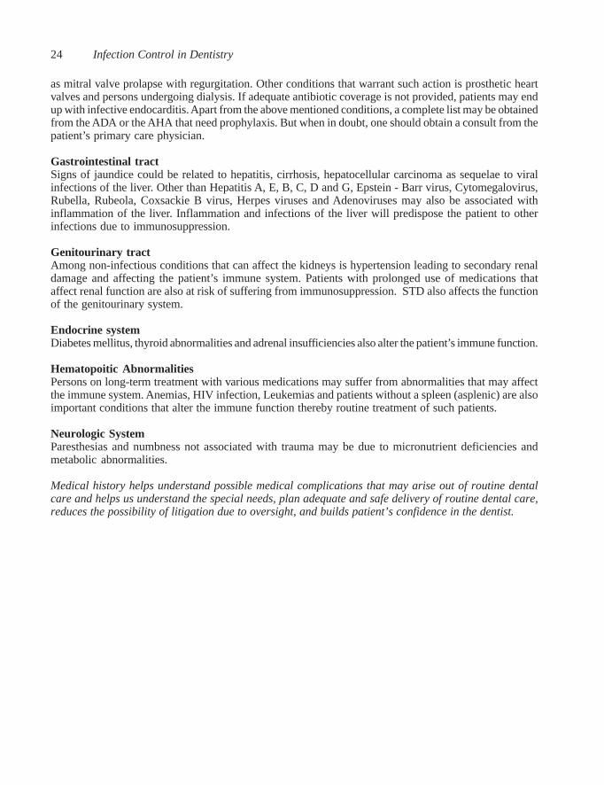
Infection Control in Dentistry24
as mitral valve prolapse with regurgitation. Other conditions that warrant such action is prosthetic heartvalves and persons undergoing dialysis. If adequate antibiotic coverage is not provided, patients may endup with infective endocarditis. Apart from the above mentioned conditions, a complete list may be obtainedfrom the ADA or the AHA that need prophylaxis. But when in doubt, one should obtain a consult from thepatient’s primary care physician.
Gastrointestinal tractSigns of jaundice could be related to hepatitis, cirrhosis, hepatocellular carcinoma as sequelae to viralinfections of the liver. Other than Hepatitis A, E, B, C, D and G, Epstein - Barr virus, Cytomegalovirus,Rubella, Rubeola, Coxsackie B virus, Herpes viruses and Adenoviruses may also be associated withinflammation of the liver. Inflammation and infections of the liver will predispose the patient to otherinfections due to immunosuppression.
Genitourinary tractAmong non-infectious conditions that can affect the kidneys is hypertension leading to secondary renaldamage and affecting the patient’s immune system. Patients with prolonged use of medications thataffect renal function are also at risk of suffering from immunosuppression. STD also affects the functionof the genitourinary system.
Endocrine systemDiabetes mellitus, thyroid abnormalities and adrenal insufficiencies also alter the patient’s immune function.
Hematopoitic AbnormalitiesPersons on long-term treatment with various medications may suffer from abnormalities that may affectthe immune system. Anemias, HIV infection, Leukemias and patients without a spleen (asplenic) are alsoimportant conditions that alter the immune function thereby routine treatment of such patients.
Neurologic SystemParesthesias and numbness not associated with trauma may be due to micronutrient deficiencies andmetabolic abnormalities.
Medical history helps understand possible medical complications that may arise out of routine dentalcare and helps us understand the special needs, plan adequate and safe delivery of routine dental care,reduces the possibility of litigation due to oversight, and builds patient’s confidence in the dentist.
Chapter 05 - NEW.p65 10/20/2007, 10:07 AM24

Immunization of Personnel Involved in Dental Care 25
Chapter 6
Immunization of Personnel Involved in Dental Care
Puttaiah & Kohli
In the developed countries and in India routine childhoodimmunization is a part of life and is taken for granted.Immunization is the first line of defense against infectiousdiseases. Even employees with minimal patient contact shouldaccept vaccination.
Table 6. Common Childhood Vaccines
Vaccine Disease
Hepatitis A Hepatitis A viral infectionHepatitis B Hepatitis B viral infectionVaricella Chicken PoxMMR Measles, Mumps and RubellaDPT Diphtheria, Pertussis and TetanusRubeola German MeaslesMeningitis MeningitisPolio Poliomyelitis
Other than the above, additional vaccines and Immunoglobulins are provided based on the geographiclocation and risks of certain endemic conditions. Bloodborne diseases such as Hepatitis C and HumanImmunodeficiency viral infection do not have vaccines.
Immunizations for Healthcare WorkersApart from the routine prescribed immunizations as seen listed in the vaccines and Immunoglobulins forchildren, influenza vaccines must be used as needed (i.e. prior to the onset of the “Flu Season”). We ashealth care workers are at a high risk of preventable infectious diseases and therefore must adopt this firstline of defense. We should be vaccinated against all immunizable infectious diseases. It may be the law insome situations, or a prescribed preventive measure within certain professions that have a potential forexposure and in institutions that immunizations as a policy are a must for employees. Irrespective of thelaw or regulations that dictate, it is the ethical and moral duty of the health care provider to stay healthy
Chapter 06 - NEW.p65 10/20/2007, 10:12 AM25

Infection Control in Dentistry26
and protect oneself against diseases through immunization. It is less expensive and less time consumingto be immunized than to be treated otherwise. If one is the head of the clinic or an institution, one mustprovide access and if possible pay for the immunization as the law or affordability may dictate.
Other than the common vaccinations (childhood vaccines), the clinicians should also be regularly vaccinatedagainst other conditions such as Influenza on an annual basis. If the clinicians live in endemic areas ofCholera, or travel frequently to endemic areas of Cholera, the clinicians should be vaccinated againstCholera.
Proposed protocol recommendation for HBV vaccine for Dental Health Care Workers in IndiaA record of immunization should be obtained while hiring a clinician. Employees should be providedimmunization against HBV as soon as possible if the employee has not been immunized. If the employeedeclines immunization, it must be documented and kept on record. The recipient of the vaccines shouldbe explained about the adverse effects if any (hypersensitivities to protein components of the vaccine,preservatives and stabilizers), prior to making a decision of administering the vaccine. Pretesting whethera non-immunized recipient is protected is not required. Post-testing (anti-HBs) to determine protection isnormally done 6 months after vaccination series. Negative can mean two outcomes— 1) Responder withlow or non-detectable levels but still protected; or 2) Recipient is a primary non-responder. If adequateprotection is not determined by post-testing, they may require additional doses of the vaccine. For thosewho have been determined to be responders (immunized), no booster doses are necessary due to anemnisticresponse or immunological memory.
Vaccines that are recommended are Rubella, Rubeola (Measles), Mumps, Poliomyelitis, Hepatitis B,Influenza, Tetanus and Diphtheria. For those not protected against Hepatitis A viral infection, immunizationis advised but not recommended. Rubella and Rubeola must not be given to immunocompromised persons.Booster doses are not required for mumps. If a health care worker who is immunized against polio isexposed to the oral secretions of a patient with active polio, Oral polio vaccine (live attenuated virus) orInactivated Polio virus vaccine boosters are needed. Hepatitis B vaccine (three 1 mL doses at 0, end of 1month and 6 months intramuscularly (deltoid muscle) is recommended. If a person is not immunizedagainst HBV and is exposed to an infected patient’s body fluids, a combination of Hepatitis B vaccineand an immunoglobulin (HBIg) must be administered immediately. If the exposed person does not wantto take the vaccine, two doses of the HBIg immunoglobulin must be given (in most cases, dose 1 within24 hours and dose 2 given 25 to 30 days after exposure). Periodic preventive programs for the clinicwould be to get every one in the clinic immunized against the seasonal influenza virus to avoid loss ofproductive work time.
Proposed protocol for HBV vaccine for Dental Undergraduate & Postgraduate students and DentalTeaching Faculty in IndiaIt is mandatory for every dental student undergraduate and postgraduate and dental teaching faculty to bevaccinated against Hepatitis B with a three dose regimen and a booster. The institute head will confirmthat the undergraduate and the postgraduate students have had a course of HBV vaccination before theadmission procedures are completed. Other than the teaching faculty all non teaching faculty whichinclude dental assistants, dental hygienists, dental mechanics and all those who come directly in contactwith patient care, cleaning and sterilization of instruments and all those who are in the clinical area or thepre clinical area of the dental teaching institution must to be vaccinated against Hepatitis B. It is alsomandatory for the institution managements including government owned institutions to provide freeHepatitis B vaccine to all its students both undergraduate and postgraduate, teaching and non teachingfaculty.
Chapter 06 - NEW.p65 10/20/2007, 10:12 AM26

Chapter 7
Handwashing and Handcare
Puttaiah & Youngblood
Patients notice most things in the clinic...from cleanliness to personal hygieneand the clinician’s professionalism. Sometimes they even take into noticewhether the clinician and staff have clean finger nails, whether they havewashed the hands with soap before donning gloves, whether the hair isunkempt and also whether the clothes are clean and presentable as a clinicstaff or dentist. Dental treatment occurs in close proximity of bodies andtherefore, body odors are an issue to the patient. Therefore, personal hygiene,cleanliness, cleanliness of the clinical area, as well as professional appearance
among others, are important issues that can affect the patient’s perception of the level of care being received.
Examination of handsBefore wearing personal protective equipment, the dentist and staff need to remove chunky jewelry andwrist watch, and examine their hands for cuts and bruises and hangnails. After handwashing, the cuts andbruises should be medicated and covered using a band aid/dressing to avoid coming in contact withpatient material.
What should one use?Hands should be washed with water and an antimicrobial soap. Some of the antimicrobial soaps thathave been cleared by the US FDA have Chlorhexidine Gluconate (CHG) at 0.75% to 4% concentrationthat may be dispensed as liquid soap or foam, Parachlorometaxylenol (PCMX) liquid, Iodine liquid orTriclosan liquid, gel or foam. CHG at 4% is marketed for surgical scrub as opposed to routine handwashingand the latter may show residual effect or substantivity (remains on the skin as a protectant) on the skinafter 4-5 repeated washes. PCMX is also an effective germicide but may lack the level of substantivity asseen in CHG. Triclosan based soaps are not as powerful as CHG, PCMX or Iodine based soaps. Soapsdispensed as foam use lesser soap than liquids (when liquid soap is mixed with air using a specializeddispenser/nozzle, foam is produced) and may be less expensive on the long term. The rationale forhandwashing is to reduce the number of microbes on the hand by the process of cleaning mainly, apartfrom the antimicrobial effect of the germicidal soaps.
When should one wash hands?Handwashing should be done at the beginning of the clinic session and soon after removal of gloves. Oneof the reasons to wash hands after removal of gloves is to reduce bacterial counts that build up on the skinduring glove use. Handwashing also helps remove glove related materials on the skin including cornstarch; other powders that are used in the gloves for easy donning hat have the potential to cause sensitizationor skin irritation.
Chapter 07 - NEW.p65 10/20/2007, 10:16 AM27

Infection Control in Dentistry28
Sequence followed in routine handwash procedures1) Remove jewelry and wrist watch and examine hands2) Wet hands with warm water3) Dispense an adequate amount of soap4) Thoroughly rub both surfaces of the hands including around the thumb and fingers for about 30 - 60
seconds5) Wash hands with warm water to remove the soap6) Dry hands with paper towels7) Examine hands for injuries such as nicks, cuts and bruises and treat as needed8) Wear single-use-disposable gloves.
The same procedure should be followed after removal of contaminated gloves. Hands should be driedusing disposable paper towels before donning a new pair of gloves because gloving wet hands is difficult.
Use of Hand SanitizersIn the recent past, hand sanitizers that are alcohol based with or without other germicideshave entered the market. These items are primarily for use when handwashing isimpractical or cannot be done (non-availability of water and antimicrobial handsoap).Hand sanitizers with alcohol have been showed to be effective in germicidal control onhands. Some hand sanitizers have been produced without alcohol, with emulsions ofnatural oils and have also reported anti-microbial efficacy. Although there is germicidalcontrol, these hand sanitizers do not clean hands adequately, and therefore, should beused in conjunction with a disposable wipe to remove the dirt and grime build-up on the
hands. Recently, hand sanitizers incorporated in wet-wipes have entered the market and show promise inboth cleaning the dirt on the hands as well as reporting antimicrobial efficacy.
Surgical HandwashingWhile planning to conduct a surgical procedure where exposing bone is involved, surgical handwashing
Rub hands thoroughly
Chapter 07 - NEW.p65 10/20/2007, 10:16 AM28

Handwashing and Handcare 29
Washing hands multiple times per day with soaps tends to make the skin dry. At the end of each session (duringluch break, or at the end of the clinic day) a good quality emolient/skin cream should be used for hand care.
procedures must be followed. All the basic precautions such as having short trimmednails, no jewelry, cuts and bruises on hands should be taken as during regularhandwashing and hygiene procedures. In addition, these procedures dictate a moreinvolved process needing additional steps. Bacteria on the skin (both patient as wellas the dental professional) can lead to wound infections if they enter the operativefield (tissue). Therefore, thorough cleaning/scrubbing of the hands immediately priorto surgery with a surgical scrub level soap (Chlorhexidine Gluconate 4%, or otherIodine based surgical soaps) are beneficial in many ways. Scrubbing hands all theway up to the elbow for about 2 to 6 minutes using a single-use disposable sponge or
a soft scrub brush removes the dead cells along with the bacteria resident on the skin of the hands.Bacteria under the gloves rapidly multiply due to the humid and warm environment as opposed to a dryand cool area. If the soap has residual properties (staying on the skin on repeated washing with theantimicrobial soap) it tends to slow the multiplication of microbes. Reducing multiplication of residentskin microbes on the hands of the clinician/surgeon for the duration of a procedure reduces the risk ofletting bacteria being released into the surgical field if gloves become compromised (torn/punctured).Surgical scrub soaps should have properties such as substantivity or residual action, must have broadspectrum antimicrobial activity, have a good kill rate (fast or rapid action and reduce the bacterial loadeffectively), be a good detergent, non-irritating to skin, and be fast-acting.
Steps involved in handwashing are as follows—1. Dispense out the needed PPE to be worn immediately after the scrub2. Remove jewelry and examine hands for cuts, bruises and hang nails (take care of them)3. Clean under the nails using a nail cleaner under running clean water4. Wet hand and apply the antimicrobial soap5. Scrub hands for 2-6 minutes (based on the length of antimicrobial exposure time per manufacturer’s
recommendation) using a sterile single-use-disposable sponge or a scrub brush6. Dry hands with sterile towelettes or paper towels7. Wear sterile surgical gloves without touching the outer surface of the gloves8. After surgery, remove gloves aseptically without letting the blood or body fluids coming in contact
with hands9. Examine hands for cuts, punctures or bruises and wash hands with an antimicrobial soap and water10. At the end of the session apply an emollient (hand lotion) that is suitable and that helps moisturize
the skin
Some institutions and recommendations advocate the use of alcohol sanitizers in addition to the scrub,but it is not widely practiced. If used, wait till the hands dry before donning surgical gloves.
Chapter 07 - NEW.p65 10/20/2007, 10:16 AM29

Infection Control in Dentistry30
Chapter 8
Personal Protective Equipment
Puttaiah
Personal Protective Equipment (PPE) commonly used in general dental care are single-use-disposablegloves (sterile or non-sterile), protective eyewear, face-shields, masks, gowns and utility gloves used toprotect personnel from blood and body fluids and chemical hazards. The main use of barriers is to controlgross contamination and not prevent spread of every single microbe. As an example, some of the virusesare smaller than the microscopic pores in latex exam gloves and therefore have a probability of passingthrough the glove material. In this instance, one may safely infer that gloves are meant for reducing theamount of exposure to the viral particles of the body fluids and not to totally prevent contact with thevirus. Therefore, handwashing with an antimicrobial soap after removing gloves is necessary and pragmatic.With respect to dentistry, there has not been a single reported and accepted case of disease transmissionlinked to passage of viral particle through the pores in the gloves.
Rationale for determining the level of PPEPPE use is determined by the anticipated exposure to blood or other potentially infectious material (BOPIM)and chemical hazards. We need use “full PPE” (protective eyewear, mask, gown and gloves) when thereis a potential for splash or spatter (use of air/water syringe, highspeed handpiece and/or scalers), or apotential for exposure to chemicals that could splash/spatter on the face or mucosa. Full PPE wouldinclude protective eyewear, mask, gown and gloves. When we anticipate no potential exposure to splash/spatter to BOPIM or chemical hazards, we could still achieve compliance by using exam gloves only (asin suture removal or cursory examination of the oral cavity).
Chapter 08 - NEW.p65 10/20/2007, 10:19 AM30
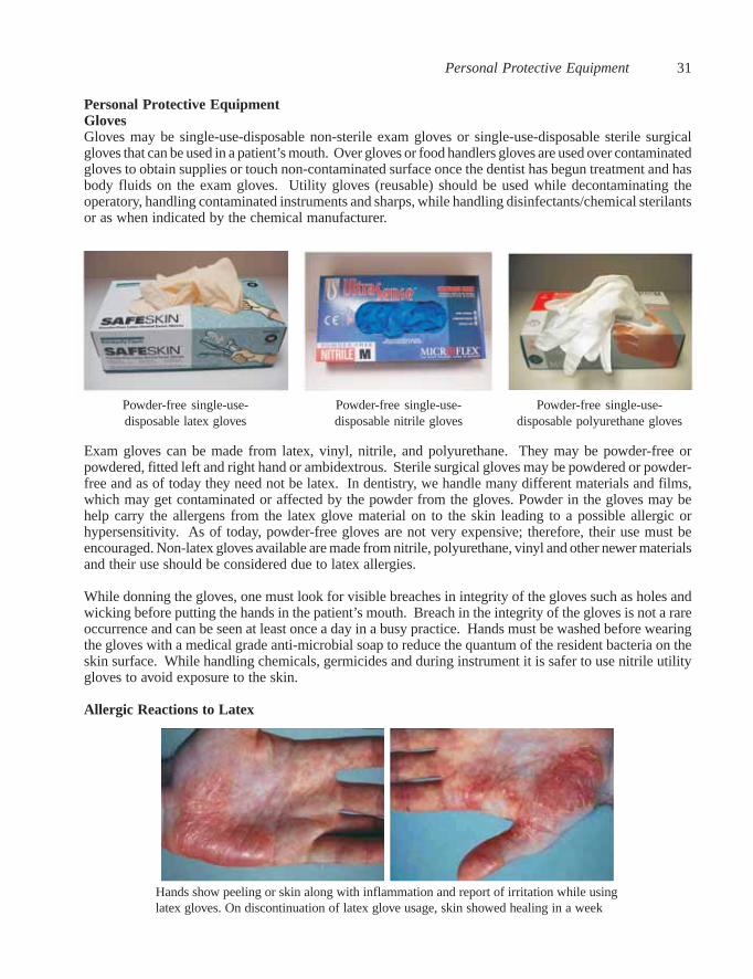
Personal Protective Equipment 31
Personal Protective EquipmentGlovesGloves may be single-use-disposable non-sterile exam gloves or single-use-disposable sterile surgicalgloves that can be used in a patient’s mouth. Over gloves or food handlers gloves are used over contaminatedgloves to obtain supplies or touch non-contaminated surface once the dentist has begun treatment and hasbody fluids on the exam gloves. Utility gloves (reusable) should be used while decontaminating theoperatory, handling contaminated instruments and sharps, while handling disinfectants/chemical sterilantsor as when indicated by the chemical manufacturer.
Exam gloves can be made from latex, vinyl, nitrile, and polyurethane. They may be powder-free orpowdered, fitted left and right hand or ambidextrous. Sterile surgical gloves may be powdered or powder-free and as of today they need not be latex. In dentistry, we handle many different materials and films,which may get contaminated or affected by the powder from the gloves. Powder in the gloves may behelp carry the allergens from the latex glove material on to the skin leading to a possible allergic orhypersensitivity. As of today, powder-free gloves are not very expensive; therefore, their use must beencouraged. Non-latex gloves available are made from nitrile, polyurethane, vinyl and other newer materialsand their use should be considered due to latex allergies.
While donning the gloves, one must look for visible breaches in integrity of the gloves such as holes andwicking before putting the hands in the patient’s mouth. Breach in the integrity of the gloves is not a rareoccurrence and can be seen at least once a day in a busy practice. Hands must be washed before wearingthe gloves with a medical grade anti-microbial soap to reduce the quantum of the resident bacteria on theskin surface. While handling chemicals, germicides and during instrument it is safer to use nitrile utilitygloves to avoid exposure to the skin.
Allergic Reactions to Latex
Hands show peeling or skin along with inflammation and report of irritation while usinglatex gloves. On discontinuation of latex glove usage, skin showed healing in a week
Powder-free single-use-disposable latex gloves
Powder-free single-use-disposable nitrile gloves
Powder-free single-use-disposable polyurethane gloves
Chapter 08 - NEW.p65 10/20/2007, 10:19 AM31
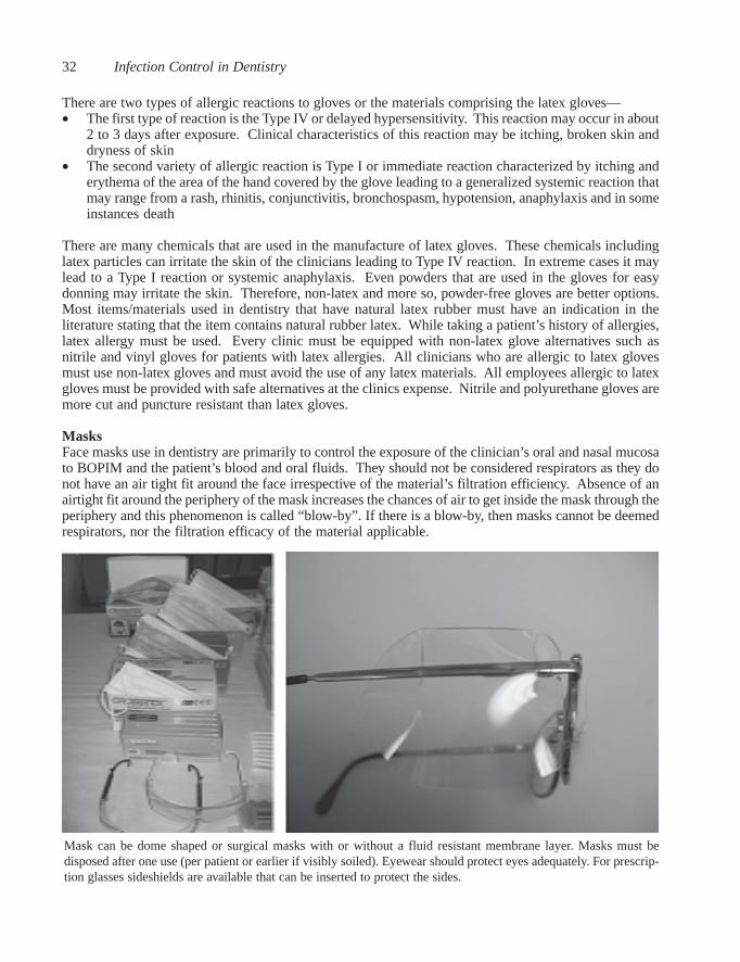
Infection Control in Dentistry32
There are two types of allergic reactions to gloves or the materials comprising the latex gloves—• The first type of reaction is the Type IV or delayed hypersensitivity. This reaction may occur in about
2 to 3 days after exposure. Clinical characteristics of this reaction may be itching, broken skin anddryness of skin
• The second variety of allergic reaction is Type I or immediate reaction characterized by itching anderythema of the area of the hand covered by the glove leading to a generalized systemic reaction thatmay range from a rash, rhinitis, conjunctivitis, bronchospasm, hypotension, anaphylaxis and in someinstances death
There are many chemicals that are used in the manufacture of latex gloves. These chemicals includinglatex particles can irritate the skin of the clinicians leading to Type IV reaction. In extreme cases it maylead to a Type I reaction or systemic anaphylaxis. Even powders that are used in the gloves for easydonning may irritate the skin. Therefore, non-latex and more so, powder-free gloves are better options.Most items/materials used in dentistry that have natural latex rubber must have an indication in theliterature stating that the item contains natural rubber latex. While taking a patient’s history of allergies,latex allergy must be used. Every clinic must be equipped with non-latex glove alternatives such asnitrile and vinyl gloves for patients with latex allergies. All clinicians who are allergic to latex glovesmust use non-latex gloves and must avoid the use of any latex materials. All employees allergic to latexgloves must be provided with safe alternatives at the clinics expense. Nitrile and polyurethane gloves aremore cut and puncture resistant than latex gloves.
MasksFace masks use in dentistry are primarily to control the exposure of the clinician’s oral and nasal mucosato BOPIM and the patient’s blood and oral fluids. They should not be considered respirators as they donot have an air tight fit around the face irrespective of the material’s filtration efficiency. Absence of anairtight fit around the periphery of the mask increases the chances of air to get inside the mask through theperiphery and this phenomenon is called “blow-by”. If there is a blow-by, then masks cannot be deemedrespirators, nor the filtration efficacy of the material applicable.
Mask can be dome shaped or surgical masks with or without a fluid resistant membrane layer. Masks must bedisposed after one use (per patient or earlier if visibly soiled). Eyewear should protect eyes adequately. For prescrip-tion glasses sideshields are available that can be inserted to protect the sides.
Chapter 08 - NEW.p65 10/20/2007, 10:19 AM32
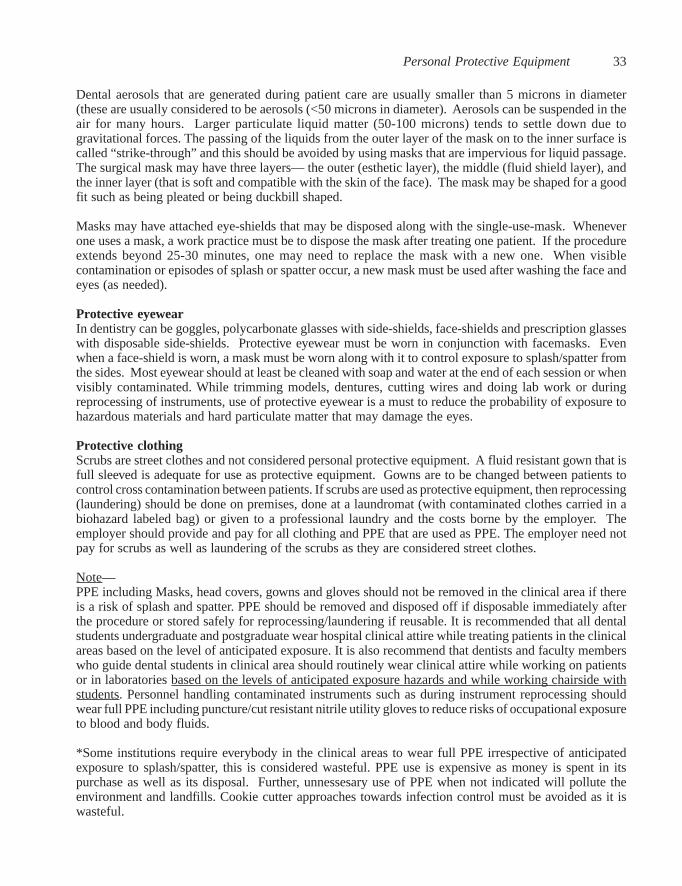
Personal Protective Equipment 33
Dental aerosols that are generated during patient care are usually smaller than 5 microns in diameter(these are usually considered to be aerosols (<50 microns in diameter). Aerosols can be suspended in theair for many hours. Larger particulate liquid matter (50-100 microns) tends to settle down due togravitational forces. The passing of the liquids from the outer layer of the mask on to the inner surface iscalled “strike-through” and this should be avoided by using masks that are impervious for liquid passage.The surgical mask may have three layers— the outer (esthetic layer), the middle (fluid shield layer), andthe inner layer (that is soft and compatible with the skin of the face). The mask may be shaped for a goodfit such as being pleated or being duckbill shaped.
Masks may have attached eye-shields that may be disposed along with the single-use-mask. Wheneverone uses a mask, a work practice must be to dispose the mask after treating one patient. If the procedureextends beyond 25-30 minutes, one may need to replace the mask with a new one. When visiblecontamination or episodes of splash or spatter occur, a new mask must be used after washing the face andeyes (as needed).
Protective eyewearIn dentistry can be goggles, polycarbonate glasses with side-shields, face-shields and prescription glasseswith disposable side-shields. Protective eyewear must be worn in conjunction with facemasks. Evenwhen a face-shield is worn, a mask must be worn along with it to control exposure to splash/spatter fromthe sides. Most eyewear should at least be cleaned with soap and water at the end of each session or whenvisibly contaminated. While trimming models, dentures, cutting wires and doing lab work or duringreprocessing of instruments, use of protective eyewear is a must to reduce the probability of exposure tohazardous materials and hard particulate matter that may damage the eyes.
Protective clothingScrubs are street clothes and not considered personal protective equipment. A fluid resistant gown that isfull sleeved is adequate for use as protective equipment. Gowns are to be changed between patients tocontrol cross contamination between patients. If scrubs are used as protective equipment, then reprocessing(laundering) should be done on premises, done at a laundromat (with contaminated clothes carried in abiohazard labeled bag) or given to a professional laundry and the costs borne by the employer. Theemployer should provide and pay for all clothing and PPE that are used as PPE. The employer need notpay for scrubs as well as laundering of the scrubs as they are considered street clothes.
Note—PPE including Masks, head covers, gowns and gloves should not be removed in the clinical area if thereis a risk of splash and spatter. PPE should be removed and disposed off if disposable immediately afterthe procedure or stored safely for reprocessing/laundering if reusable. It is recommended that all dentalstudents undergraduate and postgraduate wear hospital clinical attire while treating patients in the clinicalareas based on the level of anticipated exposure. It is also recommend that dentists and faculty memberswho guide dental students in clinical area should routinely wear clinical attire while working on patientsor in laboratories based on the levels of anticipated exposure hazards and while working chairside withstudents. Personnel handling contaminated instruments such as during instrument reprocessing shouldwear full PPE including puncture/cut resistant nitrile utility gloves to reduce risks of occupational exposureto blood and body fluids.
*Some institutions require everybody in the clinical areas to wear full PPE irrespective of anticipatedexposure to splash/spatter, this is considered wasteful. PPE use is expensive as money is spent in itspurchase as well as its disposal. Further, unnessesary use of PPE when not indicated will pollute theenvironment and landfills. Cookie cutter approaches towards infection control must be avoided as it iswasteful.
Chapter 08 - NEW.p65 10/20/2007, 10:19 AM33

Infection Control in Dentistry34
Chapter 9
Surface Barriers Versus Disinfection
Puttaiah & Kohli
Surface barriers are a practical and an easy way to contain cross-contamination. Barriers can be sterile ornon-sterile depending on whether they are used for a surgical or a non-surgical routine dental care. Mostdentists use disinfectants to clean the equipment and work surfaces between each patient. If disinfectionof work surfaces is done between each patient instead of only changing barriers, the time taken isconsiderably more due to the initial sanitization step with the disinfection (that may take between 2-5minutes) and then the disinfection step (based on the TB kill time of 5-10 minutes based on the claims ofthe disinfectant). The turn around time for an operatory if a disinfectant is used (8-15 minutes) is longerthan the time taken for removal of barriers, placing new surface barriers, disposal of the waste, return ofthe used instrument to the instrument reprocessing area (3-5 minutes). Therefore barriers make it quickerto turn around an operatory between patients. When barriers are routinely changed between patients,disinfection of the surfaces proximal to patient care may be done at the beginning of the clinic session, orat the end of the clinic session and when visibly soiled.
Work surface barriers
Light handles and light switches need to be barriered during patient use and discarded after each patient. Thinplastic bags, wraps or aluminum foil may be used
Chapter 09 -NEW.p65 10/20/2007, 10:22 AM34

Surface Barriers Versus Disinfection 35
Flat works surfaces such as the counters that are in immediate proximity to the clinician (within handsreach) and at a higher risk of contamination and touch with soiled gloves may be covered with plasticbarriers or other fluid impervious barriers such as patient bibs (one side absorbent and one side imperviousto fluids). Instead of using a patient bib, one may opt for alternatives such as a cling wrap (plastic foodwrap), a plastic bag (such as those used in the dry-cleaning industry to cover garments). Many dentalbarrier manufacturers have other preformed plastic barriers that can be used for this purpose. The brackettable is another example for barrier use. While placing contaminated items on work surfaces, it is essentialto place them on impervious barriers to control the spread of cross-contamination to the primary worksurface. The same example may be extended to work in dental laboratories or while handling contaminatedprostheses and impressions either chair side or in the laboratory. In the absence of preformed barriersthose that are designed for the food industry such as cling wrap (roll of plastic sheet that has staticelectricity and tends to cling to surfaces) may be an inexpensive alternative. Clinicians have used aluminumfoil and sterilized them between patients (today it is not acceptable and not practiced, while single-use-disposable are allowed).
Surfaces to be barrieredSingle-use-disposable barriers that are preformed or generic should be used over some of the commonlytouched surfaces—1. Dental Unit Light handles2. Dental Unit electrical or mechanical controls3. Dental Chair Head Rest4. Dental Chair Arm Rests5. Dental Unit controls including the Bracket Table6. Highspeed Handpiece couplings and hose (extended 6 inches below the coupling covering the hoses)7. Slow speed motor, coupling and hose (extended 6 inches below the coupling covering the hoses)8. Air/water syringe and hose (extended 6 inches below coupling covering the hose)9. Saliva ejector handpiece and hose (extended 6 inches below coupling covering the hose)
Air/water syringes, HVE and Saliva Ejector Syringe/couplings may be barriered to at least 6 inches below thecoupling. Other options are to disinfect between patients. Computer Keyboard and Mouse that are water resistantmay either be disinfected or barriered between patients
Chapter 09 -NEW.p65 10/20/2007, 10:22 AM35
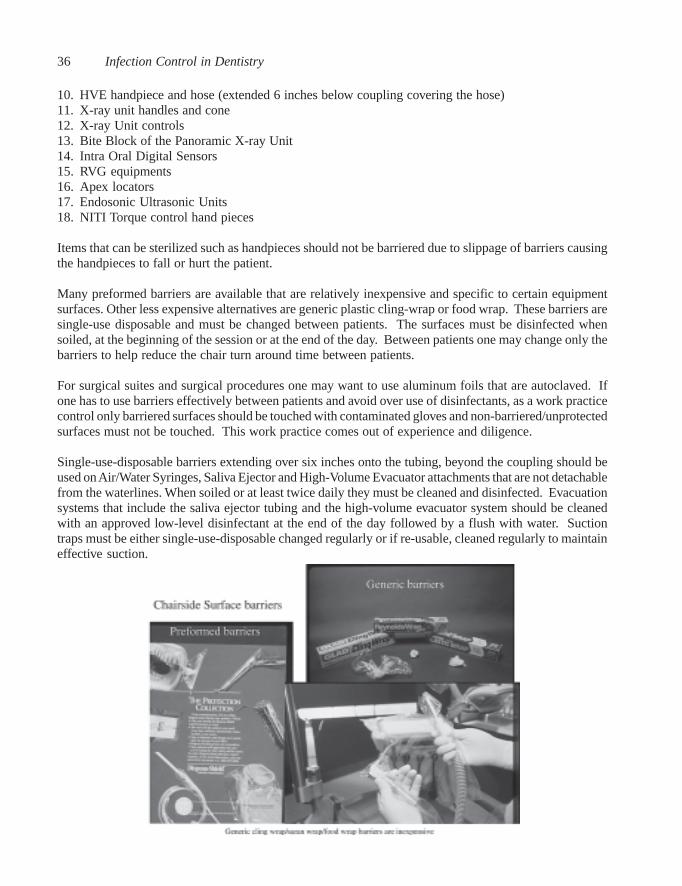
Infection Control in Dentistry36
10. HVE handpiece and hose (extended 6 inches below coupling covering the hose)11. X-ray unit handles and cone12. X-ray Unit controls13. Bite Block of the Panoramic X-ray Unit14. Intra Oral Digital Sensors15. RVG equipments16. Apex locators17. Endosonic Ultrasonic Units18. NITI Torque control hand pieces
Items that can be sterilized such as handpieces should not be barriered due to slippage of barriers causingthe handpieces to fall or hurt the patient.
Many preformed barriers are available that are relatively inexpensive and specific to certain equipmentsurfaces. Other less expensive alternatives are generic plastic cling-wrap or food wrap. These barriers aresingle-use disposable and must be changed between patients. The surfaces must be disinfected whensoiled, at the beginning of the session or at the end of the day. Between patients one may change only thebarriers to help reduce the chair turn around time between patients.
For surgical suites and surgical procedures one may want to use aluminum foils that are autoclaved. Ifone has to use barriers effectively between patients and avoid over use of disinfectants, as a work practicecontrol only barriered surfaces should be touched with contaminated gloves and non-barriered/unprotectedsurfaces must not be touched. This work practice comes out of experience and diligence.
Single-use-disposable barriers extending over six inches onto the tubing, beyond the coupling should beused on Air/Water Syringes, Saliva Ejector and High-Volume Evacuator attachments that are not detachablefrom the waterlines. When soiled or at least twice daily they must be cleaned and disinfected. Evacuationsystems that include the saliva ejector tubing and the high-volume evacuator system should be cleanedwith an approved low-level disinfectant at the end of the day followed by a flush with water. Suctiontraps must be either single-use-disposable changed regularly or if re-usable, cleaned regularly to maintaineffective suction.
Chapter 09 -NEW.p65 10/20/2007, 10:22 AM36

Chapter 10
Single-Use-Disposables
Puttaiah, Youngblood & Cederberg
In the absence of a decontamination process one can use single-use-disposable devices such as barriersfor surfaces. Today, single-use-disposable devices comprising personal protective equipment, surfacebarriers and consumables are to be used regularly in the dental practice. This section deals with thecommon disposables, proper use and the rationale for such use. Prior to addressing the issue of single-use-disposables, certain general items on infection control need to be addressed. “Sterilization”, theaccepted reprocessing method in dentistry, requires precleaning as an essential step in instrumentreprocessing. If precleaning is not achieved, sterilization can be confounded by the presence of bioburdenor inorganic material on critical and semi-critical devices. Ideally, the goal of instrument reprocessing forcritical and semi-critical items is to remove or reduce bioburden and dry instruments prior to sterilization.Reduction of bioburden on instruments and devices to be sterilized is essential to the success of anysterilization process. To overcome this problem, use of inexpensive single-use-disposables (SUDs) foreach patient is pragmatic.
Common single-use-disposable devices used in dentistryAs of today, single-use-disposable devices have become relatively inexpensive. Most single-use-devicesavailable have been cleared by governmental regulatory bodies in most developed countries. Reprocessingand reuse of SUDs is not reliable and therefore prohibited. Furthermore, these devices must strictly beused per manufacturers’ instructions and for the given purpose. The oral cavity is not a clean site, andtherefore, use of non-sterile disposable gloves are permitted as long as they have been stored hygienicallyand have not been overtly exposed to contaminants. A non-sterile glove is to protect the clinician againstpossible contaminants from the patient’s mouth.
When we anticipate exposure of deeper structures such as bone, it is pragmatic to use sterile single-use-disposable gloves as an additional measure of safety for the patient, although one may question thepresence of a very high amount of microorganisms in the oral cavity that may potentially contaminate thesurgical site. The common microorganisms found associated with surgical site infections are S. aureus,coagulase-negative Staphylococci, Enterococci, E. coli, P. aeruginosa and Enterobacteria spp., and theyare normally found on skin and fingers. The thrust of infection control in general dental procedures is tocontrol cross-infections and occupational exposure, not to eliminate or prevent.
1. Personal protective equipmenta. Exam Gloves, Surgical Gloves, Overgloves and Finger-cotsb. Side shields for prescription glassesc. Surgical masks with or without eye protection and Dome shaped Masksd. Gowns, bonnets and shoe-covers, patient bibs
2. Surface barriersa. Headrest covers, chair covers, bracket table cover, X-ray tube, X-ray switch control, and barrier
for work surface
Chapter 10 -NEW.p65 10/20/2007, 10:24 AM37
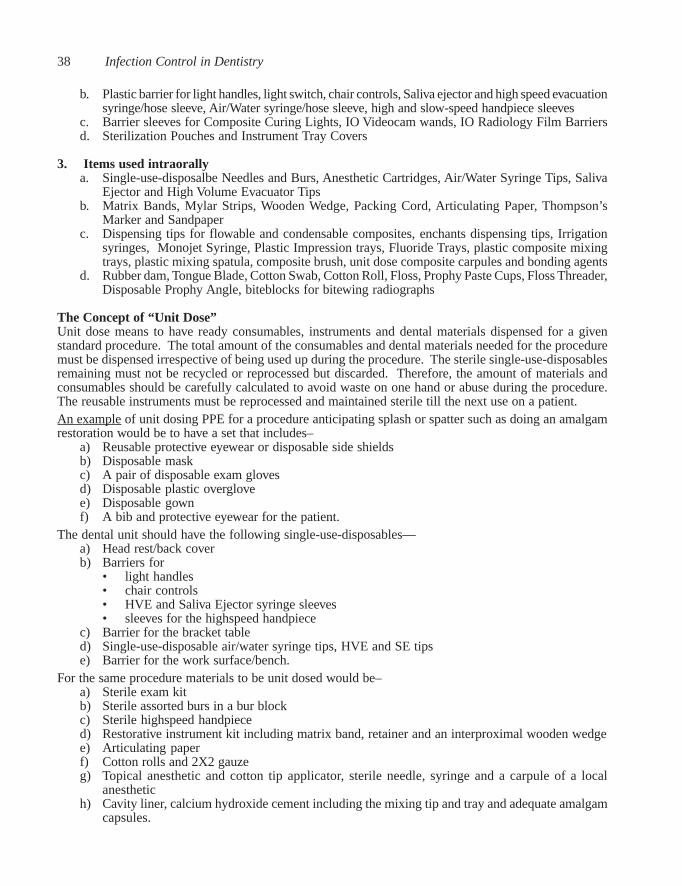
Infection Control in Dentistry38
b. Plastic barrier for light handles, light switch, chair controls, Saliva ejector and high speed evacuationsyringe/hose sleeve, Air/Water syringe/hose sleeve, high and slow-speed handpiece sleeves
c. Barrier sleeves for Composite Curing Lights, IO Videocam wands, IO Radiology Film Barriersd. Sterilization Pouches and Instrument Tray Covers
3. Items used intraorallya. Single-use-disposalbe Needles and Burs, Anesthetic Cartridges, Air/Water Syringe Tips, Saliva
Ejector and High Volume Evacuator Tipsb. Matrix Bands, Mylar Strips, Wooden Wedge, Packing Cord, Articulating Paper, Thompson’s
Marker and Sandpaperc. Dispensing tips for flowable and condensable composites, enchants dispensing tips, Irrigation
syringes, Monojet Syringe, Plastic Impression trays, Fluoride Trays, plastic composite mixingtrays, plastic mixing spatula, composite brush, unit dose composite carpules and bonding agents
d. Rubber dam, Tongue Blade, Cotton Swab, Cotton Roll, Floss, Prophy Paste Cups, Floss Threader,Disposable Prophy Angle, biteblocks for bitewing radiographs
The Concept of “Unit Dose”Unit dose means to have ready consumables, instruments and dental materials dispensed for a givenstandard procedure. The total amount of the consumables and dental materials needed for the proceduremust be dispensed irrespective of being used up during the procedure. The sterile single-use-disposablesremaining must not be recycled or reprocessed but discarded. Therefore, the amount of materials andconsumables should be carefully calculated to avoid waste on one hand or abuse during the procedure.The reusable instruments must be reprocessed and maintained sterile till the next use on a patient.An example of unit dosing PPE for a procedure anticipating splash or spatter such as doing an amalgamrestoration would be to have a set that includes–
a) Reusable protective eyewear or disposable side shieldsb) Disposable maskc) A pair of disposable exam glovesd) Disposable plastic overglovee) Disposable gownf) A bib and protective eyewear for the patient.
The dental unit should have the following single-use-disposables—a) Head rest/back coverb) Barriers for
• light handles• chair controls• HVE and Saliva Ejector syringe sleeves• sleeves for the highspeed handpiece
c) Barrier for the bracket tabled) Single-use-disposable air/water syringe tips, HVE and SE tipse) Barrier for the work surface/bench.
For the same procedure materials to be unit dosed would be–a) Sterile exam kitb) Sterile assorted burs in a bur blockc) Sterile highspeed handpieced) Restorative instrument kit including matrix band, retainer and an interproximal wooden wedgee) Articulating paperf) Cotton rolls and 2X2 gauzeg) Topical anesthetic and cotton tip applicator, sterile needle, syringe and a carpule of a local
anesthetich) Cavity liner, calcium hydroxide cement including the mixing tip and tray and adequate amalgam
capsules.
Chapter 10 -NEW.p65 10/20/2007, 10:24 AM38
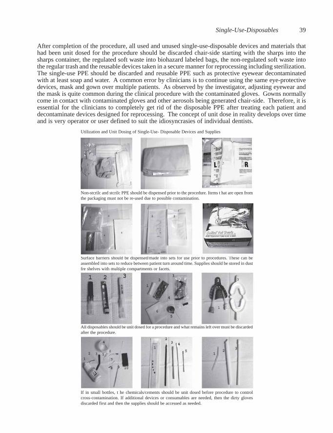
Single-Use-Disposables 39
After completion of the procedure, all used and unused single-use-disposable devices and materials thathad been unit dosed for the procedure should be discarded chair-side starting with the sharps into thesharps container, the regulated soft waste into biohazard labeled bags, the non-regulated soft waste intothe regular trash and the reusable devices taken in a secure manner for reprocessing including sterilization.The single-use PPE should be discarded and reusable PPE such as protective eyewear decontaminatedwith at least soap and water. A common error by clinicians is to continue using the same eye-protectivedevices, mask and gown over multiple patients. As observed by the investigator, adjusting eyewear andthe mask is quite common during the clinical procedure with the contaminated gloves. Gowns normallycome in contact with contaminated gloves and other aerosols being generated chair-side. Therefore, it isessential for the clinicians to completely get rid of the disposable PPE after treating each patient anddecontaminate devices designed for reprocessing. The concept of unit dose in reality develops over timeand is very operator or user defined to suit the idiosyncrasies of individual dentists.
Utilization and Unit Dosing of Single-Use- Disposable Devices and Supplies
Non-stcrilc and stcrilc PPE should be dispensed prior to the procedure. Items t hat are open fromthe packaging must not be re-used due to possible contamination.
Surface barriers should be dispensed/made into sets for use prior to procedures. These can beassembled into sets to reduce between patient turn around time. Supplies should be stored in dustfre shelves with multiple compartments or facets.
All disposables should be unit dosed for a procedure and what remains left over must be discardedafter the procedure.
If in small bottles, t he chemicals/cements should be unit dosed before procedure to controlcross-contamination. If additional devices or consumables are needed, then the dirty glovesdiscarded first and then the supplies should be accessed as needed.
Chapter 10 -NEW.p65 10/20/2007, 10:24 AM39

Infection Control in Dentistry40
Chapter 11
Chemical Germicide Use
Puttaiah
Some of the germicides commonly used in dentistry can be classified into three main categories such asliquid sterilants/high level disinfectants, intermediate & low level disinfectants surface, and antiseptics.
1. SterilantsGlutaraldehydeChlorine dioxideHydrogen Peroxide
2. Disinfectants (Intermediate and Low Level)Hydrogen peroxideSodium HypochloriteChlorine DioxideIodophorsSynthetic PhenolsQuaternary Ammonia Compounds
3. Antiseptics (for oral and non-oral use)Active Chlorine Dioxide GermicidesEssential oil compoundsIodinated CompoundsChlorhexidine CompoundsCetylpyridium compoundsSanguinarine based compoundsParachlorometaxylenol compoundsOther bacteriostatic/bactericidal compounds
Based on duration of exposure, a sterilant may be used as disinfectant (example—glutaraldehyde exposurefor 10 - 12 hours is a sterilant, while the same concentration if exposed for 30 minutes is considered adisinfectant). Sterilants should kill all bacterial endospores, vegetative microorganisms and viruses. Asterilant that is used for a short duration of contact is also called a High Level Disinfectant where allvegetative bacteria, fungi and viruses are killed including M. tuberculosis (Tuberculocidal).
Intermediate- level disinfectants should kill all vegetative microorganisms, fungi, viruses andM.tuberculosis (Tuberculocidal).
Low-level disinfectants are those that kill vegetative microorganisms, some viruses and no kill claim forM.tuberculosis.
Disinfectants are availableas concentrated liquids orthose that are ready to usein spray bottles, wipes oras immersion solutions.Read the label for usedirections and for safety inthe event of a chemicalexposure.
Chapter 11 -NEW.p65 10/20/2007, 10:26 AM40
Chemical Germicide Use 41
There are two types of viruses that the disinfectant should have kill claims on. Hydrophilic viruses arehardier to kill than the lipophilic viruses.
A hospital disinfectant is one that has to kill the following three marker organisms that are commonlyassociated with nosocomial infections—
• Staphylococcus aureus• Salmonella typhimurium• Pseudomonas aeruginosa.
In dentistry, a disinfectant must be EPA registered and have the following claims –
1. Intermediate-Level Disinfectanta. M. tuberculosis kill claimb. At least one hydrophilic virus kill claim (difficult organism to kill)
• Rotavirus WA• Rotavirus SA 11• Poliovirus Type 2
c. At least one lipophilic virus kill claim (easier organisms to kill than Hydrophilic viruses)• Herpes simplex 1• Herpes simplex 2• Influenza A
2• Human immunodeficiency virus HIV
2. Hospital Disinfectanta. Kill claims on the following organisms that are normally implicated in hospital infections (Marker
Organisms)• Staphylococcus aureus• Salmonella typhimurium• Pseudomonas aeruginosa
Ideal Disinfectant for Dental UseTherefore while choosing a disinfectant, utilize the above requirements and find out whether it is anintermediate level disinfectant with a hospital claim, and a tuberculocidal claim with kill claims on onehydrophilic and one lipophilic virus. In addition, it should have a fungal kill claim.
Use and Misuse of DisinfectantsOne must not use an immersion disinfectant/sterilant such as glutaraldehyde as a spray due to toxicityand sensitization reasons. Similarly, a disinfectant cannot be used as an antiseptic or an antiseptic as adisinfectant. Always follow the manufacturer’s instructions while using a product. All chemical germicidesmust have an Material Safety Data Sheet (MSDS) and the latter must be read and filed as per the HazardCommunication Rule pertaining to the country of practice. Misuse of germicides is dangerous and mustbe avoided. All germicides that are considered disinfectants must be cleared for market in India. Efficacyagainst bacteria, fungi and viruses and methods of use must be present on the container label. If thedisinfectant claims are not clear or if there is a controversy, one may request the manufacture to providethe regulatory or marketing approval of the product/claims. (these normally are not provided with theproduct unless and until requested by the user). Generic household bleach or other non-medical leveldomestic use germicides not cleared for use for dental office use are not medical/dental grade productsand should not be used in the dental office. These products may be used for household and janitorialneeds in the dental office.
Chapter 11 -NEW.p65 10/20/2007, 10:26 AM41
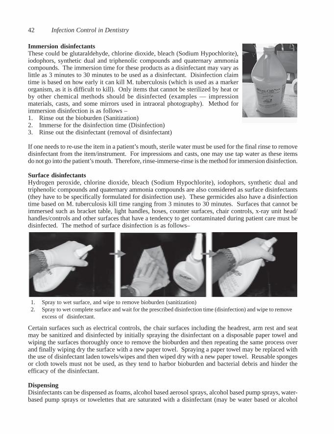
Infection Control in Dentistry42
Immersion disinfectantsThese could be glutaraldehyde, chlorine dioxide, bleach (Sodium Hypochlorite),iodophors, synthetic dual and triphenolic compounds and quaternary ammoniacompounds. The immersion time for these products as a disinfectant may vary aslittle as 3 minutes to 30 minutes to be used as a disinfectant. Disinfection claimtime is based on how early it can kill M. tuberculosis (which is used as a markerorganism, as it is difficult to kill). Only items that cannot be sterilized by heat orby other chemical methods should be disinfected (examples — impressionmaterials, casts, and some mirrors used in intraoral photography). Method forimmersion disinfection is as follows –1. Rinse out the bioburden (Sanitization)2. Immerse for the disinfection time (Disinfection)3. Rinse out the disinfectant (removal of disinfectant)
If one needs to re-use the item in a patient’s mouth, sterile water must be used for the final rinse to removedisinfectant from the item/instrument. For impressions and casts, one may use tap water as these itemsdo not go into the patient’s mouth. Therefore, rinse-immerse-rinse is the method for immersion disinfection.
Surface disinfectantsHydrogen peroxide, chlorine dioxide, bleach (Sodium Hypochlorite), iodophors, synthetic dual andtriphenolic compounds and quaternary ammonia compounds are also considered as surface disinfectants(they have to be specifically formulated for disinfection use). These germicides also have a disinfectiontime based on M. tuberculosis kill time ranging from 3 minutes to 30 minutes. Surfaces that cannot beimmersed such as bracket table, light handles, hoses, counter surfaces, chair controls, x-ray unit head/handles/controls and other surfaces that have a tendency to get contaminated during patient care must bedisinfected. The method of surface disinfection is as follows–
Certain surfaces such as electrical controls, the chair surfaces including the headrest, arm rest and seatmay be sanitized and disinfected by initially spraying the disinfectant on a disposable paper towel andwiping the surfaces thoroughly once to remove the bioburden and then repeating the same process overand finally wiping dry the surface with a new paper towel. Spraying a paper towel may be replaced withthe use of disinfectant laden towels/wipes and then wiped dry with a new paper towel. Reusable spongesor cloth towels must not be used, as they tend to harbor bioburden and bacterial debris and hinder theefficacy of the disinfectant.
DispensingDisinfectants can be dispensed as foams, alcohol based aerosol sprays, alcohol based pump sprays, water-based pump sprays or towelettes that are saturated with a disinfectant (may be water based or alcohol
1. Spray to wet surface, and wipe to remove bioburden (sanitization)2. Spray to wet complete surface and wait for the prescribed disinfection time (disinfection) and wipe to remove
excess of disinfectant.
Chapter 11 -NEW.p65 10/20/2007, 10:26 AM42
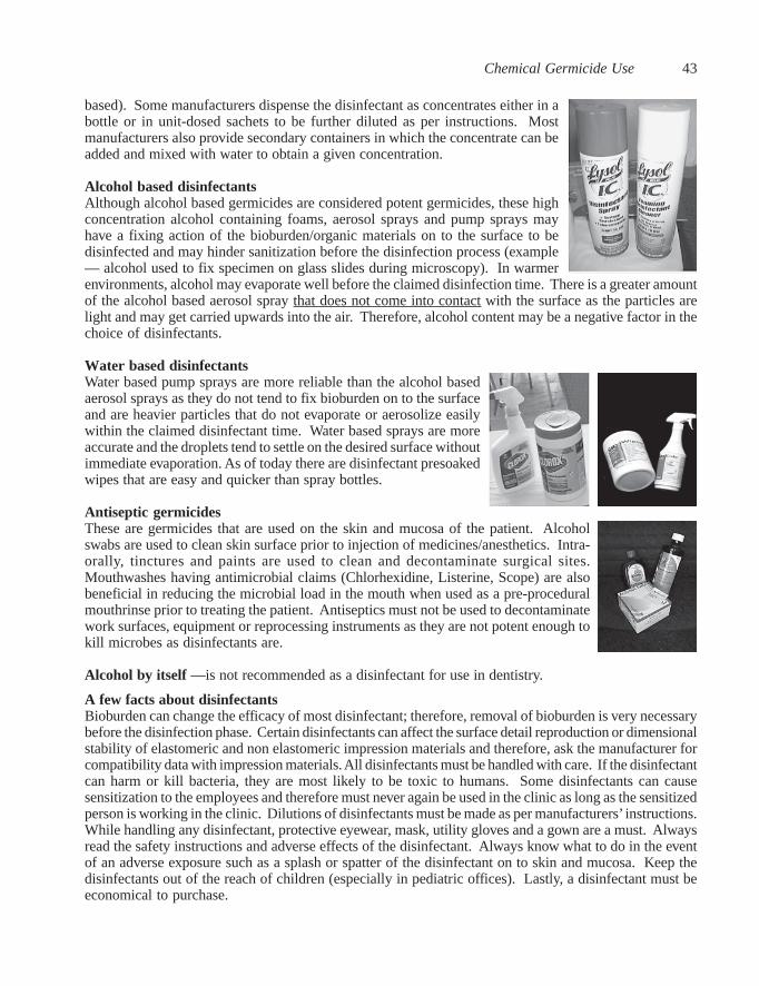
Chemical Germicide Use 43
based). Some manufacturers dispense the disinfectant as concentrates either in abottle or in unit-dosed sachets to be further diluted as per instructions. Mostmanufacturers also provide secondary containers in which the concentrate can beadded and mixed with water to obtain a given concentration.
Alcohol based disinfectantsAlthough alcohol based germicides are considered potent germicides, these highconcentration alcohol containing foams, aerosol sprays and pump sprays mayhave a fixing action of the bioburden/organic materials on to the surface to bedisinfected and may hinder sanitization before the disinfection process (example— alcohol used to fix specimen on glass slides during microscopy). In warmerenvironments, alcohol may evaporate well before the claimed disinfection time. There is a greater amountof the alcohol based aerosol spray that does not come into contact with the surface as the particles arelight and may get carried upwards into the air. Therefore, alcohol content may be a negative factor in thechoice of disinfectants.
Water based disinfectantsWater based pump sprays are more reliable than the alcohol basedaerosol sprays as they do not tend to fix bioburden on to the surfaceand are heavier particles that do not evaporate or aerosolize easilywithin the claimed disinfectant time. Water based sprays are moreaccurate and the droplets tend to settle on the desired surface withoutimmediate evaporation. As of today there are disinfectant presoakedwipes that are easy and quicker than spray bottles.
Antiseptic germicidesThese are germicides that are used on the skin and mucosa of the patient. Alcoholswabs are used to clean skin surface prior to injection of medicines/anesthetics. Intra-orally, tinctures and paints are used to clean and decontaminate surgical sites.Mouthwashes having antimicrobial claims (Chlorhexidine, Listerine, Scope) are alsobeneficial in reducing the microbial load in the mouth when used as a pre-proceduralmouthrinse prior to treating the patient. Antiseptics must not be used to decontaminatework surfaces, equipment or reprocessing instruments as they are not potent enough tokill microbes as disinfectants are.
Alcohol by itself —is not recommended as a disinfectant for use in dentistry.
A few facts about disinfectantsBioburden can change the efficacy of most disinfectant; therefore, removal of bioburden is very necessarybefore the disinfection phase. Certain disinfectants can affect the surface detail reproduction or dimensionalstability of elastomeric and non elastomeric impression materials and therefore, ask the manufacturer forcompatibility data with impression materials. All disinfectants must be handled with care. If the disinfectantcan harm or kill bacteria, they are most likely to be toxic to humans. Some disinfectants can causesensitization to the employees and therefore must never again be used in the clinic as long as the sensitizedperson is working in the clinic. Dilutions of disinfectants must be made as per manufacturers’ instructions.While handling any disinfectant, protective eyewear, mask, utility gloves and a gown are a must. Alwaysread the safety instructions and adverse effects of the disinfectant. Always know what to do in the eventof an adverse exposure such as a splash or spatter of the disinfectant on to skin and mucosa. Keep thedisinfectants out of the reach of children (especially in pediatric offices). Lastly, a disinfectant must beeconomical to purchase.
Chapter 11 -NEW.p65 10/20/2007, 10:26 AM43
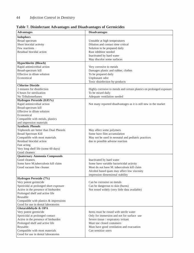
Infection Control in Dentistry44
Table 7. Disinfectant Advantages and Disadvantages of GermicidesAdvantages Disadvantages
IodophorsBroad spectrum Unstable at high temperaturesShort biocidal activity Dilution and contact time criticalFew reactions · Solution to be prepared dailyResidual biocidal action Rust inhibitor needed
Inactivated by hard waterMay discolor some surfaces
Hypochlorite (Bleach)Rapid antimicrobial action Very corrosive to metalsBroad-spectrum kill Damages plastic and rubber, clothesEffective in dilute solution To be prepared dailyEconomical Unpleasant odor
Toxic disinfection by-productsChlorine Dioxide3 minutes for disinfection Highly corrosive to metals and certain plastics on prolonged exposure6 hours for sterilization To be mixed dailyNo Trihalomethanes Adequate ventilation neededHydrogen Peroxide (0.05%)Rapid antimicrobial action Not many reported disadvantages as it is still new in the marketBroad-spectrum killEffective in dilute solutionEconomicalCompatible with metals, plasticsand impression materialsSynthetic PhenolsTriphenols are better than Dual Phenols May affect some polymersBroad Spectrum Kill Some have film accumulationCompatible with most materials May not be used in neonatal and pediatric practicesResidual biocidal action due to possible adverse reactionFast actingVery long shelf life (some 60 days)Good cleanersQuaternary Ammonia CompoundsGood cleaners Inactivated by hard waterSome have M.tuberculosis kill claim Some have variable bactericidal activityGood vacuum line cleaner Most do not have M. tuberculosis kill claim
Alcohol based quats may affect low viscosityimpression dimensional stability
Hydrogen Peroxide (7%)Very potent germicide Can be corrosive on metalsSporicidal at prolonged short exposure Can be dangerous to skin (burns)Active in the presence of bioburden Not tested widely (very little data available)Prolonged shelf and active lifeReusableCompatible with plastics & impressionsGood for use in dental laboratoriesGlutaraldehyde & OPAVery potent germicide Items must be rinsed with sterile waterSporicidal at prolonged contact Only for immersion and not for surface useActive in the presence of bioburden Severe tissue / respiratory irritantProlonged shelf and active life Must use closed containersReusable Must have good ventilation and evacuationCompatible with most materials Can sensitize usersGood for use in dental laboratories
Chapter 11 -NEW.p65 10/20/2007, 10:26 AM44

Chemical Germicide Use 45
At the end of the day, the suction lines (High-volume-evacuator, and Saliva Ejector) should be cleanedwith either a quaternary ammonia compound cleaner or an enzymatic detergent mixed in water. Thecleaner should be sucked through the lines either aerosolized or as a liquid and let sit for about 10 minutes.After the 10 minute soak, water should be sucked through the lines to wash out the cleaner along withpatient debris and other materials. The suction traps must be examined and replaced at least weekly.
Chapter 11 -NEW.p65 10/20/2007, 10:26 AM45

Infection Control in Dentistry46
Chapter 12
Instrument Reprocessing and Sterilization Monitoring
Puttaiah
Instrument reprocessing is the most important aspect of Dental Infection Control as it deals with itemsthat have the greatest potential for disease transmission during dental care. Any dental instrument thatenters the oral cavity is classified as critical or semi critical surfaces per Spaulding’s Classification andmust be sterilized. Common methods of in-office sterilization in dentistry are Autoclaving, Chemiclavingand Dry Heat. For items that are heat labile chemical immersion methods using an approved chemicalsterilant although rare, must be used. Today, all items that are used in dentistry as critical and semi-critical items are heat sterilizable or sterile-single-use disposable.
Separation of Waste and Instruments
While handling any contaminated instrument after patient care, the handler must use eye protection, amask, and a gown and wear utility gloves (heavy duty latex or nitrile) as there is a potential for exposureto patient material and sharps injuries. Immediately after patient care, the saturated waste (items that aresoaked with blood or body fluids) must be disposed off in a red/orange biohazard labeled plastic bag.Disposable sharps must be discarded into sharps containers. Other disposable items can be discarded intoregular waste. Instruments should now be taken securely to the reprocessing area and handpieces (highspeed and slow speed) removed from the tray to be processed per manufacturers’ instructions. Mosthandpiece manufacturers have specific methods of cleaning and therefore handpieces should not be soakedor sonicated. All other items that need to be reprocessed must be soaked in a holding solution or in theultrasonic machine.
Soaking or keeping bioburden moistInstruments that have been used on patients need to be place in a holding container with a detergent (soap
Chapter 12 -NEW.p65 10/20/2007, 10:36 AM46

Instrument Reprocessing and Sterilization Monitoring 47
solution) or an ultrasonic solution to keep bioburden (patient material suchas blood or tissue tags) moist. In the absence of a holding container, onemay use the ultrasonic machine with ultrasonic solution to keep instrumentsmoist. Soaking solutions or ultrasonic solutions need not be disinfectantsas the latter have no proven effect and tend to be more expensive thandetergents. Instruments should be soaked for at least 10 minutes beforesonication. Do not soak instruments overnight as even the best detergentscan be corrosive and cause pitting.
Cleaning instruments
Prior to sterilization, instruments must be cleaned to reduce bioburden. Ultrasonic cleaning (sonication)is very efficient and works by a process called cavitation where there is implosive activity or cavitationthat helps tear away dirt and debris from instrument surfaces. Sonication is a few hundred times betterthan manual cleaning with a long handle brush. Sometimes, even after an ultrasonic process patientmaterial may still be on the surface of instruments that may need to be physically removed by using along handle brush to reduce the risk of sharps injury. Sonication of loose instruments should be carriedout for 8-10 minutes and the period doubled (15-20 minutes) for instruments in cassettes. Ultrasonicsolutions that are non-ionic and some enzymatic ultrasonic solutions are less corrosive on instrumentsthan others. One should not use disinfectant solutions instead of ultrasonic solutions as there is no provenantimicrobial efficacy but leads to a false sense of security.
Inspection of cleaned instrumentsAfter the instruments are cleaned, the whole ultrasonic machines basket containing instruments must beremoved and rinsed under running water to remove the residual solution and the dirt. The basket shouldthen be tipped gently allowing the instruments to roll on to a layer of paper towels. The instrumentsshould be pat dried using a small stack of paper towels and inspected for residual bioburden or debris. Ifresidual debris is found, that particular instrument should be manually cleaned using a long handledbrush, rinsed, dried and re-inspected. The inspected instruments can now be made into sets and bagged.If cassettes are used, the cassettes need to be opened on the stack of towels and instruments inspected forresidual debris, pat dried and rearranged into the cassette to be bagged in sterilization pouches or wraps.
Packaging of instruments
Chapter 12 -NEW.p65 10/20/2007, 10:36 AM47

Infection Control in Dentistry48
1 Sterilization time is the time required for killing 1012 B. stearothermophilus spores and does not include the completesterilization cycle i.e. ramp-up time for the temperature to reach 250ºF and chamber pressure of 15 pounds per squareinch. Normally the complete cycle would take about 60-90 minutes (heat-up, sterilization and cool down)
If instruments are to be used loose or in a cassette, they should be packaged in the prescribed type ofpackaging material for sterilization (one type of material for Autoclaves and Chemiclaves and a differentone for Dry Heat). The packaging should have a chemical indicator on the packaging or a process indicatortape to show that the process has been completed (due to the color change on the indicator after sterilizationprocess). Packaged sterile instruments can be stored for as long as the integrity of the pouch/package isnot broken, damaged or affected by moisture after being sterilized and dried. There is no time limit onstorage if the package integrity is maintained (event related). If the clinic uses non-bagged sterilization ofinstruments, the instruments have to be used immediately and cannot be bagged after sterilization, preservedor considered sterile for later use.
If instruments are to be “cold sterilized” in glutaraldehyde or any approved immersion sterilant, theinstruments should not be packaged but should be rinsed with sterile water to remove residual chemicalsterilant from the surfaces of the instrument and used immediately. If immersion sterilants such asglutaraldehyde are used, the tubs should be covered and placed under an evacuation hood to reduceambient chemical vapor in the sterilization area that may affect the staff.
Common methods of heat sterilizationCommon methods of sterilization in dentistry are a) Autoclave or steam under pressure; b) Chemiclave orcombination of synergistic chemicals, heat and pressure; and c) Dry Heat.
Autoclave
Autoclaving or sterilization using steam and pressure is by far the most common method of sterilization.This is the most reliable process but could be more corrosive than the other methods for instruments thathave a high content of carbon steel (especially if packages are not adequately dried). Many autoclaveshave process/parametric information that is printed out at the end of the given cycle. Items such as water(liquids) can be sterilized other than regular instrument. Following are the most common parameters atwhich the autoclave is used—
Table 8. Parameters of Sterilization for AutoclavesParameters Standard Cycle Fast CycleSterilization Time 15 - 20 minutes1 3-5 minutesTemperature 121º Celsius (250º F) 134º C (273ºF)Pressure 15 pounds per square inch (psi) 30 pounds per square inch
Chapter 12 -NEW.p65 10/20/2007, 10:36 AM48

Instrument Reprocessing and Sterilization Monitoring 49
2 Some high-speed handpiece manufacturers recommend “not using” a rapid heat transfer method of dry heat as thehigh temperatures could breakdown the bearings of the turbine.
Even among the autoclaves there are different types. The quick turnaround sterilizers such as Statim takeone cassette with loose or packaged instruments. Loose instruments can be sterilized quickly using theFlash cycle (although not recommended for regular use), while packaged instruments take a longer time.Other efficient type is one that has a pre-vacuum and post vacuum feature. This feature allows sucking allthe air out of the chamber and is beneficial in sterilizing hollow bore instruments such as metal air/watersyringe tips and highspeed handpieces. After the vacuum is created, the steam is released into the chamberfilling the hollow bore instruments thereby increasing the efficacy of sterilization access into the mostdifficult areas of the instrument. After the sterilization process, the steam is sucked out of the chamberincluding the space in the hollow bore instruments and heat maintained to dry the instruments and thepackaging. Therefore, the pre-vacuum and post-vacuum cycle is very efficient in both sterilization anddrying of the pack. All high-speed handpieces must be sterilized between patients.
ChemiclavingChemiclaves use a combination of liquid chemicals (with <15% water) that are introduced into the chamber,heat and pressure for a sterilization cycle. The parameters for sterilization are temperature of 131ºC(270ºF), 20 psi and sterilization time of 30 minutes. Chemical Scavengers that are a part of the Chemiclavemust be replaced regularly to reduce risks of chemical exposure to employees.
Dry HeatDry heat is another common method of sterilization in dentistry. Following are the parameters for dryheat—
Table 9. Parameters of Sterilization for Dry-Heat SterilizersParameter Slow Cycle Fast Cycle Rapid Heat2
Temperature 160º C (320º F) 170º C (340º F) 190º C (375º F)Sterilization Time 120 minutes 60 minutes 6-12 minutes
Chemiclave and dry heat are less corrosive to carbon steel metals than autoclave methods. Packagingmaterials are different for dry heat vs. Chemiclave or autoclave methods. All instrument packs must beremoved from sterilizers and stored dry.
Monitoring Efficacy of Sterilizers “Weekly” monitoring of the sterilizer’s efficacy using BiologicalEquivalent monitoring devices or Sterilization Integrator (it measureswhether the parameters of temperature and pressure over time) isnecessary as this can be done in-house. The information should berecorded and maintained for a period of 3 years. The biologicalequivalents show color change when the sterilization paramenters havebeen met (the chemical stripchanges from green to greyishblack)
“Monthly” sterilization monitoringfor all sterilizers is necessary by either using a biological indicatorsuch as a spore-strip containing B. stearothermophilus and B. subtilis.These spore-strips are used for testing the efficacy of the sterilizersand both spores are available on the same spore strip called dual strips
Chapter 12 -NEW.p65 10/20/2007, 10:36 AM49

Infection Control in Dentistry50
and need to be mailed out to a monitoring service. In-office 24 hour sterilization monitoring kits areavailable that are very convenient and with little lag time for obtaining the results. When a positivegrowth occurs from a spore test or a biological equivalent test (failure of the sterilizer) the sterilizer mustbe taken out of service, tested again. The sterilizer should be reinstated only after repair and testing forefficacy. Each set of instruments being sterilized should have a chemical indicator (to show that the packhas been through a sterilization cycle).
Recommended maintenance of the reprocessing equipment is essential for proper function. Autoclavesshould use distilled water to avoid slaking and breakdown, and must be serviced per manufacturer’srecommendations for proper functioning (check the sterilizer’s manual). The chamber should be cleanedperiodically using recommended methods. Leaks in the gasket will not allow temperature and pressureparameters to be met during the sterilization cycle, thereby causing failures.
Ultrasonic SolutionsUltrasonic solutions should beprepared daily per manufacturer’sdilutions and the machine run for aminimum of 15 minutes at thebeginning of the day withoutinstruments to remove bubbles, thisprocess is call degassing. Degassingis done to remove bubbles that hinderthe process of sonication. Once amonth, an aluminum foil (to the widthof the bath) must be submerged into
the depth of the bath with water/solution and the machine run for 30 seconds. The submerged area of thefoil should show uniform pitting or pin-holes when place against a dark background. If the pitting orpinholes are not uniform, the ultrasonic machine must be serviced and the transducers that generate theultrasonic action calibrated. At the end of the day, the ultrasonic machine should be drained, the bathrinsed and wiped dry to increase the longevity of the bath.
Storage of Sterile InstrumentsSterile bagged instruments must be stored in clean dust-free shelves or cabinets. These sterile packs canbe dated. The packages are considered sterile till the packaging is compromised (damaged, torn or wet)irrespective of the date of sterilization. If the packages are compromised, all the instruments in the packmust be reprocessed again and stored sterile prior to use. If the instruments are not is sterile pack (looseinstruments not in packs/sterile pouches) they must be used immediately after sterilization. Instrumentsthat have not been sterilized in pouches cannot be stored as sterile and must be sterilized immediatelyprior to use.
Reprocessing of High-Speed, Slow-Speed Handpieces and Scaler handpiecesAll handpieces that are used in the oral cavity must be sterilized or single-usedisposable (such as in disposable contra-angle handpieces). Both, high-speedand slow-speed handpieces retract patient material and are difficult to clean anddecontaminate using chemical germicides. All re-usable handpieces must bereprocessed separately immediately after use between patient care. Highspeedhandpieces should be cleaned and lubricated per manufacturer’s recommendationand sterilized using either a Chemiclave, autoclave or dry-heat. Slow-speedhandpieces should be single-use-disposable such as the contra-angle handpiecesthat are used for polishing during oral prophylaxis. Other slow-speed handpieces
W&H Assistina is designed to cleanand lubricate the W&H Handpiecesprior to sterilization.
Chapter 12 -NEW.p65 10/20/2007, 10:36 AM50
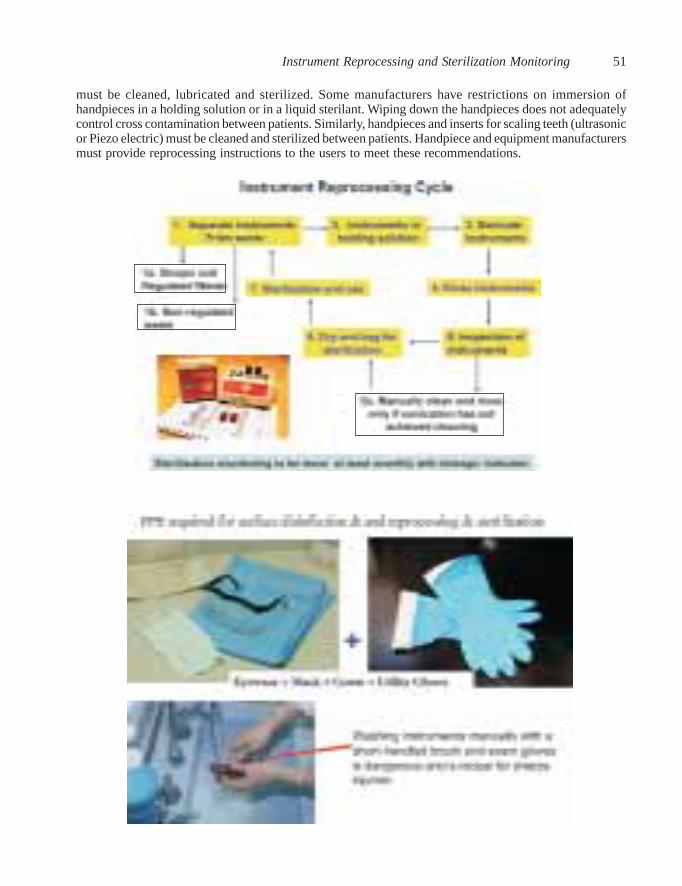
Instrument Reprocessing and Sterilization Monitoring 51
must be cleaned, lubricated and sterilized. Some manufacturers have restrictions on immersion ofhandpieces in a holding solution or in a liquid sterilant. Wiping down the handpieces does not adequatelycontrol cross contamination between patients. Similarly, handpieces and inserts for scaling teeth (ultrasonicor Piezo electric) must be cleaned and sterilized between patients. Handpiece and equipment manufacturersmust provide reprocessing instructions to the users to meet these recommendations.
Chapter 12 -NEW.p65 10/20/2007, 10:36 AM51

Infection Control in Dentistry52
Chapter 13
Dental Unit Water System Contamination Control
Puttaiah & Lin
Prior to the introduction of high-speed handpieces, dentistsdid not use water as a coolant or as an irrigant. Along withthe high-speed handpieces came the dental unit water systemthat provided water for irrigation as well as a coolant duringthe cutting of teeth. Modernization of cutting instrumentsintroduced a new problem of ‘dental unit water systemcontamination’ and ‘release of bio-aerosols’ in the dentalclinic. Most modern dental unit water systems are made upof a complex maze of waterlines, control blocks, valves,barbs and connectors that are of various sizes and composedof different metals, plastics and rubbers.
Traditionally, these water systems are directly rigged to themunicipal water system and very few as of recent years areequipped with a bottle (self-contained water system) into whichthe treatment water or irrigants are added. The design of all dentalunit water systems allows settling of contaminants from waterand air. These contaminants can be inorganic materials such assalts from the hardness of the source water that coat the lines andcause corrosion of metals and allow settling of microbes. Theother types of contaminants are organic in nature includingextremely high counts of bacteria, fungi, viruses and evennematodes. These problems of contamination have been knownby dentists and researchers for over 50 years. Only recently haveagencies such as the American Dental Association (ADA), theCenters for Disease Control and Prevention (CDC) the dentalindustry seriously broached this topic in providing somemethodologies and measures to modify the equipment, treat thecontamination and monitor the level of contamination.
Factors associated with treatment water contaminationSome factors associated with water system contamination are—1) Nutrient content of source water; 2) Extended period ofstagnation per day; 3) High surface to volume ration; 4) Low flow-rate; 5) Laminar flow; 6) Microbial quality of source water; andlastly 7) Equipment design.
Chapter 13 -NEW.p65 10/20/2007, 10:39 AM52

Dental Unit Water System Contamination Control 53
Potential RisksThis biofilm is found to be teaming with microbes and chunks of which detach contaminating the dentaltreatment water. While municipal water normally may have less than 500 “planktonic” or free-floatingmicrobes per milliliter, the water coming out of the dental handpiece and air/water syringe may havemore than a million microbes per milliliter. The units of measurement for the microbial contamination inwater is “colony forming units per milliliter” or cfu/mL. An example of usage is – 100 microbes in onemilliliter of water is referred to as 100 cfu/mL. These environmental microbes survive even under verylow nutrient conditions and may shrink to a tenth of their size, but still have the capacity to live on saltsand other contaminants.
All environmental microbes are not pathogenic, but the sheer numbers that are found in the dental treatmentwater is of concern. Most of the microbes found in the dental water system biofilms are gram negativerods. When these gram negative rods die, they release a toxin called “bacterial endotoxins”. Theseendotoxins in large amounts (as seen in un-cleaned water systems) have a potential in causing healthproblems in patients. In the first study on dental unit water system and endotoxins, investigators at BaylorCollege of Dentistry found potential of release of endotoxins to an extent of 500 endotoxin units permilliliter (EU/mL), and this amount is deemed to be quite toxic.
Among the potentially pathogenic microbes isolated in the dental unit water systems are— Pseudomonas(potential for super infections), Mycobacterium (potential for causing lung infections), Legionella (potentialfor causing Legionnaire’s Disease), and Moraxella (potential for causing bacterial endocarditis). Therefore,one has to consider the potential for being exposed to life-threatening infectious diseases from the dentaltreatment water of un-cleaned water systems. Investigators have also isolated amoebae, fungi and nematodesin the dental water systems.
Although there have not been studies linking association between the dental unit waterline contaminationand mortality/morbidity among the dental patient population, it is pragmatic to keep the water systemdecontaminated and reduce the potential risks. It is difficult to conduct either retrospective or prospectiveepidemiological studies to find association between diseases and water source due to legal issue andlogistical issues. Some studies have demonstrated significantly high titers of antibodies to Legionellaamong dental health care providers as opposed to a normal population and another study has shown thata large percentage of the dental water samples contaminated by at least one variety of Legionella.
During oral prophylaxis, use of a highspeed handpiece or even the air/water syringe we generate aerosolsthat may be laden with microbes and endotoxins. These organic matter laden aerosols are referred to as“bio-aerosols”. These aerosols may reach up to 12 – 16 feet from the source during patient care and maystay suspended in the air for over 24 hours if there is “inadequate ventilation or air exchanges” andtherefore, having a potential for exposing the employees to contaminants over time. Many studies havealso demonstrated the contamination of the water system by common oral microorganisms that couldhave been sucked back into the waterlines during patient care due to faulty antiretraction valves.Antiretraction valves are designed to prevent suck back or retraction of oral fluids during patient care,and researchers have shown that even new valves fail within few days of use leading to the risk of cross-contamination.
Disinfection-by-productsWhen certain chemicals come in contact with biofilms, they react adversely and can lead to production ofan array of toxic substances called disinfection-by-products (DBPs). When bleach comes in contact withbiofilms a group of DBPs call trihalomethanes are produced that are considered to be carcinogens. In astudy done at Baylor College of Dentistry, over 8,000 parts per billion (ppb) of DBPs were expressed indental treatment water when bleach was used to clean the lines. The permissible limit on DBPs set by the
Chapter 13 -NEW.p65 10/20/2007, 10:39 AM53

Infection Control in Dentistry54
US-EPA is 80 ppb. Therefore, use of certain agents such as bleach to constantly or periodicallydecontaminate the water system may prove to be more deleterious than beneficial. Further, we have toconsider corrosive effects of the chemical on the components of the water system as well as its safety onhuman health.
Some methods of controlMicrobial control of dental treatment water/irrigant contamination is a two pronged issue. Firstly, thedental unit water system has to be cleaned and kept free of biofilms and other inorganic contaminants byinitially and frequent periodic cleaning/disinfecting the system with a decontaminating agent. Secondly,the water/irrigant used for patient care must be microbe-free, and therefore, the water needs to be filtered,sterile, and distilled/boiled or an EPA registered and FDA approved low-grade antimicrobial added priorto use.
Biofilm formation on the surface of waterlines in the dental unit
Chapter 13 -NEW.p65 10/20/2007, 10:39 AM54

Dental Unit Water System Contamination Control 55
Periodic cleaning methods of the water system with a cleaning/decontamination agent alone may removesome amount of biofilm or inorganic materials but will not prevent re-growth of biofilms. The watersystem can get re-colonized with biofilms very soon and therefore may show high cfus/mL within daysafter periodic cleaning. Periodic cleaning products should not only disrupt and remove well establishedbiofilms but also be effective on dissolving the inorganic salts coating the water system that may act asneutralizing agents against the low-grade disinfectants/irrigants.
Selection of decontaminating agents and irrigants should entail askingmanufacturers’ information on—1. Registration of the chemicals with agencies such as the United
States Environmental Protection Agency (US-EPA) and clearanceof the United States Food and Drug Administrations (US-FDA)or agencies in the European Union (EU) for safety issues
2. Biofilms and other deposit removal efficacy3. Compatibility with metals, plastics and rubbers in the water
system4. Evidence of easy removal of periodic cleaning agent from the water system5. Biocompatibility of periodic cleaning agent and irrigant with humans6. Should not produce dangerous disinfection-by-products such as trihalomethanes7. Compatibility of irrigant with composites bonding to enamel and dentin
Examples of available control measuresThere are many different options for controlling dental treatment water system contamination. The firststep is to have a self contained reservoir (bottle system) to be retrofitted to the dental unit. Somemanufacturers of periodic cleaning agents feel that periodic cleaning is good enough in controlling thetreatment water contamination, but experimentation and science has shown otherwise. If municipal wateris used as an irrigant, the quality of such water varies from <10 cfu/mL to greater than a few thousand cfu/mL at the same location, and therefore is not reliable treatment water let alone for swimming. Therefore,water that has been treated with a physical or a chemical method or a low-grade antimicrobial must beused. Dental units with low grade antimicrobial irrigants should be used frequently to replenish the lineswith new fluid; otherwise, the active ingredient to control the microbes in the lines gets depleted. Anexample is that the same antimicrobial irrigant may show better microbial control in dental units that treatat least 5 patients per day (as in private dental clinics), in comparison with units treating about 2 patientsper day (as in a dental school setting).
Table 10. List of some periodic cleaners and irrigants being used by practitionersSome Periodic Cleaners Method of UseAlkaline Peroxide – Sterilex Ultra Full strength, overnight contact followed by flushing with hot waterChlorine Dioxide Tablets – Vista Tabs 30 – 50 ppm in water, 5-10 minutes followed by flushing with waterChlorine Dioxide 2 part Liquid - BioClenz 30 – 50 ppm in water, 5-10 minutes followed by flushing with waterElectro-chemical Oxidants – Sterilox Full Strength 30 minutes – overnight contact, followed by flush with dilute oxidantSilver Citrate Powder – specific to Pure Tube Dissolved in water with low total dissolved solids (TDS) and left over night,
followed by a water flushPeracetic Acid - TAED+Perborate (Italy/EU) 5-10 minute between patients used in the Castellini Autosteril System,
followed by sterile water flushSome Irrigants Method of UseBoiled water/Distilled/Sterile water Directly in the bottle as irrigantSilver Nitrate Tablets – ICX Dissolve in 700 ml of municipal waterSilver Citrate Tablets – BluTabs Dissolve in 700 ml of municipal waterSilver – PureTube (Sterisil) Silver ions in water with Low TDSIodine – Dentapure (DP 40, 90 and 360) Used inside the bottle replacing the intake tube, or placed in line
with water flow (3-4 ppm)Chlorine Dioxide 2 part Liquid – BioClenz 2 – 4 ppm in municipal waterElectro-chemical Oxidants – Sterilox Concentrate diluted for irrigant purposesGrapefruit Extract Botanical – Vistaclean 5 drops per 700 ml of municipal water (emulsifying agent)
Chapter 13 -NEW.p65 10/20/2007, 10:39 AM55
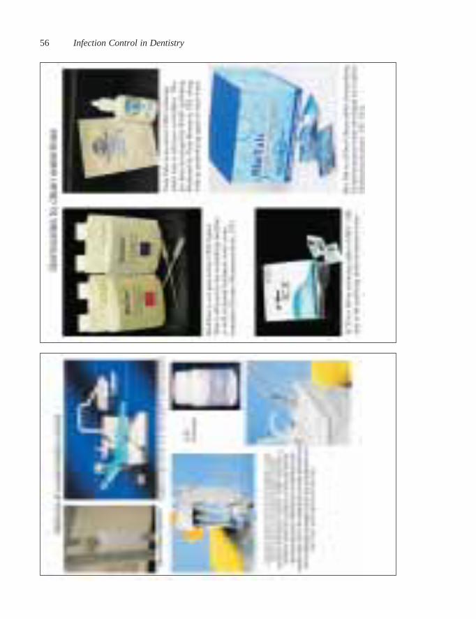
Infection Control in Dentistry56
Chapter 13 -NEW.p65 10/20/2007, 10:39 AM56

Dental Unit Water System Contamination Control 57
Monitoring the quality of treatment waterDental treatment water or irrigants should have less than 500 cfu/mL of heterotrophic mesophilicmicroorganisms. Apart from the microbial quality, the water should not have a high endotoxin contentand should at least be as clean as drinking water. Traditionally, the microbes found in dental treatmentwater are slow-growing and need low nutrient agars for growth.
The ideal agar (for providing the diagnostic or contamination) for heterotrophic counts (total counts) isR2A agar, which can grow these environmental microbes at room temperature over 1-4 weeks. Normally,after plating, the microbial colonies can be counted after one week (7 days). Prior to plating the water/irrigant samples, the latter has to be neutralized using an appropriate chemical that will neutralize theantimicrobial agent to provide a natural environment for microbial growth. Using R2A agar is not feasiblein a dental office, therefore the water collected from the water system should be sent refrigerated/on iceover night to a water quality monitoring laboratory to plate the samples.
For in-office use, there are very few microbial quality monitoring kits. One such kit is the Aquasafe™ byPall Sciences. Aquasafe™ provides heterotrophic counts of microbes in the water in about 3 days. Theother kit is made by Millipore Corporation. Millipore HPC water samplers can also be used to determinethe counts in about 7 days of incubation. Both the kits need no incubator for the growth of the microbialcolonies as the incubation temperature is room temperature. Millipore also provides neutralizers containingsodium thiosulphate that can neutralize bleach, chlorine dioxide or a few other oxidants.
Although both in office samplers are easy touse, they are not very accurate in predictingthe level of contamination, however, they arevery easy to use and not expensive, and do notneed incubation.
As of today, there are many devices availablein the market for cleaning and maintaining themicrobiological quality of dental unit watersystems. In conclusion, dental unit watersystems that have not been cleaned regularlyare highly contaminated and release highamounts of microbes and endotoxins intopatients’ oral cavities. The water system shouldbe cleaned periodically to dislodge the biofilmand should use an irrigant free of microbialcontamination. Low-grade antimicrobials mayalso be used as irrigants. Materials selectedfor cleaning/decontaminating the water systemmust be biocompatible and not harm thepatient, employee or the equipment and shouldnot affect composite bonding to enamel ordentin. Water quality should be periodicallymonitored and contamination levels keptbelow 500 cfu/mL (similar to CDC’sGuidelines). Water samples may be screenedfor contamination using in-office kits or be sentto water quality testing laboratories.
Chapter 13 -NEW.p65 10/20/2007, 10:39 AM57

Infection Control in Dentistry58
Chapter 14
Infection Control in Dental Radiology
Puttaiah & Liang
Most of oral and maxillofacial radiology normally consists of non-invasive procedures. Although exposureto blood is not common, contact with saliva does occur. Blood and saliva are the most common routes oftransmission of infectious diseases in dentistry through direct contact or through cross-contamination.Therefore unit dosing of materials and specific step-by-step infection control protocols are required.Although oral and maxillofacial radiology (OMR) procedures fall mainly in the semicritical and non-critical categories of Spaulding's Classification of inanimate objects, many contagious diseases such asinfectious mononucleosis and hepatitis-B can possibly be spread by simple contact with saliva. Therefore,whenever a potential for contamination by saliva exists during any OMR procedure, universal precautionsmust be observed including the use of adequate personal protective equipment (PPE) such as gloves, theproper handling of contaminated materials, and the decontamination of surfaces exposed to saliva orcontaminated materials. It is not usually necessary to wear PPE such as impervious gowns, long sleeves,masks and protective eyewear during routine OMR procedures when no aerosols, droplets or spatter aregenerated. It is advisable to use gowns, masks and protective eyewear while treating patients with ahistory of gag-reflex.
Following are the steps—
Table 11. Unit Dosing of MaterialsMaterials during Film Exposure Materials during Transportation1. One dose of pre-procedural anti-bacterial mouthrinse Without barrier envelopes:2. Paper towels 1. Empty paper cup3. Disinfectant 2. Cup with exposed films4. Barriers (pre-formed or a roll of plastic wrap) 3. Paper napkin/barrier5. Powder-free gloves (latex or vinyl) 4. Overgloved hands6. Radiographic films7. Sterile film holders8. Two paper cups9. Over-gloves (food handlers gloves) with wrist With barrier envelopes: area of gloves everted 1. Non-contaminated exposed film packets
10. Leaded thyroid collar and apronMasks, eyewear and protective gowns are neededwhen a patient has a known gag-reflex
Chapter 14 -NEW.p65 10/20/2007, 10:41 AM58

Infection Control in Dental Radiology 59
Table 12. Steps in Exposure and Processing
Activities During The Exposure Phase
°Pre-procedural mouth rinse with a mouthwash° Disinfect surfaces at the beginning and end of each day (between patients if visibly contaminated)°Avoid spraying electrical switches, wipe with disinfectant moistened paper towel°Apply surface covers to the yoke, tube head, cone, control unit, head rest, arm rest, and any hand held switches°Place a paper towel/surface cover on a work area to hold film packets, sterilized film holding devices, bite blocks,paper cups, and overgloves°Wear powder-free examination gloves°Place the sanitized lead apron and collar on the patient after seating the patient°Set the required mA, kVp and exposure time on the control unit and reset as required°Open the sterile pack containing the film holding devices; affix the film and position in the patients' mouth.°Position the tube head and cone by touching areas with barrier°Preferably use a foot switch or a barriered hand-held switch to trigger the unit°Drop exposed films into a paper cup without touching the outer surface of the cup°If films are pouched (ClinAsept Barriers), disinfect the envelope and/or remove the non-contaminated film packets.These can now be processed without further infection control considerations as outlined below.
Activities During The Transportation Phase
°Don over-gloves over the exam gloves now before transportation°Pick up the cup with exposed films and the extra empty cup°Carry the two paper cups to the processing area
Activities During The Film Processing Phase If Using a Daylight Loader°Open top lid of the processor with over-gloved hands°Place a paper napkin/plastic wrap on the floor of the loader's open chamber°Place the empty cup and the one with the films in chamber, keep the clean/empty paper cup on one side°Close the lid of the chamber°Insert over-gloved hands through the sleeves°Take off the over-gloves and place them on the dirty side of the paper napkin°Open the film packet by pulling gently on the outer tab to open the packet°Carefully hold the black paper tab and tease it out of the packaging, shake it gently till the film drops into the cleanpaper cup°Discard the paper, lead foil and plastic wrap on the dirty side°Once all wrappings have been removed and the films dropped into the cup, discard the powder free latex gloves onthe dirty side°With the clean bare hands pick up the films by the edges and feed them into the processor°Remove hands through the sleeves
Unit dosing of materials before the start of radiological procedures is essential for both infection control & radiology
Chapter 14 -NEW.p65 10/20/2007, 10:41 AM59

Infection Control in Dentistry60
°Wear new gloves to remove the waste from the loader chamber, remove the surface covers from the radiographicequipment, chair and control unit and discard them in to a regular waste bin°Wash hands with soap and water and dry them°With bare hands, remove the films from the processor and mount them
If Using a Darkroom Processing
°Place two paper napkins/plastic wraps on a work surface near the processing area°Place the cup with exposed films on one paper napkin°Discard the overgloves°Open each film packet by pulling gently on the outer tab°Now the inner black paper tab is seen°Hold the paper tab and tease it out of the packaging and shake it gently till the film drops onto the clean papernapkin°Discard the paper and/or plastic wrap and empty film packets onto the other paper napkin°Once all wrappers have been removed and the films dropped onto the paper napkin, discard the powder free latexgloves°With the clean bare hands pick up the films by the edges and feed films into the processor°After films are processed, with bare hands pick up the films mount them under regular lighted conditions
Chapter 14 -NEW.p65 10/20/2007, 10:41 AM60

Infection Control in Dental Radiology 61
Infection Control in Digital RadiographyHardware used in dental informatics (clinical information systems in dentistry) include digital radiographicequipment, digital intra-oral camera systems, periodontal probing devices, printers and personal computersused for management of patient information and office records. Physically this hardware could be withinthe patient care area or in the outer offices. Equipment used in patient care should be handled followinginfection control protocols to control cross-contamination.
The digital receptor is used in the patient's oral cavity needs to be sheathed with a plastic sheath extendingat least 5 inches outside the patient's mouth. The sheath needs to be changed between patients and thedigital receptor and only needs to be wiped with a disinfectant wipe if contaminated. If in the patient carearea, the CPU has to be plastic wrapped with air vents exposed (to prevent heating of the unit. Thekeyboard cannot be disinfected or sterilized due to the existing design. It is essential to obtain preformedmembrane/silicon covers to completely cover the keyboard which can be sanitized and disinfected betweenpatients. These cover needs to be changed/wiped down with a disinfectant between patients. The mouseshould not be of a trackball design as it is difficult to cover it with a membrane cover. It should be coveredwith a membrane cover which should not interfere with the movement of the mouse or create a staticelectricity interference. A joystick design for a mouse is convenient for easy application of barriers, or anelectronic pointing device (Wacom Pen and Tablet). The front monitor should also be covered withscreen shade/cover which will prevent aerosol particles settling on the screen. These screens need to besanitized when visibly soiled. Do not cover the monitor's air-vents. The printer if located within thepatient care area needs to be covered with a plastic wrap to prevent aerosol particles settling on it, and theswitches need to be covered too. The printer need not be barriered if it is located outside of the patientcare area. Apart from using barriers and disinfection techniques, the personnel should use gloves whilehandling this equipment if they are also touching the patients
Chapter 14 -NEW.p65 10/20/2007, 10:41 AM61

Infection Control in Dentistry62
Table 13. Digital Radiographic Equipment
Parts Level Barriers
1. Cart Non-critical Covers2. a. Digital Receptor Semi-critical Plastic Sheath/sleeve b. Digital Sensor Semi-critical Plastic Sleeve3. CPU Non-critical Covers4. Key board Non-critical Covers5. Mouse Non-critical Cover6. Monitor Non-critical Screen Shade7. Printer Non-critical cover controls
If any of the equipment is contaminated, disinfect using a disinfectant wipe (do not immerse or spray).Do not immerse Digital Receptors (ones with electronic leads) in disinfectant as the leaching of liquidsmay short the circuits in the receptor. Digital Sensors (that do not have leads) may be immersed in adisinfectant per manufacturer's recommendation.
Chapter 14 -NEW.p65 10/20/2007, 10:41 AM62
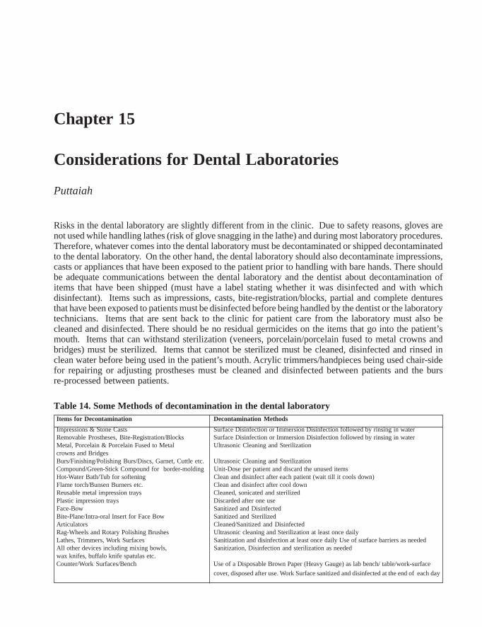
Considerations for Dental Laboratories 63
Chapter 15
Considerations for Dental Laboratories
Puttaiah
Risks in the dental laboratory are slightly different from in the clinic. Due to safety reasons, gloves arenot used while handling lathes (risk of glove snagging in the lathe) and during most laboratory procedures.Therefore, whatever comes into the dental laboratory must be decontaminated or shipped decontaminatedto the dental laboratory. On the other hand, the dental laboratory should also decontaminate impressions,casts or appliances that have been exposed to the patient prior to handling with bare hands. There shouldbe adequate communications between the dental laboratory and the dentist about decontamination ofitems that have been shipped (must have a label stating whether it was disinfected and with whichdisinfectant). Items such as impressions, casts, bite-registration/blocks, partial and complete denturesthat have been exposed to patients must be disinfected before being handled by the dentist or the laboratorytechnicians. Items that are sent back to the clinic for patient care from the laboratory must also becleaned and disinfected. There should be no residual germicides on the items that go into the patient’smouth. Items that can withstand sterilization (veneers, porcelain/porcelain fused to metal crowns andbridges) must be sterilized. Items that cannot be sterilized must be cleaned, disinfected and rinsed inclean water before being used in the patient’s mouth. Acrylic trimmers/handpieces being used chair-sidefor repairing or adjusting prostheses must be cleaned and disinfected between patients and the bursre-processed between patients.
Table 14. Some Methods of decontamination in the dental laboratoryItems for Decontamination Decontamination Methods
Impressions & Stone Casts Surface Disinfection or Immersion Disinfection followed by rinsing in waterRemovable Prostheses, Bite-Registration/Blocks Surface Disinfection or Immersion Disinfection followed by rinsing in waterMetal, Porcelain & Porcelain Fused to Metal Ultrasonic Cleaning and Sterilizationcrowns and BridgesBurs/Finishing/Polishing Burs/Discs, Garnet, Cuttle etc. Ultrasonic Cleaning and SterilizationCompound/Green-Stick Compound for border-molding Unit-Dose per patient and discard the unused itemsHot-Water Bath/Tub for softening Clean and disinfect after each patient (wait till it cools down)Flame torch/Bunsen Burners etc. Clean and disinfect after cool downReusable metal impression trays Cleaned, sonicated and sterilizedPlastic impression trays Discarded after one useFace-Bow Sanitized and DisinfectedBite-Plane/Intra-oral Insert for Face Bow Sanitized and SterilizedArticulators Cleaned/Sanitized and DisinfectedRag-Wheels and Rotary Polishing Brushes Ultrasonic cleaning and Sterilization at least once dailyLathes, Trimmers, Work Surfaces Sanitization and disinfection at least once daily Use of surface barriers as neededAll other devices including mixing bowls, Sanitization, Disinfection and sterilization as neededwax knifes, buffalo knife spatulas etc.Counter/Work Surfaces/Bench Use of a Disposable Brown Paper (Heavy Gauge) as lab bench/ table/work-surface
cover, disposed after use. Work Surface sanitized and disinfected at the end of each day
Chapter 15 -NEW.p65 10/20/2007, 10:43 AM63

Infection Control in Dentistry64
Pumice if used in the clinic while working on individual patients during try-in must be unit dosed (onlya needed amount dispensed), the pumice discarded after single use, and the container/trough/tub sanitizedand disinfected after each use.
Surface covers must be used regularly so that there is minimal contamination or dirtying of the benchtops. Laboratory work surfaces must be cleaned and disinfected at the end of each day. Before disinfection,all stone/plaster, wax, metal or other filings must be cleaned or scraped off of the surface. All sharps suchas burs, knifes and scalpels must be cleaned and decontaminated and stored safely to avoid any sharpsinjuries.
At the end of the day, the ultrasonic instrument cleaning units including the trough must be drained,wiped dry and disinfected. This will prolong the life of the ultrasonic unit by avoiding corrosion of thethrough.
Dust/metal filings/chemical vapors are generated in the dental laboratory on a regular basis. Duringtrimming of acrylic, metals, dry stone etc., the trimming should be done close to a vacuum/suction spoutto remove the inhalation risks for the staff. While handing acrylic monomer, there should be adequateventilation or evacuation of the air to reduce inhalation of the monomer. Similar safety measures shouldbe in place for other inhalation risks as needed.
PPE used in the laboratory as follows—• Mask to reduce splash/spatter from trimmers, rag-wheels etc.• Goggles/Protective Eye-Wear to protect eyes from particulate matter and flying debris• Fluid resistant Gown as needed• Heavy-duty heat resistant gloves while handling hot devices/investment flasks, hot water baths for
removal of invested wax or for curing acrylic dentures.
All Bunsen burners, torches, flame torches used in the laboratory should be cleaned, inspected and storedsafely. Any source of oxygen/gases used in the laboratory must be shut off at the end of the day. Duringperiod of extended non-use such as vacation periods (extending beyond one week) the hoses should bedisconnected and the lids screwed back on the cylinders. All gas cylinders must be harnessed securely(fixed to a wall with brackets and chains, or to a broad base) to avoid tipping over or falling.
As torches/burners/open flame is regularly used in the laboratory, fire-extinguishers should be installedand the technicians/staff trained in proper use of the extinguisher.
Dental Laboratories
Gloves cannot be used while operating the lathe due to snagging of gloves. Priordisinfection of impression, casts, dentures or other contaminated materials in needed
Chapter 15 -NEW.p65 10/20/2007, 10:43 AM64

Special Topics 65
Chapter 16
Special Topics
Puttaiah & Kohli
Considerations for extracted teeth, biopsy specimens and tissues
Extracted teeth are infectious and must be treated as medical waste if being discarded. Medical wastethat includes teeth and tissues is potentially infectious and can be a risk for personnel handling waste.Extracted teeth that are to be used in the dental laboratory for shade-matching, in research and in materialstesting laboratories, or in the preclinical laboratory for practicing cutting/restorative procedures or forpreclinical examination must be decontaminated. Methods to decontaminate teeth could be heatsterilization, immersion in a sterilant such as 5000 ppm bleach, 7% hydrogen peroxide or in Glutaraldehydefor the sterilization time. If the teeth are to be used in preclinical laboratories or for research purposes(bonding of composite to teeth for shear bond strength tests), they must be immersed in 0.05% thymolsolution in water for at least a couple of weeks with solution changed daily. Bleach should not be used inthis instance as it can dissolve the organic matter in the teeth and alter outcomes. Teeth can be autoclavedand then discarded in regular waste. Teeth with amalgam fillings should also be disposed as medicalwaste that is not incinerated (however there is very little significant evidence/scientific studies regardingmercury release beyond permissible limits due to incineration). Given the available information, teethwith amalgam fillings could be autoclaved with other saturated medical waste prior to disposal withregular waste. If patients request extracted teeth, the teeth must be washed, soft tissue tags trimmed,placed in 5000-6000 ppm (1:10 dilution in commercial liquid bleach) bleach for at least 10 minutes,rinsed again and dried using a paper towel and then provided to the patient in a plastic bag/zip-lock bag.The patient should also be informed that it is potentially infectious and not to allow others to handle thetooth/teeth. It is always better not to give it back to the patient, but dispose of the extracted teeth asmedical waste.
If extracted teeth are to be stored, the teeth should be washed, soft tissue trimmed, teeth soaked in adisinfectant for a minimum of 2 weeks (10% formalin) with the disinfectant being changed every 24hours. The jars/containers with extracted teeth must be handled with gloves and other required PPE as itis still considered potentially infectious. The teeth must be placed in leak-proof rigid container withbiological hazard labeling as well as chemical hazard labeling specifying the name and concentrationchemical. Even after disinfection, the teeth should be handled as potentially infectious (use of gloves andother PPE depending on the further use)
Biopsy specimen should be handled as potentially infectious material and should be immediately storedin 10% buffered formalin if being transported for histopathological investigation. The specimen shouldbe placed in leak-proof rigid container with biological hazard labeling as well as chemical hazard labeling
Chapter 16 -NEW.p65 10/20/2007, 10:45 AM65

Infection Control in Dentistry66
specifying the name and concentration chemical. During histopathology, the specimen must be handledwith gloves and other required PPE as it is still considered potentially infectious.
Considerations for Oral and Maxillofacial Surgery and LASER/Electrosurgery plumes
As of today, electro-surgery and the use of LASER in dentistry is becoming commonplace. Electrosurgicalprocedures tend to burn the tissue creating smoke and other possible byproducts contaminating ambientair with smoke plumes in the dental clinic. Similarly, LASERs also generate plumes or smoke in theclinic that could be laden with patient material. These gases/smoke/plumes could contain hydrogen cyanide,benzene and formaldehyde, affecting both the dental care providers and the patients. With respect todisease risks, these gases/smoke and plumes may also contain tissue debris, microbes and viruses. Someof the organisms detected in the plumes are Staphylococcus, Corynebacterium, Neisseria, Human papillomavirus and HIV. The methods of controlling risks from electrosurgical and LASER plumes is to wear FullPPE that includes well fitting (tight face-fit) high filtration masks, face shields/goggles/protective eyewear,gloves, and fluid resistant gowns. To control release of the gases/smoke/plumes into the ambient air, ahigh volume evacuation system (high-speed suction) as close as possible to the site generating the gases/smoke/plumes must be used. Further, other methods of improving air circulation in the clinic should alsobe used. This is an emerging field and is still under study with respect to risks to the dental and medicalpersonnel, and therefore, needs to be constantly researched and updated.
Considerations for new and emerging diseases including Creutzfeldt-Jakob Disease (CJD)
CJD & Prion Related DiseasesCreutzfeldt - Jakob disease (CJD) or commonly fall under a degenerative neurological diseases/disorders/conditions such as Transmissible Spongiform Encephalopathies (TSE). These conditions affect bothhumans and other mammals and are associated with a form of protein called a Prion that lacks nucleicacid but are capable of self-propagation within a host. Other than CJD and variant CJD, there are otherconditions such as kuru, Bovine Spongiform Encephalopathy (BSE) or mad cow disease, and Gerstmann-Straussler-Scheinker syndrome associated with human illness and death. The incubation period of theseprions prior to clinical signs and symptoms could be years. Death could occur within a year of theoccurrence of clinical signs and symptoms. Apart from being isolated in the brain and neurologicaltissue, prions have been identified in the tonsils, eye tissue, and pituitary glandular tissue. Prions areresistant to conventional physical and chemical decontamination processes and therefore additionalmeasures need to be taken. As of today, the potential infectivity of oral tissues with respect to prionrelated conditions is not definite. If it is a patient with known status of a prion related condition, deferroutine care and treat only urgent/emergent needs with additional precautions such as—
• single-use-disposable critical and semi-critical devices, or if possible patient-specific devices notto be used on others
• burs, endodontic files must be single-use disposable• hand instruments must be cleaned thoroughly and rinsed well before undergoing the longest
sterilization cycle• reusable instruments soaked, cleaned with ultrasonic means and rinsed thoroughly• no-flash sterilization of instruments but a complete cycle at 134°C and 15 psi for a minimum
sterilization time of 18 minutes, but the use of the longest cycle (up to 1 hour of sterilization timeand with prior rigorous cleaning of the instruments)
Chapter 16 -NEW.p65 10/20/2007, 10:45 AM66
Classification & Management of Dental Wastes and Sharps 67
Chapter 17
Classification & Management of Dental Wastes andSharps
Puttaiah & Kohli
While it is essential to protect the patient and the care provider, we do generate a lot of medical waste. With respect to dentistry,waste can be classified as follows—
1. Regulated wastea. Biological waste
i. Saturated waste soft waste (materials that are saturated blood and saliva and can express blood/saliva/body fluidwhen squeezed)Examples: —2X2 gauze and cotton rolls saturated/soaked in saliva/blood
ii. Soft tissues (oral soft tissues including biopsy specimen) and hard tissues (bone and teeth)b. Disposable Sharps
i. Examples: —Scalpel blades, needles, used carpules, orthodontic wires, disposable matrix bands, single-use-disposableburs, contaminated broken glass, wire sutures used for splinting and reducing fractures, and failed implants
c. Environmentally hazardous chemicals and metalsi. Examples:—Mercury, amalgam, Beryllium, chemicals used in processing radiographs (Silver Nitrate), formaldehydes,
glutaraldehyde, OPA, and Phenols2. Non-regulated waste
a. Examples:—Unsaturated cotton rolls, paper towels, gauze, non-sharp single-use-disposable devices (plastic AWS tips,Saliva Ejector Tip, HVE tip), disposable syringe, plastics, disposable PPE and other inanimate surface barriers
During patient care, all sharps that can be immediately discarded into a sharps container must be placed in a sharps container unlessand until needed further during the procedure. Each operatory must have a rigid approved and a labeled sharps container. Immediatelyafter patient care, all sharps must be discarded into the sharps container safely (never use a two handed technique to re-sheathneedles, just discard them into the sharps container by unscrewing the needle with a needle holder. Similarly, the scalpel blade mustbe removed from the BP handle or if using a disposable scalpel and blade, discard the whole device into the sharps container. Aftergetting rid of the sharps, the regulated soft waste that is saturated with blood/saliva must be placed in a biohazard labeled orange/redbag that needs to be disposed into a rigid larger red container as regulated waste (cumulative waste removed periodically) by acontracting agency. Waste Tissues including soft tissues and extracted teeth and bone must also be placed in the red bag and takento the rigid large container for professional disposal. The clinics should have a contract with a professional waste managementcompany that regularly removes the hazardous waste from the clinic and terminally incinerates the waste at a remote facility. Theprofessional waste management company must also be able to remove the filled sharps containers for proper professional disposal.In the absence of such services, the clinic may opt (as in the US) to place all the soft regulated waste in an autoclavable bag (withbio-hazard markings) and autoclaved/sterilized for one complete cycle before disposing into regular non-regulated waste receptacles.
Chemical wastes should not be poured down the drain as they pose a grave environmental hazard. They should be discarded basedon the local rules for hazardous chemicals. Regulated waste management companies may also be able to dispose off the wastechemical as needed. Non regulated waste such as non-saturated soft waste poses no threat to the final waste handlers and can bedisposed off similar to household waste. Waste should also be separated based on the type of material. Plastic wastes when incineratedmay generate environmentally hazardous chemicals such as dioxins that are carcinogens and therefore, it may be good to involve theinput of professional waste management companies as to possible separation of certain hazardous wastes that can be safely disposedoff without burdening the environment. While treating patients, we must also find ways to reduce waste that may harm the environment.
Chapter 17 -NEW.p65 10/20/2007, 10:46 AM67

Infection Control in Dentistry68
Chapter 18
Management of Exposure to Blood and Body Fluids
Puttaiah & Kohli
Exposure of the Dentists, Auxiliaries, Laboratory Technicians, Students and Volunteers to blood andbody fluids of patients through splash/spatter to the mucosa (eye, nose and mouth), sharps injury(percutaneous as in a needle-stick or other sharps puncturing intact skin) is possible during dental care.It is essential to know the HIV status of the patient and the DHCP during such inadvertent events. It isalso good to know other chronic infectious disease status (HBV, HCV status) of both so that appropriateand effective post exposure treatment and follow-up be initiated. One has to understand that withrespect to dentistry, saliva must be considered as potentially infectious as blood (most dental procedureswill bring about bleeding and saliva is normally mixed or contaminated with blood from the gingivaeven if blood is not visible). These exposure incidents can happen chairside while treating a patient,where the source patient can be identified; or it can happen during instrument reprocessing where theinstruments are mixed after treating multiple patients where the specific source patient is unknown.When the source patient is known it becomes easier to implement specific post-exposure testing andtherapeutic protocols. When the source is unknown, the protocols become more difficult.
Steps to be taken in the event of an exposureIn the event of an exposure, stop treatment of the patient and stabilize the patient temporarily—
a. If it was a splash or spatter exposure to the mucosa—i. Wash the area with flowing water/eyewash station for at least 10 minutesii. Contact a medical practitioner/infectious diseases specialist who can evaluate the situation,
initiate testing to find out the infectious disease status of the source patient as well as theexposed personnel
iii. nitiate treatment of the exposed personnel as needed with antibiotics and antiretroviralmedications and future follow-up of the source and the exposed
b. If it was a sharps injury (needle-stick, puncture wound from a contaminated instrument—i. Wash the injured area with an antimicrobial soap for about 2-3 minutesii. Apply first aid (medications / band-aid / suture) as needediii. Contact a medical practitioner/infectious diseases specialist who can evaluate the situation,
initiate testing to find out the infectious disease status of the source patient as well as theexposed personnel
iv. Initiate treatment of the exposed personnel as needed with antibiotics and antiretroviralmedications and future follow-up of the source and the exposed
Chapter 18 -NEW.p65 10/20/2007, 10:48 AM68

Management of Exposure to Blood and Body Fluids 69
All incidents should be documented in detail as follows—i. When did it happen?ii. How did it happen?iii. What was the type of exposure (splash/spatter or percutaneous)?iv. For percutaneous injuries describe whether it was a needle stick, puncture or laceration of
the skin with a sharp instrumentv. Whether any fluid was injected into the healthcare provider
Although there is no quantitative risk assessment of the type of exposures in causing infectious diseasesin DHCPs, the type and amount of blood and other body fluids, the type of exposure (mucous membraneexposure, non-intact skin exposure, bites or percutaneous exposures), source individuals disease status(severity of the disease) could qualitatively help in post exposure protocol with respect to HIV, HBV andHCV. Personnel exposed to HIV patients would need to undergo testing and start effective, state-of-the-art antiretroviral therapy within 24 hours of exposure. If the personnel is sero-negative for HIV and hascompleted anti-retroviral therapy, testing at 3 months and 6 months would need to be done. If the sourcepatient was sero-negative at the time of exposure, it may not be necessary to start anti-retroviral therapy.If the personnel is an employee of an organization, institution or a corporation and chooses to declinetesting and post exposure prophylaxis with anti-retroviral medications, the refusal for care should bedocumented with the personnel’s signature.
If the patient or the source has a history of other infectious diseases such as HBV, HCV or other blood-borne conditions, appropriate treatment and follow-up measures should be carried out to reduce thedisease risk to the exposed personnel.
Sample Diagram of Aseptic and Clinical Requirements during patient care
Patient factors o Medical History o Diagnostic Tests o Germicidal
Mouthrinse o Protective
eyewear
Additional Precautions
o None needed o Defer care for
infective TB patients o Only Standard
Precautions
Post TX o Asst. Separates
Waste, instruments o Removes barriers o Cleans up area o Dr. Doff Gloves o Wash hands o Dismiss patient
Clinic Preparation
o Flush lines o Surface Barriers o Unit dosing o Materials o Sterile
Instruments
Preparation of the dental Teamo Handwash o PPE based on
anticipated risk o Unit dosing of
additional PPE
STANDARD
PROCEDURES FOR THE
PREVENTION OF DISEASE
Chapter 18 -NEW.p65 10/20/2007, 10:48 AM69

Infection Control in Dentistry70
Chapter 19
Occupational Safety Directives for Dentistry (Samplebased on US OSHA Standards)
Puttaiah & Kohli
1. Bloodborne Pathogens
Scope and applicationThis directive applies to all occupational exposure to blood and other potentially infectious material(BOPIM). Examples would be blood, saliva, extracted teeth, biopsy specimen, urine and materials thatcome in contact with these potentially infectious materials. These materials may contain pathogens thattransmit disease to employees. Whenever these risks are anticipated, there must be a plan to controlexposures, and a post exposure protocol in the event of exposure to BOPIM.Exposure control• Every clinic must have a manual of Dental Infection Control & Occupational Safety• In this manual there must be an Exposure Control Plan for the specific clinic• An exposure control plan will include—
o List of categories of employees and their nameso Exposure Determination - Job responsibilities of employees and risk levels based on duties with
respect to exposure to bloodborne pathogens at worko The types of risks to BOPIM by the clinic’s employeeso After determining the risks of exposure in the duties of the employees, they need to be trained in
controlling the risks and the training documented. The training should also include evaluation ofthe exposure incident, further methods of controlling such incidents, and annual reviews/updatesor when new risks are introduced into the clinical arena
o Use of engineering and work practice controls to reduce the risk of exposureo Protocols to follow in the event of an exposureo Documentation of Initial and Periodic training in exposure control and post exposure protocols
with respect to bloodborne pathogensTasks involved in Exposure Determination• List all job categories in the clinic• Determine the categories that may have regular (High) contact with BOPIM (Dentist, hygienist,
assistant, lab-tech); Determine the categories that may have only occasional (Moderate) contacts(front office staff who may assist occasionally in patient care, clean-up etc); Determine categoriesthat have potentially no (low) contact with BOPIM (Front office staff who do not assist in clinicalwork at all).
• Each category should have a prescribed method of exposure control such as use of PPE, level of PPE(gloves, gowns, masks, eyewear, bonnets, shoe covers) for each type of task based on anticipated
Chapter 19 -NEW.p65 10/20/2007, 10:52 AM70

Occupational Safety Directives for Dentistry (Sample based on US OSHA Standards) 71
exposure, engineering controls (sharps holders; HVE and rubber dam use to control bioaerosols; useof sharps dispenser in each operatory), work-practice controls (proper or no re-sheathing of needles,safe handling of sharps during care, and instrument reprocessing). These controls may be referred toas standard operating procedures for the clinic.
Methods of ComplianceThere must be a designated safety person for a clinic whose role is to manage and implement the safetyprogram. Compliance is the “Heart of the Exposure Control Plan”. The safety person keeps a tab on thetraining, compliance during practice and documentation and should coordinate all safety efforts in theclinic to control exposures to BOPIM. Some of the common issues the safety person should look into anddocument are–• Whether there are safety protocols and acceptable methods to reduce risks in the office• Use of appropriate PPE by employees• Universal Precautions being followed in the clinic• Engineering and Work-Practice Controls being followed, including periodic testing and documentation
of the function of engineering controls• Initial and periodic immunizations against immunizable diseases• Checking to see if sharps are being handled properly during provision of care, transportation of
contaminated sharps to the reprocessing area, and during reprocessing• Safe and appropriate disposal of waste (both regulated and non-regulated waste)• Recording of Exposure incidents and logging them in prescribed OSHA format• Keeping a post-exposure protocol in place• Housekeeping issuesCommon Engineering Controls in the Exposure Control Plan• High Volume Evacuator & Rubber Dam in conjunction to control bioaerosols• Aerosol/Dust Evacuation Hood in the dental laboratory• HVAC with laminar flow of air to reduce aerosols• Sharps Containers in every operatory and in the laboratory• Splash guards/safety guards for lathes and table-top rotary devices• Crush resistance devices incorporated into dental chairs (to avoid crushing of toes and knees during
the use of chairs)These engineering controls should be examined and tested periodically, and there should be documentationof the test.Common Work practice controls• Handwashing & use of PPE• Safe handling sharps or other potentially dangerous items• Single handed recapping of needles, or use of a needle holder to grasp the cap in a two-handed
technique• Pointing the bur on the handpiece away from employees and patients while being placed in the socket• Disposing sharps and contaminated waste chair side into appropriate containers• Never overfilling a sharps containers• Prohibition of eating, drinking, smoking, application of cosmetics, handling contact lenses in
potentially hazardous areas of the clinic (during patient care, instrument reprocessing, laboratorywork where there is a risk of exposure to BOPIM)
• Minimizing splash & spatter• Safe handling of blood & saliva specimen including extracted teeth• Biohazard labeling of containers including secondary containers• Use of labeled leak free secondary containers for shipping• Labeling contaminated equipment with a biohazard label• Decontaminating contaminated equipment before use or shipping
Chapter 19 -NEW.p65 10/20/2007, 10:52 AM71

Infection Control in Dentistry72
Use of Personal Protective Equipment (PPE)All required PPE must be provided at no cost to the employees. Level and use of PPE use should bedetermined based on anticipated exposure to BOPIM and the change of PPE spelled out clearly (examplewould be changing gloves every 30 minutes for long procedures). The PPE being used should be effectiveand not overtly permeable to blood and other body fluids. They should be accessible to all employees ofall sizes and the employees should be trained in their use effectively. All the single-use-disposables mustbe discarded after one use and the reusable PPE decontaminated in an acceptable manner before beingreused. Common PPE used in the dental clinic are Face Mask, Face Shield/Goggles/Prescription Glasseswith Side-shields, Gown and Gloves used in combination while anticipating splash or spatter. While notanticipating splash or spatter, and while working intra-orally, only exam gloves may be sufficient. PPEshould not be worn outside the clinic (i.e. in the patient waiting area). Scrubs are not considered to bePPE, but are considered to be common clothes.Housekeeping-General Rules:• Work site should be clean and sanitary• Written Schedule and instructions as what to clean and with what to clean and as to who should clean
itDecontamination• Work surfaces• Equipment surfaces• Change of protective coverings• Clean bins, pails and cans periodically• Safe handling of broken glass (use PPE & mechanical devices)• Recirculation of reusable sharps (instruments)Regulated WasteItems that qualify as regulated (needs special handling including chair side as well as during disposal)• Liquid or semi liquid BOPIM• Items that would express BOPIM if compressed• Items caked with blood or saliva• Contaminated Sharps• Pathologic and microbiologic wastes containing BOPIM or derived from BOPIMSharps Containers:• Located as close as possible to dental operatory, upright and with a Biohazard label• Not to be over filled and replaced As Soon As Possible when full• Lid Closed while being moved/transported• Placed in secondary container if leaky while being transported/shippedLaundry• Contaminated laundry is that which is soiled by blood or other potentially infectious materials• Handled as little as possible• Bagged on site• Not sorted or rinsed on site• Bags labeled with Biohazard label or red bags• Handled with gloves and other PPE as neededChoices for handling laundry1. Single-Use-Disposable items better than Reusable Barriers2. Outside Professional Laundry Services3. Washer & dryer on site dedicated for contaminated laundry4. Employee(s) designated to handle (train & document)Hepatitis-B Vaccination• All employees with the risk of exposure to bloodborne pathogens
Chapter 19 -NEW.p65 10/20/2007, 10:52 AM72

Occupational Safety Directives for Dentistry (Sample based on US OSHA Standards) 73
• Vaccination offered within 15 working days, evaluated and provided by a Medical Doctorknowledgeable in infectious diseases and occupational medicine
• Vaccination Refusal Form must be signed if refused• Booster every 5 years not required (as in the US)• Records: documentation of HBV status required and maintained confidential• No prescreening required• Provide standard copy to Healthcare Professional who will administer the vaccine• Post vaccination test not a must but is desirable• Post-Exposure Evaluation and Follow-UpDefinition of an Exposure IncidentEye, mouth or mucous membrane contact with BOPIM, non-intact skin contact with BOPIM, or parenteralcontact with BOPIM.Procedures After Exposure Incident• Confidential medical examination• Post-Exposure Evaluation and Follow-UpGeneral information• Complete an exposure report• Identify Source Person• Container Labeling with the chemical name and color coded labeling for specific risks (use NFPA or
other international standard labeling format)• Understanding the MSDS with respect to chemical hazards• Employee Training in HAZCOM• Inventory of hazardous chemicals in the Clinic• Hazards involved during routine and non-routine task• Define possible chemical hazards in the clinic• Exposure of other personnel and outside contactors• Outside contactors must be informed of—
o Hazardous chemicals in the officeo Measures to reduce possibility/risks of exposureo Location of MSDSs for all hazardous chemicalso Procedures to follow if exposed
(If the contractor is interested, let them visit and evaluate the situation, the risks)• Hazardous Chemical Inventory
o Maintain a list of hazardous chemicals in the officeo List of all dental materials used in the office
• Identity or infeasibility of identifying source person• Source tested for HBV and HIV after consent with refusal if any documented in writing and Employee
provided with source’s test results• Confidentiality of the sources results/status explained to the exposed employee to avoid publicity or
any other infringement of lawEvaluating Health Care Professional (HCP)-Employee’s job responsibilities during the exposure incident-Documentation of route of exposure and circumstances in which the exposure occurred-Results of source persons blood test/status-Employee’s medical record that is maintained by the employer-The evaluating healthcare professional will do the following:• Collect and test employee’s blood after consent• If blood collected but testing for HIV is refused by employee then preserve blood for 90 days• Employee notified of all results
Chapter 19 -NEW.p65 10/20/2007, 10:52 AM73

Infection Control in Dentistry74
• Post-exposure prophylaxis provided per USPHS• Counseling regardless of testing• Illnesses evaluated that are reported by employee in the first 12 weeks irrespective of tests performed• Healthcare professionals written opinion• Made available to the employer within 15 days after completing evaluation• Diagnosis and findings unrelated to exposure incident not reported but held confidentialReport Must contain:• Whether HBV vaccine was indicated/received• Employee informed of evaluation results• Employee informed of any medical condition resulting from exposure, that require additional
evaluation and treatment• All costs should be borne by the employer or should be according to local and national labor lawsLabeling for Bloodborne RisksBiohazard warning labels affixed to:• Containers of regulated waste• Refrigerators or freezers containing BOPIM• Containers used to store or transport or ship BOPIMLabel design (use international standards/US standards as needed)• Fluorescent Orange with Biohazard Symbol and word Biohazard in a contrasting color• Affixed as close to the container by string, wire or adhesive• Red bags or containers may be substituted for labelsMedical Records• Confidential medical record established• For each employee• If the employee has not had an exposure• Name and other identification characteristics of employee• Copy of employee’s HBV vaccination status including known dates of vaccination, record related to
vaccination or ability to receive vaccination or refusal of vaccinationWhen Incident Occurs• Exposure incident report• Copy of all results, exam, testing follow-up• Written opinion of health care provider• Copy of information provided to employerRecord Keeping• Medical record maintained for the duration of employment and during the lifetime of the clinic• Available to employee upon request• If dental practice is sold• Record transferred to new ownerTraining and Training Records• Employees with occupational exposure must be provided training at no cost and during working
hours• Training at initial assignment, changes in tasks/procedures and annually• Level of material must be appropriate in content and vocabulary of the employeeContents of Training• OSHA Bloodborne Pathogen Standards• Bloodborne diseases• Exposure control plan and Emergency Procedures• Signs and labelsQuestion and answer period (necessary)
Chapter 19 -NEW.p65 10/20/2007, 10:52 AM74

Occupational Safety Directives for Dentistry (Sample based on US OSHA Standards) 75
2. Chemical hazard control
Clinic Manual should have a “Written HAZCOM Program”Issues Covered by the program should include—
List Must Have—• Name of Hazardous Chemical• Reference # for each chemical, with the same placed on each MSDS to assist employees for easy
reference in case of emergencyLocation & use of list should be known to all employees and the employees must be trained in the useof the list.
Material Safety Data Sheets• Kept in a notebook/file with index/quick ref.• Book Clearly Labeled MSDS• Each MSDS should have the following:• Chemical Common Name• Physical & Chemical Characteristics• Fire, Explosion, & reactivity• Health Hazards, Carcinogenicity (listed NTP/IARC)• Permissible exposure limits• Control Measures, PPE, Fire and general precautions• Emergency & first aid• Date prepared & changed• Name, address & telephone of manufacturer
Employees must be trained in using MSDS• How to read it• Review Health Hazard Data
MSDS should not be discarded even if chemical use in discontinuedIf material is not hazardous then .OBTAIN LETTER FROM MANUFACTURER STATING THAT
“MATERIAL IS NOT HAZARDOUS AND CONTAINS NO HAZARDOUS CHEMICALS”
Container Labeling• Each Container of Hazardous Chemical labeled or tagged with specific information• Responsibility of Both Manufacturer & Employer• Information should be:• Identity of hazardous chemical• Appropriate hazard warning including target organs• Name & address of manufacturer
Secondary containers• Identity of hazardous chemical• Hazard warning including target organs
Containers to be labeled• Disinfection tubs• X-ray fixer & developer tanks/trays• Oxygen & nitrous oxide outlets for piped-in systemsAlthough containers for immediate used need not be labeled, it is still prudent to do so in case of anexposure. Therefore, label all containers; it only takes a few seconds. Train employees in labeling or get
Chapter 19 -NEW.p65 10/20/2007, 10:52 AM75
Infection Control in Dentistry76
safety manager to label
Employee Training & Information1. Train BEFORE starting work2. When NEW HAZARDS introduced and once yearly.
Contents of Training In Reducing Risks to Chemical Hazards• Copy of the directive & explanation of contents• Location and availability of HAZCOM Program/Manual• Chemicals present in workplace• Explanation, location of MSDS and Hazardous Inventory• Effects of Hazardous chemicals• Employee Training & Information• Methods and observation techniques to determine release or presence of hazardous chemicals or
factors. example——dosimeters for formaldehyde• Instructions on reducing the risks of exposure• Use of PPE, work-practice and engineering controls• Steps taken by the office to reduce the risks• Emergency procedures to follow if Exposed• Explanation of the labeling system
Training & Documentation• Training Certificate proves training provided• Protocol in case of Exposure to Hazard. Chem.• This is an emergency situation• Determination of Hazard from the label• Application of First Aid indicated on MSDS• Obtain physicians help if it is life-threatening• Continuous Medical Re-evaluation if needed
Protocol for Chemical Spill• Determining the hazard from the label of container• Wear Appropriate PPE• Containment and removal of chemical with Spill Kit• Place contaminated materials in appropriate containers• Labeling containers• Notify the waste collector as to what is in the container• If spill is very hazardous, call manufacturer• Document the chemical spill• When, Where• What happened• Who Was Exposed, and remedial action taken• What program was implemented to reduce future exposure
Personnel Monitoring• Use of dosimeters• Personal Monitoring Badges• Potential Hazards :• Radiation• Nitrous Oxide• Formaldehyde (Chemiclave- Vaposteril->Formaldehyde)
Chapter 19 -NEW.p65 10/20/2007, 10:52 AM76

Occupational Safety Directives for Dentistry (Sample based on US OSHA Standards) 77
3. Facility and equipment safety
Contains Some Important Aspects of:• Fire Safety• Building Safety• Equipment Safety
Fire Safety• Automatic Sprinkler System for the building based on the local safety codes and that are functional
(Maintained in reliable operating condition at all times)• Written Fire Safety Policy• Use appropriate fire extinguisher (to cover all types of fires that may be encountered in the dental
office)• Portable Fire Extinguishers (with regular maintenance, training of employees in fire extinguisher use,
training of employees in maintenance of extinguisher)• Check and record function Annually & Monthly• Training in Evacuation Plan
Fire Safety PlanIn An Event of FireNotify all employees to leave premises to regroup at a safe place (please consider safety of the patients asan additional measure). Close doors on the way out. The receptionist should turn off computer and takethe appointment record. Never Use Elevators but use the stairs to evacuate. Go outside the premises andCall emergency Number (fire department and ambulance). All employees must regroup at a predeterminedsafe place for a head count and treatment of injuries before dispersion for the end of the day. Documentthe training and Post evacuation plan
Doors• Sufficient exits for easy/ smooth evacuation and prompt escape during disaster• Boldly MARKED as EXITS & must be VISIBLE• Lighted Arrow towards exit if not over door• Doors that are NOT an EXIT marked ...“NOT AN EXIT” or marked otherwise• Main Doors Should have a LIGHTED exit sign• Should not be locked during working hours or if any employee is in the building• No items should barr or block doors or evacuation in the event of an emergency
Gas Cylinders• Nitrous Oxide• Oxygen• Compressed gasesMust be maintained in safe condition and safety determined by visual inspection. Cylinders must besecured safely with chain links to a wall or an immovable surface. Lids must be secured duringtransportation or during periods the cylinders are not in use or when they are not attached to the lines.Employees must be trained in its use and training documented.
Eyewash StationsQuick Flush and Eyewash Facilities are required when employees are at risk of the eye exposure to
Chapter 19 -NEW.p65 10/20/2007, 10:52 AM77

Infection Control in Dentistry78
BOPIM, hazardous and corrosive materials. These fixed equipment must be located within 10 seconds or25 feet of reach from anyplace within the treatment facility. More than one station may be required for afacility. Eyewash Bottles are NOT acceptable. The eyewash equipment must not be connected to hotwater source and must be operable by a single movement (paddle or lever). This equipment must betested weekly. The temperature of the water must be 60 to 95 degrees Fahrenheit. Eyes must be flushedfor at least 15 minutes in case of an emergency requiring the use of the eyewash station. Water supplyshould be free of bacterial and other contaminant build-up. Drinking water or municipal water sourceshould be used. This equipment can be ordered through safety catalogues and must be installed as persafety and buildings code. Employees must be trained and the same documented.
Trash BinsWaste receptacles should be maintained clean & hygienic. Solid tight-fitting covers are needed for suchitems. Covered receptacles are safer than bins that do not have a cover. Surface should be smooth and theequipment corrosion resistant. The material should facilitate easy cleaning and disposal of contents. Thecontainers must NEVER be OVERFILLED. Never try to compress waste with hands or feet. DO NOTPUT REGULATED WASTE IN TRASH BINS
First Aid KitFirst Aid Kits must be present in each facility. OSHA requires presence of Physician Certified First AidKit especially in rural areas or areas that are reasonably far from emergency facilities. If a First Aid Kitis needed, a person trained in its use must be present. Obtain a First Aid Kit, as dictated by a Physicianand Document it with a letter from the Physician. If First Aid is available within the same businesscomplex, have written instructions on how to obtain help
ElectricalItems such as electrical cords must be in Good Condition and well maintained. No repair of cords ispermitted per safety standards (only replacements are allowed). As a temporary measure one extensioncord per appliance may be allowed that is to be detached after use. Extension cords are not a permanentsubstitute for fixed wiring. Circuit breakers and switches must be used for appliances. Only one appliancepermitted per each wall socket. No multiple appliances resembling an OCTOPUS for one socket. Circuitbreakers, switches, main switches marked boldly for ON & OFF positions, and what they control must bedisplayed clearly.
GFCI circuits within 6 feet of water sources must be used as this applies to all inside and outside outlets.
Grounding Electrical Devices is quintessential. All devices must be grounded to avoid sparking andarcing endangering the employee when he or she touches them. Three wire direct and 2 wire directsystems operating between 50 to 300 volts require grounding. Alternating current systems requiregrounding.
Hazardous area that house flammable gases, flammable liquids, vapors, dust and their storage should notdischarge static electricity, arcs or sparks must be explosion proof for gases, vapors and liquids; andignition proof for combustible dusts. Electrical devices proximal to these storage areas must meet NationalISI Codes. Repair and replacements must only be carried out by a certified electrician.
Building Safety
Dentist as a tenantEmployee complains of safety issue to the dentist and the latter who is the Renter/Leaser needs to inform
Chapter 19 -NEW.p65 10/20/2007, 10:52 AM78

Occupational Safety Directives for Dentistry (Sample based on US OSHA Standards) 79
owner of safety breaches that need to be rectified. It is the owner’s responsibility to alleviate the SafetyProblems within the premises. The employer in this case must document the problems complaints. If theowner does not respond, the employer who is the renter/leaser should inform the clinics attorney or makealternate settlement and charge the owner of neglect. The bottom-line is “safety of employees.”
Dentist as the ownerIf the dentist is the Owner and the EMPLOYEE COMPLAINS OF SAFETY ISSUES to the Dentist/Employer and Owner, the latter must take immediate action to correct problem, and correction documented.To correct any physical facility problems, Certified Agencies/Workers must be contracted to Repair orAddress Issue.
StepladdersStepladders that are Make-Shift Appliances OR Step-Stools that do not meet the ISI and internationalstandards are not to be used in the business/clinic premises. Only Stools & Ladders that meet Standardsmay be used.
Radiation safetyRadiation safety requires personal monitoringHistorically there is very little or no Hazards from routine use of Dental Radiology Employee training inRadiological Hazards and Safety and documentation of the same completed on an annual basis. Theradiological equipment used in the clinic should be monitored /checked for breaches in safety periodically.Any repairs to the equipment should be done immediately by a qualified technician.
Cardiopulmonary Resuscitation DevicesCPR devices are needed in every clinic and the clinical staff may be trained in their use. All dentists musthave a certification in CPR. Pocket Mask with a One-Way-Valve should be used. Ambu-bag devices arean alternative. All training that is provided must be documented. For the purpose of infection control andsafety, single-use-disposable CPR devices may be purchased. Reusable CPR devices with disposablebarriers or mouthpieces are an alternative. If reusable devices are used, adequate reprocessing anddecontamination of the device must be attained after use.
Air CompressorsAir compressors should be equipped with pressure release valves & gauges. Safety devices must bechecked periodically. Air intake should take only clean uncontaminated air, especially if HVE is in theroom. Employees should be prohibited from using highly compressed air for cleaning purposed. Bleedpressure before repairing the equipment is necessary.
Chapter 19 -NEW.p65 10/20/2007, 10:52 AM79
Infection Control in Dentistry80
Selected Definitions
Alcohol-based hand rub: An alcohol-containing preparation designed for reducing the number of viablemicroorganisms on the hands.
Antimicrobial soap: A detergent/soap containing an antiseptic agent.
Antiseptic: A germicide used on skin or living tissue designed to reduce microorganisms on the tissuesurface (e.g.— alcohol, chlorhexidine, hexachlorophene, povidone iodine, Parachlorometaxylenol[PCMX], some quaternary ammonium compounds, and Triclosan).
Glass Bead sterilizer: A device using glass beads 1.2—1.5 mm diameter and temperatures 2170C—2320C for brief exposures (e.g. 45 seconds) to inactivate microorganisms. (This device is not consideredto be effective as a terminal reprocessing device or Sterilizer by the US FDA)
Bioburden: The microbiological load (i.e., number of viable organisms in or on the object or surfaceincluding other organic matter prior to decontamination, or sterilization; also known as “bioload” or“microbial load”.
CDC: The Centers for Disease Control & Prevention, Atlanta, GA, USA.
Colony-forming unit (CFU): The minimum number of separable cells a bacterial growth medium(Agar) that gives rise to a visible colony of progeny. CFUs can consist of pairs, chains, and clusters, or assingle cells and are often expressed as colony-forming units per milliliter (CFUs/mL). In simple terms itis the number of microbes per milliliter of a liquid (such as water, blood, serum etc.).
Decontamination: Use of physical or chemical means to remove, inactivate, or destroy microbes orpathogens on a surface or item so that they are no longer capable of transmitting infection and devicesurface rendered safe for handling, clinical use, or disposal.
Dental treatment water: Water of good microbial quality (<200 CFU/mL) used during dental treatment,including irrigation of non-surgical operative sites and cooling of high-speed rotary and ultrasonicinstruments. This water must not be used during oral and maxillofacial surgery, surgical extraction,periodontal flap surgery where bone is exposed, or during surgical placement of implant.
Disinfectant: A chemical agent used on inanimate objects (e.g.— work surfaces in proximity to thepatient, non-critical dental equipment and surfaces, environmental surfaces such as floors, walls, or sinks)to kill vegetative microorganisms, viruses and fungi, but not necessarily bacterial endospores. U.S.Environmental Protection Agency (EPA) groups disinfectants on the basis of whether the product labelclaims limited, general, or hospital disinfectant capabilities.
Disinfection: Destruction of vegetative microbes, viruses and fungi by physical or chemical means.Disinfection is less lethal than sterilization, because it kills a majority of recognized pathogenicmicroorganisms, but not necessarily all microbial forms (e.g., bacterial spores). Disinfection does notensure the degree of safety associated with sterilization processes.
Droplet nuclei: Particle =5 µm in diameter formed by dehydration of airborne droplets containingmicroorganisms that can remain suspended in the air for long periods of time.
Chapter 19 -page 80.p65 10/20/2007, 10:54 AM80
Selected Definitions 81
DOTS: Directly Observed Treatment Strategy for tuberculosis patients. Technique pioneered by IndianHealth Authorities being observed in over 150 countries globally.
Droplets: Small particles of moisture (e.g., spatter) generated when a person coughs or sneezes, or whenwater is converted to a fine mist by an aerator, handpiece or shower head. These particles, intermediate insize between drops and droplet nucleii, can contain infectious microorganisms and tend to quickly settlefrom the air so that any risk of disease transmission is usually limited to persons in close proximity to thedroplet source.
Endotoxin: The lipopolysaccharide of gram-negative bacteria, the toxic character of which resides in thelipid protein. Endotoxins can produce pyrogenic reactions in persons exposed to their bacterial component.
EPA: Environmental Protection Agency of the United States of America.
FDA: Food and Drug Administration Agency of the United States of America.
Germicide: An agent that kills microorganisms. Terms with the suffix (e.g., virucide, fungicide, bactericide,tuberculocide, and sporicide) indicate agents that destroy the specific microorganism identified by theprefix. Germicides can be used to inactivate microorganism on or on living tissue (i.e., antiseptics) or onenvironmental surfaces (i.e., disinfectants) or to sterilize instruments when exposed to extended periodsof time (i.e. Immersion sterilant when exposed for 3-8 hours or high-level-disinfectant when exposed for30 minutes).
Hand hygiene: General term that applies to handwashing, antiseptic handwash, antiseptic hand rub, andsurgical hand antisepsis.
Health-care-associated infection: Any infection associated during providing medical or surgical care.The term Health-care-associated replaces nosocomial, which is limited to adverse infectious outcomesoccurring in hospitals.
Hepatitis B immune globulin (HBIG): Product used for prophylaxis against HBV infection. HBIG isprepared from plasma containing high titers of hepatitis B surface antibody (anti-HBs) and provides shortterm protection.
Hepatitis B surface antigen (HBsAg): Serologic maker on the surface of HBV detected in high levels inthe serum during acute or chronic hepatitis. The body normally produces antibodies to surface antigen asa part of the normal immune response to infection.
Hepatitis B e antigen (HBeAg): Secreted product of the nucleocapsid gene of HBV found in the serumduring acute and chronic HBV infection. Its presence indicates that the virus is replicating and serves asa marker of increased infectivity.
Hepatitis B surface antibody (anti-HBs): Protective antibody against HBsAg. Presence in the bloodcan indicate past infection with, and immunity to, HBV, or immune response from hepatitis B vaccine.
Heterotrophic bacteria: Those bacteria requiring an organic carbon source for growth (i.e., derive energyand carbon from organic compounds).
High-level disinfection: Disinfection process that inactivates vegetative bacteria, mycobacteria, fungiand viruses but not necessarily high numbers of bacterial spores. FDA further defines a high-level
Chapter 19 -page 80.p65 10/20/2007, 10:54 AM81

Infection Control in Dentistry82
disinfectant as a sterilant used for a shorter contact time (about 30 minutes).
Hospital disinfectant: Germicide registered by EPA for use on inanimate objects in hospitals, clinics,dental offices, and other medical-related facilities. Efficacy is demonstrated against Salmonella,Staphylococcus, and Pseudomonas species.
Iatrogenic: Induced inadvertently by HCP, medical (including dental) treatment, or diagnostic procedures.Used particularly in reference to an infectious disease or other complication of medical treatment.
Immunization: Process by which a person becomes immune, or protected against a disease. Vaccinationdefined as the process of administering a killed or weakened (attenuated) infectious organism or a toxoid;however vaccination does not always result in immunity.
Implantable device: Device placed into a surgically or naturally formed cavity of the human body if it isintended to remain there for = 30 days.
Independent water reservoir: Container used to hold water or other solutions and supply it to handpiecesand air and water syringes attached to a dental unit or ultrasonic scalers. The independent reservoir,which isolates the unit from the public/municipal water system, can be provided as original equipment oras a retrofitted device.
Intermediate-level disinfection: Disinfection process that inactivates vegetative bacteria, fungi,Mycobacteria, and viruses. Intermediate level disinfectants should kill both hydrophilic and lipophilicviruses, but not necessarily bacterial endospores.
Intermediate-level disinfectant: Liquid chemical germicide registered with US EPA as a hospitaldisinfected with a label claim of potency as tuberculocidal agent (Appendix A).
Low-level disinfection: Process that inactivates the majority of vegetative bacteria, certain fungi, andsome viruses, but cannot be relied on to inactivate resistant microorganisms (e.g., Mycobacteria or bacterialspores). This process is only used for housekeeping and environmental surfaces but not for clinical surfacessuch as non-critical surfaces in proximity to patient care.
Low-level disinfectant: Liquid chemical germicide registered with US EPA. OSHA requires low-levelhospital disinfectants also to have a label claim for potency against HIV and HBV if used for disinfectingclinical contact surfaces (Appendix A).
Microfilter: Membrane filter used to trap microorganisms suspended in water. Filters are usually installedon dental unit waterlines as a retrofit device. Micro-filtration commonly occurs at a filter pore size of 0.02to 10 microns. Sediment filters commonly found in dental unit water regulators have pore sizes of 20—90 µm and do not function as microbiological filters.
Nosocomial: Infection acquired in a hospital during provision of medical care.
Occupational exposure: Reasonably anticipated skin, eye, mucous membrane, or parenteral contactwith blood or other potentially infectious materials (BOPIM) that can result from the performance of anemployee’s duties.
OPIM: Other potentially infectious materials. OPIM is an OSHA term that refers to items other thanblood within the Bloodborne Pathogen Standards – 1) body fluids; semen, vaginal secretions, cerebrospinalfluid, synovial fluid, pleural fluid, pericardial fluid, peritoneal fluid, amniotic fluid, saliva in dental
Chapter 19 -page 80.p65 10/20/2007, 10:54 AM82
Selected Definitions 83
procedures; any body fluid visibly contaminated with blood; and all body fluids in situations wheredifferentiating between body fluids is difficult or impossible ; 2) any unfixed tissue or organ (other thanintact skin) from a human (living or dead); and 3) HIV-containing cell or tissue cultures, organ cultures;HIV-or HBV – containing culture medium or other solutions; and blood, organs, or other tissue fromexperimental animals infected with HIV or HBV.
OSHA: Occupational Safety and Health Administration of United States of America.
Parenteral: (Other than enteric or oral): Means of piercing mucous membranes or skin barrier throughsuch events as needle-sticks, human bites, cuts, and abrasions.
Persistent activity: Prolonged or extended activity that prevents or inhibits proliferation or survival ofmicroorganisms after application of a product. This activity can be demonstrated by sampling a siteseveral minutes or hours after application and demonstrating bacterial antimicrobial effectiveness whencompared with a baseline level. This term is also referred to as residual activity or substantivity ofgermicides.
Prion: Protein particle lacking nucleic acid that has been implicated/associated with certainneurodegenerative diseases (e.g., scrapie, CJD, and bovine spongiform encephalopathy [BSE]).
Retraction: Entry of oral fluids and oral microorganisms into waterlines through negative water pressureor failure of antiretraction valves in dental unit water system.
Seroconversion: The change of a serological test from negative to positive indicating the developmentof antibodies in response to infection or immunization.
Sterile: Free from all living microorganisms; usually described as a probability (e.g., the probability ofa surviving microorganism being 1 in 1 million).
Sterilization: Use of a physical or chemical process to destroy all microorganisms including bacterialendospores.
Surfactants: Surface-active agents that reduce surface tension and help cleaning by loosening, emulsifying,and holding soil in suspension, to be more readily rinsed away.
Ultrasonic cleaner: Device that removes debris by a process called cavitation or highly frequent implosiveactivity, in which waves of acoustic energy are propagated in aqueous solutions to disrupt the bonds thathold particulate matter to surface.
Vaccination: See immunization.
Vaccine: Product that induces immunity, therefore protecting the body from the disease. Vaccines areadministered through needle injections, by mouth, and by aerosols/mists.
Washer-thermal-disinfector: Automatic unit that cleans and thermally disinfects instruments, by usinga terminal high temperature cycle rather than a chemical bath.
Wicking: Absorption of a liquid by capillary action through the material (e.g., penetration of liquidsthrough microscopic pores in a glove.
Chapter 19 -page 80.p65 10/20/2007, 10:54 AM83
Infection Control in Dentistry84
Sources of information, list of readings includinginternet resources
1. American Dental Association, Infection Control Recommendations for the dental Office and DentalLaboratory, August 1992. J Am Dent Assoc, 123(8) Suppl: 1, 1992
2. Agrawal AG and Singh R. Understanding and simplifying bio-medical waste management. A trainingmanual for trainers. Toxics Link; January 2005
3. American Dental Association Research Institute Department of Toxicology: Infection hazards forboth dental personnel and patients in the operatory. J Am Dent Assoc 1988; 117:374.
4. Ahtone J, and Goodman RA. Hepatitis B and dental personnel: transmission to patients and preventionissues. J Am Dent Assoc 106, 219-22, 1983.
5. Altemeir WA, Culbertson WR. Surgical Infection. In: Moyer C. et al. (eds) Surgery, Principles andPractice. 3rd Ed. Philadelphia: JB Lippincott 1965.
6. Bachman CE, White JM, et al. Bacterial adherence and contamination during radiographicprocessing. Oral Surg Oral Med Oral Pathol 1990; 70: 669-673.
7. Bagga BSR, Murphy RA, Anderson AW, Punwani I. Contamination of dental unit cooling waterwith oral microorganisms and its prevention. J Am Dent Assoc 1984;109:712-6.
8. Basel Action Network, 1999. Eleven Recommendations for Improving Medical Waste Management.Website: http://www.ban.org
9. Bay Area Dioxins Project, 2003. Why Are Hospitals Rethinking Regulated Medical WasteManagement? http://dioxin.abag.ca.gov/
10. Bell DM. Occupational risk of human immunodeficiency virus infection in healthcare workers: Anoverview. Am J Med, 102 (suppl. 5B):9–15, 1997.
11. Benenson AS. Viral Hepatitis. Control of Communicable Diseases Manual-An official report of theAmerican Public Health Association. 16th Edition, Washington D.C.: APHA 1995: 217-233.
12. Benenson AS. Tuberculosis. Control of Communicable Diseases Manual-An official report of theAmerican Public Health Association. 16th Edition, Washington D.C.: APHA 1995: 488-499.
13. Block SS. Definition of terms. In: Disinfection, Sterilization and Preservation. Edited by BlockSS. Philadelphia: Lea & Febiger, 1991; 18-25.
14. Bolyard E. A. Hospital Infection Control Practices Advisory Committee Guidelines for InfectionControl in Health Care Personnel, 1998. Am J Infect Control 1998;26:289-354.
15. Bond WW, Favero MS, Petersen NJ, and Ebert JW. Inactivation of Hepatitis B virus by intermediateto high level disinfectant chemicals. J Clin Microbiol. 18, 535-538, 1983.
16. Bond WW, Petersen NJ, Favero MS, Ebert JW, Maynard JE. Transmission of type B viral hepatitisB via eye inoculation of a chimpanzee. J Clin Microbiol 1982;15:533-4.
Chapter 19 -page 84-91.p65 10/20/2007, 10:56 AM84

Sources of information, list of readings including internet resources 85
17. Bond WW, et al. Survival of Hepatitis B virus after drying and storage for one week. Lancet, 1,550-551, 1981.
18. Bridges BA. Survival of bacteria following exposure to ultraviolet ionizing radiation. In: The survivalof vegetative microbes; Edited by T.R.G. Gray & J.R. Postgate. Symposium 26, Society for GeneralMicrobiology, Cambridge: Cambridge Univ Press 1976; 183-209.
19. California Department of Health Services, 2000. Medical Waste Management Act California Healthand Safety Code Sections 117600 – 118360.
20. Canter J, Mackey K, Good LS, et al. An outbreak of hepatitis B associated with jet injections in aweight reduction clinic. Arch Intern Med 1990;150:1923-7.
21. Capilouto E. What is the dentist’s occupational risk of becoming infected with hepatitis B or thehuman immunodeficiency virus? Am J Public Health, 82:587-589, 1992.
22. Centers for Disease Control and Prevention. Guidelines for Infection Control in Dental Health-Care Settings — 2003. MMWR 2003; 52 (No.RR-17): 1-76.
23. Centers for Disease Control & Prevention. Guidelines for hand hygiene in healthcare settings.Recommendations of the Healthcare Infection Control Practices Advisory Committee and theHICPAC/SHEA/APIC/IDSA Hand Hygiene Task Force. October 25, 2002 / 51(RR16);1-44.
24. Centers for Disease Control and Prevention. 1995. HIV/AIDS Surveillance reports 7: 1-39.
25. Centers for Disease Control: Guideline for preventing the transmission of MycobacteriumTuberculosis in health-care facilities, 1994, Fed. Reg., 59:54242-54303, 1994.
26. Centers for Disease Control and Prevention. Recommended infection-control practices for dentistry,1993. MMWR 1993; 42 (No.RR-8): 1-20.
27. Centers for Disease Control & Prevention. Investigations of patients who have been treated byHIV-infected health-care workers-United States. MMWR, 42, 329-331, 1993.
28. CDC. Update: investigations of patients who have been treated by HIV-infected health-care workers.MMWR 1992;41:344-6.
29. Centers for Disease Control: 101 healthcare workers may have HIV from on-the-job accidents.AIDS Weekly 1992; 09 (November):12.
30. CDC. Hepatitis B virus: a comprehensive strategy for eliminating transmission in the United Statesthrough universal childhood vaccination. MMWR 1991;40(No. RR-13).
31. CDC. Recommendations for preventing transmission of human immunodeficiency virus and hepatitisB virus during exposure-prone invasive procedures. MMWR 1991;40(No. RR-8).
32. CDC. Public Health Service statement on management of occupational exposure to humanimmunodeficiency virus, including considerations regarding zidovudine post exposure use. MMWR1990;39(No. RR-1).
33. CDC. Protection against viral hepatitis: recommendations of the Immunization Practices AdvisoryCommittee (ACIP). MMWR 1990;39(No. RR-2).
Chapter 19 -page 84-91.p65 10/20/2007, 10:56 AM85

Infection Control in Dentistry86
34. CDC. Guidelines for preventing the transmission of tuberculosis in health-care settings, with specialfocus on HIV-related issues. MMWR 1990;39(No. RR-17).
35. CDC. Immunization recommendations for health-care-workers. Atlanta, GA: CDC, Division ofImmunization, Center for Prevention Services, 1989.
36. CDC. Guidelines for prevention of transmission of human immunodeficiency virus and hepatitis Bvirus to health-care and public-safety workers. MMWR 1989;38(suppl. No. S-6):1-37.
37. CDC. Update: universal precautions for prevention of transmission of human immunodeficiencyvirus, hepatitis B virus, and other bloodborne pathogens in health-care settings. MMWR 1988;37:377-82,387-8.
38. CDC. Recommendations for prevention of HIV in health-care settings. MMWR 1987;36:(No. 2S).
39. Centers for Disease Control. Outbreak of hepatitis B associated with an oral surgeon, New Hampshire.MMWR, 36,132-3, 1987.
40. CDC. Recommended infection-control practices for dentistry. MMWR 1986;35:237-42.
41. Centers for Disease Control. Hepatitis B among dental patients-Indiana. MMWR, 34:73-5, 1985
42. CDC. Recommendations for preventing transmission of infection with human T-lymphotrophicvirus type III/lymphadenopathy-associated virus in the workplace. MMWR 1985;34:682-6,691-5.
43. CDC. Hepatitis B among dental patients-Indiana. MMWR 1985;34:73-5.
44. CDC. Investigations of patients who have been treated by HIV-infected health-care workers-UnitedStates. MMWR 1993;42:329- 31, 337.
45. Chamberland ME, Bell DM. HIV transmission from health care worker to patient: what is the risk?Ann Intern Med 1992;116:871-3.
46. Ciesielski C, Marianos D, Ou D-Y, et al: Transmission of HIV in a dental practice. Ann Intern Med1992; 116:798-805.
47. Coleman GC. Patient History. In: Principles of Oral Diagnosis, edited by Coleman GC and NelsonJF. St.Louis: Mosby Year Book, 1993: 14-29. Cottone JA, Puttaiah R. Hepatitis B Virus Infection-Current Status in Dentistry. Dental Clinics of North America. Volume 40. Number 2 April 1996.
48. Cottone JA, Puttaiah R. Viral Hepatitis and Hepatitis Vaccines. In: Practical Infection Control inDentistry, 2nd Ed., edited by Cottone JA, Terezhalmy GT, Molinari JA Philadelphia: Williams &Wilkins, 1995: 15-47.
49. Cottone J, Terezhalmy G, Molinari J: Rationale for practical infection control in dentistry. In:Cottone J, Terezhalmy G, Molinari J (eds). Practical Infection Control in Dentistry. Philadelphia:Lea & Febiger, 1991:71-79
50. Crawford JJ, Young JM, and Stokes L. Proper Operatory and Instrument Recirculation. In PracticalInfection Control in Dentistry. Cottone JA, Terezhalmy GT, and Molinari JA. Lea & Febiger,Philadelphia, 149-166, 1991.
51. Cruse PJE, Foord R. The epidemiology of wound infection. A 10-year prospective study of 62,939wounds. Surg Clin North Am, 60, 27-40, 1980.
Chapter 19 -page 84-91.p65 10/20/2007, 10:56 AM86

Sources of information, list of readings including internet resources 87
52. Council on Dental Materials, Instruments, and Equipment; Dental Practice; and Dental Therapeutics.American Dental Association. Infection control recommendations for the dental office and the dentallaboratory. J Am Dent Assoc 1988;1126:241-8.
53. Crawford JJ, Broderius RK. Control of cross infection risks in the dental operatory: prevention ofwater retraction by bur cooling spray systems. J Am Dent Assoc 1988;116:685-7.
54. Dajani AS, Taubert KA et. al. Prevention of bacterial endocarditis: Recommendations by the AmericanHeart Association. JAMA, 1997, Vol 277: 1794-1801.
55. Department of Labor, OSHA. 29 CFR Part 1910. Personal Protective Equipment for GeneralIndustry; Final Rule. Federal Register Vol. 59, No. 66, April 06, 1994.
56. Department of Labor (DOL), Occupational Safety and Health Administration (OSHA). 29 CFRPart 1910.1030, occupational exposure to bloodborne pathogens; final rule. Federal Register56(235):640004-182, 1991.
57. Department of Labor, Occupational Safety and Health Administration. 1900.1200 - HazardCommunication, Standards and Interpretations; Federal Register 53, 15035:806.15-806.31, April27, 1988.
58. Favero MS, Bond WW. Sterilization, disinfection, and antisepsis in the hospital. In: Balows A,Hausler WJ, Herrmann KL, Isenberg HD, Shadomy HJ, eds. Manual of clinical microbiology, 5thed. Washington, DC: American Society for Microbiology, 1991:183-200.
59. Favero MS, and Bond WW. Chemical Disinfection of Medical and Surgical Materials. In:Disinfection, Sterilization and Preservation, 4th Edition. Block S.S. Philadelphia: Lea & Febiger,1991;17-641.
60. FDA, Office of Device Evaluation, Division of General and Restorative Devices, Infection ControlDevices Branch. Guidance on the content and format of premarket notification {510 (k)} submissionsfor liquid chemical germicides. Rockville, MD: FDA, January 31, 1992:49.
61. Garner JS. Hospital Infection Control Practices Advisory Committee (HICPAC). Guideline forisolation precautions in hospitals. Infect Control Hosp Epidemiol, 17:53–80, 1996.
62. Garner JS, Favero MS. Guideline for hand washing and hospital environmental control, 1985. Atlanta:CDC, 1985; publication no. 99-1117.
63. Garner JS. Guideline for prevention of surgical wound infections, 1985. Atlanta: CDC, 1985;publication no. 99-2381.
64. Garner JS, Simmons BP. CDC guideline for isolation precautions in hospitals. Atlanta, GA: CDC,1983; HHS publication no. (CDC)83-8314.
65. Glass BJ. Infection Control in Dental Radiology. In Practical Infection Control in Dentistry.Cottone JA, Terezhalmy GT, Molinari JA. Lea & Febiger, Philadelphia, 167-175, 1991.
66. Goaz PW, and White SC. Radiographic Infection Control. In Oral Radiology-Principles andInterpretation. Goaz PW and White SC. Mosby, St. Louis, MO, 3rd Ed., 219-226, 1994.
67. Goodwin D, Fannin SL, and McCracken BB. An oral surgeon-related hepatitis B outbreak. CalifMorbid, 14, 1976.
Chapter 19 -page 84-91.p65 10/20/2007, 10:56 AM87

Infection Control in Dentistry88
68. Goodman RA, Ahtone JL, and Finton RJ. Hepatitis B transmission from dental personnel to patients:unfinished business. Ann Intern Med, 96,119, 1982.
69. Gooch B, Marianos D, Ciesielski C, et al. Lack of evidence for patient-to-patient transmission ofHIV in a dental practice. J Am Dent Assoc 1993;124:38-44.
70. Hadler SC., Sorley DL., Acree KH., et al. An outbreak of hepatitis B in a dental practice. Ann InternMed, 5,133-8, 1981.
71. Hierholzer WJ Jr. Principles of Infectious Disease Epidemiology. In: Hospital Epidemiology andInfection Control. Edited by C. Glen Mayhall, Williams and Wilkins, Baltimore, MD, 1996; 1-10.
72. Katz JO, Cottone JA, et al. Infection control protocol for dental radiology. Gen Dent, 261-264,July-August 1990,
73. Kent GP, Brondum J, Keenlyside RA, LaFazia LM, Scott HD. A large outbreak of acupuncture-associated hepatitis B. Am J Epidemiol 1988;127:591-8.
74. Kravitz AS, and Treasure ET. The Dental Liaison Committee in the European Union, Manual ofDental Practice, May 2004: 1-318
75. Langlais RP, Langland OE, and Nortje CJ. Decision making in dental radiology. In Diagnosticimaging of the jaws. Lea & Febiger, Philadelphia, 1-17, 1995.
76. Levin M.L., Maddrey W.C., Wands J.R., et al. Hepatitis B transmission by dentists. JAMA, 228,1139-40, 1974.
77. Levin ML, Maddrey WC, Wands JR, et al. Hepatitis B transmission by dentists. JAMA1974;228:1139-40.
78. Lewis DL, Boe RK. Cross infection risks associated with current procedures for using high-speeddental handpieces. J Clin Microbiol 1992;30:401-6.
79. Lewis DL, Arens M, Appleton SS, et al. Cross-contamination potential with dental equipment.Lancet 1992;340:1252-4.
80. Lidwell OM, Lowbury EJL, Whyte W., Blowers R., Stanley SJ, and Lowe D. Airborne contaminationof wounds in joint replacement operation. The relationship to sepsis rates. J Hospital Infect, 4,111-131, 1983.
81. Marcus R, and Bell DM. Occupational risk of human immunodeficiency virus infection in healthcareworkers. In: DeVita V.T. Jr, Hellman S, Rosenberg S.A. (eds). AIDS: biology, diagnosis, treatmentand prevention. 4th Edition. Philadelphia: Lippincott-Raven Publishers, 1997; 645–54.
82. Miller C.H., Palenik C.J. Instrument Processing. In: Miller C, Palenik C (eds). Infection Controland management of Hazardous Materials for the Dental Team. St. Louis: Mosby, 1994, 132-171.
83. Miller C, Palenik C. Development of Infectious diseases. In: Miller C, Palenik C (eds). InfectionControl and management of Hazardous Materials for the Dental Team. St. Louis: Mosby, 1994:30-45.
84. Miller C, Palenik C: Sterilization, disinfection, and asepsis in dentistry. In: Block S (ed). Disinfection,Sterilization, and Preservation. Philadelphia: Lea & Febiger, 1991:676-695.
Chapter 19 -page 84-91.p65 10/20/2007, 10:56 AM88

Sources of information, list of readings including internet resources 89
85. Molinari JA, Schaefer ME, and Runnells RR. Chemical Sterilization, Disinfection and Antisepsis.In: Practical Infection Control In Dentistry, edited by Cottone J.A., Terezhalmy G.T., Molinari J.A.Baltimore: Williams & Wilkins, 1996; 161-175.
86. Molinari JA, Rosen S, and Runnells RR. Heat Sterilization and Monitoring. In: Practical InfectionControl in Dentistry, edited by Cottone J.A., Terezhalmy G.T., Molinari J.A. Baltimore: Williams& Wilkins, 1996 .
87.
88. Molinari JA, and Molinari GE. Is mouthrinising before dental procedures worthwhile? J Am DentAssoc, Vol. 123, 3:75-80, 1992.
89. Moseley BEB. The microbial response to DNA Damage FEMS Symposium No. 44, 194-205, 1988.
90. Moseley BEB. Radiation damage and its repair in non-sporulating bacteria; In: The revival ofinjured microbes; Edited by M.H.E. Andrew and A.D. Russel. Society for applied bacteriologysymposium, series No. 12, London Academic Press 1984; 147-174.
91. Moseley BEB, and Williams E. Repair of damaged DNA in bacterial. Adv Microb Physiol, 16, 99-156, 1977.
92. National Academy of Sciences - National Research Council. Post operative wound infection: Theinfluence of UV irradiation of operating room and various other factors. Ann Surg, 160 (Suppl2),1-132, 1964.
93. Nelson KE, Suriyanon V, Taylor E et. al. 1994 The incidence of HIV-1 in village populations inNorthern-Thailand. AIDS 1951-1955.
94. Occupational Safety and Health Administration, Department of Labor. 29 CFR Part 1910.1030,occupational exposure to bloodborne pathogens; final rule. Federal Register 1991.
95. Oregon Health Division. Infectious waste disposal; questions and answers pertaining to theAdministrative Rules 333-18-040 through 333-18-070. Portland, OR: Oregon Health Division, 1989.
96. Petersen NJ, Bond WW, Favero MS. Air sampling for hepatitis B surface antigen in a dental operatory.J Am Dent Assoc 1979;99:465- 7.
97. Polish LB, Shapiro CN, Bauer F, et al. Nosocomial transmission of hepatitis B virus associated withthe use of a spring-loaded finger-stick device. N Engl J Med 992;326:721-5.
98. Pollock R, Crawford JJ, and Young JM. Operatory Preparation and Instrument Recirculation. In:Practical Infection Control In Dentistry, edited by Cottone J.A., Terezhalmy G.T., Molinari J.A.Baltimore: Williams & Wilkins, 1996; 213-228.
99. Puttaiah R., Cottone JA, Guildersleeve J, Azmoudeh A, and Tenney J. Rationale for using single-use disposable air/water syringe tips. Compend Contin Educ Dent, 20, 1056 - 1064, 1999.
100. Puttaiah R & Cooley R. Infection Control & Safety in Orthodontics. In: Sachdeva R, Bartleon H-R, White L, Johnson J (eds). Orthodontics for the Next Millenium. Glendora: ORMCO, 1997: 519-540.
101. Puttaiah R & Cooley R. Infection Control & Personnel Safety in Periodontics. In: Wilson T, KornmanK (eds). Fundamentals of Periodontics. Chicago: Quintessence Publishing C, Inc., 1996: 179-194.
Chapter 19 -page 84-91.p65 10/20/2007, 10:56 AM89

Infection Control in Dentistry90
102. Puttaiah R, Langlais RP, Katz J, and Langland OE. Infection Control in Maxillofacial Radiology. JCalifornia Dent Assoc Vol. 23, No. 5, 21-28, May 1995.
103. Quinley D, Williams NJ, Ames LL, Ingram RI, and Covington J.S. The Air-water syringe:contamination and disinfection. Quintessence International, Vol. 20, No. 12, 911-916, 1989.
104. Reingold AL, Kane MA, Murphy EL, et al. Transmission of hepatitis B by an oral surgeon. J InfectDis 145, 262-268, 1982.
105. Rimland D, Parkin WE, Miller GB, et al. Hepatitis B outbreak traced to an oral surgeon. N Engl JMed, 296, 953-8, 1977.
106. Rohlin M, Åkesson L, et al. Comparison between panoramic and periapical radiography in thediagnosis of periodontal bone loss. Dentomaxillofac Radiol 18:72, 1989.
107. Runnells R: Infectious diseases important in dentistry. In: Cottone J, Terezhalmy G, Molinari J(eds). Practical Infection Control in Dentistry. Philadelphia: Lea & Febiger, 1991:1-17
108. Rutala WA. APIC guideline for selection and use of disinfectants. AJIC 1990;18:99-117.
109. Proposed American National Standard/American Dental Association Specification No. 59 for portablesteam sterilizers for use in dentistry. Chicago: ADA, April 1991.
110. Scheid RC, Kim CK, Bright JS, Whitely MS, Rosen S. Reduction of microbes in handpieces byflushing before use. J Am Dent Assoc 1982;105:658-60.
111. Shaw FE, Barrett CL, Hamm R, et al. Lethal outbreak of hepatitis B in a dental practice. JAMA,255, 3261-3264, 1986.
112. Siew C, Chang B, Gruninger SE, Verrusio AC, Neidle EA. Self-reported percutaneous injuries indentists: implications for HBV, HIV transmission risk. J Am Dent Assoc 1992;123:37-44. 16.Ahtone J, Goodman RA. Hepatitis B and dental personnel: transmission to patients and preventionissues. J Am Dent Assoc 1983;106:219-22.
113. Siew C, Gruninger SE, Mitchell EW, Burrell KH. Survey of hepatitis B exposure and vaccination involunteer dentists. J Am Dent Assoc 1987;114:457-9.
114. Spaulding E.H. Chemical disinfection of medical and surgical materials. In: Disinfection,Preservation and Sterilization. Edited by Lawrence C.A. and Block S.S. Philadelphia: Lea &Febiger 1968; 517-531.
115. Stanczyk DA, Paunovich ED, et al. Microbiologic contamination during dental radiographic filmprocessing. Oral Med Oral Surg Oral Pathol 1993; 76:112-119.
116. Strickland B. The care and handling of special medical instruments. International Association ofHospital Central Service Materiel Management. Purdue University Continuing Education Series.Vol, 2, No.3.
117. Tammisalo E, Hallikainen D, et al. Comprehensive oral X-ray diagnosis: Scanora® multimodalradiography. A preliminary description. Dentomaxillofac Radiol 21:9-15, 1992.
118. Tammisalo T, Luostarinen T, et al. Detailed rotational narrow beam radiography versus intra oralradiography in detection of periodontal lesions. Dentomaxillofac Radiol 20:49-50, (Abstracts)1991.
Chapter 19 -page 84-91.p65 10/20/2007, 10:56 AM90

Sources of information, list of readings including internet resources 91
119. Terezhalmy GT. Putting it all together: The patient health evaluation. In: Practical Infection Controlin Dentistry, 2nd Ed., edited by Cottone JA, Terezhalmy GT, Molinari JA Philadelphia: Williams &Wilkins, 1995: 103-124.
120. Terezhalmy GT, Otis LL, et al. A comparison of intra-oral bitewing with panoramic radiographsfor the detection of interproximal caries. Dentomaxillofac Radiol 18:72, 1989.
121. Tate WH, White RR. Disinfection of human teeth for educational purposes. J Dent Educ 1991;55:583-5.
122. The Michigan Medical Waste Regulatory Act of 1990, Act No. 368 of the Public Health Acts of1978, Part 138, Medical Waste, Section 13807 — Definitions.
123. UNAIDS/06.20E. 2006 Report on the Global AIDS Epidemic: Executive Summary – A UNAIDS10 anniversary special edition. WHO Library Cataloguing-in-Publication Data. “ISBN 92 9 1735116” UNAIDS20 avenue AppiaCH-1211 Geneva 27Switzerland, website: www.unaids.org
124. Underhill TE, Chilvarquer I, et al. Radiobiologic Risk Estimation from Dental Radiology Part I:Absorbed Doses to Critical Organs. Oral Surg Oral Med Oral Pathol 66:111, 1988.
125. US Department of Health and Human Services. Infection control file: practical infection control inthe dental office. Atlanta, GA/Rockville, MD:CDC/FDA, 1989. (Available through the USGovernment Printing Office, Washington, DC, or the National Technical Information Services,Springfield, VA.)
126. Wong ES. Surgical site infections. In: Hospital Epidemiology and Infection Control. Edited by C.Glen Mayhall. Baltimore: Williams and Wilkins, 1996; 154-175.
127. Yeh C-K, Puttaiah R, Prows J. HIV & AIDS. In: Practical Infection Control in Dentistry, 2nd Ed.,edited by Cottone JA, Terezhalmy GT, Molinari JA Philadelphia: Williams & Wilkins, 1995: 48-81.
128. Young JM. Aseptic consideration in Dental Equipment Selection. In Practical Infection Control inDentistry. Cottone JA, Terezhalmy GT, Molinari JA. Lea & Febiger, Philadelphia, 136-147, 1991.
Chapter 19 -page 84-91.p65 10/20/2007, 10:56 AM91

Chapter 19 -page 84-91.p65 10/20/2007, 10:56 AM92

Chapter 19 -page 84-91.p65 10/20/2007, 10:56 AM93

Chapter 19 -page 84-91.p65 10/20/2007, 10:56 AM94