Embed Size (px)
Citation preview

AQUATIC MICROBIAL ECOLOGYAquat Microb Ecol
Vol. 66: 183–197, 2012doi: 10.3354/ame01567
Published online May 31
INTRODUCTION
The southern region of Chile (41−56° S) includes avariety of embayments, fjords, and channels charac-terized by high productivity. Phytoplankton abun-dance in this area is strongly influenced by the inter-
action between silicic acid (Si(OH)4) input fromfreshwater discharge and the vertical entrainment ofnitrate (NO3) and orthophosphate (PO4) from sub-antarctic waters (Iriarte et al. 2007, Torres et al.2011). The constant supply of Si(OH)4 from river dis-charge drives the high Si(OH)4:NO3 and Si(OH)4:PO4
© Inter-Research 2012 · www.int-res.com*Email: [email protected]
Infection dynamics of Amoebophryidae parasitoids on harmful dinoflagellates in a southern
Chilean fjord dominated by diatoms
Catharina Alves-de-Souza1,2,3,8,*, Daniel Varela4, José Luis Iriarte5,6,7, Humberto E. González1,6,7, Laure Guillou2,3
1Instituto de Ciencias Marinas y Limnológicas, Universidad Austral de Chile, PO Box 567, Valdivia 5090000, Chile2Université Pierre et Marie Curie, Paris 6, Station Biologique de Roscoff, Place Georges Teissier, Roscoff 29680, France
3Centre National de la Recherche Scientifique (CNRS), UMR 7144, Laboratoire Adaptation et Diversité en Milieu Marin, Place Georges Teissier, Roscoff 29680, France
4Centro de Investigación I-mar, Universidad de los Lagos, Casilla 557, Puerto Montt 5480000, Chile5Instituto de Acuicultura, Universidad Austral de Chile, Los Pinos s/n, Puerto Montt 5480000, Chile
6Centro COPAS y COPAS Sur Austral, Universidad de Concepción, PO Box 160-C, Concepción 4030000, Chile7Centro de Investigación de Ecosistemas de la Patagonia (CIEP), Coyhaique 5950000, Chile
8Present address: Laboratório de Ficologia, Departamento de Botânica, Museu Nacional/Universidade Federal do Rio deJaneiro, Quinta da Boa Vista S/N, São Cristóvão, Rio de Janeiro, RJ 20940-040, Brasil
ABSTRACT: Parasitoids belonging to Amoebophryidae (Marine ALVeolate Group II or MALV II)infecting dinoflagellates were investigated in Reloncaví Fjord (southern Chile) in the australsummer 2009. Of the 12 dinoflagellate species recorded during monthly sampling, Prorocentrummicans, Dinophysis acuminata, and Phalacroma rotundata were infected by Amoebophrya spp. Toassess parasitoid control of host populations, the short-term dynamics of Amoebophrya spp. werefollowed for 21 d during a bloom of P. micans. Host mortality induced by Amoebophrya spp. (%hosts killed per day) was estimated by dividing prevalence by the generation time of these para-sitoids. Parasitism by Amoebophrya spp. was responsible for ~10% of P. micans mortality between8 and 17 March. The small subunit rDNA genes of individual parasitoids infecting different hostcells were sequenced, and like most environmental sequences retrieved from this ecosystem in Jan-uary and February, all belonged to the MALV II Clade 4. However, as soon as the P. micans bloomwas detected, the MALV II genetic composition changed drastically, with the detection of Clades 1,4 (a different sub-cluster than before), 5, 6, and 12. Our results suggest that different types of para-sitoids belonging to Amoebophryidae likely infect dinoflagellates in Reloncaví Fjord, offering thefirst demonstration of the presence of important genetic diversity in MALV II inhabiting an ecosys-tem where dinoflagellate hosts are not the usual dominant phytoplanktonic organisms.
KEY WORDS: Amoebophrya · MALV II · Parasitism · Harmful algal blooms
Resale or republication not permitted without written consent of the publisher

Aquat Microb Ecol 66: 183–197, 2012
ratios typically observed in these systems, whichprobably explains why diatoms are usually the pre-dominant phytoplankton group throughout the year(Alves-de-Souza et al. 2008). It was recently sug-gested that drastic reductions of precipitation insouthern Chile might result in lower flows from riversand glaciers (Lara et al. 2005). This could, in turn,reduce the silicate load and cause a decline of overallfjord productivity (e.g. Rebolledo et al. 2005, Sepul-veda et al. 2005). The lower ratios of Si(OH)4:NO3
and Si(OH)4:PO4 might favor species that do notrequire silicates, such as dinoflagellates, at theexpense of diatoms (Iriarte et al. 2010). Many dinofla-gellate species produce harmful algal blooms (HABs)that might be noxious to both humans and aquaticecosystems (Smayda 1997). Thus, the frequency ofHABs caused by dinoflagellates within Chileanfjords might be expected to increase. The maindinoflagellate species related to HABs in southernChile are Alexandrium catenella and Dinophysisspp., recurrently associated with outbreaks of para-lytic and diarrheic toxins, respectively (Lembeye etal. 1993, Guzmán et al. 2002, Molinet et al. 2003).Although less frequent, other dinoflagellate species,such as Prorocentrum micans, Gymnodinium cf.chlo rophorum, and Gymnodinium sp., have alsobeen recorded to form HABs, causing behavioralchanges and mortality in wild and farmed aquaticresources (Lembeye & Campodónico 1984, Uribe &Ruiz 2001, Iriarte et al. 2005).
Although studies of HAB dynamics usually focus onthe role of bottom-up factors, growing evidenceshows the importance of biotic factors, mainly grazingand parasitism, in the demise of blooms (e.g. Calbetet al. 2003, Montagnes et al. 2008). It has recentlybeen postulated that dinoflagellate blooms may alsoresult from the absence of efficient pathogens innewly invaded areas (Salomon et al. 2003, Chambou-vet et al. 2008), a theory known as the ‘enemy releasehypothesis’ in terrestrial ecology (Keane & Crawley2002). The major eukaryotic pathogens described fordinoflagellates are unicellular parasitoids belongingto the Phylum Alveolata (perkinsoids and Amoe-bophryidae; Park et al. 2004). Both lineages ulti -mately kill their hosts to accomplish their life cycles(Park et al. 2004), endowing these organisms with atypical parasitoid feature (i.e. parasitism is similar topredation by grazers in that a part of the host popula-tion will be killed). Although perkinsoids are rarelyreported in environmental genetic surveys, Amoe-bophryidae (synonymous to Marine ALVeolate GroupII or MALV II) is one of the most important eukaryoticgenetic lineages retrieved from marine ecosystems
(Guillou et al. 2008). To date, Amoebophryidae is onlycomposed of one genus, Amoebophrya, but thisgroup is likely much more complex in terms ofgenetic diversity. Within the genus Amoebophrya,several species have been described as infecting alarge number of dinoflagellate species (Park et al.2004). The life cycle of these parasitoids begins with asmall infective biflagellate cell (the dinospore) enter-ing the host cell (Cachon 1964). Maturation takes 2 to3 d and eventually leads to the death of the host andliberation of a long, worm-shaped, motile filament ofbiflagellate cells (the vermiform stage). Within a fewhours, the vermiform stage fragments and releaseshundreds of new infective dinospores (Coats & Bock-stahler 1994, Coats & Park 2002), each potentially ca-pable of infecting a novel host. Both field observationsand model predictions indicate that these parasitoidshave the capacity to efficiently control their dinofla-gellate host populations (Cachon 1964, Coats et al.1996, Chambouvet et al. 2008, Montagnes et al. 2008,Salomon & Stolte 2010).
Interactions between Amoebophrya spp. and theirhosts have been assessed mainly in coastal areas of thenorthern hemisphere (Cachon 1964, Taylor 1968, Fritz& Nass 1992, Coats & Bockstahler 1994, Coats et al.1996, Maranda 2001, Chambouvet et al. 2008). Thesestudies have indicated that a moderate to high preva-lence of Amoebophrya spp. (20 to 80%) is usuallyobserved during annual dinoflagellate blooms in estu-arine systems, where high nutrient concentrations andwater mass stability favor the growth of dinoflagel-lates. However, recent studies in oligotrophic coastalwaters of Brazil (Salomon et al. 2009) and ultra-olig-otrophic waters of the Mediterranean Sea (Siano et al.2011) have reported Amoebophrya spp. infections ofdinoflagellates with prevalences up to 7 and 25%,respectively, suggesting that these parasitoids couldalso be relevant at low host concentrations.
Considering the growing occurrence of HABscaused by dinoflagellates in Chile, it is important toexplore the capacity of Amoebophryidae parasitoidsto successfully infect dinoflagellates in the coastalwaters of this country. The main objectives of thisstudy were (1) to evaluate the presence of these par-asitoids in an ecosystem usually dominated bydiatoms throughout the year and (2) to assess thecapacity of Amoebophryidae to infect and controlbloom-forming dinoflagellate species. The samplingstrategy consisted of monthly monitoring at a fixedstation in Reloncaví Fjord, an ecosystem supporting ahigh salmon farming activity in southern Chile, andexploring the small subunit (SSU) rDNA geneticdiversity of Amoebophryidae using culture-indepen-
184

Alvez-de-Souza et al.: Amoebophryidae in a Chilean fjord
dent methods and the direct sequencing of infectedhost cells. As soon as a dinoflagellate bloom wasdetected, a higher frequency sampling strategy wasemployed to evaluate the capacity of these para-sitoids to efficiently propagate infections and controltheir hosts. The present work constitutes the firstrecord of Amoebophryidae infection dynamics insouthern Pacific waters.
MATERIALS AND METHODS
Study area and sample collection
Situated as the closest fjord to the equator in theworld, Reloncaví Fjord (~41.6° S) is one of the mostrepresentative estuarine systems in southern Chile(Fig. 1). It is ~60 km long, covers 170 km2, and has amaximum depth of 460 m. The present study wasperformed in austral summer 2009 at a sampling sta-tion ~20 m deep located at the head of the fjord(41° 29’ S, 72° 18’ W). Sampling was initially per-formed monthly beginning in January. Dominance ofProrocentrum micans was observed on 8 March.Samples of this bloom were then taken every 3 duntil 29 March to follow the dynamics of the infec-tions of P. micans by Amoebophrya spp. This sam-pling frequency was selected considering both theaverage daily growth rate of dinoflagellates (0.3 d−1;Smayda 1997) and the generation time of Amoe-bophrya spp. (2 to 3 d, Coats & Park 2002).
CTD (Sea Bird 19-plus) casts were used to obtain insitu vertical profiles of salinity, temperature, and fluorescence. Five depths were then selected for sam-pling. These depths were positioned (1) at the sub -surface, (2) above and (3) below the pycnocline, (4) atthe fluorescence maximum, and (5) at 16 m. Samplesfor phytoplankton, Amoebophryidae, chlo rophyll a(chl a), and macronutrient analyses were collected in 3replicates using a submergible pump (Rule 800 GPH).
Macronutrients and chl a
Water samples to determine macronutrient con-centrations (NO3, PO4, Si(OH)4) were collected in50 ml polyethylene bottles and kept frozen (−20°C)until analysis using colorimetric procedures (Strick-land & Parsons 1972). Water samples (50 ml) for chl adetermination were filtered onto Whatman GF/Fglass-fiber filters and frozen at −20°C until analysis.Chl a was extracted overnight in acetone (90%) andmeasured with a digital fluorometer (Turner DesignModel PS-700), and its concentration (µg l−1) wasdetermined using the equation recommended byParsons et al. (1984).
Phytoplankton counting
Samples were immediately fixed with Lugol’s solu-tion, and quantifications were performed using the
Utermöhl (1958) method under aninverted microscope (Nikon EclipseTS100) after sedimentation in 10 mlcolumns. Units (cells or colonies) werequantified in random fields (Uhelinger1964) under 20× magnification until atleast 100 units (p < 0.05) were enumer-ated (Lund et al. 1958). For the rarespecies, the whole bottom of the cham-ber was examined at a magnificationof 40×.
Amoebophryidae detection
To enumerate the Amoebophryidaedinospores and calculate Amoebo -phrya spp. prevalence (% of in fecteddinoflagellate hosts), 250 ml aliquotsof seawater were immediately fixedafter sampling with para form aldehyde(1% final concentration), stored for 1 h
185
Fig. 1. Sampling station (★) in Reloncaví Fjord, southern Chile

Aquat Microb Ecol 66: 183–197, 2012
in the dark at 4°C, and filtered onto 0.8 µm polycar-bonate filters (25 mm diameter) with a vacuum pump(<200 mm Hg). The filters were then dehydratedthrough an ethanol series (50, 80, and 100%; 3 mineach), dried briefly at room temperature, and storedat −80°C. The different Amoebophryidae life stageswere detected by fluorescent in situ hybridizationcoupled with tyramide signal amplification (FISH-TSA) using the ALV01 oligonucleotide probe (5’-GCC TGC CGT GAA CAC TCT-3’) specific forAmoebophryidae (Chambouvet et al. 2008) labeledwith horseradish peroxidase (Thermo fisher). TheFISH procedure was performed as described bySiano et al. (2011). After hybridization, the filterswere covered with calcofluor (for visualization ofdinoflagellate theca; 100 ng ml−1) for 10 min andwashed in distilled water. Then, they were mountedusing antifading reagent (AF1, Citifluor) with propid-ium iodide (for recognition of nuclei; 10 µg ml−1) andstored at 4°C until analysis.
All hybridized and stained filters were observedwith an Olympus BX-51 epifluorescence microscopeequipped with a 11012v2-Wide Blue filter set (Chro-maTechnology) and a CCD camera (Spot-RT, Diag-nostic Instruments). Cells were observed with fluo-rescence filter sets for calcofluor (excitation: 345 nm;emission: 455 nm), propidium iodide (excitation:536 nm; emission: 617 nm), and fluorescein tyramide(excitation: 495 nm; emission: 520 nm). Amoebo -phryidae dinospores were counted at 1000× magnifi-cation in 20 randomly chosen microscopic fields. Toestimate the prevalence of Amoebophrya spp. (% ofinfected hosts), dinoflagellates were counted at 20×or 40× magnification on the whole surface of thepiece of filter. A specimen was considered infectedwhen the nucleus of the parasitoid together with theprobe signal were clearly identifiable in the host cell.Prevalences were considered reliable when at least50 specimens of each dinoflagellate species wereobserved. Host mortality induced by Amoebophrya,i.e. the percentage of hosts killed per day, was esti-mated according to Coats & Bockstahler (1994):
Host mortality = Amoebophrya prevalence (1)Amoebophrya generation time
A generation time of 2 d was determined for Amoe-bophrya at 20°C under culture conditions (Yih &Coats 2000, Coats & Park 2002). This was adjusted to2.85 d considering the average ambient temperature(15°C) observed during this study and a Q10 of 2(Montagnes et al. 2008).
To estimate the importance of Amoebophrya-caused mortality for the decrease of a Prorocentrum
micans bloom, the in situ growth rate (μ) of thisdinoflagellate species was estimated assuming expo-nential growth (Guillard 1973):
(2)
where μ is the growth rate (d−1), and N1 and N2 werethe abundances of P. micans on 8 (t1) and 17 March(t2), respectively.
Statistical analysis
The monthly temporal distribution of phytoplank-ton assemblages related to nutrient concentrationswas evaluated by a canonical correspondence analy-sis (CCA) using CANOCO 4.5 software (ter Braak1995). Data from sampling performed on 10 January,14 February, and 8 and 26 March were previouslytransformed logarithmically (ln[x + 1]) and organizedin a ‘biological’ matrix that included the abundanceof species present in at least 10% of the samples andan ‘explanatory’ matrix including concentrations ofsilicic acid, nitrate, and orthophosphate (n = 60).Monte Carlo permutation testing (500 permutations,CANOCO 4.5) was used to determine the signifi-cance of the variables and the first 2 ordination axes.
The effect of nutrients on the short-term temporaldynamics of phytoplankton was assessed by Spear-man correlation analyses (Statistica 6.0). Given theexpectation of a time delay between the increase ofnutrients and the phytoplankton growth response(Huppert et al. 2002), the abundances of predomi-nant species observed at each sampling date be -tween 11 and 29 March were correlated with therespective nutrient concentrations measured on theprevious sampling date (between 8 and 26 March).Due to the strong vertical stratification usually ob -served in Reloncaví Fjord, correlations were estab-lished separately for each depth (n = 24) to minimizethe interference of light and salinity related to thevertical variability of these variables. Similarly, be -cause a time-delay is also expected to occur in host–parasitoid dynamics (Montagnes et al. 2008, Salomon& Stolte 2010), short-term interactions be tween Pro-rocentrum micans and parasitoids be longing to theAmoebophryidae were evaluated by correlatingdepth-integrated P. micans abundances from eachsampling with depth-integrated numbers of dino -spores and infected P. micans observed on the previ-ous sampling date (n = 24). Infected P. micans abun-dances were based on the prevalence estimated byFISH and the total P. micans enumerated in Lugol-
μ = lt t
NN2 1
2
1–ln
186

Alvez-de-Souza et al.: Amoebophryidae in a Chilean fjord
stained samples. For all analyses, data were logarith-mically transformed (ln[x + 1]).
Amoebophryidae genetic analysis
Samples were taken by net hauling (25 µm poresize), filtered through an 80 µm mesh, and stored inethanol (99%) at −20°C. Genetic characterization ofparasitoids belonging to the Amoebophryidae wasbased on 5 samples obtained on 10 January, 14 Feb-ruary, and 8, 11, and 14 March (environmental se quences). For each sample, 100 µl of 25 µm netsamples were transferred to 500 µl tubes. After cen -tri fugation (15 min at 14 000 g at 4°C), the volumewas adjusted to 10 µl. Additionally, infected dinofla-gellate cells bearing a mature Amoebophrya tro -phont were individually sorted using a glass micro -pipette and transferred to a 500 µl tube. Two infectedProrocentrum micans cells collected in the Concar-neau Bay (northwestern coast of France, AtlanticOcean) on 17 May 2011 were also se quenced. DNAwas extracted using a modified guanidinium isothio-cyanate protocol (Chomczynski & Sacchi 2006) asdescribed by Alves-de-Souza et al. (2011). Extractionproducts were used for PCR amplifications usingGoTaq DNA polymerase (Promega). For environ-mental sequences, the SSU rDNA gene was ampli-fied using the probe ALV01 as forward primer (5’-AGA GTG TTC ACG GCA GGC-3’) and the generaleukaryotic primer 1055R as reverse (5’-ACG GCCATG CAC CAC CAC CCA T-3’). For isolated cells,sequences were obtained using ALV01 as forwardprimer and 1818R as reverse (5’-ACG GAA ACCTTG TTA CG-3’). The PCR program included adenaturation step (95°C for 5 min), followed by 35cycles of denaturation (1 min at 95°C), hybridization(45 s at 55°C), and elongation (1 min 15 s at 72°C).The final elongation step lasted 7 min at 72°C. PCRproducts were cloned using the TOPO TA Cloning®
kit (Invitrogen) according to manufacturer’s recom-mendations, and selected clones were amplified byPCR following the protocol described above. PCRproducts were purified using the ExoSAP-IT kit(USB) following the manufacturer’s recommenda-tions and directly sequenced on an ABI Prism 3100automatic sequencer (Applied Biosystems). TheKeyDNAtools software (http://keydnatools.com) wasused to remove chimeras and to assign the obtainedsequences to MALV II clades as defined by Guillou etal. (2008).
Available sequences were aligned using the onlinepackage MAFFT version 6 (http:// mafft. cbrc.jp/
alignment/ software/index.html). Environmental se -quences <699 bp were not considered for phylogeny.The best nucleotide substitution model was deter-mined using MEGA5 (Tamura et al. 2011), and a gen-eral time-reversible model was selected with agamma distribution and invariant sites. Maximumlikeli hood was measured using MEGA5, and therobustness of the inferred topology was supported bybootstrap resampling (500 replicates). Gaps andmissing data were completely deleted. Perkinsozoasequences (Parvilucifera spp. and Perkinsus spp.)were used as outgroups (GenBank accession nos.X75762, AY487833, AF133909, and EU502912). Thesequences obtained during this study were depositedin GenBank (accession nos. JN998202−JN998312and JQ038241; Appendix 1).
RESULTS
Characteristics of the sampling period
The near-surface water column (0−16 m) was char-acterized by a 2-layer structure during the wholesampling period: the upper layer was characterizedby low salinities (0.6−16) and high temperatures(14−18°C) and the lower layer by higher salinities(24−32) and lower temperatures (11−14.5°C). The re-sult was a strongly stratified water column with a pro-nounced pycnocline located at 5 to 8 m (Fig. 2a,b).Strong vertical distribution was also observed for nu-trients (Fig. 2c−e). The upper layer was characterizedby higher concentrations of Si(OH)4 (20–102 µM),whereas the lower layer generally had higher valuesof NO3 (6–17 µM) and PO4 (0.5–2 µM). The highestvalues of Si(OH)4 and NO3 were observed at the be-ginning of the sampling period in January (at 2 to 3and 12 m, respectively), and the lowest con cen tra -tions were detected on 14 February and 11 March.After 20 March, Si(OH)4 (up to 56 µM) slowly in-creased in the upper layer, whereas PO4 reached thehighest concentrations observed during this study (upto 3 µM) at all depths.
Fluctuations of chl a corresponded to different phy-toplanktonic assemblages (Fig. 3a−c). The beginningof the study was characterized by moderate chl aconcentrations, with a peak observed on 10 Januaryat 8 m (6.6 µg l−1) dominated by the diatom Lepto-cylindrus minimus (13.2 × 10⁵ cells l−1). An increasein phytoplankton biomass was observed in the subse-quent months, with similar chl a concentrations ob -served on 14 February and 8 March (~12 µg l−1) at 8and 12 m, respectively. However, the specific compo-
187

Aquat Microb Ecol 66: 183–197, 2012
sition observed on these 2 sampling dates was drasti-cally different. A small centric diatom (<5 µm) wasthe predominant species on 14 February (22.2 × 10⁵
cells l−1 at 8 m), whereas the dinoflagellate Prorocen-trum micans (10.3 × 10⁴ cells l−1 at 12 m) dominatedthe phytoplankton community on 8 March. Thedetection of this dinoflagellate bloom started thehigh frequency sampling period. The P. micansabundance drastically decreased from 8 to 14 March(<1 × 10⁴ cell l−1 at all depths; Fig. 3c). From 14 to 20March, chl a values were the lowest observed duringthe monitoring survey (>4 µg l−1), with phytoplank-ton assemblages mainly composed of the diatoms
Skeletonema spp. (>4 cells × 105 cells l−1) and Thalas-sionema nitzschioides (<0.5 × 10⁵ cells l−1). Finally,the strong increase of chl a (16.5 µg l−1) culminatingon 26 March at 5 m was mainly induced by a bloomof Skeletonema spp. (29.5 × 10⁵ cells l−1).
The importance of nutrients on the overall composi-tion of phytoplankton assemblages during the studywas assessed by the CCA analysis applied to data col-lected on 10 January, 14 February, and 8 and 26March. Together, the eigenvalues of the first 2 canoni-cal axes (0.107 and 0.074, respectively) accounted for77.5% of the total variance. Species and nutrientsshowed correlation values of 0.77 and 0.64 on thecanonical Axes 1 and 2, respectively. The composi-tional Axis 1 (SPECIES AXIS 1) was correlated mainlywith PO4 (0.74), whereas Si(OH)4 (0.40) and NO3
(0.32) were correlated with the compositional Axis 2(SPECIES AXIS 2), both axes being statistically signif-icant (Monte Carlo testing, p = 0.002). The forwardstepwise model indicates that these 3 nutrients werestatistically significant (p = 0.002). The ordination dia-gram with the scores obtained is shown in Fig. 4. OnAxis 1, the small centric diatom was positively corre-lated with PO4 concentration, where as on Axis 2, Lep-tocylindrus minimus and Skeleton ema spp. were pos-
188
Fig. 2. Vertical and temporal distribution of (a) salinity, (b)temperature (°C), (c) nitrate (NO3; µM), (d) orthophosphate(PO4; µM), and (e) dissolved silicate (Si(OH)4; µM) at thesampling station in Reloncaví Fjord during the austral
summer 2009
Fig. 3. Temporal and vertical distribution of (a) chlorophyll a(µg l−1) and abundances of (b) diatoms (× 105 cells l−1) and (c)dinoflagellates (× 104 cells l−1). Peaks of most abundant spe-cies are indicated by arrows. Lmin: Leptocylindrus minimus,Cent: small unidentified centric diatom, Skel: Skeletonema
spp., Pmic: Prorocentrum micans

Alvez-de-Souza et al.: Amoebophryidae in a Chilean fjord
itively correlated with NO3 and Si(OH)4. Prorocentrummicans was also distributed on Axis 2, although it wasnot related to any environmental variable. For thehigh-frequency sampling period (8 to 29 March), aSpearman correlation analysis showed that Skele-tonema spp. abundances were positively correlatedboth with the Si(OH)4 (R = 0.52, p = 0.01) and PO4 (R =0.73, p < 0.001) levels observed 3 d before. However,no correlation was detected between the demise of P.micans and variation of NO3 and PO4 concentrations.
Infections by Amoebophryidae parasitoids
Infections, revealed by the general oligonucleotidicprobe ALV01 specific for Amoebophryidae, wererestricted to dinoflagellates. From a total of 12 re -corded dinoflagellate taxa, only Dinophysis acumi-
nata, Phalacroma rotundata, and Prorocentrummicans were infected by parasitoids belonging to theAmoebophryidae (Table 1). Different life-cyclestages of parasitoids were detected by FISH usingthe probe ALV01 but only at depths below the halo-cline (Figs. 5 & 6). Dinospores (2–3 µm) were charac-terized by a dense nucleus occupying a large part ofthe cell volume (Fig. 5a). Early infections were de -tected in close association with the host nucleus(Fig. 5b–d). Mature trophonts showed a typical bee-hive configuration (Fig. 5d,f,g), indicating their affili-ation to the genus Amoebophrya.
Infections of Dinophysis acuminata and Phala -croma rotundata were observed on 10 January and14 February, with the highest prevalence (10%)recorded on D. acuminata on the first sampling date.Infections of Prorocentrum micans were observed on8 March, with relatively low prevalence values (2%)observed at 12 m, together with the maximal abun-dance of P. micans (Fig. 6). An increase in prevalencewas observed during the 6 subsequent days and wasassociated with the rapid P. micans bloom demise, a
189
10 January 14 February 8 March
Infected dinoflagellates Dinophysis 0.012 0.020 0.019acuminata (10%; n = 55) (2%; n = 60) (0%; n = 100)
Phalachroma 0.001 0.002 0.001rotundata (>1%; n = 20) (0%; n = 30) (0%; n = 25)
Prorocentrum 2.316 51.824 87.339micans (0%; n = 50) (0%; n = 105) (2%; n = 102)
Other dinoflagellatesNeoceratium 0.340 0.049 0.043fusum
Neoceratium 0.020 0.002 0.051horridum
Neoceratium 0.140 0.008 0.026pentagonum
Dinophysis 0.000 0.002 0.008circularis
Dinophysis 0.000 0.012 0.002tripos
Dissodinium 0.220 0.008 0.003semilunula
Protoceratium 0.000 0.295 0.226reticulatum
Pyrophacus 0.000 0.088 0.049horologium
Scrippsiella 0.000 0.000 0.128cf. trochoidea
Table 1. Depth-integrated abundances (× 104 cells m−2) of in-fected and non-infected dinoflagellate species at ReloncavíFjord on 10 January, 14 February, and 8 March 2009. Amoe-bophrya spp. prevalence (% of infected cells) as detectedby oligonucleotide probe ALV01 is shown in parentheses
Fig. 4. Ordination diagram with the scores of species ob-tained in the canonical correspondence analysis related tonutrient vectors (based on data obtained on 10 January, 14February, and 8 and 26 March). NO3: nitrate, Si(OH)4: dis-solved silicate acid, PO4: ortho phosphate. d: diatoms, J: di-noflagellates. Most abundant species during the study inbold. Ast: Asteromphalus sp., Cent: small unidentified cen-tric diatom, Cosc: Coscinodiscus spp., Csub: Chaetocerossubtilis, Chae: Chaetoceros spp., Cyc: Cyclotella sp., Cyl:Cylindrotheca closterium, Euc: Eucampia sp., Gui: Guinar-dia delicatissima, Lmin: Leptocylindrus minimus, Pdel:Pseudo-nitzschia spp. group delicatissima, Paus: Pseudo-nitzschia spp. group australis, Rimb: Rhizosolenia imbricata,Rpun: Rhizosolenia pungens, Skel: Skeletonema, Thal: Tha-lassiosira spp., Tnit: Thalassionema nitzschioides, Nfus:Neoceratium fusum, Nhor: Neoceratium horridum, Npen:Neoceratium pentagonum, Pmic: Prorocentrum micans,
Pret: Protoceratium reticulatum

Aquat Microb Ecol 66: 183–197, 2012
greater number of infected cells on 11 March (0.47 ×10⁴ cells l−1 at 12 m), and an important release ofdinospores on 14 March (16.8 × 10⁵ cells l−1 at 12 m).The highest prevalence (12%) was observed on 17March at 12 m. From 14 March, P. micans was alwaysdetectable at relatively low abundances (equivalentto values observed during the 2 first samplings) untilthe end of the study. The Spearman correlationanalysis showed that the dinospore abundances oneach sampling date were positively correlated withthe numbers of infected cells observed 3 d before (R= 0.93, p < 0.001). The estimated host mortalities dueto Amoebophrya spp. were 0.7, 3.4, 4.2, and 4.5% on8, 11, 14, and 17 March, respectively. On average,Amoebophrya spp. killed ~3.2% of the P. micanspopulation each day (i.e. −0.032 d−1). Considering adecrease of −0.31 d−1 in P. micans abundance duringthe same period, we estimated that Amoebophryaspp. accounted for ~10% of the total P. micans mor-tality between 8 and 17 March.
Single-cell PCR amplifications indicated that para-sitoids belonging to Amoebophryidae parasitoidsinfecting Dinophysis acuminata (6 clones obtainedfrom 2 individuals isolated in January), Phalacromarotundata (5 clones obtained from 2 individuals iso-lated on 10 January), and Prorocentrum micans (1
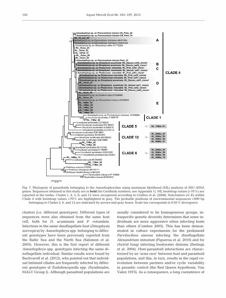
clone resulting from 1 individual iso-lated on 8 March) belonged to theMALV II Clade 4, according to theclassification proposed by Guillou etal. (2008) (see Appendix 1). A total of96 environmental sequences belong-ing to the Amoebophryidae were ob -tained from the DNA extracted on 10January, 14 February, and 8, 11, and14 March from the planktonic commu-nities >25 µm. Most of these se quen -ces belonged to Clade 4 (83 sequencesin total); the remaining 13 environ-mental se quen ces belonged to Clades5, 6, and 12 (Guillou et al. 2008). Fig. 7shows a phylogenetic analysis consid-ering all of the se quen ces obtained bysingle-cell PCR and environmental se -quen ces >699 bp. Although environ-mental se quen ces belonging to Clades5 and 12 were not included in theanalysis due their short length, theirprobable position is indicated in thephylogenetic tree. Considering boot-strap values >70%, se quen ces belong-ing to Clade 4 obtained from ReloncavíFjord during the present study were
distributed in 5 sub-clusters. Cluster A included 1environmental sequence obtained from a sample col-lected on 10 January and 2 sequences of Amoe-bophrya spp. infecting Gymnodinium instriatum(AF472554 and HM483394) published by Gundersonet al. (2002) and Coats et al. (2010). Se quences fromthe infected cells of D. acuminata and P. rotundatawere affiliated to Sub-clusters B, C, and D. Sub- cluster E was exclusively formed by environmentalsequences retrieved on 14 March and also included 2sequences of Amoebophrya spp. infecting P. micans(AY208893) and Ceratium tripos (AY208893) pub-lished by Kim et al. (2008). Although the 2 infected P.micans cells (JN998214 and JQ038241) collected inConcarneau Bay also belonged to Clade 4, they weregrouped in a sub-cluster separate from the se quen -ces obtained from Reloncaví Fjord.
DISCUSSION
Evolution of phytoplankton assemblages
Prorocentrum micans is a common species in sum-mer phytoplankton assemblages in southern Chile,although it is usually observed at low abundances
190
Fig. 5. Amoebophrya spp. life-cycle stages as detected by fluorescence in situhybridization. Cell nucleus (red), dinoflagellate theca (blue), and fluorescenceof probe ALV01 targeting Amoebophrya SSU ribosomal RNA (green) areshown. (a) Dinospores (arrows), (b) initial infection in Dinophysis acuminatanucleus, (c,d) intermediate infection in Dinophysis acuminata showing an in-cipient mastigocoel (arrows in b,c), (e) initial infection in Prorocentrum micansnucleus, and (f,g) final infection showing a mature trophont occupying the
entire intracellular space of P. micans. Scale bars = 10 µm

Alvez-de-Souza et al.: Amoebophryidae in a Chilean fjord
(<100 cells l−1, C. Alves-de-Souza unpubl. data). Thelast bloom reported in the study area was in March1983, with relatively high abundances (3.8 × 10⁷
cells l−1; Lembeye & Campodónico 1984) covering anextensive geographical area including ReloncavíFjord and part of Reloncaví Sound (both indicated inFig. 1). The magnitude and geographical extensionof the P. micans bloom observed in March 2009 wasapparently similar to that observed 26 yr before
(A. Clément pers. comm.). Maximum P. micans abun-dances occurred simultaneously with the lowestSi(OH)4 levels observed during the present study,whereas diatom blooms were positively related tohigh Si(OH)4:NO3 periods. The relative concentra-tions of Si(OH)4 and NO3 have been highlighted asthe main factor determining phytoplankton composi-tion in the fjords of southern Chile (Alves-de-Souzaet al. 2008, Torres et al. 2011), with low Si(OH)4:NO3
ratios favoring the development of non-diatom blooms(Iriarte et al. 2001, 2007).
Parasitoid and host diversity
Although 12 species of dinoflagellates were identi-fied, only 3 (Dinophysis acuminata, Phalacromarotundata, and Prorocentrum micans) were infectedby parasitoids belonging to the Amoebophryidae.Despite that, and taking into account the restrictedspatial scale considered in the present study, para-sitoids belonging to the Amoebophryidae weregenetically diverse, as they were represented by 5different clades. Clade 5, 6, and 12 are only knownfrom environmental sequences. Sequences belong-ing to these clades are probably widely distributed(detected to date in the North Sea, the northernFrench coast, the Indian Ocean, the Sargasso Sea,and the Mediterranean Sea; Guillou et al. 2008).Clade 4 has been reported to infect P. micans fromChesapeake Bay, USA (AY208893; Kim et al. 2008),and Concarneau Bay, on the northwestern coast ofFrance (JN998213 and JQ038241). This clade wasalso reported to infect other dinoflagellate species,such as Alexandrium affine, Ceratium tripos, Cer-atium lineatum (AY775284, AY208892, andAY260467; Kim et al. 2008), and Gymnodinium ins-triatum (AF472554 and HM483394; Gunderson et al.2002, Coats et al. 2010, respectively). Finally, Clade 1was also described to infect dinoflagellates such asHeterocapsa rotundata (Chambouvet et al. 2008),Prorocentrum minimum (AY208894), and Karlo-dinium veneficum (AF472553; Gunderson et al.2002). From these examples, it seems that the host-specificity of Amoebophryidae parasitoids was notlinked to its genetic filiations based upon the SSUrRNA gene (Gunderson et al. 2002, Kim et al. 2008).
Amoebophrya spp. belonging to Clade 4 were con-firmed to infect Dinophysis acuminata, Phalacromarotundata, and Prorocentrum micans based on single-cell PCR. However, phylogenetic analyses revealedthat these host species were, in fact, infected byAmoebophrya spp. belonging to different sub-
191
Fig. 6. Vertical and temporal dynamics of infection by para-sitoids belonging to the Amoebophryidae on Prorocentrummicans observed between 8 and 29 March 2009. (a) P. micansabundances (× 104 cells l−1), (b) Amoebophrya prevalences(%) on P. micans (c), number of infected P. micans (× 104
cells l−1), and (d) concentration of dinospores (× 105 cells l−1)
Aquat Microb Ecol 66: 183–197, 2012
clusters (i.e. different genotypes). Different types ofsequences were also obtained from the same hostcell, both for D. acuminata and P. rotundata.Infections in the same dinoflagellate host (Dinophysisnorvegica) by Amoebophrya spp. belonging to differ-ent genotypes have been previously reported fromthe Baltic Sea and the North Sea (Salomon et al.2003). However, this is the first report of differentAmoebophrya spp. genotypes infecting the same di-noflagellate individual. Similar results were found byBachvaroff et al. (2012), who pointed out that individ-ual tintinnid ciliates are frequently infected by differ-ent genotypes of Euduboscquella spp. (Syndiniales,MALV Group I). Although parasitoid populations are
usually considered to be homogeneous groups, in-traspecific genetic diversity determines that some in-dividuals are more aggressive when infecting hoststhan others (Combes 2005). This has been demon-strated in culture experiments for the perkinsoidParvilucifera sinerae infecting the dinoflagellateAlexandrium minutum (Figueroa et al. 2010) and forchytrid fungi infecting freshwater diatoms (Ibelingset al. 2004). Host-parasitoid interactions are charac-terized by an ‘arms race’ between host and parasitoidpopulations, and this, in turn, results in the rapid co-evolution between partners and/or cyclic variabilityin parasitic control (the Red Queen hypothesis; VanValen 1973). As a consequence, a long coexistence of
192
Fig. 7. Phylogeny of parasitoids belonging to the Amoebophryidae using maximum likelihood (ML) analyses of SSU rDNAgenes. Sequences obtained in this study are in bold (for GenBank numbers, see Appendix 1). ML bootstrap values (>70%) arereported at the nodes. Clades 1, 4, 5, 6, and 12 were recognized according to Guillou et al. (2008). Subclusters (A−E) withinClade 4 with bootstrap values >70% are highlighted in gray. The probable positions of environmental sequences <699 bp
belonging to Clades 5, 6, and 12 are indicated by arrows and gray boxes. Scale bar corresponds to 0.05% divergence

Alvez-de-Souza et al.: Amoebophryidae in a Chilean fjord
parasitoid and host populations will be reflected inhigher intraspecific genetic diversity. This would helpto explain, for example, the drastic change in the ge-netic composition of environmental sequences ob-served on 8 and 14 March during the present studyand the fact that the sequences of Amoebophrya spp.infecting P. micans from Relon caví Fjord and thosefrom other geographic areas were distributed in dif-ferent sub-clusters.
Host–Amoebophryidae dynamics
The observed Prorocentrum micans and Amoe-bophrya dynamic conforms to what has previouslybeen reported for Amoebophryidae (e.g. Coats &Park 2002, Chambouvet et al. 2008, Salomon & Stolte2010). Maximal host abundances were followed byincreased parasitoid prevalence and the consequentrelease of dinospores that reached their maximaldensity 6 d after the host peak. This burst of dino -spores declined in 3 d, an observation that correlateswith the short survival time of dinospores observed inculture (Coats & Park 2002). Estimations of the hostmortality induced by Amoebophrya spp. indicatedthat ~10% of the P. micans population was killed bythese parasitoids between 8 and 17 March. Althoughthis value only partially explains the decrease of P.micans abundance observed for the same period(94%), our results indicate that parasitism by organ-isms belonging to the Amoebophryidae likely consti-tuted an important loss for its dinoflagellate host dur-ing the study period.
The strength of infection by Amoebophrya spp. isoften related to the abundance of its dinoflagellatehosts in natural systems (Park et al. 2004). In thissense, the prevalences observed in Dinophysisacuminata and Phalacroma rotundata (<10%) wereconsistent with their low abundances and in accor-dance with the previous records of Amoebophryaspp. infections of Dinophysis species (Fritz & Nass1992, Gisselson et al. 2002, Salomon et al. 2009).However, prevalences were unexpectedly low forProrocentrum micans, with a maximum of 12%observed on 17 March. Because P. micans abun-dances were of the same order of magnitude as dur-ing other dinoflagellate blooms in which high Amoe-bophrya spp. prevalences (30 to 80%) have beenrecorded (Nishitani et al. 1985, Coats et al. 1996,Chambouvet et al. 2008), we expected to find ahigher percentage of infected cells during the pre-sent study. Nevertheless, culture studies (Yih & Coats2000, Coats & Park 2002) and mathematical simula-
tions (Salomon & Stolte 2010) have demonstratedthat the maximal prevalence of Amoebophrya spp.depends on the ratio between host and dinosporesrather than host density. Using equations given bySalomon & Stolte (2010) and considering a genera-tion time of 2.85 d, parasitoid mortality of 0.5 d−1
(Coats & Park 2002), and the conditions observed on8 March (parasitoid prevalences, host and dinosporeabundances), on 11 March, we should have observedprevalences similar to those found by Chambouvet etal. (2008) for Amoebophrya spp. infecting Alexan-drium minutum (~40%). Grazing by ciliates ondinospores has been shown to strongly suppressAmoebophrya infections under natural conditions(Johansson & Coats 2002). Unfortunately, we have noantecedents regarding ciliate abundances during thestudied period, but considering that the number ofdinospores actually increased from 8 to 11 March, itseems that dinospore mortality (by grazing or othersources) was not the main factor determining Amoe-bophrya spp. success during this 3 d period. Alterna-tively, because nutrients can have a positive effect onthe infective success and longevity of dinosporesunder culture conditions (Yih & Coats 2000), it is pos-sible that low NO3 and PO4 concentrations couldhave had an adverse effect on the quality of thedinospores produced.
The present study demonstrated for the first timethat parasitoids belonging to the Amoebophryidaecould be genetically diversified, even in ecosystemswhere their dinoflagellate hosts are not the predomi-nant phytoplankton group. Although Prorocentrummicans is a common species in southern Chile, it usu-ally occurs at very low abundances, and the forma-tion of blooms is rare. According to Chambouvet etal. (2008), the capacity of parasitoids to control hostpopulations is diminished when the excessive growthof a rare species is stimulated by environmentalchanges, when resistant host populations may actu-ally be favored. This would be particularly relevantin ecosystems like the Chilean fjords, in which cli-mate changes may favor increased dinoflagellateHABs.
Acknowledgements. We thank B. Hip and J. C. Cabrera fortheir invaluable help during sampling and in the laboratory.We are also grateful to I. Probert and D. Barriga for theirEnglish review of the manuscript and to W. Coats for hisinsightful comments on the results reported in this paper.This research was partially funded by the Dirección deInvestigación y Desarollo (Universidad Austral de Chile;DID-Uach-D-2008), the French National Research Agency(ANR) Aquaparadox (‘Biodiversity’), and EC2CO (Micro-bien, 2009−2010). This work is part of C.A.S.’s PhD thesis.She was financed by a Conicyt doctoral fellowship (Chilean
193

Aquat Microb Ecol 66: 183–197, 2012
government) and by the French ANR Paralex (‘The SixthExtinction’). Additional support for her PhD thesis was pro-vided by the Centro de Investigación de Ecosistemas de laPatagonia (CIEP).
LITERATURE CITED
Alves-de-Souza C, González MT, Iriarte JL (2008) Func-tional groups in marine phytoplankton assemblagesdominated by diatoms in fjords of southern Chile. JPlankton Res 30: 1233−1243
Alves-de-Souza C, Cornet C, Nowaczyk A, Gasparini S,Skovgaard A, Guillou L (2011) Blastodinium spp. infectcopepods in the ultra-oligotrophic marine waters of theMediterranean Sea. Biogeosciences 8: 2125−2136
Bachvaroff TR, Kim S, Guillou L, Delwiche C, Coats DW(2012) Molecular diversity of the Syndinian genus Eudu-boscquella based on single-cell PCR analysis. Appl Env-iron Microbiol 78: 334−345
Cachon J (1964) Contribution à l’étude des péridiniens par-asites. Cytologie, cycles évolutifs. Ann Sci Nat Zool 6: 1−158
Calbet A, Vaqué D, Felipe J, Vila M, Sala MM, Alcaraz M,Estrada M (2003) Relative grazing impact of microzoo-plankton and mesozooplankton on a bloom of the toxicdinoflagellate Alexandrium minutum. Mar Ecol Prog Ser259: 303−309
Chambouvet A, Morin P, Marie D, Guillou L (2008) Controlof toxic marine dinoflagellate blooms by serial parasitickillers. Science 322: 1254−1257
Chomczynski P, Sacchi N (2006) The single-step method ofRNA isolation by acid guanidinium thiocyanate-phenol-chloroform extraction: twenty-something years on. NatProtoc 1: 581−585
Coats DW, Bockstahler KR (1994) Occurrence of the para-sitic dinoflagellate Amoebophrya ceratii in ChesapeakeBay populations of Gymnodinium sanguineum. JEukaryot Microbiol 41: 586−593
Coats DW, Park MG (2002) Parasitism of photosyntheticdinoflagellates by three strains of Amoebophrya (Dino-phyta): parasite survival, infectivity, generation time, andhost specificity. J Phycol 38: 520−528
Coats DW, Adam EJ, Gallegos CL, Hedrick S (1996) Para-sitism of photosynthetic dinoflagellates in a shallowsubestuary of Chesapeake Bay, USA. Aquat Microb Ecol11: 1−9
Coats DW, Kim S, Bachvaroff TR, Handy SM, Delwiche CF(2010) Tintinnophagus acutus n. g., n. sp. (PhylumDinoflagellata), an ectoparasite of the ciliate Tintinnopsiscylindrica Daday 1887, and its relationship to Dubosc-quodinium collini Grasse 1952. J Eukaryot Microbiol 57: 468−482
Combes C (2005) The art of being a parasite. The Universityof Chicago Press, Chicago
Figueroa RI, Garcés E, Camp J (2010) Reproductive plastic-ity and local adaptation in the host-parasite systemformed by the toxic Alexandrium minutum and thedinoflagellate parasite Parvilucifera sinerae. HarmfulAlgae 10: 56−63
Fritz L, Nass M (1992) Development of two endoparasiticdinoflagellate Ameobophrya ceratii within host dinofla-gellate species. J Phycol 28: 312−320
Gisselson LA, Carlsson P, Granéli E, Pallon J (2002) Dinoph-ysis blooms in the deep euphotic zone of the Baltic Sea:
Do they grow in the dark? Harmful Algae 1: 401−418Guillard R (1973) Methods for microflagellates and nanno-
plankton. In: Stein J (ed) Handbook of phycologicalmethods: culture methods and growth measurements.Cambridge University Press, Cambridge, p 69−85
Guillou L, Viprey M, Chambouvet A, Welsh RM and others(2008) Widespread occurrence and genetic diversity ofmarine parasitoids belonging to Syndiniales (Alveolata).Environ Microbiol 10: 3349−3365
Gunderson JH, John SA, Boman WC, Coats DW (2002) Mul-tiple strains of the parasitic dinoflagellate Amoebophryaexist in Chesapeake Bay. J Eukaryot Microbiol 49: 469−474
Guzmán L, Pacheco H, Pizarro G, Alarcón C (2002) Alexan-drium catenella y veneno paralizante de los mariscos enChile. In: Sar EA, Ferrario ME, Reguera B (eds) Flo-raciones algales nocivas en el Cono Sur Americano. Insti-tuto Español de Oceanografía, Vigo, p 235−256
Huppert A, Blasius B, Stone S (2002) A model of phytoplank-ton blooms. Am Nat 159: 156−171
Ibelings BW, De Bruin A, Kagami M, Rijkeboer M, Brehm M,Van Donk E (2004) Host parasite interactions betweenfreshwater phytoplankton and chytrid fungi. J Phycol 40: 437−453
Iriarte JL, Kusch A, Ruiz M (2001) Phytoplankton biomass inthe sub-Antarctic area of the Straits of Magellan (53° S),Chile during spring-summer 1997−1998. Polar Biol 24: 154−162
Iriarte JL, Quiñones L, González RR (2005) Relationshipbetween biomass and enzymatic activity of a bloom-forming dinoflagellate (Dinophyceae) in southern Chile(41° S): a field approach. J Plankton Res 27: 159−166
Iriarte JL, González HE, Liu KK, Rivas C, Valenzuela C(2007) Spatial and temporal variability of chlorophyll andprimary productivity in surface waters of southern Chile(41.5−43° S). Estuar Coast Shelf Sci 74: 471−480
Iriarte JL, González HE, Nahuelhual L (2010) Patagonianfjord ecosystems in southern Chile as a highly vulnerableregion: problems and needs. Ambio 39: 463−466
Johansson M, Coats DW (2002) Ciliate grazing on the para-site Amoebophrya sp. decreases infection of the red-tidedinoflagellate Akashiwo sanguinea. Aquat Microb Ecol28: 69−78
Keane RM, Crawley MJ (2002) Exotic plant invasions andthe enemy release hypothesis. Trends Ecol Evol 17: 164−170
Kim S, Park MG, Kim KY, Kim CH, Yih W, Park JS, CoatsDW (2008) Genetic diversity of parasitic dinoflagellatesin the genus Amoebophrya and its relationship to para-site biology and biogeography. J Eukaryot Microbiol 55: 1−8
Lara A, Urrutia R, Luckman BH, Soto D and others (2005)The potential use of tree-rings to reconstruct streamflowand estuarine salinity in the Valdivian Rainforest eco-region. Dendrochronologia 22: 155−161
Lembeye G, Campodónico I (1984) First record bloom of thedinoflagellate Prorocentrum micans. Bot Mar 27: 491−493
Lembeye G, Yasumoto T, Zhao J, Fernández R (1993) DSPoutbreak in Chilean Fjords. In: Smayda TJ, Shimizu Y(eds) Toxic phytoplankton blooms in the sea. Elsevier,Amsterdam, p 525−529
Lund JWG, Kipling C, Lecren ED (1958) The inverted micro-scope method of estimating algal number and the statis-tical basis of estimating by counting. Hydrobiologia 11: 143−170
194
Alvez-de-Souza et al.: Amoebophryidae in a Chilean fjord
Maranda L (2001) Infection of Prorocentrum minimum(Dinophyceae) by the parasite Amoebophrya sp.(Dinoflagellea). J Phycol 37: 245−248
Molinet C, Lafon A, Lembeye G, Moreno C (2003) Spatialand temporal distribution patterns of blooms of Alexan-drium catenella (Whedon & Kofoid) Balech 1985, oninland seas of northwest Patagonia, Chile (in Spanishwith English Abstract). Rev Chil Hist Nat 76: 681−698
Montagnes DJS, Chambouvet A, Guillou L, Fenton A (2008)Responsibility of microzooplankton and parasite pres-sure for the demise of toxic dinoflagellate blooms. AquatMicrob Ecol 53: 211−225
Nishitani L, Erickson G, Chew KK (1985) Role of the para-sitic dinoflagellate Amoebophrya ceratii in control ofGonyaulax catenella populations. In: Anderson DM,White AW, Baden DG (eds) Toxic dinoflagellates. Else-vier, New York, NY, p 225−232
Park MG, Yih W, Coats DW (2004) Parasites and phyto-plankton, with special emphasis on dinoflagellate infec-tions. J Eukaryot Microbiol 51: 145−155
Parsons TR, Maita Y, Lalli CM (1984) A manual of chemicaland biological methods for seawater analysis. PergamonPress, Oxford
Rebolledo L, Lange C, Figueroa D, Pantoja S, Muñoz P, Cas-tro R (2005) 20th century fluctuations in the abundanceof siliceous microorganisms preserved in the sedimentsof the Puyuhuapi channel. Rev Chil Hist Nat 78: 469−488
Salomon PS, Stolte W (2010) Predicting the populationdynamics in Amoebophrya parasitoids and their dinofla-gellate hosts using a mathematical model. Mar Ecol ProgSer 419: 1−10
Salomon PS, Janson S, Granéli E (2003) Multiple species ofthe dinophagous dinoflagellate genus Amoebophryainfect the same host species. Environ Microbiol 5: 1046−1052
Salomon PS, Granéli E, Neves MHCB, Rodriguez EG (2009)Infection by Amoebophrya spp. parasitoids of dinoflagel-lates in a tropical marine coastal area. Aquat Microb Ecol55: 143−153
Sepulveda J, Pantoja S, Hughen K, Lange C and others(2005) Fluctuations in export productivity over the lastcentury from sediments of a southern Chilean fjord(44° S). Estuar Coast Shelf Sci 65: 587−600
Siano R, Alves-de-Souza C, Foulon E, Bendif EIM, Simon N,Guillou L, Not F (2011) Distribution and host diversity ofAmoebophryidae parasites across oligotrophic waters ofthe Mediterranean Sea. Biogeosciences 8: 267−278
Smayda TJ (1997) Harmful algal blooms: their ecophysiol-ogy and general relevance to phytoplankton blooms inthe sea. Limnol Oceanogr 42: 1137−1153
Strickland JDH, Parsons TR (1972) A practical handbook ofseawater analysis. Fisheries Research Board of Canada,Ottawa
Tamura K, Peterson D, Peterson N, Stecher G, Nei M, KumarS (2011) MEGA5: molecular evolutionary genetics analy-sis using maximum likelihood, evolutionary distance,and maximum parsimony methods. Mol Biol Evol 28: 2731−2739
Taylor FJR (1968) Parasitism of the toxin-producing dinofla-gellate Gonyaulax catenella by the endoparasiticdinoflagellate Amoebophrya ceratii. J Fish Res BoardCan 25: 2241−2245
ter Braak CJ (1995) Ordination. In: Jongman RH, ter BraakCJ, Van Tongeren OF (eds) Data analysis and commu-nity and landscape ecology. Cambridge University Press,Cambridge, p 91−73
Torres R, Frangópulos M, Hamamé M, Montecino V andothers (2011) Nitrate to silicate ratio variability and thecomposition of micro-phytoplankton blooms in the inner-fjord of Seno Ballena (Strait of Magellan, 54° S). ContShelf Res 31: 244−253
Uhelinger V (1964) Étude statistique des methods de déno-brement planctonique. Arch Sci 17: 121−223
Uribe JC, Ruiz M (2001) Gymnodinium brown tide in theMagellanic fjords, southern Chile. Rev Biol MarOceanogr 36: 155−164
Utermöhl H (1958) Zur Vervollkommnung der quantitativenPhytoplankton-Methodik. Mitt Int Ver Theor AngewLimnol 9: 1−38
Van Valen L (1973) A new evolutionary law. Evol Theory 1: 1−30
Yih W, Coats DW (2000) Infection of Gymnodinium san-guineum by the dinoflagellate Amoebophrya sp.: effectof nutrient environment on parasite generation time,reproduction, and infectivity. J Eukaryot Microbiol 47: 504−510
195

Aquat Microb Ecol 66: 183–197, 2012196
GenBank ID Organism Host Source Geographical area MALV IInumber clade
JN998202 RL_Dacum_cell1_clone01 Amoebophrya sp. Dinophysis acuminata SC Reloncaví Fjord, Chile 4JN998203 RL_Dacum_cell1_clone02 Amoebophrya sp. D. acuminata SC Reloncaví Fjord, Chile 4JN998204 RL_Dacum_cell1_clone 03 Amoebophrya sp. D. acuminata SC Reloncaví Fjord, Chile 4JN998205 RL_Dacum_cell2_clone 01 Amoebophrya sp. D. acuminata SC Reloncaví Fjord, Chile 4JN998206 RL_Dacum_cell2_clone 02 Amoebophrya sp. D. acuminata SC Reloncaví Fjord, Chile 4JN998207 RL_Dacum_cell2_clone 03 Amoebophrya sp. D. acuminata SC Reloncaví Fjord, Chile 4JN998208 RL_Prot_cell1_clone 01 Amoebophrya sp. Phalachroma rotundata SC Reloncaví Fjord, Chile 4JN998209 RL_Prot_cell1_clone 02 Amoebophrya sp. P. rotundata SC Reloncaví Fjord, Chile 4JN998210 RL_Prot_cell2_clone 01 Amoebophrya sp. P. rotundata SC Reloncaví Fjord, Chile 4JN998211 RL_Prot_cell2_clone 02 Amoebophrya sp. P. rotundata SC Reloncaví Fjord, Chile 4JN998212 RL_Prot_cell2_clone 03 Amoebophrya sp. P. rotundata SC Reloncaví Fjord, Chile 4JN998213 RL_Pmic_01 Amoebophrya sp. Prorocentrum micans SC Reloncaví Fjord, Chile 4JQ038241 CN_Pmic_01 Amoebophrya sp. P. micans SC Concarneau Bay, France 4JN998214 CN_Pmic_02 Amoebophrya sp. P. micans SC Concarneau Bay, France 4JN998215 RL_10Jan_01 Uncultured Syndiniales Unknown ES Reloncaví Fjord, Chile 4JN998216 RL_10Jan_02 Uncultured Syndiniales Unknown ES Reloncaví Fjord, Chile 4JN998217 RL_10Jan_03 Uncultured Syndiniales Unknown ES Reloncaví Fjord, Chile 4JN998218 RL_10Jan_04 Uncultured Syndiniales Unknown ES Reloncaví Fjord, Chile 4JN998219 RL_10Jan_05 Uncultured Syndiniales Unknown ES Reloncaví Fjord, Chile 4JN998220 RL_10Jan_06 Uncultured Syndiniales Unknown ES Reloncaví Fjord, Chile 4JN998221 RL_10Jan_07 Uncultured Syndiniales Unknown ES Reloncaví Fjord, Chile 4JN998222 RL_10Jan_08 Uncultured Syndiniales Unknown ES Reloncaví Fjord, Chile 4JN998223 RL_10Jan_09 Uncultured Syndiniales Unknown ES Reloncaví Fjord, Chile 4JN998224 RL_10Jan_10 Uncultured Syndiniales Unknown ES Reloncaví Fjord, Chile 4JN998225 RL_14Feb_01 Uncultured Syndiniales Unknown ES Reloncaví Fjord, Chile 4JN998226 RL_14Feb_02 Uncultured Syndiniales Unknown ES Reloncaví Fjord, Chile 4JN998227 RL_14Feb_03 Uncultured Syndiniales Unknown ES Reloncaví Fjord, Chile 4JN998228 RL_14Feb_04 Uncultured Syndiniales Unknown ES Reloncaví Fjord, Chile 4JN998229 RL_14Feb_05 Uncultured Syndiniales Unknown ES Reloncaví Fjord, Chile 4JN998230 RL_14Feb_06 Uncultured Syndiniales Unknown ES Reloncaví Fjord, Chile 4JN998231 RL_14Feb_07 Uncultured Syndiniales Unknown ES Reloncaví Fjord, Chile 4JN998232 RL_14Feb_08 Uncultured Syndiniales Unknown ES Reloncaví Fjord, Chile 4JN998233 RL_14Feb_09 Uncultured Syndiniales Unknown ES Reloncaví Fjord, Chile 4JN998234 RL_14Feb_10 Uncultured Syndiniales Unknown ES Reloncaví Fjord, Chile 4JN998235 RL_14Feb_11 Uncultured Syndiniales Unknown ES Reloncaví Fjord, Chile 4JN998236 RL_14Feb_12 Uncultured Syndiniales Unknown ES Reloncaví Fjord, Chile 4JN998237 RL_14Feb_13 Uncultured Syndiniales Unknown ES Reloncaví Fjord, Chile 4JN998238 RL_14Feb_14 Uncultured Syndiniales Unknown ES Reloncaví Fjord, Chile 4JN998239 RL_14Feb_15 Uncultured Syndiniales Unknown ES Reloncaví Fjord, Chile 4JN998240 RL_14Feb_16 Uncultured Syndiniales Unknown ES Reloncaví Fjord, Chile 4JN998241 RL_14Feb_17 Uncultured Syndiniales Unknown ES Reloncaví Fjord, Chile 4JN998242 RL_14Feb_18 Uncultured Syndiniales Unknown ES Reloncaví Fjord, Chile 4JN998243 RL_14Feb_19 Uncultured Syndiniales Unknown ES Reloncaví Fjord, Chile 4JN998244 RL_14Feb_20 Uncultured Syndiniales Unknown ES Reloncaví Fjord, Chile 4JN998245 RL_14Feb_21 Uncultured Syndiniales Unknown ES Reloncaví Fjord, Chile 4JN998246 RL_14Feb_22 Uncultured Syndiniales Unknown ES Reloncaví Fjord, Chile 4JN998247 RL_14Feb_23 Uncultured Syndiniales Unknown ES Reloncaví Fjord, Chile 4JN998248 RL_14Feb_24 Uncultured Syndiniales Unknown ES Reloncaví Fjord, Chile 4JN998249 RL_14Feb_25 Uncultured Syndiniales Unknown ES Reloncaví Fjord, Chile 4JN998250 RL_14Feb_26 Uncultured Syndiniales Unknown ES Reloncaví Fjord, Chile 4JN998251 RL_14Feb_27 Uncultured Syndiniales Unknown ES Reloncaví Fjord, Chile 4JN998252 RL_14Feb_28 Uncultured Syndiniales Unknown ES Reloncaví Fjord, Chile 4JN998253 RL_14Feb_29 Uncultured Syndiniales Unknown ES Reloncaví Fjord, Chile 4JN998254 RL_14Feb_30 Uncultured Syndiniales Unknown ES Reloncaví Fjord, Chile 4JN998255 RL_14Feb_31 Uncultured Syndiniales Unknown ES Reloncaví Fjord, Chile 4JN998256 RL_14Feb_32 Uncultured Syndiniales Unknown ES Reloncaví Fjord, Chile 4JN998257 RL_11Mar_01 Uncultured Syndiniales Unknown ES Reloncaví Fjord, Chile 5JN998258 RL_11Mar_02 Uncultured Syndiniales Unknown ES Reloncaví Fjord, Chile 6
Appendix 1. Complete list of obtained sequences. ES = environmental samples, SC = single cells

Alvez-de-Souza et al.: Amoebophryidae in a Chilean fjord 197
GenBank ID Organism Host Source Geographical area MALV IInumber clade
JN998259 RL_11Mar_03 Uncultured Syndiniales Unknown ES Reloncaví Fjord, Chile 1JN998260 RL_11Mar_04 Uncultured Syndiniales Unknown ES Reloncaví Fjord, Chile 6JN998261 RL_11Mar_05 Uncultured Syndiniales Unknown ES Reloncaví Fjord, Chile 6JN998262 RL_11Mar_06 Uncultured Syndiniales Unknown ES Reloncaví Fjord, Chile 6JN998263 RL_14Mar_01 Uncultured Syndiniales Unknown ES Reloncaví Fjord, Chile 4JN998264 RL_14Mar_02 Uncultured Syndiniales Unknown ES Reloncaví Fjord, Chile 4JN998265 RL_14Mar_03 Uncultured Syndiniales Unknown ES Reloncaví Fjord, Chile 4JN998266 RL_14Mar_04 Uncultured Syndiniales Unknown ES Reloncaví Fjord, Chile 4JN998267 RL_14Mar_05 Uncultured Syndiniales Unknown ES Reloncaví Fjord, Chile 4JN998268 RL_14Mar_06 Uncultured Syndiniales Unknown ES Reloncaví Fjord, Chile 4JN998269 RL_14Mar_07 Uncultured Syndiniales Unknown ES Reloncaví Fjord, Chile 4JN998270 RL_14Mar_08 Uncultured Syndiniales Unknown ES Reloncaví Fjord, Chile 4JN998271 RL_14Mar_09 Uncultured Syndiniales Unknown ES Reloncaví Fjord, Chile 4JN998272 RL_14Mar_10 Uncultured Syndiniales Unknown ES Reloncaví Fjord, Chile 6JN998273 RL_14Mar_11 Uncultured Syndiniales Unknown ES Reloncaví Fjord, Chile 4JN998274 RL_14Mar_12 Uncultured Syndiniales Unknown ES Reloncaví Fjord, Chile 4JN998275 RL_14Mar_13 Uncultured Syndiniales Unknown ES Reloncaví Fjord, Chile 4JN998276 RL_14Mar_14 Uncultured Syndiniales Unknown ES Reloncaví Fjord, Chile 4JN998277 RL_14Mar_15 Uncultured Syndiniales Unknown ES Reloncaví Fjord, Chile 4JN998278 RL_14Mar_16 Uncultured Syndiniales Unknown ES Reloncaví Fjord, Chile 4JN998279 RL_14Mar_17 Uncultured Syndiniales Unknown ES Reloncaví Fjord, Chile 4JN998280 RL_14Mar_18 Uncultured Syndiniales Unknown ES Reloncaví Fjord, Chile 4JN998281 RL_14Mar_19 Uncultured Syndiniales Unknown ES Reloncaví Fjord, Chile 4JN998282 RL_14Mar_20 Uncultured Syndiniales Unknown ES Reloncaví Fjord, Chile 4JN998283 RL_14Mar_21 Uncultured Syndiniales Unknown ES Reloncaví Fjord, Chile 4JN998284 RL_14Mar_22 Uncultured Syndiniales Unknown ES Reloncaví Fjord, Chile 4JN998285 RL_14Mar_23 Uncultured Syndiniales Unknown ES Reloncaví Fjord, Chile 4JN998286 RL_08Mar_01 Uncultured Syndiniales Unknown ES Reloncaví Fjord, Chile 4JN998287 RL_08Mar_02 Uncultured Syndiniales Unknown ES Reloncaví Fjord, Chile 4JN998288 RL_08Mar_03 Uncultured Syndiniales Unknown ES Reloncaví Fjord, Chile 12JN998289 RL_08Mar_04 Uncultured Syndiniales Unknown ES Reloncaví Fjord, Chile 6JN998290 RL_08Mar_05 Uncultured Syndiniales Unknown ES Reloncaví Fjord, Chile 12JN998291 RL_08Mar_06 Uncultured Syndiniales Unknown ES Reloncaví Fjord, Chile 12JN998292 RL_08Mar_07 Uncultured Syndiniales Unknown ES Reloncaví Fjord, Chile 12JN998293 RL_08Mar_08 Uncultured Syndiniales Unknown ES Reloncaví Fjord, Chile 6JN998294 RL_08Mar_09 Uncultured Syndiniales Unknown ES Reloncaví Fjord, Chile 6JN998295 RL_08Mar_10 Uncultured Syndiniales Unknown ES Reloncaví Fjord, Chile 6JN998296 RL_08Mar_11 Uncultured Syndiniales Unknown ES Reloncaví Fjord, Chile 4JN998297 RL_08Mar_12 Uncultured Syndiniales Unknown ES Reloncaví Fjord, Chile 4JN998298 RL_08Mar_13 Uncultured Syndiniales Unknown ES Reloncaví Fjord, Chile 4JN998299 RL_08Mar_14 Uncultured Syndiniales Unknown ES Reloncaví Fjord, Chile 4JN998300 RL_08Mar_15 Uncultured Syndiniales Unknown ES Reloncaví Fjord, Chile 4JN998301 RL_08Mar_16 Uncultured Syndiniales Unknown ES Reloncaví Fjord, Chile 4JN998302 RL_08Mar_17 Uncultured Syndiniales Unknown ES Reloncaví Fjord, Chile 4JN998303 RL_08Mar_18 Uncultured Syndiniales Unknown ES Reloncaví Fjord, Chile 4JN998304 RL_08Mar_19 Uncultured Syndiniales Unknown ES Reloncaví Fjord, Chile 4JN998305 RL_08Mar_20 Uncultured Syndiniales Unknown ES Reloncaví Fjord, Chile 4JN998306 RL_08Mar_21 Uncultured Syndiniales Unknown ES Reloncaví Fjord, Chile 4JN998307 RL_08Mar_22 Uncultured Syndiniales Unknown ES Reloncaví Fjord, Chile 4JN998308 RL_08Mar_23 Uncultured Syndiniales Unknown ES Reloncaví Fjord, Chile 4JN998309 RL_08Mar_24 Uncultured Syndiniales Unknown ES Reloncaví Fjord, Chile 4JN998310 RL_08Mar_25 Uncultured Syndiniales Unknown ES Reloncaví Fjord, Chile 4JN998311 RL_08Mar_26 Uncultured Syndiniales Unknown ES Reloncaví Fjord, Chile 4JN998312 RL_08Mar_27 Uncultured Syndiniales Unknown ES Reloncaví Fjord, Chile 4
Appendix 1 (continued)
Editorial responsibility: Tom Fenchel, Helsingør, Denmark
Submitted: September 14, 2011; Accepted: March 29, 2012Proofs received from author(s): May 24, 2012