Embed Size (px)
Citation preview

Coffee Science, Lavras, v. 14, n. 1, p. 127 - 130, jan./mar. 2019
Paula, P. V. A. A. et al.127 NOTA PRÉVIA
INFECTION PROCESS OF Cercospora coffeicola IN IMMATURE COFFEE FRUITS
Paulo Victor Augusto Azevedo de Paula1, Edson Ampélio Pozza2, Eduardo Alves3, Silvino Intra Moreira4, Júlio Cesar Azevedo Paula5, Leandro Alvarenga Santos6
(Received: September 20, 2018; accepted: December 06, 2018)
ABSTRACT: Cercosporiosis is caused by Cercospora coffeicola and represents a very important coffee plants phytosanitary problem. Catuaí IAC 144 and Topázio cultivars at the F2 stage were inoculated with a conidial suspension. Samples were collected at 4, 8, 12, 24, 36, 48, 72, 96, and 168 hours after inoculation (hai) for scanning electron microscope studies. Fungal germination in epidermal surface occurred four hai; penetration only through epidermal wounds, without appressoria, started at 12 hai while the mycelial colonization occurred at 72 hai. The infection in immature coffee fruits implies brown eyespot control management anticipation need, even before the first symptoms.
Index terms: Brown eyespot, Coffea arabica, scanning electron microscopy.
PROCESSO INFECCIOSO DE Cercospora coffeicola EM FRUTOS DE CAFÉ IMATUROS
RESUMO: Cercosporiose é causada por Cercospora coffeicola e representa grave problema fitossanitário do cafeeiro. Frutos de cv. Catuaí IAC 144 e Topázio no estádio F2 foram inoculados com suspensão conidial. Amostras foram coletadas 4, 8, 12, 24, 36, 48, 72, 96, e 168 horas após a inoculação (hai) para estudos de microscopia eletrônica de varredura. A germinação conidial na superfície epidérmica ocorreu quatro hai; a penetração na epiderme ocorreu através de ferimentos, sem formação de apressórios, 12 hai, já a colonização micelial interna ocorreu 72 hai. A infecção de frutos imaturos implica na necessidade de antecipação no manejo da mancha de olho pardo, mesmo antes dos primeiros sintomas.
Termos para indexação: Mancha de olho pardo, Coffea arabica, microscopia eletrônica de varredura.
One of the main phytosanitary problems of coffee is cercosporiosis, also known as brown eye spot disease, whose etiological agent is the fungus Cercospora coffeicola Berkeley & Cooke. The pathogen can infect leaves and fruits of coffee tree, causing losses of 15% to 30% (GUIMARÃES; MENDES; BALIZA, 2010), and can affect the final product quality: consumers’ cup of coffee (POZZA et al., 2010). This adverse effect on quality is due to change in staining and, as proportion of diseased grains increases, a reduction in grain size, total sugars, total solids, and pH (LIMA; POZZA; SANTOS, 2012).
In order to reduce this infection and to guarantee quality to final product, adequate management strategies for cercosporiosis control must be employed. Therefore, it is necessary to know how the pathogen germinates and penetrates the fruit, whether by stomata or wounds, or by appressoria formation. The infection process of C. coffeicola has been frequently studied and characterized in foliar tissues of coffee (SOUZA et al., 2011) but little is known about how this
1,2,3,4,5Universidade Federal de Lavras/UFLA - Departamento de Fitopatologia/DFP - Cx. P. 3037 - 37.200-000 - Lavras-MG - [email protected], [email protected], [email protected], [email protected], [email protected] Estadual do Centro-Oeste/UNICENTRO - Departamento de Agronomia - 85.040-080 - Guarapuava PR - [email protected]
process occurs in fruits. Infections occurrence during the early flowering stages has been frequently reported in the field; however there is not scientific confirmation. Our results explain how this pathogen penetrates the coffee fruit and helps to determine when coffee grain disease monitoring and biological control agents and fungicides application should start. The objective in this study was to describe the infection process of Cercospora coffeicola in coffee fruit.
The isolate of C. coffeicola used was CML 2489 and was deposited in mycological collection of Federal University of Lavras (UFLA) in Lavras, Minas Gerais, Brazil. Its sporulation was induced as described by Souza et al. (2011), using a conidia suspension of 6×10⁴ conidia.mL-1 to inoculate the pathogen. The trials were carried out with fruit obtained from detached branches of the middle third of cv. Catuaí IAC 144 and cv. Topázio sampled from the UFLA Coffee Growing Field in November 2013. It was used two branches of each cultivar with 10 fruit rosettes in F2 stage (‘pin head’ stage, with green color and 3.1 to 4 mm). It was

Coffee Science, Lavras, v. 14, n. 1, p. 127 - 130 jan./mar. 2019
Infection process of Cercospora coffeicola in ... 128
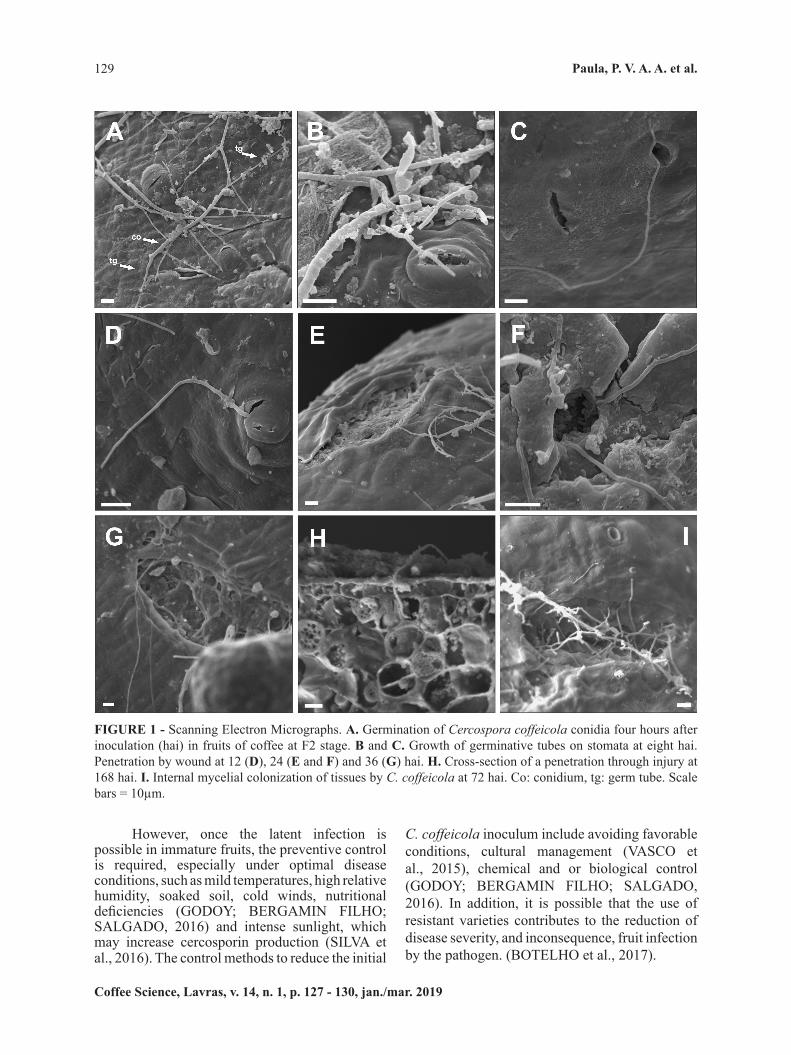
(Figures 1 E and F), 36 hai (Figure 1 G), and 168 hai (Figure 1 H). It was not observed appressoria formation, reinforcing the passive penetration hypothesis, by wounding. Samples evaluated at 168 hai had internal mycelial tissue colonization (Figure 1 H). No symptoms were observed during the evaluation.
In our work, the presence of germinative tubes in fruits at four hai was verified on the fruit epidermal surface. Similarly, Souza et al. (2011) observed C. coffeicola conidial germination both on adaxial and abaxial in coffee leaves surfaces at four hai. These authors also observed the growth of the germinative tubes towards stomatal openings, as occurred in the fruit; however, they observed penetration by stomata. They also verified that penetration into coffee leaves occurred at 36 hai and was mainly through the stomata on the epidermal surface or, occasionally, due to wounds in the epidermis, also without the appressoria formation. Some Cercospora species penetrate leaf tissues in this way, such as C. arachidicola (SMITH; PAUER; SHOKES, 1992), and C. moricola (GUPTA et al., 1995); however, C. henningsii penetrates directly through the epidermis of leaf tissues, and also without appressoria formation (BABU et al., 2009; PAULA et al., 2015). In this way, the infection process of Cercospora spp. may differ depending on the organ of the plant or the studied pathosystem.
The importance of infection process of C. coffeicola studies in immature fruit at young stages, shortly after the dry petals fall, is evidenced with the present results because certain species of cercosporioides fungi can go through long periods of latency; as example, Pseudocercospora musae has a latency periods around 40 days (FREITAS et al., 2017). That is, penetration occurs and symptoms are observed days or months after this process. Therefore, it is recommended starting control measures in the early fruiting stages from F2 to F5. The germination of C. coffeicola on the surface of the fruit from coffee cv. Catuaí IAC 144 and Topázio in the F2 stage is observed at four hai and its penetration only occurs by wounds in the epidermis and after 12 hai. Additionally, its colonization in the fruit’s internal tissues occurs after 72 hai.
In order to evaluate brown eye spot in yellow and red coffee fruits, diagrammatic scales were validated by Paula et al. (2016). These scales are important to assessing the severity progress in already infected fields and compare control treatments.
performed the fruits disinfestation by immersion in 1 mg L-1 sodium hypochlorite (twice), followed by rinsing with distilled water and drying on filter paper in a laminar flow chamber for 2 hours; afterwards, the samples were placed in a plastic tray.
The fruit branch cut site was introduced in moist phenolic foam and wrapped in plastic film, with nutrient solution from Hoagland and Arnon (1950), to ensure nutrition for the fruit during the experiment period. The inoculation was performed using 100μL of conidial suspension, applied to F2 stage fruits surface, just after petals fell. The samples were incubated at 25 ± 2 °C and 90 ± 5% relative humidity with continuous white fluorescent light. The samples were processed to electron microscope analysis as done by Silva et al. (2017). Ten fruit units from each cultivar were collected at 4, 8, 12, 24, 36, 48, 72, 96, and 168 hours after inoculation (hai); fragments of 25 mm2 were fixed with Karnovsky solution (pH 7.2) and post-fixed with 1% osmium tetroxide solution and dehydrated with acetone series. The specimens were dried in a Critical Point Apparatus (Bal-tec CPD 030 Balzers) followed by gold coating on a Sputter Coater Evaporator (Bal-tec SCD 050 Balzers). The acquisition of the images was performed with at the Electron Microscopy Lab in Federal University of Lavras using the Zeiss LEO EVO 40 XVP MEV and Smart Sem software, at 20 Kv and 9 mm working distance. The images obtained were edited using Corel Draw software.
Our analysis showed the germination of some C. coffeicola conidia on the coffee fruit surface at four hai (Figure 1A) and this was verified for both cultivars; germination occasionally showed two germ tubes (Figure 1A, arrows) and most of the conidia germinated at eight hai. The germ tubes grew towards the stomata in an attempt to penetrate them, but as they not clearly penetrate directed the sub-stomatal cavity, demonstrating a possibly absence of chemotropism to the stomata (Figures 1 B and C). However it was observed that the germ tubes clearly grew towards the wounds (Figures 1 D to G), thus showing a probable wounds chemotropism. In fact, the penetration of the pathogen occurred only through lesions on the fruits epidermis (Figures 1 D to G), for both coffee cultivars. Germ tubes developed and moved on open stomata but did not penetrate these natural openings (Figures 1 B and C). The penetration of C. coffeicola into the fruit occurred at 12 hai (Figure 1 D) and in subsequent evaluations: 24 hai
Coffee Science, Lavras, v. 14, n. 1, p. 127 - 130, jan./mar. 2019
Paula, P. V. A. A. et al.129
However, once the latent infection is possible in immature fruits, the preventive control is required, especially under optimal disease conditions, such as mild temperatures, high relative humidity, soaked soil, cold winds, nutritional deficiencies (GODOY; BERGAMIN FILHO; SALGADO, 2016) and intense sunlight, which may increase cercosporin production (SILVA et al., 2016). The control methods to reduce the initial
FIGURE 1 - Scanning Electron Micrographs. A. Germination of Cercospora coffeicola conidia four hours after inoculation (hai) in fruits of coffee at F2 stage. B and C. Growth of germinative tubes on stomata at eight hai. Penetration by wound at 12 (D), 24 (E and F) and 36 (G) hai. H. Cross-section of a penetration through injury at 168 hai. I. Internal mycelial colonization of tissues by C. coffeicola at 72 hai. Co: conidium, tg: germ tube. Scale bars = 10µm.
C. coffeicola inoculum include avoiding favorable conditions, cultural management (VASCO et al., 2015), chemical and or biological control (GODOY; BERGAMIN FILHO; SALGADO, 2016). In addition, it is possible that the use of resistant varieties contributes to the reduction of disease severity, and inconsequence, fruit infection by the pathogen. (BOTELHO et al., 2017).

Coffee Science, Lavras, v. 14, n. 1, p. 127 - 130 jan./mar. 2019
Infection process of Cercospora coffeicola in ... 130
HOAGLAND D. R.; ARNON D. I. The water-culture method for growing plants without soil. Circular 347. San Diego: Ed. University of California, 1950.
LIMA L. M; POZZA E. A.; SANTOS F. S. Relationship between Incidence of Brown Eye Spot of Coffee Cherries and the Chemical Composition of Coffee Beans. Journal of Phytopathology, Göttingen, v. 160, p. 209-211, 2012.
PAULA P. V. A. A., et al. Formas de Penetração do Gênero Cercospora. Nucleus, Ituverava, v. 12, p. 271-279,Oct. 2015.
PAULA P. V. A. A., et al. Diagrammatic Scales for Assessing Brown Eye Spot (Cercospora coffeicola) in Red and Yellow Coffee Cherries. Journal of Phytopathology, Göttingen, v. 164, p. 791-800, Oct. 2015.
SILVA, F. F., et al. Emergência e análise ultraestrutural de plântulas de soja inoculadas com Sclerotinia sclerotiorum sob efeito da aplicação de Trichoderma harzianum. Summa Phytopathologica, Botucatu, v. 43, n. 1, p. 41-45,Jan./Mar. 2017.
SILVA M. G., et al. Effect of light and temperature on Cercospora coffeicola and Coffea arabica pathosystem. Coffee Science, Lavras, v. 11, n. 2, p. 148-160, June 2016.
SMITH D. H.; PAUER G. D. C.; SHOKES F. M. Cercosporidium and Cercospora leaf spots of peanut. In: CHAUBE H.S., et al. (Eds.). Plant diseases of international importance v. 2. New Jersey: Prentica Hall Inc., 1992. p. 285-304.
SOUZA A. G. C., et al. Infection process of Cercospora coffeicola on coffee leaf. Journal of Phytopathology, Göttingen, v.159, p. 6-11, 2011.
VASCO G. B. et al. Incidência da cercosporiose em frutos de cafeeiro: diferentes densidades de plantio e manejos de irrigação. Coffee Science, Lavras, v. 10, n. 1, p. 38-45, Mar. 2015.
The Cercospora coffeicola infection in immature coffee fruits is very important, and implies in brown eyespot control management anticipation need, even before the first symptoms. Thus, the control actions should be preventive, since it is only possible to observe this infection through microscope.
ACKNOWLEDGMENTSThe authors thanks the FAPEMIG (Minas
Gerais Research Foundation), the CAPES (Brazilian Federal Agency for the Support and Evaluation of Graduate Education), and the CNPq (National Council of Technological and Scientific Development) for financial support.
REFERENCESBABU A. M., et al. Scanning electron microscopy of the infection process of Cercospora henningsii on cassava leaves. Journal of Phytopathology, Göttingen, v. 157, n. 2, p. 57-62, Aug 2008.
BOTELHO D. M. S., et al. Cercosporiosis resistance in coffee germplasm collection. Euphytica, Wageningen, v. 213, n. 2, p. 117-129, May 2017.
FREITAS, A. S., et al. Infection process of Pseudocercospora musae on banana leaf. Phytoparasitica, Rotterdam, v. 45, n. 3, p. 317–324, June 2017.
GODOY C. V.; BERGAMIN FILHO A.; SALGADO C. L. Doenças do cafeeiro (Coffea arabica L.). In: AMORIM L.; REZENDE J. A. M.; CAMARGO L. F. A. (Eds.) Manual de Fitopatologia v.2, 5ª Ed. Ouro Fino: Agronômica Ceres, 2016. p. 178-193.
GUIMARÃES R. J.; MENDES A. N. G.; BALIZA D. P. Semiologia do cafeeiro: Sintomas de desordens nutricionais, fitossanitárias e fisiológicas. Lavras: Ed. UFLA, 2010.
GUPTA V., et al. Observations on the surface ultrastructre of conidial stage of Cercospora moricola and its infection process in mulberry. Sericologia, Lyon, v. 35, n. 4, p. 123-131, Oct. 1995.